an ontological view of cardiovascular … · "limpio", hemos utilizado ratones...
TRANSCRIPT

CENTRO DE INVESTIGACIÓN DEL CÁNCER INSTITUTO DE BIOLOGÍA MOLECULAR Y CELULAR DEL CÁNCER
CSIC – UNIVERSIDAD DE SALAMANCA
AN ONTOLOGICAL VIEW OF CARDIOVASCULAR AND METABOLIC DISEASE PROGRESSION
USING GENETICALLY MODIFIED MICE
TESIS DOCTORAL
SALVATORE FABBIANO Salamanca, 2014


El Dr. XOSÉ RAMÓN GARCÍA BUSTELO, Profesor de Investigación del Consejo Superior de Investigaciones Científicas en el Instituto de Biología Molecular y Celular del Cáncer de Salamanca y el Dr. MAURICIO ARIEL MENACHO MÁRQUEZ, Doctor por la Universidad Politécnica de Valencia e Investigador Postdoctoral en el Centro de Investigación del Cáncer de Salamanca, CERTIFICAN: Que el trabajo de tesis titulado “An ontological view of cardiovascular and metabolic disease progression using genetically modified mice”, presentado por Don SALVATORE FABBIANO para optar al grado de Doctor por la Universidad de Salamanca, ha sido realizado bajo nuestra dirección en el Centro de Investigación del Cáncer (CSIC-USAL). Considerando que cumple con las condiciones necesarias, autorizamos su presentación a fin que pueda ser defendido ante el tribunal correspondiente. Y para que conste a los efectos oportunos, expedimos y firmamos el presente certificado en Salamanca, a 23 de Junio de 2014. Fdo. Dr Xosé Ramón García Bustelo Fdo. Dr. Mauricio Ariel Menacho Márquez


Este trabajo de tesis ha sido financiado por:
• Una beca FPI del Ministerio de Economia y Competitividad (2010-2014)
• Red Temática de Investigación Cooperativa de Cáncer (RD06/0020/001). Proyecto financiado por el Instituto de Salud Carlos III (Ministerio de Sanidad y Consumo) (2007-2012)
• Importancia de la GTPasa oncogénica TC21 en cáncer de mama (CSI039A12-1). Consejería de Educación de la Junta de Castilla y León (2012)
• Importancia de la GTPasa oncogénica TC21 en procesos tumorigénicos. Proyecto de Investigación financiado por la Fundación Científica de la Asociación Española contra el Cáncer (Convocatoria de Grupos Estables de Investigación) (2009-2014)
• Oncoproteína de la familia Vav: nuevos avances sobre su regulación, señalización y valor potencial como dianas terapéuticas en enfermedades de alta incidencia (SAF2012-31371). Programa de Biomedicina del Ministerio de Economia y Competitividad (2013-2015)
• Red Temática de Investigación Cooperativa de Cáncer (RD12/0036/0002). Proyecto financiado por el Instituto de Salud Carlos III (Ministerio de Sanidad y Consumo) (2013-2016)


Whatever it is you’re seeking won’t come in the form you’re expecting
Haruki Murakami

ii
Indice de la tesis traducido al castellano
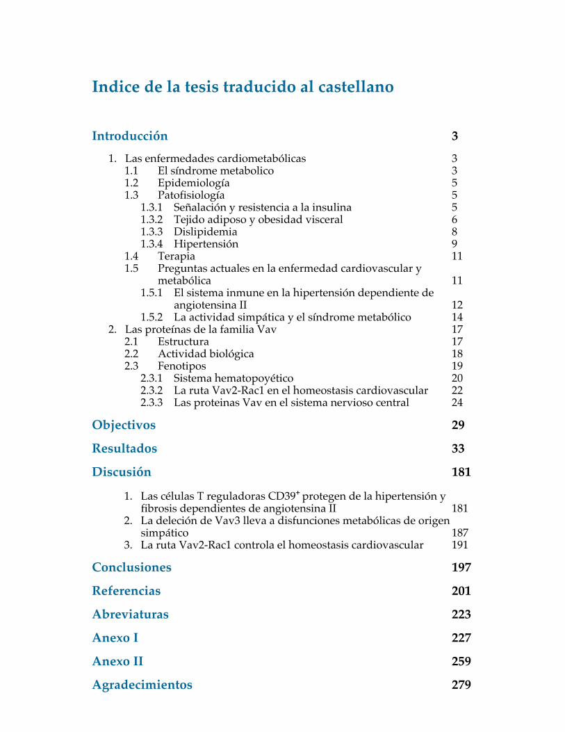
Introducción 3 1. Las enfermedades cardiometabólicas 3
1.1 El síndrome metabolico 3 1.2 Epidemiología 5 1.3 Patofisiología 5
1.3.1 Señalación y resistencia a la insulina 5 1.3.2 Tejido adiposo y obesidad visceral 6 1.3.3 Dislipidemia 8 1.3.4 Hipertensión 9
1.4 Terapia 11 1.5 Preguntas actuales en la enfermedad cardiovascular y
metabólica 11 1.5.1 El sistema inmune en la hipertensión dependiente de
angiotensina II 12 1.5.2 La actividad simpática y el síndrome metabólico 14
2. Las proteínas de la familia Vav 17 2.1 Estructura 17 2.2 Actividad biológica 18 2.3 Fenotipos 19
2.3.1 Sistema hematopoyético 20 2.3.2 La ruta Vav2-Rac1 en el homeostasis cardiovascular 22 2.3.3 Las proteinas Vav en el sistema nervioso central 24
Objectivos 29
Resultados 33
Discusión 181
1. Las células T reguladoras CD39+ protegen de la hipertensión y fibrosis dependientes de angiotensina II 181
2. La deleción de Vav3 lleva a disfunciones metabólicas de origen simpático 187
3. La ruta Vav2-Rac1 controla el homeostasis cardiovascular 191
Conclusiones 197
Referencias 201
Abreviaturas 223
Anexo I 227
Anexo II 259
Agradecimientos 279


Resumen en castellano
Introducción
El síndrome metabólico
Las enfermedades metabólicas tienen una tendencia a ocurrir conjuntamente.
Cuando esto sucede, el riesgo de desarrollar otras enfermedades relacionadas
aumenta de manera significativa. Síndrome metabólico fue descrito inicialmente
como Síndrome X por Gerald Reaven de la Universidad de Stanford en 1988, aunque
antes varios autores venían advirtiendo sobre el riesgo cardiovascular que implicaba
tener dislipidemias, obesidad, hipertensión arterial e intolerancia a la glucosa, por
lo cual se los llamaba el cuarteto de la muerte entre otros. Sin embargo fue el
grupo de Reaven el que confirmó la asociación de estas alteraciones metabólicas con
la resistencia a la insulina, inclusive en personas aparentemente sanas y delgadas
(Reaven, 1988). Desde entonces varias definiciones se han propuesto en el tiempo
y actualmente se habla de "síndrome metabólico", definido como una constelación
de factores metabólicos interconectados que aumentan directamente el riesgo de
desarrollar enfermedad cardiovascular y diabetes de tipo 2. Los elementos centrales
de este síndrome, según las últimas definiciones, son la resistencia a la insulina, la
obesidad visceral, la dislipidemia y la hipertensión (Duvnjak and Duvnjak, 2009).
El síndrome metabólico tiene actualmente una incidencia elevada tanto en países
desarrollados como en vías de desarrollo. La Federación Internacional de Diabetes
ha estimado que alrededor del 25% de la población mundial padece de este síndrome.
263

Resumen en castellano
Patofisiología del síndrome metabólico
Resistencia a la insulina
La insulina es una hormona secretada por las células beta del páncreas en respuesta a
un aumento de los niveles de glucosa en sangre. Su función es regular el metabolismo
de glucosa, lípidos y proteínas en numerosos tejidos como hígado, riñón, cerebro,
músculo, endotelio y tejido adiposo. A través de la interacción con su receptor
de membrana, la insulina favorece la captación de glucosa e inhibe la lipólisis,
además de promover el gasto energético y enviar señales al cerebro para disminuir
la sensación de hambre (Bunner et al., 2014). La resistencia a la insulina es una
condición patológica en la cual esta hormona no es capaz de producir su efecto
fisiológico en los órganos diana. Esto lleva a una reducción en la respuesta a la
glucosa que causa un consecuente aumento de producción de insulina para recuperar
los niveles normales de glicemia. Asimismo, la hiperinsulinemia producida tiene
un efecto perjudicial para muchos órganos, donde rutas de señalización resistentes
y sensibles a insulina se ven desreguladas y conducen a defectos a nivel vascular,
adiposo y hepático. Estas disfunciones aumentan de manera importante el riesgo de
desarrollar diabetes, aterosclerosis y enfermedad del hígado graso (Wang et al., 2004;
Gill et al., 2005).
Obesidad
La obesidad es el factor patogénico más importante, junto con la resistencia a
insulina, en el desarrollo del síndrome metabólico. El aumento de la masa adiposa
lleva a una mayor producción de adipoquinas proinflamatorias, una serie de
moléculas que contribuyen a otras alteraciones metabólicas como resistencia a la
insulina, aterosclerosis, desregulación de la actividad simpática y menor sensación
264

Resumen en castellano
de saciedad, que causa una disminución del gasto energético y una ingesta calórica
excesiva (Enriori et al., 2007). Juntas, estas disfunciones aumentan de manera
significativa el riesgo de sufrir eventos cardiovasculares (Neels and Olefsky, 2006).
Dislipidemia
La dislipidemia es una alteración en la estructura, el metabolismo y la actividad de
lipoproteínas pro- y anti-aterogénicas. Esta condición se caracteriza por un aumento
en los niveles de lipoproteínas de muy baja densidad debido a su mayor producción
y menor degradación. Al mismo tiempo, disminuyen los niveles de lipoproteínas de
alta densidad, que tienen un importante papel antiaterogénico, aumentando así el
riesgo de disfunciones vasculares (Giugliano et al., 2006).
Hipertensión
La hipertensión es una condición patológica donde una desregulación en el
gasto cardíaco o la resistencia vascular producen una tensión arterial elevada,
caracterizada en humanos por valores de presión sistólica ≥ a 140 mmHg y/o
diastólica ≥ 90 mmHg. Es una enfermedad multifactorial en la cual componentes
simpáticos, cardiovasculares y renales tienen un papel regulador muy importante
y se afectan mutuamente. Sin embargo, en la mayoría de los casos la hipertensión
no tiene una causa identificable, y se denomina entonces hipertensión esencial
o idiopática (Bolívar, 2013). Si no se controla, la hipertensión lleva a daño
hemodinámico y disfunción cardíaca y renal.
265

Resumen en castellano
Cuestiones abiertas en las enfermedades cardiometabólicas
El sistema inmune en el desarrollo de la hipertensión
El papel del sistema inmune en la hipertensión ha sido objeto de muchos estudios
en los últimos años, con un particular enfoque sobre el rol de los linfocitos T en
la hipertensión dependiente de angiotensina II (Guzik et al., 2007; Crowley et al.,
2010) y el posible efecto beneficioso de fármacos inmunosupresores en el tratamiento
de la enfermedad cardiovascular (Rodríguez-Iturbe et al., 2001; De Miguel et al.,
2010; Boesen et al., 2010). Las poblaciones proinflamatorias TH1 y TH17 han
sido investigadas como posibles mediadores de la hipertensión dependiente de
angiotensina II y de daño cardiovascular (Shao et al., 2003; Madhur et al., 2010a). Por
otro lado se ha sugerido un papel de los linfocitos T reguladores, una población con
actividad inmunotolerante, en el control de la enfermedad cardiovascular (Kvakan
et al., 2009; Barhoumi et al., 2011). Sin embargo, a pesar de las evidencias
obtenidas hasta la fecha, los mecanismos y las poblaciones inmunes involucradas
en el desarrollo de la hipertensión siguen siendo objeto de discusión.
La actividad simpática en el síndrome metabólico
El sistema nervioso simpático regula varias funciones metabólicas a través de la
liberación a nivel local de catecolaminas en los terminales nerviosos o aumentando
su concentración en circulación por estimulación de la glándula suprarrenal. Las
catecolaminas se unen a receptores adrenérgicos α y β que se encuentran expresados
en el sistema cardiovascular, en el páncreas, en el hígado y en el tejido adiposo
entre otros. Por medio de esta señalización, el sistema nervioso simpático regula
la actividad metabólica basal y responde a cambios en la necesidad energética del
organismo (Kelley et al., 2005; Asensio et al., 2005). Estudios epidemiológicos
sugieren una conexión entre una actividad simpática sostenida en el tiempo y el
266

Resumen en castellano
desarrollo de trastornos metabólicos. Sin embargo, la heterogeneidad de los grupos
de individuos usados en los estudios ha llevado a resultados controvertidos y poco
claros (Guyenet, 2006). Para abordar estas cuestiones de un modo genéticamente
"limpio", hemos utilizado ratones transgénicos con defectos a nivel del sistema
inmune, de la actividad simpática y de la homeostasis cardiovascular, debido a la
ausencia de las proteínas de la familia Vav.
Las proteínas Vav
Las proteínas de la familia Vav son un grupo de moléculas intercambiadoras de
nucleótido de guanina para las GTPasas de las familias Rho y Rac cuya activación
es modulada por fosforilación. En vertebrados existen tres miembros llamados Vav1,
Vav2 y Vav3 (Bustelo, 2000). La función principal de estas proteínas es el intercambio
de GDP por GTP que permite la activación de las GTPasas Rho/Rac. A través de esta
activación, las proteínas Vav median procesos como cambios a nivel citoesquelético,
activan rutas de señalización y factores de transcripción. Además, poseen funciones
independientes de la actividad de intercambio (Bustelo, 2000). Estas proteínas se
regulan por múltiples eventos de fosforilación que permiten la transición de una
conformación cerrada autoinhibitoria a una abierta y catalíticamente competente.
Fenotipos
Vav1 tiene un patrón de expresión restringido al sistema hematopoyético, mientras
que Vav2 y Vav3 se expresan de manera casi ubicua. Los ratones deficientes en
cada una de las tres proteínas y los knock-out doble y triple son viables y no tienen
defectos en crecimiento o fertilidad. Sin embargo, muchos procesos fisiológicos
se ven afectados por la ausencia de las proteínas Vav. Algunos fenotipos son de
particular interés para nuestro estudio.
267
Resumen en castellano
Desarrollo del sistema hematopoyético
Las proteínas Vav están involucradas en el desarrollo y la función de las células del
sistema hematopoyético. Vav1 en particular participa en procesos implicados en
el desarrollo linfocitario y tienen un papel clave en las respuestas del receptor de
células T (Fischer et al., 1995; Turner et al. 1997). Ratones KO para Vav1 sufren
defectos en la selección positiva y negativa durante la timopoyesis, lo que resulta en
una acumulación de timocitos CD4−CD8− doble negativos y una menor producción
de linfocitos T maduros CD4+ y CD8+ con la subsecuente linfopenia. Vav3 compensa
de manera parcial este defecto, puesto que ratones que carecen de Vav1 y Vav3 tienen
un defecto aun mayor (Fujukawa et al., 2003).
Vav2 en la homeostasis cardiovascular
Vav2 tiene un papel clave en la regulación de la contractilidad arterial en respuesta
al óxido nítrico, una molécula con un importante efecto vasodilatador. Por un lado,
el óxido nítrico aumenta los niveles de GMP cíclico en la célula, un evento que
permite la activación de la quinasa I dependiente de cGMP, que inactiva a RhoA
por fosforilación. Este estímulo está controlado por la proteína fosfodiesterasa 5
(PDE5), que hidroliza el GMP cíclico a GMP. Por otro lado, el óxido nítrico estimula la
fosforilación de Vav2 por quinasas de la familia Src. La activación de Rac1 por Vav2
promueve la inhibición de la PDE5 a través de su efector Pak1. De esta manera,
en respuesta al óxido nítrico, se bloquea la degradación del cGMP y se obtiene
una inhibición de RhoA que permite una vasodilatación a nivel tisular (Sauzeau
et al., 2010b). En ausencia de Vav2, el tejido muscular no se relaja de manera
correcta. En ratones Vav2−/−, esto conduce a hipertensión, aumento de los niveles
de angiotensina II y catecolaminas en sangre, y fibrosis cardiorenal (Sauzeau et al.,
2007). El tratamiento de ratones KO para Vav2 con el inhibidor de PDE5 sildenafilo
268

Resumen en castellano
previene la hipertensión, la fibrosis cardiorenal y el aumento en catecolaminas. Sin
embargo, parte de fenotipo no está afectado por este tratamiento, indicando que hay
otros componentes no limitados al sistema vascular en el fenotipo de los ratones
Vav2−/−. Para corroborar la especificidad del fenotipo vascular en el desarrollo de
la hipertensión detectada en los ratones Vav2−/− hace falta entonces un mutante
restringido al tejido muscular liso.
Rol de Vav3 en el control de la actividad simpática
Vav3 se expresa en distintas regiones del cerebro y está implicado en numerosos
procesos de migración y desarrollo neuronal (Cowan et al., 2005; Yamauchi et
al., 2004; Quevedo et al., 2010). De particular relevancia es su expresión en la
médula ventrolateral caudal. Esta región tiene una función inhibitoria a través de
las neuronas GABAérgicas sobre el área rostral de la médula ventrolateral, el sitio
principal de control de las neuronas eferentes implicadas en la regulación de la
presión arterial. En ausencia de Vav3 los axones GABAérgicos que provienen de
la región caudal no consiguen formar las sinapsis adecuadas para la señalización a la
región rostral (Sauzeau et al., 2010a). Al no poderse inhibir la actividad de la médula
ventrolateral rostral, el tono simpático sube y las funciones cardíaca y respiratoria
se encuentran activadas de manera excesiva. Los ratones Vav3−/− presentan como
consecuencia una mayor actividad simpática que lleva a la producción de mayores
niveles de catecolaminas y angiotensina II, hipertensión, daño cardiovasculo-renal
y aumento del ritmo respiratorio (Sauzeau et al., 2006). Esta actividad simpática
crónica encontrada en los ratones Vav3−/− los convierte en un buen modelo para
el análisis del papel de la excitación simpática en el desarrollo de enfermedades
metabólicas.
269

Resumen en castellano
270

Resumen en castellano
Conclusiones
1. Las células T reguladoras CD39+ protegen de la enfermedad cardiovascular
dependiente de angiotensina II induciendo apoptosis en los neutrófilos. La
supresión de esta población proinflamatoria previene la fibrosis cardiorenal y
el aumento excesivo de presión arterial.
2. Un aumento en la relación T reguladoras/T helper les permite a las primeras
llevar a cabo su actividad inmunomoduladora. Aumentar esta relación in vivo
es beneficioso en ratones inmunocompetentes.
3. Usando ratones KO para Vav3 observamos que una actividad simpática crónica
es responsable de efectos dependientes de la dieta, llevando a enfermedad de
hígado graso y resistencia a la insulina en dieta estándar pero protegiendo del
desarrollo de síndrome metabólico en dieta rica en grasa.
4. La ruta Vav2-Rac1 es importante en el músculo liso para migración,
proliferación y respuesta al óxido nítrico. La ausencia de Rac1 en este tejido
causa hipertensión dependiente de angiotensina II.
277

References
Afshari, F. T., Kwok, J. C., and Fawcett, J. W. (2010). Astrocyte-produced ephrins
inhibit schwann cell migration via VAV2 signaling. The Journal of neuroscience : the
official journal of the Society for Neuroscience, 30(12):4246–4255.
Alberti, K. G. and Zimmet, P. Z. (1998). Definition, diagnosis and classification
of diabetes mellitus and its complications. Part 1: diagnosis and classification of
diabetes mellitus provisional report of a WHO consultation. Diabetic medicine : a
journal of the British Diabetic Association, 15(7):539–553.
Alvarez, P. A., Egozcue, J., Sleiman, J., Moretti, L., Di Girolamo, G., and Keller, G. A.
(2012). Severe neutropenia in a renal transplant patient suggesting an interaction
between mycophenolate and fenofibrate. Current drug safety, 7(1):24–29.
Asensio, C., Jimenez, M., Kühne, F., Rohner-Jeanrenaud, F., and Muzzin, P. (2005).
The lack of beta-adrenoceptors results in enhanced insulin sensitivity in mice
exhibiting increased adiposity and glucose intolerance. Diabetes, 54(12):3490–3495.
Barhoumi, T., Kasal, D. A., Li, M. W., Shbat, L., Laurant, P., Neves, M. F., Paradis,
P., and Schiffrin, E. L. (2011). T regulatory lymphocytes prevent angiotensin
II-induced hypertension and vascular injury. Hypertension, 57(3):469–476.
Berndt, N., Hamilton, A. D., and Sebti, S. M. (2011). Targeting protein prenylation
for cancer therapy. Nature reviews. Cancer, 11(11):775–791.
201

References
Blessing, W. W. (1988). Depressor neurons in rabbit caudal medulla act via GABA
receptors in rostral medulla. The American journal of physiology, 254(4 Pt 2):H686–92.
Boesen, E. I., Williams, D. L., Pollock, J. S., and Pollock, D. M. (2010).
Immunosuppression with mycophenolate mofetil attenuates the development of
hypertension and albuminuria in deoxycorticosterone acetate-salt hypertensive
rats. Clinical and experimental pharmacology & physiology, 37(10):1016–1022.
Bolívar, J. J. (2013). Essential Hypertension: An Approach to Its Etiology and
Neurogenic Pathophysiology. International journal of hypertension, 2013:547809.
Bond, M., Wu, Y.-J., Sala-Newby, G. B., and Newby, A. C. (2008). Rho GTPase,
Rac1, regulates Skp2 levels, vascular smooth muscle cell proliferation, and intima
formation in vitro and in vivo. Cardiovascular research, 80(2):290–298.
Borsellino, G., Kleinewietfeld, M., Di Mitri, D., Sternjak, A., Diamantini, A.,
Giometto, R., Höpner, S., Centonze, D., Bernardi, G., Dell’Acqua, M. L.,
Rossini, P. M., Battistini, L., Rötzschke, O., and Falk, K. (2007). Expression of
ectonucleotidase CD39 by Foxp3+ Treg cells: hydrolysis of extracellular ATP and
immune suppression. Blood, 110(4):1225–1232.
Brantley-Sieders, D. M., Zhuang, G., Vaught, D., Freeman, T., Hwang, Y., Hicks, D.,
and Chen, J. (2009). Host deficiency in Vav2/3 guanine nucleotide exchange factors
impairs tumor growth, survival, and angiogenesis in vivo. Molecular cancer research
: MCR, 7(5):615–623.
Brown, M. S. and Goldstein, J. L. (2008). Selective versus total insulin resistance: a
pathogenic paradox. Cell metabolism, 7(2):95–96.
Bubeck Wardenburg, J., Pappu, R., Bu, J. Y., Mayer, B., Chernoff, J., Straus, D., and
202

References
Chan, A. C. (1998). Regulation of PAK activation and the T cell cytoskeleton by the
linker protein SLP-76. Immunity, 9(5):607–616.
Bunner, A. E., Chandrasekera, P. C., and Barnard, N. D. (2014). Knockout mouse
models of insulin signaling: Relevance past and future. World journal of diabetes,
5(2):146–159.
Bustelo, X. R. (2000). Regulatory and signaling properties of the Vav family. Molecular
and cellular biology, 20(5):1461–1477.
Bustelo, X. R. (2001). Vav proteins, adaptors and cell signaling. Oncogene,
20(44):6372–6381.
Bustelo, X. R. (2002). Understanding Rho/Rac biology in T-cells using animal
models. BioEssays : news and reviews in molecular, cellular and developmental biology,
24(7):602–612.
Bustelo, X. R. (2012). Vav Family. Encyclopedia of Signaling Molecules.
Caballero, A. E. (2004). Endothelial dysfunction, inflammation, and insulin
resistance: a focus on subjects at risk for type 2 diabetes. Current diabetes reports,
4(4):237–246.
Caloca, M. J., Zugaza, J. L., and Bustelo, X. R. (2008). Mechanistic analysis
of the amplification and diversification events induced by Vav proteins in
B-lymphocytes. The Journal of biological chemistry, 283(52):36454–36464.
Cameron, A. J., Shaw, J. E., and Zimmet, P. Z. (2004). The metabolic syndrome:
prevalence in worldwide populations. Endocrinology and metabolism clinics of North
America, 33(2):351–75– table of contents.
Cannon, B. and Nedergaard, J. (2004). Brown adipose tissue: function and
physiological significance. Physiological reviews, 84(1):277–359.
203

References
Cella, M., Fujikawa, K., Tassi, I., Kim, S., Latinis, K., Nishi, S., Yokoyama, W.,
Colonna, M., and Swat, W. (2004). Differential requirements for Vav proteins
in DAP10- and ITAM-mediated NK cell cytotoxicity. The Journal of experimental
medicine, 200(6):817–823.
Chen, X., Priatel, J. J., Chow, M. T., and Teh, H.-S. (2008). Preferential development of
CD4 and CD8 T regulatory cells in RasGRP1-deficient mice. Journal of immunology
(Baltimore, Md. : 1950), 180(9):5973–5982.
Chen, Y., Corriden, R., Inoue, Y., Yip, L., Hashiguchi, N., Zinkernagel, A., Nizet, V.,
Insel, P. A., and Junger, W. G. (2006). ATP release guides neutrophil chemotaxis via
P2Y2 and A3 receptors. Science (New York, N.Y.), 314(5806):1792–1795.
Chrousos, G. P. (2009). Stress and disorders of the stress system. Nature reviews.
Endocrinology, 5(7):374–381.
Cinti, S., Mitchell, G., Barbatelli, G., Murano, I., Ceresi, E., Faloia, E., Wang, S., Fortier,
M., Greenberg, A. S., and Obin, M. S. (2005). Adipocyte death defines macrophage
localization and function in adipose tissue of obese mice and humans. Journal of
lipid research, 46(11):2347–2355.
Colomba, A., Giuriato, S., Dejean, E., Thornber, K., Delsol, G., Tronchère, H.,
Meggetto, F., Payrastre, B., and Gaits-Iacovoni, F. (2011). Inhibition of Rac controls
NPM-ALK-dependent lymphoma development and dissemination. Blood cancer
journal, 1(6):e21.
Costello, P. S., Walters, A. E., Mee, P. J., Turner, M., Reynolds, L. F., Prisco, A., Sarner,
N., Zamoyska, R., and Tybulewicz, V. L. (1999). The Rho-family GTP exchange
factor Vav is a critical transducer of T cell receptor signals to the calcium, ERK, and
NF-kappaB pathways. Proceedings of the National Academy of Sciences of the United
States of America, 96(6):3035–3040.
204

References
Cowan, C. W., Shao, Y. R., Sahin, M., Shamah, S. M., Lin, M. Z., Greer, P. L., Gao,
S., Griffith, E. C., Brugge, J. S., and Greenberg, M. E. (2005). Vav family GEFs link
activated Ephs to endocytosis and axon guidance. Neuron, 46(2):205–217.
Crespo, P., Bustelo, X. R., Aaronson, D. S., Coso, O. A., López-Barahona, M., Barbacid,
M., and Gutkind, J. S. (1996). Rac-1 dependent stimulation of the JNK/SAPK
signaling pathway by Vav. Oncogene, 13(3):455–460.
Crowley, S. D., Song, Y.-S., Lin, E. E., Griffiths, R., Kim, H.-S., and Ruiz, P.
(2010). Lymphocyte responses exacerbate angiotensin II-dependent hypertension.
American journal of physiology. Regulatory, integrative and comparative physiology,
298(4):R1089–97.
De Miguel, C., Das, S., Lund, H., and Mattson, D. L. (2010). T lymphocytes mediate
hypertension and kidney damage in Dahl salt-sensitive rats. American journal of
physiology. Regulatory, integrative and comparative physiology, 298(4):R1136–42.
Deaglio, S., Dwyer, K. M., Gao, W., Friedman, D., Usheva, A., Erat, A., Chen, J.-F.,
Enjyoji, K., Linden, J., Oukka, M., Kuchroo, V. K., Strom, T. B., and Robson,
S. C. (2007). Adenosine generation catalyzed by CD39 and CD73 expressed
on regulatory T cells mediates immune suppression. The Journal of experimental
medicine, 204(6):1257–1265.
Deaglio, S. and Robson, S. C. (2011). Ectonucleotidases as regulators of purinergic
signaling in thrombosis, inflammation, and immunity. Advances in pharmacology
(San Diego, Calif.), 61:301–332.
Desroches, S. and Lamarche, B. (2007). The evolving definitions and increasing
prevalence of the metabolic syndrome. Applied physiology, nutrition, and metabolism
= Physiologie appliquée, nutrition et métabolisme, 32(1):23–32.
205
References
Duvnjak, L. and Duvnjak, M. (2009). The metabolic syndrome - an ongoing story.
Journal of physiology and pharmacology : an official journal of the Polish Physiological
Society, 60 Suppl 7:19–24.
Eckel, R. H., Alberti, K. G. M. M., Grundy, S. M., and Zimmet, P. Z. (2010). The
metabolic syndrome. Lancet, 375(9710):181–183.
Eckel, R. H., Grundy, S. M., and Zimmet, P. Z. (2005). The metabolic syndrome.
Lancet, 365(9468):1415–1428.
Enriori, P. J., Evans, A. E., Sinnayah, P., Jobst, E. E., Tonelli-Lemos, L., Billes, S. K.,
Glavas, M. M., Grayson, B. E., Perello, M., Nillni, E. A., Grove, K. L., and Cowley,
M. A. (2007). Diet-induced obesity causes severe but reversible leptin resistance in
arcuate melanocortin neurons. Cell metabolism, 5(3):181–194.
Faccio, R., Teitelbaum, S. L., Fujikawa, K., Chappel, J., Zallone, A., Tybulewicz, V. L.,
Ross, F. P., and Swat, W. (2005). Vav3 regulates osteoclast function and bone mass.
Nature medicine, 11(3):284–290.
Fischer, K. D., Zmuldzinas, A., Gardner, S., Barbacid, M., Bernstein, A., and
Guidos, C. (1995). Defective T-cell receptor signalling and positive selection of
Vav-deficient CD4+ CD8+ thymocytes. Nature, 374(6521):474–477.
Fujikawa, K., Miletic, A. V., Alt, F. W., Faccio, R., Brown, T., Hoog, J., Fredericks, J.,
Nishi, S., Mildiner, S., Moores, S. L., Brugge, J., Rosen, F. S., and Swat, W. (2003).
Vav1/2/3-null mice define an essential role for Vav family proteins in lymphocyte
development and activation but a differential requirement in MAPK signaling in
T and B cells. The Journal of experimental medicine, 198(10):1595–1608.
Gakidis, M. A. M., Cullere, X., Olson, T., Wilsbacher, J. L., Zhang, B., Moores, S. L.,
Ley, K., Swat, W., Mayadas, T., and Brugge, J. S. (2004). Vav GEFs are required
206

References
for beta2 integrin-dependent functions of neutrophils. The Journal of cell biology,
166(2):273–282.
Gasmi, L., McLennan, A. G., and Edwards, S. W. (1996). The diadenosine
polyphosphates Ap3A and Ap4A and adenosine triphosphate interact with
granulocyte-macrophage colony-stimulating factor to delay neutrophil apoptosis:
implications for neutrophil: platelet interactions during inflammation. Blood,
87(8):3442–3449.
Gehrig, S., Duerr, J., Weitnauer, M., Wagner, C. J., Graeber, S. Y., Schatterny, J.,
Hirtz, S., Belaaouaj, A., Dalpke, A. H., Schultz, C., and Mall, M. A. (2014).
Lack of Neutrophil Elastase Reduces Inflammation, Mucus Hypersecretion, and
Emphysema, but Not Mucus Obstruction, in Mice with Cystic Fibrosis-like Lung
Disease. American journal of respiratory and critical care medicine, 189(9):1082–1092.
Gill, H., Mugo, M., Whaley-Connell, A., Stump, C., and Sowers, J. R. (2005). The key
role of insulin resistance in the cardiometabolic syndrome. The American journal of
the medical sciences, 330(6):290–294.
Ginsberg, H. N., Zhang, Y.-L., and Hernandez-Ono, A. (2005). Regulation of
plasma triglycerides in insulin resistance and diabetes. Archives of medical research,
36(3):232–240.
Giorda, C., Appendino, M., Mason, M. G., Imperiale, E., and Pagano, G. (1995).
Alpha 1-blocker doxazosin improves peripheral insulin sensitivity in diabetic
hypertensive patients. Metabolism: clinical and experimental, 44(5):673–676.
Giugliano, D., Ceriello, A., and Esposito, K. (2006). The effects of diet on
inflammation: emphasis on the metabolic syndrome. Journal of the American College
of Cardiology, 48(4):677–685.
207

References
Goldberg, I. J., Eckel, R. H., and Abumrad, N. A. (2009). Regulation of fatty acid
uptake into tissues: lipoprotein lipase- and CD36-mediated pathways. Journal of
lipid research, 50 Suppl:S86–90.
Guyenet, P. G. (2006). The sympathetic control of blood pressure. Nature reviews.
Neuroscience, 7(5):335–346.
Guzik, T. J., Hoch, N. E., Brown, K. A., McCann, L. A., Rahman, A., Dikalov, S.,
Goronzy, J., Weyand, C., and Harrison, D. G. (2007). Role of the T cell in the genesis
of angiotensin II induced hypertension and vascular dysfunction. The Journal of
experimental medicine, 204(10):2449–2460.
Hale, C. F., Dietz, K. C., Varela, J. A., Wood, C. B., Zirlin, B. C., Leverich, L. S., Greene,
R. W., and Cowan, C. W. (2011). Essential role for vav Guanine nucleotide exchange
factors in brain-derived neurotrophic factor-induced dendritic spine growth and
synapse plasticity. The Journal of neuroscience : the official journal of the Society for
Neuroscience, 31(35):12426–12436.
Hall, A. B., Gakidis, M. A. M., Glogauer, M., Wilsbacher, J. L., Gao, S., Swat, W., and
Brugge, J. S. (2006). Requirements for Vav guanine nucleotide exchange factors and
Rho GTPases in FcgammaR- and complement-mediated phagocytosis. Immunity,
24(3):305–316.
Hinz, B. (2009). Tissue stiffness, latent TGF-beta1 activation, and mechanical signal
transduction: implications for the pathogenesis and treatment of fibrosis. Current
rheumatology reports, 11(2):120–126.
Hori, S., Nomura, T., and Sakaguchi, S. (2003). Control of regulatory T cell
development by the transcription factor Foxp3. Science (New York, N.Y.),
299(5609):1057–1061.
208
References
Hunter, S. G., Zhuang, G., Brantley-Sieders, D., Swat, W., Cowan, C. W., and Chen, J.
(2006). Essential role of Vav family guanine nucleotide exchange factors in EphA
receptor-mediated angiogenesis. Molecular and cellular biology, 26(13):4830–4842.
Iannitti, R. G., Carvalho, A., Cunha, C., De Luca, A., Giovannini, G., Casagrande,
A., Zelante, T., Vacca, C., Fallarino, F., Puccetti, P., Massi-Benedetti, C., Defilippi,
G., Russo, M., Porcaro, L., Colombo, C., Ratclif, L., De Benedictis, F. M., and
Romani, L. (2013). Th17/Treg imbalance in murine cystic fibrosis is linked to
indoleamine 2,3-dioxygenase deficiency but corrected by kynurenines. American
journal of respiratory and critical care medicine, 187(6):609–620.
Isomaa, B., Almgren, P., Tuomi, T., Forsén, B., Lahti, K., Nissén, M., Taskinen, M. R.,
and Groop, L. (2001). Cardiovascular morbidity and mortality associated with the
metabolic syndrome. Diabetes care, 24(4):683–689.
Junger, W. G. (2011). Immune cell regulation by autocrine purinergic signalling.
Nature reviews. Immunology, 11(3):201–212.
Jüttner, B., Bencel, M., Weissig, A., Studzinski, A., Stenger, K., and Scheinichen,
D. (2009). Mycophenolic acid inhibits PMA-induced activation of the neutrophil
respiratory burst. Transplant infectious disease : an official journal of the Transplantation
Society, 11(3):235–240.
Kajantie, E. and Räikkönen, K. (2010). Early life predictors of the physiological stress
response later in life. Neuroscience and biobehavioral reviews, 35(1):23–32.
Kannel, W. B. (2000). Elevated systolic blood pressure as a cardiovascular risk factor.
The American journal of cardiology, 85(2):251–255.
Kaur, J. (2014). A Comprehensive Review on Metabolic Syndrome. Cardiology research
and practice, 2014:943162.
209

References
Kelley, A. E., Baldo, B. A., Pratt, W. E., and Will, M. J. (2005).
Corticostriatal-hypothalamic circuitry and food motivation: integration of
energy, action and reward. Physiology & behavior, 86(5):773–795.
Klop, B., Elte, J. W. F., and Cabezas, M. C. (2013). Dyslipidemia in obesity:
mechanisms and potential targets. Nutrients, 5(4):1218–1240.
Kohno, Y., Sei, Y., Koshiba, M., Kim, H. O., and Jacobson, K. A. (1996).
Induction of apoptosis in HL-60 human promyelocytic leukemia cells by adenosine
A(3) receptor agonists. Biochemical and biophysical research communications,
219(3):904–910.
Kolovou, G. D., Anagnostopoulou, K. K., Salpea, K. D., and Mikhailidis, D. P. (2007).
The prevalence of metabolic syndrome in various populations. The American
journal of the medical sciences, 333(6):362–371.
Koziak, K., Sévigny, J., Robson, S. C., Siegel, J. B., and Kaczmarek, E. (1999).
Analysis of CD39/ATP diphosphohydrolase (ATPDase) expression in endothelial
cells, platelets and leukocytes. Thrombosis and haemostasis, 82(5):1538–1544.
Kvakan, H., Kleinewietfeld, M., Qadri, F., Park, J.-K., Fischer, R., Schwarz, I., Rahn,
H.-P., Plehm, R., Wellner, M., Elitok, S., Gratze, P., Dechend, R., Luft, F. C., and
Muller, D. N. (2009). Regulatory T cells ameliorate angiotensin II-induced cardiac
damage. Circulation, 119(22):2904–2912.
Lambert, G. W., Straznicky, N. E., Lambert, E. A., Dixon, J. B., and Schlaich,
M. P. (2010). Sympathetic nervous activation in obesity and the metabolic
syndrome–causes, consequences and therapeutic implications. Pharmacology &
therapeutics, 126(2):159–172.
210
References
Lifton, R. P., Gharavi, A. G., and Geller, D. S. (2001). Molecular mechanisms of human
hypertension. Cell, 104(4):545–556.
Locke, R. M., Rial, E., Scott, I. D., and Nicholls, D. G. (1982). Fatty acids as acute
regulators of the proton conductance of hamster brown-fat mitochondria. European
journal of biochemistry / FEBS, 129(2):373–380.
Loirand, G., Sauzeau, V., and Pacaud, P. (2013). Small G proteins in the
cardiovascular system: physiological and pathological aspects. Physiological
reviews, 93(4):1659–1720.
Loirand, G., Scalbert, E., Bril, A., and Pacaud, P. (2008). Rho exchange factors in the
cardiovascular system. Current opinion in pharmacology, 8(2):174–180.
Lorell, B. H. and Carabello, B. A. (2000). Left ventricular hypertrophy: pathogenesis,
detection, and prognosis. Circulation, 102(4):470–479.
Madden, C. J. (2012). Glucoprivation in the ventrolateral medulla decreases brown
adipose tissue sympathetic nerve activity by decreasing the activity of neurons in
raphe pallidus. American journal of physiology. Regulatory, integrative and comparative
physiology, 302(2):R224–32.
Madhur, M. S., Lob, H. E., McCann, L. A., Iwakura, Y., Blinder, Y., Guzik, T. J.,
and Harrison, D. G. (2010a). Interleukin 17 promotes angiotensin II-induced
hypertension and vascular dysfunction. Hypertension, 55(2):500–507.
Madhur, M. S., Lob, H. E., McCann, L. A., Iwakura, Y., Blinder, Y., Guzik, T. J.,
and Harrison, D. G. (2010b). Interleukin 17 promotes angiotensin II-induced
hypertension and vascular dysfunction. Hypertension, 55(2):500–507.
Malhotra, A., Kang, B. P., Cheung, S., Opawumi, D., and Meggs, L. G. (2001).
211
References
Angiotensin II promotes glucose-induced activation of cardiac protein kinase C
isozymes and phosphorylation of troponin I. Diabetes, 50(8):1918–1926.
Maliszewski, C. R., Delespesse, G. J., Schoenborn, M. A., Armitage, R. J., Fanslow,
W. C., Nakajima, T., Baker, E., Sutherland, G. R., Poindexter, K., and Birks,
C. (1994). The CD39 lymphoid cell activation antigen. Molecular cloning
and structural characterization. Journal of immunology (Baltimore, Md. : 1950),
153(8):3574–3583.
Manetz, T. S., Gonzalez-Espinosa, C., Arudchandran, R., Xirasagar, S., Tybulewicz,
V., and Rivera, J. (2001). Vav1 regulates phospholipase cgamma activation and
calcium responses in mast cells. Molecular and cellular biology, 21(11):3763–3774.
Marvar, P. J., Thabet, S. R., Guzik, T. J., Lob, H. E., McCann, L. A., Weyand, C.,
Gordon, F. J., and Harrison, D. G. (2010). Central and peripheral mechanisms
of T-lymphocyte activation and vascular inflammation produced by angiotensin
II-induced hypertension. Circulation research, 107(2):263–270.
Maser, R. E., Lenhard, M. J., Kolm, P., and Edwards, D. G. (2013). Direct renin
inhibition improves parasympathetic function in diabetes. Diabetes, obesity &
metabolism, 15(1):28–34.
Menacho-Márquez, M., García-Escudero, R., Ojeda, V., Abad, A., Delgado, P., Costa,
C., Ruiz, S., Alarcón, B., Paramio, J. M., and Bustelo, X. R. (2013). The Rho exchange
factors Vav2 and Vav3 favor skin tumor initiation and promotion by engaging
extracellular signaling loops. PLoS biology, 11(7):e1001615.
Mervaala, E., Müller, D. N., Park, J. K., Dechend, R., Schmidt, F., Fiebeler, A.,
Bieringer, M., Breu, V., Ganten, D., Haller, H., and Luft, F. C. (2000). Cyclosporin A
protects against angiotensin II-induced end-organ damage in double transgenic
212

References
rats harboring human renin and angiotensinogen genes. Hypertension, 35(1 Pt
2):360–366.
Miletic, A. V., Graham, D. B., Montgrain, V., Fujikawa, K., Kloeppel, T., Brim, K.,
Weaver, B., Schreiber, R., Xavier, R., and Swat, W. (2007). Vav proteins control
MyD88-dependent oxidative burst. Blood, 109(8):3360–3368.
Morrison, S. F. (1999). RVLM and raphe differentially regulate sympathetic outflows
to splanchnic and brown adipose tissue. The American journal of physiology, 276(4
Pt 2):R962–73.
Morrison, S. F., Nakamura, K., and Madden, C. J. (2008). Central control of
thermogenesis in mammals. Experimental physiology, 93(7):773–797.
Muller, D. N., Shagdarsuren, E., Park, J.-K., Dechend, R., Mervaala, E., Hampich,
F., Fiebeler, A., Ju, X., Finckenberg, P., Theuer, J., Viedt, C., Kreuzer, J., Heidecke,
H., Haller, H., Zenke, M., and Luft, F. C. (2002). Immunosuppressive treatment
protects against angiotensin II-induced renal damage. The American journal of
pathology, 161(5):1679–1693.
Murata, T., Ohnishi, H., Okazawa, H., Murata, Y., Kusakari, S., Hayashi, Y.,
Miyashita, M., Itoh, H., Oldenborg, P.-A., Furuya, N., and Matozaki, T. (2006).
CD47 promotes neuronal development through Src- and FRG/Vav2-mediated
activation of Rac and Cdc42. The Journal of neuroscience : the official journal of the
Society for Neuroscience, 26(48):12397–12407.
Neels, J. G. and Olefsky, J. M. (2006). Inflamed fat: what starts the fire? The Journal of
clinical investigation, 116(1):33–35.
Nogueras, F., Espinosa, M. D., Mansilla, A., Torres, J. T., Cabrera, M. A., and
213

References
Martín-Vivaldi, R. (2005). Mycophenolate mofetil-induced neutropenia in liver
transplantation. Transplantation proceedings, 37(3):1509–1511.
O’Rourke, L. M., Tooze, R., Turner, M., Sandoval, D. M., Carter, R. H., Tybulewicz,
V. L., and Fearon, D. T. (1998). CD19 as a membrane-anchored adaptor protein of
B lymphocytes: costimulation of lipid and protein kinases by recruitment of Vav.
Immunity, 8(5):635–645.
Pang, J., Chan, D. C., and Watts, G. F. (2014). Origin and therapy for
hypertriglyceridaemia in type 2 diabetes. World journal of diabetes, 5(2):165–175.
Pardo, A., Barrios, R., Gaxiola, M., Segura-Valdez, L., Carrillo, G., Estrada, A.,
Mejía, M., and Selman, M. (2000). Increase of lung neutrophils in hypersensitivity
pneumonitis is associated with lung fibrosis. American journal of respiratory and
critical care medicine, 161(5):1698–1704.
Pearce, A. C., Senis, Y. A., Billadeau, D. D., Turner, M., Watson, S. P., and Vigorito,
E. (2004). Vav1 and vav3 have critical but redundant roles in mediating platelet
activation by collagen. The Journal of biological chemistry, 279(52):53955–53962.
Platten, M., Youssef, S., Hur, E. M., Ho, P. P., Han, M. H., Lanz, T. V., Phillips, L. K.,
Goldstein, M. J., Bhat, R., Raine, C. S., Sobel, R. A., and Steinman, L. (2009).
Blocking angiotensin-converting enzyme induces potent regulatory T cells and
modulates TH1- and TH17-mediated autoimmunity. Proceedings of the National
Academy of Sciences of the United States of America, 106(35):14948–14953.
Quevedo, C., Sauzeau, V., Menacho-Márquez, M., Castro-Castro, A., and Bustelo,
X. R. (2010). Vav3-deficient mice exhibit a transient delay in cerebellar
development. Molecular biology of the cell, 21(6):1125–1139.
Rahmouni, K., Correia, M. L. G., Haynes, W. G., and Mark, A. L. (2005).
214

References
Obesity-associated hypertension: new insights into mechanisms. Hypertension,
45(1):9–14.
Reaven, G. M. (1988). Banting lecture 1988. Role of insulin resistance in human
disease. Diabetes, 37(12):1595–1607.
Reynolds, L. F., Smyth, L. A., Norton, T., Freshney, N., Downward, J., Kioussis, D.,
and Tybulewicz, V. L. J. (2002). Vav1 transduces T cell receptor signals to the
activation of phospholipase C-gamma1 via phosphoinositide 3-kinase-dependent
and -independent pathways. The Journal of experimental medicine, 195(9):1103–1114.
Robidoux, J., Martin, T. L., and Collins, S. (2004). Beta-adrenergic receptors and
regulation of energy expenditure: a family affair. Annual review of pharmacology
and toxicology, 44(1):297–323.
Rodríguez-Iturbe, B., Pons, H., Quiroz, Y., Gordon, K., Rincón, J., Chávez, M., Parra,
G., Herrera-Acosta, J., Gómez-Garre, D., Largo, R., Egido, J., and Johnson, R. J.
(2001). Mycophenolate mofetil prevents salt-sensitive hypertension resulting from
angiotensin II exposure. Kidney international, 59(6):2222–2232.
Sauzeau, V., Horta-Junior, J. A. C., Riolobos, A. S., Fernández, G., Sevilla, M. A.,
López, D. E., Montero, M. J., Rico, B., and Bustelo, X. R. (2010a). Vav3 is involved in
GABAergic axon guidance events important for the proper function of brainstem
neurons controlling cardiovascular, respiratory, and renal parameters. Molecular
biology of the cell, 21(23):4251–4263.
Sauzeau, V., Jerkic, M., López-Novoa, J. M., and Bustelo, X. R. (2007). Loss of Vav2
proto-oncogene causes tachycardia and cardiovascular disease in mice. Molecular
biology of the cell, 18(3):943–952.
Sauzeau, V., Sevilla, M. A., Montero, M. J., and Bustelo, X. R. (2010b). The Rho/Rac
215

References
exchange factor Vav2 controls nitric oxide-dependent responses in mouse vascular
smooth muscle cells. The Journal of clinical investigation, 120(1):315–330.
Sauzeau, V., Sevilla, M. A., Rivas-Elena, J. V., de Alava, E., Montero, M. J.,
López-Novoa, J. M., and Bustelo, X. R. (2006). Vav3 proto-oncogene deficiency
leads to sympathetic hyperactivity and cardiovascular dysfunction. Nature
medicine, 12(7):841–845.
Saveliev, A., Vanes, L., Ksionda, O., Rapley, J., Smerdon, S. J., Rittinger, K., and
Tybulewicz, V. L. J. (2009). Function of the nucleotide exchange activity of vav1
in T cell development and activation. Science signaling, 2(101):ra83–ra83.
Savontaus, E., Fagerholm, V., Rahkonen, O., and Scheinin, M. (2008). Reduced
blood glucose levels, increased insulin levels and improved glucose tolerance
in alpha2A-adrenoceptor knockout mice. European journal of pharmacology,
578(2-3):359–364.
Shao, J., Nangaku, M., Miyata, T., Inagi, R., Yamada, K., Kurokawa, K., and Fujita, T.
(2003). Imbalance of T-cell subsets in angiotensin II-infused hypertensive rats with
kidney injury. Hypertension, 42(1):31–38.
Shevach, E. M. (2009). Mechanisms of foxp3+ T regulatory cell-mediated
suppression. Immunity, 30(5):636–645.
Shin, E. Y., Lee, C. S., Park, M. H., Kim, D. J., Kwak, S. J., and Kim, E. G. (2009).
Involvement of betaPIX in angiotensin II-induced migration of vascular smooth
muscle cells. Experimental & molecular medicine, 41(6):387–396.
Smith, E., von Vietinghoff, S., Stark, M. A., Zarbock, A., Sanders, J. M., Duley, A.,
Rivera-Nieves, J., Bender, T. P., and Ley, K. (2009). T-lineage cells require the
216

References
thymus but not VDJ recombination to produce IL-17A and regulate granulopoiesis
in vivo. Journal of immunology (Baltimore, Md. : 1950), 183(9):5685–5693.
Spurrell, D. R., Luckashenak, N. A., Minney, D. C., Chaplin, A., Penninger, J. M.,
Liwski, R. S., Clements, J. L., and West, K. A. (2009). Vav1 regulates the migration
and adhesion of dendritic cells. Journal of immunology (Baltimore, Md. : 1950),
183(1):310–318.
Suzuki, E., Mellins, E. D., Gershwin, M. E., Nestle, F. O., and Adamopoulos,
I. E. (2014). The IL-23/IL-17 axis in psoriatic arthritis. Autoimmunity reviews,
13(4-5):496–502.
Swaidani, S., Bulek, K., Kang, Z., Liu, C., Lu, Y., Yin, W., Aronica, M., and Li, X. (2009).
The critical role of epithelial-derived Act1 in IL-17- and IL-25-mediated pulmonary
inflammation. Journal of immunology (Baltimore, Md. : 1950), 182(3):1631–1640.
Tanaka, Y., So, T., Lebedeva, S., Croft, M., and Altman, A. (2005). Impaired IL-4
and c-Maf expression and enhanced Th1-cell development in Vav1-deficient mice.
Blood, 106(4):1286–1295.
Tarakhovsky, A., Turner, M., Schaal, S., Mee, P. J., Duddy, L. P., Rajewsky, K., and
Tybulewicz, V. L. (1995). Defective antigen receptor-mediated proliferation of B
and T cells in the absence of Vav. Nature, 374(6521):467–470.
Tedford, K., Nitschke, L., Girkontaite, I., Charlesworth, A., Chan, G., Sakk, V.,
Barbacid, M., and Fischer, K. D. (2001). Compensation between Vav-1 and
Vav-2 in B cell development and antigen receptor signaling. Nature immunology,
2(6):548–555.
Tiringer, K., Treis, A., Fucik, P., Gona, M., Gruber, S., Renner, S., Dehlink, E.,
Nachbaur, E., Horak, F., Jaksch, P., Döring, G., Crameri, R., Jung, A., Rochat, M. K.,
217

References
Hörmann, M., Spittler, A., Klepetko, W., Akdis, C. A., Szépfalusi, Z., Frischer, T.,
and Eiwegger, T. (2013). A Th17- and Th2-skewed cytokine profile in cystic fibrosis
lungs represents a potential risk factor for Pseudomonas aeruginosa infection.
American journal of respiratory and critical care medicine, 187(6):621–629.
Trayhurn, P. and Wood, I. S. (2004). Adipokines: inflammation and the pleiotropic
role of white adipose tissue. The British journal of nutrition, 92(3):347–355.
Tsai, S., Clemente-Casares, X., and Santamaria, P. (2011). CD8(+) Tregs in
autoimmunity: learning "self"-control from experience. Cellular and molecular life
sciences : CMLS, 68(23):3781–3795.
Turner, M., Mee, P. J., Walters, A. E., Quinn, M. E., Mellor, A. L., Zamoyska, R., and
Tybulewicz, V. L. (1997). A requirement for the Rho-family GTP exchange factor
Vav in positive and negative selection of thymocytes. Immunity, 7(4):451–460.
Vaz, M., Jennings, G., Turner, A., Cox, H., Lambert, G., and Esler, M. (1997). Regional
sympathetic nervous activity and oxygen consumption in obese normotensive
human subjects. Circulation, 96(10):3423–3429.
Verberne, A. J. M. and Sartor, D. M. (2010). Rostroventrolateral medullary
neurons modulate glucose homeostasis in the rat. American journal of physiology.
Endocrinology and metabolism, 299(5):E802–7.
Vignali, D. A. A., Collison, L. W., and Workman, C. J. (2008). How regulatory T cells
work. Nature reviews. Immunology, 8(7):523–532.
Vigorito, E., Gambardella, L., Colucci, F., McAdam, S., and Turner, M. (2005). Vav
proteins regulate peripheral B-cell survival. Blood, 106(7):2391–2398.
Vigorito, E., Kovesdi, D., and Turner, M. (2006). Synergistic activation of PKD by the
218

References
B cell antigen receptor and CD19 requires PI3K, Vav1 and PLCgamma. Cellular
signalling, 18(9):1455–1460.
von Boehmer, H. (2005). Mechanisms of suppression by suppressor T cells. Nature
immunology, 6(4):338–344.
Wang, C. C. L., Goalstone, M. L., and Draznin, B. (2004). Molecular mechanisms of
insulin resistance that impact cardiovascular biology. Diabetes, 53(11):2735–2740.
Wang, R., Zhao, N., Li, S., Fang, J.-H., Chen, M.-X., Yang, J., Jia, W.-H., Yuan, Y., and
Zhuang, S.-M. (2013). MicroRNA-195 suppresses angiogenesis and metastasis of
hepatocellular carcinoma by inhibiting the expression of VEGF, VAV2, and CDC42.
Hepatology, 58(2):642–653.
Wirth, A., Benyó, Z., Lukasova, M., Leutgeb, B., Wettschureck, N., Gorbey, S., Orsy,
P., Horváth, B., Maser-Gluth, C., Greiner, E., Lemmer, B., Schütz, G., Gutkind, J. S.,
and Offermanns, S. (2008). G12-G13-LARG-mediated signaling in vascular smooth
muscle is required for salt-induced hypertension. Nature medicine, 14(1):64–68.
Wu, J.-H., Fanaroff, A. C., Sharma, K. C., Smith, L. S., Brian, L., Eipper, B. A.,
Mains, R. E., Freedman, N. J., and Zhang, L. (2013). Kalirin promotes neointimal
hyperplasia by activating Rac in smooth muscle cells. Arteriosclerosis, thrombosis,
and vascular biology, 33(4):702–708.
Wynn, T. A. and Ramalingam, T. R. (2012). Mechanisms of fibrosis: therapeutic
translation for fibrotic disease. Nature medicine, 18(7):1028–1040.
Yamauchi, J., Chan, J. R., and Shooter, E. M. (2004). Neurotrophins regulate Schwann
cell migration by activating divergent signaling pathways dependent on Rho
GTPases. Proceedings of the National Academy of Sciences of the United States of
America, 101(23):8774–8779.
219

References
Yoo, A. S. and Greenwald, I. (2005). LIN-12/Notch activation leads to
microRNA-mediated down-regulation of Vav in C. elegans. Science (New York,
N.Y.), 310(5752):1330–1333.
Zhang, R., Alt, F. W., Davidson, L., Orkin, S. H., and Swat, W. (1995). Defective
signalling through the T- and B-cell antigen receptors in lymphoid cells lacking the
vav proto-oncogene. Nature, 374(6521):470–473.
Zins, K., Lucas, T., Reichl, P., Abraham, D., and Aharinejad, S. (2013). A Rac1/Cdc42
GTPase-specific small molecule inhibitor suppresses growth of primary human
prostate cancer xenografts and prolongs survival in mice. PloS one, 8(9):e74924.
220