an orientation the human body - anatomy and...
TRANSCRIPT
Lecture Presentation by Patty Bostwick-Taylor
Florence-Darlington Technical College
Chapter 1
The Human Body: An Orientation
© 2015 Pearson Education, Inc.

© 2015 Pearson Education, Inc.
The Human Body—An Orientation
Anatomy▪Study of the structure and shape of the body and its parts▪Observation is used to see sizes and relationships of parts

© 2015 Pearson Education, Inc.
Anatomy—Levels of Study
▪Gross anatomy▪ Large structures▪Easily observable

© 2015 Pearson Education, Inc.
The Human Body—An Orientation
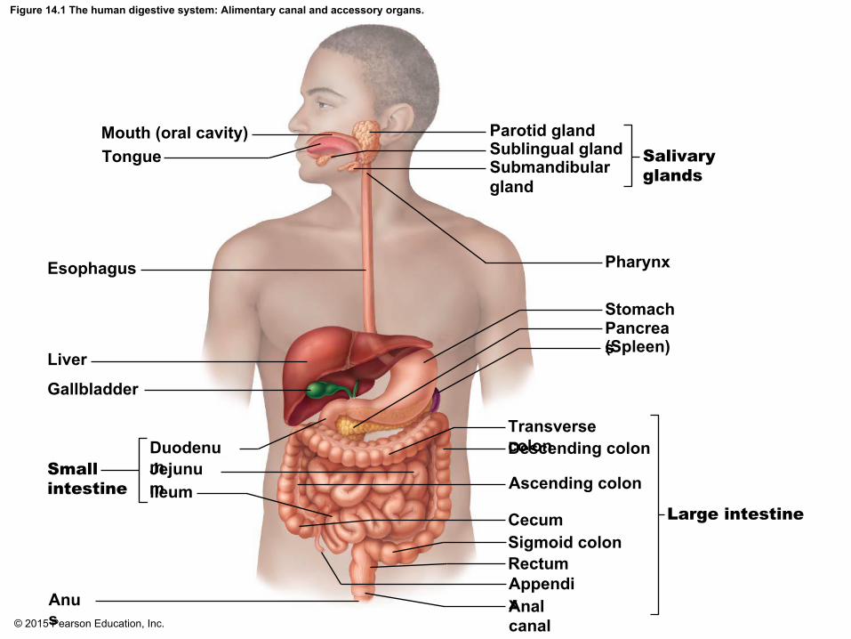
▪Let’s look at an example of gross anatomy using the digestive system organs
© 2015 Pearson Education, Inc.
Figure 14.1 The human digestive system: Alimentary canal and accessory organs.
Mouth (oral cavity)Tongue
Esophagus
Parotid glandSublingual glandSubmandibulargland
Salivary glands
Pharynx
StomachPancreas(Spleen)
Transverse colonDescending colon
Ascending colon
CecumSigmoid colonRectumAppendixAnal canal
Anus
Smallintestine
DuodenumJejunumIleum
Liver
Gallbladder
Large intestine

© 2015 Pearson Education, Inc.
Anatomy—Levels of Study
▪Microscopic anatomy▪Structures cannot be seen with the naked eye▪Structures can be viewed only with a microscope

© 2015 Pearson Education, Inc.
Figure 14.4c Anatomy of the stomach.
Pyloricsphincter
Gastric pits
Gas
tric
pi
tG
astr
ic g
land
Surfaceepithelium
Mucousneck cells
Parietal cells
GastricglandsChief cells
(c)

© 2015 Pearson Education, Inc.
Figure 14.4d Anatomy of the stomach.
Chief cells
Enteroendocrinecell
(d)
PepsinogenHCI
Pepsin
Parietal cells

© 2015 Pearson Education, Inc.
The Human Body—An Orientation
Physiology▪Study of how the body and its parts work or function

© 2015 Pearson Education, Inc.
Relationship between Anatomy and Physiology▪Structure determines what functions can occur▪ If structure changes, the function must also change

© 2015 Pearson Education, Inc.

© 2015 Pearson Education, Inc.
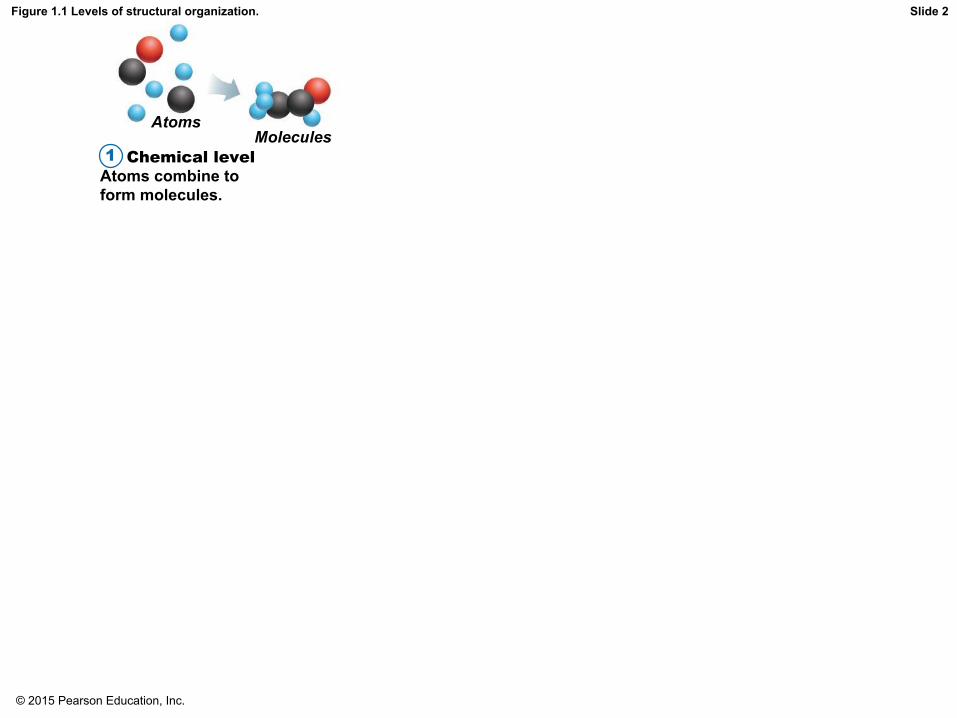
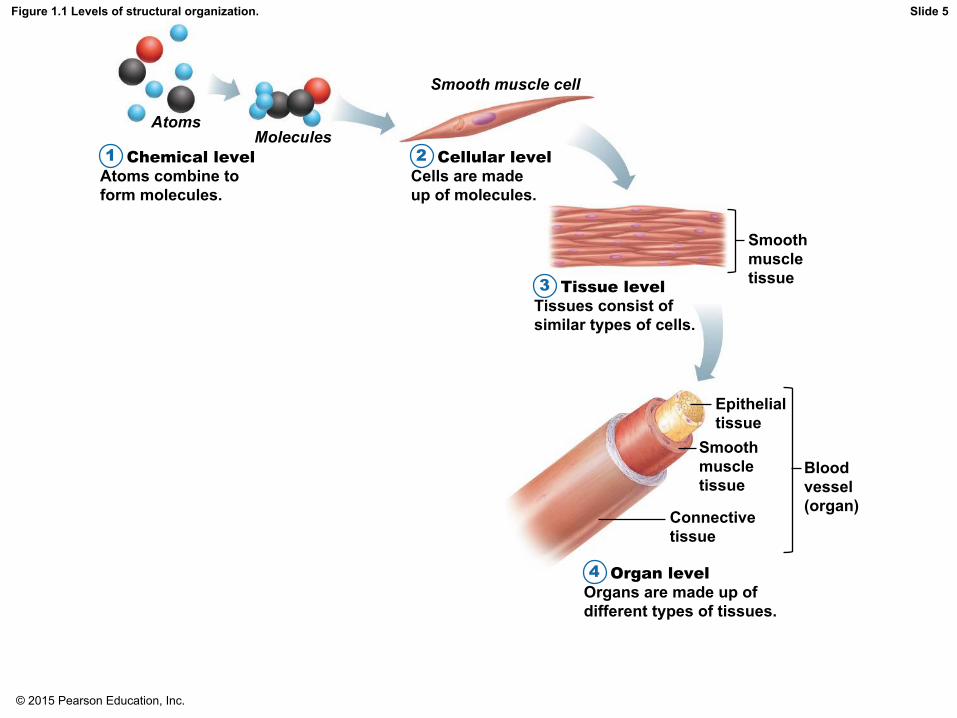
Levels of Structural Organization
▪Six levels of structural organization1. Atoms2. Cells3. Tissues4. Organs5. Organ systems6. Organisms

© 2015 Pearson Education, Inc.
Figure 1.1 Levels of structural organization.
Chemical levelAtoms combine toform molecules.
1
AtomsMolecules
Smooth muscle cell
Cellular levelCells are madeup of molecules.
2
Tissue levelTissues consist ofsimilar types of cells.
3
Organ levelOrgans are made up ofdifferent types of tissues.
4
Organ system levelOrgan systems consist ofdifferent organs that worktogether closely.
5 Organismal levelHuman organisms aremade up of many organsystems.
6
Smoothmuscletissue
Bloodvessel(organ)
Epithelialtissue
Smoothmuscletissue
Connectivetissue
Bloodvessels
Heart
Cardio–vascularsystem
Slide 1
© 2015 Pearson Education, Inc.
Figure 1.1 Levels of structural organization.
Chemical levelAtoms combine toform molecules.
1
AtomsMolecules
Slide 2

© 2015 Pearson Education, Inc.
Figure 1.1 Levels of structural organization.
Chemical levelAtoms combine toform molecules.
1
AtomsMolecules
Smooth muscle cell
Cellular levelCells are madeup of molecules.
2
Slide 3

© 2015 Pearson Education, Inc.
Figure 1.1 Levels of structural organization.
Chemical levelAtoms combine toform molecules.
1
AtomsMolecules
Smooth muscle cell
Cellular levelCells are madeup of molecules.
2
Tissue levelTissues consist ofsimilar types of cells.
3
Smoothmuscletissue
Slide 4
© 2015 Pearson Education, Inc.
Figure 1.1 Levels of structural organization.
Chemical levelAtoms combine toform molecules.
1
AtomsMolecules
Smooth muscle cell
Cellular levelCells are madeup of molecules.
2
Tissue levelTissues consist ofsimilar types of cells.
3
Organ levelOrgans are made up ofdifferent types of tissues.
4
Smoothmuscletissue
Bloodvessel(organ)
Epithelialtissue
Smoothmuscletissue
Connectivetissue
Slide 5

© 2015 Pearson Education, Inc.
Figure 1.1 Levels of structural organization.
Chemical levelAtoms combine toform molecules.
1
AtomsMolecules
Smooth muscle cell
Cellular levelCells are madeup of molecules.
2
Tissue levelTissues consist ofsimilar types of cells.
3
Organ levelOrgans are made up ofdifferent types of tissues.
4
Organ system levelOrgan systems consist ofdifferent organs that worktogether closely.
5
Smoothmuscletissue
Bloodvessel(organ)
Epithelialtissue
Smoothmuscletissue
Connectivetissue
Bloodvessels
Heart
Cardio–vascularsystem
Slide 6

© 2015 Pearson Education, Inc.
Figure 1.1 Levels of structural organization.
Chemical levelAtoms combine toform molecules.
1
AtomsMolecules
Smooth muscle cell
Cellular levelCells are madeup of molecules.
2
Tissue levelTissues consist ofsimilar types of cells.
3
Organ levelOrgans are made up ofdifferent types of tissues.
4
Organ system levelOrgan systems consist ofdifferent organs that worktogether closely.
5 Organismal levelHuman organisms aremade up of many organsystems.
6
Smoothmuscletissue
Bloodvessel(organ)
Epithelialtissue
Smoothmuscletissue
Connectivetissue
Bloodvessels
Heart
Cardio–vascularsystem
Slide 7

© 2015 Pearson Education, Inc.
Organ System Overview
▪ Integumentary system▪ Forms the external body covering (skin)▪Protects deeper tissue from injury▪Helps regulate body temperature▪ Location of cutaneous nerve receptors

© 2015 Pearson Education, Inc.
Figure 1.2a The body’s organ systems.
(a) Integumentary System
Forms the external body covering; protectsdeeper tissue from injury; synthesizesvitamin D; location of cutaneous receptors(pain, pressure, etc.) and sweat and oil glands.
Skin

© 2015 Pearson Education, Inc.
Organ System Overview
▪Skeletal system▪Consists of bones, cartilages, ligaments, and joints▪Supports the body▪Provides muscle attachment for movement▪Site of blood cell formation (hematopoiesis)▪Stores minerals

© 2015 Pearson Education, Inc.
Figure 1.2b The body’s organ systems.
(b) Skeletal System
Protects and supports body organs; provides aframework the muscles use to cause movement;blood cells are formed within bones; storesminerals.
Joint
Cartilages
Bones

© 2015 Pearson Education, Inc.
Organ System Overview
▪Muscular system▪Skeletal muscles contract or shorten▪Produces movement of bones

© 2015 Pearson Education, Inc.
Figure 1.2c The body’s organ systems.
Skeletalmuscles
(c) Muscular System
Allows manipulation of the environment,locomotion, and facial expression; maintainsposture; produces heat.

© 2015 Pearson Education, Inc.
Organ System Overview
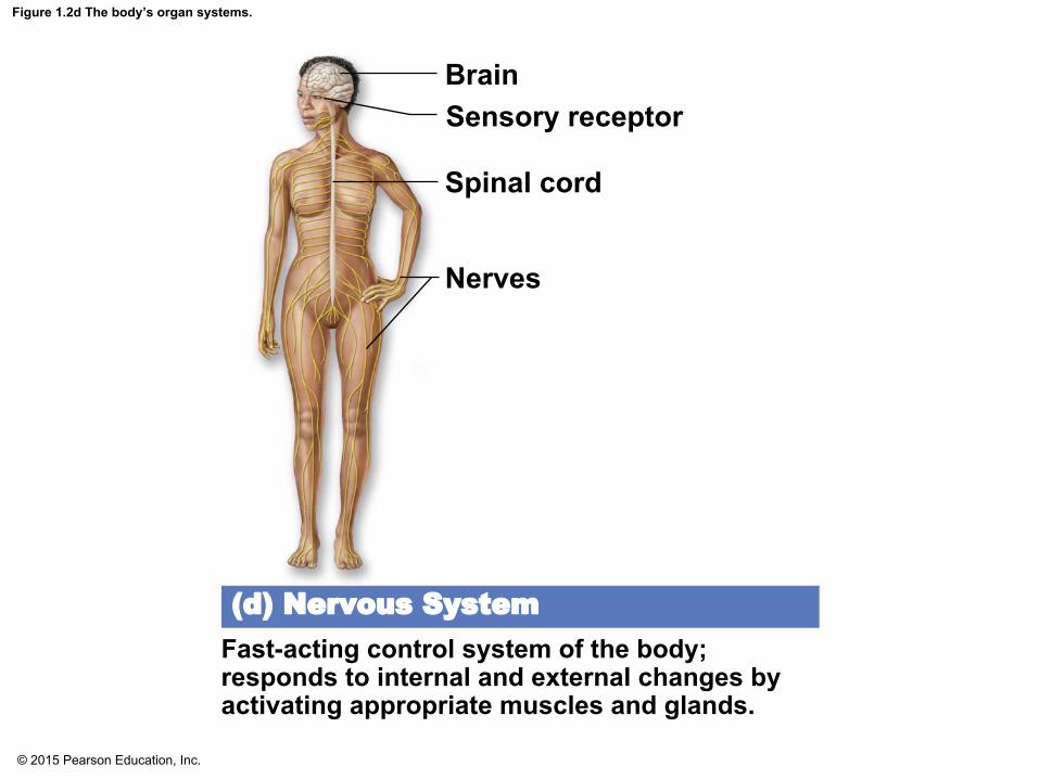
▪Nervous system▪ Fast-acting control system▪Consists of brain, spinal cord, nerves, and sensory receptors▪Responds to internal and external change▪Sends messages via nerve impulses to central nervous system ▪Central nervous system activates effectors (muscles and glands)
© 2015 Pearson Education, Inc.
Figure 1.2d The body’s organ systems.
BrainSensory receptor
Spinal cord
Nerves
(d) Nervous System
Fast-acting control system of the body;responds to internal and external changes byactivating appropriate muscles and glands.

© 2015 Pearson Education, Inc.
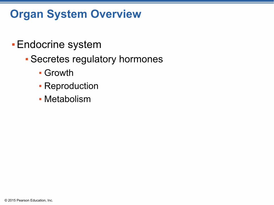
Organ System Overview
▪Endocrine system▪Endocrine glands include:
▪ Pituitary gland▪ Thyroid and parathyroids▪ Adrenal glands▪ Thymus▪ Pancreas▪ Pineal gland▪ Ovaries (females) and testes (males)
© 2015 Pearson Education, Inc.
Organ System Overview
▪Endocrine system▪Secretes regulatory hormones
▪ Growth▪ Reproduction▪ Metabolism

© 2015 Pearson Education, Inc.
Figure 1.2e The body’s organ systems.
(e) Endocrine System
Glands secrete hormones that regulateprocesses such as growth, reproduction, andnutrient use by body cells.
Pineal glandPituitary gland
Thyroid gland (parathyroidglands on posterior aspect)
Thymus glandAdrenal glandsPancreasTestis (male)Ovary (female)

© 2015 Pearson Education, Inc.
Organ System Overview
▪Cardiovascular system▪ Includes heart and blood vessels
▪ Heart pumps blood▪ Vessels transport blood to tissues
▪ Transports materials in body via blood pumped by heart▪ Oxygen and carbon dioxide▪ Nutrients▪ Wastes

© 2015 Pearson Education, Inc.
Figure 1.2f The body’s organ systems.
Heart
Bloodvessels
(f) Cardiovascular System
Blood vessels transport blood, which carriesoxygen, carbon dioxide, nutrients, wastes, etc.;the heart pumps blood.
© 2015 Pearson Education, Inc.
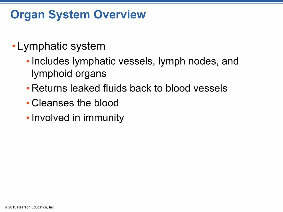
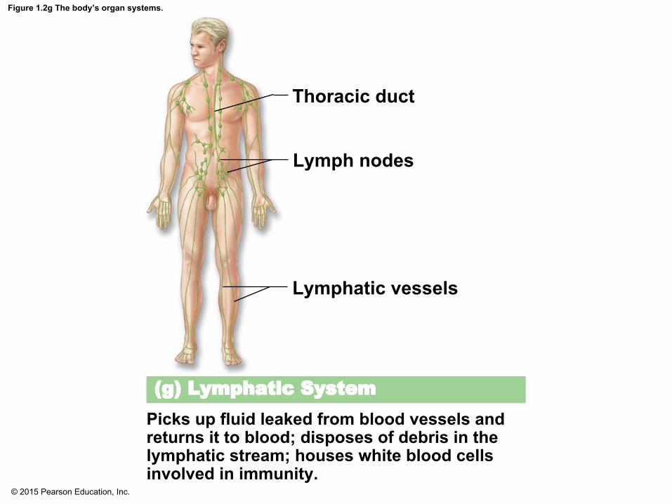
Organ System Overview
▪Lymphatic system▪ Includes lymphatic vessels, lymph nodes, and lymphoid organs▪Returns leaked fluids back to blood vessels▪Cleanses the blood▪ Involved in immunity
© 2015 Pearson Education, Inc.
Figure 1.2g The body’s organ systems.
(g) Lymphatic System
Picks up fluid leaked from blood vessels andreturns it to blood; disposes of debris in thelymphatic stream; houses white blood cellsinvolved in immunity.
Thoracic duct
Lymph nodes
Lymphatic vessels

© 2015 Pearson Education, Inc.
Organ System Overview
▪Respiratory system▪ Includes the nasal passages, pharynx, larynx, trachea, bronchi, and lungs▪Supplies blood with oxygen▪Removes carbon dioxide

© 2015 Pearson Education, Inc.
Figure 1.2h The body’s organ systems.
(h) Respiratory System
Keeps blood constantly supplied with oxygenand removes carbon dioxide; the gaseousexchanges occur through the walls of the airsacs of the lungs.
Nasal cavityPharynxLarynxTracheaBronchusLeft lung

© 2015 Pearson Education, Inc.
Organ System Overview
▪Digestive system▪ Includes the oral cavity, esophagus, stomach, small and large intestines, and accessory organs▪Breaks down food▪Allows for nutrient absorption into blood▪Eliminates indigestible material as feces

© 2015 Pearson Education, Inc.
Figure 1.2i The body’s organ systems.
(i) Digestive System
Breaks food down into absorbable units thatenter the blood for distribution to body cells;indigestible foodstuffs are eliminated as feces.
Anus
Rectum
Large intestine
Small intestineStomach
EsophagusOral cavity

© 2015 Pearson Education, Inc.
Organ System Overview
▪Urinary system▪ Includes the kidneys, ureters, urinary bladder, and urethra▪Eliminates nitrogenous wastes▪Maintains acid-base balance▪Regulates water and electrolytes

© 2015 Pearson Education, Inc.
Figure 1.2j The body’s organ systems.
Kidney
UreterUrinary bladder
Urethra
(j) Urinary System
Eliminates nitrogen-containing wastes fromthe body; regulates water, electrolyte, andacid-base balance of the blood.

© 2015 Pearson Education, Inc.
Organ System Overview
▪Reproductive system▪ For males, includes the testes, scrotum, penis, accessory glands, and duct system▪ Testes produce sperm▪ Duct system carries sperm to exterior
▪ For females, includes the ovaries, uterine tubes, uterus, and vagina▪ Ovaries produce eggs▪ Uterus provides site of development for fetus

© 2015 Pearson Education, Inc.
Figure 1.2k-l The body’s organ systems.
(k) Male Reproductive System
Overall function of the reproductive system is production of offspring. Testes producesperm and male sex hormone; ducts and glands aid in delivery of viable sperm to thefemale reproductive tract. Ovaries produce eggs and female sex hormones; remainingstructures serve as sites for fertilization and development of the fetus. Mammaryglands of female breast produce milk to nourish the newborn.
(l) Female Reproductive System
Prostate gland
Vas deferens
TestisScrotum
Seminalvesicles
Penis
Mammary glands(in breasts)Uterine tube
OvaryUterus
Vagina
© 2015 Pearson Education, Inc.
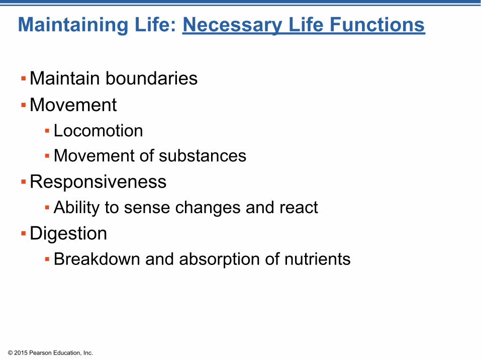
Maintaining Life: Necessary Life Functions
▪Maintain boundaries▪Movement
▪ Locomotion▪Movement of substances
▪Responsiveness▪Ability to sense changes and react
▪Digestion▪Breakdown and absorption of nutrients

© 2015 Pearson Education, Inc.
Necessary Life Functions
▪Metabolism—chemical reactions within the body▪Break down complex molecules into smaller ones▪Build larger molecules from smaller ones▪Produces energy▪Regulated by hormones
▪Excretion▪Eliminates waste from metabolic reactions▪Wastes may be removed in urine or feces

© 2015 Pearson Education, Inc.
Necessary Life Functions
▪Reproduction▪Occurs on cellular level or organismal level▪Produces future generation
▪Growth▪ Increases cell size and number of cells

© 2015 Pearson Education, Inc.
Survival Needs
▪Nutrients▪Chemicals for energy and cell building▪ Includes carbohydrates, proteins, lipids, vitamins, and minerals
▪Oxygen▪Required for chemical reactions
© 2015 Pearson Education, Inc.
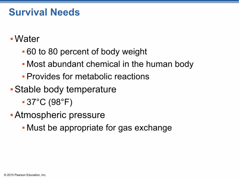
Survival Needs
▪Water▪ 60 to 80 percent of body weight▪Most abundant chemical in the human body▪Provides for metabolic reactions
▪Stable body temperature▪ 37°C (98°F)
▪Atmospheric pressure ▪Must be appropriate for gas exchange

© 2015 Pearson Education, Inc.
Figure 1.3 Examples of selected interrelationships among body organ systems.
Digestive systemTakes in nutrients, breaks them down,and eliminates unabsorbed matter (feces)
Respiratory systemTakes in oxygen and eliminatescarbon dioxide
Cardiovascular systemVia the blood, distributes oxygenand nutrients to all body cells anddelivers wastes and carbondioxide to disposal organs
Urinary systemEliminatesnitrogen-containingwastes andexcess ions
Food O2 CO2
CO2O2
Blood
Heart
Interstitial fluid
Nutrients
Nutrients and wastes passbetween blood and cellsvia the interstitial fluid
Feces UrineIntegumentary systemProtects the body as a wholefrom the external environment
© 2015 Pearson Education, Inc.
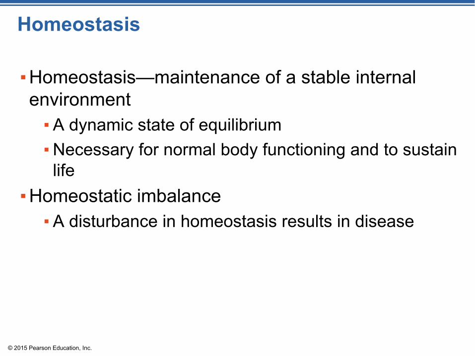
Homeostasis
▪Homeostasis—maintenance of a stable internal environment ▪A dynamic state of equilibrium▪Necessary for normal body functioning and to sustain life
▪Homeostatic imbalance▪A disturbance in homeostasis results in disease
© 2015 Pearson Education, Inc.
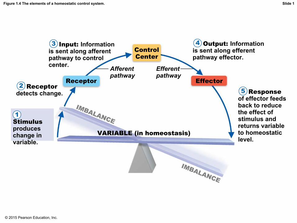
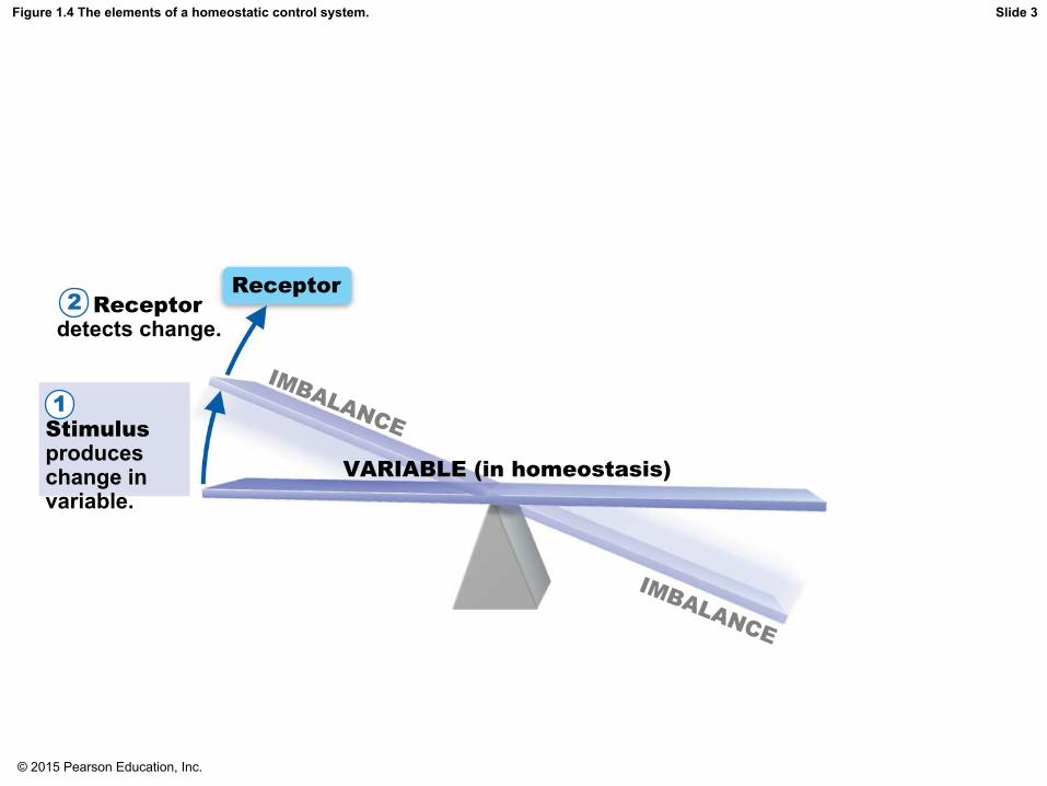
Figure 1.4 The elements of a homeostatic control system.
Stimulusproduceschange invariable.
1
Receptordetects change.2
Input: Informationis sent along afferentpathway to controlcenter.
3
Receptor
Afferentpathway
ControlCenter
Efferentpathway
Effector
Output: Informationis sent along efferentpathway effector.
4
Responseof effector feedsback to reducethe effect ofstimulus andreturns variableto homeostaticlevel.
5
VARIABLE (in homeostasis)
IMBALANCE
IMBALANCE
Slide 1

© 2015 Pearson Education, Inc.
Figure 1.4 The elements of a homeostatic control system.
Stimulusproduceschange invariable.
1
VARIABLE (in homeostasis)
IMBALANCE
IMBALANCE
Slide 2
© 2015 Pearson Education, Inc.
Figure 1.4 The elements of a homeostatic control system.
Stimulusproduceschange invariable.
1
Receptordetects change.2
Receptor
VARIABLE (in homeostasis)
IMBALANCE
IMBALANCE
Slide 3

© 2015 Pearson Education, Inc.
Figure 1.4 The elements of a homeostatic control system.
Stimulusproduceschange invariable.
1
Receptordetects change.2
Input: Informationis sent along afferentpathway to controlcenter.
3
Receptor
Afferentpathway
ControlCenter
VARIABLE (in homeostasis)
IMBALANCE
IMBALANCE
Slide 4

© 2015 Pearson Education, Inc.
Figure 1.4 The elements of a homeostatic control system.
Stimulusproduceschange invariable.
1
Receptordetects change.2
Input: Informationis sent along afferentpathway to controlcenter.
3
Receptor
Afferentpathway
ControlCenter
Efferentpathway
Effector
Output: Informationis sent along efferentpathway effector.
4
VARIABLE (in homeostasis)
IMBALANCE
IMBALANCE
Slide 5

© 2015 Pearson Education, Inc.
Figure 1.4 The elements of a homeostatic control system.
Stimulusproduceschange invariable.
1
Receptordetects change.2
Input: Informationis sent along afferentpathway to controlcenter.
3
Receptor
Afferentpathway
ControlCenter
Efferentpathway
Effector
Output: Informationis sent along efferentpathway effector.
4
Responseof effector feedsback to reducethe effect ofstimulus andreturns variableto homeostaticlevel.
5
VARIABLE (in homeostasis)
IMBALANCE
IMBALANCE
Slide 6

© 2015 Pearson Education, Inc.
Maintaining Homeostasis
▪The body communicates through neural and hormonal control systems▪Receptor
▪ Responds to changes in the environment (stimuli)▪ Sends information to control center along an afferent
pathway

© 2015 Pearson Education, Inc.
Maintaining Homeostasis
▪Control center▪ Determines set point▪ Analyzes information▪ Determines appropriate response
▪Effector▪ Provides a means for response to the stimulus▪ Information flows from control center to effector along
efferent pathway

© 2015 Pearson Education, Inc.
Feedback Mechanisms
▪Negative feedback▪ Includes most homeostatic control mechanisms▪Shuts off the original stimulus or reduces its intensity▪Works like a household thermostat
© 2015 Pearson Education, Inc.
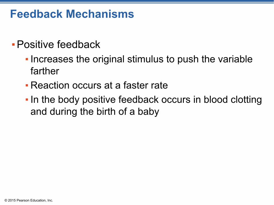
Feedback Mechanisms
▪Positive feedback▪ Increases the original stimulus to push the variable farther▪Reaction occurs at a faster rate▪ In the body positive feedback occurs in blood clotting and during the birth of a baby

© 2015 Pearson Education, Inc.
The Language of Anatomy
▪Special terminology is used to prevent misunderstanding▪Exact terms are used for:
▪Position▪Direction▪Regions▪Structures

© 2015 Pearson Education, Inc.
The Language of Anatomy
▪Anatomical position▪Standard body position used to avoid confusion▪ Terminology refers to this position regardless of actual body position▪Stand erect, feet parallel, arms hanging at the sides with palms facing forward

Anatomical Position▪Same body position and reference points ▪Anatomical position:
▪Standing erect▪ Face forward▪Arms at the sides▪Palms and toes directed forward

© 2015 Pearson Education, Inc.
Anatomical Position

© 2015 Pearson Education, Inc.
Directional TermsExplains location of one body structure in relation to another

© 2015 Pearson Education, Inc.
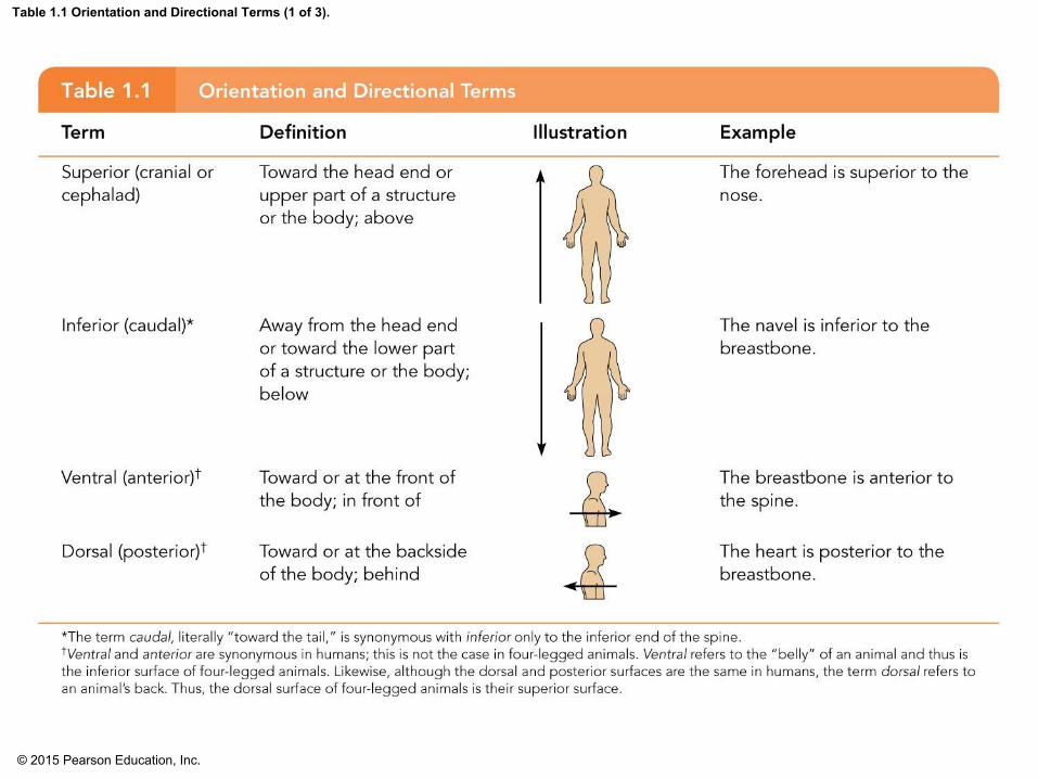
Directional Terms
▪Superior (cranial or cephalad): toward the head or upper part of a structure or the body; above ▪ Inferior (caudal): away from the head or toward the lower part of a structure or the body; below

© 2015 Pearson Education, Inc.
Directional Terms
▪Ventral (anterior): toward or at the front of the body; in front of▪Dorsal (posterior): toward or at the backside of the body; behind
© 2015 Pearson Education, Inc.
Table 1.1 Orientation and Directional Terms (1 of 3).

© 2015 Pearson Education, Inc.
Directional Terms
▪Medial: toward or at the midline of the body; on the inner side of▪Lateral: away from the midline of the body; on the outer side of▪ Intermediate: between a more medial and a more lateral structure

© 2015 Pearson Education, Inc.
Directional Terms
▪Proximal: close to the origin of the body part or point of attachment to a limb to the body trunk▪Distal: farther from the origin of a body part or the point of attachment of a limb to the body trunk

© 2015 Pearson Education, Inc.
Table 1.1 Orientation and Directional Terms (2 of 3).

© 2015 Pearson Education, Inc.
Directional Terms
▪Superficial (external): toward or at the body surface▪Deep (internal): away from the body surface; more internal

© 2015 Pearson Education, Inc.
Table 1.1 Orientation and Directional Terms (3 of 3).
© 2015 Pearson Education, Inc.
Regional Terms
▪Anterior (ventral) body landmarks

© 2015 Pearson Education, Inc.
Figure 1.5a Regional terms used to designate specific body areas.
FrontalCephalic
OrbitalNasalBuccalOralMental
CervicalThoracicSternalAxillary
AbdominalUmbilical
PelvicInguinal(groin)
Pubic (genital)
KEY:ThoraxAbdomen
(a) Anterior/Ventral
Upper limbUpper limbAcromialDeltoidBrachial (arm)Antecubital
Antebrachial(forearm)Carpal (wrist)
Manus (hand)Digital
Lower limbCoxal (hip)Femoral (thigh)Patellar
Crural (leg)
FibularPedal (foot)Tarsal (ankle)
Digital

© 2015 Pearson Education, Inc.
Regional Terms
▪Posterior (dorsal) body landmarks

© 2015 Pearson Education, Inc.
Figure 1.5b Regional terms used to designate specific body areas.
(b) Posterior/Dorsal
Upper limbUpper limbAcromial
Brachial (arm)
OlecranalAntebrachial(forearm)
Manus (hand)Digital
Femoral (thigh)
Popliteal
Fibular
Pedal (foot)Calcaneal
Plantar
Occipital(back of head)
Cephalic
Cervical
Back (dorsal)Scapular
Vertebral
Lumbar
Sacral
Gluteal
Sural (calf) KEY:
Back (Dorsum)

© 2015 Pearson Education, Inc.
Body Planes and Sections
▪Sections are cuts along imaginary lines known as planes▪Three types of planes or sections exist as right angles to one another
▪ http://www.madsci.org/~lynn/VH/planes.html
© 2015 Pearson Education, Inc.
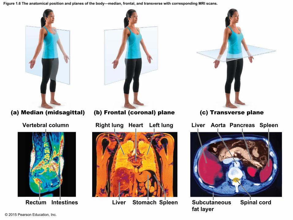
Figure 1.6 The anatomical position and planes of the body—median, frontal, and transverse with corresponding MRI scans.
(a) Median (midsagittal) (b) Frontal (coronal) plane (c) Transverse plane
Vertebral column
Rectum Intestines
Right lung Left lungHeart
Liver Stomach Spleen
Liver Aorta Pancreas Spleen
Spinal cordSubcutaneousfat layer

© 2015 Pearson Education, Inc.
Body Planes and Sections
▪A sagittal section divides the body (or organ) into left and right parts▪A midsagittal, section divides the body (or organ) into equal left and right parts▪A frontal, or coronal, section divides the body (or organ) into anterior and posterior parts ▪A transverse, or cross, section divides the body (or organ) into superior and inferior parts

© 2015 Pearson Education, Inc.
Body Cavities
▪Two body cavities▪Dorsal▪Ventral
▪Body cavities provide protection to organs within them

© 2015 Pearson Education, Inc.
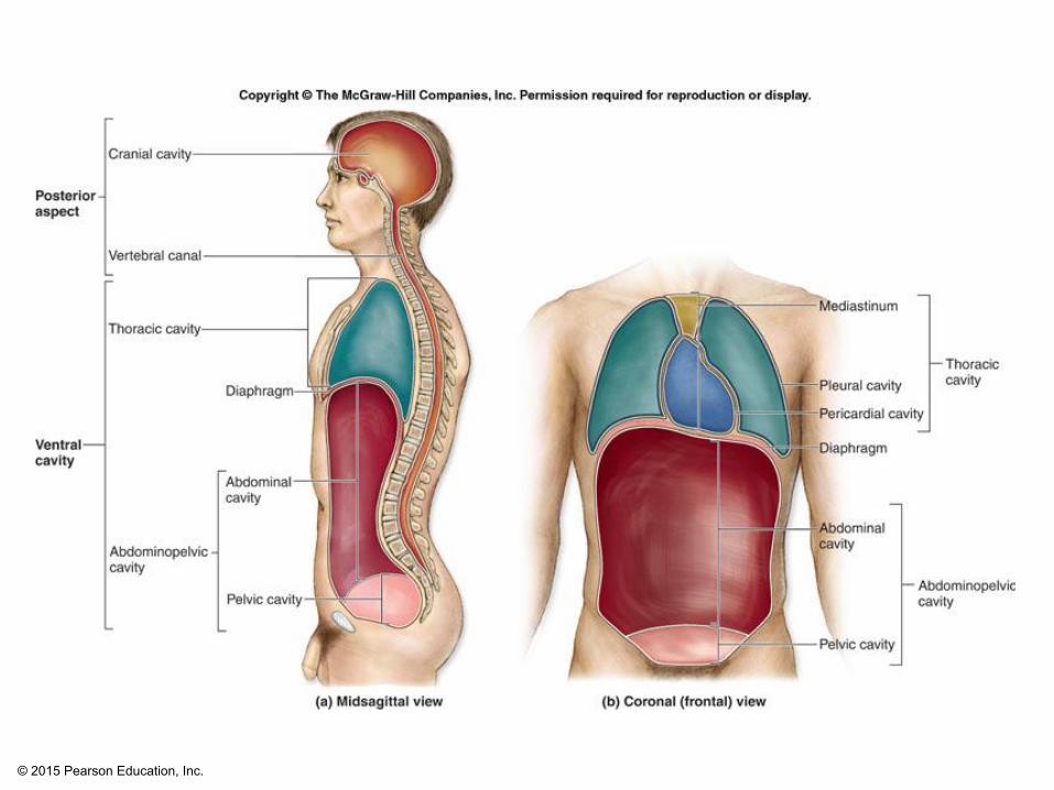
Figure 1.7 Body cavities.
Cranialcavity
Thoraciccavity
Diaphragm
AbdominalcavitySpinal
cavity
Pelviccavity A
bd
om
inope
lvic
cavit
y
KEY:Dorsal body cavity Ventral body cavity
© 2015 Pearson Education, Inc.

© 2015 Pearson Education, Inc.
Body Cavities
▪Dorsal body cavity has two subdivisions1. Cranial cavity
▪ Houses the brain▪ Protected by the skull
2. Spinal cavity ▪ Houses the spinal cord▪ Protected by the vertebrae
Membranes that line the dorsal body cavity and cover the organs within are called MENINGES

© 2015 Pearson Education, Inc.
Body Cavities
▪Ventral body cavity has two subdivisions separated by the diaphragm1. Thoracic cavity2. Abdominopelvic cavity

© 2015 Pearson Education, Inc.
Body Cavities
▪Thoracic cavity ▪Cavity superior to the diaphragm▪Houses heart, lungs, and other organs▪Mediastinum, the central region, houses heart, trachea, and other organs
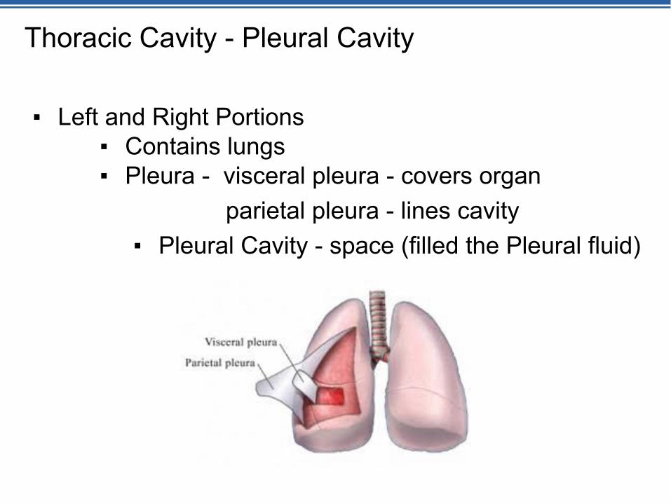
Thoracic Cavity - Pleural Cavity
▪ Left and Right Portions▪ Contains lungs▪ Pleura - visceral pleura - covers organ
parietal pleura - lines cavity▪ Pleural Cavity - space (filled the Pleural fluid)

© 2015 Pearson Education, Inc.
Body Cavities
▪Medial Portion - Mediastinum ▪Contains the heart, thymus gland, trachea, esophagus…..▪Pericardium - Visceral and Parietal▪Pericardial cavity - space filled with pericardial fluid

© 2015 Pearson Education, Inc.
Body Cavities
▪Abdominopelvic cavity▪Cavity inferior to the diaphragm▪Superior abdominal cavity contains the stomach, liver, and other organs▪ Protected only by trunk muscles
▪ Inferior pelvic cavity contains reproductive organs, bladder, and rectum ▪ Protected somewhat by bony pelvis
▪No physical structure separates abdominal from pelvic cavities
Membranes that line the abdominopelvic cavity and cover the organs within are called PERITONEUM.

© 2015 Pearson Education, Inc.

© 2015 Pearson Education, Inc.
Review: Membranes of body cavities
▪ If lining the cavity it’s a parietal membrane▪ Lining thoracic cavity = parietal pleura▪ Lining abdominopelvic cavity-parietal peritoneum▪ Lining the pericardial cavity – parietal pericardium
▪ If covering an organ within a cavity it’s a visceral membrane▪Covering the lungs in the thoracic cavity = visceral pleura▪Covering the stomach in the abdominopelvic cavity = visceral peritoneum▪Covering the heart = visceral pericardium

© 2015 Pearson Education, Inc.
Body cavities
▪Space between the visceral and parietal membranes ▪ In thoracic cavity = pleural fluid▪ In abdominopelvic cavity = peritoneal fluid
This is serous fluid that is there for cushioning, protection, and to reduce friction so organs can move around a little, expand, etc.

Greater Omentum
▪a peritoneal membrane▪ “apron of fat” ▪Covers the anterior aspect of the small intestine

Lesser Omentum
▪Peritoneal membrane▪Suspends the superior border of the stomach to the liver

Mesentery
▪ Encircles and holds the small intestine to the dorsal wall of the abdominopelvic cavity

© 2015 Pearson Education, Inc.
Body Cavities
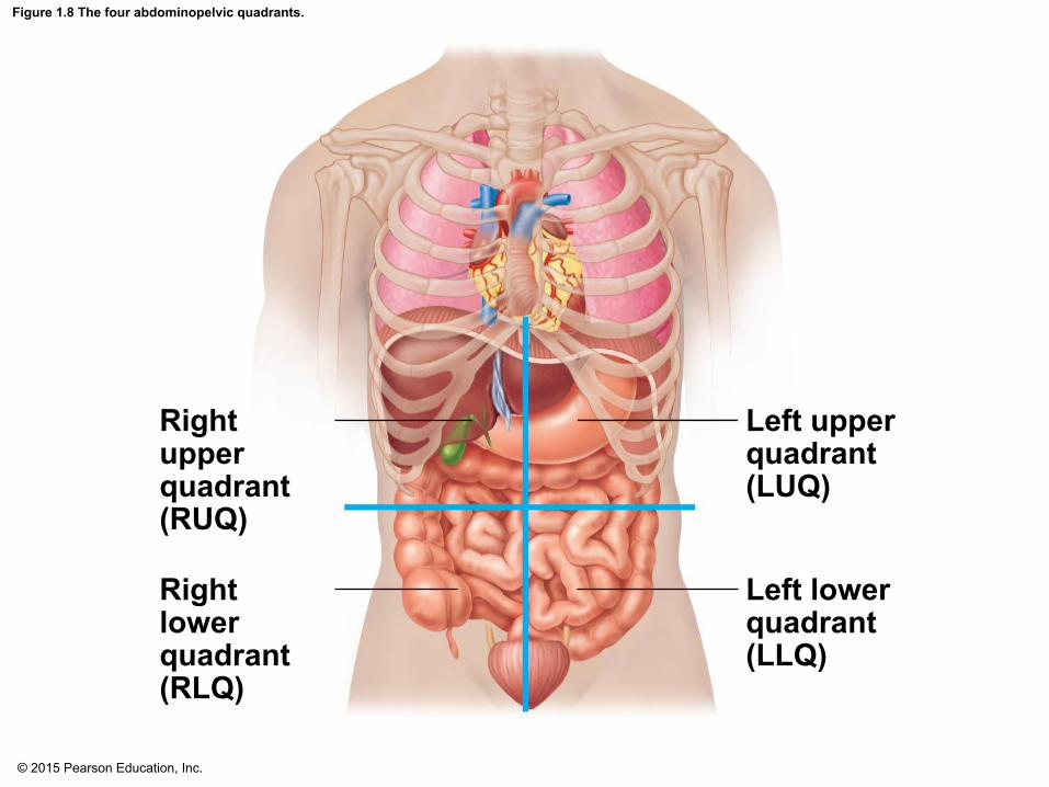
▪Abdominopelvic cavity subdivisions▪ Four quadrants▪Nine regions
© 2015 Pearson Education, Inc.
Figure 1.8 The four abdominopelvic quadrants.
Left upperquadrant(LUQ)
Left lowerquadrant(LLQ)
Right upperquadrant(RUQ)
Right lowerquadrant(RLQ)

© 2015 Pearson Education, Inc.
Figure 1.9 The nine abdominopelvic regions.
Righthypochondriac
region
Lefthypochondriac
region
Epigastricregion
Umbilicalregion
Rightlumbarregion
Leftlumbarregion
Right iliac(inguinal)
region
Left iliac(inguinal)
region
Hypo-gastric(pubic)region
(a) Nine regions delineated by four planes
(b) Anterior view of the nine regions showing the superficial organs
Liver
Gallbladder
Ascendingcolon oflarge intestineSmall intestineCecumAppendix
DiaphragmStomachTransversecolon of largeintestineDescendingcolon of largeintestineInitial partof sigmoidcolonUrinarybladder


© 2015 Pearson Education, Inc.
Body Cavities
▪Other body cavities include:▪Oral and digestive cavities▪Nasal cavity▪Orbital cavities▪Middle ear cavities