an update on novel therapeutic warfronts of extracellular
TRANSCRIPT

Review ArticleAn Update on Novel Therapeutic Warfronts of ExtracellularVesicles (EVs) in Cancer Treatment: WhereWe Are Standing RightNow and Where to Go in the Future
Muhammad Babar Khawar ,1,2,3 Muddasir Hassan Abbasi ,3,4 Zerwa Siddique,5
Amin Arif,3 and Nadeem Sheikh 3
1State Key Laboratory of Stem Cell and Reproductive Biology, Institute of Zoology, Chinese Academy of Sciences,Beijing 100101, China2University of Chinese Academy of Sciences, Beijing 100049, China3Cell & Molecular Biology Lab, Department of Zoology, University of the Punjab, Lahore, Pakistan4Department of Zoology, University of Okara, Okara, Pakistan5Centre for Applied Molecular Biology (CAMB), University of the Punjab, Lahore, Pakistan
Correspondence should be addressed to Muddasir Hassan Abbasi; [email protected] Nadeem Sheikh; [email protected]
Received 18 March 2019; Revised 3 June 2019; Accepted 4 July 2019; Published 25 July 2019
Academic Editor: Fabio Altieri
Copyright © 2019 Muhammad Babar Khawar et al. This is an open access article distributed under the Creative CommonsAttribution License, which permits unrestricted use, distribution, and reproduction in any medium, provided the original workis properly cited.
Extracellular vesicles (EVs) are a heterogeneous group of membrane-bounded vesicles that are believed to be produced and secretedby presumably all cell types under physiological and pathological conditions, including tumors. EVs are very important vehicles inintercellular communications for both shorter and longer distances and are able to deliver a wide range of cargos including proteins,lipids, and various species of nucleic acids effectively. EVs have been emerging as a novel biotherapeutic platform to efficientlydeliver therapeutic cargos to treat a broad range of diseases including cancer. This vast potential of drug delivery lies in theirabilities to carry a variety of cargos and their ease in crossing the biological membranes. Similarly, their presence in a variety ofbody fluids makes them a potential biomarker for early diagnosis, prognostication, and surveillance of cancer. Here, we discussthe relatively least and understudied aspects of EV biology and tried to highlight the obstacles and limitations in their clinicalapplications and also described most of the new warfronts to beat cancer at multiple stages. However, much more challengesstill remain to evaluate EV-based therapeutics, and we are very much hopeful that the current work prompts further discovery.
1. Introduction
A bidirectional communication exists between cells and theirimmediate surroundings that ensures the survival of cells andis an essential factor for both normal and pathophysiologicalcircumstances. Traditionally, such crosstalk was believed tooccur via the release of soluble cellular factors (i.e., chemo-kines, cytokines, and growth factors) [1–3] or via directcell-cell contact; however, involvement of the extracellularvesicle (EV) in cellular communication has changed thenotion over the past decade [4, 5]. Various eukaryotic celltypes secrete these EVs in vitro, and their presence has been
reported in a variety of body fluids including blood, bile,milk, and urine as well as in fecal matter [6]. A number offactors can induce the release of EVs, i.e., change in pH,stress, damage, irradiation, lack of oxygen, exposure to com-plement proteins, and also as a result of cell activation (i.e.,platelet activation) [7, 8]. Secretion of EVs by plants andnumerous pathogens including bacteria, Archaea, mycobac-teria, and fungi is suggestive of an efficient evolutionarilyconserved intercellular communication mechanism [9, 10].
Intercellular communication is an important phenome-non in multicellular organisms and usually mediated viadirect contacts between the cells or via transfer of secretory
HindawiOxidative Medicine and Cellular LongevityVolume 2019, Article ID 9702562, 21 pageshttps://doi.org/10.1155/2019/9702562

molecules. Some of these secretory molecules are packagedinto small lipid bilayer vesicles, known as extracellular vesi-cles (EVs), identified as a new means of intercellular commu-nication. These EVs are major players in tumor progressionand have shown a greater potential in therapeutic applica-tions [11]. These diverse collections of vesicles are secretedby almost every type of cells [12]. Wolf identified these EVsfor the first time in 1967 [13]. They successfully isolated min-ute particulate material from plasma free of platelets viaultracentrifugation. They originally named it as “plateletdust” [13] which was later on replaced by the currentlyknown term “extracellular vesicles.” Afterward in 1981, cul-tured normal and neoplastic cells were found to secretemembranous vesicles which were suspected to take part inphysiological processes [14]. In the following few years, asimilar kind of small vesicles, named “exosomes,” was alsofound to be produced and secreted by reticulocytes in vitro[15]. Though only a little work was done in the early yearsfollowing their discovery, recent rediscovery of EVs by cancerscientists has thrown the research into gear, and now, EVstudy is an exciting and rapidly growing field. The presentterm “EVs” was assigned by the International Society ofExtracellular Vesicles (ISEV).
EVs play significant roles in multiple physiological pro-cesses including stem cell differentiation [16], autophagy[17], blood clotting [18], angiogenesis [19], immunity (innateand acquired) and immunomodulation [20, 21], pregnancy[22], embryo implantation [23], reproduction, placentalphysiology, semen regulatory function [24], and tissue regen-eration [19]. Furthermore, the role of EVs in neuronal regen-eration and in the development and functioning of thenervous system has also been anticipated as novel arbitratorsof communication between the cells [25, 26]. Besides theircontribution in normal physiology, EVs are also the keycomponents in various pathologies like cancer [27–29] anddevelopment of multiple neurodegenerative diseases [30].EVs mediate a variety of processes involved in cancer pro-gression including inflammation, lymphogenesis, cell prolif-eration, epithelial-to-mesenchymal transition, angiogenesis,migration, suppression of the immune system, and metasta-sis—all of which are the so called “hallmarks of cancer”[31]. These extraordinary organelles have been associatedwith a number of aspects of cancer development and progres-sion [32, 33], hence have a great potential to be used asbiomarkers and ideal targets for novel future therapies forcancer treatment.
2. EV Diversity and Classification
EVs are currently classified as exosomes, microvesicles,microparticles, ectosomes, oncosomes, and apoptotic bodies[34] on the basis of their size, origin, and characteristics[35] (Figures 1 and 2). However, this classification is consid-ered insufficient to cover the heterogeneity that lies in cargoand their uncountable roles [36]. Exosomes are the best char-acterized and are less variable in size (ranging 40-150 nm)than other subtypes. Exosomes are produced as a result ofmembrane invaginations of endosomes resulting in the for-mation of multivesicular bodies (MVBs) and are stored as
intraluminal vesicles (ILVs) that are secreted out of the cellonce MVB fused with the plasma membrane at a certainpoint [37–39] (Figure 1).
Microvesicles (MVs), sometimes also referred to asectosomes or microparticles or membrane particles, arelarger and more heterogeneous in size (ranging from100 nm to several microns) than exosomes [40, 41]. Thesecell surface-derived EVs are produced as a result of bulgingof the plasma membrane and are ultimately shed from thecell surface as these blebs undergo fission upon proper stim-ulation [40, 41]. MVs are known to be full of phosphatidyl-serine and have several other lipid components [42]. MVswere firstly identified to be released from RBCs [43] and acti-vated blood platelets [44] and were thought to contribute inthe regulation of coagulation cascade [45]. Their formationis endorsed by increased Ca2+ levels that do so by alteringthe phospholipid distribution within the plasma membrane.A steady state or classical asymmetric lipid compositionexists along the plasma membrane and is characterized bythe existence of aminophospholipids towards the cytoplas-mic side while phospholipids are on the side facing extracel-lular environment [46]. A variety of membrane enzymesincluding floppase, flippase, translocase, and scramblase helpin the maintenance of this intricate balance. Increased intra-cellular Ca2+ in association with cytoskeleton modification[44] and recruitment of scramblase (a Ca2+-dependentenzyme) [47] relocate phosphatidylserine from the inner sideto the outer side [48] in an ATP-dependent manner [49].ADP-ribosylation factor 6- (ARF6-) mediated invasion andcytoskeletal remodeling in prostate and breast cancers areaccused of the shedding MVs and all other classes of EVs[50] (Figure 1). Considering the heterogeneity in the size ofMVs, the presence of numerous subpopulations can be spec-ulated within this subdivision of EVs. The term “oncosomes”was coined to describe the MVs or EVs secreted by cancercells [51]. It is noteworthy that the term “large oncosomes(LO)” is referred to as a specific class of EVs and used todescribe a comparatively larger size (1-10 μm) of subtypeMVs, which originate directly from the plasma membrane,produced by cancer cells [52, 53].
Apoptotic bodies, range in size from 800 to 5000 nm, areproduced during cellular blebbing and released by cellsundergoing programmed cell death [41]. However, how theyaffect other cells is not well-studied yet.
EVs are usually classified on the basis of their origin;however, this classification and the current techniquesemployed for the identification of these EVs are insufficientto distinguish clearly each type of EV separately [54]. Somevesicles also originated from the nanotubular projectionspresent on plasma membranes [55]. Recently, the presenceof very small nanovesicles (8-12 nm) has been reported inperipheral blood [56] that was errantly separated along withexosomes. It would not be wrong to say that the currentnomenclature has not been without debate, and size attri-butes of EV subtypes are ambiguous to fully distinguish themseparately [6, 57]. As a matter of fact, the isolation techniquesand protocols are still in the development process, andconsensus best practices have not yet been achieved resultingin differences in EV subpopulations being evaluated between
2 Oxidative Medicine and Cellular Longevity

investigations [36, 58]. Therefore, the mode of biogenesis andcargo within EVs may prove crucial in establishing a moreproper criterion for their classification. In line with thisspeculation, EV subpopulations have been characterizedrecently on the basis of their size, density, cargo, and theireffects on target cells [36, 59, 60]. Therefore, new EV classesand modifications in the current classification criteria areexpected to emerge as the field progresses. Additional vesicletypes used to study lipid and protein behavior have also beenreported, however not classified along with the above givenvesicles. For instance, a number of stimuli, i.e., some chemi-cals (paraformaldehyde and dithiothreitol), salts, and lasertreatment may result in the formation of giant plasmamembrane vesicles (GPMVs) in live cells [61–63]. These ves-icles are large sized and help in understanding the lipid and
protein organization existing along the plasma membrane[62, 64, 65]. There are many other additional vesicle typesincluding small, large, and giant uni- and multilamellar lipidvesicles [66] along with artificial plasma membrane vesicles[67] that are exclusively synthesized in vitro. These extraordi-nary vesicle species are employed therapeutically to enhancethe stability and uptake of drugs and other biological mole-cules probably via the same mechanism adapted by naturalEVs [68, 69]. Currently, most, if not all, cell types are believedto produce EVs [70, 71]; however, diseased states and stressnot only upregulate their production but also alter the cargocontained within [72].
Considering the unexpected pace in the growth of EV-related information, Minimal Information for Studies ofExtracellular Vesicles (“MISEV”) guidelines were proposed
Exosomes
Microvesicles
Proteins
mRNA
Lipids
miRNA
Fragmented DNA
Endosome
Multivesicularbodies (MVBs)
MVBs
Nucleus
Endoplasmic reticulum
Figure 1: Biogenesis of exosomes and microvesicles: a schematic representation of endosome formation by internalizing the extracellularsubstances by invagination and pinching of the plasma membrane via endocytosis. These endosomes are transformed to multivesicularbodies (MVBs) by taking up a variety of cytosolic contents (proteins, nucleic acids, and various metabolites) via inward budding of lateendosomes. Later, these MVBs may fuse with the plasma membrane at certain points to release the internal vesicles named as “exosomes.”In contrast, microvesicles are formed due to outward protrusion/blebbing of the plasma membrane. A diverse array of cargos is packedinto these protrusions which pinched off the parent cell giving rise to microvesicles.
3Oxidative Medicine and Cellular Longevity

by ISEV five years ago and have been updated last year onthe basis of evolution of the collective knowledge of thefield in these past four years. These guidelines equippedthe EV biologist with a collection of standardized proto-cols and procedures for a better documenting of EV-associated functions [34].
3. Therapeutic Approaches to Target EVs
As described before, EVs are major players in tumor progres-sion via the transfer of cargo within them. As a matter of fact,the pathways targeted by EVs differ largely from conven-tional methods, i.e., chemotherapy or molecular targetingdrugs. Therefore, three potential therapeutic approaches areproposed in this regard: (i) inhibition of EV formation, (ii)eradication of circulating EVs, and (iii) ablation of EVabsorption [73] (Figure 2). A large number of investigationsboth in vivo and in vitro have stressed the efficacy ofinhibiting EV production in cancer reduction. For instance,blockade of EV secretion and miR-210-3p transfer and sub-sequently suppression of angiogenesis and metastasis wereobserved, in a xenograft mouse model, as a result ofnSMase2-knockdown [74]. In another study, repression ofovarian cancer dissemination, as a result of inhibition ofEV production, was observed upon nSMase2-knockdown[75]. In addition to it, to date, a large number of other mole-
cules involved in EV production, i.e., RAB27A, RAB27B, andTSG101, have been exploited to reduce the production ofcancer-derived EVs [76, 77]. Although suppression of EVproduction seems quite an efficient strategy in cancer treat-ment, targeting genes involved in this pathway will affectmany of the important biological activities of normal cellsas they are the mode of intercellular communication [73].For instance, nSMase2 has been found to express in normalneural cells [78]. Furthermore, downregulation of these genesshowed different extent of their inhibitory effects on EV pro-duction among different cancer types. For example, there wasno effect on EV production upon ablation of nSMase2 in theprostate cancer cell line [79]. Therefore, it is the need of thecurrent time and would be a future challenge to identifygenes associated with cancer type-specific EV production.
A novel therapeutic strategy to remove the circulatingEVs was devised byMarleau and coworkers in 2012. A hemo-filtration system was developed that was able to specificallytrap the circulating cancer cell-derived HER2-expressingEVs [80]. These HER2-positive EVs hinder the availabletherapies and subsequently promote cancer development[81]; therefore, selectively targeting the HER-2-expressingEVs may prove a better approach in breast cancer treatment.In line with it, the circulating EVs have also been found toestablish a premetastatic niche and subsequently promotecancer metastasis [33]. Hence, it can be speculated easily that
A
B
C
EV-producing cell
Recipient cell
Exosomes
Microvesicles
Proteins
mRNA
Lipids
miRNA
Fragmented DNA
Figure 2: Therapeutic strategies to target EVs for cancer treatment. There are a number of potential ways to target the EV-mediatedintercellular communication. (A) EV biogenesis or release can be targeted via interfering the specific components involved in EVproduction or surface shedding. (B) EVs can be targeted and specifically removed from the circulation using different substances, i.e.,specific antibodies. (C) EV uptake/internalization by the recipient cells can be interrupted by targeting the EV ligands or cell surface receptors.
4 Oxidative Medicine and Cellular Longevity

elimination of these circulating EVs may help in the preven-tion of cancer metastasis. Recently, a new idea has beenadapted to target circulating EVs in a human breast cancerxenograft mouse model [82]. In this study, a pronounceddecrease in metastatic activity was observed upon the admin-istration of anti-CD9 and CD63 (two of the most enrichedreceptors on the EV surface) antibodies [82]. However, therewere found no prominent effects on primary site growth.Moreover, macrophages were utilized to internalize the EVstagged by anti-CD9 and CD63 and were not allowed topromote cancer progression. Furthermore, more in-depthstudies are required as anti-CD9 and CD63 antibodies areunable to selectively target the cancer-derived EVs inhumans. However, identification of cancer-specific mole-cules on the EV surface and development of specific antibod-ies against them can likely help in eliminating the circulatingEVs and subsequently prove effective in cancer treatment.Therefore, that investigation was believed to devise a noveltreatment strategy for cancer.
Microvesicle cargo is simply internalized directly into thecytoplasm via plasma membrane-EV fusion while intactvesicles are taken up via several ways for transferring to thelysosomal or endosomal pathway [83–85]. The multiple waysof EV internalization include micropinocytosis [86, 87], cla-thrin and caveolin-mediated endocytosis, phagocytosis, andlipid raft endocytosis [88, 89]. In addition to it, another keyfactor that favors the fusion of EVmembranes with the recip-ient cells is the low pH conditions produced by the tumormicroenvironment [88]. Therefore, disruption of EV inter-nalization may help in the formulation of new and bettertherapeutic approaches to prevent tumor progression andin cancer treatment. Recently, heparan sulfate proteoglycans(HSPGs) were found to act as a receptor of GBM-derivedEVs [89]. A dose-dependent inhibition of EV uptake and aclear suppression of EV-dependent cell migration were seenin GBM in the presence of an HS mimetic, heparin. A num-ber of molecules capable of EV internalization have beendescribed to date, and manymore are expected to be reportedin the near future [84, 90, 91]. Despite the identification ofseveral molecules responsible for EV internalization, themechanism involved in this internalization is not very clear.However, caveolin-dependent endocytosis has been reportedas a primary route for internalization of multiple myelomacell-derived exosomes while some of the exosomes weretaken up via macropinocytosis and membrane fusion [92].Glycans are involved in energy storage and also serve asstructural components that have been recently described toplay an important role in several molecular recognitionevents. Glycans not only modulate recognition at the celllevel but also regulate the intracellular traffic and folding ofindividual proteins [93, 94]. Abnormal glycosylation usuallyinterrupts these crucial recognition events and may lead tocancer or other disorders like lysosomal storage diseases[94]. Although we are not familiar with the function of glyco-conjugates in EV biology at present, several novel strategiesthat utilize EV glycosylation have already been emerging.Indeed, an extraordinary interest has been emerging recentlyin studying the effects of glycosylation modulation of EVsand their cargo. Glycoengineering is a promising field that
is highly exploited to optimize the stability and alter the phar-macokinetics of protein-based drugs [95, 96]. This conceptwas proved for EVs by engineering Lamp2b protein (exoso-mal membrane protein) [97]. Furthermore, N-glycosylationwas found to protect the peptides from lysosomal proteolysis[97]. In fact, a twofold increase in the efficiency of exosomedelivery was found in the nervous system upon glycosylationof the desired peptides. Therefore, this strategy can bespeculated the best choice to improve the uptake efficiencyof peptide-targeted vesicles. Neuroblastoma-derived exo-somes were found highly enriched in glycosphingolipids,and these glycans showed a huge therapeutic potentialagainst Alzheimer’s disease as they were capable of scaveng-ing the β-amyloid [98]. The formation of hybrid exosomeshaving unique lipid components with the liposomes [99]has shown an enormous therapeutic potential of the glyco-lipid cargoes. These few experiments are the EV glycoengi-neering efforts that advocate a promising platform andfuture directions. Briefly, glycosylation can be exploited inmanipulating the cargo protein recruitment and offers noveltherapeutic targeting approaches [100, 101]. One moreaspect that can be utilized to modulate the physicochemicalcharacteristics of EVs is the sialylation status as it is capableof altering the vesicle charge [102]. This approach is yet tobe achieved, but the availability of massive information onglycoengineering can be applied to EVs [103].
Unluckily, currently, there are no data available about theeffects of inhibition of EV internalization under in vivo con-ditions. One of the prime reasons behind this insufficiency ofinvestigations is that the cancer cell-associated EV uptakepathway is not quite clear yet. To ensure the integrity of nor-mal cell homeostasis, it is necessary to fully understand thecancer-specific EV uptake pathways by the cancer biologistsfor therapeutic development. The presence of different EVprotein markers has been recently described in different frac-tions of EVs; furthermore, these fractions were found to havedifferent molecular and biological characteristics [36]. Like-wise, immature dendritic cells have been found to releasetwo EV subpopulations (small and large EVs) that were fur-ther found to affect the T helper cell in a different manner[104]. Therefore, identifying the EV subpopulations, theireffects on target cells, and specific internalization pathwaywould be the best approach for better future therapies. Onthe basis of the above given contexts, inhibition of EV trans-fer could be speculated to be employed as a novel therapeuticapproach in suppressing the tumor progression. Despite agreat number of challenges, outstanding advances and pacein understanding of EVs are promising a better future [11].
4. EVs as Potent Novel Drug Delivery Systems
EVs have been emerging as attractive novel entities for drugdelivery because of their structural analogy with liposomes[105]. Liposomes have proven their efficiency as a novel drugcarrier and have been widely employed for drug delivery asthey are very similar in composition with the plasma mem-branes [106]. Since that time, multiple commercializedliposome-based products like Myocet (an approved nonpe-gylated liposomal doxorubicin highly practiced against
5Oxidative Medicine and Cellular Longevity

metastatic breast cancer), DaunoXome (an approved lipo-some employed against advanced HIV-associated Kaposi’ssarcoma to deliver daunorubicin (DNR)), and Depocyt(approved against lymphomatous meningitis) have beenintroduced for therapeutic purposes [107]. Liposomalresearch has laid down the foundation to explore their phys-icochemical properties and stability for their employment asnovel drug delivery agents [108–110]. Exploiting EVs is abetter choice and more advantageous than liposomes as theyare naturally produced by the cells and can easily transfer thedesired drugs. These properties make EVs the best choice tobe utilized as drug delivery agents even across the blood-brain barrier (BBB) [111]. Some of the potential EVs thathave been used recently as drug delivery vehicles in differenttypes of cancer are summarized in Table 1.
5. EVs in Cancer Treatment
A huge number of studies have provided the evidences of theuse of these EVs as a splendid tool to deliver small interferingRNAs and other synthetic molecules for therapeutic pur-poses [123]. EVs have been employed in a number of animalmodel studies developed for different diseases as potent ther-apeutic DDSs [124]. Moreover, they are splendid antitumorDDSs as EVs are capable of passively targeting tumorsbecause of their enhanced permeation and retention [125].It is of great interest that genetically engineered EVs as tar-geted DDSs offer a dynamic and handy platform for specificand target-oriented drug delivery with better therapeuticoutcomes. Recently, an efficient DDS was developed for thesuccessful transfer of siRNA to the CNS via modified den-dritic cell- (DC-) derived EVs [111]. The DCs, isolated frommice, were transected with a plasmid expressing EV surfaceprotein, lysosome-associated membrane glycoprotein 2b(Lamp2b), along with rabies viral glycoprotein (RVG) thathelps in binding with acetylcholine receptor. An efficientbrain-targeting gene knockdown was observed by GAPDHsiRNA-loaded DC-derived EVs, signifying their prospect aseffective targeted DDSs. Effective delivery of both genes andproteins represents the potential of these extraordinary EVsto be served as cell-derived liposome-like nanoplatforms tocure various diseases including cancer [5, 40, 41, 126, 127].Moreover, a zip code-like 25-nt sequence has been found toenhance the packaging of miRNAs into EVs and has pushedthe research one step forward by guaranteeing high-yieldingEVs loaded with various RNAs [128]. Amazingly, siRNAhave also been utilized as therapeutics against tumors viabacterial outer membrane vesicles (OMVs). This study hashighlighted the significance of bacteria in the production ofbiological nanovesicles and their application in drug delivery[129]. Furthermore, they are also being employed to transferchemotherapeutic agents in addition to biomolecule-baseddrugs to enhance their efficiency and to minimize the possi-ble side effects associated with them. For instance, an effec-tive inhibition and successful reduction of breast and coloncancers have been achieved by encapsulation of the EVs withdoxorubicin and curcumin [130, 131]. The outcomes of theseinvestigations suggest that EVs offer an effective way tosuppress cancerous tumors by delivering a wide range of
the chemotherapeutics drugs. Recently, doxorubicin (chemo-therapeutic drug) have been successfully delivered, via an i.v.injection, to αv integrin-positive breast cancer cells viaexosomes isolated from Lamp2b-iRGD peptide expressingengineered immature mouse DCs (imDCs), and a remark-able suppression in tumor growth was observed [130]. More-over, these therapeutic exosomes were less toxic and veryeffectual in cancer inhibition. Furthermore, their effective-ness in delivering the therapeutic cargo has also been authen-ticated employing multiple tumor models [132] includinghepatocarcinoma [133], lymphocytic leukemia [134], andpancreas [135] and prostate [136] cancers.
Some of the potent EVs that have been utilized recently indifferent types of cancer are summarized in Table 2.
6. EV-Associated Antitumor ncRNAs
A number of attributes, including their release from theparent cells, delivery via the circulatory system, targetedcell uptake, and selective cargo transport, make them apromising and sizzling object for the selective drug deliverycarrier [73, 144]. Therefore, investigators are proposinginnovative and dynamic approaches for the modification ofEVs specifically exosomes to cope with the current clinicalchallenges and therapeutic needs [145–147]. One of suchapproaches involves the direct modification of the contentsof isolated exosomes. For instance, siRNAs and shRNAshave been incorporated into fibroblast-like mesenchymalstem cell-derived exosomes via electroporation to targetKRASG12D mutation of pancreatic cancer [143]. Therapeu-tic use of natural exosomes is highly advantageous forseveral reasons compared to synthetic liposomes. Forinstance, exosomes are prevented from being phagocytosedby monocytes and macrophages due to the presence ofCD47 on the exosomal membrane. Furthermore, the accu-mulation and uptake of exosomes by cancerous tissues arefacilitated by some yet unknown native proteins presenton the exosomal surface. Consequently, these “chimeric”exosomes were found to effectively execute an enhancedsurvival and reduced metastasis [143]. Another approachis to stimulate the parental cells to release modified exo-somes. For example, exosomes containing miR-143 wereobtained from MSCs pretreated with medium containingsynthetic miR-143 and were found to successfully deliverthese miRNAs to osteosarcoma cells to hamper their met-astatic activity in vitro [148].
7. Application of EVs as Cancer Vaccines
Considering their production by every cell and theirimmune-modulatory effects, they can be employed for diag-nostic purposes. Similarly, exosomes have shown antigen-presenting and immune-stimulatory potential and are beingutilized for triggering antitumor responses. Moreover, releaseof exosomes from tumor cells is suggestive of their involve-ment in tumor microenvironments [149]. Cancer- andimmune cell-derived EVs are capable of inducing immunos-timulation to recipient cells. This prospect can presume theuse of EVs as cancer vaccines, either derived from APCs or
6 Oxidative Medicine and Cellular Longevity
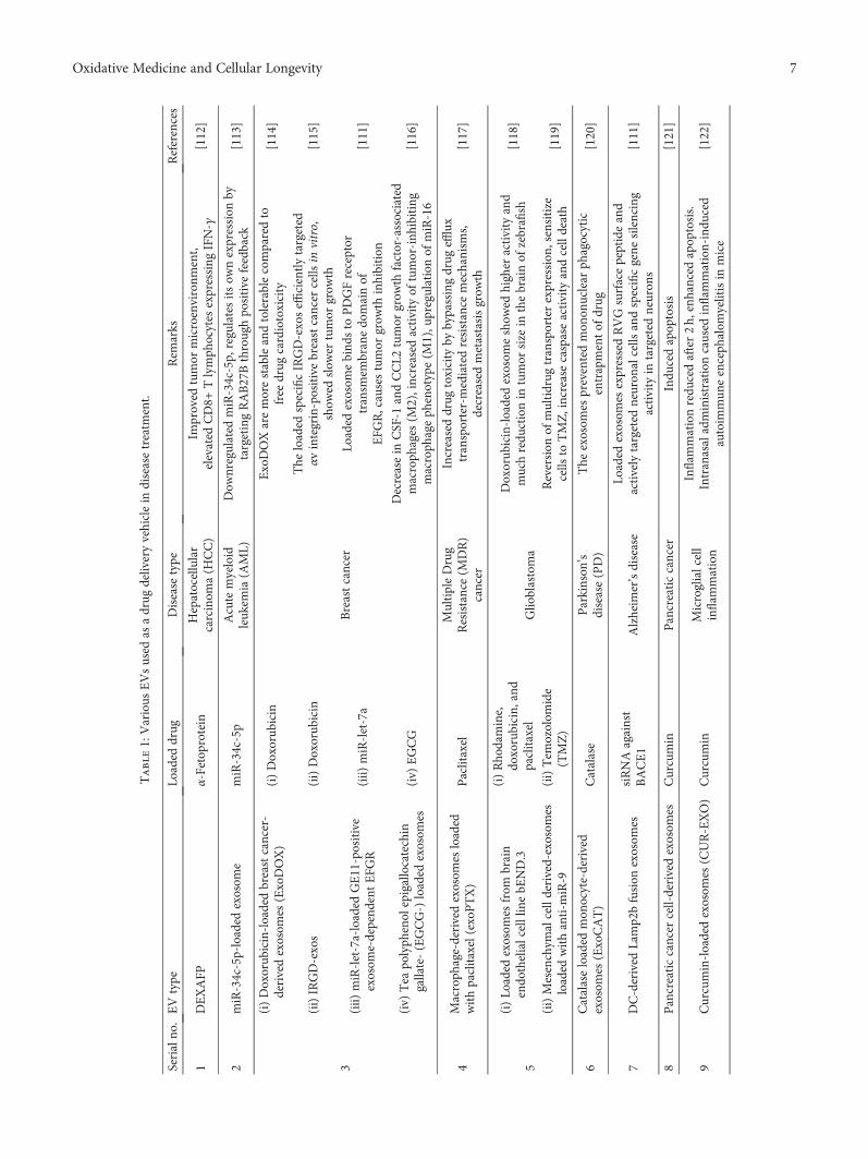
Table1:Various
EVsused
asadrug
deliveryvehiclein
diseasetreatm
ent.
Serialno
.EVtype
Loaded
drug
Disease
type
Rem
arks
References
1DEXAFP
α-Fetop
rotein
Hepatocellular
carcinom
a(H
CC)
Improved
tumor
microenvironm
ent,
elevated
CD8+
Tlymph
ocytes
expressing
IFN-γ
[112]
2miR-34c-5p-loaded
exosom
emiR-34c-5p
Acutemyeloid
leuk
emia(A
ML)
Dow
nregulated
miR-34c-5p,
regulatesitsow
nexpression
bytargetingRAB27Bthroughpo
sitive
feedback
[113]
3
(i)Doxorub
icin-loadedbreastcancer-
derivedexosom
es(ExoDOX)
(i)Doxorub
icin
Breastcancer
ExoDOXaremorestableandtolerablecomparedto
free
drug
cardiotoxicity
[114]
(ii)IRGD-exos
(ii)Doxorub
icin
The
loaded
specificIRGD-exoseffi
cientlytargeted
αvintegrin-positivebreastcancer
cells
invitro,
show
edslow
ertumor
grow
th[115]
(iii)
miR-let-7a-loaded
GE11-positive
exosom
e-depend
entEFG
R(iii)
miR-let-7a
Loaded
exosom
ebind
sto
PDGFreceptor
transm
embranedo
mainof
EFG
R,causestumor
grow
thinhibition
[111]
(iv)
Tea
polyph
enol
epigallocatechin
gallate-(EGCG-)loaded
exosom
es(iv)
EGCG
Decreasein
CSF-1
andCCL2
tumor
grow
thfactor-associated
macroph
ages
(M2),increased
activity
oftumor-inh
ibiting
macroph
ageph
enotype(M
1),u
pregulationof
miR-16
[116]
4Macroph
age-derivedexosom
esloaded
withpaclitaxel(exoPTX)
Paclitaxel
MultipleDrug
Resistance(M
DR)
cancer
Increaseddrug
toxicity
bybypassingdrug
efflux
transporter-mediatedresistance
mechanism
s,decreasedmetastasisgrow
th[117]
5
(i)Lo
aded
exosom
esfrom
brain
endo
thelialcelllinebE
ND.3
(i)Rho
damine,
doxorubicin,
and
paclitaxel
Glio
blastoma
Doxorub
icin-loadedexosom
eshow
edhigher
activity
and
muchredu
ctionin
tumor
size
inthebrainof
zebrafish
[118]
(ii)Mesenchym
alcellderived-exosom
esloaded
withanti-m
iR-9
(ii)Tem
ozolom
ide
(TMZ)
Reversion
ofmultidrug
transporterexpression
,sensitize
cells
toTMZ,increasecaspaseactivity
andcelldeath
[119]
6Catalaseloaded
mon
ocyte-derived
exosom
es(ExoCAT)
Catalase
Parkinson
’sdisease(PD)
The
exosom
espreventedmon
onuclear
phagocytic
entrapmentof
drug
[120]
7DC-derived
Lamp2bfusion
exosom
essiRNAagainst
BACE1
Alzheim
er’sdisease
Loaded
exosom
esexpressedRVGsurfacepeptideand
activelytargeted
neuron
alcells
andspecificgene
silencing
activity
intargeted
neuron
s[111]
8Pancreaticcancer
cell-derivedexosom
esCurcumin
Pancreaticcancer
Indu
cedapop
tosis
[121]
9Curcumin-loadedexosom
es(CUR-EXO)
Curcumin
Microglialcell
inflam
mation
Inflam
mationredu
cedafter2h,
enhanced
apop
tosis.
Intranasaladministrationcaused
inflam
mation-indu
ced
autoim
mun
eenceph
alom
yelitisin
mice
[122]
7Oxidative Medicine and Cellular Longevity
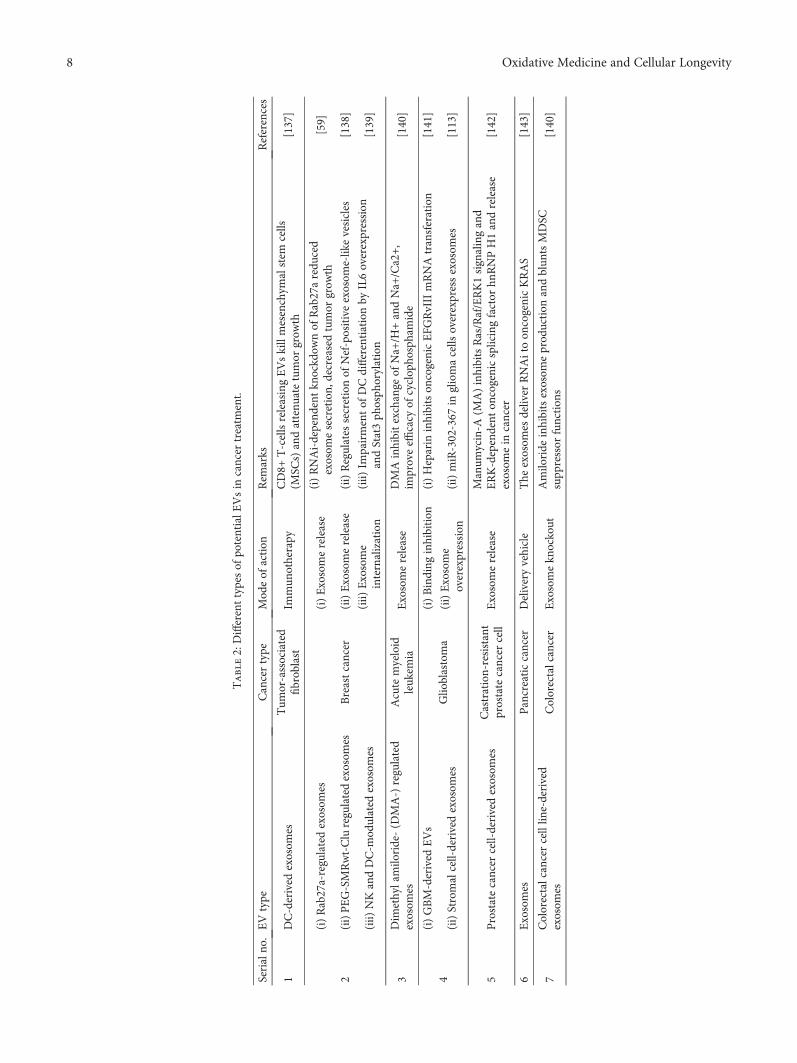
Table2:Differenttypesof
potentialE
Vsin
cancer
treatm
ent.
Serialno
.EVtype
Cancertype
Mod
eof
action
Rem
arks
References
1DC-derived
exosom
esTum
or-associated
fibroblast
Immun
otherapy
CD8+
T-cellsreleasingEVskillmesenchym
alstem
cells
(MSC
s)andattenu
atetumor
grow
th[137]
2
(i)Rab27a-regulatedexosom
es
Breastcancer
(i)Exosomerelease
(i)RNAi-depend
entkn
ockd
ownof
Rab27aredu
ced
exosom
esecretion,
decreasedtumor
grow
th[59]
(ii)PEG-SMRwt-Cluregulatedexosom
es(ii)Exosomerelease
(ii)Regulates
secretionof
Nef-positiveexosom
e-likevesicles
[138]
(iii)
NKandDC-m
odulated
exosom
es(iii)
Exosome
internalization
(iii)
Impairmentof
DCdifferentiationby
IL6overexpression
andStat3ph
osph
orylation
[139]
3Dim
ethylamilo
ride-(D
MA-)regulated
exosom
esAcutemyeloid
leuk
emia
Exosomerelease
DMAinhibitexchange
ofNa+
/H+andNa+
/Ca2+,
improveeffi
cacy
ofcyclop
hosphamide
[140]
4(i)GBM-derived
EVs
Glio
blastoma
(i)Binding
inhibition
(i)Heparin
inhibitson
cogenicEFG
RvIIImRNAtransferation
[141]
(ii)Stromalcell-derivedexosom
es(ii)Exosome
overexpression
(ii)miR-302-367
ingliomacells
overexpressexosom
es[113]
5Prostatecancer
cell-derivedexosom
esCastration-resistant
prostatecancer
cell
Exosomerelease
Manum
ycin-A
(MA)inhibitsRas/Raf/ERK1signalingand
ERK-dependent
oncogenicsplicingfactor
hnRNPH1andrelease
exosom
ein
cancer
[142]
6Exosomes
Pancreaticcancer
Deliveryvehicle
The
exosom
esdeliver
RNAitoon
cogenicKRAS
[143]
7Colorectalcancercelllin
e-derived
exosom
esColorectalcancer
Exosomekn
ockout
Amilo
ride
inhibitsexosom
eprod
uction
andblun
tsMDSC
supp
ressor
function
s[140]
8 Oxidative Medicine and Cellular Longevity

derived by tumors themselves [150]. These cancer-derivedEVs are believed as potential proimmune elements becauseof the presence of several stimulatory molecules, i.e., heatshock proteins [151, 152] and numerous tumor antigens ontheir surface [151, 153, 154]. There is an ample amount ofdata available in favor of EVs as potent immune-suppressive agents [150, 155, 156]. Therefore, an encounterbetween the immune system and tumor EVs takes place inan immune-stimulatory vs. an immune-suppressive context[150], and by considering their immune-stimulatory fea-tures, these tumor EVs have been employed clinically ascancer vaccines [157]. In addition to a previous report ofKunigelis and Graner [150], another trial is also availableon “clinicaltrials.gov” (NCT01550523). This trial was per-formed on patients who had resected the tumors and failedthe prior therapies; an antisense construct against IGF1Rwas used to induce apoptotic cell death in autologous tumorcells. The cells were positioned in a biodiffusion chamber,and soluble ingredients, for the induction of immuneresponse, were allowed to be released. Next, the chamberwas inserted in the rectus sheath and was detached after 24hours. Some of the subjects were found to develop deep veinthrombosis and were subjected to enoxaparin treatment toget rid of this problem. Except this minor trouble, the therapywas believed to be safe as some subjects were found to showcomplete response and some were found to show partialresponse under two (2) to twenty-seven- (27-) week time-span [158]. In the second phase I trial, the glioma cell-derived exosomes were referred to as immune stimulatorsby the authors [159]. The tumor-challenged mice were safe-guarded via implanting chamber-based vaccine in this caseprobably due to the formation and release of antigen-bearing immunostimulatory exosomes. In line with theseinvestigations, DC-derived EVs/exosomes (also referred toas dexosomes/DEX) have also been subjected to phase IItrials [150]. For this purpose, multiple types of antigens (pep-tides, proteins, and tumor lysate) are loaded to DCs isolatedfrom patients. Subsequently, the exosomes produced by thecells in the culture supernatants are utilized as cell-free can-cer vaccines. Recently, inoperable non-small-cell lung cancerpatients were subjected to chemotherapy followed by DEX-based immunotherapy for maintenance. For this purpose,MAGE/NY ESO/MART1 peptides were introduced in DCsvia the pulse and cultured with gamma interferon (IFNG).Subsequently, DEX were isolated, and 1-27 injections ofDEX were given to the patients. A median overall survivalof fifteen (15) months along with median progression-freesurvival (PFS) of 2.2 months was found in the treatedpatients. An increased number of NK cells and an enhancedactivity associated with NKp30 (NK surface ligand) werefound in subjects with >2.2 month PFS [160]. It is of greatinterest that NK cell activation was found in an earlier trial[161]; in addition, an improvement of T cell responses butno induction of tumor-specific T cells was found upon IFNGaddition. Upon cessation of chemotherapy, about 50% of thepatients with PFS did not reach primary endpoint at 4months with this trial; however, large-scale DEX productioncould be adapted to treat very advanced cancer. The updatesabout the use of either tumor-derived or immune cell-
derived EVs to promote antitumor responses and cancer sup-pression are continuing to grow. A huge number of investiga-tions are available in favor of tumor EV-driven immunesuppression [150, 156, 162–164]. Inflammation is a majorcontributor in immune-mediated progression and tumorsuppression [165], and nucleotide receptors are commonmediators of inflammatory reaction [166] and cancer [167].One of such receptor families is the purinoreceptor familythat participates in immune responses mediated by EVs inimmunity, inflammation, and cancer settings [168].
Radiotherapy is a new choice in enhancing the immuno-therapy effects; however, radiations led to the oxidation,degradation, and accumulation of the DNA in the cytosol.This accumulation of DNA encourages the release ofinterferon-b from tumor cells via activation of the cGAS/ST-ING-mediated DNA-sensing pathway. STING is a key sig-naling component that responds to pathogen-derived DNAby inducing the production of a variety of cytokines andtype-I IFNs upon activation by its ligand, cyclic GMP-AMP(cGAMP). Cyclic GMP-AMP synthase (cGAS) associateswith the pathogenic DNA and led to the formation ofcGAMP from GTP and ATP. The resultant cGAS-STINGaxis stimulates the production of inflammatory cytokinesand type-I IFNs via activating the NF-κB and IRF3, respec-tively [169]. Interestingly, tumor growth was augmented dur-ing radiotherapy in the mouse model lacking STING becauseof the attenuation of antitumor T-cell activation [170, 171].In addition to it, tumoral growth was restricted in a murinemelanoma model upon intratumoral administration ofcGAMP [172]. Therefore, the cGAS/STING signaling path-way is an attractive therapeutic approach in inducing theefficient immune responses against tumors.
In another study, tumor cell-derived microparticles (T-MPs) were described to be used as cell-free tumor vaccinerecently. T-MP-based vaccinations were found effectiveagainst several tumor types, and T-MP-loaded dendritic cells(DC) were also found very fruitful in a number of tumormodels [173, 174]. In these models, T-MPs efficiently deliv-ered the DNA fragments to DCs that subsequently inducedthe expression of type I IFN via activating the cGAS/STINGsignaling pathway. Furthermore, the subsequent increase inthe IFN level enhanced the antitumor immunity by promot-ing the presentation of tumor antigens to T-cells and matura-tion of DC. Indeed, this study represents a novel tumor cell-free vaccine strategy of high therapeutic potential [173].
8. EVs as Cancer Biomarkers
A substantial interest has been growing, in the past few years,in investigating the potential of tumor-associated EVs fordiagnostic purposes and their exploitation for disease moni-toring. EVs derived from a number of tumor types arebelieved to contain specific cargo including nucleic acid andvarious proteins [175]. The presence of tumor-derived EVsin circulating bodily fluids including cerebrospinal fluid(CSF), urine, and blood makes them an easy and readilyaccessible battery of biomarkers. Therefore, these tumor-derived EVs are speculated to be specifically served for longi-tudinal disease monitoring and early relapse detection [176].
9Oxidative Medicine and Cellular Longevity

A few of EV-associated cargo (particularly nucleic acid andproteins) are also capable of predicting the therapeuticresponse of a specific treatment. Collectively, EVs have beenproven by a growing body of evidence as a new representativeclass of rich and readily accessible cancer biomarkers. Theirpotential as a cancer biomarker was explored for the very firsttime by comparing the contents of EVs derived from glio-blastoma and from the cells of origin [32]. In this report,authors found tumor-specific RNA and protein species,reflective of the parental cell, enriched in the released EVs[32]. Accordingly, a vast assortment of tumor-specific speciesincluding various nucleic acid species such as lncRNA [177],miRNA [178], and mRNA [32, 179] and multiple posttrans-lational protein modifications [179] have been well recog-nized. The diagnostic and predictive values of these EVshave been utilized in multiple studies with different cancertypes and further strengthened recently by the massive profil-ing of sixty cancer cell lines [175]. EV proteome, from all thetested samples, was reported to reflect the cellular proteomeand transcriptome. EV proteomic data helps in their exem-plification by hierarchical clustering and categorization ofthe basis of the originating cell [175]. This correlationbetween tumor-associated EVs and contents of secreting cellsis highly important for brain and CNS-associated tumorswhere conducting a tissue biopsy is a limitation. For instance,an upregulation of miR21 was observed in glioblastomamultiforme-associated EVs in CSF compared to healthy con-trols [180, 181]. Moreover, a positive correlation was notedbetween the level of EV-miR21 and tumor burden. Conse-quently, a huge number of tumor-derived EV-miRNA hav-ing prognostic and diagnostic values have been described inother types of cancer including pancreatic [182], colorectal[183], and non-small-cell lung [184] cancers. In line with it,several candidate mRNAs (C-MYC, BCL-6, and PTEN),characterized with diagnostic value to predict progression-free survival, have been found in plasma-derived EVs frompatients of non-Hodgkin’s lymphoma [185]. Therefore, thesereports have opened a new potential of tumor-associated EVsfor noninvasive longitudinal disease monitoring [186]. Thecancer-related EVs may also be helpful in early disease detec-tion. For instance, it was shown in an in vivo pancreatic can-cer model that particular EVs expressing a marker proteinwere upregulated even at the time when the tumor cannotbe detected by conventional imaging techniques [187]. Inaddition to it, AML-EVs can be detected in the blood of acutemyeloid leukemia (AML) patients even prior to the release ofleukemic blasts in the blood [176]. Moreover, tumor-associated EVs have also been employed in the predictionof response to a specific treatment. Interestingly, tumor-associated EVs are capable of transferring resistance fromdrug-resistant to drug-sensitive cells via specific miRNAsand various protein species carried by them. Several therapiesfor a number of cancer types including pazopanib (chemo-therapy) in soft tissue sarcoma [188], tamoxifen (antiestro-gen) in breast cancer [189], and cetuximab (anti-EGFR)therapy in colon cancer [190] have been found to show thesame resistance transfer phenomenon. Surprisingly, in all ofthese investigations, disruption of sensitivity to a specificdrug and development of resistance were observed upon
exposure to EVs from the resistant cells. Moreover, authorsalso illustrated a possible mechanism for Trastuzumab(anti-HER2) therapy in breast cancer [81]. Astonishingly,EV-associated HER2 was found to reduce the therapeuticeffects of this drug as it is able to bind and decrease the avail-able concentration of Trastuzumab [81]. Collectively, theabove cited literature is suggestive of the prognostic, diagnos-tic, and predictive values of tumor-associated EVs [191].
Some of the potent EVs that have been recently found aspotent biomarkers in different types of cancer are summa-rized in Table 3.
9. Conclusion and Future Perspectives
EVs are the potent carriers of cargo molecules includingfunctional RNA species, many therapeutic agents like miR-NAs, mRNAs, proteins, and peptides, and synthetic drugs.These small vesicles loaded with therapeutic agents are highlyadvantageous in terms of their biocompatibility, low immu-nogenicity, and innate ability to interact with target cells.Many futuristic approaches can be implicated from ex vivoand in vivo studies. However, due to complexity of EVs,many questions must be addressed prior to opting these mol-ecules in clinics.
Advances in isolation and characterization techniqueswill allow more insight understanding and hence will providea platform to develop EV-based therapeutic and diagnostictools. An inexpensive, reliable method of isolations must berecognized and implemented to ensure that the optimal yieldof EVs is being obtained in a safe and repeatable manner.Ultimately, a preferred method of isolating intact EVs mustbe identified and scaled so that EV-based options can bedeveloped into a clinically viable therapy.
Upcoming research would completely get benefit of exo-somes’ ubiquitous occurrence in eukaryotic cells as theyappear to provide an excess for anticancer therapy. Exosomeshave been in attention for their role in the TME, and TDEs,in particular, provide a hopeful way for cancer remedies asmechanisms for superior drug delivery, tumor suppression,and immune regulation due to their appropriate dimension,composition, and homing capabilities. The progress of can-cer encompasses the difficult and intricate communicationof cells and signaling molecules in the TME, and exosomeshave been shown to advance tumor growth through the inhi-bition of antitumor immunity and the development ofangiogenesis.
Existing investigations in EVs are inadequate to thein vitro system. More in vivo studies must be conducted, liketransgenic models of the breast cancer system, which helps usto have a better understanding of breast cancer cell-derivedEVs. By in vivo imaging, we can know the source of EVs, theirkinetics, numbers, the recipient cell types, and the evenrelationship between EVs and soluble factors. Efforts in thisarea to understand the biodistribution and bioavailabilityin vivo include elaborating the type and nature of interactionsbetween EVs and the extracellular matrix and more pro-nounced in vivomodels to test the relevance of in vitro obser-vations. Improvement is being made here also, with growing
10 Oxidative Medicine and Cellular Longevity

Table3:Severalp
otentialEVbiom
arkersfoun
din
differentbody
fluids.
Cancertype
Bod
yfluid
Biomarker
Metho
d/techniqu
eClin
icalsignificance
Reference
Breastcancer
Plasm
aserum
Integrins
ELISA
Srcactivation
andup
regulation
ofproinfl
ammatoryS100
genes
[100]
miR-9
qRT-PCR
HER2,CD47,D
el-1,
miR-1246,miR-21
Microfluidicchip
Adaptivedynamicartificial
polyligandtargeting
(ADAPT)
Upregulated,m
iR-1246attacks
CCNG2andprom
otes
drug
resistance
againstcancer
[192]
Glutathione
S-transferaseP1
(GST
P1),
ubiquitincarboxylterm
inal
hydrolase-1(U
CH-L1),N
ANOG,
NEUROD1,HER2,KDR,C
D49d,
CXCR4,CD44,m
iR-340-5p,
miR-130a-3p,m
iR-93-5p,m
iR-17-5p
qRT-PCR
FCM
PCRarray
Partialremission
(PR)/complete
remission
(CR)
Progression
-freesurvival(PFS)
Disease-freesurvival(D
FS)
Distant
organmetastasis
Recurrence
[193]
TRPC5
Con
focalanalysis,
Western
blot
Elevatedlevelsshow
poor
progno
sis
[194]
Survivin
ELISA
Elevatedsurvivin-ΔEx3
splicevariant,
whiledifferentialexpression
ofsurvivin-2Bin
BCpatients
[195]
Periostin
Nanop
articletracking
analysis
Higherlevelsin
patientswith
lymph
node
metastasis
[196]
miR-373
qRT-PCR
Con
troversialeffecton
BC
[197]
Glio
blastoma
Cerebrospinalfluid(CSF)
Serum
EGFR
vIII
Western
blot
Mutated
EGFR
vIIIcause:increase
mitogenicfactor
Akt,sup
press
apop
tosis,do
wnregulateBcl-2
[198]
PTRF/caveolin-1,m
iR-21
qRT-PCR
Elevation
leadsto
recurrence
[181]
DNM3,p65,p53
Microarray
Elevatedlevelsin
prim
aryand
recurrentGBC
[199]
Melanom
aSerum
Plasm
a
MDA-9,G
RP78
Western
blot
Elevation
inmetastasis
[200]
miR-125b
Western
blot
Melanom
apatientPDSand
OSpatientsshow
edhigh
survival
[201]
PD-1,C
D28
qRT-PCR
Dow
nregulationleadsto
MM
progression
[202]
MIA
,S-100
Immun
oaffinity
capture
InteractionwithECM
proteins:
prom
otemetastasis
[203]
CD63,caveolin
-1In-hou
sesand
wich
ELISA
(Exotest)
Elevatedlevelsin
MP
[204]
11Oxidative Medicine and Cellular Longevity

Table3:Con
tinu
ed.
Cancertype
Bod
yfluid
Biomarker
Metho
d/techniqu
eClin
icalsignificance
Reference
Hepatocellular
carcinom
a(H
CC)
Serum
TAK1
Microarrayanalysis
Upregulation
[205]
miR-320a
qRT-PCR
Cancersupp
ression
[206]
miR-122
qRT-PCR
Upregulateseptin-9:taxol
resistance
[207]
miR-21,211,222,224
qRT-PCR
Upregulationin
cancer
patients
[208]
miR-718,1246
qRT-PCR
Dow
nregulationcauses
HCC
progression,
miR-718
targetsEGR-3
andincreasesproliferation
[209]
Ovarian
cancer
Plasm
aSerum
miR-21,141,200a,200c,
200b,203,205,214,222-3p
miRNAarray
Supp
ress
apop
tosisthroughbind
ingto
APAF1,grant
paclitaxelresistance.
miR-21targetsBcl-2,T
PM1,PDCD4,
maspin,
andPTEN
leadingto
tumor
proliferation
.miR-200
attacksZEB1/2leadingto
EM.
miR-205
targetstheHER2pathway
causingtumor
supp
ression
[210]
Pho
sphatidylserine
Nanop
articletracking
analysis
Elevatedlevelsin
cancer
patients
[211]
Claud
in-4
Western
blot
Upregulationin
OCpatients
[212]
Multiplemyeloma
Serum
MSC
-derived
miR-15a,
Let-7b,m
iR-18a
qRT-PCRarrayanalysis
Supp
ressor
ofMM
Elevatedlevelsin
cancer
patients
[213]
Colorectalcancer
(CRC)
Serum
miR-9
qRT-PCR
Inhibitssupp
ressiveexpression
SOCS5,
upregulateendo
thelialcellm
igration
[214]
CRNDE-h,m
iR-19a-3p,
21-5p,
425-5p,17-92a
ExoScreen
Upregulationin
cancer
patients
[215]
miR-4772-3p
qRT-PCR
Lower
levelcausescancer
recurrence
[216]
let-7a,m
iR150,1246,
1229,223,21,23a
qRT-PCR
let-7a
bind
sKRASandinhibitscancer,
miR-21do
wnregulates
p53
[217]
Prostatecancer
Serum
Urine
Plasm
a
LncR
NA-p21,m
iR-21,375
miR-1290
qRT-PCR
Elevation
inprostatecancer
patients
[218]
PSA
,PSM
A,P
-glycoprotein
qRT-PCR
Elevation
causes
castration
-resident
prostatecancer
Elevation
indo
cetaxel-resistantpatients
[219]
miR-1246
Western
blot
Overexpressionleadsto
positive
metastasis
[220]
TM256,LA
MTOR1,VATL,
ADIRF,
survivin
qRT-PCR
ELISA
VATLincreasesmetastasis,LA
MTOR1
regulatesmTORsignaling
[221]
12 Oxidative Medicine and Cellular Longevity

Table3:Con
tinu
ed.
Cancertype
Bod
yfluid
Biomarker
Metho
d/techniqu
eClin
icalsignificance
Reference
Pancreaticcancer
Serum
Glypican-1
miR-1246,4644,3976,4306
GFP
qRT-PCR
Increasedlevelsin
pancreaticcancer
patient’s
upregulation
in83%
ofpancreaticcancer
patients
[222]
CD44,V
6,Tspan8,
EpC
AM,C
D104
ELISA
CD44v6
targetMETandVEFG
R-2
pathwaysto
prom
otemetastasis
[223]
Non
-small-celllung
cancer
(NSC
LC)
Plasm
amiR-302c,302a,126
Western
blot
Elevation
incancer
patients
[224]
Broncho
alveolar
lavage
EML4
-ALK
Electronmicroscop
y
miR
181-5p,30a-3p,
361-5p,
15b-5p,320b,30e-3p
Nanop
articletracking
analysis
EpC
AM,N
Y-ESO
-1,A
lix,
PLA
P,m
iR-24
miRNA-seq
EFG
Rmutationleadsto
metastasis,
miR-24targetsJab1/CSN
5to
prom
otetumorigenesis
[225]
Acutemyeloid
leuk
emia(A
ML)
Plasm
aCD34
Immun
oaffinity
capture
ElevatedCD34+exosom
esin
AMLpatients
[226]
Cervicalcancer
Cervicovaginallavage
specim
ens
miR-21,146a
qRT-PCR
Elevation
inHPV+patients
[227]
Survivin
qRT-PCR
Supp
ressgeno
toxic-indu
cedstress
apop
tosisandenhanceproliferation
[228]
Bladd
ercancer
Serum
Urine
Lnc-UCA1
qRT-PCR
Elevatedexpression
inBCpatients
[229]
EDIL-3
Western
blot
Tum
orprogressionviaactivation
ofEFG
R[230]
13Oxidative Medicine and Cellular Longevity

in vivo imaging techniques enabling visualization of EV pro-duction and distribution in vivo.
To develop EV-mediated therapeutic, efficient, andscalable bioengineering solutions are required; again, prog-ress is being made, but there remain technical challenges.Given the pace of advances in the EV field over the pastdecade, it is likely that rapid progress will be made in address-ing these challenges, and the promise of EV clinical transla-tion will begin to become a reality. We hope that in theforthcoming years, research and trials can make availablemore efficient EV-based therapeutic options.
Conflicts of Interest
The authors declare no conflict of interests.
Authors’ Contributions
Muhammad Babar Khawar and Muddasir Hassan Abbasicontributed equally to this work.
Acknowledgments
The authors are thankful to the Vice Chancellor, Universityof the Punjab, Lahore, Pakistan, for providing financialsupport for the accomplishment of this review.
References
[1] K. A. Ahmed and J. Xiang, “Mechanisms of cellular commu-nication through intercellular protein transfer,” Journal ofCellular and Molecular Medicine, vol. 15, no. 7, pp. 1458–1473, 2011.
[2] J. M. Pitt, G. Kroemer, and L. Zitvogel, “Extracellular vesicles:masters of intercellular communication and potential clinicalinterventions,” The Journal of Clinical Investigation, vol. 126,no. 4, pp. 1139–1143, 2016.
[3] H. Peinado, S. Lavotshkin, and D. Lyden, “The secretedfactors responsible for pre-metastatic niche formation: oldsayings and new thoughts,” Seminars in Cancer Biology,vol. 21, no. 2, pp. 139–146, 2011.
[4] R. Xu, D. W. Greening, H. J. Zhu, N. Takahashi, and R. J.Simpson, “Extracellular vesicle isolation and characteriza-tion: toward clinical application,” The Journal of ClinicalInvestigation, vol. 126, no. 4, pp. 1152–1162, 2016.
[5] M. Colombo, G. Raposo, and C. Thery, “Biogenesis, secre-tion, and intercellular interactions of exosomes and otherextracellular vesicles,” Annual Review of Cell and Develop-mental Biology, vol. 30, no. 1, pp. 255–289, 2014.
[6] K. W. Witwer, E. I. Buzás, L. T. Bemis et al., “Standardizationof sample collection, isolation and analysis methods in extra-cellular vesicle research,” Journal of Extracellular Vesicles,vol. 2, no. 1, p. 20360, 2013.
[7] I. Parolini, C. Federici, C. Raggi et al., “MicroenvironmentalpH is a key factor for exosome traffic in tumor cells,” Journalof Biological Chemistry, vol. 284, no. 49, pp. 34211–34222,2009.
[8] M. Mittelbrunn, C. Gutiérrez-Vázquez, C. Villarroya-Beltriet al., “Unidirectional transfer of microRNA-loaded exo-somes from T cells to antigen-presenting cells,” Nature Com-munications, vol. 2, no. 1, article 282, 2011.
[9] Q. An, A. J. E. van Bel, and R. Hückelhoven, “Do plant cellssecrete exosomes derived from multivesicular bodies?,” PlantSignaling & Behavior, vol. 2, no. 1, pp. 4–7, 2007.
[10] B. L. Deatherage and B. T. Cookson, “Membrane vesiclerelease in bacteria, eukaryotes, and archaea: a conserved yetunderappreciated aspect of microbial life,” Infection andImmunity, vol. 80, no. 6, pp. 1948–1957, 2012.
[11] F. Urabe, N. Kosaka, Y. Yoshioka, S. Egawa, and T. Ochiya,“The small vesicular culprits: the investigation of extracellularvesicles as new targets for cancer treatment,” Clinical andTranslational Medicine, vol. 6, no. 1, p. 45, 2017.
[12] K. Denzer, M. J. Kleijmeer, H. F. Heijnen, W. Stoorvogel, andH. J. Geuze, “Exosome: from internal vesicle of the multivesi-cular body to intercellular signaling device,” Journal of CellScience, vol. 113, Part 19, pp. 3365–3374, 2000.
[13] P. Wolf, “The nature and significance of platelet products inhuman plasma,” British Journal of Haematology, vol. 13,no. 3, pp. 269–288, 1967.
[14] E. G. Trams, C. J. Lauter, J. Norman Salem, and U. Heine,“Exfoliation of membrane ecto-enzymes in the form ofmicro-vesicles,” Biochimica et Biophysica Acta (BBA) - Bio-membranes, vol. 645, no. 1, pp. 63–70, 1981.
[15] R. M. Johnstone, M. Adam, J. R. Hammond, L. Orr, andC. Turbide, “Vesicle formation during reticulocyte matura-tion. Association of plasmamembrane activities with releasedvesicles (exosomes),” The Journal of Biological Chemistry,vol. 262, no. 19, pp. 9412–9420, 1987.
[16] R. Nair, L. Santos, S. Awasthi et al., “Extracellular vesiclesderived from preosteoblasts influence embryonic stem celldifferentiation,” Stem Cells and Development, vol. 23, no. 14,pp. 1625–1635, 2014.
[17] F. Baixauli, C. López-Otín, and M. Mittelbrunn, “Exosomesand autophagy: coordinated mechanisms for the mainte-nance of cellular fitness,” Frontiers in Immunology, vol. 5,p. 403, 2014.
[18] H. F. Heijnen, A. E. Schiel, R. Fijnheer, H. J. Geuze, andJ. J. Sixma, “Activated platelets release two types of mem-brane vesicles: microvesicles by surface shedding and exo-somes derived from exocytosis of multivesicular bodiesand alpha-granules,” Blood, vol. 94, no. 11, pp. 3791–3799,1999.
[19] X. Teng, L. Chen, W. Chen, J. Yang, Z. Yang, andZ. Shen, “Mesenchymal stem cell-derived exosomesimprove the microenvironment of infarcted myocardiumcontributing to angiogenesis and anti-inflammation,” Cellu-lar Physiology and Biochemistry, vol. 37, no. 6, pp. 2415–2424, 2015.
[20] D. W. Greening, S. K. Gopal, R. Xu, R. J. Simpson, andW. Chen, “Exosomes and their roles in immune regulationand cancer,” Seminars in Cell & Developmental Biology,vol. 40, pp. 72–81, 2015.
[21] P. D. Robbins and A. E. Morelli, “Regulation of immuneresponses by extracellular vesicles,” Nature Reviews Immu-nology, vol. 14, no. 3, pp. 195–208, 2014.
[22] M. D. Mitchell, H. N. Peiris, M. Kobayashi et al., “Placentalexosomes in normal and complicated pregnancy,” AmericanJournal of Obstetrics and Gynecology, vol. 213, no. 4, Supple-ment, pp. S173–S181, 2015.
[23] D. W. Greening, H. P. T. Nguyen, K. Elgass, R. J. Simpson,and L. A. Salamonsen, “Human endometrial exosomescontain hormone-specific cargo modulating trophoblast
14 Oxidative Medicine and Cellular Longevity

adhesive capacity: insights into endometrial-embryo interac-tions,” Biology of Reproduction, vol. 94, no. 2, pp. 1–15, 2016.
[24] C. Simon, D. W. Greening, D. Bolumar, N. Balaguer, L. A.Salamonsen, and F. Vilella, “Extracellular vesicles in humanreproduction in health and disease,” Endocrine Reviews,vol. 39, no. 3, pp. 292–332, 2018.
[25] C. Frühbeis, D. Fröhlich, W. P. Kuo et al., “Neurotransmitter-triggered transfer of exosomes mediates oligodendrocyte–neuron communication,” PLoS Biology, vol. 11, no. 7, articlee1001604, 2013.
[26] C. Fruhbeis, D. Frohlich, W. P. Kuo, and E. M. Kramer-Albers, “Extracellular vesicles as mediators of neuron–gliacommunication,” Frontiers in Cellular Neuroscience, vol. 7,p. 182, 2013.
[27] H. Peinado, H. Zhang, I. R. Matei et al., “Pre-metastaticniches: organ-specific homes for metastases,” Nature ReviewsCancer, vol. 17, no. 5, pp. 302–317, 2017.
[28] S. L. N. Maas, X. O. Breakefield, and A. M.Weaver, “Extracel-lular vesicles: unique intercellular delivery vehicles,” Trendsin Cell Biology, vol. 27, no. 3, pp. 172–188, 2017.
[29] S. K. Gopal, D. W. Greening, A. Rai et al., “Extracellular ves-icles: their role in cancer biology and epithelial-mesenchymaltransition,” Biochemical Journal, vol. 474, no. 1, pp. 21–45,2017.
[30] V. Budnik, C. Ruiz-Canada, and F. Wendler, “Extracellularvesicles round off communication in the nervous system,”Nature ReviewsNeuroscience, vol. 17, no. 3, pp. 160–172, 2016.
[31] D. Hanahan and R. A. Weinberg, “Hallmarks of cancer: thenext generation,” Cell, vol. 144, no. 5, pp. 646–674, 2011.
[32] J. Skog, T. Würdinger, S. van Rijn et al., “Glioblastomamicro-vesicles transport RNA and proteins that promote tumourgrowth and provide diagnostic biomarkers,”Nature Cell Biol-ogy, vol. 10, no. 12, pp. 1470–1476, 2008.
[33] H. Peinado, M. Alečković, S. Lavotshkin et al., “Melanomaexosomes educate bone marrow progenitor cells toward apro-metastatic phenotype through MET,” Nature Medicine,vol. 18, no. 6, pp. 883–891, 2012.
[34] C. Théry, K. W. Witwer, E. Aikawa et al., “Minimal informa-tion for studies of extracellular vesicles 2018 (MISEV2018): aposition statement of the International Society for Extracellu-lar Vesicles and update of the MISEV2014 guidelines,” Jour-nal of Extracellular Vesicles, vol. 7, no. 1, article 1535750,2018.
[35] G. Raposo and W. Stoorvogel, “Extracellular vesicles: exo-somes, microvesicles, and friends,” The Journal of Cell Biol-ogy, vol. 200, no. 4, pp. 373–383, 2013.
[36] J. Kowal, G. Arras, M. Colombo et al., “Proteomic compari-son defines novel markers to characterize heterogeneous pop-ulations of extracellular vesicle subtypes,” Proceedings of theNational Academy of Sciences of the United States of America,vol. 113, no. 8, pp. E968–E977, 2016.
[37] N. P. Hessvik and A. Llorente, “Current knowledge on exo-some biogenesis and release,” Cellular and Molecular Life Sci-ences, vol. 75, no. 2, pp. 193–208, 2018.
[38] M. Yáñez-Mó, P. R. M. Siljander, Z. Andreu et al., “Biologicalproperties of extracellular vesicles and their physiologicalfunctions,” Journal of Extracellular Vesicles, vol. 4, no. 1,p. 27066, 2015.
[39] E. Cocucci and J. Meldolesi, “Ectosomes and exosomes: shed-ding the confusion between extracellular vesicles,” Trends inCell Biology, vol. 25, no. 6, pp. 364–372, 2015.
[40] E. Cocucci, G. Racchetti, and J. Meldolesi, “Shedding micro-vesicles: artefacts no more,” Trends in Cell Biology, vol. 19,no. 2, pp. 43–51, 2009.
[41] C. D'Souza-Schorey and J. W. Clancy, “Tumor-derivedmicrovesicles: shedding light on novel microenvironmentmodulators and prospective cancer biomarkers,” Genes &Development, vol. 26, no. 12, pp. 1287–1299, 2012.
[42] V. Muralidharan-Chari, J. W. Clancy, A. Sedgwick, andC. D'Souza-Schorey, “Microvesicles: mediators of extracellu-lar communication during cancer progression,” Journal ofCell Science, vol. 123, no. 10, pp. 1603–1611, 2010.
[43] O. Fourcade, M. F. Simon, C. Viodé et al., “Secretory phos-pholipase A2 generates the novel lipid mediator lysopho-sphatidic acid in membrane microvesicles shed fromactivated cells,” Cell, vol. 80, no. 6, pp. 919–927, 1995.
[44] J. E. Fox, C. D. Austin, J. K. Boyles, and P. K. Steffen, “Role ofthe membrane skeleton in preventing the shedding ofprocoagulant-rich microvesicles from the platelet plasmamembrane,” The Journal of Cell Biology, vol. 111, no. 2,pp. 483–493, 1990.
[45] I. Del Conde, C. N. Shrimpton, P. Thiagarajan, and J. A.Lopez, “Tissue-factor-bearing microvesicles arise from lipidrafts and fuse with activated platelets to initiate coagulation,”Blood, vol. 106, no. 5, pp. 1604–1611, 2005.
[46] R. F. Zwaal and E. M. Bevers, “Platelet phospholipid asymme-try and its significance in hemostasis,” Subcellular Biochemis-try, vol. 9, pp. 299–334, 1983.
[47] D. W. Dekkers, P. Comfurius, W. M. Vuist et al., “ImpairedCa2+-induced tyrosine phosphorylation and defective lipidscrambling in erythrocytes from a patient with Scott syn-drome: a study using an inhibitor for scramblase that mimicsthe defect in Scott syndrome,” Blood, vol. 91, no. 6, pp. 2133–2138, 1998.
[48] A. Piccin, W. G. Murphy, and O. P. Smith, “Circulatingmicroparticles: pathophysiology and clinical implications,”Blood Reviews, vol. 21, no. 3, pp. 157–171, 2007.
[49] Z. Beleznay, A. Zachowski, P. F. Devaux, M. P. Navazo, andP. Ott, “ATP-dependent aminophospholipid translocationin erythrocyte vesicles: stoichiometry of transport,” Biochem-istry, vol. 32, no. 12, pp. 3146–3152, 1993.
[50] V. Muralidharan-Chari, J. Clancy, C. Plou et al., “ARF6-reg-ulated shedding of tumor cell-derived plasma membranemicrovesicles,” Current Biology, vol. 19, no. 22, pp. 1875–1885, 2009.
[51] B. Meehan, J. Rak, and D. Di Vizio, “Oncosomes – large andsmall: what are they, where they came from?,” Journal ofExtracellular Vesicles, vol. 5, no. 1, article 33109, 2016.
[52] V. R. Minciacchi, S. You, C. Spinelli et al., “Large oncosomescontain distinct protein cargo and represent a separate func-tional class of tumor-derived extracellular vesicles,” Oncotar-get, vol. 6, no. 13, pp. 11327–11341, 2015.
[53] D. di Vizio, M. Morello, A. C. Dudley et al., “Large onco-somes in human prostate cancer tissues and in the circulationof mice with metastatic disease,” The American Journal ofPathology, vol. 181, no. 5, pp. 1573–1584, 2012.
[54] J. Van Deun, P. Mestdagh, R. Sormunen et al., “The impact ofdisparate isolation methods for extracellular vesicles ondownstream RNA profiling,” Journal of Extracellular Vesicles,vol. 3, no. 1, p. 24858, 2014.
[55] K. Rilla, H. Siiskonen, M. Tammi, and R. Tammi, “ChapterFive - Hyaluronan-coated extracellular vesicles—a novel link
15Oxidative Medicine and Cellular Longevity

between hyaluronan and cancer,” Advances in CancerResearch, vol. 123, pp. 121–148, 2014.
[56] H. G. Zhang, P. Cao, Y. Teng et al., “Isolation, identification,and characterization of novel nanovesicles,” Oncotarget,vol. 7, no. 27, pp. 41346–41362, 2016.
[57] S. J. Gould and G. Raposo, “As we wait: coping with animperfect nomenclature for extracellular vesicles,” Journalof Extracellular Vesicles, vol. 2, no. 1, article 20389, 2013.
[58] I. Furi, F. Momen-Heravi, and G. Szabo, “Extracellular vesicleisolation: present and future,” Annals of Translational Medi-cine, vol. 5, no. 12, p. 263, 2017.
[59] A. Bobrie, M. Colombo, S. Krumeich, G. Raposo, andC. Thery, “Diverse subpopulations of vesicles secreted bydifferent intracellular mechanisms are present in exosomepreparations obtained by differential ultracentrifugation,”Journal of Extracellular Vesicles, vol. 1, no. 1, article 18397,2012.
[60] E. Willms, H. J. Johansson, I. Mäger et al., “Cells release sub-populations of exosomes with distinct molecular and biolog-ical properties,” Scientific Reports, vol. 6, no. 1, article 22519,2016.
[61] S. Sarabipour, R. B. Chan, B. Zhou, G. Di Paolo, andK. Hristova, “Analytical characterization of plasmamembrane-derived vesicles produced via osmotic and chem-ical vesiculation,” Biochimica et Biophysica Acta (BBA) - Bio-membranes, vol. 1848, no. 7, pp. 1591–1598, 2015.
[62] E. Sezgin, H. J. Kaiser, T. Baumgart, P. Schwille, K. Simons,and I. Levental, “Elucidating membrane structure and proteinbehavior using giant plasmamembrane vesicles,”Nature Pro-tocols, vol. 7, no. 6, pp. 1042–1051, 2012.
[63] N. Del Piccolo, J. Placone, L. He, S. C. Agudelo, andK. Hristova, “Production of plasma membrane vesicles withchloride salts and their utility as a cell membrane mimeticfor biophysical characterization of membrane protein inter-actions,” Analytical Chemistry, vol. 84, no. 20, pp. 8650–8655, 2012.
[64] T. Baumgart, A. T. Hammond, P. Sengupta et al., “Large-scalefluid/fluid phase separation of proteins and lipids in giantplasma membrane vesicles,” Proceedings of the NationalAcademy of Sciences of the United States of America,vol. 104, no. 9, pp. 3165–3170, 2007.
[65] I. Levental, F. J. Byfield, P. Chowdhury, F. Gai, T. Baumgart,and P. A. Janmey, “Cholesterol-dependent phase separationin cell-derived giant plasma-membrane vesicles,” Biochemi-cal Journal, vol. 424, no. 2, pp. 163–167, 2009.
[66] C. Schmitt, A. H. Lippert, N. Bonakdar, V. Sandoghdar, andL. M. Voll, “Compartmentalization and transport in syntheticvesicles,” Frontiers in Bioengineering and Biotechnology,vol. 4, p. 19, 2016.
[67] Q. Lin and E. London, “Preparation of artificial plasma mem-brane mimicking vesicles with lipid asymmetry,” PloS One,vol. 9, no. 1, article e87903, 2014.
[68] A. Akbarzadeh, R. Rezaei-Sadabady, S. Davaran et al., “Lipo-some: classification, preparation, and applications,” Nano-scale Research Letters, vol. 8, no. 1, p. 102, 2013.
[69] T. M. Allen and P. R. Cullis, “Liposomal drug delivery sys-tems: from concept to clinical applications,” Advanced DrugDelivery Reviews, vol. 65, no. 1, pp. 36–48, 2013.
[70] D. M. Pegtel, L. Peferoen, and S. Amor, “Extracellular vesiclesas modulators of cell-to-cell communication in the healthyand diseased brain,” Philosophical Transactions of the Royal
Society B: Biological Sciences, vol. 369, no. 1652, article20130516, 2014.
[71] C. Ciardiello, L. Cavallini, C. Spinelli et al., “Focus on extra-cellular vesicles: new frontiers of cell-to-cell communicationin cancer,” International Journal of Molecular Sciences,vol. 17, no. 2, p. 175, 2016.
[72] B. T. Kreger, A. L. Dougherty, K. S. Greene, R. A. Cerione,and M. A. Antonyak, “Microvesicle cargo and functionchanges upon induction of cellular transformation,” Journalof Biological Chemistry, vol. 291, no. 38, pp. 19774–19785,2016.
[73] N. Kosaka, Y. Yoshioka, Y. Fujita, and T. Ochiya, “Versatileroles of extracellular vesicles in cancer,” The Journal ofClinical Investigation, vol. 126, no. 4, pp. 1163–1172, 2016.
[74] N. Kosaka, H. Iguchi, K. Hagiwara, Y. Yoshioka, F. Takeshita,and T. Ochiya, “Neutral sphingomyelinase 2 (nSMase2)-dependent exosomal transfer of angiogenic microRNAs regu-late cancer cell metastasis,” Journal of Biological Chemistry,vol. 288, no. 15, pp. 10849–10859, 2013.
[75] A. Yokoi, Y. Yoshioka, Y. Yamamoto et al., “Malignant extra-cellular vesicles carrying MMP1 mRNA facilitate peritonealdissemination in ovarian cancer,” Nature Communications,vol. 8, no. 1, article 14470, 2017.
[76] M. Ostrowski, N. B. Carmo, S. Krumeich et al., “Rab27a andRab27b control different steps of the exosome secretion path-way,” Nature Cell Biology, vol. 12, no. 1, pp. 19–30, 2010.
[77] M. F. Baietti, Z. Zhang, E. Mortier et al., “Syndecan–synte-nin–ALIX regulates the biogenesis of exosomes,” Nature CellBiology, vol. 14, no. 7, pp. 677–685, 2012.
[78] K. Yuyama, H. Sun, S. Mitsutake, and Y. Igarashi, “Sphingo-lipid-modulated exosome secretion promotes clearance ofamyloid-β by microglia,” Journal of Biological Chemistry,vol. 287, no. 14, pp. 10977–10989, 2012.
[79] S. Phuyal, N. P. Hessvik, T. Skotland, K. Sandvig, andA. Llorente, “Regulation of exosome release by glycosphingo-lipids and flotillins,” The FEBS Journal, vol. 281, no. 9,pp. 2214–2227, 2014.
[80] A. M. Marleau, C. S. Chen, J. A. Joyce, and R. H. Tullis, “Exo-some removal as a therapeutic adjuvant in cancer,” Journal ofTranslational Medicine, vol. 10, no. 1, p. 134, 2012.
[81] V. Ciravolo, V. Huber, G. C. Ghedini et al., “Potential role ofHER2-overexpressing exosomes in countering trastuzumab-based therapy,” Journal of Cellular Physiology, vol. 227,no. 2, pp. 658–667, 2012.
[82] N. Nishida-Aoki, N. Tominaga, F. Takeshita, H. Sonoda,Y. Yoshioka, and T. Ochiya, “Disruption of circulating extra-cellular vesicles as a novel therapeutic strategy against cancermetastasis,” Molecular Therapy, vol. 25, no. 1, pp. 181–191,2017.
[83] K. J. McKelvey, K. L. Powell, A. W. Ashton, J. M. Morris, andS. A. McCracken, “Exosomes: mechanisms of uptake,” Jour-nal of Circulating Biomarkers, vol. 4, p. 7, 2015.
[84] L. A. Mulcahy, R. C. Pink, and D. R. F. Carter, “Routes andmechanisms of extracellular vesicle uptake,” Journal of Extra-cellular Vesicles, vol. 3, no. 1, p. 24641, 2014.
[85] K. C. French, M. A. Antonyak, and R. A. Cerione, “Extracel-lular vesicle docking at the cellular port: extracellular vesiclebinding and uptake,” Seminars in Cell & Developmental Biol-ogy, vol. 67, pp. 48–55, 2017.
[86] H. Costa Verdera, J. J. Gitz-Francois, R. M. Schiffelers, andP. Vader, “Cellular uptake of extracellular vesicles is mediated
16 Oxidative Medicine and Cellular Longevity

by clathrin-independent endocytosis and macropinocytosis,”Journal of Controlled Release, vol. 266, pp. 100–108, 2017.
[87] D. J. Schneider, J. M. Speth, L. R. Penke, S. H.Wettlaufer, J. A.Swanson, and M. Peters-Golden, “Mechanisms and modula-tion of microvesicle uptake in a model of alveolar cellcommunication,” Journal of Biological Chemistry, vol. 292,no. 51, pp. 20897–20910, 2017.
[88] A. E. Sedgwick and C. D'Souza-Schorey, “The biology ofextracellular microvesicles,” Traffic, vol. 19, no. 5, pp. 319–327, 2018.
[89] H. C. Christianson, K. J. Svensson, T. H. van Kuppevelt, J. P.Li, and M. Belting, “Cancer cell exosomes depend on cell-surface heparan sulfate proteoglycans for their internaliza-tion and functional activity,” Proceedings of the NationalAcademy of Sciences of the United States of America,vol. 110, no. 43, pp. 17380–17385, 2013.
[90] K. J. Svensson, H. C. Christianson, A. Wittrup et al., “Exo-some uptake depends on ERK1/2-heat shock protein 27 sig-naling and lipid raft-mediated endocytosis negativelyregulated by caveolin-1,” Journal of Biological Chemistry,vol. 288, no. 24, pp. 17713–17724, 2013.
[91] T. Kawamoto, N. Ohga, K. Akiyama et al., “Tumor-derivedmicrovesicles induce proangiogenic phenotype in endothelialcells via endocytosis,” PloS One, vol. 7, no. 3, article e34045,2012.
[92] Y. Zheng, C. Tu, J. Zhang, and J. Wang, “Inhibition of multi-ple myeloma‑derived exosomes uptake suppresses the func-tional response in bone marrow stromal cell,” InternationalJournal of Oncology, vol. 54, no. 3, pp. 1061–1070, 2019.
[93] H. Ghazarian, B. Idoni, and S. B. Oppenheimer, “A glycobiol-ogy review: carbohydrates, lectins and implications in cancertherapeutics,” Acta Histochemica, vol. 113, no. 3, pp. 236–247, 2011.
[94] K. Ohtsubo and J. D. Marth, “Glycosylation in cellular mech-anisms of health and disease,” Cell, vol. 126, no. 5, pp. 855–867, 2006.
[95] R. J. Sola and K. Griebenow, “Glycosylation of therapeuticproteins: an effective strategy to optimize efficacy,” BioDrugs,vol. 24, no. 1, pp. 9–21, 2010.
[96] R. J. Solá and K. A. I. Griebenow, “Effects of glycosylation onthe stability of protein pharmaceuticals,” Journal of Pharma-ceutical Sciences, vol. 98, no. 4, pp. 1223–1245, 2009.
[97] M. E. Hung and J. N. Leonard, “Stabilization of exosome-targeting peptides via engineered glycosylation,” Journal ofBiological Chemistry, vol. 290, no. 13, pp. 8166–8172, 2015.
[98] K. Yuyama, H. Sun, S. Sakai et al., “Decreased amyloid-βpathologies by intracerebral loading of glycosphingolipid-enriched exosomes in Alzheimer model mice,” Journal ofBiological Chemistry, vol. 289, no. 35, pp. 24488–24498, 2014.
[99] Y. T. Sato, K. Umezaki, S. Sawada et al., “Engineering hybridexosomes by membrane fusion with liposomes,” ScientificReports, vol. 6, no. 1, article 21933, 2016.
[100] A. Hoshino, B. Costa-Silva, T. L. Shen et al., “Tumour exo-some integrins determine organotropic metastasis,” Nature,vol. 527, no. 7578, pp. 329–335, 2015.
[101] A. Shimoda, Y. Tahara, S. I. Sawada, Y. Sasaki, andK. Akiyoshi, “Glycan profiling analysis using evanescent-field fluorescence-assisted lectin array: importance of sugarrecognition for cellular uptake of exosomes from mesenchy-mal stem cells,” Biochemical and Biophysical Research Com-munications, vol. 491, no. 3, pp. 701–707, 2017.
[102] T. Akagi, K. Kato, M. Kobayashi, N. Kosaka, T. Ochiya, andT. Ichiki, “On-chip immunoelectrophoresis of extracellularvesicles released from human breast cancer cells,” PloS One,vol. 10, no. 4, article e0123603, 2015.
[103] C. Williams, F. Royo, O. Aizpurua-Olaizola et al., “Glycosyl-ation of extracellular vesicles: current knowledge, tools andclinical perspectives,” Journal of Extracellular Vesicles,vol. 7, no. 1, article 1442985, 2018.
[104] M. Tkach, J. Kowal, A. E. Zucchetti et al., “Qualitative differ-ences in T‐cell activation by dendritic cell‐derived extracellu-lar vesicle subtypes,” The EMBO Journal, vol. 36, no. 20,pp. 3012–3028, 2017.
[105] R. vanderMeel,M.H.A.M.Fens, P.Vader,W.W.vanSolinge,O. Eniola-Adefeso, and R. M. Schiffelers, “Extracellularvesicles as drug delivery systems: lessons from the liposomefield,” Journal of Controlled Release, vol. 195, pp. 72–85, 2014.
[106] Y. Lee and D. H. Thompson, “Stimuli-responsive liposomesfor drug delivery,” Wiley Interdisciplinary Reviews: Nanome-dicine and Nanobiotechnology, vol. 9, no. 5, article e1450,2017.
[107] H.-I. Chang and M. K. Yeh, “Clinical development of lipo-some based drugs: formulation, characterization, and thera-peutic efficacy,” International Journal of Nanomedicine,vol. 7, pp. 49–60, 2012.
[108] S. M. van Dommelen, P. Vader, S. Lakhal et al., “Microvesi-cles and exosomes: opportunities for cell-derived membranevesicles in drug delivery,” Journal of Controlled Release,vol. 161, no. 2, pp. 635–644, 2012.
[109] S. el Andaloussi, S. Lakhal, I. Mäger, and M. J. A. Wood,“Exosomes for targeted siRNA delivery across biological bar-riers,” Advanced Drug Delivery Reviews, vol. 65, no. 3,pp. 391–397, 2013.
[110] N. Kosaka, F. Takeshita, Y. Yoshioka et al., “Exosomal tumor-suppressive microRNAs as novel cancer therapy: “exocure” isanother choice for cancer treatment,” Advanced Drug Deliv-ery Reviews, vol. 65, no. 3, pp. 376–382, 2013.
[111] L. Alvarez-Erviti, Y. Seow, H. F. Yin, C. Betts, S. Lakhal, andM. J. A. Wood, “Delivery of siRNA to the mouse brain by sys-temic injection of targeted exosomes,” Nature Biotechnology,vol. 29, no. 4, pp. 341–345, 2011.
[112] Z. Lu, B. Zuo, R. Jing et al., “Dendritic cell-derived exosomeselicit tumor regression in autochthonous hepatocellular car-cinoma mouse models,” Journal of Hepatology, vol. 67,no. 4, pp. 739–748, 2017.
[113] M. Fareh, F. Almairac, L. Turchi et al., “Cell-based therapyusing miR-302-367 expressing cells represses glioblastomagrowth,” Cell Death & Disease, vol. 8, no. 3, article e2713,2017.
[114] M. Hadla, S. Palazzolo, G. Corona et al., “Exosomes increasethe therapeutic index of doxorubicin in breast and ovariancancer mouse models,” Nanomedicine, vol. 11, no. 18,pp. 2431–2441, 2016.
[115] M. Julsgaard, L. A. Christensen, P. R. Gibson et al., “Concen-trations of adalimumab and infliximab in mothers and new-borns, and effects on infection,” Gastroenterology, vol. 151,no. 1, pp. 110–119, 2016.
[116] M. Karin, “NF-κB as a critical link between inflammation andcancer,” Cold Spring Harbor Perspectives in Biology, vol. 1,no. 5, article a000141, 2009.
[117] M. S. Kim, M. J. Haney, Y. Zhao et al., “Development ofexosome-encapsulated paclitaxel to overcome MDR in
17Oxidative Medicine and Cellular Longevity

cancer cells,” Nanomedicine: Nanotechnology, Biology andMedicine, vol. 12, no. 3, pp. 655–664, 2016.
[118] T. Yang, P. Martin, B. Fogarty et al., “Exosome delivered anti-cancer drugs across the blood-brain barrier for brain cancertherapy in Danio Rerio,” Pharmaceutical Research, vol. 32,no. 6, pp. 2003–2014, 2015.
[119] J. L. Munoz, S. A. Bliss, S. J. Greco, S. H. Ramkissoon, K. L.Ligon, and P. Rameshwar, “Delivery of functional anti-miR-9 by mesenchymal stem cell-derived exosomes to glioblas-toma multiforme cells conferred chemosensitivity,” Molecu-lar Therapy - Nucleic Acids, vol. 2, p. e126, 2013.
[120] M. J. Haney, N. L. Klyachko, Y. Zhao et al., “Exosomes asdrug delivery vehicles for Parkinson’s disease therapy,” Jour-nal of Controlled Release, vol. 207, pp. 18–30, 2015.
[121] C. J. D. Osterman, J. C. Lynch, P. Leaf et al., “Curcuminmodulates pancreatic adenocarcinoma cell-derived exoso-mal function,” PloS One, vol. 10, no. 7, article e0132845,2015.
[122] X. Zhuang, X. Xiang, W. Grizzle et al., “Treatment of braininflammatory diseases by delivering exosome encapsulatedanti-inflammatory drugs from the nasal region to the brain,”Molecular Therapy, vol. 19, no. 10, pp. 1769–1779, 2011.
[123] X. C. Jiang and J. Q. Gao, “Exosomes as novel bio-carriers forgene and drug delivery,” International Journal of Pharmaceu-tics, vol. 521, no. 1-2, pp. 167–175, 2017.
[124] L. Barile and G. Vassalli, “Exosomes: therapy delivery toolsand biomarkers of diseases,” Pharmacology & Therapeutics,vol. 174, pp. 63–78, 2017.
[125] H. Maeda, “Macromolecular therapeutics in cancer treat-ment: the EPR effect and beyond,” Journal of ControlledRelease, vol. 164, no. 2, pp. 138–144, 2012.
[126] G. Camussi, M. C. Deregibus, S. Bruno, C. Grange,V. Fonsato, and C. Tetta, “Exosome/microvesicle-mediatedepigenetic reprogramming of cells,” American Journal ofCancer Research, vol. 1, no. 1, pp. 98–110, 2011.
[127] C. Thery, M. Boussac, P. Veron et al., “Proteomic analysis ofdendritic cell-derived exosomes: a secreted subcellular com-partment distinct from apoptotic vesicles,” The Journal ofImmunology, vol. 166, no. 12, pp. 7309–7318, 2001.
[128] M. F. Bolukbasi, A. Mizrak, G. B. Ozdener et al., “miR-1289 and “zipcode”-like sequence enrich mRNAs in micro-vesicles,” Molecular Therapy - Nucleic Acids, vol. 1, p. e10,2012.
[129] V. Gujrati, S. Kim, S. H. Kim et al., “Bioengineered bacterialouter membrane vesicles as cell-specific drug-deliveryvehicles for cancer therapy,” ACS Nano, vol. 8, no. 2,pp. 1525–1537, 2014.
[130] Y. Tian, S. Li, J. Song et al., “A doxorubicin delivery platformusing engineered natural membrane vesicle exosomes fortargeted tumor therapy,” Biomaterials, vol. 35, no. 7,pp. 2383–2390, 2014.
[131] S. C. Jang, O. Y. Kim, C. M. Yoon et al., “Bioinspiredexosome-mimetic nanovesicles for targeted delivery of che-motherapeutics to malignant tumors,” ACS Nano, vol. 7,no. 9, pp. 7698–7710, 2013.
[132] E. J. Bunggulawa, W. Wang, T. Yin et al., “Recent advance-ments in the use of exosomes as drug delivery systems,” Jour-nal of Nanobiotechnology, vol. 16, no. 1, p. 81, 2018.
[133] K. Tang, Y. Zhang, H. Zhang et al., “Delivery of chemothera-peutic drugs in tumour cell-derived microparticles,” NatureCommunications, vol. 3, no. 1, article 1282, 2012.
[134] F. Guo, C. K. Chang, H. H. Fan et al., “Anti-tumour effects ofexosomes in combination with cyclophosphamide andpolyinosinic-polycytidylic acid,” Journal of InternationalMedical Research, vol. 36, no. 6, pp. 1342–1353, 2008.
[135] L. Pascucci, V. Coccè, A. Bonomi et al., “Paclitaxel is incorpo-rated by mesenchymal stromal cells and released in exosomesthat inhibit in vitro tumor growth: a new approach for drugdelivery,” Journal of Controlled Release, vol. 192, pp. 262–270, 2014.
[136] H. Saari, E. Lazaro-Ibanez, T. Viitala, E. Vuorimaa-Laukka-nen, P. Siljander, and M. Yliperttula, “Microvesicle- andexosome-mediated drug delivery enhances the cytotoxicityof Paclitaxel in autologous prostate cancer cells,” Journal ofControlled Release, vol. 220, Part B, pp. 727–737, 2015.
[137] N. Seo, Y. Shirakura, Y. Tahara et al., “Activated CD8+ T cellextracellular vesicles prevent tumour progression by target-ing of lesional mesenchymal cells,” Nature Communications,vol. 9, no. 1, article 435, 2018.
[138] M. B. Huang, R. R. Gonzalez, J. Lillard, and V. C. Bond,“Secretion modification region-derived peptide blocks exo-some release and mediates cell cycle arrest in breast cancercells,” Oncotarget, vol. 8, no. 7, pp. 11302–11315, 2017.
[139] S. Yu, C. Liu, K. Su et al., “Tumor exosomes inhibit differen-tiation of bone marrow dendritic cells,” The Journal of Immu-nology, vol. 178, no. 11, pp. 6867–6875, 2007.
[140] F. Chalmin, S. Ladoire, G. Mignot et al., “Membrane-associ-ated Hsp72 from tumor-derived exosomes mediatesSTAT3-dependent immunosuppressive function of mouseand human myeloid-derived suppressor cells,” The Journalof Clinical Investigation, vol. 120, no. 2, pp. 457–471, 2010.
[141] N. A. Atai, L. Balaj, H. van Veen et al., “Heparin blocks trans-fer of extracellular vesicles between donor and recipientcells,” Journal of Neuro-Oncology, vol. 115, no. 3, pp. 343–351, 2013.
[142] A. Datta, H. Kim, M. Lal et al., “Manumycin A suppressesexosome biogenesis and secretion via targeted inhibition ofRas/Raf/ERK1/2 signaling and hnRNP H1 in castration-resistant prostate cancer cells,” Cancer Letters, vol. 408,pp. 73–81, 2017.
[143] S. Kamerkar, V. S. LeBleu, H. Sugimoto et al., “Exosomesfacilitate therapeutic targeting of oncogenic KRAS in pan-creatic cancer,” Nature, vol. 546, no. 7659, pp. 498–503,2017.
[144] M. Tkach and C. Thery, “Communication by extracellularvesicles: where we are and where we need to go,” Cell,vol. 164, no. 6, pp. 1226–1232, 2016.
[145] J. M. Pitt, F. Andre, S. Amigorena et al., “Dendritic cell-derived exosomes for cancer therapy,” The Journal of ClinicalInvestigation, vol. 126, no. 4, pp. 1224–1232, 2016.
[146] R. S. Conlan, S. Pisano, M. I. Oliveira, M. Ferrari, andI. Mendes Pinto, “Exosomes as reconfigurable therapeuticsystems,” Trends in Molecular Medicine, vol. 23, no. 7,pp. 636–650, 2017.
[147] N. L. Syn, L. Wang, E. K.-H. Chow, C. T. Lim, and B. C. Goh,“Exosomes in cancer nanomedicine and immunotherapy:prospects and challenges,” Trends in Biotechnology, vol. 35,no. 7, pp. 665–676, 2017.
[148] K. Shimbo, S. Miyaki, H. Ishitobi et al., “Exosome-formedsynthetic microRNA-143 is transferred to osteosarcoma cellsand inhibits their migration,” Biochemical and BiophysicalResearch Communications, vol. 445, no. 2, pp. 381–387, 2014.
18 Oxidative Medicine and Cellular Longevity

[149] A. T. Jan, S. Rahman, S. Khan, S. A. Tasduq, and I. Choi,“Biology, pathophysiological role, and clinical implicationsof exosomes: a critical appraisal,” Cells, vol. 8, no. 2, p. 99,2019.
[150] K. Kunigelis and M. Graner, “The dichotomy of tumor exo-somes (TEX) in cancer immunity: is it all in the ConTEXt?,”Vaccines, vol. 3, no. 4, pp. 1019–1051, 2015.
[151] M. W. Graner, O. Alzate, A. M. Dechkovskaia et al., “Proteo-mic and immunologic analyses of brain tumor exosomes,”The FASEB Journal, vol. 23, no. 5, pp. 1541–1557, 2009.
[152] S. G. Griffiths, M. T. Cormier, A. Clayton, and A. A. Douc-ette, “Differential proteome analysis of extracellular vesiclesfrom breast cancer cell lines by chaperone affinity enrich-ment,” Proteomes, vol. 5, no. 4, p. 25, 2017.
[153] J. Wolfers, A. Lozier, G. Raposo et al., “Tumor-derived exo-somes are a source of shared tumor rejection antigens forCTL cross-priming,” Nature Medicine, vol. 7, no. 3,pp. 297–303, 2001.
[154] S. L. Altieri, A. N. H. Khan, and T. B. Tomasi, “Exosomesfrom plasmacytoma cells as a tumor vaccine,” Journal ofImmunotherapy, vol. 27, no. 4, pp. 282–288, 2004.
[155] T. L. Whiteside, “Exosomes in cancer: another mechanism oftumor-induced immune suppression,” in Tumor ImmuneMicroenvironment in Cancer Progression and Cancer Ther-apy, P. Kalinski, Ed., vol. 1036 of Advances in ExperimentalMedicine and Biology, pp. 81–89, Springer, Cham, 2017.
[156] L. Czernek and M. Duchler, “Functions of cancer-derivedextracellular vesicles in immunosuppression,” ArchivumImmunologiae et Therapiae Experimentalis, vol. 65, no. 4,pp. 311–323, 2017.
[157] S. Dai, D. Wei, Z. Wu et al., “Phase I clinical trial of autolo-gous ascites-derived exosomes combined with GM-CSF forcolorectal cancer,” Molecular Therapy, vol. 16, no. 4,pp. 782–790, 2008.
[158] D. W. Andrews, M. Resnicoff, A. E. Flanders et al., “Results ofa pilot study involving the use of an antisense oligodeoxynu-cleotide directed against the insulin-like growth factor type Ireceptor in malignant astrocytomas,” Journal of ClinicalOncology, vol. 19, no. 8, pp. 2189–2200, 2001.
[159] L. A. Harshyne, K. M. Hooper, E. G. Andrews et al., “Glio-blastoma exosomes and IGF-1R/AS-ODN are immunogenicstimuli in a translational research immunotherapy para-digm,” Cancer Immunology, Immunotherapy, vol. 64, no. 3,pp. 299–309, 2015.
[160] B. Besse, M. Charrier, V. Lapierre et al., “Dendritic cell-derived exosomes as maintenance immunotherapy after firstline chemotherapy in NSCLC,” OncoImmunology, vol. 5,no. 4, article e1071008, 2015.
[161] B. Escudier, T. Dorval, N. Chaput et al., “Vaccination of met-astatic melanoma patients with autologous dendritic cell(DC) derived-exosomes: results of thefirst phase I clinicaltrial,” Journal of Translational Medicine, vol. 3, no. 1, p. 10,2005.
[162] W. Chen, J. Jiang, W. Xia, and J. Huang, “Tumor-relatedexosomes contribute to tumor-promoting microenviron-ment: an immunological perspective,” Journal of Immunol-ogy Research, vol. 2017, Article ID 1073947, 10 pages,2017.
[163] T. L. Whiteside, “Exosomes carrying immunoinhibitory pro-teins and their role in cancer,” Clinical & ExperimentalImmunology, vol. 189, no. 3, pp. 259–267, 2017.
[164] T. L. Whiteside, “Exosomes and tumor-mediated immunesuppression,” The Journal of Clinical Investigation, vol. 126,no. 4, pp. 1216–1223, 2016.
[165] X. Qu, Y. Tang, and S. Hua, “Immunological approachestowards cancer and inflammation: a cross talk,” Frontiers inImmunology, vol. 9, p. 563, 2018.
[166] M. Idzko, D. Ferrari, and H. K. Eltzschig, “Nucleotide signal-ling during inflammation,” Nature, vol. 509, no. 7500,pp. 310–317, 2014.
[167] S. Roger, B. Jelassi, I. Couillin, P. Pelegrin, P. Besson, andL. H. Jiang, “Understanding the roles of the P2X7 receptorin solid tumour progression and therapeutic perspectives,”Biochimica et Biophysica Acta (BBA) - Biomembranes,vol. 1848, no. 10, pp. 2584–2602, 2015.
[168] M. W. Graner, “Extracellular vesicles in cancer immuneresponses: roles of purinergic receptors,” Seminars in Immu-nopathology, vol. 40, no. 5, pp. 465–475, 2018.
[169] X. Cai, Y. H. Chiu, and Z. J. Chen, “The cGAS-cGAMP-STING pathway of cytosolic DNA sensing and signaling,”Molecular Cell, vol. 54, no. 2, pp. 289–296, 2014.
[170] S. R. Woo, M. B. Fuertes, L. Corrales et al., “STING-depen-dent cytosolic DNA sensing mediates innate immune recog-nition of immunogenic tumors,” Immunity, vol. 41, no. 5,pp. 830–842, 2014.
[171] L. Deng, H. Liang, M. Xu et al., “STING-dependent cytosolicDNA sensing promotes radiation-induced type I interferon-dependent antitumor immunity in immunogenic tumors,”Immunity, vol. 41, no. 5, pp. 843–852, 2014.
[172] O. Demaria, A. de Gassart, S. Coso et al., “STING activationof tumor endothelial cells initiates spontaneous and thera-peutic antitumor immunity,” Proceedings of the NationalAcademy of Sciences of the United States of America,vol. 112, no. 50, pp. 15408–15413, 2015.
[173] H. Zhang, K. Tang, Y. Zhang et al., “Cell-free tumor micro-particle vaccines stimulate dendritic cells via cGAS/STINGsignaling,” Cancer Immunology Research, vol. 3, no. 2,pp. 196–205, 2015.
[174] H. Zhang and B. Huang, “Tumor cell-derived microparticles:a new form of cancer vaccine,” OncoImmunology, vol. 4,no. 8, article e1017704, 2015.
[175] S. N. Hurwitz, M. A. Rider, J. L. Bundy, X. Liu, R. K. Singh,and D. G. Meckes Jr., “Proteomic profiling of NCI-60 extra-cellular vesicles uncovers common protein cargo and cancertype-specific biomarkers,” Oncotarget, vol. 7, no. 52,pp. 86999–87015, 2016.
[176] N. I. Hornick, J. Huan, B. Doron et al., “Serum exosomemicroRNA as a minimally-invasive early biomarker ofAML,” Scientific Reports, vol. 5, no. 1, article 11295, 2015.
[177] T. Kogure, I. K. Yan, W. L. Lin, and T. Patel, “Extracellularvesicle-mediated transfer of a novel long noncoding RNATUC339: a mechanism of intercellular signaling in humanhepatocellular cancer,” Genes & Cancer, vol. 4, no. 7-8,pp. 261–272, 2013.
[178] G. Rabinowits, C. Gercel-Taylor, J. M. Day, D. D. Taylor, andG. H. Kloecker, “Exosomal microRNA: a diagnostic markerfor lung cancer,” Clinical Lung Cancer, vol. 10, no. 1,pp. 42–46, 2009.
[179] I. H. Chen, L. Xue, C. C. Hsu et al., “Phosphoproteins inextracellular vesicles as candidate markers for breast cancer,”Proceedings of the National Academy of Sciences of the UnitedStates of America, vol. 114, no. 12, pp. 3175–3180, 2017.
19Oxidative Medicine and Cellular Longevity

[180] J. C. Akers, V. Ramakrishnan, R. Kim et al., “miR-21 in theextracellular vesicles (EVs) of cerebrospinal fluid (CSF): aplatform for glioblastoma biomarker development,” PloSOne, vol. 8, no. 10, article e78115, 2013.
[181] R. Shi, P. Y.Wang, X. Y. Li et al., “Exosomal levels of miRNA-21 from cerebrospinal fluids associated with poor prognosisand tumor recurrence of glioma patients,” Oncotarget,vol. 6, no. 29, pp. 26971–26981, 2015.
[182] T. Goto, M. Fujiya, H. Konishi et al., “An elevated expressionof serum exosomal microRNA-191, -21,-451a of pancreaticneoplasm is considered to be efficient diagnostic marker,”BMC Cancer, vol. 18, no. 1, p. 116, 2018.
[183] F. Fu, W. Jiang, L. Zhou, and Z. Chen, “Circulating exosomalmiR-17-5p and miR-92a-3p predict pathologic stage andgrade of colorectal cancer,” Translational oncology, vol. 11,no. 2, pp. 221–232, 2018.
[184] Q. Liu, Z. Yu, S. Yuan et al., “Circulating exosomal micro-RNAs as prognostic biomarkers for non-small-cell lungcancer,” Oncotarget, vol. 8, no. 8, pp. 13048–13058, 2017.
[185] M. Provencio, M. Rodríguez, B. Cantos et al., “mRNA in exo-somas as a liquid biopsy in non-Hodgkin lymphoma: a multi-centric study by the Spanish Lymphoma Oncology Group,”Oncotarget, vol. 8, no. 31, pp. 50949–50957, 2017.
[186] T. L. Whiteside, “The potential of tumor-derived exosomesfor noninvasive cancer monitoring,” Expert Review of Molec-ular Diagnostics, vol. 15, no. 10, pp. 1293–1310, 2015.
[187] S. A. Melo, L. B. Luecke, C. Kahlert et al., “Glypican-1 iden-tifies cancer exosomes and detects early pancreatic cancer,”Nature, vol. 523, no. 7559, pp. 177–182, 2015.
[188] K. Shiozawa, J. Shuting, Y. Yoshioka, T. Ochiya, andT. Kondo, “Extracellular vesicle-encapsulated microRNA-761 enhances pazopanib resistance in synovial sarcoma,” Bio-chemical and Biophysical Research Communications, vol. 495,no. 1, pp. 1322–1327, 2018.
[189] Y. Wei, X. Lai, S. Yu et al., “Exosomal miR-221/222 enhancestamoxifen resistance in recipient ER-positive breast cancercells,” Breast Cancer Research and Treatment, vol. 147,no. 2, pp. 423–431, 2014.
[190] S. Zhang, Y. Zhang, J. Qu et al., “Exosomes promote cetuxi-mab resistance via the PTEN/Akt pathway in colon cancercells,” Brazilian Journal of Medical and Biological Research,vol. 51, no. 1, 2018.
[191] R. E. Lane, D. Korbie, M. M. Hill, and M. Trau, “Extracellularvesicles as circulating cancer biomarkers: opportunities andchallenges,” Clinical and Translational Medicine, vol. 7,no. 1, p. 14, 2018.
[192] L. Gerratana, B. Toffoletto, M. Bulfoni et al., “A37Metastaticbreast cancer and circulating exosomes. Hints from anexploratory analysis,” Annals of Oncology, vol. 26, Supple-ment 6, pp. vi14.3–vivi14, 2015.
[193] B. C. Melnik and G. Schmitz, “Exosomes of pasteurized milk:potential pathogens of Western diseases,” Journal of Transla-tional Medicine, vol. 17, no. 1, p. 3, 2019.
[194] T. Wang, K. Ning, T. Lu et al., “Increasing circulatingexosomes-carrying TRPC5 predicts chemoresistance in met-astatic breast cancer patients,” Cancer Science, vol. 108, no. 3,pp. 448–454, 2017.
[195] I. Vardaki, S. Ceder, D. Rutishauser, G. Baltatzis, T. Foukakis,and T. Panaretakis, “Periostin is identified as a putativemetastatic marker in breast cancer-derived exosomes,” Onco-target, vol. 7, no. 46, pp. 74966–74978, 2016.
[196] S. Khan, H. F. Bennit, D. Turay et al., “Early diagnostic valueof survivin and its alternative splice variants in breast cancer,”BMC Cancer, vol. 14, no. 1, p. 176, 2014.
[197] C. Eichelser, I. Stuckrath, V. Muller et al., “Increased serumlevels of circulating exosomal microRNA-373 in receptor-negative breast cancer patients,” Oncotarget, vol. 5, no. 20,pp. 9650–9663, 2014.
[198] H. Xu, H. Zong, C. Ma et al., “Epidermal growth factor recep-tor in glioblastoma,” Oncology Letters, vol. 14, no. 1, pp. 512–516, 2017.
[199] J. K. Yang, J. Song, H. R. Huo et al., “DNM3, p65 and p53from exosomes represent potential clinical diagnosismarkers for glioblastoma multiforme,” TherapeuticAdvances in Medical Oncology, vol. 9, no. 12, pp. 741–754,2017.
[200] X. Ma, W. Guo, S. Yang, X. Zhu, J. Xiang, and H. Li, “SerumGRP78 as a tumor marker and its prognostic significance innon-small cell lung cancers: a retrospective study,” DiseaseMarkers, vol. 2015, Article ID 814670, 6 pages, 2015.
[201] E. Alegre, M. F. Sanmamed, C. Rodriguez, O. Carranza,S. Martín-Algarra, and Á. González, “Study of circulatingmicroRNA-125b levels in serum exosomes in advanced mel-anoma,” Archives of Pathology & Laboratory Medicine,vol. 138, no. 6, pp. 828–832, 2014.
[202] M. Tucci, A. Passarelli, F. Mannavola et al., “Serum exosomesas predictors of clinical response to ipilimumab in metastaticmelanoma,” OncoImmunology, vol. 7, no. 2, article e1387706,2018.
[203] A. K. Bosserhoff and R. Buettner, “Expression, function andclinical relevance of MIA (melanoma inhibitory activity),”Histology and Histopathology, vol. 17, no. 1, pp. 289–300,2002.
[204] M. Logozzi, A. de Milito, L. Lugini et al., “High levels ofexosomes expressing CD63 and caveolin-1 in plasma ofmelanoma patients,” PloS One, vol. 4, no. 4, article e5219,2009.
[205] H. L. Sun, R. Cui, J. K. Zhou et al., “ERK activation globallydownregulates miRNAs through phosphorylating exportin-5,” Cancer Cell, vol. 30, no. 5, pp. 723–736, 2016.
[206] H. Wang, L. Hou, A. Li, Y. Duan, H. Gao, and X. Song,“Expression of serum exosomal microRNA-21 in humanhepatocellular carcinoma,” BioMed Research International,vol. 2014, Article ID 864894, 5 pages, 2014.
[207] W. Sohn, J. Kim, S. H. Kang et al., “Serum exosomal micro-RNAs as novel biomarkers for hepatocellular carcinoma,”Experimental & Molecular Medicine, vol. 47, no. 9, articlee184, 2015.
[208] K. Sugimachi, T. Matsumura, H. Hirata et al., “Identificationof a bona fide microRNA biomarker in serum exosomes thatpredicts hepatocellular carcinoma recurrence after livertransplantation,” British Journal of Cancer, vol. 112, no. 3,pp. 532–538, 2015.
[209] Z.-d. Wang, F.-y. Qu, Y.-y. Chen, Z.-s. Ran, H.-y. Liu, andH.-d. Zhang, “Involvement of microRNA-718, a new regu-lator of EGR3, in regulation of malignant phenotype ofHCC cells,” Journal of Zhejiang University-SCIENCE B,vol. 18, no. 1, pp. 27–36, 2017.
[210] C. L. Au Yeung, N. N. Co, T. Tsuruga et al., “Exosomal trans-fer of stroma-derived miR21 confers paclitaxel resistance inovarian cancer cells through targeting APAF1,” Nature Com-munications, vol. 7, no. 1, article 11150, 2016.
20 Oxidative Medicine and Cellular Longevity

[211] D. D. Taylor and C. Gercel-Taylor, “MicroRNA signatures oftumor-derived exosomes as diagnostic biomarkers of ovariancancer,” Gynecologic Oncology, vol. 110, no. 1, pp. 13–21,2008.
[212] J. Li, C. A. Sherman-Baust, M. Tsai-Turton, R. E. Bristow,R. B. Roden, and P. J. Morin, “Claudin-containing exosomesin the peripheral circulation of women with ovarian cancer,”BMC Cancer, vol. 9, no. 1, p. 244, 2009.
[213] S. Manier, C. J. Liu, H. Avet-Loiseau et al., “Prognostic role ofcirculating exosomal miRNAs in multiple myeloma,” Blood,vol. 129, no. 17, pp. 2429–2436, 2017.
[214] W. Guo, Y. Gao, N. Li et al., “Exosomes: new players in cancer(Review),” Oncology Reports, vol. 38, no. 2, pp. 665–675,2017.
[215] M. Zhu, Z. Huang, D. Zhu et al., “A panel of microRNA sig-nature in serum for colorectal cancer diagnosis,” Oncotarget,vol. 8, no. 10, pp. 17081–17091, 2017.
[216] C. Liu, C. Eng, J. Shen et al., “Serum exosomal miR-4772-3p isa predictor of tumor recurrence in stage II and III coloncancer,” Oncotarget, vol. 7, no. 46, pp. 76250–76260, 2016.
[217] M. Agostini, S. Pucciarelli, F. Calore, C. Bedin, M. Enzo, andD. Nitti, “miRNAs in colon and rectal cancer: a consensus fortheir true clinical value,” Clinica Chimica Acta, vol. 411,no. 17-18, pp. 1181–1186, 2010.
[218] M. Işın, E. Uysaler, E. Özgür et al., “Exosomal lncRNA-p21levels may help to distinguish prostate cancer from benigndisease,” Frontiers in Genetics, vol. 6, p. 168, 2015.
[219] T. Kato, K. Mizutani, K. Kameyama et al., “Serum exosomalP-glycoprotein is a potential marker to diagnose docetaxelresistance and select a taxoid for patients with prostate can-cer,” Urologic Oncology: Seminars and Original Investiga-tions, vol. 33, no. 9, pp. 385.e15–385.e20, 2015.
[220] D. Bhagirath, T. L. Yang, N. Bucay et al., “MicroRNA-1246 isan exosomal biomarker for aggressive prostate cancer,”Cancer Research, vol. 78, no. 7, pp. 1833–1844, 2018.
[221] Q. Yang, M. P. Diamond, and A. Al-Hendy, “The emergingrole of extracellular vesicle-derived miRNAs: implication incancer progression and stem cell related diseases,” Journalof Clinical Epigenetics, vol. 2, no. 1, 2016.
[222] B. Madhavan, S. Yue, U. Galli et al., “Combined evaluation ofa panel of protein and miRNA serum-exosome biomarkersfor pancreatic cancer diagnosis increases sensitivity and spec-ificity,” International Journal of Cancer, vol. 136, no. 11,pp. 2616–2627, 2015.
[223] A. Matzke-Ogi, K. Jannasch, M. Shatirishvili et al., “Inhibi-tion of tumor growth and metastasis in pancreatic cancermodels by interference with CD44v6 signaling,” Gastroenter-ology, vol. 150, no. 2, pp. 513–525.e10, 2016.
[224] M. Rodríguez, J. Silva, A. López-Alfonso et al., “Differentexosome cargo from plasma/bronchoalveolar lavage in non-small-cell lung cancer,” Genes, Chromosomes and Cancer,vol. 53, no. 9, pp. 713–724, 2014.
[225] E. Brambilla and A. Gazdar, “Pathogenesis of lung cancersignalling pathways: roadmap for therapies,” European Respi-ratory Journal, vol. 33, no. 6, pp. 1485–1497, 2009.
[226] C. S. Hong, L. Muller, M. Boyiadzis, and T. L. Whiteside,“Isolation and characterization of CD34+ blast-derivedexosomes in acute myeloid leukemia,” PloS One, vol. 9,no. 8, article e103310, 2014.
[227] Z. Zhao, D. C. Muth, K. Mulka et al., “miRNA profiling ofprimate cervicovaginal lavage and extracellular vesiclesreveals miR-186-5p as a potential retroviral restriction factorin macrophages,” January 2018, bioRxiv: 263947.
[228] S. Khan, J. R. Aspe, M. G. Asumen et al., “Extracellular, cell-permeable survivin inhibits apoptosis while promoting pro-liferative and metastatic potential,” British Journal of Cancer,vol. 100, no. 7, pp. 1073–1086, 2009.
[229] M. Xue, W. Chen, A. Xiang et al., “Hypoxic exosomes facili-tate bladder tumor growth and development through trans-ferring long non-coding RNA-UCA1,” Molecular Cancer,vol. 16, no. 1, p. 143, 2017.
[230] C. J. Beckham, J. Olsen, P. N. Yin et al., “Bladder cancer exo-somes contain EDIL-3/Del1 and facilitate cancer progres-sion,” The Journal of Urology, vol. 192, no. 2, pp. 583–592,2014.
21Oxidative Medicine and Cellular Longevity

Stem Cells International
Hindawiwww.hindawi.com Volume 2018
Hindawiwww.hindawi.com Volume 2018
MEDIATORSINFLAMMATION
of
EndocrinologyInternational Journal of
Hindawiwww.hindawi.com Volume 2018
Hindawiwww.hindawi.com Volume 2018
Disease Markers
Hindawiwww.hindawi.com Volume 2018
BioMed Research International
OncologyJournal of
Hindawiwww.hindawi.com Volume 2013
Hindawiwww.hindawi.com Volume 2018
Oxidative Medicine and Cellular Longevity
Hindawiwww.hindawi.com Volume 2018
PPAR Research
Hindawi Publishing Corporation http://www.hindawi.com Volume 2013Hindawiwww.hindawi.com
The Scientific World Journal
Volume 2018
Immunology ResearchHindawiwww.hindawi.com Volume 2018
Journal of
ObesityJournal of
Hindawiwww.hindawi.com Volume 2018
Hindawiwww.hindawi.com Volume 2018
Computational and Mathematical Methods in Medicine
Hindawiwww.hindawi.com Volume 2018
Behavioural Neurology
OphthalmologyJournal of
Hindawiwww.hindawi.com Volume 2018
Diabetes ResearchJournal of
Hindawiwww.hindawi.com Volume 2018
Hindawiwww.hindawi.com Volume 2018
Research and TreatmentAIDS
Hindawiwww.hindawi.com Volume 2018
Gastroenterology Research and Practice
Hindawiwww.hindawi.com Volume 2018
Parkinson’s Disease
Evidence-Based Complementary andAlternative Medicine
Volume 2018Hindawiwww.hindawi.com
Submit your manuscripts atwww.hindawi.com