analternativepathwayforokazakifragmentprocessing · okazaki fragment initiated with a stable...
TRANSCRIPT

An Alternative Pathway for Okazaki Fragment ProcessingRESOLUTION OF FOLD-BACK FLAPS BY Pif1 HELICASE*□S
Received for publication, May 20, 2010, and in revised form, October 13, 2010 Published, JBC Papers in Press, October 19, 2010, DOI 10.1074/jbc.M110.146894
Jason E. Pike‡, Ryan A. Henry‡, Peter M. J. Burgers§, Judith L. Campbell¶, and Robert A. Bambara‡1
From the ‡Department of Biochemistry and Biophysics, University of Rochester School of Medicine and Dentistry, Rochester,New York 14642, the §Department of Biochemistry and Molecular Biophysics, Washington University School of Medicine, St. Louis,Missouri 63110, and the ¶Braun Laboratories, California Institute of Technology, Pasadena, California 91125
Two pathways have been proposed for eukaryotic Okazakifragment RNA primer removal. Results presented here provideevidence for an alternative pathway. Primer extension by DNApolymerase � (pol �) displaces the downstream fragment intoan RNA-initiated flap. Most flaps are cleaved by flap endonu-clease 1 (FEN1) while short, and the remaining nicks joined inthe first pathway. A small fraction escapes immediate FEN1cleavage and is further lengthened by Pif1 helicase. Long flapsare bound by replication protein A (RPA), which inhibitsFEN1. In the second pathway, Dna2 nuclease cleaves an RPA-bound flap and displaces RPA, leaving a short flap for FEN1.Pif1 flap lengthening creates a requirement for Dna2. This re-lationship should not have evolved unless Pif1 had an impor-tant role in fragment processing. In this study, biochemicalreconstitution experiments were used to gain insight into thisrole. Pif1 did not promote synthesis through GC-rich se-quences, which impede strand displacement. Pif1 was also un-able to open fold-back flaps that are immune to cleavage byeither FEN1 or Dna2 and cannot be bound by RPA. However,Pif1 working with pol � readily unwound a full-length Okazakifragment initiated by a fold-back flap. Additionally, a fold-back in the template slowed pol � synthesis, so that the frag-ment could be removed before ligation to the lagging strand.These results suggest an alternative pathway in which Pif1 re-moves Okazaki fragments initiated by fold-back flaps in vivo.
During eukaryotic DNA replication, the lagging strand issynthesized in a series of segments, each �150 nucleotides(nt)2 long, called Okazaki fragments (1). An Okazaki fragmentis initiated by DNA polymerase �/primase (pol �), which syn-thesizes a primer beginning with 10–12 nt of RNA followedby �20 nt of DNA (2). After primer synthesis, the slidingclamp proliferating cell nuclear antigen (PCNA) is loaded onthe primer-template DNA by replication factor C (RFC).
DNA polymerase � (pol �) then conjugates with PCNA andcontinues rapid and efficient extension of the primer. Uponreaching the downstream Okazaki fragment, pol � displacesits 5� end into a single-stranded flap that must be removed bynucleases (3, 4). Cleavage of the flap produces a nick thatDNA ligase I (LigI) will seal to complete the continuous DNAstrand.Two pathways are proposed to process Okazaki flaps. In
the first pathway, only one nuclease, flap endonuclease I(FEN1), is employed. In reconstitution studies, pol � displacesshort flaps, �1–5 nt long, that are efficiently cleaved by FEN1to produce a nicked intermediate (5–7). FEN1 binds the 5�end of the flap, tracks down the flap, and cleaves once at thebase (8, 9). PCNA binds and stimulates both pol � and FEN1,allowing for tight coordination between flap displacement andcleavage (10). This cooperation keeps flaps short, and theFEN1-only pathway has the potential to process virtually allflaps. However, reconstitutions have shown that some flapscan escape immediate cleavage and become long (11–13).When flaps become �25–30 nt long, the eukaryotic singlestrand-binding protein replication protein A (RPA) can bindthe flap stably (14). RPA binding inhibits FEN1 cleavage (15),necessitating the second pathway.This second, or two-nuclease, pathway was proposed to
utilize Dna2 in addition to FEN1 to process long RPA-boundflaps (15). Dna2 displays both 5�–3� helicase and endonucle-ase activities (16–18). Dna2, like FEN1, cleaves a 5� flap struc-ture by binding the 5� end and tracking toward the base (19).However, Dna2 cleaves multiple times before approaching thebase, finally leaving a short flap of �5–10 nt (20). Dna2 is ca-pable of cleaving an RPA-bound flap by displacing the RPA asit tracks (15, 21). Dna2 cleavage ultimately produces a shortflap that RPA can no longer bind. FEN1 will then completecleavage of the short flap, leaving a nick to be sealed by LigI.The importance of Dna2 cleavage is highlighted by the obser-vation that Dna2 nuclease is essential in Saccharomyces cer-evisiae (17, 22). In the absence of Dna2, it is likely that longRPA-bound flaps cannot be properly processed, leading togenomic instability and cell death.Genetic evidence suggests that Pif1 helicase influences the
pathway chosen for flap processing by lengthening displacedflaps. Deletion of PIF1 rescues the lethality of dna2� in S. cer-evisiae (23, 24), suggesting that in the absence of Pif1, flaps donot become long enough to require cleavage by Dna2. Ourbiochemical studies support this conclusion (11, 13). Using anOkazaki fragment processing reconstitution system, we
* This work was supported, in whole or in part, by National Institutes ofHealth Grants GM024441 (to R. A. B.), GM078666 (to J. L. C.), andGM032431 (to P. M. J. B.).
□S The on-line version of this article (available at http://www.jbc.org) con-tains supplemental Fig. 1.
1 To whom correspondence should be addressed: Dept. of Biochemistryand Biophysics, University of Rochester School of Medicine and Dentistry,601 Elmwood Ave., Box 712, Rochester, NY 14642. Tel.: 585-275-3269;Fax: 585-275-6007; E-mail: [email protected].
2 The abbreviations used are: nt, nucleotide; pol �, DNA polymerase �/pri-mase; PCNA, proliferating cell nuclear antigen; RFC, replication factor C;pol �, DNA polymerase �; LigI, DNA ligase I; FEN1, flap endonuclease 1;RPA, replication protein A.
THE JOURNAL OF BIOLOGICAL CHEMISTRY VOL. 285, NO. 53, pp. 41712–41723, December 31, 2010© 2010 by The American Society for Biochemistry and Molecular Biology, Inc. Printed in the U.S.A.
41712 JOURNAL OF BIOLOGICAL CHEMISTRY VOLUME 285 • NUMBER 53 • DECEMBER 31, 2010
at Washington U
niversity, on June 16, 2011w
ww
.jbc.orgD
ownloaded from
http://www.jbc.org/content/suppl/2010/10/19/M110.146894.DC1.html Supplemental Material can be found at:
showed that in the absence of Pif1 virtually all flaps remaintoo short for RPA to bind. When Pif1 is included, longer flapsare created, and their cleavage is inhibited by RPA (11). Cleav-age of these flaps and ligation of the nicked intermediates re-quires Dna2, demonstrating that Pif1 directs flaps to the two-nuclease pathway (13). Additionally, Pif1 stimulates stranddisplacement synthesis by pol �, further supporting the hy-pothesis that Pif1 binds short flaps as they are displaced andlengthens them (13). The small portion of flaps lengthened byPif1 implies that Pif1 binds flaps that escape immediate FEN1cleavage.The precise role Pif1 plays in Okazaki fragment processing
remains unknown. If virtually all flaps are capable of beingprocessed by the FEN1-only pathway in the absence of Pif1(11), then Pif1 activity at Okazaki fragments merely promotesinefficient energy use by requiring the action of Dna2. It isreasonable to assume that if Pif1 were not important forproper Okazaki fragment processing, evolution would havedriven Pif1 to localize solely to telomeres and mitochondria,where it plays important roles in limiting telomere growthand maintaining mitochondrial DNA stability (25). Therefore,Pif1 likely also plays a biologically important role at Okazakifragments. We considered the possibility that Pif1 is requiredfor efficient synthesis and flap processing at specific se-quences, such as regions of high GC content or having thepotential to form fold-back flaps.pol � does not displace through a sequence of high GC con-
tent as well as through a sequence of comparatively low GCcontent (12), presumably because stable hydrogen bondingproduces an energy barrier to strand separation. We consid-ered that helicase activity of Pif1 might permit more rapidsynthesis through such sequences. Pif1 is known to efficientlyunwind G-quadruplexes (26), consistent with an ability todestabilize structures that might produce barriers to primerextension. Observations in vivo support this interpretation, asPif1 is important in maintaining genomic stability at locilikely to form G-quadruplexes (26).We also anticipated that Pif1 would be needed for replica-
tion of sequences that have the potential to form stable fold-back flaps. Neither FEN1 nor Dna2 can cleave such flaps (20).If the fold-back is relatively weak and initiated with a 5� sin-gle-stranded tail, Dna2 helicase activity can unwind the fold-back and allow cleavage of the flap. However, if no tail is pres-ent or the structure is very stable, Dna2 will not be able toenter to affect cleavage. Additionally, RPA strand melting ac-tivity can unwind weak structure in flaps and permit Dna2cleavage, but RPA is unable to unwind flaps with strong sec-ondary structure (21, 27). Thus, flaps that form strong fold-backs, likely to occur in certain sequences such as triplet re-peat regions, cannot be processed by either pathway. Weconsidered that Pif1 might unwind such flaps and permitFEN1 cleavage, Dna2 cleavage, or RPA binding.In this study, we examined potential biologically important
roles for Pif1 in Okazaki fragment processing. We first exam-ined possible stimulation of synthesis through a GC-rich se-quence. Next, we asked whether Pif1 is capable of unwindinga fold-back flap and allowing FEN1 or Dna2 to cleave or RPAto bind. Finally, we used a reconstitution system to examine
the effect of Pif1 on strand displacement synthesis through anOkazaki fragment initiated with a stable fold-back flap. Ourresults provide evidence for an alternative Okazaki fragment-processing pathway, in which Pif1 promotes removal of anentire fragment initiated by a fold-back flap from the templateDNA.
EXPERIMENTAL PROCEDURES
Materials—Radioactive nucleotides [�-32P]ATP and[�-32P]dCTP were obtained from PerkinElmer Life Sciences.Oligonucleotide primers were synthesized by Integrated DNATechnologies (Coralville, IA) or Midland Certified ReagentsCo. (Midland, TX). The primers and their sequences are listedin Table 1. Streptavidin, Escherichia coli DNA polymerase IKlenow fragment, and polynucleotide kinase were obtainedfrom Roche Applied Science. Other reagents were the bestgrade commercially available.Enzyme Expression and Purification—S. cerevisiae pol �
(28) and LigI (5) were overexpressed in S. cerevisiae and puri-fied as described previously. S. cerevisiae PCNA (12), RFC(29), FEN1 (30), RPA (31), and Pif1 and helicase-deficient Pif1K264A (11) were overexpressed in E. coli and purified as de-scribed previously. PCNA and FEN1 recombinant proteinshad C-terminal His6 tags. Pif1 recombinant protein had anN-terminal His6 tag. S. cerevisiae Dna2 was overexpressed andpurified from baculovirus High Five cells as described previ-ously (17).Oligonucleotide Substrates—Substrates composed of the
oligonucleotides listed in Table 1 were designed to simulateintermediates of Okazaki fragment processing. The 5� ends ofprimers U2, U3, and I1 were radiolabeled with [�-32P]ATPusing polynucleotide kinase. Primers D1, D4, D5, D6, D7, D8,D9, D10, D11, or D12 were annealed at the 3� end to a 20-ntlabeling template with a 5�-GCTA overhang and radiolabeledwith [�-32P]dCTP using Klenow polymerase. The radiola-beled primers were separated by running the reactions on a15%, 7 M urea polyacrylamide gel. The radiolabeled productswere then gel-purified. To anneal substrates, component oli-gonucleotides were mixed in annealing buffer (50 mM Tris-HCl, pH 8.0, 50 mM NaCl, 1 mM dithiothreitol), heated at95 °C for 5 min, adjusted to 70 °C, and slowly cooled to roomtemperature. When the upstream primer was labeled, the oli-gonucleotides were annealed at a 1:2:4 ratio of upstreamprimer to template to downstream primer. When the down-stream primer was labeled, the oligonucleotides were an-nealed at a 1:2:4 ratio of downstream primer to template toupstream primer. When the internal primer was labeled, theoligonucleotides were annealed at a 1:2:4:4 ratio of internalprimer to template to upstream primer to downstreamprimer.Four sets of substrates were used in the following experi-
ments. The first set consisted of 10 fixed flap configurationsthat were designed to examine Pif1 helicase activity and Pif1stimulation of FEN1 cleavage, Dna2 cleavage, and RPA bind-ing of fixed fold-back flaps with certain structural elements.The first fixed flap substrate had a 30-nt unstructured controlflap and consisted of a 71-nt upstream primer (U1) and a70-nt downstream primer (D1) annealed to a 110-nt template
Alternative Pathway for Okazaki Fragment Processing
DECEMBER 31, 2010 • VOLUME 285 • NUMBER 53 JOURNAL OF BIOLOGICAL CHEMISTRY 41713
at Washington U
niversity, on June 16, 2011w
ww
.jbc.orgD
ownloaded from

(T1). This will be referred to as the 30-nt flap substrate in thetext. The next three fixed flap substrates had an 18-, 15-, or12-nt fold-back flap with a 12-nt 5� tail and a 6-nt gap be-tween the fold-back and the downstream annealed region.These consisted of a 71-nt upstream primer (U1) and a 100-,94-, or 88-nt downstream primer (D4, D5, or D6, respectively)annealed to a 110-nt template (T1). In the text, these will bereferred to as the 18-, 15-, and 12-nt fold-back flap substrates,respectively. The next three flap substrates were identical tothe fold-back flap substrates but lacked the 6-nt gap betweenthe fold-back and the downstream annealed region. Theseconsisted of a 71-nt upstream primer (U1) and a 94-, 88-, or
82-nt downstream primer (D7, D8, or D9, respectively) an-nealed to a 110-nt template (T1). In the text, these will be re-ferred to as the 18-, 15-, and 12-nt fold-back –G flap sub-strates, respectively. The final three flap substrates wereidentical to the fold-back flap substrates but lacked both the12-nt 5� tail and the 6-nt gap between the fold-back anddownstream annealed region. These consisted of a 71-nt up-stream primer (U1) and an 82-, 76-, or 70-nt downstreamprimer (D10, D11, or D12, respectively) annealed to a 110-nttemplate (T1). In the text, these will be referred to as the 18-,15-, and 12-nt fold-back –G–T flap substrates, respectively.The second set of substrates was designed to examine the
effect of Pif1 on pol � strand displacement synthesis. Bothsubstrates in this set were identical in structure but differentin sequence. The first substrate consisted of a 44-nt upstreamprimer (U2) and a 60-nt downstream primer (D2) annealed toa 110-nt template (T1), leaving a 6-nt gap between the up-stream and downstream primers. This substrate has beenused in previous reconstitution experiments (11, 13) and willbe referred to as the standard-44 substrate in the text. Thesecond substrate consisted of the same 44-nt upstreamprimer (U2) and a different 60-nt downstream primer (D3)annealed to a different 110-nt template (T2), also leaving a6-nt gap between the upstream and downstream primers.This substrate will be referred to as the GC-44 substrate inthe text. The downstream annealed region of the GC-44 sub-strate was identical in nucleotide composition to that of thestandard-44 substrate but different in sequence. The first 12nts of the GC-44 downstream annealed region were 75% GCas opposed to 50% in the standard-44 substrate.The third single-member substrate set was designed to ex-
amine strand displacement synthesis through, and cleavageand ligation of, an Okazaki fragment initiated by a pre-created18-nt fold-back flap. The substrate consisted of a 25-nt up-stream primer (U3), a 100-nt internal primer (I1), and a 30-ntdownstream primer (D13) annealed to a 110-nt template (T1),forming a 2-nt gap between the upstream and internal prim-ers and a nick between the internal and downstream primers.The internal primer approximated a full-length Okazaki frag-ment, and the upstream and downstream primers representthe adjacent fragments. This substrate will be referred to asthe internal 18-nt fold-back substrate.The final single-member substrate set was designed to ex-
amine synthesis through a fold-back in the template DNA.The substrate consisted of a 44-nt upstream primer (U2) an-nealed to the 3� end of a 110-nt template (T3). The templatehas an 18-nt fold-back 10 nt downstream of the upstreamprimer. This substrate will be referred to as the template fold-back substrate. The standard-44 substrate lacking the down-stream primer was used as an unstructured control.Strand Displacement Synthesis Assays—Five fmol of radio-
labeled biotinylated substrate were first incubated on ice with500 fmol of streptavidin for 20 min. Streptavidin complexeswith biotin on the template ends, blocking the substrate endsand requiring that RFC loads PCNA. For simplicity, theblocked ends are not depicted in the figures. Streptavidin-conjugated substrate was then incubated with various combi-nations and amounts of pol �, PCNA, RFC, FEN1, Dna2, RPA,
TABLE 1Oligonucleotide sequences
a Underline indicates a nucleotide with a 3�-phosphate.b Boldface indicates fold-back region.c Underline and italics indicates a nucleotide with a 5�-phosphate.d Templates T1, T2, T3, and T4 are biotinylated at both the 5� and 3� ends.
Alternative Pathway for Okazaki Fragment Processing
41714 JOURNAL OF BIOLOGICAL CHEMISTRY VOLUME 285 • NUMBER 53 • DECEMBER 31, 2010
at Washington U
niversity, on June 16, 2011w
ww
.jbc.orgD
ownloaded from

LigI, and Pif1 for 10 min at 30 °C in 20 �l of reconstitutionbuffer (50 mM Tris-HCl, pH 7.5, 2 mM dithiothreitol, 25�g/ml bovine serum albumin, 50 �M dNTPs, 1 mM ATP, 4mM MgCl2, and 75 mM NaCl). Reactions were stopped with 20�l of 2� termination dye (90% formamide (v/v), 10 mM
EDTA, 0.01% bromphenol blue, and 0.01% xylene cyanole),followed by heating for 5 min at 95 °C. Reaction productswere separated by electrophoresis on a 22.5%, 7 M urea poly-acrylamide gel for 1 h and 30 min at 80 watts. The gel wasdried and exposed to a phosphor screen, which was scannedwith a GE Healthcare PhosphorImager and analyzed usingImageQuant version 1.2 software.For the kinetic experiment shown in Fig. 5, the reactions
were initiated in a total of 120 �l of reconstitution buffer andat given time points (0, 0.5, 1, 2.5, 5, and 10 min), a 20-�l sam-ple was removed from each reaction, added to 20 �l of 2�termination dye, and heated for 5 min at 95 °C. Products werethen separated by electrophoresis and analyzed as describedabove.Cleavage Assays—For the strand displacement-coupled
cleavage assay shown in Fig. 4A, reactions were run and ana-lyzed as described above. For fixed fold-back flap cleavageassays, 5 fmol of radiolabeled substrate were incubated witheither FEN1 or Dna2 and various amounts of Pif1 for 10 minat 30 °C in 20 �l of reaction buffer (same as reconstitutionbuffer described above but without dNTPs). Reactions werestopped, separated by electrophoresis, and analyzed as de-scribed above.Electrophoretic Mobility Shift Assays—Five fmol of radiola-
beled substrate were incubated with RPA and variousamounts of Pif1 for 10 min at 30 °C in 20 �l of reaction buffer.Reaction samples were loaded onto a 12% native polyacryl-amide gel, and products were separated by electrophoresis for2 h at 250 V. The gel was dried, scanned, and analyzed as de-scribed above.Helicase Assays—Five fmol of radiolabeled substrate were
incubated with either various amounts of Pif1 or pol �, PCNA,and RFC with increasing amounts of Pif1 for 10 min at 30 °Cin 20 �l of reaction buffer. Reactions were stopped by adding4 �l of 6� helicase dye (50 mM EDTA, 0.9% SDS, 30% glyc-erol, 0.125% bromphenol blue, and 0.125% xylene cyanole).Reactions to be boiled were further incubated for 5 min at95 °C. Reaction samples were loaded onto a 12% native poly-acrylamide gel, and products were separated by electrophore-sis for 2 h at 250 V. The gel was dried, scanned, and analyzedas described above.The amount of each protein used in each experiment is
given in the corresponding figure legend. All experimentswere performed at least in triplicate, and a representative gelis shown in the corresponding figure.
RESULTS
Pif1 Does Not Stimulate Displacement Synthesis throughGC-rich Sequences—Our goal in this study was to determinewhether there are specific substrate structures on which Pif1promotes Okazaki fragment processing. One possibility wasthat Pif1 stimulation of synthesis is necessary for rapid andefficient synthesis through GC-rich sequences, as these
sequences inhibit strand displacement by pol � (12). Wetherefore examined Pif1 stimulation of synthesis using areconstitution substrate with a relatively GC-rich region atthe 5� end of the downstream primer, the GC-44 substrate(Fig. 1). The standard-44 substrate served as a control. Thefirst 12 nt of the downstream primer of the GC-44 sub-strate were 75% G or C, and they were 50% G or C in thestandard-44 substrate. As expected, Pif1 stimulated full-length synthesis with the standard-44 substrate (Fig. 1,lanes 6–8). Stimulation of synthesis by FEN1 was greatlyreduced on the GC-44 substrate (Fig. 1, lane 13 comparedwith lane 5), as observed previously (12). Interestingly, Pif1did not stimulate full-length synthesis with the GC-44 sub-strate (Fig. 1, lanes 14–16). This suggests that Pif1 doesnot stimulate synthesis through sequences of stable struc-ture even though it has been shown to unwind stable G-rich structures, such as G-quadruplexes (26).Pif1 Does Not Resolve a Fold-back Flap to Permit Processing—
Another possibility was that Pif1 activity is required forproper processing of fold-back flaps. Triplet repeats, espe-cially CTG repeats, are particularly prone to form such struc-tures (20, 32). Fold-back flaps present a challenge to the flap-processing pathways. Neither FEN1 nor Dna2 can cleave afold-back (20). Additionally, RPA does not bind double-stranded DNA. Therefore, fold-back flaps are inert to all ofthe components of either flap-processing pathway. As a 5�–3�helicase, Pif1 would appear to be a promising candidate forthe critical component necessary to open fold-backs to permitprocessing. To examine this hypothesis, we designed a set offixed fold-back flap substrates. Each had a fold-back flap ofdifferent length, a 12-nt 5� tail on which Pif1 will load, and a6-nt gap between the fold-back and the downstream annealedregion. The fold-backs were 18, 15, or 12 nt long, allowing usto examine various self-annealing stabilities. We hypothesizedthat Pif1 would unwind the tailed fold-back and stimulatecleavage by FEN1 and Dna2 and binding by RPA. As the sta-bility of the fold-back decreased, we expected the level of
FIGURE 1. Pif1 does not stimulate strand displacement synthesisthrough GC-rich sequences. Strand displacement synthesis by pol � (23fmol) was assayed on the standard-44 substrate (U2:T1:D2) (lanes 1– 8) andthe GC-44 substrate (U2:T2:D3) (lanes 9 –16) in the presence of various com-binations of PCNA (25 fmol), RFC (25 fmol), FEN1 (20 fmol), and increasingamounts of Pif1 (50, 100, or 200 fmol) as indicated in the figure and as de-scribed under “Experimental Procedures.” The substrates are depictedabove the figures, with the asterisk denoting location of the radiolabel. �indicates the presence and � indicates the absence of the given enzyme.The dotted line in the second substrate depiction denotes the 12-nt GC-richstretch.
Alternative Pathway for Okazaki Fragment Processing
DECEMBER 31, 2010 • VOLUME 285 • NUMBER 53 JOURNAL OF BIOLOGICAL CHEMISTRY 41715
at Washington U
niversity, on June 16, 2011w
ww
.jbc.orgD
ownloaded from
stimulation to increase, as Pif1 should more easily unwind afold-back of lower stability. We used the 30-nt flap substrateas an unstructured control.We first examined Pif1 stimulation of FEN1 on the fold-
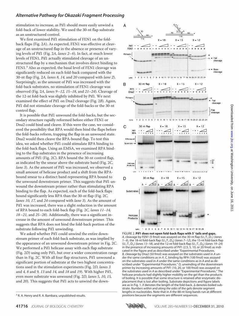
back flaps (Fig. 2A). As expected, FEN1 was effective at cleav-age of an unstructured flap in the absence or presence of vary-ing levels of Pif1 (Fig. 2A, lanes 2–6). In fact, at much lowerlevels of FEN1, Pif1 actually stimulated cleavage of an un-structured flap by a mechanism that involves direct binding toFEN1.3 Also as expected, the basal level of FEN1 cleavage wassignificantly reduced on each fold-back compared with the30-nt flap (Fig. 2A, lanes 8, 14, and 20 compared with lane 2).Surprisingly, as the amount of Pif1 was increased with thefold-back substrates, no stimulation of FEN1 cleavage wasobserved (Fig. 2A, lanes 9–12, 15–18, and 21–24). Cleavage ofthe 12-nt fold-back was slightly inhibited by Pif1. We nextexamined the effect of Pif1 on Dna2 cleavage (Fig. 2B). Again,Pif1 did not stimulate cleavage of the fold-backs or the 30-ntcontrol flap.It is possible that Pif1 unwound the fold-backs, but the sec-
ondary structure rapidly reformed before either FEN1 orDna2 could bind and cleave. If this were the case, we consid-ered the possibility that RPA would then bind the flaps beforethe fold-backs reform, trapping the flap in an unwound state.Dna2 would then cleave the RPA-bound flap. To test thisidea, we asked whether Pif1 could stimulate RPA binding tothe fold-back flaps. Using an EMSA, we examined RPA bind-ing to the flap substrates in the presence of increasingamounts of Pif1 (Fig. 2C). RPA bound the 30-nt control flap,as indicated by the smear above the substrate band (Fig. 2C,lane 3). As the amount of Pif1 was increased, we observed asmall amount of helicase product and a shift from the RPA-bound smear to a distinct band representing RPA bound tothe unwound downstream primer. This suggests that Pif1 un-wound the downstream primer rather than stimulating RPAbinding to the flap. As expected, each of the fold-back flapsbound significantly less RPA than the 30-nt flap (Fig. 2C,lanes 10, 17, and 24 compared with lane 3). As the amount ofPif1 was increased, there was a slight reduction in the amountof RPA bound to each fold-back flap (Fig. 2C, lanes 11–14,18–21, and 25–28). Additionally, there was a significant in-crease in the amount of unwound downstream primer. Thissuggests that RPA does not bind the fold-back portion of thesubstrate following Pif1 unwinding.We asked whether Pif1 could unwind the entire down-
stream primer of each fold-back substrate, as was implied bythe appearance of an unwound downstream primer in Fig. 2C.We performed a Pif1 helicase assay with each flap substrate(Fig. 2D) using only Pif1, but over a wider concentration rangethan in Fig. 2C. With all four flap structures, Pif1 unwound asignificant portion of substrate at the two highest concentra-tions used in the stimulation experiments (Fig. 2D, lanes 3and 4, 8 and 9, 13 and 14, and 18 and 19). With higher Pif1,even more substrate was unwound (Fig. 2D, lanes 5, 10, 15,and 20). This suggests that Pif1 acts to unwind the down-
3 R. A. Henry and R. A. Bambara, unpublished results.
FIGURE 2. Pif1 does not open fold-back flaps with 5� tails and gaps.A, cleavage by FEN1 (5 fmol) was assayed on the 30-nt flap (U1:T1:D1) (lanes1– 6), the 18-nt fold-back flap (U1:T1:D4) (lanes 7–12), the 15-nt fold-back flap(U1:T1:D5) (lanes 13–18), and the 12-nt fold-back flap (U1:T1:D6) (lanes 19 –24)in the presence of increasing amounts of Pif1 (2.5, 5, 10, or 20 fmol) as indi-cated in the figure and as described under “Experimental Procedures.B, cleavage by Dna2 (50 fmol) was assayed on the substrates used in A un-der the same conditions as in A. C, binding by RPA (100 fmol) was assayedon the substrates used in A under the same conditions as in A and as de-scribed under “Experimental Procedures.” D, unwinding of the downstreamprimer by increasing amounts of Pif1 (10, 20, or 100 fmol) was assayed onthe substrates used in A as described under “Experimental Procedures.” Thehelicase products had slightly higher mobility on the gel than the productsof boiling. It is possible that some structure is retained after enzymatic dis-placement that is lost after boiling. Substrate depictions and figure labelsare as in Fig. 1. X denotes the length of the fold-back. � denotes boiled sub-strate. Numbers within and along the sides of the gels denote segmentlengths in nucleotides. Note that in A the 88-nt-long bands run at differentpositions because the segments are different sequences.
Alternative Pathway for Okazaki Fragment Processing
41716 JOURNAL OF BIOLOGICAL CHEMISTRY VOLUME 285 • NUMBER 53 • DECEMBER 31, 2010
at Washington U
niversity, on June 16, 2011w
ww
.jbc.orgD
ownloaded from

stream primer of each fold-back substrate rather than openthe fold-back, which would have allowed FEN1, Dna2, andRPA to access the flap.The gap region of the fold-back substrates is most likely the
initial binding site for Pif1. Pfh1, the Schizosaccharomycespombe homolog of Pif1, has been shown to unwind the down-stream primer of a fold-back flap substrate if there is a gapbetween the fold-back and the downstream annealed region(32). We wondered whether removing the gap from the fold-back flaps would prevent Pif1 from unwinding the down-stream primers and promote unwinding of the fold-back it-self. If so, Pif1 should then stimulate FEN1 and Dna2 cleavageand RPA binding. We designed fold-back substrates similar tothose used in Fig. 2 with the exception that there were nogaps between the fold-backs and the downstream annealedregions. Surprisingly, we again observed no stimulation ofFEN1 (Fig. 3A), Dna2 (Fig. 3B), or RPA (Fig. 3C) by Pif1 onthese fold-back substrates. In a helicase assay, Pif1 again un-wound the downstream primers of all three fold-back sub-strates in addition to the 30-nt control flap (Fig. 3D).This result suggests two mechanisms, which are not mutu-
ally exclusive, by which Pif1 could unwind the downstreamprimers of the fold-backs. First, Pif1 could bind the 5� tail ofeach fold-back and unwind both the fold-back itself and thedownstream annealed region. Second, Pif1 could bind a tran-sient gap that would form as a result of reversible melting atthe junction point between the fold-back and the downstreamannealed region. To determine the mechanism used, we de-signed a third set of fold-back flap substrates, which lack boththe gap region and the 5� tail. If Pif1 were to unwind thesesubstrates, it likely binds a transient gap region. If Pif1 did notunwind these substrates, it likely binds the 5� tail when one ispresent. Pif1 unwound the downstream primers of these fold-backs to an extent significantly less than it did with the otherfold-back structures (Fig. 3E). Only at the highest concentra-tion of Pif1 was any unwinding observed. This suggests thatPif1 binds the 5� tail of each fold-back and unwinds both thefold-back and the entire downstream annealed region.Pif1 Removes an Okazaki Fragment Initiated by a
Fold-back—The helicase activity observed on the fold-backflaps with a 5� tail but no gap (Fig. 3D) is evidence that Pif1
FIGURE 3. Pif1 does not open fold-back flaps with 5� tails but withoutgaps. A, cleavage by FEN1 (5 fmol) was assayed on the 30-nt flap (U1:T1:D1)(lanes 1– 6), the 18-nt fold-back –G flap (U1:T1:D7) (lanes 7–12), the 15-nt
fold-back –G flap (U1:T1:D8) (lanes 13–18), and the 12-nt fold-back –G flap(U1:T1:D9) (lanes 19 –24) in the presence of increasing amounts of Pif1 (2.5, 5,10, or 20 fmol) as indicated in the figure and as described under “Experi-mental Procedures.” B, cleavage by Dna2 (50 fmol) was assayed on the sub-strates used in A under the same conditions as in A. C, binding by RPA (100fmol) was assayed on the substrates used in A under the same conditions asin A and as described under “Experimental Procedures.” D, unwinding of thedownstream primer by increasing amounts of Pif1 (10, 20, or 100 fmol) wasassayed on the substrates used in A as described under “Experimental Pro-cedures.” E, unwinding of the downstream primer by increasing amounts ofPif1 (10, 20, or 100 fmol) was assayed on the 30-nt flap (U1:T1:D1) (lanes 1–5),the 18-nt fold-back –G–T flap (U1:T1:D10) (lanes 6 –10), the 15-nt fold-back–G–T flap (U1:T1:D11) (lanes 11–15), and the 12-nt fold-back –G–T flap (U1:T1:D12) (lanes 16 –20) as described under “Experimental Procedures.” As in Fig.2D, the helicase products had slightly higher mobility than the boiled prod-ucts. Substrate depictions and figure labels are as in Fig. 1. X denotes thelength of the fold-back. � denotes boiled substrate. Numbers within andalong the sides of the gels denote segment lengths in nucleotides. Note thatin A the 82-nt-long bands run at different positions because the segmentsare different sequences.
Alternative Pathway for Okazaki Fragment Processing
DECEMBER 31, 2010 • VOLUME 285 • NUMBER 53 JOURNAL OF BIOLOGICAL CHEMISTRY 41717
at Washington U
niversity, on June 16, 2011w
ww
.jbc.orgD
ownloaded from

displays a much higher processivity than the previous mea-surement of �30 nt (33). A previous study found that Pfh1unwound the downstream primer of flaps initiated by a fold-back as long as there was a gap on which Pfh1 could load (32).Ryu et al. (32) suggested that Pif1 family helicases removeentire Okazaki fragments initiated by fold-back flaps that can-not be processed by either conventional pathway. pol � wouldthen synthesize through the resulting gap, and ligation to thenext downstream fragment would complete replication of theregion with the fold-back sequence. However, the substratesin the previous study were too short to simulate full-lengthOkazaki fragments, and no reconstitution experiments wereperformed. We wished to use our reconstitution system todetermine whether Pif1 can indeed unwind an entire Okazakifragment initiated by a fold-back.We designed an internal fold-back substrate that simulates
a full-length Okazaki fragment initiated by a fold-back flapand flanked by the adjacent Okazaki fragments. Three oligo-nucleotides were annealed to a 110-nt template primer. The25-nt upstream primer would be extended by pol � displacingthe 100-nt internal primer. If any extended upstream primersfully displace the internal primer, LigI would join them to the30-nt downstream primer. The internal primer simulated afull-length Okazaki fragment with the first 42 nt formed intoan 18-nt fold-back flap. This left 58 nt annealed to the tem-plate. There was a 2-nt gap between the upstream and inter-nal primers and a nick between the internal and downstreamprimers. The 5� end of the downstream primer had a phos-phate attached to allow ligation to the extended upstreamprimer. The 3� end of the internal primer had a 3� phosphategroup to prevent ligation to the downstream primer. Thedownstream primer also had a 5-nt 3� overhang to allow us todistinguish between a full-length synthesis product and a liga-tion product. The full-length synthesis product was predictedto run at 110 nt and the ligation product at 115 nt.First, we verified that neither FEN1 nor Dna2 could cleave
the fold-back in the internal fold-back substrate (Fig. 4A). Theinternal primer was labeled at the 5� end to allow us to visual-ize cleavage products. In the absence of synthesis, neitherFEN1 nor Dna2 cleaved the flap (Fig. 4A, lanes 2 and 3). Fullcleavage reconstitution with FEN1, Dna2, and RPA also didnot permit cleavage (Fig. 4A, lane 4). When cleavage was ex-amined in the context of synthesis by pol �, PCNA, and RFC,we again saw no cleavage by FEN1, Dna2, or the combinationof FEN1, Dna2, and RPA (Fig. 4A, lanes 6–8). Interestingly, inthe presence of FEN1, we observed some extension of the in-ternal primer by pol � to the end of the template (Fig. 4A,lanes 6 and 8). This suggests that the 3� exonuclease of pol �
FIGURE 4. Pif1 removes an Okazaki fragment initiated by a fold-backflap. A, cleavage by FEN1 (5 fmol) and Dna2 (50 fmol) was assayed on theinternal 18-nt fold-back substrate (U3:I1:T1:D13) in the presence of variouscombinations of RPA (100 fmol), pol � (23 fmol), PCNA (25 fmol), and RFC(25 fmol) as indicated in the figure and as described under “ExperimentalProcedures.” Lane 9 contains a radiolabeled 42-nt oligomer, the expectedlength of a FEN1 cleavage product. B, strand displacement synthesis by pol� (23 fmol) and ligation by LigI (25 fmol) were assayed on the internal 18-ntfold-back substrate in the presence of various combinations of PCNA (25fmol), RFC (25 fmol), FEN1 (5 fmol), Dna2 (50 fmol), RPA (100 fmol), and
increasing amounts of Pif1 (50, 100, or 200 fmol) as indicated in the figureand as described under “Experimental Procedures.” C, magnification andoverexposure of the boxed portion of B are shown. D, unwinding of thedownstream primer by increasing amounts of Pif1 (50, 100, 200, 300, or 500fmol) in the presence of pol � (23 fmol), PCNA (250 fmol), and RFC (25 fmol)was assayed on the internal 18-nt fold-back substrate (U3:I1:T1:D13) as de-scribed under “Experimental Procedures.” The control in lane 2 contains 500fmol of Pif1. dNTPs were included in the reaction buffer to allow for activesynthesis. Substrate depictions and figure labels are as in Fig. 1. P denotesthe presence of a phosphate group. � denotes boiled substrate. Numbersalong the sides of the gel denote segment lengths in nucleotides.
Alternative Pathway for Okazaki Fragment Processing
41718 JOURNAL OF BIOLOGICAL CHEMISTRY VOLUME 285 • NUMBER 53 • DECEMBER 31, 2010
at Washington U
niversity, on June 16, 2011w
ww
.jbc.orgD
ownloaded from

activated some of the ends by cleaving the 3� phosphate.pol � then extended those internal primers, and FEN1 wasnecessary to stimulate strand displacement through thedownstream primer. The crucial observation is that thefold-back was immune to cleavage under all conditions,demonstrating that an Okazaki fragment initiated by afold-back could be immune to processing by both flap-pro-cessing pathways.We then asked whether Pif1 could stimulate pol � synthesis
through the internal primer and thus permit resolution of thefold-back by removing the entire Okazaki fragment. With theupstream primer 5�-labeled, we examined synthesis and liga-tion within the internal fold-back substrate (Fig. 4, B and C).pol � alone strand-displaced a short distance into the internalprimer (Fig. 4, B and C, lane 3). When PCNA and RFC werepresent, strand displacement was stimulated, as expected((Fig. 4, B and C, lane 4). A small amount of an 85-nt productwas observed. This represents synthesis through the entireinternal primer and pausing at the 5� end of the downstreamprimer. Interestingly, addition of LigI slightly stimulated syn-thesis up to the pause point (Fig. 4, B and C, lane 5). Additionof FEN1 stimulated even more synthesis to the pause point(Fig. 4, B and C, lane 6). Given that FEN1 does not cleave thefold-back flap (Fig. 4A), this stimulation by FEN1 must havebeen cleavage-independent. Neither Dna2 (Fig. 4, B and C,lane 7) nor RPA (lane 8) stimulated synthesis. All possiblecombinations of FEN1, Dna2, and RPA did not stimulate syn-thesis beyond what was observed with FEN1 alone (Fig. 4, Band C, lanes 9–12). Importantly, no synthesis beyond thepause point or ligation of the extended upstream primer tothe downstream primer was observed in any of these reac-tions. However, when Pif1 was added in the absence of FEN1,Dna2, and RPA, synthesis up to the pause point was stimu-lated, and full-length synthesis and ligation products appeared(Fig. 4, B and C, lanes 13–15). Notably, Pif1 has an ability topromote ligation in our assays. Inclusion of RPA with Pif1 didnot further improve stimulation of synthesis and ligation (Fig.4, B and C, lanes 16–18). When all proteins were present,synthesis and ligation were stimulated to the highest amountobserved (Fig. 4, B and C, lanes 19–21). This suggests thatPif1 bound the gap created on the internal primer as pol �strand displaced. Pif1 then unwound the entire internalprimer, allowing pol � to synthesize through the gap. Whenpol � reached the downstream primer, either LigI sealed thenick between the extended upstream primer and downstreamprimer or pol � continued strand displacement through thedownstream primer. Pif1 thus allowed a fold-back flap, im-mune to the two pathways of cleavage, to be processed by re-moval of the entire Okazaki fragment.This experiment was repeated utilizing a helicase-deficient
mutant of Pif1, which was unable to stimulate the formationof full-length products (data not shown). This demonstratesthat the helicase activity of Pif1 facilitates the removal of andsynthesis through the fold-back flap. To further demonstratethis point, a helicase assay was performed using the samefold-back substrate as in Fig. 4B. Although pol �, PCNA, andRFC alone were unable to remove the fold-back, increasingamounts of Pif1 promoted fold-back flap removal (Fig. 4D),
further suggesting the importance of Pif1 for proper removalof fold-back flaps.Template Secondary Structure Slows pol � Synthesis—Oka-
zaki fragments do not have phosphates at their 3� ends in vivo.Therefore, it is possible that a full-length fragment initiatedby a fold-back flap could be ligated to the downstream frag-ment, before effective removal by Pif1. Once this happens invivo, the fold-back would be locked into the intact laggingstrand in a way that would be inaccessible to currently under-stood DNA replication mechanisms. It would have to be han-dled by repair systems as a site of DNA damage.We considered the possibility that Okazaki fragments initi-
ated with a fold-back might be extended more slowly thanmost and would therefore be delayed for ligation with the ad-jacent fragment. Why would this be the case? In the positionof the sequence with the potential to form a fold-back flap,the complementary template sequence will also have the po-tential to form secondary structure. We asked whether tem-plate secondary structure could slow down pol � so that syn-thesis would not reach the downstream primer before Pif1could remove the fragment with the fold-back.We designed the template fold-back substrate, which con-
sists of an upstream primer annealed to a template with an18-nt template fold-back downstream of the 3� end of the up-stream primer. As a control, we used the standard-44 sub-strate, also lacking the downstream primer. The template inthe control substrate has no significant secondary structure.We incubated each substrate with pol �, PCNA, and RFC forincreasing amounts of time (Fig. 5). With the control sub-strate, pol � synthesized some full-length product after only30 s, and the amount increased with time (Fig. 5, lanes 1–6).With the template fold-back substrate, synthesis was slowercompared with the control substrate (Fig. 5, lanes 13–18).Surprisingly, no full-length product was evident, even at thelongest time. Synthesis reached a maximum at �5 min anddid not increase further after longer incubation. When utiliz-ing this substrate, synthesis halts near the end of the fold-backsequence.RPA has previously been shown to stimulate synthesis by
pol � through hairpin structures in the template, presumablyby binding the template and melting those structures (34). Wewanted to determine whether RPA would have this same ef-fect on our template fold-back substrate and permit full-length synthesis by pol �. On the control substrate, RPA stim-ulated synthesis slightly (Fig. 5, lanes 7–12). On the templatefold-back substrate, synthesis was stimulated, and there wassome shift toward longer products (Fig. 5, lanes 19–24). How-ever, RPA was not able to promote full-length synthesis.To further characterize the effect of a fold-back within
the template, we utilized a second fold-back template. Thissubstrate consists of an upstream primer annealed to atemplate with only a 16-nt template fold-back downstreamof the 3� end of the upstream primer. Additionally, we de-creased the amount of GC pairs found in the fold-back todetermine whether a weaker fold-back region would im-prove pol � synthesis. Interestingly, when utilizing thissubstrate, we were able to observe full-length synthesis(supplemental Fig. 1, lanes 1–6). We again saw a slight
Alternative Pathway for Okazaki Fragment Processing
DECEMBER 31, 2010 • VOLUME 285 • NUMBER 53 JOURNAL OF BIOLOGICAL CHEMISTRY 41719
at Washington U
niversity, on June 16, 2011w
ww
.jbc.orgD
ownloaded from

stimulation of synthesis by RPA (supplemental Fig. 1, lanes7–12).The position at which synthesis halts is very important to
the interpretation of these results. In the presence of the 18-ntfold-back, synthesis continued through the fold-back, allow-ing the fold-back flap to be made, but it then slowed so theOkazaki fragment with the fold-back was not rapidly ex-tended to a potential point of ligation. Some synthesis termi-nated within the hairpin, some near the end of the hairpin,and a small amount beyond the hairpin. The range of termi-nation is broad, producing a variety of intermediate struc-tures. When utilizing the 16-nt fold-back, a range of productswas created throughout the fold-back region, with only a fewproducts reaching full length. This would indicate that pol �slowed as it synthesized through the fold-back region, result-ing in a number of intermediates.Upon strand displacement, some products are expected to
form weak structure, susceptible to FEN1, although otherswould form more stable structure, allowing loading of Pif1and displacement. The slowing of pol � through the fold-backtemplate would allow the fold-backs to be displaced by Pif1before they can be joined to the continuous lagging strand.
DISCUSSION
Our previous reconstitution analyses of eukaryotic laggingstrand DNA replication suggest that in vivo Pif1 helicaselengthens Okazaki flaps that escape immediate FEN1 cleavage(11). These long flaps are processed by the two-nuclease path-way, which requires Dna2 helicase/nuclease in addition toFEN1 (13). Although Pif1 has several known cellular roles, ourresults would appear to indicate that at Okazaki fragmentsPif1 only serves to create a requirement for Dna2. If Pif1 didnot influence Okazaki fragment maturation, the multisteppathway involving Dna2 might not be required. It is unlikelythat the fragment maturation process would have evolved thisway if the only role of Pif1 were to promote the secondarypathway. Therefore, we hypothesized that Pif1 plays an im-
portant but previously unnoticed role at Okazaki fragments.In this study, we attempted to determine the nature of thisrole.Duplexes that are highly GC-rich inhibit strand displace-
ment synthesis by pol � (12). We hypothesized that Pif1would improve strand displacement through such sequencesby unwinding GC-rich duplexes and alleviating the energeticbarrier to strand displacement. However, we found that Pif1did not stimulate synthesis on a reconstitution substrate witha downstream primer containing a GC-rich 5� end (Fig. 1).This result is not entirely surprising. Pif1 requires pol � todisplace at least a short flap before Pif1 can create a long flap(13). pol � rarely displaces a flap longer than 1-nt upon en-countering a GC-rich sequence, a flap that is likely too shortfor Pif1 to bind. Without proper initiation, Pif1 would notunwind the downstream primer and so could not stimulatesynthesis.Some sequences, for example triplet repeats, have the po-
tential to form stable secondary structures when they are dis-placed into flaps. We hypothesized that Pif1 would open suchfold-back flaps. However, Pif1 was not able to stimulate FEN1cleavage, Dna2 cleavage, or RPA binding of fold-back flapswith 5� tails. Rather than open the fold-back, Pif1 unwoundthe downstream primer (Fig. 2). We reasoned that Pif1 boundthe gap between the fold-back and the downstream annealedregion. However, removal of the gap did not permit stimula-tion of FEN1, Dna2, or RPA, and Pif1 still unwound thedownstream primers. When the 5� tails were then removed aswell, Pif1 was no longer able to unwind the downstream prim-ers effectively (Fig. 3). These results collectively imply that thetail is a sufficient binding site for Pif1 and that Pif1, oncebound, is able to unwind both the fold-back and the down-stream annealed region. Additional results with Pfh1 suggestthat Pif1 will bind a gap as well, if available (32). Binding ofPif1 to either site results in removal of the downstreamprimer.
FIGURE 5. Template secondary structure slows synthesis by pol �. Synthesis by pol � (23 fmol) was assayed on the standard-44 substrate lacking thedownstream primer (U2:T1) (lanes 1–12) and the template fold-back substrate (U2:T3) (lanes 13–24) in the presence of PCNA (25 fmol) and RFC (25 fmol) andin the absence or presence of RPA (200 fmol) for increasing amounts of time (0, 0.5, 1, 2.5, 5, and 10 min), as indicated in the figure and as described under“Experimental Procedures.” Substrate depictions and figure labels are as in Fig. 1.
Alternative Pathway for Okazaki Fragment Processing
41720 JOURNAL OF BIOLOGICAL CHEMISTRY VOLUME 285 • NUMBER 53 • DECEMBER 31, 2010
at Washington U
niversity, on June 16, 2011w
ww
.jbc.orgD
ownloaded from

On the 18-nt fold-back –G flap substrate, Pif1 must unwind58 nt to remove the downstream primer by the mechanismdescribed above. This is almost twice the length of the previ-ously measured processivity of Pif1, �30 nt (33). It is possiblethat under our reaction conditions Pif1 has a higher proces-sivity than has been measured. Alternatively, the flap struc-ture may stimulate Pif1 processivity to the level we observed.Pif1 may also unwind the flap in a cooperative fashion, inwhich one molecule unwinds a given distance, dissociates,and then another Pif1 molecule binds and continuesunwinding.The high processivity of Pif1 that we observed led us to ask
whether Pif1 is capable of removing an entire Okazaki frag-ment that is initiated by a fold-back flap. This concept wasoriginally proposed by Ryu et al. (32) when they performedsimilar helicase assays with Pfh1. However, they did not teststimulation of FEN1, Dna2, or RPA, and their flaps did notsimulate full-length Okazaki fragments. We felt that our re-constitution system is ideal to test this proposal. We designeda substrate that simulated a full-length Okazaki fragment witha 5� fold-back flap and tested synthesis through the fragmentin the presence of components of the two flap-processingpathways (Fig. 4). We observed slight stimulation of synthesisby LigI alone and by FEN1, although FEN1 did not cleave. Toour knowledge, there are no previous reports of stimulation ofsynthesis by LigI or cleavage-independent stimulation byFEN1. PCNA is believed to bind one molecule each of pol �,FEN1, and LigI (10). It is possible that formation of the com-plete complex stimulates each individual enzyme. This wouldexplain how LigI and FEN1 could stimulate pol �. It will beinteresting to examine whether such a PCNA-dependentmechanism of stimulation exists.Most significantly, there was no stimulation of synthesis by
either Dna2 or RPA, and the combinations of FEN1, Dna2,and RPA did not stimulate synthesis above that observed withFEN1 alone. This implies that the components of the twoflap-processing pathways are incapable of alleviating the blockto synthesis presented by the fold-back flap. Therefore, if sucha flap were to form in vivo, the fragment would not be cleavedand could cause genome instability through chromosomebreaks or strand invasions. When Pif1 was present, we ob-served increased amounts of intermediate synthesis productsand synthesis through the entire internal and downstreamprimers. In addition, we observed ligation of the extendedupstream primer to the downstream primer. This implies thatPif1 stimulates LigI, consistent with previous evidence (13).Both full-length synthesis and ligation imply successful com-plete displacement of the fold-back flap-initiated Okazakifragment. Because this fold-back did not have a 5� tail, Pif1likely bound the gap that would emerge as pol � displaced afew nucleotides into the internal annealed region. A 5� tail invivo would also allow Pif1 binding, but the 5� tail configura-tion is a substrate for FEN1, which would be likely to cleavebefore Pif1 could bind (Figs. 2A and 3A).RPA did not enhance stimulation of synthesis by Pif1; in
fact, there was a slight decrease in the stimulation. It is possi-ble that as Pif1 unwinds the internal primer, RPA binds thegrowing gap between the fold-back and the remaining an-
nealed portion of the internal primer. If Pif1 unwinds longstretches via the cooperative mechanism described above, theRPA bound to the gap may prevent the second Pif1 moleculefrom binding. Specific interactions between RPA and Pif1must be examined to fully understand this result.The greatest stimulation of synthesis and ligation was ob-
served when all proteins were present. This phenomenon wasalso observed when we examined processing of a long flapfollowing Pif1 lengthening (13). The reason for this is notknown. Numerous studies have shown that individual pairs ofreplication proteins interact with and stimulate each other.Perhaps when the entire system is reconstituted, these inter-actions combine to produce the most efficient synthesis andflap processing possible.Overall, results suggest a unique and important role for Pif1
in Okazaki fragment processing. Namely, it fully displacesOkazaki fragments that form flap intermediates that are re-fractory to cleavage by either the FEN1-only or two-nucleasepathways. We were able to test this directly utilizing a helicaseassay (Fig. 4D). We observed that Pif1 is able to unwind thefold-back flap both on its own and in the presence of pol �(Fig. 4D, lanes 3 and 9, respectively). We believe Pif1 is able tounwind the fold-back on its own because of breathing of theannealed region, exposing a few nucleotides of single-stranded DNA. Pif1 could bypass the fold-back and bind tothis single-stranded region to unwind. In the presence of thereplication complex, we observe that unwinding of the sub-strate decreases. It is likely that the replication complex occu-pies the region at the base of the flap. This would physicallyprevent Pif1 from binding to any single-stranded region thatresults from DNA breathing. Thus, Pif1 would have to waituntil pol � displaces an adequate amount of single-strandedDNA before Pif1 could bind to the flap. Importantly, the addi-tion of Pif1 displaces these fold-back flaps, which pol � alonecannot accomplish (Fig. 4D, lanes 4 and 9).A potential conceptual flaw, however, in this proposed role
for Pif1 is the apparent likelihood that by the time Pif1 coulddisplace the undesirable fold-back fragment, it would bejoined to the adjacent downstream fragment. Once the fold-back fragment became part of the continuous lagging strand,no amount of displacement could remove it. Our substratewas designed to prevent this reaction, but the possibility ex-ists in vivo, as natural Okazaki fragments evolved for ligation.We considered that the template sequence at a fold-back
flap could also form secondary structure. Such a structurecould slow primer elongation. However, hairpin-type second-ary structures in the template usually pause polymerases atthe base of the stem but not after the hairpin has been partlyor fully copied. Such an outcome would pause Okazaki frag-ment elongation before the fold-back flap was made or dis-placed and would not be relevant. A mechanism in which anOkazaki fragment was made and then displaced into a fold-back flap, but never elongated enough for ligation, would in-volve slowing of primer elongation over the template withinor just beyond the fold-back.When we tested the progress of pol �-catalyzed primer
elongation over a stable fold-back in the template, synthesisslowed as the fold-back was copied and terminated in a dif-
Alternative Pathway for Okazaki Fragment Processing
DECEMBER 31, 2010 • VOLUME 285 • NUMBER 53 JOURNAL OF BIOLOGICAL CHEMISTRY 41721
at Washington U
niversity, on June 16, 2011w
ww
.jbc.orgD
ownloaded from

fuse region within or beyond the fold-back (Fig. 5 and supple-mental Fig. 1). Although RPA stimulated synthesis, it allowedonly a moderate shift toward longer products. As expected,we see that the presence of a fold-back within the templateslows pol � synthesis. This would allow the upstream Okazakifragment to be extended while pol � is still in the process ofsynthesizing through the fold-back in the template. This isexactly the behavior that would allow Pif1 to effectively dis-place a fold-back fragment before it was ligated. Further stud-ies on the importance of fold-back length and stability and theinfluence of Pif1 and other replication proteins will provideinsight into how the polymerase-PCNA complex interactswith template fold-backs.Based on these results, we propose an alternative Okazaki
fragment-processing pathway, specifically for fragments thatform fold-back flaps (Fig. 6). As the template DNA is openedby a replicative helicase, it forms a fold-back. An Okazakifragment initiated a short distance upstream of the templatefold-back will be extended through the fold-back, but the pol�-PCNA processive complex will be slowed by the presence ofa fold-back in the template. Meanwhile, another molecule ofpol � will be extending the upstream Okazaki fragment. Elon-gation of the upstream fragment begins to displace the down-stream fragment, and if the flap becomes long, a fold-backflap will form. The fold-back cannot be processed by eitherthe FEN1-only or two-nuclease pathway. As pol � displacesthrough the fold-back flap, a gap is created either in the loopor just past the stem, and Pif1 binds the gap. Pif1 then un-winds and removes the incomplete, cleavage-resistant frag-ment, allowing pol � to continue extending the upstreamprimer through the gap. Upon reaching the next downstreamfragment, standard flap processing and ligation will completesynthesis.Slowing of pol � during synthesis of the template fold-back
might serve two important roles. First, a gap is left betweenthe point of dissociation and the next downstream Okazakifragment, so the two fragments cannot be ligated. This alsoprevents Pif1 from having to unwind a full-length Okazakifragment. Second, it converts the template fold-back into ashort double-stranded region. pol �, now working with Pif1,can then readily synthesize through the region to the nextdownstream primer for ligation.Although it is energetically wasteful to synthesize a long
DNA fragment only to have it displaced and presumably de-graded, use of this pathway should be a rare event in DNAreplication. There are likely to be very few flaps that form dis-ruptive secondary structure in vivo. We have suggested thatonly a small fraction of flaps become long via Pif1 lengthening(13). However, other mechanisms are likely to produce longflaps. The histone acetylase p300 has been reported to acety-late FEN1, reducing its nuclease activity (35), and to acetylateDna2, greatly stimulating its nuclease and helicase activities(36). We have proposed that this is a regulatory process thatleads to more flap displacement and use of the two-nucleasepathway in regions of actively transcribed chromatin (36).Nevertheless, only a small fraction of these flaps is likely toform in regions that allow for stable fold-backs. However,there are hundreds of thousands of Okazaki fragments pro-
cessed each replication cycle in yeast (37) and many more inhumans. If only a few flaps form secondary structure, then thecell must be prepared to deal with them to maintain genomestability. Therefore, the alternative pathway is likely a rarelyused but critically important mechanism for genomemaintenance.We cannot rule out the possibility that Pif1 plays other im-
portant roles at Okazaki fragments. Through its flap length-ening capacity, Pif1 may act as part of a correction mecha-nism for mutations introduced by pol �. The DNA patch
FIGURE 6. Model for an alternative Okazaki fragment-processing path-way in vivo. A, during DNA replication, at a sequence that has the potentialto form secondary structure, a fold-back will form in the template DNA. TheOkazaki fragment downstream of the fold-back is synthesized normally. Aprimer is laid down upstream of the template secondary structure. Notethat the RNA portion of the primer is not depicted for simplicity. B, pol � willbe slowed as it extends the primer through the secondary structure. A gapbetween the extended primer and the downstream primer will remain.C, meanwhile, the fragment upstream of the internal fragment will be syn-thesized normally. D, when it reaches the internal fragment, flap lengthen-ing factors, such as Pif1 activity or acetylation of the nucleases, may create along flap. The long flap will then fold-back on itself. Neither the FEN1-onlypathway nor the two-nuclease pathway can process the fold-back flap.E, Pif1 will bind a gap between the fold-back and the remaining down-stream annealed region and, working with strand displacement synthesisfrom the upstream primer, unwind the entire internal Okazaki fragment.This synthesis will convert the fold-back region of the template to doublestrands that will no longer be capable of forming secondary structure. F, pol� will then continue synthesis through the gap until it reaches the down-stream fragment. G, conventional flap processing will produce a nick thatLigI will then seal. The red lines represent segments with potential to formsecondary structures.
Alternative Pathway for Okazaki Fragment Processing
41722 JOURNAL OF BIOLOGICAL CHEMISTRY VOLUME 285 • NUMBER 53 • DECEMBER 31, 2010
at Washington U
niversity, on June 16, 2011w
ww
.jbc.orgD
ownloaded from

synthesized and displaced by pol � is estimated to be too shortto remove the entire RNA/DNA primer synthesized by pol �(24). As mentioned previously, pol � is a low fidelity polymer-ase. All pol �-synthesized bases must be removed to ensurethe best fidelity of DNA replication. Perhaps Pif1-promotedflap lengthening is required at locations of pol � mistakes.The long flap would include all mismatched nucleotides atdisplacement distances normally not achieved by pol �. Addi-tional promotion of flap displacement by acetylation of FEN1and Dna2 should augment this effect, making complete re-moval of pol � errors even more certain in active chromatin(36). Our reconstitution system is well designed to addressthis issue. The downstream primer of the substrate could in-clude a mismatch at variable distances from the 5� end, andincorporation of the mismatch into a final ligated productfollowing strand displacement synthesis could be measured.We could then test the effect of Pif1 and acetylation of thenucleases on this process. These and similar biochemical ex-periments will be crucial to further reveal the biological rele-vance of Pif1 in Okazaki fragment processing.
Acknowledgments—We thank the members of the Bambara labora-tory for helpful discussions and suggestions. We thank Dr. MarcWold for providing us with purified RPA.
REFERENCES1. Kornberg, A., and Baker, T. A. (1992) DNA Replication, 2nd Ed.,
pp. 113–195, W. H. Freeman & Co., New York2. Bambara, R. A., Murante, R. S., and Henricksen, L. A. (1997) J. Biol.
Chem. 272, 4647–46503. Liu, Y., Kao, H. I., and Bambara, R. A. (2004) Annu. Rev. Biochem. 73,
589–6154. Rossi, M. L., Purohit, V., Brandt, P. D., and Bambara, R. A. (2006) Chem.
Rev. 106, 453–4735. Ayyagari, R., Gomes, X. V., Gordenin, D. A., and Burgers, P. M. (2003)
J. Biol. Chem. 278, 1618–16256. Garg, P., Stith, C. M., Sabouri, N., Johansson, E., and Burgers, P. M.
(2004) Genes Dev. 18, 2764–27737. Jin, Y. H., Ayyagari, R., Resnick, M. A., Gordenin, D. A., and Burgers,
P. M. (2003) J. Biol. Chem. 278, 1626–16338. Harrington, J. J., and Lieber, M. R. (1994) EMBO J. 13, 1235–12469. Murante, R. S., Rust, L., and Bambara, R. A. (1995) J. Biol. Chem. 270,
30377–3038310. Kao, H. I., and Bambara, R. A. (2003) Crit. Rev. Biochem. Mol. Biol. 38,
433–45211. Rossi, M. L., Pike, J. E., Wang, W., Burgers, P. M., Campbell, J. L., and
Bambara, R. A. (2008) J. Biol. Chem. 283, 27483–2749312. Rossi, M. L., and Bambara, R. A. (2006) J. Biol. Chem. 281, 26051–2606113. Pike, J. E., Burgers, P. M., Campbell, J. L., and Bambara, R. A. (2009)
J. Biol. Chem. 284, 25170–2518014. Fanning, E., Klimovich, V., and Nager, A. R. (2006) Nucleic Acids Res.
34, 4126–413715. Bae, S. H., Bae, K. H., Kim, J. A., and Seo, Y. S. (2001) Nature 412,
456–46116. Bae, S. H., and Seo, Y. S. (2000) J. Biol. Chem. 275, 38022–3803117. Budd, M. E., Choe, W., and Campbell, J. L. (2000) J. Biol. Chem. 275,
16518–1652918. Bae, S. H., Choi, E., Lee, K. H., Park, J. S., Lee, S. H., and Seo, Y. S. (1998)
J. Biol. Chem. 273, 26880–2689019. Kao, H. I., Campbell, J. L., and Bambara, R. A. (2004) J. Biol. Chem. 279,
50840–5084920. Kao, H. I., Veeraraghavan, J., Polaczek, P., Campbell, J. L., and Bambara,
R. A. (2004) J. Biol. Chem. 279, 15014–1502421. Stewart, J. A., Miller, A. S., Campbell, J. L., and Bambara, R. A. (2008)
J. Biol. Chem. 283, 31356–3136522. Lee, K. H., Kim, D. W., Bae, S. H., Kim, J. A., Ryu, G. H., Kwon, Y. N.,
Kim, K. A., Koo, H. S., and Seo, Y. S. (2000) Nucleic Acids Res. 28,2873–2881
23. Budd, M. E., Reis, C. C., Smith, S., Myung, K., and Campbell, J. L. (2006)Mol. Cell. Biol. 26, 2490–2500
24. Stith, C. M., Sterling, J., Resnick, M. A., Gordenin, D. A., and Burgers,P. M. (2008) J. Biol. Chem. 283, 34129–34140
25. Boule, J. B., and Zakian, V. A. (2006) Nucleic Acids Res. 34, 4147–415326. Ribeyre, C., Lopes, J., Boule, J. B., Piazza, A., Guedin, A., Zakian, V. A.,
Mergny, J. L., and Nicolas, A. (2009) PLoS Genet. 5, e100047527. Bartos, J. D., Willmott, L. J., Binz, S. K., Wold, M. S., and Bambara, R. A.
(2008) J. Biol. Chem. 283, 21758–2176828. Burgers, P. M., and Gerik, K. J. (1998) J. Biol. Chem. 273, 19756–1976229. Gerik, K. J., Gary, S. L., and Burgers, P. M. (1997) J. Biol. Chem. 272,
1256–126230. Kao, H. I., Henricksen, L. A., Liu, Y., and Bambara, R. A. (2002) J. Biol.
Chem. 277, 14379–1438931. Sibenaller, Z. A., Sorensen, B. R., and Wold, M. S. (1998) Biochemistry
37, 12496–1250632. Ryu, G. H., Tanaka, H., Kim, D. H., Kim, J. H., Bae, S. H., Kwon, Y. N.,
Rhee, J. S., MacNeill, S. A., and Seo, Y. S. (2004) Nucleic Acids Res. 32,4205–4216
33. Lahaye, A., Leterme, S., and Foury, F. (1993) J. Biol. Chem. 268,26155–26161
34. Fortune, J. M., Stith, C. M., Kissling, G. E., Burgers, P. M., and Kunkel,T. A. (2006) Nucleic Acids Res. 34, 4335–4341
35. Hasan, S., Stucki, M., Hassa, P. O., Imhof, R., Gehrig, P., Hunziker, P.,Hubscher, U., and Hottiger, M. O. (2001)Mol. Cell 7, 1221–1231
36. Balakrishnan, L., Stewart, J., Polaczek, P., Campbell, J. L., and Bambara,R. A. (2010) J. Biol. Chem. 285, 4398–4404
37. Burgers, P. M. (2009) J. Biol. Chem. 284, 4041–4045
Alternative Pathway for Okazaki Fragment Processing
DECEMBER 31, 2010 • VOLUME 285 • NUMBER 53 JOURNAL OF BIOLOGICAL CHEMISTRY 41723
at Washington U
niversity, on June 16, 2011w
ww
.jbc.orgD
ownloaded from

Lane
– RPA + RPA
Time
44 nt
110 nt
54 nt
Full-length
Intermediates
*16
1410
6
44
1 2 3 4 5 6 7 8 9 10 11 12
Primer
Supplemental Figure 1
Supplemental Figure 1. Template secondary structure slows synthesis by pol δ on a shorter fold-back. Synthesis by pol δ (23 fmol) wasassayed on a 16 nt template fold-back substrate (U2:T4) in the presence of PCNA (25 fmol) and RFC (25 fmol) and in the absence or presence ofRPA (200 fmol) for increasing amounts of time (0, 0.5, 1, 2.5, 5, and 10 minutes) as indicated in the figure and as described under ExperimentalProcedures. Substrate depictions and figure labels are as in Fig. 1. Numbers along the sides of the gel denote segment lengths in nucleotides.
at Washington U
niversity, on June 16, 2011w
ww
.jbc.orgD
ownloaded from