analysis and isolation of stem cells using flow … and isolation of stem cells using flow cytometry...
TRANSCRIPT

Stem Cell Research
Analysis and Isolation of Stem Cells Using Flow Cytometry

2 Cover Illustration: Fairman Studios, LLC.

3
Recent discoveries in stem cell biology have amplified the importance of pluripotent stem cells in therapeutics and have improved researchers’ understanding of normal and disease processes. Still, the inherent heterogeneous nature of differentiating cultures remains a primary challenge in stem cell research.
Flow cytometry is a powerful methodology for characterizing, analyzing, and isolating stem cells and their derivatives.
Flow cytometry is unique in its ability to investigate large cell populations at the single-cell level. In contrast to methods such as Western blot and cellular imaging, multicolor flow cytometry enables researchers to interrogate heterogeneous cell populations and analyze their subpopulations. Using multiple fluorescent-labeled antibodies, researchers can obtain robust, multiparametric data and population-based statistics on differentiating stem cell cultures—and can isolate stem cells and their derivatives from primary tissue and diverse in vitro populations.
Flow cytometry has been used for decades by biologists studying hematopoietic stem cells to address the challenge of heterogeneity. New methods and tools are enabling researchers to employ this powerful technique to make key discoveries about other stem cell types and their respective lineages.
Researchers can use fluorochrome-conjugated antibodies to either cell surface or intracellular biomarkers to verify that stem cells have maintained pluripotency. Since stem cells differentiate into the three primary germ layers and into differentiated tissue, antibodies can monitor their changing expression patterns. Analysis based on cell surface markers can preserve cell viability for use in additional experiments. BD Lyoplate™ cell surface marker screening panels provide a powerful method for discovering surface marker signatures that can be used to explore these cells in depth.
BD Biosciences offers a diverse set of tools including high-quality antibodies, buffers, protocols, and instrumentation to support stem cell research. This evolving toolset combines the power of advanced technologies and world-class service to support investigators in characterizing, analyzing, and sorting heterogeneous stem cell populations.

4
Human Markers Mouse Markers BD Stemflow™ or other Kit Cat. No.
Pluripotent Stem Cells (ESCs and iPSCs)
Positive: Alkaline Phosphatase, SSEA-4, SSEA-3, TRA-1-81, TRA-1-60
Negative: SSEA-1
Positive: SSEA-1 Human iPSC Sorting and Analysis Kit 562626
Human Pluripotent Stem Cell Sorting and Analysis Kit
560461
Human and Mouse Pluripotent Stem Cell Analysis Kit
560477
Hematopoietic Stem Cells (HSCs)
Positive: CD34, CD49f, CD90
Negative: CD38, CD45RA, Lineage*
Positive: CD150, c-Kit, Sca1
Negative: CD34, CD41, CD48, Lineage
BD Pharmingen™ Human Lineage Cocktail 4 562722
Mouse Hematopoietic Stem Cell Isolation Kit 560492
Mesenchymal Stem Cells (MSCs)
Positive: CD44, CD73, CD90, CD105, CD146, CD271
Negative: CD11b, CD19, CD31, CD34, CD45, CD144, HLA-DR
Positive: CD29, CD44, CD90, CD105, CD106, Sca-1
Negative: CD11b, CD31, CD45, Ter-119
Human MSC Analysis Kit 562245
Human Mesenchymal Stem Cell Lineage Antibody Cocktail
562530
Neural Stem Cells (NSCs)
Positive: CD15mid, CD24, CD184
Negative: CD44, CD271
– Human Neural Cell Sorting Kit 562271
Neurons
Positive: CD15low, CD24
Negative: CD44, CD184
– Human Neural Cell Sorting Kit 562271
Cancer
CD15, CD24, CD34, CD44, CD45, CD49f, CD166, CD326, CD338, Her-2/Neu, Lgr5
– – –
* Human lineage (lin) markers: CD2, CD3, CD4, CD7, CD8, CD10, CD11b, CD14, CD19, CD20, CD56, CD235a
Each type of stem cell or derivative expresses characteristic surface and intracellular proteins that can be used for identification (Tables 1 and 2). Because intracellular analysis requires permeabilization, surface markers are essential when researchers want to isolate live cell populations for further analysis. Fluorescence-activated cell sorting, or FACS, can be used to sort cells of interest in bulk or in single-cell depositions for downstream applications. To analyze cells for surface marker expression, a single-cell suspension must be stained with fluorescent-labeled antibodies and analyzed or sorted on a flow cytometer.
Sample preparationStem cells tend to be adherent and can grow as three-dimensional structures. To prepare a single-cell suspension for flow cytometric analysis, enzymatic digestion (with BD™ Accutase cell detachment solution or trypsin) or mechanical scraping can be used. Since enzymatic methods might cleave or modify some protein epitopes during the
digestion process, preventing antibody labeling, they must be evaluated for each surface marker being measured. BD Accutase tends to be more broadly applicable than trypsin and yields a more consistent single-cell suspension than does scraping.
Monoclonal antibodiesOnce cells are harvested and the dissociation buffer is removed, the cells are ready to be stained with antibodies. BD Biosciences offers an extensive reagent selection of antibodies against hundreds of stem cell markers conjugated to a variety of fluorochromes for flexibility in experimental design. For analysis of rare events and low-density antigens, the BD Horizon™ Brilliant Violet™ family of reagents can increase brightness and resolution. For ease of use, BD Stemflow™ kits and cocktails contain standard antibody panels for analysis or sorting of different stem cell types, as shown in Table 1.
Cell surface staining for analysis and sorting of live cells
Isolation of live cell populations
Table 1.Representative surface markers of selected stem cells and derivatives.

H E T E R O G E N E I T Y
5
BV
605
CD
9010
5
PE CD49f
CD49f+
104
103
102
0-1
02-5
31
102-105 0 103 104 105
Tube: enriched
Population
All EventsScatter
Lineage-ViableCD34+CD38-
CD90+CD45RA-CD49f+
#Events %Parent %Total
184 20.9 0.1879 18.9 0.4
4,650 9.6 2.348,194 33.1 23.9
145,671 72.2 72.2201,698 #### 100.0
BV
421
CD
3810
5
APC CD34
CD34+CD38-
104
103
102
102 103 104 105B
V60
5 C
D90
105APC-H7 CD45RA
CD90+CD45RA-
104
103
102
-531
0-1
02-650 -102 1031020 104 105
SSC
-A(x
1,0
00)
(x 1,000)250
250
FSC-A
Scatter
200
150
100
5020015010050
0 101
FITC Lineage Cocktail 4
Lineage-Viable
7-A
AD
105
104
103
102
-124
0
102 103 104 105
Hematopoietic stem cell phenotypingCells of the hematopoietic system are well characterized with respect to surface marker expression, which is often used to isolate and characterize subsets of cells during hematopoiesis. Hematopoietic stem cells (HSCs), the source of these hematopoietic cells, are currently a focus area in stem cell biology because they can be used to replenish normal bone marrow function.
Historically, among a pool of cells, HSCs were identified as lineage-negative cells that expressed CD90 and CD34.1 Recently, researchers have used additional markers to enrich pools of long-term HSCs (LT-HSCs) capable of self-renewal. These markers include CD38,2 CD45RA,3 and most recently CD49f.4 Reportedly, about 10% of cells with a Lin–CD34+CD38–CD90+CD45RA–CD49f+ phenotype are able to provide long-term repopulating capacity in mouse models.4
Gating strategy for LT-HSCs.Frozen human cord blood mononuclear cells (Stem Cell Technologies) were enriched using the BD IMag™ human lineage cell depletion set – DM (Cat. No. 560030), stained with antibodies, and acquired and analyzed on a BD LSRFortessa™ flow cytometer. A. First, cells were gated based on light-scatter properties to screen out debris. B. Next, viable, lineage-negative (CD2, CD3, CD4, CD7, CD8, CD10, CD11b, CD14, CD19, CD20, CD56, CD235a) cells were gated based on the viability dye 7-AAD and the BD Pharmingen human lineage cocktail 4 kit (Cat. No. 562722). To identify a highly enriched LT-HSC population, (C) cells were gated on CD34+CD38–; (D) then (in a child gate) on CD90+CD45RA–; and (E) finally on CD49f+. This combination of cell surface markers, summarized in (F) the complete gating hierarchy, results in a population rich in LT-HSCs.
B
D
F
A
C
E

6
Cell Type Intracellular Markers BD Stemflow Kit Cat. No.
Embryonic stem cells (ESCs)
Induced pluripotent stem cells (iPSCs)
Nanog, Oct3/4, Sox2 Human Pluripotent Stem Cell Transcription Factor Analysis Kit
560589
Mouse Pluripotent Stem Cell Transcription Factor Analysis Kit
560585
Neural stem cells (NSCs) Nestin, Pax6, Sox1, Sox2 Human Neural Lineage Analysis Kit 561526
Astrocytes GFAP Human Neural Lineage Analysis Kit 561526
Neurons Doublecortin Human Neural Lineage Analysis Kit 561526
Early pancreatic endoderm FoxA2, Pax6, Pdx1, Sox17 Human Definitive and Pancreatic Endoderm Analysis Kit
562496
Late pancreatic endoderm NeuroD1, Nkx6.1 – –
Cardiac cTNI, GATA4, Islet-1, Myosin Heavy Chain
– –
Hepatic AFP, GATA4 – –
Intracellular staining allows researchers to extend the speed and statistical relevance of flow cytometry to the investigation of functional proteins inside the cell. It can be used in combination with surface staining to identify critical time points, markers, and proportions of cells moving along particular differentiation pathways. Intracellular staining protocols require the cells to be fixed and permeabilized so that antibodies can access the cytoplasm and nucleus. Since fixation effectively kills the cells, intracellular staining is not compatible with live-cell sorting.
Sample preparationFor intracellular staining, as with surface staining, a single-cell suspension must be prepared using enzymatic or mechanical methods. BD Accutase is recommended since it helps to prevent cell clumping and can preserve surface proteins for simultaneous analysis. After optional surface staining, cells must be fixed and permeabilized to enable antibodies to enter. The cells are then stained with fluorescent-labeled antibodies to intracellular antigens and analyzed on a flow cytometer.
To optimize permeabilization and staining conditions, BD has developed several kits for the detection of key stem cell transcription factors. The kits contain optimized antibodies and buffer systems to characterize stem cells as well as their differentiation into various lineages.
Intracellular staining for transcription factor analysis
Study of differentiation pathways
Table 2.Representative intracellular markers of selected stem cells and derivatives.

7
I N T R A C E L L U L A R
Stem cell differentiationThe ability of human pluripotent stem cells to differentiate into various cell lineages is a central topic in developmental biology and has applications for regenerative medicine and cellular therapy. As pluripotent cells differentiate into different lineages, the expression of transcription factors and other proteins can change. Multiparametric flow cytometry is an excellent method for determining the relative numbers of cells expressing markers of interest, and can be used to optimize, quantitate, and compare differentiation protocols and differentiation potential.
For example, in mammalian embryonic development, the definitive endoderm generates the liver, pancreas, and intestine.5 During lineage specification into definitive endoderm, the levels of transcription factors Sox17 and FoxA2 increase, while pluripotency markers such as Nanog decrease.6
The differentiation of neural stem cells to neural lineages can also be monitored using multicolor flow analysis. As neural stem cells (NSCs) differentiate into neurons, they gradually express less Nestin and more of the early neuronal marker doublecortin (DCX). A subpopulation of cells that continues to express Nestin further delineates into a glial cell population that expresses CD44.
Changes in intracellular and surface markers in neural cell differentiation.NSCs derived from H9 hESCs using the serum-free embryoid body (SFEB) method were differentiated into neurons and glia and monitored on a BD LSRFortessa flow cytometer using the BD Stemflow human neural lineage analysis kit (Cat. No. 561526). A. As differentiation progressed, cells expressed less Nestin and more DCX. B. Nestin+ cells were further delineated into a glial cell population that expressed both CD44 and Nestin over time.
Changes in transcription factors in definitive endoderm development.H9 hESCs (WiCell) were differentiated into definitive endoderm according to D’Amour, et al6 and monitored on a BD LSRFortessa flow cytometer using the BD Stemflow™ definitive and pancreatic endoderm analysis kit (Cat. No. 562496). As embryonic stem cells (A) differentiated toward definitive endoderm (B), more cells expressed both Sox17 and FoxA2, while Nanog expression decreased.
Day 0
DC
X
Nestin
6.5%17.3%
73.7%
5.3% 35.4%
40.0%
49.8%90.5%
Day 14 Day 27
CD
44
Nestin
M
M
105
104
103
102
102-3
84
-42
0-1
02
103
104
105
0
105
104
103
102
102-1
24
-3
-3
0
103
104
105
101
105
104
103
102
102-3
84
-42
-42
0
103
104
105
0
105
104
103
102
102-3
84
-42
0-1
02
103
104
105
0
105
104
103
102
102-1
2410
10
103
104
105
101
105
104
103
102
102-3
840
103
104
105
0
Day 0: H9 hESCs
Day 3: Definitive Endoderm
FoxA2
Sox1
7
Sox1
7
Nanog
92%
24%
57%
1%Sox17+FoxA2+
Sox17+FoxA2+
Nanog+
Nanog+
FoxA2
Sox1
7
Sox1
7
Nanog
105
104
103
102
102
0
101
-63
-68
0
103
104
105
105
104
103
102
102
0
101
-52
-42
0
101
103
104
105
105
104
103
102
102
0
101
-52
-113
0
103
104
105
105
104
103
102
102
0
101
-63
-149
0
103
104
105
B
B
A
A
8
Property of sample Flow cytometry Imaging
Suspension cells X
Rare cell populations X
Subpopulation analysisCo-staining with multiple markers
X
Reporter lines X X
Specific morphology changes X
Limited number of cells X
A major challenge facing stem cell biology is the heterogeneous nature of cultures and differentiations. To sort viable cells to purify for use in later experiments, one must know the cell surface signature for the particular cell of interest. Since different types of related cells may share markers, researchers must find a unique multimarker signature for each, while other markers may be useful in distinguishing subpopulations of a particular type of cell.
BD Lyoplate cell surface marker screening panels provide a comprehensive and efficient solution for profiling stem cells and their derivatives for hundreds of human or mouse cell surface markers by flow cytometry or cellular imaging. Deciphering the cell surface proteome enables researchers to define strategies for the analysis and isolation of targeted cells from heterogeneous populations for functional studies, drug screening, in vivo animal studies, and cell therapy research.
The hundreds of monoclonal antibodies in each panel constitute one of the most cost-effective screening tools available for cellular analysis. To simplify the transition to more targeted, larger-scale experiments, all antibodies included in the screening panels are available in the BD Biosciences catalog.
Both the human (Cat. No. 560747) and mouse (Cat. No. 562208) panels contain three plates. Each well contains lyophilized, purified antibody to one cell surface marker or isotype control. Following reconstitution, the cellular samples are stained with purified antibodies, and detection reagents included with the panel are added. Finally, samples are analyzed by flow cytometry or imaging.
To provide flexibility while simplifying workflow, open wells allow the panel to be expanded to include additional markers. Powerful BD Biosciences analysis tools facilitate data mining and heatmap generation.
Rapid and efficient surface marker screening
Unique multimarker signatures
Table 3.Considerations for flow cytometry vs image screening with BD Lyoplate panels.
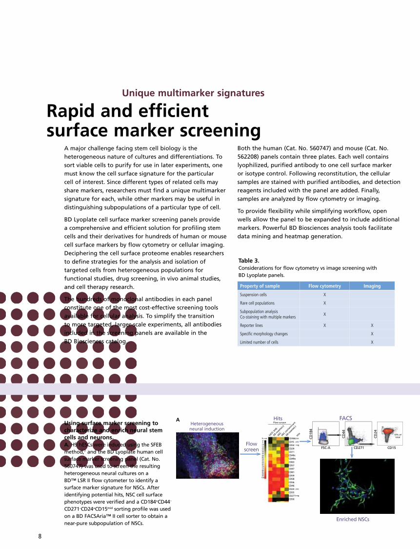
Using surface marker screening to characterize and enrich neural stem cells and neurons.A. H9 hESCs were induced using the SFEB method,7 and the BD Lyoplate human cell surface marker screening panel (Cat. No. 560747) was used to screen the resulting heterogeneous neural cultures on a BD™ LSR II flow cytometer to identify a surface marker signature for NSCs. After identifying potential hits, NSC cell surface phenotypes were verified and a CD184+CD44–
CD271–CD24+CD15mid sorting profile was used on a BD FACSAria™ II cell sorter to obtain a near-pure subpopulation of NSCs.
CD184+ve%05
101520253035404550556065707580859095
100
Flow screen
HitshESC (H
9)
EB-ro
sette
(+)
EB-ro
sette
(-)
NSC NSC co
ntam
inan
t
pos
CD15 pos
CD24CD55
CD50
pos
CD271neg
CD54
CD29CD71CD49cCD49eCD63CD57CD47CD81
CD59CD58
CD56CD46
neg
P3
P4
M
P5
i
ii
i
10% oftotal
iiiHeterogeneousneural induction
Flowscreen
Heterogeneous neuronaldifferentiation
Imagescreen
Hits
Enriched NSCs
Near-pure neurons
FACS
Hits FACS
CD184 CD15
CD
24
CD
44
FSC-A CD271 CD15
CD
24
CD
44
CD
184
A

9
D I S C O V E R Y
Screening of neural populationsNeural cell populations derived from pluripotent stem cells are important for studying human disease and development. Pluripotent stem cells can be differentiated into self-renewing NSCs, which can be further differentiated into heterogeneous populations of neurons and glia.7 A key to further research is to identify surface marker signatures for each of these cell types.
In the example, the BD Lyoplate human cell surface marker screening panel (Cat. No. 560747) was used to identify cell surface phenotypes for NSCs and neurons. In panel A, heterogeneous neural induction cultures were screened by flow cytometry, and potential NSC markers were identified on a heatmap. A resulting NSC cell surface phenotype of CD184+CD44–CD271–CD24+CD15mid was verified using intracellular NSC markers. The surface phenotype was used to sort a near-pure subpopulation of NSCs, the ability of which to differentiate both in vivo and in vitro was later confirmed.
B. The purified NSCs were differentiated into a mixed culture of neuronal and glial cell populations, which was screened using the same panel to identify a surface marker signature for neurons. An imaging screen was chosen due to the unique morphology of neurons and the ability to co-stain with a neuron-specific marker. Potential hits were verified by flow cytometry, and a CD44–
CD184–CD24+CD15low sorting profile was used to purify neurons.7
CD184+ve%05
101520253035404550556065707580859095
100
Flow screen
HitshESC (H
9)
EB-ro
sette
(+)
EB-ro
sette
(-)
NSC NSC co
ntam
inan
t
pos
CD15 pos
CD24CD55
CD50
pos
CD271neg
CD54
CD29CD71CD49cCD49eCD63CD57CD47CD81
CD59CD58
CD56CD46
neg
P3
P4
M
P5
i
ii
i
10% oftotal
iiiHeterogeneousneural induction
Flowscreen
Heterogeneous neuronaldifferentiation
Imagescreen
Hits
Enriched NSCs
Near-pure neurons
FACS
Hits FACS
CD184 CD15
CD
24
CD
44
FSC-A CD271 CD15C
D24
CD
44
CD
184
In panel B, the purified NSCs were differentiated into neuronal and glial cell populations, which were screened by imaging using the same panel to identify surface markers for isolating neurons. An imaging screen was chosen due to the unique morphology of neurons and the ability to co-stain with a neuronal-specific marker. A potential neuronal surface phenotype of CD44–CD184–CD24+CD15low was verified by flow cytometry and used to purify neurons.
In addition to neural cells, the BD Lyoplate human cell surface marker screening panel has also been used to identify cell surface markers of cardiomyocytes derived from pluripotent stem cells.8 Most recently, this powerful methodology was used to develop a human stem cell model of Alzheimer’s disease.9
B

10
S E R V I C E S
References1. Baum CM, Weissman IL, Tsukamoto
AS, Buckle AM, Peault B. Isolation of a candidate human hematopoietic stem-cell population. Proc Natl Acad Sci U S A. 1992;89:2804-2808.
2. Bhatia M, Wang JC, Kapp U, Bonnet D, Dick JE. Purification of primitive human hematopoietic cells capable of repopulating immune-deficient mice. Proc Natl Acad Sci U S A. 1997;94:5320-5325.
3. Majeti R, Park CY, Weissman IL. Identification of a hierarchy of multipotent hematopoietic progenitors in human cord blood. Cell Stem Cell. 2007;1:635-645.
4. Notta F, Doulatov S, Laurenti E, Poeppl A, Jurisica I, Dick JE. Isolation of single human hematopoietic stem cells capable of long-term multilineage engraftment. Science. 2011;333:218-221.
5. Murry CE, Keller G. Differentiation of embryonic stem cells to clinically relevant populations: lessons from embryonic development. Cell. 2008;132:661-680.
6. D’Amour KA, Agulnick AD, Eliazer S, Kelly OG, Kroon E, Baetge EE. Efficient differentiation of human embryonic stem cells to definitive endoderm. Nat Biotechnol. 2005;23:1534-1541.
7. Yuan SH, Martin J, Elia J, et al. Cell-surface marker signatures for the isolation of neural stem cells, glia and neurons derived from human pluripotent stem cells. PLoS One. 2011;6:e17540.
8. Uosaki H, Fukushima H, Takeuchi A, et al. Efficient and scalable purification of cardiomyocytes from human embryonic and induced pluripotent stem cells by VCAM1 surface expression. PLoS One. 2011;6:e23657.
9. Israel MA, Yuan SH, Bardy C, et al. Probing sporadic and familial Alzheimer’s disease using induced pluripotent stem cells. Nature. 2012;482:216-220.
For more than 25 years, BD has actively worked with stem cell researchers to develop tools that help improve workflow, ease of use, and performance. This in-depth knowledge and experience is available to customers through comprehensive training, application and technical support, and expert field service.
TrainingHeld at BD training centers worldwide, BD Biosciences flow cytometry training courses combine theory and hands-on practice to provide participants with the skills and experience they need to take full advantage of the capabilities of their instrument.
Technical Applications SupportBD Biosciences technical applications support specialists are available to provide field- or phone-based assistance and advice. Expert in a diverse array of topics, BD technical application specialists are well equipped to address customer needs in both instrument and application support.
Field Service EngineersBD Biosciences field service engineers are located across the world. When instrument installation or service is required, a BD Biosciences Technical Field Service Engineer can be dispatched to the customer site. On-site service and maintenance agreements are available to provide long-term support.
Special Order Research ProductsIn addition to other services, BD instruments can be customized to me et specific customer requirements via the special order research program.
Custom ServicesMobilizing technology for research applications requires close collaboration. The Custom Technology Team (CTT) at BD Biosciences works with customers to provide solutions through custom reagents, panels, or assay protocols. Staffed by leading scientists with a breadth and depth of scientific and technical expertise, the CTT team will coordinate with researchers to study the problem at hand, make recommendations, and help implement solutions. In this way, BD Biosciences technical know-how is translated into practical solutions that allow customers to focus on research.
Services and Support

11

12
23-15142-00
For Research Use Only. Not for use in diagnostic or therapeutic procedures.
BD flow cytometers are Class 1 Laser Products.
BD, BD Logo and all other trademarks are property of Becton, Dickinson and Company. © 2013 BD
BD Biosciences Regional Offices
Office locations are available on our websites.
AustraliaToll Free 1800 656 100Tel 61.2.8875.7000Fax 61.2.8875.7200bdbiosciences.com/anz
CanadaTel 866.979.9408Fax 888.229.9918 bdbiosciences.com/ca
ChinaTel 86.21.3210.4610Fax 86.21.5292.5191bdbiosciences.com/cn
EuropeTel 32.2.400.98.95Fax 32.2.401.70.94 bdbiosciences.com/eu
IndiaTel 91.124.2383566Fax 91.124.2383224/25/26bdbiosciences.com/in
JapanNippon Becton DickinsonToll Free 0120.8555.90Fax 81.24.593.3281bd.com/jp
Latin America/CaribbeanTel 55.11.5185.9995Fax 55.11.5185.9895bdbiosciences.com/br
New ZealandToll Free 0800 572.468 Tel 64.9.574.2468Fax 64.9.574.2469bdbiosciences.com/anz
SingaporeTel 65.6861.0633Fax 65.6860.1593bdbiosciences.com/sg
United StatesUS Orders 855.236.2772Technical Service 877.232.8995Fax 800.325.9637bdbiosciences.com