analysis of the golden proportion and widthheight.pdf
TRANSCRIPT

Analysis of the Golden Proportion and Width/HeightRatios of Maxillary Anterior Dentition in Patients withLateral Incisor Agenesisjerd_533 402..414
NÚBIA PAVESI PINI, DDS*, LUCIANA MANZOTTI DE-MARCHI, DDS, MS†,‡,
BRUNO FRAZÃO GRIBEL, DDS, MsC§,||, ADRIANA LEMOS MORI UBALDINI, DDS**,
RENATA CORRÊA PASCOTTO, DDS, MS, PhD††
ABSTRACT
Objective: The purpose of this study was to assess the presence of the golden proportion (GP) in the facial viewtooth-to-tooth width proportion of the six maxillary anterior teeth and to evaluate the width/height (W/H) ratios ofthe incisors of patients with maxillary lateral incisor (LI) agenesis treated either with implants or orthodontically (bymoving canines into the position of the laterals, recontouring them, and placing composite restorations over therepositioned teeth).
Materials and Methods: Forty-eight patients with LI agenesis were divided into four experimental groups: unilateralrecontouring group (N = 10), bilateral recontouring group (BRG, N = 18), unilateral implant group (UIG, N = 10),bilateral implant group (N = 10), and a control group (CG, N = 25) of patients without agenesis. GP ratios weredetermined on patients’ dental casts placed over Levin’s grids, whereas W/H ratios were measured directly on thecasts and a millimeter ruler to determine these distances. Statistical analysis was performed with Shapiro–Wilk,Kruskal–Wallis, Mann–Whitney, Friedman, and Wilcoxon tests (p < 0.05).
Results: The incidence of GP in the tooth-to-tooth width proportions was significantly different between groups andmore commonly found between centrals and laterals than between laterals and canines.The GP was more likely to beobserved in the BRG, UIG, and CG.The results demonstrated that the GP was not found to be present in themajority of the cases treated with maxillary agenesis, regardless of the method of treatment.The mean W/H ratios ofthe laterals ranged between 0.75 and 0.90.
Conclusion: Although the GP may be a useful diagnostic guide, it was not observed in the majority of estheticoutcomes of patients treated with maxillary LI agenesis in this study.
CLINICAL SIGNIFICANCE
The assessment of the golden proportion and width/height ratio of upper anterior teeth in patients with upper lateralincisor agenesis treated with either implants or tooth re-contouring may assist dentists and patients in deciding thebest treatment option based on the peculiarities of each case.
(J Esthet Restor Dent 24:402–416, 2012)
*MS Applicant in Restorative Dentistry, Department of Restorative Dentistry, Piracicaba Dental School, State University of Campinas, Piracicaba, SP, Brazil†Specialist in Orthodontics, University Center of Maringá, Maringá, PR, Brazil‡Professor of Orthodontics, University Center of Maringá, Maringá, PR, Brazil§Specialist in Orthodontics, Department of Radiology, Piracicaba Dental School, State University of Campinas, Piracicaba, SP, Brazil||PhD Applicant in Radiology, Department of Radiology, Piracicaba Dental School, State University of Campinas, Piracicaba, SP, Brazil
**MS Applicant in Integrated Dentistry, State University of Maringá, Maringá, PR, Brazil††Professor of Dentistry, Department of Dentistry, State University of Maringá, Maringá, PR, Brazil
RESEARCH ARTICLE
Vol 24 • No 6 • 402–414 • 2012 Journal of Esthetic and Restorative Dentistry DOI 10.1111/j.1708-8240.2012.00533.x © 2012 Wiley Periodicals, Inc.402

INTRODUCTION
Agenesis of maxillary lateral incisors (LIs) is the thirdmost common dental agenesis, excluding third molars.It is preceded by maxillary and mandibular premolarsand comprises approximately 20% of all anomalies.1This condition is more prevalent in females, and theabsence of the LI is more frequently bilateral thanunilateral.1–5 There may be significant demand foresthetic treatment in such cases, as this condition canaffect the harmony and balance of the dentofacialcomplex, thereby affecting the patient’s self-esteem andsocial relationships.1,5
To assist dental professionals in the planning of cases ofcongenital maxillary LI agenesis, a number of studieshave analyzed the functional performance oftreatments.1,6–9 However, to the best of our knowledge,there are no studies that comparatively address the finalesthetic outcomes of different treatments. Althoughseveral authors have studied esthetic principles, such asthe golden proportion (GP) and the width/height(W/H) ratios of the maxillary anterior teeth,10–26 there islittle information on how these parameters have beenapplied in esthetic restorative treatments in cases ofmaxillary LI agenesis.
The GP was described by Pythagoras, an ancient Greekmathematician, as an attempt to correlate science withbeauty. It was used to design the Parthenon, and laterto label dimensions in da Vinci’s classic drawings ofhuman anatomy. The ratio is approximately 0.618 to 1,whereby the height of the shorter object divided by theheight of the longer one is identical to the height of thelonger object divided by the sum of the shorter plusthe longer objects.16,25,26 Levin recommends the width ofthe maxillary LI be in GP to the width of the maxillarycentral incisor (CI) when viewing from the front.27
However, a range of studies20,22,23,25,26 have not foundthis proportion to exist in a majority of patients in thegeneral population.
Two procedures are commonly used in cases ofunilateral or bilateral agenesis: space closure by mesiallyrepositioning the canine (C) and recontouring it, orspace opening or its maintenance in order for implants
or dentures to replace the missing LI.5,28,29 For estheticrehabilitation, the GP and the W/H ratio of the teethmay be useful guides in the reestablishment of anattractive smile.19,23,30,31
The aim of the present study was twofold: (1) todetermine the presence of the GP between the anteriorteeth as measured in pairs (CI:LI and LI:C), and (2) todetermine the W/H ratio means of each anteriormaxillary tooth, with particular emphasis onthe results and discussion of the results of the W/Hof the LI.
MATERIALS AND METHODS
In the experimental groups, patients presented withmaxillary LI agenesis treated with either recontouringor implants, in contrast to a control group (CG) ofpatients with no history of orthodontic treatment oragenesis. Patients’ selection criteria excluded those withprosthesis or implants for replacement of other teeth,such as Cs, CIs, and premolars, as well as patientswhose agenesis treatment had not involved orthodonticrepositioning.
The 48 patients (39 women, 9 men, aged between 18and 45 years) had either unilateral (N = 20) or bilateral(N = 28) maxillary LI agenesis. Agenesis treatmentconsisted of either space closure involving mesialmovement of Cs and remodeling of anterior teeth withcomposite resin (N = 28), or orthodontic LI spaceopening followed by implant placement in the region ofthe agenesis (N = 20).
In order to minimize differences between the widthsand heights of an LI replacement and those of a naturaltooth and to maintain the gingival height, all cases wereorthodontically treated with the incisors being lined upat the gingival margin. For the recontouring cases, theCs were moved mesially and the cusps and the widthsremodeled. For the implant cases, as much as needed,bone and connective tissue were surgically placed, sothat the gingival height was kept similar to that of anatural LI.
GP AND W/H RATIO ANALYSIS OF ANTERIOR DENTITION IN LI AGENESIS PATIENTS Pini et al
© 2012 Wiley Periodicals, Inc. DOI 10.1111/j.1708-8240.2012.00533.x Journal of Esthetic and Restorative Dentistry Vol 24 • No 6 • 402–414 • 2012 403

The patients were divided into four groups based ontype of treatment: unilateral recontouring group (URG,N = 10), bilateral recontouring group (BRG, N = 18),unilateral implant group (UIG, N = 10), and bilateralimplant group (BIG, N = 10). The CG consisted of 25volunteers with no agenesis (except for third molars),good tooth alignment, no maxillary anterior toothrestorations affecting the relative size of the teeth, nolaminate veneers or other prosthesis, no noticeabletooth wear, no history of orthodontic treatment, no useof a biteplate, and no bone base discrepancies. Thestudy was approved by the local ethical committee(672/2008).
Concerning GP or successive tooth-to-tooth widthproportion analyses, the apparent widths of each toothwere measured over a Levin’s grid registered on a blankcard and viewed from the front (Figure 1). Levin arguedthat successive GP occurs when both theCI and LI, and the LI and C relations are in agreementwith the golden number, that is, 0.618. In other words,the width of the CI is in GP to the width of the LI,which is also in GP to the width of the C. The apparentwidths were measured with the use of a caliper and a1-mm increment ruler (Figure 2). The successivetooth-to-tooth width proportions between CIs and LIs(CI:LI), and between LIs and Cs (LI:C) were calculatedby dividing the smaller width by the larger one. In thepresent study, ratios between 60% and 64% wereconsidered to be within the range of the GP, asestablished by Preston.26
With reference to the W/H evaluation, the real widthand height of the teeth were measured directly overeach subject’s dental cast, parallel to the facial surfacesof the teeth, with the use of the same caliper andruler used to measure the successive tooth-to-toothwidth proportions. The W/H ratios were calculatedby dividing each tooth’s width by its height(Figure 3).
FIGURE 1. Levin’s grid.
A B
FIGURE 2. A, Measurement ofLevin’s grid with caliper andB, millimeter ruler.
A B
FIGURE 3. A, Measurement ofheight and B, width of anteriorteeth.
GP AND W/H RATIO ANALYSIS OF ANTERIOR DENTITION IN LI AGENESIS PATIENTS Pini et al
Vol 24 • No 6 • 402–414 • 2012 Journal of Esthetic and Restorative Dentistry DOI 10.1111/j.1708-8240.2012.00533.x © 2012 Wiley Periodicals, Inc.404

Both the GP or tooth-to-tooth width proportion andthe W/H ratios were further employed in order toanalyze the correspondence between the measures ofthe right and the left side of the arch. These measureswere used with different purposes depending on thetype of LI agenesis: for the groups with unilateralagenesis, the proportion for each pair of teeth (CI:LIand LI:C) was used for within-groupcorrespondences—the mean tooth-to-tooth widthproportion in one side were compared with theircorrespondent measures on the contralateral side. Forthe groups with bilateral agenesis, apart from verifyingwithin-group correspondences, the mean of the grouptreated with implants was compared with the means ofthe group treated with recontouring, as well as inrelation to the CG. With relation to the W/H ratios, themeasures were used to verify whether individual teethratios (CI, LI, and C) corresponded on both sides of thearch within the same type of treatment and alsobetween different types of treatment. Bearing in mindthat the GP deals with apparent widths only and giventhat the height of the built LI could vary whether it wasrecontoured or implanted, longer in the former case,one could find correspondence between right and leftGP measures but not necessarily in the W/H ratios.
A further investigation of the two ways of measuringonly the widths of the LIs was examined: (1) theapparent width as estimated over a Levin’s grid, withthe purpose of verifying whether the posterior–anteriortransition of measures were reestablished, and (2) thereal width verified directly over the cast, with the aim ofanalyzing whether the teeth proportion was preservedaccording to different types of treatment.
In order to guarantee the reliability of the examiner’sassessments, the analyses of proportionality, both theGP/tooth-to-tooth width proportions and the W/Hratio, were performed twice by a single examiner, witha 30-day interval between the two moments. In theResults section, tables and figures express the data ofthe first analysis.
Statistical analyses were run with the use of theSoftware R 2.10.1 (R Foundation for StatisticalComputing, Vienna, Austria) and level of significance at
p < 0.05. The results, reported as mean, standarddeviation, and percentages, were checked for normalityof distribution with Shapiro–Wilk test. Wilcoxon testwas used to determine the statistical significancebetween the two moments in which the GP and theW/H were evaluated. Kruskal–Wallis test was used toverify between-group comparison of means, followed byMann–Whitney U post hoc test in order to determinedifferences between two independent samples.Friedman test and post hoc Wilcoxon test were used forwithin-group comparison of means.
RESULTS
Analysis of the data involved 73 patients: 48 having LIagenesis and 25 having incisors not missing (control).With regard to the intraclass correlation analysis(Wilcoxon test), no significant difference was foundbetween the two moments of measurements, either forthe GP or the W/H ratio (p > 0.05). The results showedthat the prevalence of missing right LI was higher in theURG (70%) when compared with that in the UIG (50%)(Figure 4). Concerning the presence of the GP or theother tooth-to-tooth width proportion prevalent,Table 1 displays all groups’ minimum, maximum, andmean values (in mm), separated by the right and leftside of measurements, as well as by the pairanalysis—CI:LI and LI:C.
Keeping in mind that the present study adoptedPreston’s26 GP range (60–64%), the mean proportions
FIGURE 4. Distribution of unilateral agenesis by group.UIG = unilateral implant group; URG = unilateral recontouringgroup.
GP AND W/H RATIO ANALYSIS OF ANTERIOR DENTITION IN LI AGENESIS PATIENTS Pini et al
© 2012 Wiley Periodicals, Inc. DOI 10.1111/j.1708-8240.2012.00533.x Journal of Esthetic and Restorative Dentistry Vol 24 • No 6 • 402–414 • 2012 405

between CI:LI in all groups varied from 60% to 66%with a range of 40% to 87.5%, whereas between LI:C,they varied from 76% to 86% with a range of 53.85% to120%. In other words, in all groups, the mean widthproportion between the CI:LI was closer to the 62%, avalue closer to GP than the width proportions betweenthe LI:C. This finding was more evident within the CGand the two groups treated with implants (BIG and
UIG), whereas the groups treated with recontouring(BRG and URG) showed more variation. In addition,the successive GP as suggested by Levin, in which thewidth relations between both the CI:LI and the LI:C arein agreement with the golden number, was not found inany group.
The tooth-to-tooth width proportion mean values werecompared between the groups with bilateral incisoragenesis (BRG ¥ BIG), between the groups withunilateral agenesis (URG ¥ UIG), and between each ofthese groups (BRG, BIG, URG, or UIG) and the control(Table 2). No statistical difference was found betweendifferent types of treatment within the same type ofagenesis. Significant differences were found betweenBRG and the CG concerning CI:LI on the left side andLI:C on both sides. As for BIG and CG groups,significant difference was found for the relation LI:C onthe left side. No significant differences were foundbetween CG and URG or CG and UIG.
The analysis of the percentage of patients whodemonstrated the GP as defined by Preston (60–64%) ineach of the experimental groups was averaged into asingle group defined as agenesis group (AG). In doingso, the percentages of the CG and of the AG could becontrasted with those of previous studies. Figure 5
TABLE 1. Golden proportion analysis: minimum, maximum,mean, and standard deviation values by groups, side, and pairinvestigation
Group Side Pair Minimum Maximum Mean Standarddeviation
BRG Right CI:LI 0.5263 0.7500 0.6575 0.0589
LI:C 0.6667 0.9000 0.7597 0.0744
Left CI:LI 0.5263 0.7500 0.6575 0.0589
LI:C 0.6667 0.9091 0.7799 0.0879
BIG Right CI:LI 0.5000 0.7647 0.6075 0.0802
LI:C 0.6667 0.8889 0.7991 0.0566
Left CI:LI 0.5556 0.8750 0.6306 0.1126
LI:C 0.6429 0.9000 0.7812 0.0753
URG Right CI:LI 0.5556 0.8235 0.6667 0.0990
LI:C 0.6667 1.0000 0.8165 0.1102
Left CI:LI 0.5556 0.7778 0.6482 0.0683
LI:C 0.5385 0.9091 0.7839 0.1196
UIG Right CI:LI 0.5000 0.7222 0.6241 0.0625
LI:C 0.6154 1.0000 0.7902 0.1191
Left CI:LI 0.5000 0.7500 0.6359 0.0805
LI:C 0.6154 1.0000 0.7877 0.1402
CG Right CI:LI 0.4000 0.7500 0.6186 0.0765
LI:C 0.6364 1.2500 0.8649 0.1596
Left CI:LI 0.5000 0.7500 0.6211 0.0648
LI:C 0.5714 1.2000 0.8662 0.1395
BIG = bilateral implant group; BRG = bilateral recontouring group;C = canine; CG = control group; CI = central incisor ; LI = lateralincisor ; UIG = unilateral implant group; URG = unilateral recontouringgroup.
TABLE 2. Golden proportion analysis: mean comparisons(p-value) between different types of treatment. Significantdifferences in boldface
Right Left
CI:LI LI:C CI:LI LI:C
BRG ¥ CG 0.0555 0.0228 0.0360 0.0361
BIG ¥ CG 0.5538 0.1555 0.6566 0.0200
URG ¥ CG 0.3207 0.4189 0.3118 0.1183
UIG ¥ CG 0.9853 0.1570 0.6465 0.1183
BRG ¥ BIG 0.1033 0.1956 0.2310 0.7700
URG ¥ UIG 0.5691 0.5690 0.8495 0.8197
BIG = bilateral implant group; BRG = bilateral recontouring group;C = canine; CG = control group; CI = central incisor ; LI = lateralincisor ; UIG = unilateral implant group; URG = unilateral recontouringgroup.
GP AND W/H RATIO ANALYSIS OF ANTERIOR DENTITION IN LI AGENESIS PATIENTS Pini et al
Vol 24 • No 6 • 402–414 • 2012 Journal of Esthetic and Restorative Dentistry DOI 10.1111/j.1708-8240.2012.00533.x © 2012 Wiley Periodicals, Inc.406
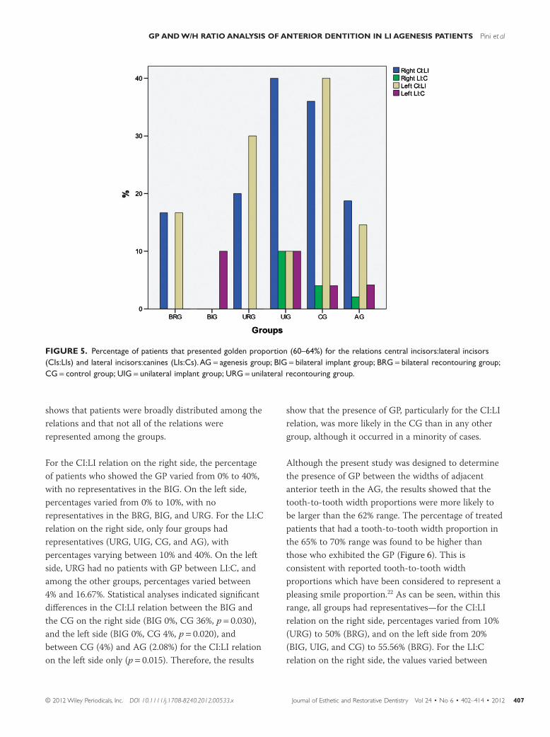
shows that patients were broadly distributed among therelations and that not all of the relations wererepresented among the groups.
For the CI:LI relation on the right side, the percentageof patients who showed the GP varied from 0% to 40%,with no representatives in the BIG. On the left side,percentages varied from 0% to 10%, with norepresentatives in the BRG, BIG, and URG. For the LI:Crelation on the right side, only four groups hadrepresentatives (URG, UIG, CG, and AG), withpercentages varying between 10% and 40%. On the leftside, URG had no patients with GP between LI:C, andamong the other groups, percentages varied between4% and 16.67%. Statistical analyses indicated significantdifferences in the CI:LI relation between the BIG andthe CG on the right side (BIG 0%, CG 36%, p = 0.030),and the left side (BIG 0%, CG 4%, p = 0.020), andbetween CG (4%) and AG (2.08%) for the CI:LI relationon the left side only (p = 0.015). Therefore, the results
show that the presence of GP, particularly for the CI:LIrelation, was more likely in the CG than in any othergroup, although it occurred in a minority of cases.
Although the present study was designed to determinethe presence of GP between the widths of adjacentanterior teeth in the AG, the results showed that thetooth-to-tooth width proportions were more likely tobe larger than the 62% range. The percentage of treatedpatients that had a tooth-to-tooth width proportion inthe 65% to 70% range was found to be higher thanthose who exhibited the GP (Figure 6). This isconsistent with reported tooth-to-tooth widthproportions which have been considered to represent apleasing smile proportion.22 As can be seen, within thisrange, all groups had representatives—for the CI:LIrelation on the right side, percentages varied from 10%(URG) to 50% (BRG), and on the left side from 20%(BIG, UIG, and CG) to 55.56% (BRG). For the LI:Crelation on the right side, the values varied between
FIGURE 5. Percentage of patients that presented golden proportion (60–64%) for the relations central incisors:lateral incisors(CIs:LIs) and lateral incisors:canines (LIs:Cs).AG = agenesis group; BIG = bilateral implant group; BRG = bilateral recontouring group;CG = control group; UIG = unilateral implant group; URG = unilateral recontouring group.
GP AND W/H RATIO ANALYSIS OF ANTERIOR DENTITION IN LI AGENESIS PATIENTS Pini et al
© 2012 Wiley Periodicals, Inc. DOI 10.1111/j.1708-8240.2012.00533.x Journal of Esthetic and Restorative Dentistry Vol 24 • No 6 • 402–414 • 2012 407

10% (BIG) and 22.22% (BRG), and on the left sidebetween 4% (CG) and 30% (UIG). Significantdifferences were found between the BRG and the CGfor the CI:LI relation on the left side (BRG 55.56%, CG20%, p = 0.017), and for the LI:C relation on the left side(BRG 10%, CG 4%, p = 0.028), as well as between theUIG (30%) and the CG (4%) in the LI:C relation on theleft side (p = 0.031).
The means of the W/H ratios of each maxillary anteriorare shown in Table 3, separated by group, side, andtooth under analysis. As can be seen, means variedbetween 80% and 88% for most incisors and groups,with the exception of the left LI in the BIG (78%) andthe right LI in the URG (90%). Regarding the LIcomparison of means, no significant differences werefound between the results of the CG and those of thetwo groups with implanted LI (BIG and UIG). Similarly,no differences were found between the means of theCG and those of recontoured groups (BRG and URG),with the exception of the right LI between the CG(82%) and the URG (90%).
With regard to the W/H ratio of the LI, thepercentages of patients within a 10% range scale from≥65 to ≤96 showed variation between 0% and 60%(Figure 7). As can be seen, most patients seem tohave fallen within the range of 76% to 85% and 86% to95%. However, between-group comparison ofpercentages showed significant differences only betweenthe URG and the CG on the right side for the W/Hratio above 96%. Similarly, within-group comparisonof percentages did not show differences for mostgroups—only the UIG within the W/H ratio of 86%to 95% yielded significance over the otherpercentages.
With the aim at verifying whether the W/H ratio meansvaried with regard to the type of treatment and within atype of agenesis, between-group comparison of meanswas run with the bilateral AGs (BRG ¥ BIG), theunilateral AGs (URG ¥ UIG), and between the CG andeach of these groups. Significant differences were foundonly between the URG and the CG for the means of theright LI (Table 4).
FIGURE 6. Percentage of patients that presented a 65% to 70% golden proportion between central incisors:lateral incisors(CIs:LIs) and lateral incisors:canines (LIs:Cs).AG = agenesis group; BIG = bilateral implant group; BRG = bilateral recontouring group;CG = control group; UIG = unilateral implant group; URG = unilateral recontouring group.
GP AND W/H RATIO ANALYSIS OF ANTERIOR DENTITION IN LI AGENESIS PATIENTS Pini et al
Vol 24 • No 6 • 402–414 • 2012 Journal of Esthetic and Restorative Dentistry DOI 10.1111/j.1708-8240.2012.00533.x © 2012 Wiley Periodicals, Inc.408
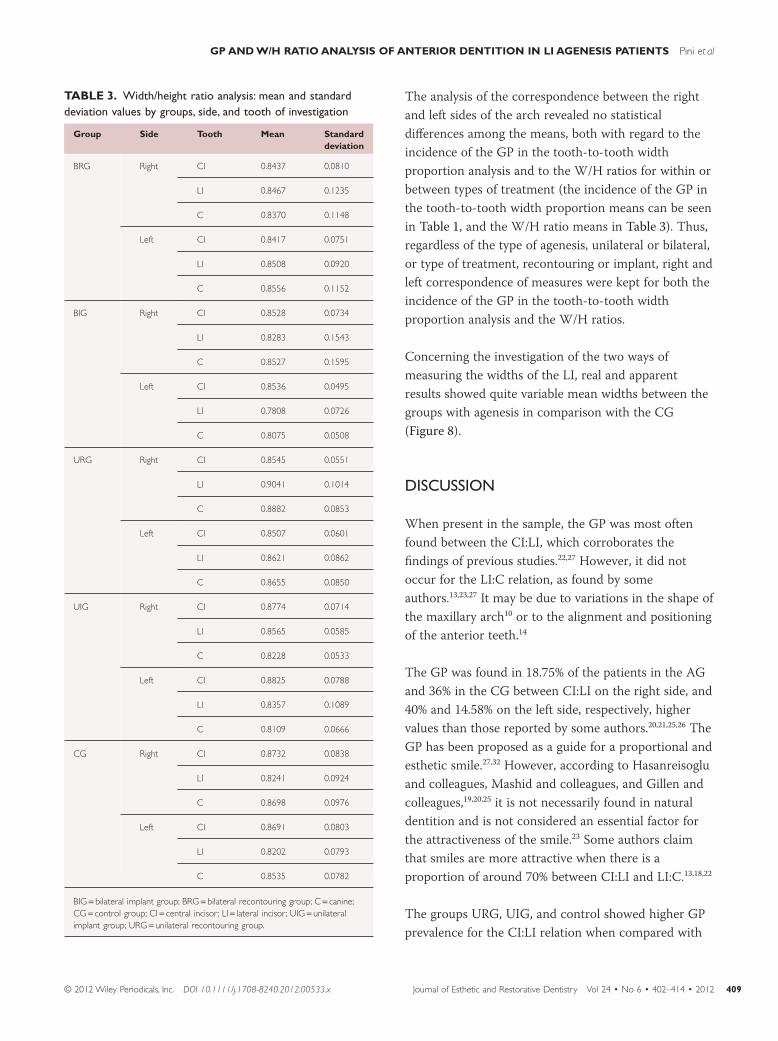
The analysis of the correspondence between the rightand left sides of the arch revealed no statisticaldifferences among the means, both with regard to theincidence of the GP in the tooth-to-tooth widthproportion analysis and to the W/H ratios for within orbetween types of treatment (the incidence of the GP inthe tooth-to-tooth width proportion means can be seenin Table 1, and the W/H ratio means in Table 3). Thus,regardless of the type of agenesis, unilateral or bilateral,or type of treatment, recontouring or implant, right andleft correspondence of measures were kept for both theincidence of the GP in the tooth-to-tooth widthproportion analysis and the W/H ratios.
Concerning the investigation of the two ways ofmeasuring the widths of the LI, real and apparentresults showed quite variable mean widths between thegroups with agenesis in comparison with the CG(Figure 8).
DISCUSSION
When present in the sample, the GP was most oftenfound between the CI:LI, which corroborates thefindings of previous studies.22,27 However, it did notoccur for the LI:C relation, as found by someauthors.13,23,27 It may be due to variations in the shape ofthe maxillary arch10 or to the alignment and positioningof the anterior teeth.14
The GP was found in 18.75% of the patients in the AGand 36% in the CG between CI:LI on the right side, and40% and 14.58% on the left side, respectively, highervalues than those reported by some authors.20,21,25,26 TheGP has been proposed as a guide for a proportional andesthetic smile.27,32 However, according to Hasanreisogluand colleagues, Mashid and colleagues, and Gillen andcolleagues,19,20,25 it is not necessarily found in naturaldentition and is not considered an essential factor forthe attractiveness of the smile.23 Some authors claimthat smiles are more attractive when there is aproportion of around 70% between CI:LI and LI:C.13,18,22
The groups URG, UIG, and control showed higher GPprevalence for the CI:LI relation when compared with
TABLE 3. Width/height ratio analysis: mean and standarddeviation values by groups, side, and tooth of investigation
Group Side Tooth Mean Standarddeviation
BRG Right CI 0.8437 0.0810
LI 0.8467 0.1235
C 0.8370 0.1148
Left CI 0.8417 0.0751
LI 0.8508 0.0920
C 0.8556 0.1152
BIG Right CI 0.8528 0.0734
LI 0.8283 0.1543
C 0.8527 0.1595
Left CI 0.8536 0.0495
LI 0.7808 0.0726
C 0.8075 0.0508
URG Right CI 0.8545 0.0551
LI 0.9041 0.1014
C 0.8882 0.0853
Left CI 0.8507 0.0601
LI 0.8621 0.0862
C 0.8655 0.0850
UIG Right CI 0.8774 0.0714
LI 0.8565 0.0585
C 0.8228 0.0533
Left CI 0.8825 0.0788
LI 0.8357 0.1089
C 0.8109 0.0666
CG Right CI 0.8732 0.0838
LI 0.8241 0.0924
C 0.8698 0.0976
Left CI 0.8691 0.0803
LI 0.8202 0.0793
C 0.8535 0.0782
BIG = bilateral implant group; BRG = bilateral recontouring group; C = canine;CG = control group; CI = central incisor ; LI = lateral incisor ; UIG = unilateralimplant group; URG = unilateral recontouring group.
GP AND W/H RATIO ANALYSIS OF ANTERIOR DENTITION IN LI AGENESIS PATIENTS Pini et al
© 2012 Wiley Periodicals, Inc. DOI 10.1111/j.1708-8240.2012.00533.x Journal of Esthetic and Restorative Dentistry Vol 24 • No 6 • 402–414 • 2012 409

the other groups, suggesting that treatment withrecontouring or implants in cases of bilateral agenesisdoes not follow a clear GP pattern. The differencesfound between the BIG and the CG may be due to thefact that patients with agenesis also present othermorphological differences in relation to those withoutagenesis, as the crowns are usually reduced in size, and,because of this, patients from the BIG might havedifferent tooth-to-tooth width proportion from those ofthe CG.
However, comparing the CI:LI means, the groups thatcame closest to the GP were the BIG, the UIG, and theCG. The similarity to the CG’s results may be duethe need of orthodontic treatment in order to adjust thespace to the diameter of the tooth to be replaced
FIGURE 7. Width/height (W/H) analysis of the lateral incisors (LIs): percentage of patients within different W/H ratios of the rightand the left LI.AG = agenesis group; BIG = bilateral implant group; BRG = bilateral recontouring group; CG = control group;UIG = unilateral implant group; URG = unilateral recontouring group.
TABLE 4. Width/height ratio analysis: mean comparisons(p-value) between different types of treatment
Right Left
CI LI C CI LI C
BRG ¥ CG 0.5196 0.3356 0.2176 0.4125 0.1745 0.8244
BIG ¥ CG 0.7681 0.7826 0.4981 0.7392 0.1990 0.0671
URG ¥ CG 0.7120 0.0383 0.5332 0.6438 0.2196 0.5458
UIG ¥ CG 0.7690 0.2848 0.1287 0.7002 0.9562 0.1429
BRG ¥ BIG 0.8273 0.3852 0.7729 0.5937 0.0537 0.3247
URG ¥ UIG 0.5167 0.2399 0.0951 0.4471 0.4247 0.1107
BIG = bilateral implant group; BRG = bilateral recontouring group;C = canine; CG = control group; CI = central incisor ; LI = lateral incisor ;UIG = unilateral implant group; URG = unilateral recontouring group.
GP AND W/H RATIO ANALYSIS OF ANTERIOR DENTITION IN LI AGENESIS PATIENTS Pini et al
Vol 24 • No 6 • 402–414 • 2012 Journal of Esthetic and Restorative Dentistry DOI 10.1111/j.1708-8240.2012.00533.x © 2012 Wiley Periodicals, Inc.410

(as for the case of the BIG), or to the diameter of theexisting LI (as for the case of the UIG). The means ofthe BRG and of the URG differed the most from theGP/tooth-to-tooth width proportion. This may be dueto the orthodontic treatment and transformation of theCs into LI, a procedure that usually requires therecontouring of the other anterior teeth, such as that ofthe CI, which contributes to the existing proportion inthe anterior segment of the smile.
Regarding the alternative analysis of GP ortooth-to-tooth width proportion that were foundto fall within the 65% to 70% range, the differencesfor the CI:LI relation between the BRG and theCG on the left side and for the LI:C between the UIGand the CG on the left side, also the side in whichagenesis was more frequent, can be explained bythe fact that the recontoured Cs are wider than anatural LI.
With regard to the W/H ratios of the LIs, the BIG andUIG were the groups that came closest to themeasurements found in the CG. This may be due to thetype of treatment, implants, for which the agenesisspace is adjusted to that of a natural LI. In the case ofthe UIG, this explanation applies only to the left side,which corresponds to the patients with agenesis of thetooth #22 treated with implants. The BRG and the URGhad the highest LI means, which suggests that thewidth of this tooth is similar to its height, resulting in a
high W/H quotient. In addition, again it is important tokeep in mind that the recontoured Cs are wider than anatural LI. The intention in comparing the right andleft sizes of the smile was to verify if dentists areworried about reestablishing the symmetry in thetreatment, mainly in the cases of unilateral agenesis inwhich the patients have a natural LI as a pattern tofollow with implants or recontouring of the Csinto LIs.
Regardless the type of treatment, it is important to takeinto consideration a multidisciplinary approachinvolving Orthodontics, even for cases treated withimplants. The correct alignment of the gingival contouris essential for the reestablishment of a natural W/Hratio, both for implants and recontoured Cs in patientswith maxillary LI agenesis.28
The prevalence of the W/H ratio of the LI was thesame (76–85) for the right and left sides only in theBIG, which is in accord with previous findings.12,24 Thissymmetry is provided by the type of treatment as bothsides are orthodontically adjusted to receive prosthesisto replace the missing LI. Only the prevalence of theW/H found in the BRG, UIG, and URG on the rightside, was similar to that of the CG (86–95). Theseresults were according to the previous studies.19,25 Forthe BRG, this indicates that even with anatomicdifferences between the Cs and LIs, recontouringtreatment seems to be able to reestablish a natural
FIGURE 8. Comparison of realand apparent (Ap) widths of upperlateral incisors. BIG = bilateralimplant group; BRG = bilateralrecontouring group; CG = controlgroup; LLI = left lateral incisor;RLI = right lateral incisor;UIG = unilateral implant group;URG = unilateral recontouringgroup.
GP AND W/H RATIO ANALYSIS OF ANTERIOR DENTITION IN LI AGENESIS PATIENTS Pini et al
© 2012 Wiley Periodicals, Inc. DOI 10.1111/j.1708-8240.2012.00533.x Journal of Esthetic and Restorative Dentistry Vol 24 • No 6 • 402–414 • 2012 411

proportion when compared with individuals withnatural LIs, such as those from the CG. The samereasoning applies to the UIG, as the implant is adjustedto the existing LI, which reestablishes a naturalproportion similar to that found in the CG. Theprevalence of the W/H in the URG, to whom therecontoured C was the treatment, was inconsistent onthe right side, with a W/H ratio greater than 90, a valuethat has not been previously reported. For the left side,there was found a proportion of 75 to 80, as reportedpreviously.12,24
Comparing the GP/tooth-to-tooth width proportionand W/H ratios between the right and left sides of arch,all individuals in all groups exhibited symmetry(p > 0.05). This comparison shows the reestablishmentof symmetry and balance of the smile in therehabilitation of cases of agenesis. This aspect isespecially important for individuals with unilateralagenesis, for whom treatment should involve symmetrywith the existing LI.
The comparison between the real and the apparentwidths of the LIs revealed significant discrepanciesbetween the patients with agenesis and the CG. Theapparent dimensions of the anterior teeth seem to bemore important than the real measurements, becausethe proportional ratio of the anterior segment of thesmile is based on the perceptible size of the teeth ratherthan the real size.13,20,23 The BRG and URG were thegroups that most differed from the CG, which isprobably due to the presence of recontoured Cs inthese groups. The UIG showed the smallest realand apparent widths. This may be related to theprevalence of the GP in this group, that may be due tothe size and shape of the teeth, which is usuallynarrower when the GP is present, as reported by someauthors.13,24
This is a pioneering study in the analysis of estheticproportions of the smile in patients with maxillary LIagenesis treated with either orthodontic space closurefollowed by tooth recontouring or implant placement.In the present study, a Levin’s grid was used for eachindividual measurement in order to determine theproportion between the maxillary anterior teeth and to
investigate the application of this device in thetreatment of patients with maxillary LI agenesis. Forsuch cases, the use of a Levin’s grid as a tool to measurethe apparent widths, and thus to obtain the GP as aguide for the planning of esthetic rehabilitation, isproposed in the literature.32 However, research suggeststhat a Levin’s grid is not sufficiently precise to confirmthe existence of the GP or other smile proportions.21,33
Whereas several authors defend the application of theGP in dentistry,27,31,32 others believe that the existence ofexact smile proportions is not necessarily an importantconcept for the symmetry and esthetics of anteriorteeth.14,15,19,20,33
The limitations and the differences between the resultsof the present study and those of previousinvestigations may be attributed to several factors, suchas variations in the methodology (use of casts orphotographs, rulers, compasses, or calipers)10,17,20,25,26
and ethnic background of the patients.15,26 Castro andcolleagues16 performed measurements in threeways—directly on patients’ teeth using a millimeterperiodontal probe and digital calipers, as well as onphotographs. They17 found consistent results betweenmethods regarding the prevalence of the GP betweenCI:LI. Studying Americans, Preston26 found aprevalence of 17% for the GP between CI:LI, whereasFayyad and colleagues15 studying Arabs, found a higherprevalence (38%). In a smile analysis using a digitalsoftware program, Basting and colleagues17 found aprevalence of 19% for the GP of smiles consideredesthetically pleasing. However, Mashid and colleagues20
and Gillen and colleagues25 did not find this proportionin patients with smiles also considered estheticallypleasing.
Taking the results of the present study and theconflicting previous findings into consideration, furtherstudies should investigate the application of estheticproportions in the treatment of patients with maxillaryLI agenesis. A larger number of patients and differentmethods of analysis are needed so that differencesbetween groups and patterns of treatment can beestablished. The follow-up of these patients is alsonecessary in order to assess long-term esthetic resultsof the treatment.
GP AND W/H RATIO ANALYSIS OF ANTERIOR DENTITION IN LI AGENESIS PATIENTS Pini et al
Vol 24 • No 6 • 402–414 • 2012 Journal of Esthetic and Restorative Dentistry DOI 10.1111/j.1708-8240.2012.00533.x © 2012 Wiley Periodicals, Inc.412
CONCLUSION
Although the GP may be advocated by someresearchers as a guide for the determination of thewidth of the missing maxillary LI, it was not found inthe majority of cases in this study. Although the groupsdid not present a high prevalence of the GP in anteriorsuccessive tooth-to-tooth width proportions and theGP in W/H ratios, the smiles created were pleasing.Therefore, it is not believed to be necessary to recreatesmiles exhibiting the GP between the views successivewidths of the maxillary anterior teeth for the esthetictreatment of patients with LI agenesis.
REFERENCES
1. Robertsson S, Mohlin B. The congenitally missing upperlateral incisor. A retrospective study of orthodontic spaceclosure versus restorative treatment. Eur J Orthod2005;22:697–710.
2. Fekonja A. Hypodontia in orthodontically treatedchildren. Eur J Orthod 2005;27:457–60.
3. Pinho T, Tavares P, Maciel P, Pollmann C.Developmental absence of maxillary lateral incisorsin the Portuguese population. Eur J Orthod2005;27:457–60.
4. Altug-Atac AT, Erdem D. Prevalence and distribution ofdental anomalies in orthodontic patients. Am J OrthodDentofacial Orthop 2007;131:510–4.
5. Araújo EA, Oliveira DS, Araújo MT. Diagnostic protocolin cases of congenitally missing maxillary lateral incisors.World J Orthod 2006;7:376–88.
6. Senty EL. The maxillary cuspid and missing lateralincisors: esthetics and occlusion. Angle Orthod1976;46:365–71.
7. Nordquist CG, McNeill RW. Orthodontic vs. restorativetreatment of the congenitally absent lateralincisor—longterm periodontal and occlusal evaluation.J Periodontol 1975;46:139–43.
8. Tuverson DL. Orthodontic treatment using canines inplace of missing lateral incisors. Am J Orthod DentofacialOrthop 1970;58:109–27.
9. Carlson H. Suggested treatment for missing lateralincisors cases. Angle Orthod 1952;22:205–16.
10. Nikgoo A, Alavi K, Alavi K, Mirfazaelian A. Assessmentof the golden ratio in pleasing smiles. World J Orthod2009;10:224–8.
11. Murthy BVS, Ramani N. Evaluation of natural smile:golden ratio, RED or golden percentage. J Conserv Dent2008;1:16–21.
12. Zlataric DK, Kristek E, Celebic A. Analysis ofwidth/length ratios of normal clinical crowns of themaxillary anterior dentition: correlation between dentalproportions and facial measurements. Int J Prosthodont2007;20:313–5.
13. Ward DH. A study of dentists’ preferred maxillaryanterior tooth width proportions: comparing therecurring esthetic dental proportion to othermathematical and naturally occurring proportions.J Esthet Restor Dent 2007;19:324–39.
14. Ong E, Brown RA, Richmond S. Peer assessment ofdental attractiveness. Am J Orthod Dentofacial Orthop2006;130:163–9.
15. Fayyad MAI, Jamani KD, Agrabawi J. Geometric andmathematical proportions and their relations to maxillaryanterior teeth. J Contemp Dent Pract 2006;5:62–70.
16. Castro MVM, Santos NCM, Ricardo LH. Assessment ofthe “golden ratio” in agreeable smiles. Quintessence Int2006;37:597–604.
17. Basting RT, Trindade RS, Flório FM. Comparative studyof smile analysis by subjective and computerizedmethods. Oper Dent 2006;6:652–9.
18. Wolfart S, Thormann H, Freitag S, Kern M. Assessmentof dental appearance following changes in incisorproportions. Eur J Oral Sci 2005;114:159–65.
19. Hasanreisoglu U, Berskun S, Aras K, Arslan I. An analysisof maxillary anterior teeth: facial and dental proportions.J Prosthet Dent 2005;94:530–8.
20. Mashid M, Khoshvaghti A, Varshosaz M, Vallaei N.Evaluation of “golden ratio” in individuals with an estheticsmile. J Esthet Restor Dent 2004;16:185–93.
21. Magne P, Galluci GO, Belser UC. Anatomic crownwidth/length ratios of unworn and worn teeth in whitesubjects. J Prosthet Dent 2003;89:453–61.
22. Ward DH. Proportional smile design using the recurringesthetic dental (RED) proportion. Dent Clin North Am2002;45:143–54.
23. Rosentiel SF, Ward DH, Rashid RG. Dentists’ preferencesof anterior tooth proportion—a web based study.J Prosthodont 2000;9:123–36.
24. Sterret JD, Oliver T, Robinson F, et al. Width/lengthratios of normal clinical crowns of the maxillary anteriordentition in man. J Clin Periodontol 1999;26:153–7.
25. Gillen RJ, Schwartz RS, Hilton TJ, Evans DB. An analysisof selected normative tooth proportions. Int JProsthodont 1994;7:410–7.
26. Preston JD. The golden ratio revisited. J Esthet Dent1993;5:247–51.
27. Levin EI. Dental esthetics and the golden ratio. J ProsthetDent 1978;3:244–52.
28. Kinzer GA, Kokich VO Jr. Managing congenitally missinglateral incisors—part 1: canine substitution. J EsthetRestor Dent 2007;3:2–6.
GP AND W/H RATIO ANALYSIS OF ANTERIOR DENTITION IN LI AGENESIS PATIENTS Pini et al
© 2012 Wiley Periodicals, Inc. DOI 10.1111/j.1708-8240.2012.00533.x Journal of Esthetic and Restorative Dentistry Vol 24 • No 6 • 402–414 • 2012 413
29. Turpin LT. Treatment of missing lateral incisors. Am JOrthod Dentofacial Orthop 2004;125:125–29.
30. Snow SR. Esthetic smile analysis of maxillary anteriortooth width: the golden percentage. J Esthet Dent1999;11:177–84.
31. Ricketts RM. The biologic significance of the divineproportion and Fibonacci series. Am J OrthodDentofacial Orthop 1982;5:351–70.
32. Lombardi RE. The principles of visual perception andtheir clinical application to denture esthetics. J ProsthetDent 1973;4:358–82.
33. Javaheri DS, Shahnavaz S. Utilizing the concept of thegolden ratio. Dent Today 2002;6:96–101.
Reprint requests: Núbia Pavesi Pini, DDS, MS Applicant in Restorative
Dentistry, Avenida Limeira, 901, Piracicaba, SP CEP 13414-903, Brazil;Tel.:
19-2106-5340/19-8300-4142; Fax: 19-3421-0144; email:
This article is accompanied by commentary, Analysis of the Golden
Proportion and Width/Height Ratios of Maxillary Anterior Dentition in
Patients with Lateral Incisor Agenesis, Daniel H. Ward, DDS
DOI 10.1111/j.1708-8240.2012.00534.x
GP AND W/H RATIO ANALYSIS OF ANTERIOR DENTITION IN LI AGENESIS PATIENTS Pini et al
Vol 24 • No 6 • 402–414 • 2012 Journal of Esthetic and Restorative Dentistry DOI 10.1111/j.1708-8240.2012.00533.x © 2012 Wiley Periodicals, Inc.414