anatomy ata glance
TRANSCRIPT
Anatomy at a Glance
Omar FaizDavid Moffat
Blackwell Science

Anatomy at a Glance
OMAR FAIZBSc (Hons), FRCS (Eng)Specialist Registrar in General Surgery
DAVID MOFFATVRD, MD, FRCSEmeritus Professor of AnatomyUniversity of Cardiff
BlackwellScience
AAAA01 21/5/05 10:34 AM Page 1

© 2002 by Blackwell Science Ltda Blackwell Publishing companyEditorial Offices:Osney Mead, Oxford OX2 0EL, UK
Tel: +44 (0)1865 206206Blackwell Science, Inc., 350 Main Street, Malden, MA 02148-5018, USA
Tel: +1 781 388 8250Blackwell Science Asia Pty, 54 University Street, Carlton, Victoria 3053, Australia
Tel: +61 (0)3 9347 0300Blackwell Wissenschafts Verlag, Kurfürstendamm 57, 10707 Berlin, Germany
Tel: +49 (0)30 32 79 060
The right of the Authors to be identified as the Authors of this Work has been asserted in accordancewith the Copyright, Designs and Patents Act 1988.
All rights reserved. No part of this publication may be reproduced, stored in a retrieval system, ortransmitted, in any form or by any means, electronic, mechanical, photocopying, recording orotherwise, except as permitted by the UK Copyright, Designs and Patents Act 1988, without the priorpermission of the publisher.
First published 2002 by Blackwell Science LtdReprinted 2002
Library of Congress Cataloging-in-Publication DataFaiz, Omar.
Anatomy at a glance / Omar Faiz, David Moffatp. cm.
Includes index.ISBN 0-632-05934-6 (pbk.)1. Human anatomy—Outlines, syllabi, etc. I. Moffat, David, MD. II. Title.[DNLM: 1: Anatomy. QS 4 F175a 2002]QM31 .F33 2002611—dc21 2001052646
ISBN 0-632-05934-6
A Catalogue record for this title is available from the British Library.
Set in 9/11A pt Times by Graphicraft Limited, Hong KongPrinted and bound in Italy by G. Canale & C. SpA, Turin
For further information onBlackwell Science, visit our website:www.blackwell-science.com
AAAA01 21/5/05 10:34 AM Page 2

Contents 3
Preface, 5
The thorax1 The thoracic wall I, 62 The thoracic wall II, 83 The mediastinum Iathe contents of the
mediastinum, 104 The mediastinum IIathe vessels of the thorax, 125 The pleura and airways, 146 The lungs, 167 The heart I, 188 The heart II, 229 The nerves of the thorax, 24
10 Surface anatomy of the thorax, 26
The abdomen and pelvis11 The abdominal wall, 2812 The arteries of the abdomen, 3113 The veins and lymphatics of the abdomen, 3414 The peritoneum, 3615 The upper gastrointestinal tract I, 3816 The upper gastrointestinal tract II, 4017 The lower gastrointestinal tract, 4218 The liver, gall-bladder and biliary tree, 4419 The pancreas and spleen, 4620 The posterior abdominal wall, 4821 The nerves of the abdomen, 5022 Surface anatomy of the abdomen, 5223 The pelvis Iathe bony and ligamentous pelvis, 5424 The pelvis IIathe contents of the pelvis, 5625 The perineum, 5826 The pelvic viscera, 60
The upper limb27 The osteology of the upper limb, 6228 Arteries of the upper limb, 6629 The venous and lymphatic drainage of the upper limb and the
breast, 6830 Nerves of the upper limb I, 7031 Nerves of the upper limb II, 7232 The pectoral and scapular regions, 7433 The axilla, 7634 The shoulder (gleno-humeral) joint, 7835 The arm, 8036 The elbow joint and cubital fossa, 8237 The forearm, 8438 The carpal tunnel and joints of the wrist and hand, 86
39 The hand, 8840 Surface anatomy of the upper limb, 90
The lower limb41 The osteology of the lower limb, 9242 The arteries of the lower limb, 9443 The veins and lymphatics of the lower limb, 9644 The nerves of the lower limb I, 9845 The nerves of the lower limb II, 10046 The hip joint and gluteal region, 10247 The thigh, 10648 The knee joint and popliteal fossa, 10949 The leg, 11250 The ankle and foot I, 11451 The ankle and foot II, 11652 Surface anatomy of the lower limb, 118
The autonomic nervous system53 The autonomic nervous system, 120
The head and neck54 The skull I, 12255 The skull II, 12456 Spinal nerves and cranial nerves I–IV, 12657 The trigeminal nerve (V), 12858 Cranial nerves VI–XII, 13059 The arteries I, 13260 The arteries II and the veins, 13461 Anterior and posterior triangles, 13662 The pharynx and larynx, 13863 The root of the neck, 14064 The oesophagus and trachea and the thyroid gland, 14265 The upper part of the neck and the submandibular
region, 14466 The mouth, palate and nose, 14667 The face and scalp, 14868 The cranial cavity, 15269 The orbit and eyeball, 15470 The ear, and lymphatics and surface anatomy of the head and
neck, 156
The spine and spinal cord71 The spine, 15872 The spinal cord, 160
Muscle index, 162Index, 168
Contents
AAAA01 21/5/05 10:34 AM Page 3

AAAA01 21/5/05 10:34 AM Page 4

The study of anatomy has changed enormously in the last few decades.No longer do medical students have to spend long hours in the dissect-ing room searching fruitlessly for the otic ganglion or tracing the smallarteries that form the anastomosis round the elbow joint. They nowneed to know only the basic essentials of anatomy with particularemphasis on their clinical relevance and this is a change that is longoverdue. However, students still have examinations to pass and in thisbook the authors, a surgeon and an anatomist, have tried to provide ameans of rapid revision without any frills. To this end, the book followsthe standard format of the at a Glance series and is arranged in short,easily digested chapters, written largely in note form, with the appro-priate illustrations on the facing page. Where necessary, clinical appli-cations are included in italics and there are a number of clinicalillustrations. We thus hope that this book will be helpful in revising andconsolidating the knowledge that has been gained from the dissectingroom and from more detailed and explanatory textbooks.
The anatomical drawings are the work of Jane Fallows, with helpfrom Roger Hulley, who has transformed our rough sketches into thefinished pages of illustrations that form such an important part of thebook and we should like to thank her for her patience and skill in carry-ing out this onerous task. Some of the drawings have been borrowed oradapted from Professor Harold Ellis’s superb book Clinical Anatomy(9th edn) and we are most grateful to him for his permission to do this.We should also like to thank Dr Mike Benjamin of Cardiff Universityfor the surface anatomy photographs. Finally, it is a pleasure to thankall the staff at Blackwell Science who have had a hand in the prepara-tion of this book, particularly Fiona Goodgame and Jonathan Rowley.
Omar FaizDavid Moffat
Preface 5
Preface
AAAA01 21/5/05 10:34 AM Page 5
6 Thorax
1 The thoracic wall I
Cervicalrib
Scalenusanterior
Brachialplexus
Subclavianartery
Subcostal groove
Tubercle
NeckHead
Facet forvertebral body
First rib
Thoracic outlet (inlet)
Suprasternal notch
Manubrium
Third rib
Body of sternum
Intercostal space
Xiphisternum
Costal cartilage
Floating ribs
Angle
Sternocostaljoint
6th rib
Costochondraljoint
Shaft
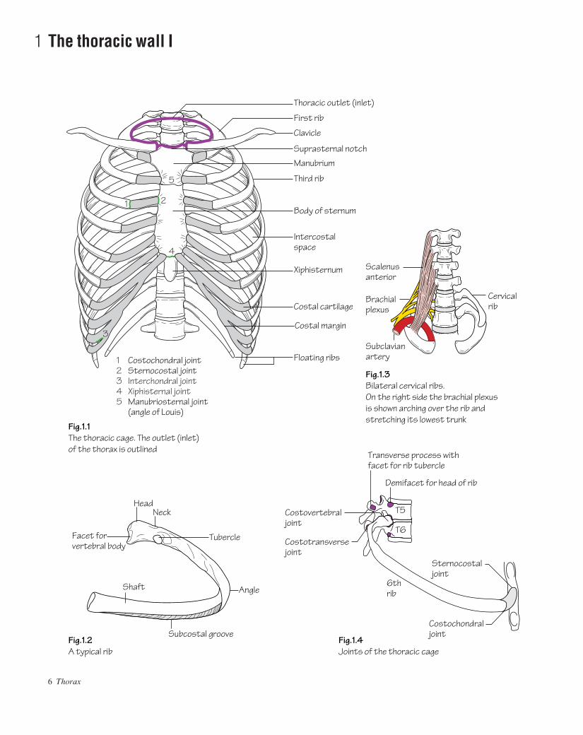
Fig.1.2A typical rib
Fig.1.1The thoracic cage. The outlet (inlet)of the thorax is outlined
Fig.1.4Joints of the thoracic cage
Fig.1.3Bilateral cervical ribs.On the right side the brachial plexus is shown arching over the rib and stretching its lowest trunk
T5
T6
Demifacet for head of rib
Transverse process withfacet for rib tubercle
Costovertebraljoint
1 2
5
3
4
12345
Costochondral jointSternocostal jointInterchondral jointXiphisternal jointManubriosternal joint(angle of Louis)
Clavicle
Costal margin
Costotransversejoint
AAAC01 21/5/05 10:38 AM Page 6

The thoracic cageThe thoracic cage is formed by the sternum and costal cartilages infront, the vertebral column behind and the ribs and intercostal spaceslaterally.
It is separated from the abdominal cavity by the diaphragm and com-municates superiorly with the root of the neck through the thoracicinlet (Fig. 1.1).
The ribs (Fig. 1.1)• Of the 12 pairs of ribs the first seven articulate with the vertebrae pos-teriorly and with the sternum anteriorly by way of the costal cartilages(true ribs).• The cartilages of the 8th, 9th and 10th ribs articulate with the carti-lages of the ribs above ( false ribs).• The 11th and 12th ribs are termed ‘floating’ because they do not articu-late anteriorly ( false ribs).
Typical ribs (3rd–9th)These comprise the following features (Fig. 1.2):• A head which bears two demifacets for articulation with the bodiesof: the numerically corresponding vertebra, and the vertebra above(Fig. 1.4).• A tubercle which comprises a rough non-articulating lateral facet aswell as a smooth medial facet. The latter articulates with the transverseprocess of the corresponding vertebra (Fig. 1.4).• A subcostal groove: the hollow on the inferior inner aspect of theshaft which accommodates the intercostal neurovascular structures.
Atypical ribs (1st, 2nd, 10th, 11th, 12th)• The 1st rib (see Fig. 63.2) is short, flat and sharply curved. The headbears a single facet for articulation. A prominent tubercle (scalenetubercle) on the inner border of the upper surface represents the inser-tion site for scalenus anterior. The subclavian vein passes over the 1strib anterior to this tubercle whereas the subclavian artery and lowesttrunk of the brachial plexus pass posteriorly.
A cervical rib is a rare ‘extra’ rib which articulates with C7 poster-iorly and the 1st rib anteriorly. A neurological deficit as well as vascu-lar insufficiency arise as a result of pressure from the rib on the lowesttrunk of the brachial plexus (T1) and subclavian artery, respectively(Fig. 1.3).
• The 2nd rib is less curved and longer than the 1st rib.• The 10th rib has only one articular facet on the head.• The 11th and 12th ribs are short and do not articulate anteriorly.They articulate posteriorly with the vertebrae by way of a single faceton the head. They are devoid of both a tubercle and a subcostal groove.
The sternum (Fig. 1.1)The sternum comprises a manubrium, body and xiphoid process.• The manubrium has facets for articulation with the clavicles, 1stcostal cartilage and upper part of the 2nd costal cartilage. It articulatesinferiorly with the body of the sternum at the manubriosternal joint.• The body is composed of four parts or sternebrae which fuse between15 and 25 years of age. It has facets for articulation with the lower partof the 2nd and the 3rd to 7th costal cartilages.• The xiphoid articulates above with the body at the xiphisternal joint.The xiphoid usually remains cartilaginous well into adult life.
Costal cartilagesThese are bars of hyaline cartilage which connect the upper seven ribsdirectly to the sternum and the 8th, 9th and 10th ribs to the cartilageimmediately above.
Joints of the thoracic cage (Figs 1.1 and 1.4)• The manubriosternal joint is a symphysis. It usually ossifies after theage of 30.• The xiphisternal joint is also a symphysis.• The 1st sternocostal joint is a primary cartilaginous joint. The rest(2nd to 7th) are synovial joints. All have a single synovial joint exceptfor the 2nd which is double.• The costochondral joints (between ribs and costal cartilages) are prim-ary cartilaginous joints.• The interchondral joints (between the costal cartilages of the 8th, 9thand 10th ribs) are synovial joints.• The costovertebral joints comprise two synovial joints formed by thearticulations of the demifacets on the head of each rib with the bodies ofits corresponding vertebra together with that of the vertebra above. The1st and 10th–12th ribs have a single synovial joint with their corres-ponding vertebral bodies.• The costotransverse joints are synovial joints formed by the articula-tions between the facets on the rib tubercle and the transverse processof its corresponding vertebra.
The thoracic wall I 7
AAAC01 21/5/05 10:38 AM Page 7
8 Thorax
2 The thoracic wall II
VeinArteryNerve
ExternalInternal Intercostal muscles
Intercostal
Innermost
Xiphisternum
Internalthoracic artery
Lateral branchlateral
anterior
Cutaneousbranches
Pleural andperitonealsensorybranches
Intercostalnerve
Posterior ramus Posterior intercostalartery
Anterior intercostalartery
Aorta
Spinalbranch
Costal margin
Central tendon
Inferior vena cava
Oesophagus
Aorta
T8
Vertebrallevels
Lateral arcuate ligamentMedial arcuate ligamentRight crus
Psoas majorQuadratus lumborum
Third lumbar vertebra
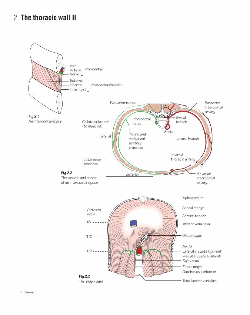
Fig.2.1An intercostal space
Fig.2.3The diaphragm
Fig.2.2The vessels and nervesof an intercostal space
T10
T12
Collateral branch(to muscles)
AAAC02 21/5/05 10:38 AM Page 8
The intercostal space (Fig. 2.1)Typically, each space contains three muscles comparable to those ofthe abdominal wall. These include the:• External intercostal: this muscle fills the intercostal space from thevertebra posteriorly to the costochondral junction anteriorly where itbecomes the thin anterior intercostal membrane. The fibres run down-wards and forwards from rib above to rib below.• Internal intercostal: this muscle fills the intercostal space from thesternum anteriorly to the angles of the ribs posteriorly where it becomesthe posterior intercostal membrane which reaches as far back as thevertebral bodies. The fibres run downwards and backwards.• Innermost intercostals: this group comprises the subcostal musclesposteriorly, the intercostales intimi laterally and the transversus thor-acis anteriorly. The fibres of these muscles span more than one inter-costal space.
The neurovascular space is the plane in which the neurovascularbundle (intercostal vein, artery and nerve) courses. It lies between theinternal intercostal and innermost intercostal muscle layers.
The intercostal structures course under cover of the subcostalgroove. Pleural aspiration should be performed close to the upper bor-der of a rib to minimize the risk of injury.
Vascular supply and venous drainage of the chest wallThe intercostal spaces receive their arterial supply from the anteriorand posterior intercostal arteries.• The anterior intercostal arteries are branches of the internal thoracicartery and its terminal branch the musculophrenic artery. The lowesttwo spaces have no anterior intercostal supply (Fig. 2.2).• The first 2–3 posterior intercostal arteries arise from the superiorintercostal branch of the costocervical trunk, a branch of the 2nd part ofthe subclavian artery (see Fig. 60.1). The lower nine posterior inter-costal arteries are branches of the thoracic aorta. The posterior inter-costal arteries are much longer than the anterior intercostal arteries(Fig. 2.2).
The anterior intercostal veins drain anteriorly into the internal thor-acic and musculophrenic veins. The posterior intercostal veins draininto the azygos and hemiazygos systems (see Fig. 4.2).
Lymphatic drainage of the chest wallLymph drainage from the:• Anterior chest wall: is to the anterior axillary nodes.• Posterior chest wall: is to the posterior axillary nodes.• Anterior intercostal spaces: is to the internal thoracic nodes.• Posterior intercostal spaces: is to the para-aortic nodes.
Nerve supply of the chest wall (Fig. 2.2)The intercostal nerves are the anterior primary rami of the thoracic seg-mental nerves. Only the upper six intercostal nerves run in their inter-costal spaces, the remainder gaining access to the anterior abdominalwall.
Branches of the intercostal nerves include:• Cutaneous anterior and lateral branches.
• A collateral branch which supplies the muscles of the intercostalspace (also supplied by the main intercostal nerve).• Sensory branches from the pleura (upper nerves) and peritoneum(lower nerves).
Exceptions include:• The 1st intercostal nerve is joined to the brachial plexus and has noanterior cutaneous branch.• The 2nd intercostal nerve is joined to the medial cutaneous nerve ofthe arm by the intercostobrachial nerve branch. The 2nd intercostalnerve consequently supplies the skin of the armpit and medial side ofthe arm.
The diaphragm (Fig. 2.3)The diaphragm separates the thoracic and abdominal cavities. It is com-posed of a peripheral muscular portion which inserts into a centralaponeurosisathe central tendon.
The muscular part has three component origins:• A vertebral part: this comprises the crura and arcuate ligaments.
The right crus arises from the front of the L1–3 vertebral bodies andintervening discs. Some fibres from the right crus pass around the loweroesophagus.
The left crus originates from L1 and L2 only.The medial arcuate ligament is made up of thickened fascia which
overlies psoas major and is attached medially to the body of L1 and lat-erally to the transverse process of L1. The lateral arcuate ligament ismade up of fascia which overlies quadratus lumborum from the trans-verse process of L1 medially to the 12th rib laterally.
The median arcuate ligament is a fibrous arch which connects leftand right crura.• A costal part: attached to the inner aspects of the lower six ribs.• A sternal part: consists of two small slips arising from the deep sur-face of the xiphoid process.
Openings in the diaphragmStructures traverse the diaphragm at different levels to pass from thoracic to abdominal cavities and vice versa. These levels are as follows:• T8, the opening for the inferior vena cava: transmits the inferior venacava and right phrenic nerve.• T10, the oesophageal opening: transmits the oesophagus, vagi andbranches of the left gastric artery and vein.• T12, the aortic opening: transmits the aorta, thoracic duct and azygosvein.
The left phrenic nerve passes into the diaphragm as a solitary structure.
Nerve supply of the diaphragm• Motor supply: the entire motor supply arises from the phrenic nerves(C3,4,5). Diaphragmatic contraction is the mainstay of inspiration.• Sensory supply: the periphery of the diaphragm receives sensoryfibres from the lower intercostal nerves. The sensory supply from thecentral part is carried by the phrenic nerves.
The thoracic wall II 9
AAAC02 21/5/05 10:38 AM Page 9
10 Thorax
3 The mediastinum Icthe contents of the mediastinum
Jugular lymph trunks
Thoracic duct
From lower limbs
Superior vena cava
From chest wall (right)
From chest wall (left)
Middle mediastinumHeart and roots of great vesselsPericardium
Superior mediastinumGreat vesselsTracheaOesophagusThymus, etc.
Anterior mediastinumThymus
Posterior mediastinumOesophagusDescending thoracic aortaThoracic ductAzygos and hemiazygos veinsSympathetic trunk, etc.
DiaphragmL1
L2Cisterna chyli
From abdominalviscera
Thoracic duct
Recurrent laryngeal nerve
OesophagusTrachea
Left vagus
Anterior pulmonaryplexus
Oesophageal plexus
Anterior vagal trunk
Oesophageal opening (T10)
Aortic opening (T12)
Left crus
Rightvagus
Azygosvein
Diaphragm
Rightcrus
Subclavian lymph trunkBronchomediastinallymph trunk
Right lymph duct
From kidneys andabdominal wall
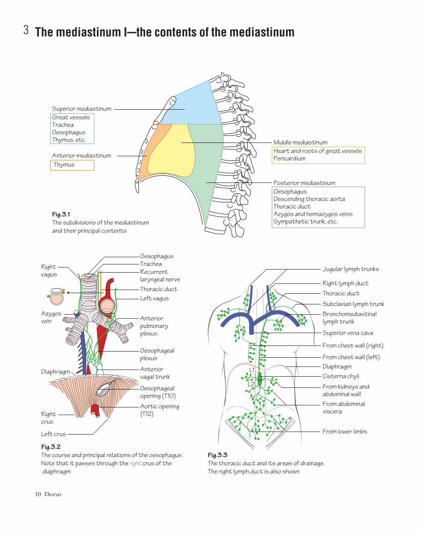
Fig.3.2The course and principal relations of the oesophagus.Note that it passes through the right crus of the diaphragm
Fig.3.3The thoracic duct and its areas of drainage.The right lymph duct is also shown
Fig.3.1The subdivisions of the mediastinum and their principal contents
AAAC03 21/5/05 10:38 AM Page 10

Subdivisions of the mediastinum (Fig. 3.1)The mediastinum is the space located between the two pleural sacs. Fordescriptive purposes it is divided into superior and inferior mediastinalregions by a line drawn backwards horizontally from the angle of Louis(manubriosternal joint) to the vertebral column (T4/5 intervertebral disc).
The superior mediastinum communicates with the root of the neckthrough the ‘thoracic inlet’. The latter opening is bounded anteriorly bythe manubrium, posteriorly by T1 vertebra and laterally by the 1st rib.
The inferior mediastinum is further subdivided into the:• Anterior mediastinum: the region in front of the pericardium.• Middle mediastinum: consists of the pericardium and heart.• Posterior mediastinum: the region between the pericardium and vertebrae.
The contents of the mediastinum (Figs 3.1 and 3.2)The oesophagus• Course: the oesophagus commences as a cervical structure at thelevel of the cricoid cartilage at C6 in the neck. In the thorax the oesoph-agus passes initially through the superior and then the posterior medi-astina. Having deviated slightly to the left in the neck the oesophagusreturns to the midline in the thorax at the level of T5. From here, itpasses downwards and forwards to reach the oesophageal opening inthe diaphragm (T10).• Structure: the oesophagus is composed of four layers:
• An inner mucosa of stratified squamous epithelium.• A submucous layer.• A double muscular layeralongitudinal outer layer and circular
inner layer. The muscle is striated in the upper two-thirds andsmooth in the lower third.
• An outer layer of areolar tissue.• Relations: the relations of the oesophagus are shown in Fig. 3.2. Onthe right side the oesophagus is crossed only by the azygos vein and theright vagus nerve and hence this forms the least hazardous surgicalapproach.• Arterial supply and venous drainage: owing to the length of thisstructure (25 cm), the oesophagus receives arterial blood from variedsources throughout its course:
• Upper thirdainferior thyroid artery.• Middle thirdaoesophageal branches of thoracic aorta.• Lower thirdaleft gastric branch of coeliac artery.
Similarly the venous drainage varies throughout its length:• Upper thirdainferior thyroid veins.• Middle thirdaazygos system.• Lower thirdaboth the azygos (systemic system) and left gastric
veins (portal system).The dual drainage of the lower third forms a site of portal-systemic
anastomosis. In advanced liver cirrhosis, portal pressure rises result-ing in back-pressure on the left gastric tributaries at the lower oesoph-agus. These veins become distended and fragile (oesophageal varices).They are predisposed to rupture, causing potentially life-threatening haemorrhage.• Lymphatic drainage: this is to a peri-oesophageal lymph plexus andthen to the posterior mediastinal nodes. From here lymph drains intosupraclavicular nodes. The lower oesophagus also drains into the nodesaround the left gastric vessels.
Carcinoma of the oesophagus carries an extremely poor prognosis.Two main histological typesbsquamous and adenocarcinomab
account for the majority of tumours. The incidence of adenocarcinoma ofthe lower third of the oesophagus is currently increasing for unknownreasons. Most tumours are unresectable at the time of diagnosis. Theinsertion of stents and use of lasers to pass through tumour obstructionhave become the principal methods of palliation.
The thoracic duct (Fig. 3.3)• The cisterna chyli is a lymphatic sac that receives lymph from theabdomen and lower half of the body. It is situated between the abdom-inal aorta and the right crus of the diaphragm.• The thoracic duct carries lymph from the cisterna chyli through thethorax to drain into the left brachiocephalic vein. It usually receivestributaries from the left jugular, subclavian and mediastinal lymphtrunks, although they may open into the large neck veins directly.• On the right side the main lymph trunks from the right upper bodyusually join and drain directly through a common tributary, the rightlymph duct, into the right brachiocephalic vein.
The thymus gland• This is an important component of the lymphatic system. It usuallylies behind the manubrium (in the superior mediastinum) but canextend to about the 4th costal cartilage in the anterior mediastinum.After puberty the thymus is gradually replaced by fat.
The mediastinum Ibthe contents of the mediastinum 11
AAAC03 21/5/05 10:38 AM Page 11

12 Thorax
4 The mediastinum IIcthe vessels of the thorax
Thyrocervical trunkSuprascapular
Inferior thyroidSuperficial cervical
Scalenus anteriorDorsal scapularSubclavian
Anterior intercostalsInternal thoracic (mammary)
MusculophrenicSuperior epigastric
Inferior thyroid
Deep cervical
Left internal jugularThoracic duct
VertebralLeft subclavianInternal thoracicLeft superior intercostal
Vagus nervePhrenic nerve
Crossing archof the aorta
Posterior intercostal
Posterior intercostals(also supply spinal cord)BronchialOesophagealMediastinal
Aortic opening in diaphragm
Aortic opening in diaphragm(T12)
branches
Right lymph duct
Left brachiocephalic
Costocervical trunk
Thyroidea ima
Superior intercostalUpper two posterior intercostals
Brachiocephalic
Inferior laryngeal
Right brachiocephalic
Superior vena cava
Right atriumAzygos
Diaphragm
Accessory hemiazygosT7
T8
Hemiazygos
Fig.4.2The principal veins of the thorax
Fig.4.1The branches of the arch and the descending thoracic aorta
Vertebral
Subcostal
AAAC04 21/5/05 10:37 AM Page 12

The thoracic aorta (Fig. 4.1)The ascending aorta arises from the aortic vestibule behind theinfundibulum of the right ventricle and the pulmonary trunk. It is con-tinuous with the aortic arch. The arch lies posterior to the lower half ofthe manubrium and arches from front to back over the left mainbronchus. The descending thoracic aorta is continuous with the archand begins at the lower border of the body of T4. It initially lies slightlyto the left of the midline and then passes medially to gain access to theabdomen by passing beneath the median arcuate ligament of thediaphragm at the level of T12. From here it continues as the abdominalaorta.
The branches of the ascending aorta are the:• Right and left coronary arteries.
The branches of the aortic arch are the:• Brachiocephalic artery: arises from the arch behind the manubriumand courses upwards to bifurcate into right subclavian and right com-mon carotid branches posterior to the right sternoclavicular joint.• Left common carotid artery: see p. 133.• Left subclavian artery.• Thyroidea ima artery.
The branches of the descending thoracic aorta include the:• Oesophageal, bronchial, mediastinal, posterior intercostal and sub-costal arteries.
The subclavian arteries (see Fig. 60.1)The subclavian arteries become the axillary arteries at the outer bor-der of the 1st rib. Each artery is divided into three parts by scalenusanterior:• 1st part: the part of the artery that lies medial to the medial border ofscalenus anterior. It gives rise to three branches, the: vertebral artery(p. 135), thyrocervical trunk and internal thoracic (mammary) artery.The latter artery courses on the posterior surface of the anterior chestwall one fingerbreadth from the lateral border of the sternum. Along its course it gives off anterior intercostal, thymic and perforatingbranches. The ‘perforators’ pass through the anterior chest wall to
supply the breast. The internal thoracic artery divides behind the 6thcostal cartilage into superior epigastric and musculophrenic branches.The thyrocervical trunk terminates as the inferior thyroid artery.• 2nd part: the part of the artery that lies behind scalenus anterior. Itgives rise to the costocervical trunk (see Fig. 60.1).• 3rd part: the part of the artery that lies lateral to the lateral border ofscalenus anterior. This part gives rise to the dorsal scapular artery.
The great veins (Fig. 4.2)The brachiocephalic veins are formed by the confluence of the subcla-vian and internal jugular veins behind the sternoclavicular joints. Theleft brachiocephalic vein traverses diagonally behind the manubrium tojoin the right brachiocephalic vein behind the 1st costal cartilage thusforming the superior vena cava. The superior vena cava receives onlyone tributaryathe azygos vein.
The azygos system of veins (Fig. 4.2)• The azygos vein: commences as the union of the right subcostal veinand one or more veins from the abdomen. It passes through the aorticopening in the diaphragm, ascends on the posterior chest wall to thelevel of T4 and then arches over the right lung root to enter the superiorvena cava. It receives tributaries from the: lower eight posterior inter-costal veins, right superior intercostal vein and hemiazygos veins.• The hemiazygos vein: arises on the left side in the same manner as theazygos vein. It passes through the aortic opening in the diaphragm andup to the level of T9 from where it passes diagonally behind the aortaand thoracic duct to drain into the azygos vein at the level of T8. Itreceives venous blood from the lower four left posterior intercostalveins.• The accessory hemiazygos vein: drains blood from the middle poster-ior intercostal veins (as well as some bronchial and mid-oesophagealveins). The accessory hemiazygos crosses to the right to drain into theazygos vein at the level of T7.• The upper four left intercostal veins drain into the left brachio-cephalic vein via the left superior intercostal vein.
The mediastinum IIbthe vessels of the thorax 13
AAAC04 21/5/05 10:37 AM Page 13

14 Thorax
5 The pleura and airways
Apical
Apical
Anterior
Right main bronchusLeft main bronchus
Posterior
Middle
Anterior
Lingular
Anterior basalLateral basal
Posterior basal
Trachea
Anterior basal
Lateral basal
Apical oflower lobe
Medial basal
Posterior basal
Posterior
Cricoid (C6)
Apico-posterior
Pulmonary arteryBronchusPulmonary veinsLymph nodeCut edge of pleuraPulmonary ligament
Fig. 5.1The principal structuresin the hilum of the lung
Fig. 5.2The trachea and main bronchi
Brachiocephalicartery
Superiorvena cava
Rightpulmonaryartery
Thyroidisthmus
Left brachiocephalicvein
Aortic arch
Fig. 5.3The anterior relations of the trachea
AAAC05 23/05/2005 2:59 PM Page 14

The respiratory tract is most often discussed in terms of upper andlower parts. The upper respiratory tract relates to the nasopharynx andlarynx whereas the lower relates to the trachea, bronchi and lungs.
The pleurae• Each pleura consists of two layers: a visceral layer which is adherentto the lung and a parietal layer which lines the inner aspect of the chestwall, diaphragm and sides of the pericardium and mediastinum.• At the hilum of the lung the visceral and parietal layers become con-tinuous. This cuff hangs loosely over the hilum and is known as the pul-monary ligament. It permits expansion of the pulmonary veins andmovement of hilar structures during respiration (Fig. 5.1).• The two pleural cavities do not connect.• The pleural cavity contains a small amount of pleural fluid which actsas a lubricant decreasing friction between the pleurae.• During maximal inspiration the lungs almost fill the pleural cavities.In quiet inspiration the lungs do not expand fully into the costo-diaphragmatic and costomediastinal recesses of the pleural cavity.• The parietal pleura is sensitive to pain and touch (carried by the inter-costal and phrenic nerves). The visceral pleura is sensitive only tostretch (carried by autonomic afferents from the pulmonary plexus).
Air can enter the pleural cavity following a fractured rib or a tornlung (pneumothorax). This eliminates the normal negative pleuralpressure, causing the lung to collapse.
Inflammation of the pleura (pleurisy) results from infection of theadjacent lung (pneumonia). When this occurs the inflammatory processrenders the pleura sticky. Under these circumstances a pleural rub canoften be auscultated over the affected region during inspiration andexpiration. Pus in the pleural cavity (secondary to an infective process)is termed an empyema.
The trachea (Fig. 5.2)• Course: the trachea commences at the level of the cricoid cartilage inthe neck (C6). It terminates at the level of the angle of Louis (T4/5)where it bifurcates into right and left main bronchi.
• Structure: the trachea is a rigid fibroelastic structure. Incom-plete rings of hyaline cartilage continuously maintain the patency of the lumen. The trachea is lined internally with ciliated columnar epithelium.• Relations: behind the trachea lies the oesophagus. The 2nd, 3rd and4th tracheal rings are crossed anteriorly by the thyroid isthmus (Figs 5.3and 64.1).• Blood supply: the trachea receives its blood supply from branches ofthe inferior thyroid and bronchial arteries.
The bronchi and bronchopulmonary segments (Fig. 5.2)• The right main bronchus is shorter, wider and takes a more verticalcourse than the left. The width and vertical course of the right mainbronchus account for the tendency for inhaled foreign bodies to prefer-entially impact in the right middle and lower lobe bronchi.• The left main bronchus enters the hilum and divides into a superiorand inferior lobar bronchus. The right main bronchus gives off thebronchus to the upper lobe prior to entering the hilum and once into thehilum divides into middle and inferior lobar bronchi.• Each lobar bronchus divides within the lobe into segmental bronchi.Each segmental bronchus enters a bronchopulmonary segment.• Each bronchopulmonary segment is pyramidal in shape with its apexdirected towards the hilum (see Fig. 6.1). It is a structural unit of a lobethat has its own segmental bronchus, artery and lymphatics. If onebronchopulmonary segment is diseased it may be resected with pre-servation of the rest of the lobe. The veins draining each segment areintersegmental.
Bronchial carcinoma is the commonest cancer among men in theUnited Kingdom. Four main histological types occur of which smallcell carries the worst prognosis. The overall prognosis remainsappalling with only 10% of sufferers surviving 5 years. It occurs mostcommonly in the mucous membranes lining the major bronchi near thehilum. Local invasion and spread to hilar and tracheobronchial nodesoccurs early.
The pleura and airways 15
AAAC05 23/05/2005 2:59 PM Page 15

16 Thorax
6 The lungs
12
6
10 7
3
57
8 9
10
6
21
4
3
589
3
4
5
54
7
8
9 10
6
23
29 10
1
3
54
7
8
910
6
6
21
3
1
2
6
109 8
4 5
1234 and 5678910
ApicalPosterior (1 and 2 from a common apico-posterior stem on the left side)AnteriorLateral and medial middle lobe (superior and inferior lingular on left side)Superior (apical)Medial basal (cardiac on left)Anterior basal (7 and 8 often by a common stem on left)Lateral basalPosterior basal
Upper lobe Middle lobe Lower lobe
LEFT LUNG RIGHT LUNG
Fig. 6.1The segmental bronchi (viewed fromthe lateral side) and the broncho-pulmonary segments, with theirstandard numbering
Fig. 6.2P–A. Chest X-ray
Trachea
Arch of aorta
Lung hilum
Left ventricle
Costophrenic angleBreast shadow
Right atrium
Diaphragm
AAAC06 21/5/05 10:36 AM Page 16

The lungs (Fig. 6.1)• The lungs provide an alveolar surface area of approximately 40 m2
for gaseous exchange.• Each lung has: an apex which reaches above the sternal end of the 1strib; a costovertebral surface which underlies the chest wall; a baseoverlying the diaphragm and a mediastinal surface which is moulded toadjacent mediastinal structures.• Structure: the right lung is divided into upper, middle and lowerlobes by oblique and horizontal fissures. The left lung has only anoblique fissure and hence no middle lobe. The lingular segment repres-ents the left sided equivalent of the right middle lobe. It is, however, ananatomical part of the left upper lobe.
Structures enter or leave the lungs by way of the lung hilum which,as mentioned earlier, is ensheathed in a loose pleural cuff (see Fig. 5.1).• Blood supply: the bronchi and parenchymal tissue of the lungs aresupplied by bronchial arteriesabranches of the descending thoracicaorta. Bronchial veins, which also communicate with pulmonary veins,drain into the azygos and hemiazygos. The alveoli receive deoxy-genated blood from terminal branches of the pulmonary artery and oxy-genated blood returns via tributaries of the pulmonary veins. Twopulmonary veins return blood from each lung to the left atrium.• Lymphatic drainage of the lungs: lymph returns from the peripherytowards the hilar tracheobronchial groups of nodes and from here tomediastinal lymph trunks.• Nerve supply of the lungs: a pulmonary plexus is located at the rootof each lung. The plexus is composed of sympathetic fibres (from thesympathetic trunk) and parasympathetic fibres (from the vagus).Efferent fibres from the plexus supply the bronchial musculature andafferents are received from the mucous membranes of bronchioles andfrom the alveoli.
The mechanics of respiration• A negative intrapleural pressure keeps the lungs continuously par-tially inflated.
• During normal inspiration: contraction of the upper external inter-costals increases the A-P diameter of the upper thorax; contraction ofthe lower external intercostals increases the transverse diameter of thelower thorax; and contraction of the diaphragm increases the verticallength of the internal thorax. These changes serve to increase lung vol-ume and thereby result in reduction of intrapulmonary pressure causingair to be sucked into the lungs. In deep inspiration the sternocleidomas-toid, scalenus anterior and medius, serratus anterior and pectoralismajor and minor all aid to maximize thoracic capacity. The latter aretermed collectivelyathe accessory muscles of respiration.• Expiration is mostly due to passive relaxation of the muscles of inspira-tion and elastic recoil of the lungs. In forced expiration the abdominalmusculature aids ascent of the diaphragm.
The chest X-ray (CXR) (Fig. 6.2)The standard CXR is the postero-anterior (PA) view. This is taken withthe subject’s chest touching the cassette holder and the X-ray beamdirected anteriorly from behind.
Structures visible on the chest X-ray include the:• Heart borders: any significant enlargement of a particular chambercan be seen on the X-ray. In congestive cardiac failure all four cham-bers of the heart are enlarged (cardiomegaly). This is identified on thePA view as a cardiothoracic ratio greater than 0.5. This ratio is calcu-lated by dividing the width of the heart by the width of the thoracic cav-ity at its widest point.• Lungs: the lungs are radiolucent. Dense streaky shadows, seen at thelung roots, represent the blood-filled pulmonary vasculature.• Diaphragm: the angle made between the diaphragm and chest wall istermed the costophrenic angle. This angle is lost when a pleural effu-sion collects.• Mediastinal structures: these are difficult to distinguish as there isconsiderable overlap. Clearly visible, however, is the aortic archwhich, when pathologically dilated (aneurysmal), creates the impres-sion of ‘widening’ of the mediastinum.
The lungs 17
AAAC06 21/5/05 10:36 AM Page 17

18 Thorax
7 The heart I
Right vagus
Right phrenic
Brachiocephalic artery
Rightbrachiocephalic vein
Right pulmonary veins
Right atrium
Inferior vena cava
Superior vena cava
Inferior thyroid veinsLeft subclavian artery
Left common carotid artery
Left vagus
Left phrenic
Left brachiocephalic vein
Left pulmonary artery
Left recurrent laryngeal
Left bronchus
Left pulmonary veins
Thyroid
Pulmonary veins
Pericardium Heart
Back of left atriumBack of right atriumInferior vena cavaParietal pericardiumVisceral pericardium
Arrow in transverse sinus
Pulmonary trunk
Arrow in oblique sinus
Aorta
Right recurrent laryngeal
Right recurrent laryngeal
Fig.7.1The heart and the great vessels
Fig.7.2The sinuses of the pericardium. The heart has been removed from the pericardial cavity and turned over to show its posterior aspect. The red line shows the cut edges where the visceral pericardium is continuous with the parietal pericardium. Visceral layer: blue, parietal layer: red
AAAC07 21/5/05 10:36 AM Page 18

The heart I 19
• Blood supply: from the pericardiacophrenic branches of the internalthoracic arteries.• Nerve supply: the fibrous pericardium and the parietal layer ofserous pericardium are supplied by the phrenic nerve.
Following thoracic trauma blood can collect in the pericardialspace (haemopericardium) which may, in turn, lead to cardiac tam-ponade. This manifests itself clinically as shock, distended neck veinsand muffled/absent heart sounds (Beck’s triad). This condition is fatalunless pericardial decompression is effected immediately.
The heart surfaces• The anterior (sternocostal ) surface comprises the: right atrium, atri-oventricular groove, right ventricle, a small strip of left ventricle andthe auricle of the left atrium.• The inferior (diaphragmatic) surface comprises the: right atrium,atrioventricular groove and both ventricles separated by the interven-tricular groove.• The posterior surface (base) comprises the left atrium receiving thefour pulmonary veins.
The heart, pericardium, lung roots and adjoining parts of the great ves-sels constitute the middle mediastinum (Figs 3.1 and 7.1).
The pericardiumThe pericardium comprises fibrous and serous components. Thefibrous pericardium is a strong layer which covers the heart. It fuseswith the roots of the great vessels above and with the central tendon ofthe diaphragm below. The serous pericardium lines the fibrous peri-cardium (parietal layer) and is reflected at the vessel roots to cover theheart surface (visceral layer). The serous pericardium provides smoothsurfaces for the heart to move against. Two important sinuses arelocated between the parietal and visceral layers. These are the:
• Transverse sinusalocated between the superior vena cava and leftatrium posteriorly and the pulmonary trunk and aorta anteriorly (Fig. 7.2).
• Oblique sinusabehind the left atrium, the sinus is bounded by theinferior vena cava and the pulmonary veins (Fig. 7.2).
AAAC07 21/5/05 10:36 AM Page 19
20 Thorax
Portion of right atrium derived from sinusvenosus
Crista terminalis
Inferior vena cava
Fossa ovalis
Opening ofcoronary sinusValve of thecoronary sinus
Valve of the inferior vena cava
Musculipectinati
Superior vena cava
Limbusfossa ovalis
Pulmonary valve(posterior, anterolateraland anteromedial cusps)
Mitralvalve
Opening of right coronary arteryAortic valve(Anterior (right coronary) cusp,Left posterior (left coronary) cusp,right posterior (non-coronary) cusp)
Right atrium
Left atrium
Tricuspid valve
Posteriorcusp
Posteriorcusp
Anteriorcusp
AnteriorcuspSeptal
cusp
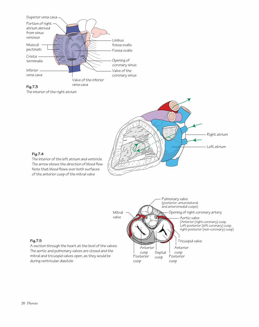
Fig.7.3The interior of the right atrium
Fig.7.4The interior of the left atrium and ventricle.The arrow shows the direction of blood flow.Note that blood flows over both surfacesof the anterior cusp of the mitral valve
Fig.7.5A section through the heart at the level of the valves.The aortic and pulmonary valves are closed and the mitral and tricuspid valves open, as they would be during ventricular diastole
AAAC07 21/5/05 10:36 AM Page 20

The heart chambersThe right atrium (Fig. 7.3)• Receives deoxygenated blood from the inferior vena cava below andfrom the superior vena cava above.• Receives the coronary sinus in its lower part (p. 23).• The upper end of the atrium projects to the left of the superior venacava as the right auricle.• The sulcus terminalis is a vertical groove on the outer surface of theatrium. This groove corresponds internally to the crista terminalisaamuscular ridge which separates the smooth walled atrium (derivedfrom the sinus venosus) from the rest of the atrium (derived from thetrue fetal atrium). The latter contains horizontal ridges of musclea
musculi pectinati.• Above the coronary sinus the interatrial septum forms the posteriorwall. The depression in the septumathe fossa ovalisarepresents thesite of the foramen ovale. Its floor is the fetal septum primum. Theupper ridge of the fossa ovalis is termed the limbus, which representsthe septum secundum. Failure of fusion of the septum primum with theseptum secundum gives rise to a patent foramen ovale (atrial septaldefect) but as long as the two septa still overlap, there will be no func-tional disability. A patent foramen gives rise to a left–right shunt.
The right ventricle• Receives blood from the right atrium through the tricuspid valve (seebelow). The edges of the valve cusps are attached to chordae tendineaewhich are, in turn, attached below to papillary muscles. The latter areprojections of muscle bundles on the ventricular wall.• The wall of the right ventricle is thicker than that of the atria but notas thick as that of the left ventricle. The wall contains a mass of muscu-lar bundles known as trabeculae carneae. One prominent bundle pro-jects forwards from the interventricular septum to the anterior wall.This is the moderator band (or septomarginal trabecula) and is ofimportance in the conduction of impulses as it contains the right branchof the atrioventricular bundle.
• The infundibulum is the smooth walled outflow tract of the right ventricle.• The pulmonary valve (see below) is situated at the top of theinfundibulum. It is composed of three semilunar cusps. Blood flowsthrough the valve and into the pulmonary arteries via the pulmonarytrunk to be oxygenated in the lungs.
The left atrium• Receives oxygenated blood from four pulmonary veins which drainposteriorly.• The cavity is smooth walled except for the atrial appendage.• On the septal surface a depression marks the fossa ovalis.• The mitral (bicuspid) valve guards the passage of blood from the leftatrium to the left ventricle.
The left ventricle (Fig. 7.4)• The wall of the left ventricle is considerably thicker than that of theright ventricle but the structure is similar. The thick wall is necessary topump oxygenated blood at high pressure through the systemic circula-tion. Trabeculae carneae project from the wall with papillary musclesattached to the mitral valve cusp edges by way of chordae tendineae.• The vestibule is a smooth walled part of the left ventricle which islocated below the aortic valve and constitutes the outflow tract.
The heart valves (Fig. 7.5)• The purpose of valves within the heart is to maintain unidirectional flow.• The mitral (bicuspid) and tricuspid valves are flat. During ventricularsystole the free edges of the cusps come into contact and eversion isprevented by the pull of the chordae. Papillary muscle rupture canoccur as a complication of myocardial infarction. This is evident clin-ically by a pansystolic murmur representing regurgitant flow of bloodfrom ventricle to atrium.• The aortic and pulmonary valves are composed of three semilunarcusps which are cup shaped. During ventricular diastole back-pressureof blood above the cusps forces them to fill and hence close.
The heart I 21
AAAC07 21/5/05 10:36 AM Page 21

22 Thorax
8 The heart II
Left coronaryartery
Posteriorinterventricularbranch
Marginalartery
50
40
3525
1235
55
65
15
0
Right coronaryartery
Anteriorinterventricularbranch
S–A node
Atrial conduction
Ventricular conduction
A–V node
Coronarysinus
Smallcardiacvein
Middlecardiacvein
Greatcardiacvein
QRS TP
Fig.8.1The coronary arteries.Variations are common
Fig.8.3The direction and timing of the spread of action potential in the conductingsystem of the heart.Times are in msec
Fig.8.2The venous drainage of the heart
Fig.8.4An electrocardiogram
AAAC08 23/05/2005 3:06 PM Page 22
The grooves between the four heart chambers represent the sites thatoffer the least stretch during systole and, for this reason, are where mostof the vessels supplying the heart are situated.
The arterial supply of the heart (Fig. 8.1)The coronary arteries are responsible for supplying the heart itself withoxygenated blood.
The coronary arteries are functional end-arteries and hence follow-ing a total occlusion, the myocardium supplied by the blocked artery isdeprived of its blood supply (myocardial infarction). When the vessellumen gradually narrows due to atheromatous change of the walls,patients complain of gradually increasing chest pain on exertion(angina). Under these conditions the increased demand placed on themyocardium cannot be met by the diminished arterial supply. Anginathat is not amenable to pharmacological control can be relieved bydilating (angioplasty), or surgically bypassing (coronary artery bypassgrafting), the arterial stenosis. The latter procedure is usually per-formed using a reversed length of great saphenous vein anastomosed tothe proximal aorta and then distally to the coronary artery beyond thestenosis. Ischaemic heart disease is the leading cause of death in thewestern world and consequently a thorough knowledge of the coronaryanatomy is essential.
The origins of the coronary arteries are as follows:• The left coronary artery arises from the aortic sinus immediatelyabove the left posterior cusp of the aortic valve (see Fig. 7.5).• The right coronary artery arises from the aortic sinus immediatelyabove the anterior cusp of the aortic valve (see Fig. 7.5).
There is considerable variation in size and distribution zones of thecoronary arteries. For example, in some people the posterior interven-tricular branch of the right coronary artery is large and supplies a largepart of the left ventricle whereas in the majority this is supplied by theanterior interventricular branch of the left coronary.
Similarly, the sinu-atrial node is usually supplied by a nodal branchof the right coronary artery but in 30–40% of the population it receivesits supply from the left coronary.
The venous drainage of the heart (Fig. 8.2)The venous drainage systems in the heart include:• The veins which accompany the coronary arteries and drain into the
right atrium via the coronary sinus. The coronary sinus drains into theright atrium to the left of and superior to the opening of the inferior venacava. The great cardiac vein follows the anterior interventricularbranch of the left coronary and then sweeps backwards to the left in theatrioventricular groove. The middle cardiac vein follows the posteriorinterventricular artery and, along with the small cardiac vein which fol-lows the marginal artery, drains into the coronary sinus. The coronarysinus drains the vast majority of the heart’s venous blood.• The venae cordis minimi: these are small veins which drain directlyinto the cardiac chambers.• The anterior cardiac veins: these are small veins which cross the atri-oventricular groove to drain directly into the right atrium.
The conducting system of the heart (Figs 8.3 and 8.4)• The sinu-atrial (SA) node is the pacemaker of the heart. It is situatednear the top of the crista terminalis, below the superior vena cavalopening into the right atrium. Impulses generated by the SA node areconducted throughout the atrial musculature to effect synchronousatrial contraction. Disease or degeneration of any part of the conduc-tion pathway can lead to dangerous interruption of heart rhythm.Degeneration of the SA node leads to other sites of the conduction path-way taking over the pacemaking role, albeit usually at a slower rate.• Impulses reach the atrioventricular (AV) node which lies in theinteratrial septum just above the opening for the coronary sinus. Fromhere the impulse is transmitted to the ventricles via the atrioventricularbundle (of His) which descends in the interventricular septum.• The bundle of His divides into right and left branches which sendPurkinje fibres to lie within the subendocardium of the ventricles. Theposition of the Purkinje fibres accounts for the almost synchronouscontraction of the ventricles.
The nerve supply of the heartThe heart receives both a sympathetic and a parasympathetic nervesupply so that heart rate can be controlled to demand.• The parasympathetic supply (bradycardic effect): is derived from thevagus nerve (p. 25).• The sympathetic supply (tachycardic and positively inotropic effect):is derived from the cervical and upper thoracic sympathetic ganglia byway of superficial and deep cardiac plexuses (p. 25).
The heart II 23
AAAC08 23/05/2005 3:06 PM Page 23
24 Thorax
9 The nerves of the thorax
Subclavian artery
Superior intercostal veinArch of aortaLeft recurrent laryngeal nerveLeft pulmonary artery
Posterior pulmonary plexus
Descending aorta
Oesophageal plexus on oesophagus
Sympathetic trunk
Greater splanchnic nerve
C3C4C5
Thoracic duct on side of oesophagus
Central tendon of diaphragm
Inferior vena cava
Branches to fibrousand parietal pericardium
Mediastinal pleura
Scalenus anterior
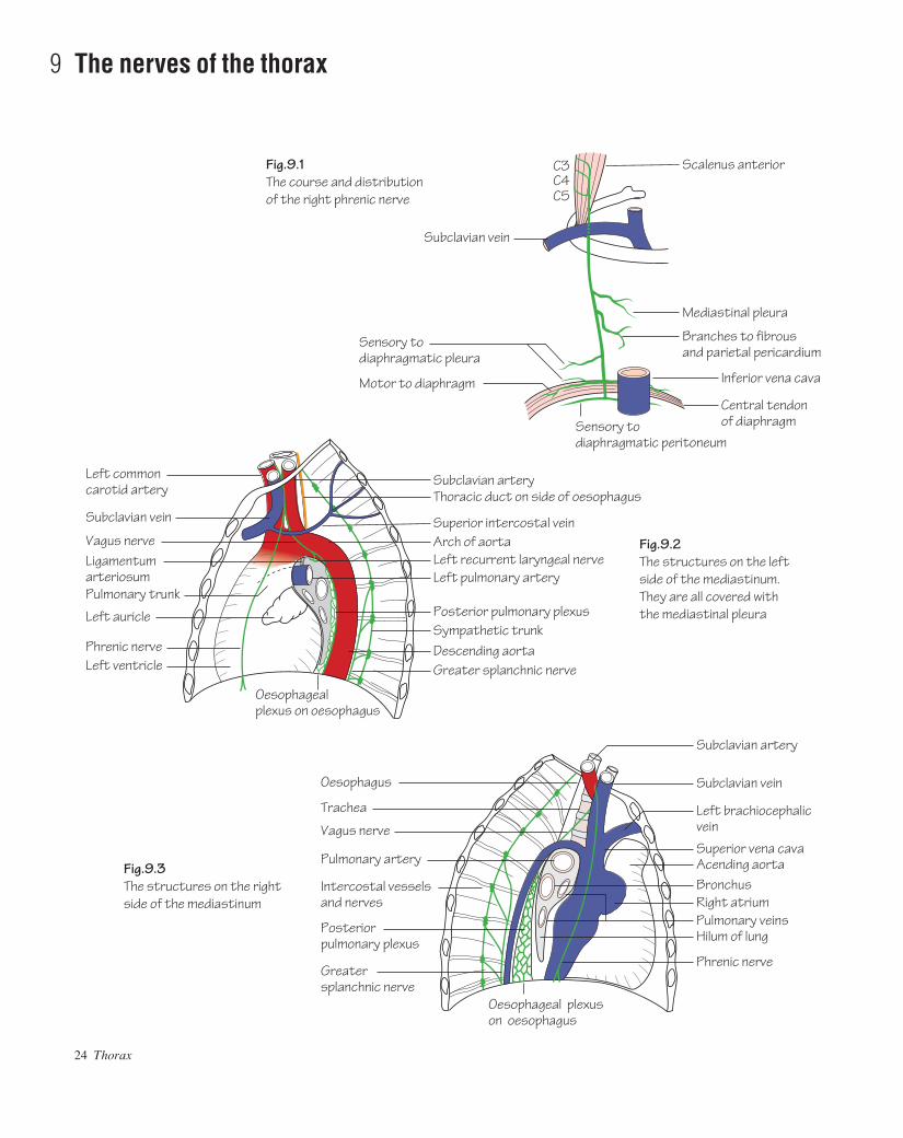
Fig.9.2The structures on the left side of the mediastinum.They are all covered with the mediastinal pleura
Fig.9.1The course and distribution of the right phrenic nerve
Fig.9.3The structures on the right side of the mediastinum
Subclavian artery
Subclavian vein
Left brachiocephalicvein
Superior vena cavaAcending aortaBronchus
Pulmonary veinsHilum of lung
Phrenic nerve
Oesophagus
Trachea
Vagus nerve
Intercostal vesselsand nerves
Posteriorpulmonary plexus
Greatersplanchnic nerve
Oesophageal plexus on oesophagus
Right atrium
Pulmonary artery
Subclavian vein
Sensory todiaphragmatic pleura
Sensory todiaphragmatic peritoneum
Motor to diaphragm
Left commoncarotid artery
Subclavian vein
Vagus nerveLigamentumarteriosumPulmonary trunk
Left auricle
Phrenic nerveLeft ventricle
AAAC09 21/5/05 10:35 AM Page 24

The phrenic nervesThe phrenic nerves arise from the C3, C4 and C5 nerve roots in theneck.• The right phrenic nerve (Fig. 9.1) descends along a near verticalpath, anterior to the lung root, lying on sequentially: the right brachio-cephalic vein, the superior vena cava, and the right atrium before pass-ing to the inferior vena caval opening in the diaphragm (T8). Here theright phrenic enters the caval opening and immediately penetrates thediaphragm which it supplies.• The left phrenic nerve (Fig. 9.2) descends alongside the left subcla-vian artery. On the arch of the aorta it passes over the left superior inter-costal vein to descend in front of the left lung root onto the pericardiumoverlying the left ventricle. The left phrenic then pierces the musculardiaphragm as a solitary structure. Note: the phrenic nerves do not passbeyond the undersurface of the diaphragm.• The phrenic nerves are composed mostly of motor fibres which supplythe diaphragm. However, they also transmit fibres which are sensory to the fibrous pericardium, mediastinal pleura and peritoneum as wellas the central part of the diaphragm.
Irritation of the diaphragmatic peritoneum is usually referred to theC4 dermatome. Hence, upper abdominal pathology such as a perfor-ated duodenal ulcer often results in pain felt at the shoulder tip.
The vagiThe vagi are the 10th cranial nerves (p. 145).• The right vagus nerve (Figs 9.3 and 3.2) descends adherent to the thor-acic trachea prior to passing behind the lung root to form the posteriorpulmonary plexus. It finally reaches the lower oesophagus where itforms an oesophageal plexus with the left vagus. From this plexus,anterior and posterior vagal trunks descend (carrying fibres from bothleft and right vagi) on the oesophagus to pass into the abdomen throughthe oesophageal opening in the diaphragm at the level of T10.• The left vagus nerve (Fig. 9.2) crosses the arch of the aorta and its branches. It is itself crossed here by the left superior intercostal vein. Below, it descends behind the lung root to reach the oesophaguswhere it contributes to the oesophageal plexus mentioned above (seeFig. 3.2).
Vagal branches• The left recurrent laryngeal nerve arises from the left vagus belowthe arch of the aorta. It hooks around the ligamentum arteriosum andascends in the groove between the trachea and the oesophagus to reachthe larynx (p. 139).• The right recurrent laryngeal nerve arises from the right vagus in theneck and hooks around the right subclavian artery prior to ascending inthe groove between the trachea and the oesophagus before finallyreaching the larynx.
• The recurrent laryngeal nerves supply the mucosa of the upper tra-chea and oesophagus as well as providing a motor supply to all of themuscles of the larynx (except cricothyroid) and sensory fibres to thelower larynx.• The vagi also contribute branches to the cardiac and pulmonaryplexuses.
The thoracic sympathetic trunk (Figs 9.2 and 9.3, and Chapter 53)• The thoracic sympathetic chain is a continuation of the cervicalchain. It descends in the thorax behind the pleura immediately lateral tothe vertebral bodies and passes under the medial arcuate ligament of thediaphragm to continue as the lumbar sympathetic trunk.• The thoracic chain bears a ganglion for each spinal nerve; the firstfrequently joins the inferior cervical ganglion to form the stellate gan-glion. Each ganglion receives a white ramus communicans containingpreganglionic fibres from its corresponding spinal nerve and sendsback a grey ramus, bearing postganglionic fibres.
Upper limb sympathectomy is used for the treatment of hyperhidro-sis and Raynaud syndrome. Surgical sympathectomy involves excisionof part of the thoracic sympathetic chain (usually for two interspaces)below the level of the stellate ganglion. The latter structure must beidentified on the neck of the 1st rib.Branches:• Sympathetic fibres are distributed to the skin with each of the thor-acic spinal nerves.• Postganglionic fibres from T1–5 are distributed to the thoracic visceraathe heart and great vessels, the lungs and the oesophagus.• Mainly preganglionic fibres from T5–12 form the splanchnic nerves,which pierce the crura of the diaphragm and pass to the coeliac andrenal ganglia from which they are relayed as postganglionic fibres tothe abdominal viscera (cf. fibres to the suprarenal medulla which arepreganglionic). These splanchnic nerves are the: greater splanchnic(T5–10), lesser splanchnic (T10–11) and lowest splanchnic (T12).They lie medial to the sympathetic trunk on the bodies of the thoracicvertebrae and are quite easily visible through the parietal pleura.
The cardiac plexusThis plexus is for descriptive purposes divided into superficial and deepparts. It consists of sympathetic and parasympathetic efferents as wellas afferents.• Cardiac branches from the plexus supply the heart where they:accompany coronary arteries for vasomotor control and supply thesinu-atrial and atrioventricular nodes for cardio-inhibitory and cardio-acceleratory purposes.• Pulmonary branches supply the bronchial wall smooth muscle (con-trolling diameter) and pulmonary blood vessels for vasomotor control.
The nerves of the thorax 25
AAAC09 21/5/05 10:35 AM Page 25

26 Thorax
10 Surface anatomy of the thorax
2
4
6
8
6 6
8 8
10 1012 12
10
2
4
6
8
10
Cervical plexus
Cardiac notch of lungTransverse fissure
Oblique fissure
Costodiaphragmatic recess
Apex of lower lung
Oblique fissure
Beginning of transverse fissure
Costodiaphragmatic recess
Mid-clavicular line
Fig.10.1The surface markings of the lungs and pleural cavities
Fig.10.2The surface markings of the heart.The areas of auscultation for the aortic, pulmonary, mitral and tricuspid valves are indicated by letters
1
2
3
6
5
1
2
PA
TM
AAAC10 21/5/05 10:35 AM Page 26

The anterior thoraxLandmarks of the anterior thorax include:• The angle of Louis (sternal angle): formed by the joint between themanubrium and body of the sternum. It is an important landmark as the2nd costal cartilages articulate on either side and by following this lineonto the 2nd rib, further ribs and intercostal spaces can be identified.The sternal angle corresponds to a horizontal point level with the inter-vertebral disc between T4 and T5.• The suprasternal notch: situated in the midline between the medialends of the clavicles and above the upper edge of the manubrium.• The costal margin: formed by the lower borders of the cartilages ofthe 7th, 8th, 9th and 10th ribs and the ends of the 11th and 12th ribs.• The xiphisternal joint: formed by the joint between the body of thesternum and xiphisternum.
The posterior thoraxLandmarks of the posterior thorax include:• The first palpable spinous process is that of C7 (vertebra prominens).C1–6 vertebrae are covered by the thick ligamentum nuchae. Thespinous processes of the thoracic vertebrae can be palpated and countedin the midline posteriorly.• The scapula is located on the upper posterior chest wall. In slim sub-jects the superior angle, inferior angle, spine and medial (vertebral)border of the scapula are easily palpable.
Lines of orientationThese are imaginary vertical lines used to describe locations on thechest wall. These include:• The mid-clavicular line: a vertical line from the midpoint of the clav-icle downwards.• The anterior and posterior axillary lines: from the anterior and poster-ior axillary folds, respectively, vertically downwards.• The mid-axillary line: from the midpoint between anterior and poster-ior axillary lines vertically downwards.
Vertebral levelsPalpable bony prominences can be used to identify the location ofimportant underlying structures. The following bony landmarks andtheir corresponding vertebral levels are given:• Suprasternal notch: T2/3.• Sternal angle (angle of Louis): T4/5.• Superior angle of the scapula: T2.• Inferior angle of the scapula: T8.• Xiphisternal joint: T9.• Subcostal plane (lowest part of the costal margin): L3.
The surface markings of thoracic structuresThe tracheaThe trachea commences at the lower border of the cricoid cartilage (C6vertebral level). It runs downwards in the midline and ends slightly tothe right by bifurcating into the left and right main bronchi. The bifurca-tion occurs at the level of the sternal angle (T4/5).
The pleura (Fig. 10.1)The apex of the pleura projects about 2.5 cm above the medial third ofthe clavicle. The lines of pleural reflection pass behind the sternoclavicu-lar joints to meet in the midline at the level of the sternal angle. Theright pleura then passes downwards to the 6th costal cartilage. The left
pleura passes laterally for a small distance at the 4th costal cartilage anddescends vertically lateral to the sternal border to the 6th costal cartil-age. From these points both pleurae pass posteriorly and in so doingcross the 8th rib in the mid-clavicular line, the 10th rib in the mid-axillary line and finally reach the level of the 12th rib posteriorly.
The lungs (Fig. 10.1)The apex and mediastinal border of the right lung follow the pleuraloutline. In mid-inspiration the right lung lower border crosses the 6thrib in the mid-clavicular line, the 8th rib in the mid-axillary line andreaches the level of the 10th rib posteriorly. The left lung borders aresimilar to those of the right except that the mediastinal border archeslaterally (the cardiac notch) but then resumes the course mentionedabove.• The oblique fissure: is represented by an oblique line drawn from apoint 2.5 cm lateral to the 5th thoracic spinous process to the 6th costalcartilage anteriorly. The oblique fissures separate the lungs into upperand lower lobes.• The transverse fissure: is represented by a line drawn horizontallyfrom the 4th costal cartilage to a point where it intersects the obliquefissure. The fissure separates the upper and middle lobes of the rightlung.
The heart• The borders of the heart are illustrated by joining the four pointsshown (Fig. 10.2).• The apex of the left ventricle corresponds to where the apex beat ispalpable. The surface marking for the apex beat is in the 5th intercostalspace in the mid-clavicular line.• See Fig. 10.2 for optimal sites of valvular auscultation.
The great vessels• The aortic arch: arches antero-posteriorly behind the manubrium.The highest point of the arch reaches the midpoint of the manubrium.• The brachiocephalic artery and left common carotid artery: ascendposterior to the manubrium.• The brachiocephalic veins: are formed by the confluence of the inter-nal jugular and subclavian veins. This occurs posterior to the sterno-clavicular joints.• The superior vena cava: is formed by the confluence of the left andright brachiocephalic veins between the 2nd and 3rd right costal cartil-ages at the right border of the sternum.
The breastThe base of the breast (p. 69) is constant, overlying the 2nd to the 6thribs and costal cartilages anteriorly and from the lateral border of thesternum to the mid-axillary line. The position of the nipple is variablein the female but in the man it is usually in the 4th intercostal space inthe mid-clavicular line.
The internal thoracic vesselsThese arteries and veins descend 1 cm lateral to the edge of the sternum.
The diaphragmIn mid-inspiration the highest part of the right dome reaches as far asthe upper border of the 5th rib in the mid-clavicular line. The left domereaches only the lower border of the 5th rib.
Surface anatomy of the thorax 27
AAAC10 21/5/05 10:35 AM Page 27

28 Abdomen and pelvis
11 The abdominal wall
Lineasemilunaris
Serratusanterior
Superficial inguinal ring
A
A
A
B
C
Cut edge of external oblique
Fig.11.1Two muscles of the anterior abdominal wall.The external oblique (on the right) and the internal oblique (on the left)
Fig.11.2The fibrous layer of superficial fascia can be likened to a pair of bathing trunks sewn to the thigh below the inguinal ligament and clinging to the penis andscrotum (except for the glans)
Fig.11.3Transverse sections through the rectus sheath.A: above the costal marginB: above the umbilicusC: above the pubic symphysis
Linea alba
Cut edge of external oblique
Internal oblique
Anterior superior iliac spine
Inguinal ligament
Inguinalligament
Conjoint tendonPubic tubercle
Rectus abdominis
Dartosmuscle
Rectus abdominisExternal oblique
Linea alba
Costal cartilages
External obliqueInternal obliqueTransversus abdominis
Transversalis fascia
Superiorepigastricartery
Deep layer ofsuperficial fascia
Fascia penis
Colles' fascia
External obliqueInternal obliqueTransversus abdominis
Peritoneum
Inferiorepigastricartery
AAAC11 21/5/05 10:43 AM Page 28
The abdominal wall 29
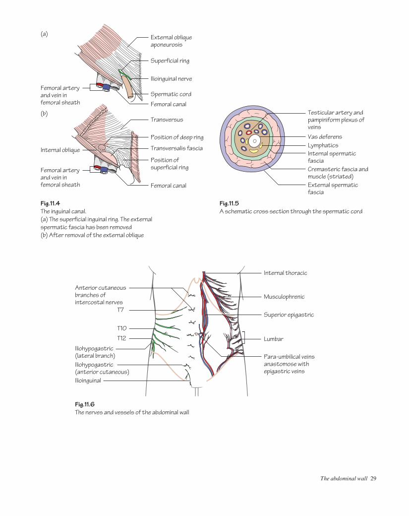
Fig.11.4The inguinal canal.(a) The superficial inguinal ring. The external spermatic fascia has been removed(b) After removal of the external oblique
Fig.11.6The nerves and vessels of the abdominal wall
Fig.11.5A schematic cross section through the spermatic cord
Superficial ring
Femoral arteryand vein infemoral sheath
External obliqueaponeurosis
Transversus
Transversalis fascia
Position of deep ring
Position of superficial ring
Ilioinguinal nerve
Spermatic cord
Femoral canal
Femoral canal
Femoral arteryand vein infemoral sheath
Internal oblique
(b)
(a)
Testicular artery andpampiniform plexus ofveins
Lymphatics
Internal thoracic
Musculophrenic
T7
Vas deferens
External spermatic fascia
Cremasteric fascia andmuscle (striated)
Internal spermatic fascia
Superior epigastric
Para-umbilical veinsanastomose withepigastric veins
Lumbar
T10
T12
Ilioinguinal
Anterior cutaneousbranches ofintercostal nerves
Iliohypogastric(lateral branch)Iliohypogastric(anterior cutaneous)
AAAC11 21/5/05 10:43 AM Page 29
deep circumflex iliac artery (a branch of the external iliac artery) an-teriorly. The two lower intercostal and four lumbar arteries supply thewall posterolaterally.
Veins of the abdominal wall (Fig. 11.6)The abdominal wall is a site of porto-systemic anastomosis. The lateralthoracic, lumbar and superficial epigastric tributaries of the systemiccirculation anastomose around the umbilicus with the para-umbilicalveins which accompany the ligamentum teres and drain into the portalcirculation.
Lymph drainage of the abdominal wallSee p. 35.
The inguinal canal (Fig. 11.4)The canal is approximately 4 cm long and allows the passage of thespermatic cord (round ligament in the female) through the lower ab-dominal wall. The canal passes obliquely from the deep inguinal ringin a medial direction to the superficial inguinal ring.• The deep ring: is an opening in the transversalis fascia. It lies half-way between the anterior superior iliac spine and the pubic tubercle.The inferior epigastric vessels pass medial to the deep ring.• The superficial ring: is not a ring but a triangular-shaped defect inthe external oblique aponeurosis lying above and medial to the pubictubercle.
The walls of the inguinal canal (Fig. 11.4)• Anterior: external oblique covers the length of the canal anteriorly.It is reinforced in its lateral third by internal oblique.• Superior: internal oblique arches posteriorly to form the roof of thecanal.• Posterior: transversalis fascia forms the lateral part of the posteriorwall. The conjoint tendon (the combined common insertion of the inter-nal oblique and transversus into the pectineal line) forms the medialpart of the posterior wall.• Inferior: the inguinal ligament.
Contents of the inguinal canal• The spermatic cord (or round ligament in the female).• The ilioinguinal nerve (L1).
The spermatic cord (Fig. 11.5)The spermatic cord is covered by three layers which arise from the layers of the lower abdominal wall as the cord passes through theinguinal canal. These are the:• External spermatic fascia: from the external oblique aponeurosis.• Cremasteric fascia and muscle: from the internal oblique aponeurosis.• Internal spermatic fascia: from the transversalis fascia.The contents of the spermatic cord include the:• Ductus (vas) deferens (or round ligament).• Testicular artery: a branch of the abdominal aorta.• Pampiniform plexus of veins: these coalesce to form the testicularvein in the region of the deep ring.• Lymphatics: from the testis and epididymis draining to the pre-aortic nodes.• Autonomic nerves.
30 Abdomen and pelvis
The anterior abdominal wall comprises: skin, superficial fascia, abdom-inal muscles (and their respective aponeuroses), transversalis fascia,extraperitoneal fat, and parietal peritoneum.
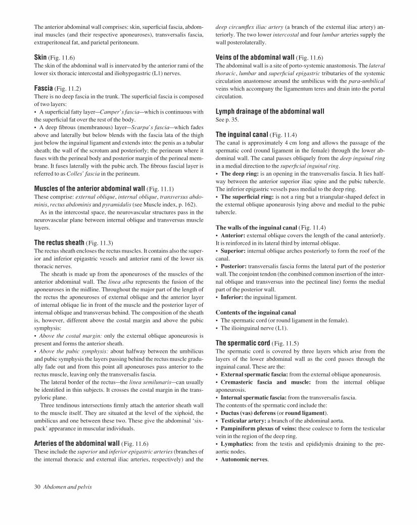
Skin (Fig. 11.6)The skin of the abdominal wall is innervated by the anterior rami of thelower six thoracic intercostal and iliohypogastric (L1) nerves.
Fascia (Fig. 11.2)There is no deep fascia in the trunk. The superficial fascia is composedof two layers:• A superficial fatty layeraCamper’s fasciaawhich is continuous withthe superficial fat over the rest of the body.• A deep fibrous (membranous) layeraScarpa’s fasciaawhich fadesabove and laterally but below blends with the fascia lata of the thighjust below the inguinal ligament and extends into: the penis as a tubularsheath; the wall of the scrotum and posteriorly; the perineum where itfuses with the perineal body and posterior margin of the perineal mem-brane. It fuses laterally with the pubic arch. The fibrous fascial layer isreferred to as Colles’ fascia in the perineum.
Muscles of the anterior abdominal wall (Fig. 11.1)These comprise: external oblique, internal oblique, transversus abdo-minis, rectus abdominis and pyramidalis (see Muscle index, p. 162).
As in the intercostal space, the neurovascular structures pass in theneurovascular plane between internal oblique and transversus musclelayers.
The rectus sheath (Fig. 11.3)The rectus sheath encloses the rectus muscles. It contains also the super-ior and inferior epigastric vessels and anterior rami of the lower six thoracic nerves.
The sheath is made up from the aponeuroses of the muscles of theanterior abdominal wall. The linea alba represents the fusion of theaponeuroses in the midline. Throughout the major part of the length ofthe rectus the aponeuroses of external oblique and the anterior layer of internal oblique lie in front of the muscle and the posterior layer ofinternal oblique and transversus behind. The composition of the sheathis, however, different above the costal margin and above the pubicsymphysis:• Above the costal margin: only the external oblique aponeurosis ispresent and forms the anterior sheath.• Above the pubic symphysis: about halfway between the umbilicusand pubic symphysis the layers passing behind the rectus muscle gradu-ally fade out and from this point all aponeuroses pass anterior to the rectus muscle, leaving only the transversalis fascia.
The lateral border of the rectusathe linea semilunarisacan usuallybe identified in thin subjects. It crosses the costal margin in the trans-pyloric plane.
Three tendinous intersections firmly attach the anterior sheath wallto the muscle itself. They are situated at the level of the xiphoid, theumbilicus and one between these two. These give the abdominal ‘six-pack’ appearance in muscular individuals.
Arteries of the abdominal wall (Fig. 11.6)These include the superior and inferior epigastric arteries (branches ofthe internal thoracic and external iliac arteries, respectively) and the
AAAC11 21/5/05 10:43 AM Page 30
The arteries of the abdomen 31
12 The arteries of the abdomen
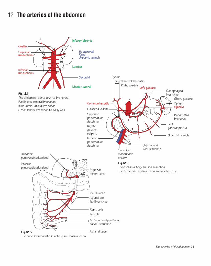
Fig.12.1The abdominal aorta and its branches.Red labels: ventral branchesBlue labels: lateral branchesGreen labels: branches to body wall
Fig.12.2The coeliac artery and its branches.The three primary branches are labelled in red
Fig.12.3The superior mesenteric artery and its branches
Inferior phrenic
Suprarenal
Coeliac
Superiormesenteric
Inferiormesenteric
RenalUreteric branch
Lumbar
Median sacral
Gonadal
Oesophagealbranches
Left gastricRight gastric
Right and left hepatic
Superiormesentericartery
Jejunal andileal branches
Cystic
Common hepatic
Gastroduodenal
Omental branch
SpleenSplenic
Short gastric
Jejunal andileal branches
Ileocolic Right colic
Middle colic
Superiormesenteric
Appendicular
Anterior and posterior caecal branches
Superiorpancreatico-duodenal
Superiorpancreaticoduodenal
Inferiorpancreaticoduodenal
Rightgastro-epiploicInferiorpancreatico-duodenal
Leftgastroepiploic
Pancreatic branches
AAAC12 21/5/05 10:43 AM Page 31
32 Abdomen and pelvis
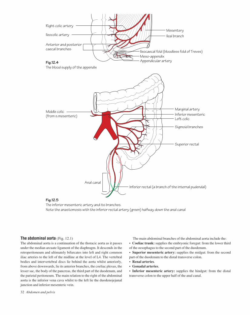
Fig.12.4The blood supply of the appendix
Fig.12.5The inferior mesenteric artery and its branches.Note the anastomosis with the inferior rectal artery (green) halfway down the anal canal
Ileocolic artery
Right colic arteryMesenteryIleal branch
Inferior rectal (a branch of the internal pudendal)Anal canal
Middle colic (from s.mesenteric)
Superior rectal
Appendicular arteryMeso-appendix
Anterior and posterior caecal branches
Ileocaecal fold (bloodless fold of Treves)
Marginal artery
Sigmoid branches
Left colicInferior mesenteric
The main abdominal branches of the abdominal aorta include the:• Coeliac trunk: supplies the embryonic foregut: from the lower thirdof the oesophagus to the second part of the duodenum.• Superior mesenteric artery: supplies the midgut: from the secondpart of the duodenum to the distal transverse colon.• Renal arteries.• Gonadal arteries.• Inferior mesenteric artery: supplies the hindgut: from the distaltransverse colon to the upper half of the anal canal.
The abdominal aorta (Fig. 12.1)The abdominal aorta is a continuation of the thoracic aorta as it passesunder the median arcuate ligament of the diaphragm. It descends in theretroperitoneum and ultimately bifurcates into left and right commoniliac arteries to the left of the midline at the level of L4. The vertebralbodies and intervertebral discs lie behind the aorta whilst anteriorly,from above downwards, lie its anterior branches, the coeliac plexus, thelesser sac, the body of the pancreas, the third part of the duodenum, andthe parietal peritoneum. The main relation to the right of the abdominalaorta is the inferior vena cava whilst to the left lie the duodenojejunaljunction and inferior mesenteric vein.
AAAC12 21/5/05 10:43 AM Page 32
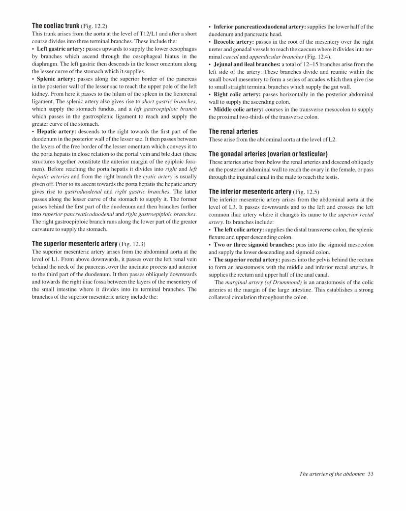
The arteries of the abdomen 33
The coeliac trunk (Fig. 12.2)This trunk arises from the aorta at the level of T12/L1 and after a shortcourse divides into three terminal branches. These include the:• Left gastric artery: passes upwards to supply the lower oesophagusby branches which ascend through the oesophageal hiatus in thediaphragm. The left gastric then descends in the lesser omentum alongthe lesser curve of the stomach which it supplies.• Splenic artery: passes along the superior border of the pancreas in the posterior wall of the lesser sac to reach the upper pole of the leftkidney. From here it passes to the hilum of the spleen in the lienorenalligament. The splenic artery also gives rise to short gastric branches,which supply the stomach fundus, and a left gastroepiploic branchwhich passes in the gastrosplenic ligament to reach and supply thegreater curve of the stomach.• Hepatic artery: descends to the right towards the first part of theduodenum in the posterior wall of the lesser sac. It then passes betweenthe layers of the free border of the lesser omentum which conveys it tothe porta hepatis in close relation to the portal vein and bile duct (thesestructures together constitute the anterior margin of the epiploic fora-men). Before reaching the porta hepatis it divides into right and lefthepatic arteries and from the right branch the cystic artery is usuallygiven off. Prior to its ascent towards the porta hepatis the hepatic arterygives rise to gastroduodenal and right gastric branches. The latterpasses along the lesser curve of the stomach to supply it. The formerpasses behind the first part of the duodenum and then branches furtherinto superior pancreaticoduodenal and right gastroepiploic branches.The right gastroepiploic branch runs along the lower part of the greatercurvature to supply the stomach.
The superior mesenteric artery (Fig. 12.3)The superior mesenteric artery arises from the abdominal aorta at thelevel of L1. From above downwards, it passes over the left renal veinbehind the neck of the pancreas, over the uncinate process and anteriorto the third part of the duodenum. It then passes obliquely downwardsand towards the right iliac fossa between the layers of the mesentery ofthe small intestine where it divides into its terminal branches. Thebranches of the superior mesenteric artery include the:
• Inferior pancreaticoduodenal artery: supplies the lower half of theduodenum and pancreatic head.• Ileocolic artery: passes in the root of the mesentery over the rightureter and gonadal vessels to reach the caecum where it divides into ter-minal caecal and appendicular branches (Fig. 12.4).• Jejunal and ileal branches: a total of 12–15 branches arise from theleft side of the artery. These branches divide and reunite within thesmall bowel mesentery to form a series of arcades which then give riseto small straight terminal branches which supply the gut wall.• Right colic artery: passes horizontally in the posterior abdominalwall to supply the ascending colon.• Middle colic artery: courses in the transverse mesocolon to supplythe proximal two-thirds of the transverse colon.
The renal arteriesThese arise from the abdominal aorta at the level of L2.
The gonadal arteries (ovarian or testicular)These arteries arise from below the renal arteries and descend obliquelyon the posterior abdominal wall to reach the ovary in the female, or passthrough the inguinal canal in the male to reach the testis.
The inferior mesenteric artery (Fig. 12.5)The inferior mesenteric artery arises from the abdominal aorta at thelevel of L3. It passes downwards and to the left and crosses the leftcommon iliac artery where it changes its name to the superior rectalartery. Its branches include:• The left colic artery: supplies the distal transverse colon, the splenicflexure and upper descending colon.• Two or three sigmoid branches: pass into the sigmoid mesocolonand supply the lower descending and sigmoid colon.• The superior rectal artery: passes into the pelvis behind the rectumto form an anastomosis with the middle and inferior rectal arteries. Itsupplies the rectum and upper half of the anal canal.
The marginal artery (of Drummond) is an anastomosis of the colicarteries at the margin of the large intestine. This establishes a strongcollateral circulation throughout the colon.
AAAC12 21/5/05 10:43 AM Page 33

34 Abdomen and pelvis
13 The veins and lymphatics of the abdomen
Fig.13.1The inferior vena cava and its tributaries
Fig.13.2The portal system.Note the anastomoses with the systemic system (orange) in the oesophagus and the anal canal
Inferior phrenic
SuprarenalUreteric branchRenal
Lumbar
Median sacral
Common iliac
Gonadal
Pancreaticoduodenal
Right colic
Middle colic
CysticOesophageal branches
Right gastric
Left gastric
Right gastroepiploicSpleen
SplenicInferior mesentericSuperior mesenteric
Left colic
Sigmoid branches
Superior rectal
Portal vein
AAAC13 21/5/05 10:42 AM Page 34

The portal vein (Fig. 13.2)The portal venous system receives blood from the length of gut fromthe lower third of the oesophagus to the upper half of the anal canal aswell as the spleen, pancreas and gall-bladder. It serves to transfer bloodto the liver where the products of digestion can be metabolized andstored. Blood from the liver ultimately gains access to the inferior venacava by way of the hepatic veins. The portal vein is formed behind theneck of the pancreas by the union of the superior mesenteric and splenicveins. It passes behind the first part of the duodenum in front of the in-ferior vena cava and enters the free border of the lesser omentum. Thevein then ascends towards the porta hepatis in the anterior margin of theepiploic foramen (of Winslow) in the lesser omentum. At the porta hep-atis it divides into right and left branches. The veins that correspond tothe branches of the coeliac and superior mesenteric arteries drain intothe portal vein or one of its tributaries. The inferior mesenteric veindrains into the splenic vein adjacent to the fourth part of the duodenum.
Porto-systemic anastomosesA number of connections occur between the portal and systemic circula-tions. When the direct pathway through the liver becomes congested(such as in cirrhosis) the pressure within the portal vein rises and underthese circumstances the porto-systemic anastomoses form an alternat-ive route for the blood to take. The sites of porto-systemic anastomosisinclude:• The lower oesophagus (p. 11): formed by tributaries of the left gas-tric (portal) and oesophageal veins (systemic via the azygos and hemi-azygos veins).• The anal canal: formed by the superior rectal (portal) and middleand inferior rectal veins (systemic).• The bare area of the liver: formed by the small veins of the portalsystem and the phrenic veins (systemic).• The periumbilical region: formed by small paraumbilical veinswhich drain into the left portal vein and the superficial veins of the anter-ior abdominal wall (systemic).
The inferior vena cava (Fig. 13.1)The inferior vena cava is formed by the union of the common iliac veinsin front of the body of L5. It ascends in the retroperitoneum on the rightside of the abdominal aorta. Along its course, from below upwards, itforms the posterior wall of the epiploic foramen of Winslow and isembedded in the bare area of the liver in front of the right suprarenalgland. The inferior vena cava passes through the caval opening in thediaphragm at the level of T8 and drains into the right atrium.
The lymphatic drainage of the abdomen and pelvisThe abdominal wallLymph from the skin of the anterolateral abdominal wall above thelevel of the umbilicus drains to the anterior axillary lymph nodes. Effer-ent lymph from the skin below the umbilicus drains to the superficialinguinal nodes.
The lymph nodes and trunksThe two main lymph node groups of the abdomen are closely related tothe aorta. These comprise the pre-aortic and para-aortic groups.• The pre-aortic nodes are arranged around the three ventral branchesof the aorta and consequently receive lymph from the territories that aresupplied by these branches. This includes most of the gastrointestinaltract, liver, gall-bladder, spleen and pancreas. The efferent vessels fromthe pre-aortic nodes coalesce to form a variable number of intestinaltrunks which deliver the lymph to the cisterna chyli.• The para-aortic nodes are arranged around the lateral branches of theaorta and drain lymph from their corresponding territories, i.e. the kid-neys, adrenals, gonads, and abdominal wall as well as the common iliacnodes. The efferent vessels from the para-aortic nodes coalesce to forma variable number of lumbar trunks which deliver the lymph to the cis-terna chyli.
Cisterna chyliThe cisterna chyli is a lymphatic sac that lies anterior to the bodies ofthe 1st and 2nd lumbar vertebrae. It is formed by the confluence of theintestinal trunks, the lumbar trunks and lymphatics from the lower tho-racic wall. It serves as a receptacle for lymph from the abdomen andlower limbs which is then relayed to the thorax by the thoracic duct (p. 11).
The lymphatic drainage of the stomachLymph from the stomach drains to the coeliac nodes. For the purposesof description, the stomach can be divided into four quarters wherelymph drains to the nearest appropriate group of nodes.
The lymphatic drainage of the testesLymph from the skin of the scrotum and the tunica albuginea drains tothe superficial inguinal nodes. Lymph from the testes, however, drainsalong the course of the testicular artery to the para-aortic group ofnodes. Hence, a malignancy of the scrotal skin might result in palpableenlargement of the superficial inguinal nodes whereas testiculartumours metastasize to the para-aortic nodes.
The veins and lymphatics of the abdomen 35
AAAC13 21/5/05 10:42 AM Page 35

36 Abdomen and pelvis
14 The peritoneum
Fig.14.1A vertical section through the abdomen to show theperitoneal relations.Lesser sacGreater sac
Fig.14.2A horizontal section through the abdomen.Note how the epiploic foramen lies between two major veins
Fig.14.3The peritoneal relations of the liver(a) Seen from in front(b) The same liver rotated in the direction of the arrow to show the upper and posterior surfaces.The narrow spaces between the liver and the diaphragm labelled A and B are the right and left subphrenic spaces
Upper recess ofomental bursa
Diaphragm
Transverse mesocolon
Small intestineMesentery
Liver
Epiploic foramen(in the distance)
Greater omentum
Subphrenic space
Epiploic foramen (of Winslow)
Aorta
Left kidneySplenic arteryLienorenal ligamentSpleenShort gastric vesselsGastrosplenicligamentStomachLesser omentum
Duodenum (third part)Transverse colon
Fusion between layersof greater omentum
Lesser omentum
PancreasStomach
Omental bursa
Portal veinInferior vena cava
Hepatic arteryCommon bile duct
Liver
(a) (b)
Peritoneumcoveringcaudate lobe
Lower layer ofcoronary ligament
Righttriangularligament
Left triangularligament
Inferior vena cava
Upper layer ofcoronary ligamentUpper layer of
coronary ligament Bare area
Falciform ligament
Gall bladder
Ligamentum teres
Position of umbilicus
Ligamentum teres
Portal vein, hepaticartery and bile duct in free edge of lesseromentum leading toporta hepatisCut edge of lesseromentum
A B
Fissure for ligamentum venosum
Left triangularligament
Fundus ofgall bladder
AAAC14 21/5/05 10:42 AM Page 36

The mesenteries and layers of the peritoneumThe transverse colon, stomach, spleen and liver each have attached tothem two ‘mesenteries’adouble layers of peritoneum containing arteriesand their accompanying veins, nerves and lymphaticsawhile the smallintestine and sigmoid colon have only one. All the other viscera are re-troperitoneal. The mesenteries and their associated arteries are as follows:• The colon (Fig. 14.1): (1) The transverse mesocolon (the middlecolic artery). (2) The posterior two layers of the greater omentum.• The stomach (Fig. 14.1): (1) The lesser omentum (the left and rightgastric arteries and in its free border, the hepatic artery, portal vein andbile duct). (2) The anterior two layers of the greater omentum (the rightand left gastroepiploic arteries and their omental branches).• The spleen (Fig. 14.2): (1) The lienorenal ligament (the splenicartery). (2) The gastrosplenic ligament (the short gastric and left gas-troepiploic arteries).• The liver (Fig. 14.3): (1) The falciform ligament and the two layersof the coronary ligament with their sharp edges, the left and right trian-gular ligaments. This mesentery is exceptional in that the layers of thecoronary ligament are widely separated so that the liver has a bare areadirectly in contact with the diaphragm (the obliterated umbilical arteryin the free edge of the falciform ligament and numerous small veins inthe bare area, p. 35). (2) The lesser omentum (already described).• The small intestine (Fig. 14.1): (1) The mesentery of the small intes-tine (the superior mesenteric artery and its branches).• The sigmoid colon: (1) The sigmoid mesocolon (the sigmoid arteriesand their branches).
The peritoneal cavity (Figs 14.1 and 14.2)• The complications of the peritoneal cavity may best be described bystarting at the transverse mesocolon. Its two layers are attached to theanterior surface of the pancreas, the second part of the duodenum andthe front of the left kidney. They envelop the transverse colon and con-tinue downwards to form the posterior two layers of the greater omen-tum, which hangs down over the coils of the small intestine. They thenturn back on themselves to form the anterior two layers of the omentumand these reach the greater curvature of the stomach. The four layers ofthe omentum are fused and impregnated with fat. The greater omentumplays an important role in limiting the spread of infection in the peri-toneal cavity.• From its attachment to the pancreas, the lower layer of the transversemesocolon turns downwards to become the parietal peritoneum of theposterior abdominal wall from which it is reflected to form the mesen-tery of the small intestine and the sigmoid mesocolon.• The upper layer of the transverse mesocolon passes upwards to formthe parietal peritoneum of the posterior abdominal wall, covering theupper part of the pancreas, the left kidney and its suprarenal, the aortaand the origin of the coeliac artery (the ‘stomach bed’). It thus forms theposterior wall of the omental bursa. It then covers the diaphragm andcontinues onto the anterior abdominal wall.• From the diaphragm and anterior abdominal wall it is reflected ontothe liver to form its ‘mesentery’ in the form of the two layers of the fal-ciform ligament. At the liver, the left layer of the falciform ligamentfolds back on itself to form the sharp edge of the left triangular liga-
ment while the right layer turns back on itself to form the upper andlower layers of the coronary ligament with its sharp-edged right tri-angular ligament. The layers of the coronary ligament are widely separated so that a large area of liver between themathe bare areaa
is directly in contact with the diaphragm. The inferior vena cava isembedded in the bare area (Fig. 14.3).• From the undersurface of the liver another ‘mesentery’ passes fromthe fissure for the ligamentum venosum to the lesser curvature of thestomach to form the lesser omentum.• The lesser omentum splits to enclose the stomach and is continuouswith the two layers of the greater omentum already described. Thelesser omentum has a right free border which contains the portal vein,the hepatic artery and the common bile duct.• In the region of the spleen there are two more ‘mesenteries’ which arecontinuous with the lesser and greater omenta. These are the lienorenalligament, a double layer of peritoneum reflected from the front of theleft kidney to the hilum of the spleen, and the gastrosplenic ligamentwhich passes from the hilum of the spleen to the greater curvature of thestomach (Fig. 14.2).• The mesentery of the small intestine is attached to the posterior ab-dominal wall from the duodenojejunal flexure to the ileocolic junction.• The sigmoid mesocolon passes from a V-shaped attachment on theposterior abdominal wall to the sigmoid colon.• The general peritoneal cavity comprises the main cavityathe greatersacaand a diverticulum from itathe omental bursa (lesser sac). Theomental bursa lies between the stomach and the stomach bed to allowfree movement of the stomach. It lies behind the stomach, the lesseromentum and the caudate lobe of the liver and in front of the structuresof the stomach bed. The left border is formed by the hilum of the spleenand the lienorenal and gastrosplenic ligaments.• The communication between the greater and lesser sacs is the epi-ploic foramen ( foramen of Winslow). It lies behind the free border ofthe lesser omentum and its contained structures, below the caudate pro-cess of the liver, in front of the inferior vena cava and above the firstpart of the duodenum.• The subphrenic spaces are part of the greater sac that lies between thediaphragm and the upper surface of the liver. There are right and leftspaces, separated by the falciform ligament.• In the pelvis the parietal peritoneum covers the upper two-thirds ofthe rectum whence it is reflected, in the female, onto the posteriorfornix of the vagina and the back of the uterus to form the recto-uterinepouch (pouch of Douglas). In the male it passes onto the back of thebladder to form the rectovesical pouch.
The anterior abdominal wall• The peritoneum of the deep surface of the anterior abdominal wallshows a central ridge from the apex of the bladder to the umbilicus pro-duced by the median umbilical ligament. This is the remains of theembryonic urachus. Two medial umbilical ligaments converge to theumbilicus from the pelvis. They represent the obliterated umbilicalarteries of the fetus. The ligamentum teres is a fibrous band in the freemargin of the falciform ligament. It represents the obliterated leftumbilical vein.
The peritoneum 37
AAAC14 21/5/05 10:42 AM Page 37

38 Abdomen and pelvis
15 The upper gastrointestinal tract I
Fig.15.1The subdivisions of the stomach
Fig.15.2The stomach bed. For more detail see fig.19.1.The stomach is outlined but the shape is by no means constant
Duodenum
Right crus of diaphragm
Suprarenal
Pyloric sphincter
Angular incisure
Lesser curvature
Cardiac notch
Fundus
Body
Greater curvature
Pyloric antrum
Right kidney
Spleen
Aorta
Splenic artery
Splenic flexure of colon
Pancreas
Left kidney
Descending colon
Hepaticflexure
Ascendingcolon
AAAC15 21/5/05 10:42 AM Page 38

The embryonic gut is divided into foregut, midgut and hindgut, sup-plied, respectively, by the coeliac, superior mesenteric and inferiormesenteric arteries. The foregut extends from the oesophagus to theentrance of the common bile duct into the second part of the duodenum.The midgut extends down to two-thirds of the way along the transversecolon. It largely develops outside the abdomen until this congenital‘umbilical hernia’ is reduced during the 8th–10th week of gestation.The hindgut extends down to include the upper half of the anal canal.
The abdominal oesophagus• The abdominal oesophagus measures approximately 1 cm in length.• It is accompanied by the anterior and posterior vagal trunks from theleft and right vagi and the oesophageal branches of the left gastricartery.• The lower third of the oesophagus is a site of porto-systemic venousanastomosis. This is formed between tributaries of the left gastric andazygos veins (p. 11).
The stomach (Figs 15.1 and 15.2)• The notch on the lesser curve, at the junction of the body and pyloricantrum, is the incisura angularis.• The pyloric sphincter controls the release of stomach contents intothe duodenum. The sphincter is composed of a thickened layer of circu-lar smooth muscle which acts as an anatomical, as well as physiolo-gical, sphincter. The junction of the pylorus and duodenum can be seenexternally as a constriction with an overlying veinathe prepyloric vein(of Mayo).• The cardiac orifice represents the point of entry for oesophageal con-tents into the stomach. The cardiac sphincter acts to prevent reflux ofstomach contents into the oesophagus. Unlike the pylorus there is nodiscrete anatomical sphincter at the cardia; however, multiple factorscontribute towards its mechanism. These include: the arrangement ofmuscle fibres at the cardiac orifice acting as a physiological sphincter;the angle at which the oesophagus enters the stomach producing a valveeffect; the right crus of the diaphragm surrounding the oesophagus andcompression of the short segment of intra-abdominal oesophagus by in-creases in intra-abdominal pressure during straining, preventing reflux.• The lesser omentum is attached to the lesser curvature and the greateromentum to the greater curvature. The omenta contain the blood andlymphatic supply to the stomach.• The mucosa of the stomach is thrown into foldsarugae.• Blood supply (see Fig. 12.2): the arterial supply to the stomach isexclusively from branches of the coeliac axis. Venous drainage is to theportal system (see Fig. 13.2).• Nerve supply: the anterior and posterior vagal trunks arise from theoesophageal plexuses and enter the abdomen through the oesophagealhiatus. The hepatic branches of the anterior vagus pass to the liver. Thecoeliac branch of the posterior vagus passes to the coeliac ganglionfrom where it proceeds to supply the intestine down to the distal trans-
verse colon. The anterior and posterior vagal trunks descend along thelesser curve as the anterior and posterior nerves of Latarjet from whichterminal branches arise to supply the stomach. The vagi provide amotor and secretory supply to the stomach. The latter includes a supplyto the acid-secreting partathe body.
The duodenum (Figs 19.1 and 19.2)The duodenum is the first part of the small intestine. It is approximately25 cm long and curves around the head of the pancreas. Its primaryfunction is in the absorption of digested products. Despite its relativelyshort length the surface area is greatly enhanced by the mucosa beingthrown into folds bearing villi which are visible only at a microscopiclevel. With the exception of the first 2.5 cm, which is completely cov-ered by peritoneum, the duodenum is a retroperitoneal structure. It isconsidered in four parts:
• First part (5 cm).• Second part (7.5 cm)athis part descends around the head of the
pancreas. Internally, in the mid-section, a small eminence may befound on the posteromedial aspect of the mucosaathe duodenalpapilla. This structure represents the site of the common openingof the bile duct and main pancreatic duct (of Wirsung). The sphinc-ter of Oddi guards this common opening. A smaller subsidiarypancreatic duct (of Santorini) opens into the duodenum a smalldistance above the papilla.
• Third part (10 cm)athis part is crossed anteriorly by the root ofthe mesentery and superior mesenteric vessels.
• Fourth part (2.5 cm)athis part terminates as the duodenojejunaljunction. The termination of the duodenum is demarcated by a peritoneal fold stretching from the junction to the right crus of the diaphragm covering the suspensory ligament of Treitz. The terminal part of the inferior mesenteric vein lies adjacent to theduodenojejunal junction and serves as a useful landmark.
• Blood supply (see Fig. 12.2): the superior and inferior pancreatico-duodenal arteries supply the duodenum and run between this structureand the pancreatic head. The superior artery arises from the coeliac axisand the inferior from the superior mesenteric artery.
Peptic ulcer diseaseMost peptic ulcers occur in the stomach and proximal duodenum. Theyarise as a result of an imbalance between acid secretion and mucosaldefences. Helicobacter pylori infection is a significant aetiological factor and the eradication of this organism, as well as the attenuationof acid secretion, form the cornerstones of medical treatment. In aminority of cases the symptoms are not controlled by medical treatmentalone and surgery is required. ‘Very highly selective vagotomy’ is atechnique where only the afferent vagal fibres to the acid-secretingbody are denervated thus not compromising the motor supply to thestomach and hence bypassing the need for a drainage procedure (e.g.gastrojejunostomy).
The upper gastrointestinal tract I 39
AAAC15 21/5/05 10:42 AM Page 39

40 Abdomen and pelvis
16 The upper gastrointestinal tract II
Thicker wall(feels full)
Jejunal branches
Redderwall
Ileal branches
From superior mesenteric artery
Thinner wall(feels empty)
Simple arcades Multiple arcades
Fig.16.1The jejunum and the ileum can be distinguished by their colour, feel and the complexity of the arterial arcades
Fig.16.2Small bowel obstruction, showing dilated bowel loops
Jejunum Ileum
AAAC16 21/5/05 10:41 AM Page 40
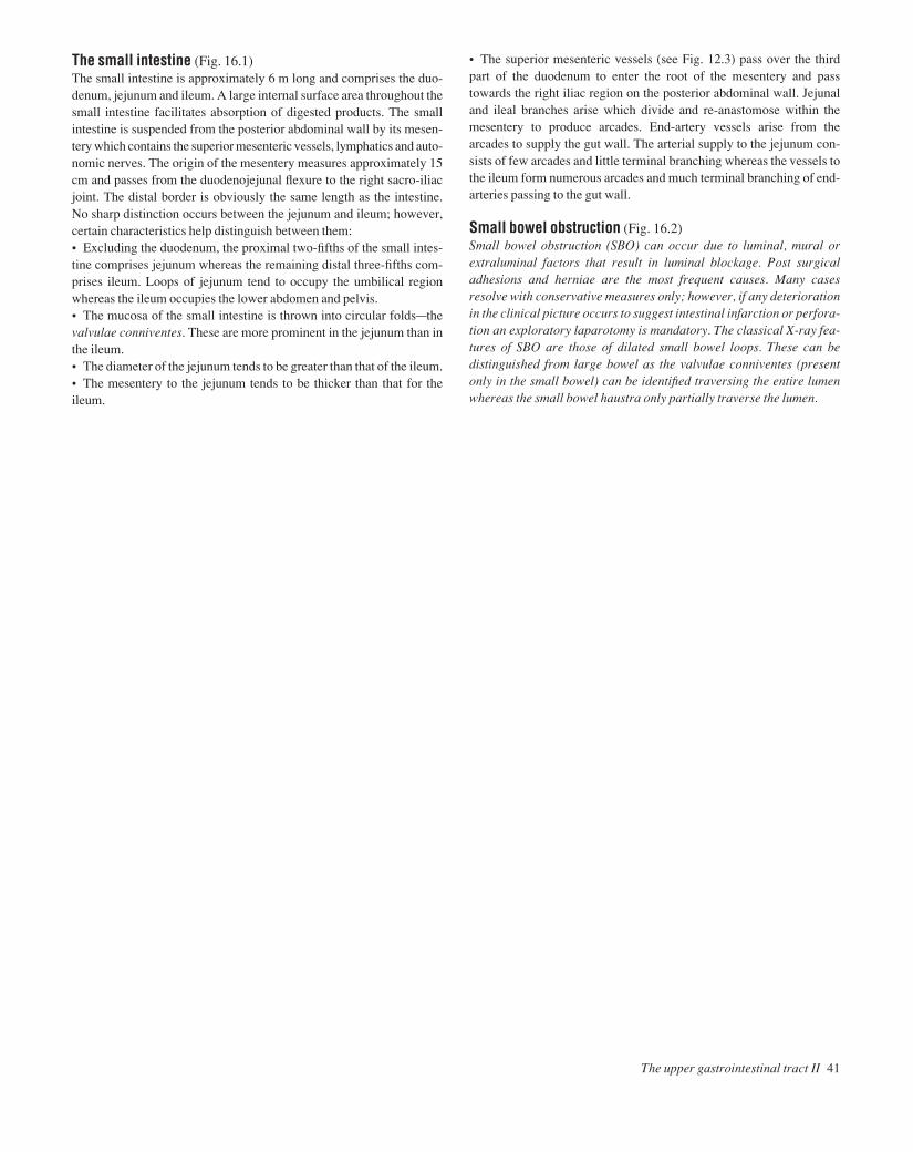
The small intestine (Fig. 16.1)The small intestine is approximately 6 m long and comprises the duo-denum, jejunum and ileum. A large internal surface area throughout thesmall intestine facilitates absorption of digested products. The smallintestine is suspended from the posterior abdominal wall by its mesen-tery which contains the superior mesenteric vessels, lymphatics and auto-nomic nerves. The origin of the mesentery measures approximately 15cm and passes from the duodenojejunal flexure to the right sacro-iliacjoint. The distal border is obviously the same length as the intestine. No sharp distinction occurs between the jejunum and ileum; however,certain characteristics help distinguish between them:• Excluding the duodenum, the proximal two-fifths of the small intes-tine comprises jejunum whereas the remaining distal three-fifths com-prises ileum. Loops of jejunum tend to occupy the umbilical regionwhereas the ileum occupies the lower abdomen and pelvis.• The mucosa of the small intestine is thrown into circular foldsathevalvulae conniventes. These are more prominent in the jejunum than inthe ileum.• The diameter of the jejunum tends to be greater than that of the ileum.• The mesentery to the jejunum tends to be thicker than that for theileum.
• The superior mesenteric vessels (see Fig. 12.3) pass over the thirdpart of the duodenum to enter the root of the mesentery and passtowards the right iliac region on the posterior abdominal wall. Jejunaland ileal branches arise which divide and re-anastomose within themesentery to produce arcades. End-artery vessels arise from thearcades to supply the gut wall. The arterial supply to the jejunum con-sists of few arcades and little terminal branching whereas the vessels tothe ileum form numerous arcades and much terminal branching of end-arteries passing to the gut wall.
Small bowel obstruction (Fig. 16.2)Small bowel obstruction (SBO) can occur due to luminal, mural orextraluminal factors that result in luminal blockage. Post surgicaladhesions and herniae are the most frequent causes. Many casesresolve with conservative measures only; however, if any deteriorationin the clinical picture occurs to suggest intestinal infarction or perfora-tion an exploratory laparotomy is mandatory. The classical X-ray fea-tures of SBO are those of dilated small bowel loops. These can bedistinguished from large bowel as the valvulae conniventes (presentonly in the small bowel) can be identified traversing the entire lumenwhereas the small bowel haustra only partially traverse the lumen.
The upper gastrointestinal tract II 41
AAAC16 21/5/05 10:41 AM Page 41

42 Abdomen and pelvis
17 The lower gastrointestinal tract
Teniae coli
Fig.17.2A coronal section through the pelvis to show theanal sphincters and the ischiorectal fossa
Fig.17.1The various positions in which the appendix may be found.In the pelvic position the appendix may be close to the ovary in the female
Appendicesepiploicae
Retrocaecal
Pelvic
Subcaecal
Ovary in female
RectumLongitudinal muscle
Submucosa
Sphincter ani internus
Pre-ileal
Retro-ileal
Retrocolic
Circular muscle
Levator ani
Obturator internus
Fat of ischiorectal fossa
Sphincteraniexternus
DeepSuperficialSubcutaneous
Pudendal canal
Adductor muscles
Inferior rectalvesels and nerve
AAAC17 21/5/05 10:40 AM Page 42

The caecum and colon (Figs 17.1, 12.3, 12.5)In adults, the large bowel measures approximately 1.5 m. The caecum,ascending, transverse, descending and sigmoid colon have similarcharacteristic features. These are that they possess:• Appendices epiploicae (Fig. 17.1): these are fat-laden peritoneal tagspresent over the surface of the caecum and colon.• Teniae coli (Fig. 17.1): these are three flattened bands representingthe condensed longitudinal muscular coat of the large intestine. Theycourse from the base of the appendix (and form a useful way of locatingthis structure at operation) to the recto-sigmoid junction.• Sacculations: because the teniae are shorter than the bowel itself thecolon takes on a sacculated appearance. These sacculations are visiblenot only at operation but also radiographically. On a plain abdominal X-ray, the colon, which appears radiotranslucent because of the gas within,has shelf-like processes (haustra) which partially project into the lumen.
The transverse and sigmoid colon are each attached to the posteriorabdominal wall by their respective mesocolons and are covered en-tirely by peritoneum. Conversely, the ascending and descending colonnormally possess no mesocolon. They are adherent to the posteriorabdominal wall and covered only anteriorly by peritoneum.
The appendix (Fig. 17.1)The appendix varies enormously in length but in adults it is approxim-ately 5–15 cm long. The base of the appendix arises from the postero-medial aspect of the caecum; however, the lie of the appendix itself ishighly variable. In most cases the appendix lies in the retrocaecal posi-tion but other positions frequently occur. The appendix has the follow-ing characteristic features:• It has a small mesentery which descends behind the terminal ileum.The only blood supply to the appendix, the appendicular artery (abranch of the ileocolic), courses within its mesentery (see Fig. 12.4). Incases of appendicitis the appendicular artery ultimately thromboses.When this occurs, gangrene and perforation of the appendix inevitablysupervene.• The appendix has a lumen which is relatively wide in infants and gra-dually narrows throughout life, often becoming obliterated in the elderly.• The teniae coli of the caecum lead to the base of the appendix.• The bloodless fold of Treves (ileocaecal fold) is the name given to asmall peritoneal reflection passing from the anterior terminal ileum tothe appendix. Despite its name it is not an avascular structure!
Appendicectomy is performed most commonly through a grid-ironmuscle-splitting incision. The appendix is first located and then deliv-ered into the wound. The mesentery of the appendix is then divided andligated. The appendix is then tied at its base, excised and removed.Most surgeons still opt to invaginate the appendix stump as a precau-tionary measure against slippage of the stump ligature.
The rectum (Figs 17.2, 12.5)• The rectum measures 10–15 cm in length. It commences in front ofthe 3rd sacral vertebra as a continuation of the sigmoid colon and fol-lows the curve of the sacrum anteriorly. It turns backwards abruptly infront of the coccyx to become the anal canal.• The mucosa of the rectum is thrown into three horizontal folds thatproject into the lumenathe valves of Houston.• The rectum lacks haustrations. The teniae coli fan out over the rec-tum to form anterior and posterior bands.• The rectum is slightly dilated at its lower endathe ampulla, and issupported laterally by the levator ani.• Peritoneum covers the upper two-thirds of the rectum anteriorly butonly the upper third laterally. In the female it is reflected forwards ontothe uterus forming the recto-uterine pouch (pouch of Douglas). Therectum is separated from anterior structures by a tough fascial sheetathe rectovesical (Denonvilliers) fascia.
The anal canal (Fig. 17.2)The anorectal junction is slung by the puborectalis component of lev-ator ani which pulls it forwards. The canal is approximately 4 cm longand angled postero-inferiorly. Developmentally the midpoint of theanal canal is represented by the dentate line. This is the site where theproctodeum (ectoderm) meets endoderm. This developmental implica-tion is reflected by the following characteristics of the anal canal:• The epithelium of the upper half of the anal canal is columnar. In con-trast the epithelium of the lower half of the anal canal is squamous. Themucosa of the upper canal is thrown into vertical columns (of Mor-gagni). At the bases of the columns are valve-like folds (valves of Ball).The level of the valves is termed the dentate line.• The blood supply to the upper anal canal (see Fig. 12.5) is from thesuperior rectal artery (derived from the inferior mesenteric artery)whereas the lower anal canal is supplied by the inferior rectal artery(derived from the internal iliac artery). As mentioned previously, thevenous drainage follows suit and represents a site of porto-systemicanastomosis (see p. 35).• The upper anal canal is insensitive to pain as it is supplied by auto-nomic nerves only. The lower anal canal is sensitive to pain as it is sup-plied by somatic innervation (inferior rectal nerve).• The lymphatics from the upper canal drain upwards along the super-ior rectal vessels to the internal iliac nodes whereas lymph from thelower anal canal drains to the inguinal nodes.
The anal sphincterSee Chapter 25.
The lower gastrointestinal tract 43
AAAC17 21/5/05 10:40 AM Page 43

44 Abdomen and pelvis
18 The liver, gall-bladder and biliary tree
Fig.18.1The venous circulation through the liver.The transmission of blood from the portal system to the inferior vena cavais via the liver lobules (fig. 18.2)
Fig.18.2(a) A liver lobule to show the direction of blood flow from the portal system to the centrilobular veins and thence to the inferior vena cava(b) The blood flow through the sinusoids of the liver lobule and the passage of bile from the bilecanaliculi to the bile ducts
Opening in central tendonof diaphragm
Portal vein
Liver
Spleen
Splenic vein
Bile canaliculiBile ductBranch of hepatic arteryBranch of portal vein
Periportalconnectivetissue
Direction of bile flow
Bile duct
Inferior mesenteric vein
Superior mesenteric vein
(a) (b)
Hepatic vein
Direction of blood flowArtery
Sinusoids
Vein
Central vein
Central vein
AAAC18 21/5/05 10:40 AM Page 44
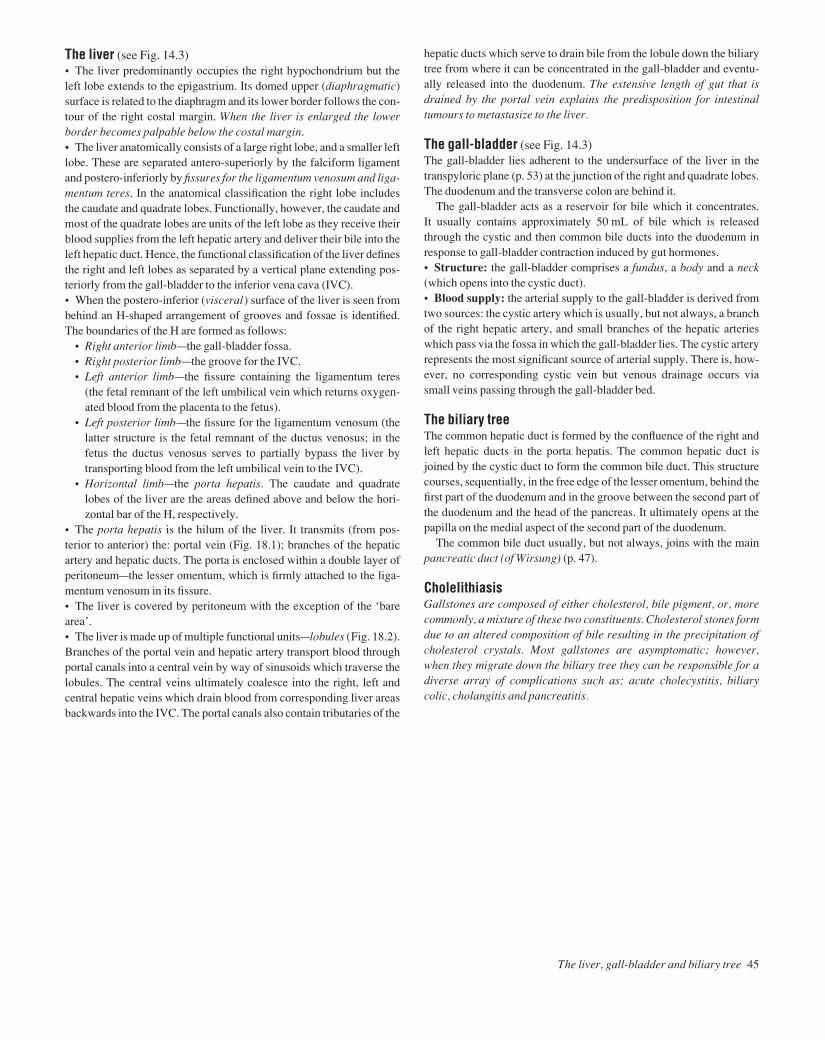
The liver (see Fig. 14.3)• The liver predominantly occupies the right hypochondrium but theleft lobe extends to the epigastrium. Its domed upper (diaphragmatic)surface is related to the diaphragm and its lower border follows the con-tour of the right costal margin. When the liver is enlarged the lowerborder becomes palpable below the costal margin.• The liver anatomically consists of a large right lobe, and a smaller leftlobe. These are separated antero-superiorly by the falciform ligamentand postero-inferiorly by fissures for the ligamentum venosum and liga-mentum teres. In the anatomical classification the right lobe includesthe caudate and quadrate lobes. Functionally, however, the caudate andmost of the quadrate lobes are units of the left lobe as they receive theirblood supplies from the left hepatic artery and deliver their bile into theleft hepatic duct. Hence, the functional classification of the liver definesthe right and left lobes as separated by a vertical plane extending pos-teriorly from the gall-bladder to the inferior vena cava (IVC).• When the postero-inferior (visceral) surface of the liver is seen frombehind an H-shaped arrangement of grooves and fossae is identified.The boundaries of the H are formed as follows:
• Right anterior limbathe gall-bladder fossa.• Right posterior limbathe groove for the IVC.• Left anterior limbathe fissure containing the ligamentum teres
(the fetal remnant of the left umbilical vein which returns oxygen-ated blood from the placenta to the fetus).
• Left posterior limbathe fissure for the ligamentum venosum (thelatter structure is the fetal remnant of the ductus venosus; in thefetus the ductus venosus serves to partially bypass the liver bytransporting blood from the left umbilical vein to the IVC).
• Horizontal limbathe porta hepatis. The caudate and quadratelobes of the liver are the areas defined above and below the hori-zontal bar of the H, respectively.
• The porta hepatis is the hilum of the liver. It transmits (from pos-terior to anterior) the: portal vein (Fig. 18.1); branches of the hepaticartery and hepatic ducts. The porta is enclosed within a double layer ofperitoneumathe lesser omentum, which is firmly attached to the liga-mentum venosum in its fissure.• The liver is covered by peritoneum with the exception of the ‘barearea’.• The liver is made up of multiple functional unitsalobules (Fig. 18.2).Branches of the portal vein and hepatic artery transport blood throughportal canals into a central vein by way of sinusoids which traverse thelobules. The central veins ultimately coalesce into the right, left andcentral hepatic veins which drain blood from corresponding liver areasbackwards into the IVC. The portal canals also contain tributaries of the
hepatic ducts which serve to drain bile from the lobule down the biliarytree from where it can be concentrated in the gall-bladder and eventu-ally released into the duodenum. The extensive length of gut that isdrained by the portal vein explains the predisposition for intestinaltumours to metastasize to the liver.
The gall-bladder (see Fig. 14.3)The gall-bladder lies adherent to the undersurface of the liver in thetranspyloric plane (p. 53) at the junction of the right and quadrate lobes.The duodenum and the transverse colon are behind it.
The gall-bladder acts as a reservoir for bile which it concentrates. It usually contains approximately 50 mL of bile which is releasedthrough the cystic and then common bile ducts into the duodenum inresponse to gall-bladder contraction induced by gut hormones.• Structure: the gall-bladder comprises a fundus, a body and a neck(which opens into the cystic duct).• Blood supply: the arterial supply to the gall-bladder is derived fromtwo sources: the cystic artery which is usually, but not always, a branchof the right hepatic artery, and small branches of the hepatic arterieswhich pass via the fossa in which the gall-bladder lies. The cystic arteryrepresents the most significant source of arterial supply. There is, how-ever, no corresponding cystic vein but venous drainage occurs viasmall veins passing through the gall-bladder bed.
The biliary treeThe common hepatic duct is formed by the confluence of the right andleft hepatic ducts in the porta hepatis. The common hepatic duct isjoined by the cystic duct to form the common bile duct. This structurecourses, sequentially, in the free edge of the lesser omentum, behind thefirst part of the duodenum and in the groove between the second part ofthe duodenum and the head of the pancreas. It ultimately opens at thepapilla on the medial aspect of the second part of the duodenum.
The common bile duct usually, but not always, joins with the mainpancreatic duct (of Wirsung) (p. 47).
CholelithiasisGallstones are composed of either cholesterol, bile pigment, or, morecommonly, a mixture of these two constituents. Cholesterol stones formdue to an altered composition of bile resulting in the precipitation ofcholesterol crystals. Most gallstones are asymptomatic; however,when they migrate down the biliary tree they can be responsible for adiverse array of complications such as: acute cholecystitis, biliarycolic, cholangitis and pancreatitis.
The liver, gall-bladder and biliary tree 45
AAAC18 21/5/05 10:40 AM Page 45

46 Abdomen and pelvis
19 The pancreas and spleen
Fig.19.1The relations of the pancreas
Fig.19.2The ducts of the pancreas and the biliary system
Fig.19.3The relations of the spleen
Inferior vena cava
Right and left hepatic ducts
Right gastroepiploic artery
Superiorpancreaticoduodenal artery
Inferiorpancreaticoduodenal artery
Superior mesentericartery and vein
Portal veinCommon bile ductHepatic arteryRight gastric arteryGastroduodenal artery
Left gastric arteryCoeliac artery
Splenic artery
Inferior mesentericartery and vein
Cystic ductRight hepatic artery
Pancreatic duct(of Wirsung)
Tail of pancreasBodyNeck
Superior mesenteric vessels
Common bile ductNeckBody
Gall bladder(displaced)
Uncinate process
Fundus
Duodenal papilla(of Vater)
Accessory duct(of Santorini)
Spleen
Diaphragm
Ribs 9,10 and 11Splenic
artery
Tail ofpacreas
Left kidneyand suprarenal
Splenic flexure of colon
Cystic artery
AAAC19 21/5/05 10:39 AM Page 46

The pancreas (Figs 19.1 and 19.2)The pancreas has a: head, neck, body and tail. It is a retroperitonealorgan which lies roughly along the transpyloric plane. The head isbound laterally by the curved duodenum and the tail extends to thehilum of the spleen in the lienorenal ligament. The superior mesentericvessels pass behind the pancreas, then anteriorly, over the uncinateprocess and third part of the duodenum into the root of the small bowelmesentery. The inferior vena cava, aorta, coeliac plexus, left kidney(and its vessels) and the left adrenal gland are posterior pancreatic rela-tions. In addition, the portal vein is formed behind the pancreatic neckby the confluence of the splenic and superior mesenteric veins. Thelesser sac and stomach are anterior pancreatic relations.• Structure: the main pancreatic duct (of Wirsung) courses the lengthof the gland, ultimately draining pancreatic secretions into the ampullaof Vater, together with the common bile duct, and thence into the sec-ond part of the duodenum. An accessory duct (of Santorini) drains theuncinate process of the pancreas, opening slightly proximal to theampulla into the second part of the duodenum.• Blood supply: the pancreatic head receives its supply from the superior and inferior pancreaticoduodenal arteries. The splenic arterycourses along the upper border of the body of the pancreas which it sup-plies by means of a large branchathe arteria pancreatica magnaaandnumerous smaller branches.• Function: the pancreas is a lobulated structure which performs bothexocrine and endocrine functions. The exocrine secretory glands drainpancreatic juice into the pancreatic ducts and, from there, ultimatelyinto the duodenum. The secretion is essential for the digestion andabsorption of proteins, fats and carbohydrates. The endocrine pancreasis responsible for the production and secretion of glucagon and insulin,which take place in specialized cells of the islets of Langerhans.
Acute pancreatitisThe presence of gallstones and a history of excessive alcohol intake arethe predominant associations for pancreatitis. The mechanism bywhich these aetiological factors result in pancreatic injury is unknown;however, they both appear to result in activation of pancreatic exocrinepro-enzymes with resultant autodigestion. Even today, the mortalityrate for severe acute pancreatitis remains in the region of 20%.
The spleen (Fig. 19.3)The spleen is approximately the size of a clenched fist and lies directlybelow the left hemidiaphragm which, in addition to the pleura, separ-ates it from the overlying 9th, 10th and 11th ribs.• Peritoneal attachments: the splenic capsule is fibrous with peri-toneum adherent to its surface. The gastrosplenic and lienorenal liga-ments attach it to the stomach and kidney, respectively. The formerligament carries the short gastric and left gastroepiploic vessels to thefundus and greater curvature of the stomach, and the latter ligamentcarries the splenic vessels and tail of the pancreas towards the left kidney.• Blood supply: is from the splenic artery to the hilum of the spleen.Venous drainage is to the splenic vein, thence to the portal vein.• Structure: the spleen is a highly vascular reticulo-endothelial organ.It consists of a thin capsule from which trabeculae extend into thesplenic pulp. In the spleen, the immunological centres, i.e. the lym-phoid follicles (the white pulp), are scattered throughout richly vascu-larized sinusoids (the red pulp).
SplenectomyAs the spleen is a highly vascular organ, any injury to it can be life-threatening. Under these circumstances splenectomy must be carriedout urgently. The technique used differs slightly when the procedure isperformed for emergency as opposed to elective indications, but theprinciples are similar. Splenectomy involves: ligature of the splenicvessels approaching the hilum (taking care not to injure the tail of thepancreas or colon); and dissection of the splenic pediclesbthe gastro-splenic (including the short gastric vessels) and lienorenal ligaments.As the spleen is an important immunological organ, postsplenectomypatients are rendered immunocompromised to capsulated bacteria.The latter organisms (e.g. meningococcus, pneumococcus) requireopsonization for elimination and splenic lymphoid follicles are theprincipal sites where this takes place. Hence, following splenectomy,all patients are routinely vaccinated against the capsulated bacteria,and children, who are the group most at risk of sepsis, are maintainedon long-term antibiotic prophylaxis.
The pancreas and spleen 47
AAAC19 21/5/05 10:39 AM Page 47
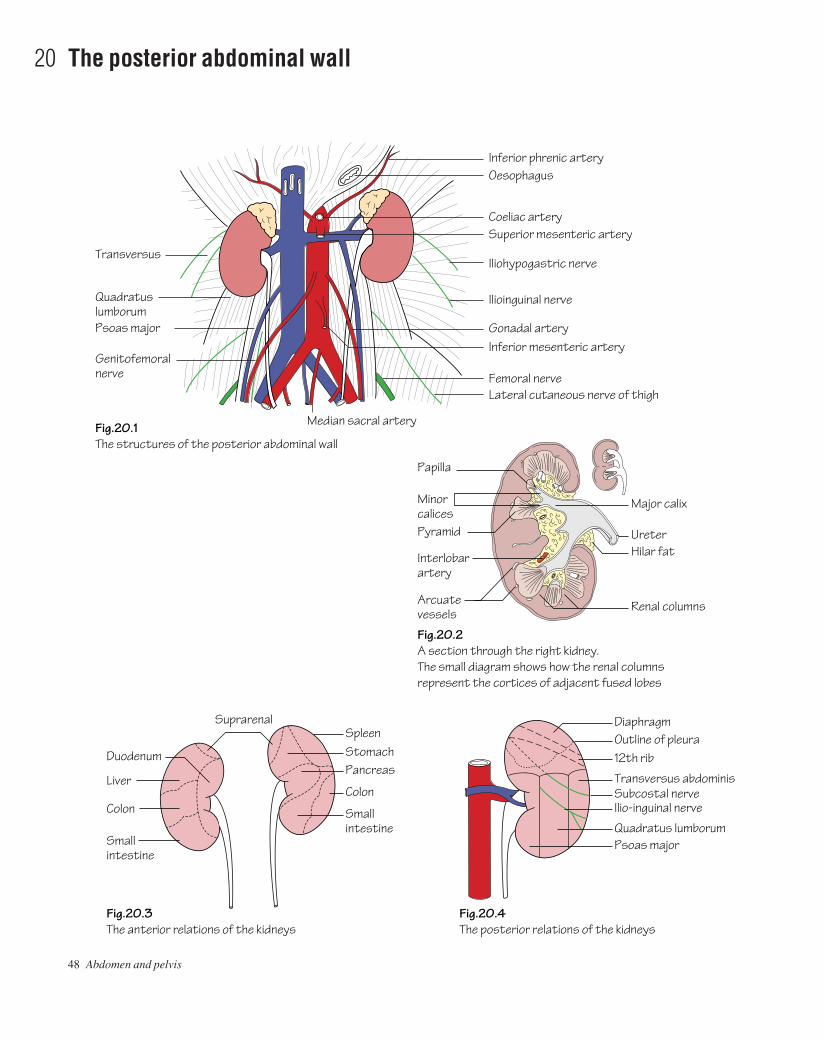
48 Abdomen and pelvis
20 The posterior abdominal wall
Fig.20.1The structures of the posterior abdominal wall
Fig.20.2A section through the right kidney.The small diagram shows how the renal columns represent the cortices of adjacent fused lobes
Fig.20.3The anterior relations of the kidneys
Fig.20.4The posterior relations of the kidneys
Inferior phrenic arteryOesophagus
Coeliac arterySuperior mesenteric artery
Iliohypogastric nerve
Ilioinguinal nerve
Inferior mesenteric artery
Femoral nerveLateral cutaneous nerve of thigh
QuadratuslumborumPsoas major
Genitofemoralnerve
Median sacral artery
Major calix
Gonadal artery
Outline of pleura12th rib
Diaphragm
UreterHilar fat
Renal columnsArcuatevessels
Interlobarartery
Pyramid
Minorcalices
Papilla
SpleenSuprarenal
StomachPancreas
ColonLiver
Colon
Smallintestine
Duodenum
Smallintestine
Transversus abdominis
Ilio-inguinal nerve
Quadratus lumborumPsoas major
Subcostal nerve
Transversus
AAAC20 21/5/05 10:39 AM Page 48

The structures of the posterior abdominal wall (Fig. 20.1)These include:• Muscles: including psoas major and quadratus lumborum (seeMuscle index, p. 162).• The abdominal aorta and its branches: see p. 32.• The inferior vena cava (IVC) and its tributaries: see p. 35.• The kidneys.• The ureters.• The adrenal (suprarenal) glands.• The lumbar sympathetic trunks and plexuses and the lumbar plexus(see p. 51).
The kidneys (Fig. 20.2)• Structure: the kidney has its own fibrous capsule and is surroundedby perinephric fat which, in turn, is enclosed by renal fascia. Each kid-ney is approximately 10–12 cm long and consists of an outer cortex, aninner medulla and a pelvis.
The hilum of the kidney is situated medially and transmits from frontto back the: renal vein, renal artery, ureteric pelvis as well as lymphaticsand sympathetic vasomotor nerves.
The renal pelvis divides into two or three major calices and these, inturn, divide into minor calices which receive urine from the medullarypyramids by way of the papillae.• Position: the kidneys lie in the retroperitoneum against the posteriorabdominal wall. The right kidney lies approximately 1 cm lower thanthe left.• Relations: See Figs 20.3 and 20.4.• Blood supply: the renal arteries arise from the aorta at the level ofL2. Together, the renal arteries direct 25% of the cardiac outputtowards the kidneys. Each renal artery divides into five segmentalarteries at the hilum which, in turn, divide sequentially into lobar,interlobar, arcuate and cortical radial branches. The cortical radialbranches give rise to the afferent arterioles which supply the glomeruliand go on to become efferent arterioles. The differential pressuresbetween afferent and efferent arterioles lead to the production of anultrafiltrate which then passes through, and is modified by, the nephronto produce urine.
The right renal artery passes behind the IVC. The left renal vein islong as it courses in front of the aorta to drain into the IVC.• Lymphatic drainage: to the para-aortic lymph nodes.
The ureter (Fig. 20.1)The ureter is considered in abdominal, pelvic and intravesical portions.• Structure: the ureter is approximately 20–30 cm long and coursesfrom the hilum of the kidney to the bladder. It has a muscular wall andis lined by transitional epithelium. At operation it can be recognized byits peristalsis.
• Course: from the renal pelvis at the hilum the course of the ureter canbe summarized as follows:
• It passes along the medial part of psoas major behind, but adherentto, the peritoneum.
• It then crosses the common iliac bifurcation anterior to the sacro-iliac joint and courses over the lateral wall of the pelvis to theischial spine.
• At the ischial spine the ureter passes forwards and medially to enterthe bladder obliquely. The intravesical portion of the ureter isapproximately 2 cm long and its passage through the bladder wallproduces a sphincter-like effect. In the male the ureter is crossedsuperficially near its termination by the vas deferens. In the femalethe ureter passes above the lateral fornix of the vagina but belowthe broad ligament and uterine vessels.
• Blood supply: as the ureter is an abdominal and pelvic structure itreceives a blood supply from multiple sources:
• The upper ureterareceives direct branches from the aorta, renaland gonadal arteries.
• The lower ureterareceives branches of the internal iliac and in-ferior vesical arteries.
Ureteric stonesMost ureteric calculi arise for unknown reasons, although inadequateurinary drainage, the presence of infected urine, and hypercalcaemiaare definite predisposing factors. The presence of an impacted uretericstone is characterized by haematuria and agonizing colicky pain(ureteric colic), which classically radiates from loin to groin. Largeimpacted stones can lead to hydronephrosis and/or infection of theaffected kidney and consequently need to be broken up or removed byinterventional or open procedures.
Adrenal (suprarenal) glands (Fig. 20.1)The adrenal glands comprise an outer cortex and inner medulla. Thecortex is derived from mesoderm and is responsible for the productionof steroid hormones (glucocorticoids, mineralocorticoids and sexsteroids). The medulla is derived from ectoderm (neural crest) and actsas a part of the autonomic nervous system. It receives sympathetic preganglionic fibres from the greater splanchnic nerves which stimulate the medulla to secrete noradrenaline and adrenaline into thebloodstream.• Position: the adrenals are small glands which lie in the renal fasciaon the upper poles of the kidneys. The right gland lies behind the rightlobe of the liver and immediately posterolateral to the IVC. The leftadrenal is anteriorly related to the lesser sac and stomach.• Blood supply: the phrenic, renal arteries and aorta all contributebranches to the adrenal glands. Venous drainage is on the right to theIVC and on the left to the left renal vein.
The posterior abdominal wall 49
AAAC20 21/5/05 10:39 AM Page 49

50 Abdomen and pelvis
21 The nerves of the abdomen
Fig.21.1The lumbar plexus
Fig.21.2The sympathetic system in the abdomen and pelvis
Nerves
T12 (subcostal)
L1
L2
L3
L4
L5Lateralcutaneousof thigh
Genitofemoral
Femoral
Obturator
Lumbosacral trunk
Ilioinguinal
Iliohypogastric
Subcostal
Coeliac ganglia
Suprarenal branch
Cut psoas majorSympathetic trunkLumbar sympathetic ganglia(usually 4)
Superior hypogastric plexus(presacral nerves)
Pelvic sympathetic gangliaGrey rami
Grey rami
Grey and white rami(white rami on firsttwo lumbar nerves only)
Medial arcuateligament
Inferior hypogastric plexus
AAAC21 21/5/05 10:47 AM Page 50

The lumbar plexus (Fig. 21.1)• The lumbar plexus is formed from the anterior primary rami of L1–4.The trunks of the plexus lie within the substance of psoas major and,with the exceptions of the obturator and genitofemoral nerves, emergeat its lateral border.• The 12th intercostal nerve is also termed the subcostal nerve as it hasno intercostal space but, instead, runs below the rib in the neurovascu-lar plane to supply the abdominal wall.• The iliohypogastric nerve is the main trunk of the 1st lumbar nerve. Itsupplies the skin of the upper buttock, by way of a lateral cutaneousbranch, and terminates by piercing the external oblique above thesuperficial inguinal ring where it supplies the overlying skin of themons pubis. The ilioinguinal nerve is the collateral branch of the iliohy-pogastric. The ilioinguinal runs in the neurovascular plane of theabdominal wall to emerge through the superficial inguinal ring to pro-vide a cutaneous supply to the skin of the medial thigh, the root of thepenis and anterior one third of the scrotum (or labium majus in thefemale).• The genitofemoral nerve (L1,2) emerges from the anterior surface ofpsoas major. It courses inferiorly and divides into: a genital componentthat enters the spermatic cord and supplies the cremaster (in the male),and a femoral component that supplies the skin of the thigh overlyingthe femoral triangle.• The lateral cutaneous nerve of the thigh (L2,3), having emergedfrom the lateral border of psoas major, encircles the iliac fossa to passunder the inguinal ligament (p. 99).• The femoral nerve (L2–4, posterior division): see p. 99.• The obturator nerve (L2–4, anterior division): see p. 99.• A large part of L4 joins with L5 to contribute to the sacral plexus asthe lumbosacral trunk.
Lumbar sympathetic chain (Fig. 21.2)• Sympathetic supply: the lumbar sympathetic chain is a continuationof the thoracic sympathetic chain as it passes under the medial arcuateligament of the diaphragm. The chain passes anterior to the lumbar ver-tebral bodies and usually carries four ganglia which send grey ramicommunicans to the lumbar spinal nerves. The upper two gangliareceive white rami from L1 and L2.
The lumbar sympathetic chain, the splanchnic nerves and the vaguscontribute sympathetic and parasympathetic branches to plexuses(coeliac, superior mesenteric, renal and inferior mesenteric) around theabdominal aorta. In addition, other branches continue inferiorly to formthe superior hypogastric plexus (presacral nerves) from where theybranch into right and left inferior hypogastric plexuses. The latter alsoreceive a parasympathetic supply from the pelvic splanchnic nerves. Thebranches from the inferior hypogastric plexuses are distributed to thepelvic viscera along the course and branches of the internal iliac artery.
The coeliac ganglia are prominent and lie around the origins of thecoeliac and superior mesenteric arteries.• Parasympathetic supply: to the pelvic viscera arises from the anter-ior primary rami of S2,3,4athe pelvic splanchnic nerves. The latterparasympathetic supply reaches proximally as far as the junctionbetween the hindgut and midgut on the transverse colon.
Lumbar sympathectomyThis procedure is performed in cases of severe peripheral vascular dis-ease of the lower limbs where vascular reconstructive surgery is notpossible and skin necrosis is imminent. The operation involves excisionof the 2nd to 4th lumbar ganglia with the intermediate chain.
The nerves of the abdomen 51
AAAC21 21/5/05 10:47 AM Page 51
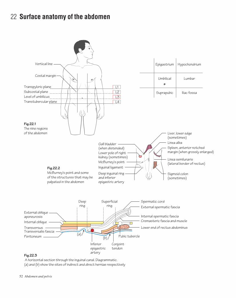
52 Abdomen and pelvis
22 Surface anatomy of the abdomen
L1L2L3L4
Transpyloric plane
Costal margin
Vertical line
Subcostal planeLevel of umbilicusTranstubercular plane
(a)(b) Pubic tubercle
Conjointtendon
Inferiorepigastricartery
Deepring
Superficialring
External obliqueaponeurosis
Spermatic cordExternal spermatic fascia
Internal spermatic fascia
Internal oblique
Transversus
Fig.22.1The nine regions of the abdomen
Fig.22.2McBurney's point and someof the structures that may bepalpated in the abdomen
Fig.22.3 A horizontal section through the inguinal canal. Diagrammatic.(a) and (b) show the sites of indirect and direct herniae respectively
Cremasteric fascia and muscle
Lower end of rectus abdominusTransversalis fasciaPeritoneum
Linea alba
Epigastrium
Umbilical
Suprapubic
Hypochondrium
Lumbar
Iliac fossa
Liver, lower edge(sometimes)
Sigmoid colon(sometimes)
Linea semilunaris(lateral border of rectus)
Spleen, anterior notchedmargin (when grossly enlarged)
Gall bladder(when distended)Lower pole of right kidney (sometimes)McBurney's pointInguinal ligament
Deep inguinal ringand inferiorepigastric artery
AAAC22 21/5/05 10:47 AM Page 52

Vertebral levels (Fig. 22.1)(In each case the lower border is referred to.)• T9: xiphoid process.• L1: the transpyloric plane (of Addison). This horizontal plane passesapproximately through the tip of the 9th costal cartilage, the pylorus,pancreatic neck, duodenojejunal flexure, the gall-bladder fundus andthe hila of the kidneys. This plane also corresponds to the level at whichthe spinal cord terminates and the lateral edge of rectus abdominiscrosses the costal margin.• L2: the subcostal plane. This plane corresponds to a line joining thelowest points of the thoracic cageathe lower margin of the 10th rib laterally.• L3: the level of the umbilicus (in a young slim person).• L4: the transtubercular plane. This corresponds to a line which joinsthe tubercles of the iliac crests.
Lines of orientationVertical lines: these are imaginary and most often used with the sub-costal and intertubercular planes, for purposes of description, to subdi-vide the abdomen into nine regions (Fig. 22.1). They pass vertically, oneither side, through the point halfway between the anterior superioriliac spine and the pubic tubercle. More commonly used, for descrip-tion of pain location, are quadrants. The latter are imaginary lines aris-ing by the bisection of the umbilicus by vertical and horizontal lines.
Surface markings of the abdominal wall• The costal margin (Fig. 22.1) is the inferior margin of the thoraciccage. It includes the costal cartilages anteriorly, the 7th–10th ribs later-ally and the cartilages of the 11th and 12th ribs posteriorly.• The symphysis pubis is an easily palpable secondary cartilaginousjoint which lies between the pubic bones in the midline. The pubictubercle is an important landmark and is identifiable on the superiorsurface of the pubis.• The inguinal ligament (Figs 11.1 and 22.2) is attached laterally to theanterior superior iliac spine and medially to the pubic tubercle.• The superficial inguinal ring (see Fig. 11.1) is a triangular-shapeddefect in the external oblique aponeurosis. It is situated above andmedial to the pubic tubercle.• The spermatic cord can be felt passing medial to the pubic tubercleand descending into the scrotum.• The deep inguinal ring (Fig. 22.3) lies halfway along a line from theanterior superior iliac spine to the pubic tubercle.• The linea alba (see Fig. 11.1) is formed by the fusion of the aponeu-roses of the muscles of the anterior abdominal wall. It extends as a de-pression in the midline from the xiphoid process to the symphysis pubis.
• The linea semilunaris is the lateral edge of the rectus abdominis muscle. It crosses the costal margin at the tip of the 9th costal cartilage.
Inguinal herniae (Figs 22.3 and 52.1)• Indirect inguinal herniae: arise as a result of persistence of the pro-cessus vaginalis of the embryo. Abdominal contents bulge through thedeep inguinal ring, into the canal, and eventually into the scrotum. Thishernia can be controlled by digital pressure over the deep ring.• Direct inguinal herniae: arise as a result of weakness in the poster-ior wall of the inguinal canal. This hernia cannot be controlled by digital pressure over the deep ring and only rarely does the hernia passinto the scrotum.
The clinical distinction between direct and indirect inguinal herniascan be difficult. At operation, however, the relation of the hernial neckto the inferior epigastric artery defines the hernia type, i.e. the neck ofthe sac of an indirect hernia lies lateral to the artery whereas that of adirect type always lies medial to it.
Surface markings of the abdominal viscera (Fig. 22.2)• Liver: the lower border of the liver is usually just palpable on deepinspiration in slim individuals. The upper border follows the undersur-face of the diaphragm and reaches a level just below the nipple on eachside.• Spleen: this organ lies below the left hemidiaphragm deep to the 9th,10th and 11th ribs posteriorly. The anterior notch reaches the mid-axillary line anteriorly.• Gall-bladder: the fundus of the gall-bladder lies in the transpyloricplane (L1). The surface marking corresponds to a point where the lat-eral border of rectus abdominis (linea semilunaris) crosses the costalmargin.• Pancreas: the pancreatic neck lies on the level of the transpyloricplane (L1). The pancreatic head lies to the right and below the neckwhereas the body and tail pass upwards and to the left.• Aorta: the aorta bifurcates to the left of the midline at the level of L4.• Kidneys: the kidney hila lie on the level of the transpyloric plane(L1). The lower pole of the right kidney usually extends 3 cm below thelevel of the left and is often palpable in slim subjects.• Appendix: McBurney’s point represents the surface marking for thebase of the appendix. This point lies one third of the way along a linejoining the anterior superior iliac spine and the umbilicus. McBurney’spoint is important surgically as it represents the usual site of maximaltenderness in appendicitis and also serves as the central point for theincision made when performing an appendicectomy.• Bladder: in adults the bladder is a pelvic organ and can be palpatedabove the symphysis pubis only when full or enlarged.
Surface anatomy of the abdomen 53
AAAC22 21/5/05 10:47 AM Page 53

54 Abdomen and pelvis
23 The pelvis Icthe bony and ligamentous pelvis
Iliac crest
Anterior gluteal line
Posterior gluteal line
Greater sciatic notch
Lesser sciatic notch
Iliac fossa
Auricularsurface
Iliopectinealline
Pubicsymphysis
Pubictubercle
Spine of ischium
Ischial tuberosity
Ramus of ischiumInferior ramus
Body of pubis
Pubic crest
Acetabulum
Anterior inferioriliac spine
Anterior superioriliac spine
Inferior gluteal line
Pubic tubercleObturator foramen
Transverse tubercleAuricular surface
Posterior sacral foramen
Posterior superioriliac spine
Fig.23.1The lateral surface of the left hip bone
Fig.23.3The posterior surface of the sacrum
Fig.23.4The sacrospinous and sacrotuberous ligaments resistrotation of the sacrum due to the body weight
Fig.23.5The male pelvic floor from above.The blue line represents the originof levator ani from the obturatorfascia
Fig.23.2The medial surface of the left hip bone
Sacral hiatus
Body weight
Greater sciatic foramen
Articular tubercleAla
Superior articular facet
Spinous tubercle
Lesser sciatic foramenSacrospinous ligament
False pelvis
Pelvic brimTrue pelvis
Sacrotuberous ligament
Perineal bodyProstate Obturator fascia
Obturator internusLevator prostatae
PiriformisSacral nerves
Spine of ischium
Puborectalis
Anterior edgeof levator ani
Coccygeus
Recto-anal junctionIschiococcygeus
AAAC23 21/5/05 10:46 AM Page 54

The pelvis is bounded posteriorly by the sacrum and coccyx and antero-laterally by the innominate bones.
Os innominatum (hip bone) (Figs 23.1 and 23.2)This bone comprises three component parts, the: ilium, ischium andpubis. By adulthood the constituent bones have fused together at theacetabulum. Posteriorly each hip bone articulates with the sacrum at thesacro-iliac joint (a synovial joint).• Ilium: the iliac crest forms the upper border of the bone. It runs back-wards from the anterior superior iliac spine to the posterior superioriliac spine. Below each of these bony landmarks are the correspondinginferior spines. The outer surface of the ilium is termed the gluteal sur-face as it is where the gluteal muscles are attached. The inferior, anter-ior and posterior gluteal lines demarcate the bony attachments of theglutei. The inner surface of the ilium is smooth and hollowed out toform the iliac fossa. It gives attachment to the iliacus muscle. Theauricular surface of the ilium articulates with the sacrum at the sacro-iliac joints (synovial joints). Posterior, interosseous and anterior sacro-iliac ligaments strengthen the sacro-iliac joints. The iliopectineal linecourses anteriorly on the inner surface of the ilium from the auricularsurface to the pubis. It forms the lateral margin of the pelvic brim (seebelow).• Ischium: comprises a spine on its posterior part which demarcatesthe greater (above) and lesser sciatic (below) notches. The ischialtuberosity is a thickening on the lower part of the body of the ischiumwhich bears weight in the sitting position. The ischial ramus projectsforwards from the tuberosity to meet and fuse with the inferior pubicramus.• Pubis: comprises a body and superior and inferior pubic rami. Itarticulates with the pubic bone of the other side at the symphysis pubis(a secondary cartilaginous joint). The superior surface of the bodybears the pubic crest and the pubic tubercle (Fig. 23.1).
The obturator foramen is a large opening bounded by the rami of thepubis and ischium.
The sacrum and coccyx (Fig. 23.3)• The sacrum comprises five fused vertebrae. The anterior and lateralaspects of the sacrum are termed the central and lateral masses, respect-ively. The upper anterior part is termed the sacral promontory. Fouranterior sacral foramina on each side transmit the upper four sacralanterior primary rami. Posteriorly, the fused pedicles and laminae formthe sacral canal representing a continuation of the vertebral canal.Inferiorly, the canal terminates at the sacral hiatus. Sacral cornuabound the hiatus inferiorly on either side. The subarachnoid space ter-minates at the level of S2. The sacrum is tilted anteriorly to form thelumbosacral angle with the lumbar vertebra.• The coccyx articulates superiorly with the sacrum. It comprisesbetween three and five fused rudimentary vertebrae.
The obturator membraneThe obturator membrane is a sheet of fibrous tissue which covers theobturator foramen with the exception of a small area for the passage ofthe obturator nerve and vessels which traverse the canal to pass fromthe pelvis to gain access to the thigh.
The pelvic cavityThe pelvic brim (also termed the pelvic inlet) separates the pelvis intothe false pelvis (above) and the true pelvis (below). The brim is formedby the sacral promontory behind, the iliopectineal lines laterally and thesymphysis pubis anteriorly. The pelvic outlet is bounded by the coccyxbehind, the ischial tuberosities laterally and the pubic arch anteriorly.The true pelvis (pelvic cavity) lies between the inlet and outlet. Thefalse pelvis is best considered as part of the abdominal cavity.
The ligaments of the pelvis (Fig. 23.4)These include the:• Sacrotuberous ligament: extends from the lateral part of the sacrumand coccyx to the ischial tuberosity.• Sacrospinous ligament: extends from the lateral part of the sacrumand coccyx to the ischial spine.
The above ligaments, together with the sacro-iliac ligaments, bindthe sacrum and coccyx to the os and prevent excessive movement at thesacro-iliac joints. In addition, these ligaments create the greater andlesser sciatic foramina with the greater and lesser sciatic notches.
The pelvic floor (Fig. 23.5)The pelvic floor muscles: support the viscera; produce a sphincteraction on the rectum and vagina and help to produce increases in intra-abdominal pressure during straining. The rectum, urethra and vagina(in the female) traverse the pelvic floor to gain access to the exterior.The levator ani and coccygeus muscles form the pelvic floor, while piri-formis covers the front of the sacrum.• Levator ani: arises from the posterior aspect of the pubis, the fasciaoverlying obturator internus on the side wall of the pelvis and theischial spine. From this broad origin fibres sweep backwards towardsthe midline as follows:
• Anterior fibres (sphincter vaginae or levator prostatae)athesefibres surround the vagina in the female (prostate in the male) andinsert into the perineal body. The latter structure is a fibromuscularnode which lies anterior to the anal canal.
• Intermediate fibres (puborectalis)athese fibres surround theanorectal junction and also insert into the deep part of the analsphincter. They provide an important voluntary sphincter action atthe anorectal junction.
• Posterior fibres (iliococcygeus)athese fibres insert into the lateralaspect of the coccyx and a median fibrous raphe (the anococcygealbody).
• Coccygeus: arises from the ischial spine and inserts into the lowersacrum and coccyx.
Sex differences in the pelvisThe female pelvis differs from that of the male for the purpose of child-bearing. The major sex differences include:1 The pelvic inlet is oval in the female. In the male the sacral promon-tory is prominent, producing a heart-shaped inlet.2 The pelvic outlet is wider in females as the ischial tuberosities areeverted.3 The pelvic cavity is more spacious in the female than in the male.4 The false pelvis is shallow in the female.5 The pubic arch (the angle between the inferior pubic rami) is widerand more rounded in the female when compared with that of the male.
The pelvis Ibthe bony and ligamentous pelvis 55
AAAC23 21/5/05 10:46 AM Page 55
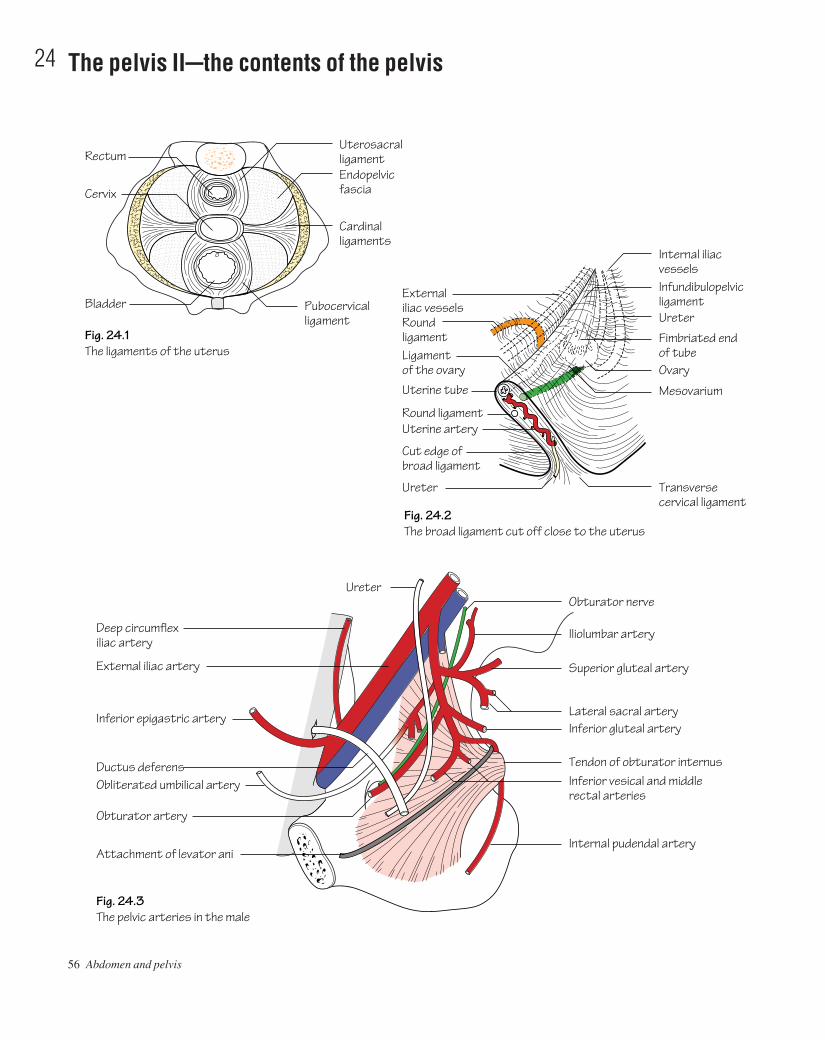
56 Abdomen and pelvis
24 The pelvis IIcthe contents of the pelvis
Externaliliac vesselsRound ligamentLigament of the ovary
Round ligamentUterine artery
Cut edge of broad ligament
Ureter Transversecervical ligament
Internal iliac vessels
Uterine tube
Uterosacralligament
Pubocervicalligament
Bladder
Rectum
CervixEndopelvicfascia
Cardinalligaments
Infundibulopelvic ligamentUreter
Fimbriated end of tubeOvary
Internal pudendal arteryAttachment of levator ani
Mesovarium
Obturator artery
Ductus deferens
Inferior epigastric artery
External iliac artery
Deep circumflexiliac artery
Ureter
Obliterated umbilical artery Inferior vesical and middle rectal arteries
Tendon of obturator internus
Inferior gluteal artery
Superior gluteal artery
Obturator nerve
Iliolumbar artery
Lateral sacral artery
Fig. 24.2The broad ligament cut off close to the uterus
Fig. 24.1The ligaments of the uterus
Fig. 24.3The pelvic arteries in the male
AAAC24 21/5/05 10:46 AM Page 56
Pelvic fascia (Fig. 24.1)The pelvic fascia is the term given to the connective tissue that lines thepelvis covering levator ani and obturator internus. It is continuous withthe fascial layers of the abdominal wall above and the perineum below.Endopelvic fascia is the term given to the loose connective tissue thatcovers the pelvic viscera. The endopelvic fascia is condensed into fas-cial ligaments which act as supports for the cervix and vagina. Theseligaments include the:• Cardinal (Mackenrodt’s) ligaments: pass laterally from the cervixand upper vagina to the pelvic side walls.• Utero-sacral ligaments: pass backwards from the cervix and va-ginal fornices to the fascia overlying the sacro-iliac joints.• Pubocervical ligaments: extend anteriorly from the cardinal liga-ments to the pubis (puboprostatic in the male).• Pubovesical ligaments: from the back of the symphysis pubis to thebladder neck.
The broad and round ligaments (Fig. 24.2)• Broad ligament: is a double fold of peritoneum which hangsbetween the lateral aspect of the uterus and the pelvic side walls. Theureter passes forwards under this ligament, but above and lateral to thelateral fornix of the vagina, to gain access to the bladder. The broad liga-ment contains the following structures:
• Fallopian tube.• Ovary.• Ovarian ligament.• Round ligament (see below).• Uterine and ovarian vessels.• Nerves and lymphatics.
• Round ligament: is a cord-like fibromuscular structure which is thefemale equivalent of the gubernaculum in the male. It passes from thelateral angle of the uterus to the labium majus by coursing in the broadligament and then through the inguinal canal (p. 30).
Arteries of the pelvis (Fig. 24.3)• Common iliac arteries: arise from the aortic bifurcation to the left ofthe midline at the level of the umbilicus. These arteries, in turn, bifur-cate into external and internal iliac branches anterior to the sacro-iliacjoints on either side.• External iliac artery: courses from its origin (described above) tobecome the femoral artery as it passes under the inguinal ligament atthe mid-inguinal point. The external iliac artery gives rise to brancheswhich supply the anterior abdominal wall. These include the: deep cir-
cumflex iliac artery and inferior epigastric artery. The latter branchgains access to the rectus sheath, which it supplies, and eventuallyanastomoses with the superior epigastric artery.• Internal iliac artery: courses from its origin (described above) to divide into anterior and posterior trunks at the level of the greater sciatic foramen.
Branches of the anterior trunk• Obturator artery: passes with the obturator nerve through the obtur-ator canal to enter the thigh.• Umbilical artery: although the distal part is obliterated the proximalpart is patent and gives rise to the superior vesical artery which con-tributes a supply to the bladder.• Inferior vesical artery: as well as contributing a supply to the blad-der it also gives off a branch to the vas deferens (in the male).• Middle rectal artery: anastomoses with the superior and inferiorrectal arteries to supply the rectum.• Internal pudendal artery: is the predominant supply to the per-ineum. It exits the pelvis briefly through the greater sciatic foramen butthen re-enters below piriformis through the lesser sciatic foramen toenter the pudendal canal together with the pudendal nerve.• Uterine artery: passes medially on the pelvic floor and then over theureter and lateral fornix of the vagina to ascend the lateral aspect of theuterus between the layers of the broad ligament.• Inferior gluteal artery: passes out of the pelvis through the greatersciatic foramen to the gluteal region which it supplies.• Vaginal artery.
Branches of the posterior trunk• Superior gluteal artery: contributes a supply to the gluteal muscles.It leaves the pelvis through the greater sciatic foramen.• Ilio-lumbar artery.• Lateral sacral artery.
Veins of the pelvisThe right and left common iliac veins join to form the inferior venacava behind the right common iliac artery but anterolateral to the bodyof L5. The overall arrangement of pelvic venous drainage reciprocatesthat of the arterial supply.
Nerves of the pelvisSacral plexus (see p. 100).
The pelvis IIbthe contents of the pelvis 57
AAAC24 21/5/05 10:46 AM Page 57
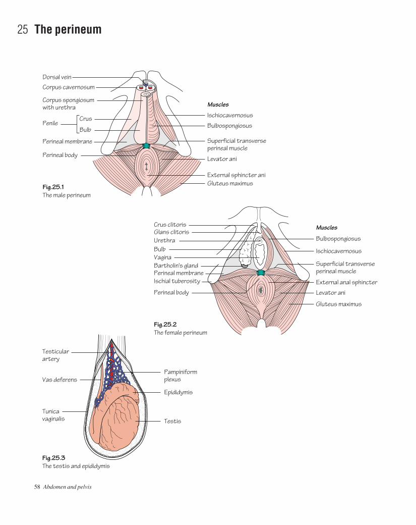
58 Abdomen and pelvis
25 The perineum
Fig.25.3The testis and epididymis
Fig.25.2The female perineum
Fig.25.1The male perineum
Crus clitorisGlans clitoris
Pampiniformplexus
Epididymis
Testis
Testicularartery
Vas deferens
Tunicavaginalis
Bulbospongiosus
Muscles
Ischiocavernosus
Superficial transverseperineal muscle
External anal sphincter
Levator ani
Gluteus maximus
UrethraBulbVaginaBartholin's glandPerineal membrane
Perineal body
Ischial tuberosity
Bulbospongiosus
Muscles
Ischiocavernosus
Superficial transverseperineal muscle
External sphincter ani
Levator ani
Gluteus maximus
Dorsal vein
Corpus cavernosum
Corpus spongiosumwith urethra
PenileCrus
Bulb
Perineal membrane
Perineal body
AAAC25 21/5/05 10:46 AM Page 58

The perineum lies below the pelvic diaphragm. It forms a diamond-shaped area when viewed from below that can be divided into an anter-ior urogenital region and a posterior anal region by a line joining theischial tuberosities horizontally.
Anal region (Figs 25.1 and 17.2)The anal region contains the anal canal and ischiorectal fossae.• Anal canal: is described earlier (p. 43).• Anal sphincter: comprises external and internal sphincter compon-ents. The internal anal sphincter is a continuation of the inner circularsmooth muscle of the rectum. The external anal sphincter is a skeletalmuscular tube which, at its rectal end, blends with puborectalis to forman area of palpable thickening termed the anorectal ring. The compet-ence of the latter is fundamental to anal continence.• Ischiorectal fossae: lie on either side of the anal canal. The medialand lateral walls of the ischiorectal fossa are the levator ani and analcanal and the obturator internus, respectively. The fossae are filled withfat. The anococcygeal body separates the fossae posteriorly; however,infection in one fossa can spread anteriorly to the contralateral fossaforming a horseshoe abscess. The pudendal (Alcock’s) canal is a sheathin the lateral wall of the ischiorectal fossa. It conveys the pudendalnerve and internal pudendal vessels from the lesser sciatic notch to thedeep perineal pouch (see below). The inferior rectal branches of thepudendal nerve and internal pudendal vessels course transverselyacross the fossa to reach the anus.
Urogenital regionThe urogenital region is triangular in shape. The perineal membrane isa strong fascial layer that is attached to the sides of the urogenital tri-angle. In the male it is pierced by the urethra and, in females, by the urethra and vagina.
(a) In the female (Fig. 25.2)• Vulva: is the term given to the female external genitalia. The monspubis is the fatty protuberance overlying the pubic symphysis andpubic bones. The labia majora are fatty hair-bearing lips that extendposteriorly from the mons. The labia minora lie internal to the labiamajora and unite posteriorly at the fourchette. Anteriorly, the labiaminora form the prepuce and split to enclose the clitoris. The clitoriscorresponds to the penis in the male. It has a similar structure in that it ismade up of three masses of erectile tissue: the bulb (corresponding tothe penile bulb) and right and left crura covered by similar but smallermuscles than those in the male. As in the male, these form the contentsof the superficial perineal pouch. The deep perineal pouch, however,contains the vagina as well as part of the urethra and sphincter urethraeand internal pudendal vessels. The vestibule is the area enclosed by thelabia minora and contains the urethral and vaginal orifices. Deep to theposterior aspect of the labia majoris lie Bartholin’s glandsaa pair ofmucus-secreting glands that drain anteriorly. They are not palpable inhealth but can become grossly inflamed when infected.• Urethra: is short in the female (3–4 cm). This factor contributestowards the predisposition to urinary tract infection due to upwardspread of bowel organisms. The urethra extends from the bladder neckto the external meatus. The meatus lies between the clitoris and vagina.• Vagina: measures approximately 8–12 cm in length. It is a musculartube that passes upwards and backwards from the vaginal orifice. Thecervix projects into the upper anterior aspect of the vagina creating fornices anteriorly, posteriorly and laterally. Lymph from the uppervagina drains into the internal and external iliac nodes. Lymph from the
lower vagina drains to the superficial inguinal nodes. The blood supplyto the vagina is from the vaginal artery (branch of the internal iliacartery) and the vaginal branch of the uterine artery.
(b) In the male (Fig. 25.1)The external urethral sphincter (striated muscle) lies deep to the per-ineal membrane within a fascial capsule termed the deep perinealpouch. In addition to the sphincter, two glands of Cowper are also contained within the deep pouch. The ducts from these glands pass forwards to drain into the bulbous urethra. Inferior to the perineal mem-brane is the superficial perineal pouch which contains the:• Superficial transverse perineal muscles: run from the perinealbody to the ischial ramus.• Bulbo-spongiosus muscle: covers the corpus spongiosum. The lat-ter structure covers the spongy urethra.• Ischio-cavernosus muscle: arises on each side from the ischialramus to cover the corpus cavernosum. It is the engorgement of venoussinuses within these cavernosa that generate and maintain an erection.
Hence, the penile root comprises a well-vascularized bulb and twocrura which are supplied by branches of the internal pudendal artery.The erectile penile tissue is enclosed within a tubular fascial sheath. Atthe distal end of the penis the corpus spongiosum expands to form theglans penis. On the tip of the glans the urethra opens as the externalurethral meatus. The foreskin is attached to the glans below the meatusby a fold of skinathe frenulum.
The scrotumThe skin of the scrotum is thin, rugose and contains many sebaceousglands. A longitudinal median raphe is visible in the midline. Beneath theskin lies a thin layer of involuntary dartos muscle. The terminal spermaticcords, the testes and their epididymes are contained within the scrotum.
Testis and epididymis (Fig. 25.3)The testes are responsible for spermatogenesis. Their descent to an extra-abdominal position favours optimal spermatogenesis as the ambientscrotal temperature is approximately 3°C lower than body temperature.• Structure: the testis is divided internally by a series of septa intoapproximately 200 lobules. Each lobule contains 1–3 seminiferoustubules which anastomose into a plexus termed the rete testis. Eachtubule is coiled when in situ, but when extended measures approxim-ately 60 cm. Efferent ducts connect the rete testis to the epididymalhead. They serve to transmit sperm from the testicle to the epididymis.
• The tunica vaginalis, derived from the peritoneum, is a double covering into which the testis is invaginated.
• The tunica albuginea is a tough fibrous capsule that covers the testis.• The epididymis lies along the posterolateral and superior borders of
the testicle. The tunica vaginalis covers the epididymis with theexception of the posterior border.
• The upper poles of both the testis and epididymis bear an appendixtestis and appendix epididymis (hydatid of Morgagni), respectively.
• Blood supply: is from the testicular artery (a branch of the abdom-inal aorta, p. 32). Venous drainage from the testicle is to the pampini-form plexus of veins. The latter plexus lies within the spermatic cordbut coalesces to form a single vein at the internal ring. The left testicu-lar vein drains to the left renal vein whereas the right testicular veindrains directly to the inferior vena cava.• Lymphatic drainage: is to the para-aortic lymph nodes.• Nerve supply: is from T10 sympathetic fibres via the renal and aorticplexuses.
The perineum 59
AAAC25 21/5/05 10:46 AM Page 59

60 Abdomen and pelvis
26 The pelvic viscera
Contents of the pelvic cavity (see Fig. 24.1)• Sigmoid colon: see p. 43.• Rectum: see p. 43.• Ureters: see p. 49.• Bladder (Fig. 26.2): see below.
BladderIn adults the bladder is a pelvic organ. It lies behind the pubis and iscovered superiorly by peritoneum. It acts as a receptacle for urine andhas a capacity of approximately 500 mL.
• Structure: the bladder is pyramidal in shape. The apex of the pyra-mid points forwards and from it a fibrous cord, the urachus, passesupwards to the umbilicus as the median umbilical ligament. The base(posterior surface) is triangular. In the male, the seminal vesicles lie onthe outer posterior surface of the bladder and are separated by the vasdeferens. The rectum lies behind. In the female, the vagina intervenesbetween the bladder and rectum. The inferolateral surfaces are relatedinferiorly to the pelvic floor and anteriorly to the retropubic fat pad andpubic bones. The bladder neck fuses with the prostate in the malewhereas it lies directly on the pelvic fascia in the female. The pelvic
Body
Internal osSupravaginal cervix
Intravaginal cervixCervical canal
Lateral fornixExternal osVagina
Fallopian tube
Bladder
Spongioseurethra
UreterVaginal artery
Uterineartery
Ovarianartery
Lateralcornu
Rectovesicalpouch
CavityFundus
Fig.26.3A vertical section through the uterus and vagina.Note the relation of the uterine artery to the ureter
Fig.26.1Sagittal sections through the male and female pelves
Prostate
Ureter
Ductusdeferens
Seminalvesicle
Urachus
Fig.26.2The bladder and prostate
Ampulla
Prostate
Urethra
Trigone
Ureter
Sphincterurethrae
Prostate
RectumUterovesicalpouch Recto-uterine pouch
Anal canalPerineal body
ProstaticurethraMembranousurethra
Suspensoryligament
BladderUrethraVaginaVestibule
Perineal body
Posterior fornix of vaginaCervix of uterusSphincter ani externusAnal canal
AAAC26 21/5/05 10:45 AM Page 60
fascia is thickened in the form of the puboprostatic ligaments (male)and pubovesical ligaments to hold the bladder neck in position. Themucous membrane of the bladder is thrown into folds when the bladderis empty with the exception of the membrane overlying the base(termed the trigone) which is smooth. The superior angles of thetrigone mark the openings of the ureteric orifices. A muscular eleva-tion, the interureteric ridge, runs between the ureteric orifices. Theinferior angle of the trigone corresponds to the internal urethral mea-tus. The muscle coat of the bladder is composed of a triple layer of tra-beculated smooth muscle known as the detrusor (muscle). The detrusoris thickened at the bladder neck to form the sphincter vesicae.• Blood supply: is from the superior and inferior vesical arteries(branches of the internal iliac artery, p. 57). The vesical veins coalescearound the bladder to form a plexus that drains into the internal iliacvein.• Lymph drainage: is to the para-aortic nodes.• Nerve supply: motor input to the detrusor muscle is from efferentparasympathetic fibres from S2–4. Fibres from the same source conveyinhibitory fibres to the internal sphincter so that co-ordinated micturi-tion can occur. Conversely, sympathetic efferent fibres inhibit thedetrusor and stimulate the sphincter.
The male pelvic organsThe prostate (Fig. 26.2)In health the prostate is approximately the size of a walnut. It surroundsthe prostatic urethra and lies between the bladder neck and the urogen-ital diaphragm. The apex of the prostate rests on the external urethralsphincter of the bladder. It is related anteriorly to the pubic symphysisbut separated from it by extraperitoneal fat in the retropubic space(cave of Retzius). Posteriorly, the prostate is separated from the rectumby the fascia of Denonvilliers.• Structure: the prostate comprises anterior, posterior, middle and lat-eral lobes. On rectal examination a posterior median groove can be pal-pated between the lateral lobes. The prostatic lobes contain numerousglands producing an alkaline secretion which is added to the seminalfluid at ejaculation. The prostatic glands open into the prostatic sinus.The ejaculatory ducts, which drain both the seminal vesicles and thevas, enter the upper part of the prostate and then the prostatic urethra atthe verumontanum.• Blood supply: is from the inferior vesical artery (branch of the inter-nal iliac artery, p. 57). A prostatic plexus of veins is situated betweenthe prostatic capsule and the outer fibrous sheath. The plexus receivesthe dorsal vein of the penis and drains into the internal iliac veins.
The vas deferensThe vas deferens conveys sperm from the epididymis to the ejaculatoryduct from which it can be passed to the urethra. The vas arises from thetail of the epididymis and traverses the inguinal canal to the deep ring,passes downwards on the lateral wall of the pelvis almost to the ischialtuberosity and turns medially to reach the base of the bladder where itjoins with the duct of the seminal vesicle to form the ejaculatory duct.
The seminal vesicles (Fig. 26.2)The seminal vesicles consist of lobulated tubes which lie extraperi-toneally on the bladder base lateral to the vas deferens.
The urethra (Fig. 26.1)The male urethra is approximately 20 cm long (4 cm in the female). Itis considered in three parts:
• Prostatic urethra (3 cm): bears a longitudinal elevation (urethralcrest) on its posterior wall. On either side of the crest a shallow depres-sion, the prostatic sinus, marks the drainage point for 15–20 prostaticducts. The prostatic utricle is a 5 mm blind ending tract which opensinto an eminence in the middle of the crestathe verumontanum. Theejaculatory ducts open on either side of the utricle.• Membranous urethra (2 cm): lies in the urogenital diaphragm andis surrounded by the external urethral sphincter (sphincter urethrae).• Penile urethra (15 cm): traverses the corpus spongiosum of thepenis (see perineum, p. 59) to the external urethral meatus.
The female pelvic organsThe vaginaSee perineum, p. 59.
The uterus and fallopian tubes (Fig. 26.3)• Structure: the uterus measures approximately 8 cm in length in thenulliparous female. It comprises a: fundus (part lying above theentrance of the fallopian tubes), body and cervix. The cervix is sunkeninto the anterior wall of the vagina and is consequently divided intosupravaginal and vaginal parts. The internal cavity of the cervix com-municates with the cavity of the body at the internal os and with thevagina at the external os. The fallopian tubes lie in the free edges of thebroad ligaments and serve to transmit ova from the ovary to the cornuaof the uterus. They comprise an: infundibulum, ampulla, isthmus andinterstitial part. The uterus is made up of a thick muscular wall(myometrium) and lined by a mucous membrane (endometrium). Theendometrium undergoes massive cyclical change during menstruation.• Relations: the uterus and cervix are related to the uterovesical pouchand superior surface of the bladder anteriorly. The recto-uterine pouch(of Douglas), which extends down as far as the posterior fornix of thevagina, is a posterior relation. The broad ligament is the main lateralrelation of the uterus.• Position: in the majority, the uterus is anteverted, i.e. the axis of thecervix is bent forward on the axis of the vagina. In some women theuterus is retroverted.• Blood supply: is predominantly from the uterine artery (a branch ofthe internal iliac artery, p. 57). It runs in the broad ligament and, at thelevel of the internal os, crosses the ureter at right angles to reach, andsupply, the uterus before anastomosing with the ovarian artery (abranch of the abdominal aorta, p. 32).• Lymph drainage: lymphatics from the fundus accompany the ovar-ian artery and drain into the para-aortic nodes. Lymphatics from thebody and cervix drain to the internal and external iliac lymph nodes.
The ovaryEach ovary contains a number of primordial follicles which develop inearly fetal life and await full development into ova. In addition to theproduction of ova, the ovaries are also responsible for the production ofsex hormones. Each ovary is surrounded by a fibrous capsule, thetunica albuginea.• Attachments: the ovary lies next to the pelvic side wall and issecured in this position by two structures: the broad ligament whichattaches the ovary posteriorly by the mesovarium; and the ovarian liga-ment which secures the ovary to the cornu of the uterus.• Blood supply: is from the ovarian artery (a branch of the abdominalaorta). Venous drainage is to the inferior vena cava on the right and tothe left renal vein on the left.• Lymphatic drainage: is to the para-aortic nodes.
The pelvic viscera 61
AAAC26 21/5/05 10:45 AM Page 61

The clavicle (Fig. 27.1)• The clavicle is the first bone to ossify in the fetus (6 weeks).• It develops in membrane and not in cartilage.• It is subcutaneous throughout its length and transmits forces from thearm to the axial skeleton.• The medial two-thirds are circular in cross-section and curved con-vex forwards. The lateral third is flat and curved convex backwards.• The clavicle articulates medially with the sternum and 1st costal car-tilage at the sternoclavicular joint. The clavicle is also attached medi-ally to the 1st rib by strong costoclavicular ligaments and to thesternum by sternoclavicular ligaments.
62 Upper limb
27 The osteology of the upper limb
Fig.27.1The upper and lower surfaces of the left clavicle
Fig.27.2X-ray of a fractured clavicle
Facet for acromion
Medial (sternal) end
Trapezoid line
Tubercle for costo-clavicular ligament
Conoid tubercule
• The clavicle articulates laterally with the acromion process of thescapulaathe acromioclavicular joint. The coracoclavicular ligamentssecure the clavicle inferolaterally to the coracoid process of thescapula. This ligament has two componentsathe conoid and trapezoidligaments which are attached to the conoid tubercle and trapezoid lineof the clavicle, respectively.• The clavicle is the most commonly fractured bone in the body. Theweakest point of the bone is the junction of the middle and outer thirds(Fig. 27.2).
AAAC27 21/5/05 10:45 AM Page 62

• The greater and lesser tubercles provide attachment for the rotatorcuff muscles. The tubercles are separated by the intertubercular sulcusin which the long head of biceps tendon courses.• A faint spiral groove is visible on the posterior aspect of the humeralshaft traversing obliquely downwards and laterally. The medial and lat-eral heads of triceps originate on either side of this groove. The radialnerve passes between the two heads.• The ulnar nerve winds forwards in a groove behind the medial epicondyle.• At the elbow joint: the trochlea articulates with the trochlear notch ofthe ulna; and the rounded capitulum with the radial head. The medialborder of the trochlea projects inferiorly a little further than the lateralborder. This accounts for the carrying angle, i.e. the slight lateral anglemade between the arm and forearm when the elbow is extended.
The osteology of the upper limb 63
The scapula (Fig. 27.3)• The scapula is triangular in shape. It provides an attachment fornumerous muscles.• The glenoid fossa articulates with the humeral head (gleno-humeraljoint), and the acromion process with the clavicle (acromioclavicularjoint).
The humerus (Fig. 27.4)• The humeral head consists of one third of a sphere. The rounded headarticulates with the shallow glenoid. This arrangement permits a widerange of shoulder movement.• The anatomical neck separates the head from the greater and lessertubercles. The surgical neck lies below the anatomical neck betweenthe upper end of the humerus and shaft. The axillary nerve and cir-cumflex vessels wind around the surgical neck of the humerus. Theseare at risk of injury in shoulder dislocations and humeral neck fractures(see Fig. 34.3).
Fig.27.3Posterior and anterior views of the left scapula
Fig.27.4Anterior and posterior views of the left humerus
Acromion
Coracoid processSuperior angle
Medial border
Greater tubercle
Glenoid fossa
Acromion
Spine Head
Supraspinousfossa
Infraspinousfossa
Suprascapular notchSpinoglenoid notch
Intertubercularsulcus
Medial supracondylarridge
Medial epicondyle
Lessertubercle
Subscapular fossa
Lateral border
Anatomical neckSurgical neck
Deltoidtuberosity
Spiral groove
Olecranonfossa
TrochleaCapitulum
AAAC27 21/5/05 10:45 AM Page 63

The radius and ulna (Fig. 27.5)• Both the radius and ulna have interosseous, anterior and posteriorborders.• The biceps tendon inserts into the roughened posterior part of theradial tuberosity. The anterior part of the tuberosity is smooth where itis covered by a bursa.• The radial head is at its proximal end whilst the ulnar head is at itsdistal end.• The lower end of the radius articulates with the scaphoid and lunatecarpal bones at the wrist joint. The distal ulna does not participatedirectly in the wrist joint.• The dorsal tubercle (of Lister) is located on the posterior surface ofthe distal radius.
64 Upper limb
• In pronation/supination movements the radial head rotates in theradial notch of the ulna and the radial shaft pivots around the relativelyfixed ulna (connected by the interosseous ligament). The distal radiusrotates around the head of the ulna.
A Colles fracture is a common injury occurring at the wrist in theelderly and usually osteoporotic population. It classically follows a fallon the outstretched hand. The fracture line is usually about 2.5 cmproximal to the wrist and the distal fragment displaces posteriorly (din-nerfork deformity when viewed from the side) and radially. Somedegree of shortening often occurs due to impaction of the componentparts (Fig. 27.6).
Fig.27.6X-ray of a fracture of the lower endof the radius (Colles' fracture)
Radial styloidFig.27.5The left radius and ulna in (a) supination and (b) pronation
Supinator crest
Trochlearnotch
Olecranon
Head of ulna
Head ofradius
Coronoid process
Ulna styloid
Radialtuberosity
Tuberosityof the ulna
Interosseousborders
Dorsaltubercle
Attachmentof pronator teres
AAAC27 21/5/05 10:45 AM Page 64

The hand (Fig. 27.7)The carpal bones are arranged into two rows. The palmar aspect of thecarpus is concave. This is brought about by the shapes of the con-stituent bones and the flexor retinaculum bridging the bones anteriorlyto form the carpal tunnel (see Fig. 38.1).
The scaphoid may be fractured through a fall on the outstretchedhand. This injury is common in young adults and must be suspectedclinically when tenderness is elicited by deep palpation in the anatomi-
cal snuffbox. Radiographic changes are often not apparent and, ifeffective treatment is not implemented, permanent wrist weakness andsecondary osteoarthritis may follow. The blood supply to the scaphoidenters via its proximal and distal ends. However, in as many as onethird of cases the blood supply enters only from the distal end. Underthese circumstances the proximal scaphoid fragment may be deprivedof arterial supply and undergo avascular necrosis.
The osteology of the upper limb 65
Fig.27.7The skeleton of the left hand, holding a cross-section through the carpal tunnel
Triquetral
Lunate Tubercle of scaphoid
Hook of hamate
Pisiform
Trapezium
Pisiform
Triquetral
Flexor retinaculum
Scaphoid
Lunate
TrapezoidCapitate
AAAC27 21/5/05 10:45 AM Page 65

66 Upper limb
28 Arteries of the upper limb
Flexor carpi radialis
Radial artery
Ulnarnerve
Radialisindicis
Radial artery
Princepspollicis
Ulnarartery
Deep palmar archPalmar metacarpal
Superficialpalmar arch
Ulnar arteryUlnar nervePisiform
Deep branch of ulnar
Flexor retinaculum
Lateral thoracic
Radialnerve
Radial nerve
Common interosseousPosterior interosseous
Anterior interosseous
Median nerve
Profunda brachii
Circumflex scapularAxillary nerve
Subscapular
Thoracoacromialartery
Fig.28.1The arterial system of the upper limb.The main nerves that are related tothe arteries are shown in green.Only the major arterial branchesare labelled
AAAC28 21/5/05 10:44 AM Page 66

The axillary artery• Course: the axillary artery commences at the lateral border of the 1strib as a continuation of the subclavian artery (see Fig. 60.1) and ends atthe inferior border of teres major where it continues as the brachialartery. The axillary vein is a medial relation throughout its course. It iscrossed anteriorly by pectoralis minor which subdivides it into threeparts:
• First part (medial to pectoralis minor).• Second part (behind pectoralis minor)agives off the lateral
thoracic artery (which helps to supply the breast) and the thora-coacromial artery (p. 75).
• Third part (lateral to pectoralis minor)agives off the subscapularartery which follows the lateral border of the scapula and gives offthe circumflex scapular artery.
The brachial artery• Course: the brachial artery commences at the inferior border of teresmajor as a continuation of the axillary artery and ends by bifurcatinginto the radial and ulnar arteries at the level of the neck of the radius. Itlies immediately below the deep fascia throughout its course. Thebrachial artery is crossed superficially by the median nerve in the mid-arm from lateral to medial and hence lies between the median nerve(medial relation) and biceps tendon (lateral relation) in the cubital fossa(see Fig. 36.3).• Branches:
• Profunda brachiiaarises near the origin of the brachial artery andwinds behind the humerus with the radial nerve in the spiral groovebefore taking part in the anastomosis around the elbow joint.
• Other branchesainclude a nutrient artery to the humerus andsuperior and inferior ulnar collateral branches which ultimatelytake part in the anastomosis around the elbow.
‘Volkman’s ischaemic contracture’ is a deformity arising as a resultof brachial artery spasm following injury (usually from fractures of thedistal humerus). The reduced arterial flow results in ischaemic necro-sis of the forearm musculature which is eventually replaced by short-ened fibrous tissue. In the classical deformity the forearm is wasted andthe wrist joint flexed with the fingers extended. When the wrist isextended the fingers flex.
The radial artery• Course: the radial artery arises at the level of the neck of the radiusfrom the bifurcation of the brachial artery. It passes over the biceps ten-don to lie firstly on supinator then descends on the radial side of theforearm, lying under the edge of brachioradialis in the upper half of its course and then between the tendons of brachioradialis and flexorcarpi radialis in the lower forearm. The radial artery passes sequentiallyover supinator, pronator teres, the radial head of flexor digitorumsuperficialis, flexor pollicis longus and pronator quadratus. At the wrist
the artery lies on the distal radius lateral to the tendon of flexor carpiradialis. This is where the radial pulse is best felt.• Branches:
• Palmar and dorsal carpal branches are given off at the wrist.• A superficial palmar branch arises at the wrist which supplies the
thenar muscles and consequently anastomoses with the superficialpalmar branch of the ulnar artery to form the superficial palmararch.
• The radial artery passes backwards under the tendons of abductorpollicis longus and extensor pollicis brevis to enter the anatomicalsnuffbox. It consequently passes over the scaphoid and trapeziumin the snuffbox and exits by passing between the two heads ofadductor pollicis to enter the palm and forms the deep palmar archwith a contribution from the ulnar artery (deep palmar branch). Itgives off the princeps pollicis to the thumb and the radialis indicisto the index finger.
• The deep palmar arch gives off three palmar metacarpal arterieswhich subsequently join the common palmar digital arteries (fromthe superficial arch) to supply the digits.
The ulnar artery• Course: the ulnar artery commences as the terminal bifurcation ofthe brachial artery at the level of the neck of the radius. It passes deep tothe deep head of pronator teres and deep to the fibrous arch of flexordigitorum superficialis and descends on flexor digitorum profunduswith the ulnar nerve lying medial to it and flexor carpi ulnaris overlap-ping it in the proximal half of the forearm.
At the wrist both the ulnar artery and nerve lie lateral (radial) toflexor carpi ulnaris and pass over the flexor retinaculum giving carpalbranches which contribute to the dorsal and palmar carpal arches.• Branches:
• A deep palmar branch completes the deep palmar arch (see above)and the ulnar artery continues as the superficial palmar arch whichis completed by the superficial palmar branch of the radial artery.
• The common interosseous artery (see below).
The common interosseous arteryThe common interosseous artery is the first ulnar branch to arise and itsubdivides into the:• Anterior interosseous artery: descends with the interosseous branchof the median nerve on the anterior surface of the interosseous membrane. It predominantly supplies the flexor compartment of theforearm.• Posterior interosseous artery: passes above the upper border of theinterosseous membrane to enter the extensor compartment where itruns with the deep branch of the radial nerve supplying the extensormuscles of the forearm, eventually anastomosing with the anteriorinterosseous artery.
Arteries of the upper limb 67
AAAC28 21/5/05 10:44 AM Page 67

68 Upper limb
29 The venous and lymphatic drainage of the upper limb and the breast
Fig. 29.2The lymph nodes of the axilla and the lymphatic drainage of the breast
Fig. 29.3A section through the breast to show its major relations.The inset shows the radial arrangement of thelactiferous ducts
Fig. 29.1The superficial venous system of the upper limb and two majorcutaneous nerves
Cephalic vein piercesclavipectoral fascia
Deltoid
Pectoralis majorPectoralisminor
Pectoralismajor
Cephalic vein
Median cubital vein
Tendon of biceps
Lateral cutaneous nerve of forearm
Medial cutaneous nerveof forearm
Brachial arteryMedian nerve
Bicipital aponeurosis
Basilic vein
Right lymph duct
Lateral group
Central group
Axillary vessels
Supratrochlear group
Inferior deep cervical(supraclavicular) group
Delto-pectoral group
Apical group
Posterior group
Anterior (pectoral) group
Parasternal group(inside chest)
Suspensory ligament
Intercostals
Rib
Alveoli of breast
Lactiferous duct
Lactiferous sinusNippleAreolaMontgomery'sgland
Fat
AAAC29 21/5/05 10:44 AM Page 68

Venous drainage of the upper limb (Fig. 29.1)As in the lower limb the venous drainage comprises interconnectedsuperficial and deep systems.• The superficial system: comprises the cephalic and basilic veins.
• The cephalic vein commences from the lateral end of the dorsalvenous network overlying the anatomical snuffbox. It ascends thelateral, then anterolateral, aspects of the forearm and arm andfinally courses in the deltopectoral groove to pierce the clavipec-toral fascia and drain into the axillary vein.
• The basilic vein commences from the medial end of the dorsalvenous network. It ascends along the medial then anteromedialaspects of the forearm and arm to pierce the deep fascia (in theregion of the mid-arm) to join with the venae comitantes of thebrachial artery to form the axillary vein.
The two superficial veins are usually connected by a median cubitalvein in the cubital fossa.• The deep veins: consist of venae comitantes (veins which accom-pany arteries).
The superficial veins of the upper limb are of extreme clinical import-ance for phlebotomy and peripheral venous access. The most com-monly used sites are the median cubital vein in the antecubital fossa andthe cephalic vein in the forearm.
Lymphatic drainage of the chest wall and upper limb (Fig. 29.2)Lymph from the chest wall and upper limb drains centrally via axillary,supratrochlear and infraclavicular lymph nodes.
Axillary lymph node groupsThere are approximately 30–50 lymph nodes in the axilla. They arearranged into five groups:• Anterior (pectoral) group: these lie along the anterior part of themedial wall of the axilla. They receive lymph from the upper anteriorpart of the trunk wall and breast.• Posterior (subscapular) group: these lie along the posterior part ofthe medial wall of the axilla. They receive lymph from the upper pos-terior trunk wall down as far as the iliac crest.• Lateral group: these lie immediately medial to the axillary vein.They receive lymph from the upper limb and the breast.• Central group: these lie within the fat of the axilla. They receivelymph from all of the groups named above.• Apical group: these lie in the apex of the axilla. They receive lymphfrom all of the groups mentioned above. From here lymph is passed tothe thoracic duct (on the left) or right lymphatic trunks (see Fig. 3.3),with some passing to the inferior deep cervical (supraclavicular)group.
Lymph node groups in the arm• The supratrochlear group of nodes lie subcutaneously above the
medial epicondyle. They drain lymph from the ulnar side of the forearmand hand. Lymph from this group passes to the lateral group of axillarylymph nodes and thence drains centrally.• A small amount of lymph from the radial side of the upper limbdrains directly into the infraclavicular group of nodes. This group isarranged around the cephalic vein in the deltopectoral groove. Fromthis point the efferent vessels pass through the clavipectoral fascia todrain into the apical group of axillary nodes and thence centrally.
This information can be applied to the clinical scenario. If a patientpresents with an infected insect bite of the thumb, the infraclavicularnodes would reactively enlarge. If, however, infection occurred on thepatient’s little finger, lymphadenopathy of the supratrochlear nodeswould result.
The breast (Fig. 29.3)The breasts are present in both sexes and have similar characteristicsuntil puberty when, in the female, they enlarge and develop the capac-ity for milk production. The breasts are essentially specialized skinglands comprising fat, glandular and connective tissue. The base of thebreast lies in a constant position on the anterior chest wall. It extendsfrom the 2nd to 6th ribs anteriorly and from the lateral edge of the ster-num to the mid-axillary line laterally. A part of the breast, the axillarytail, extends laterally through the deep fascia beneath pectoralis to enterthe axilla. Each breast comprises 15–30 functional ducto-lobular unitsarranged radially around the nipple. The lobes are separated by fibroussepta (suspensory ligaments) which pass from the deep fascia to theoverlying skin thereby giving the breast structure. A lactiferous ductarises from each lobe and converges on the nipple. In its terminal por-tion the duct is dilated (lactiferous sinus) and thence continues to thenipple from where milk can be expressed. The areola is the darkenedskin that surrounds the nipple. Its surface is usually irregular due tomultiple small tuberclesaMontgomery’s glands.• Blood supply: is from the perforating branches of the internal thoracic artery (p. 13) and the lateral thoracic and thoracoacromialbranches of the axillary artery (p. 67). The venous drainage corres-ponds to the arterial supply.• Lymphatic drainage: from the lateral half of the breast is to theanterior axillary nodes. Lymph from the medial breast drains into theinternal mammary nodes (adjacent to the internal thoracic vesselsbeneath the chest wall).
Lymph drainage in carcinoma of the breastThe axillary lymph nodes represent an early site of metastasis from prim-ary breast malignancies and their surgical removal and subsequentexamination provide important prognostic information as well as abasis for choice of adjuvant treatment. Damage to axillary lymphaticsduring surgical clearance of axillary nodes or resulting from radio-therapy to the axilla increases the likelihood of subsequent upper limblymphoedema.
The venous and lymphatic drainage of the upper limb and the breast 69
AAAC29 21/5/05 10:44 AM Page 69
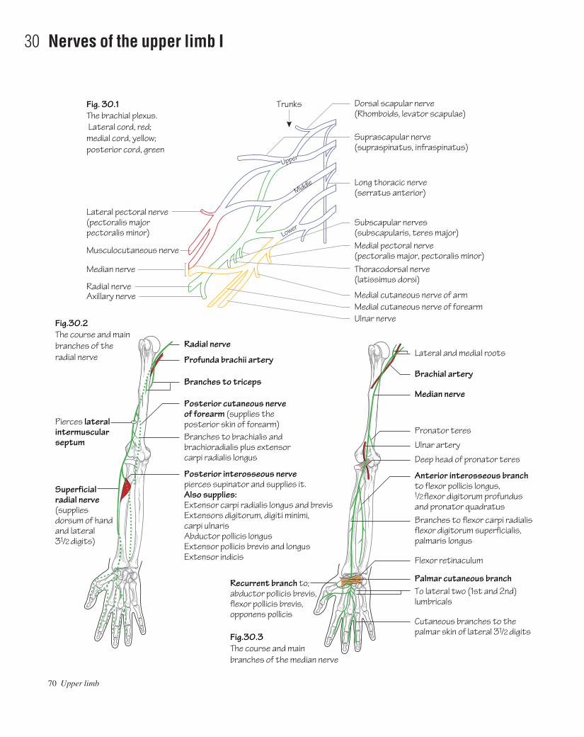
70 Upper limb
30 Nerves of the upper limb I
Fig. 30.1The brachial plexus. Lateral cord, red; medial cord, yellow; posterior cord, green
Dorsal scapular nerve(Rhomboids, levator scapulae)
Trunks
Upper
Middle
Lower
Suprascapular nerve(supraspinatus, infraspinatus)
Long thoracic nerve(serratus anterior)
Medial pectoral nerve(pectoralis major, pectoralis minor)Thoracodorsal nerve(latissimus dorsi)
Medial cutaneous nerve of arm
Subscapular nerves(subscapularis, teres major)
Lateral pectoral nerve(pectoralis majorpectoralis minor)
Musculocutaneous nerve
Radial nerveAxillary nerve
Radial nerve
Profunda brachii artery
Branches to triceps
Pierces lateralintermuscularseptum
Superficialradial nerve(supplies dorsum of handand lateral31/2 digits)
Posterior cutaneous nerveof forearm (supplies theposterior skin of forearm)Branches to brachialis andbrachioradialis plus extensorcarpi radialis longus
Posterior interosseous nerve pierces supinator and supplies it.Also supplies:Extensor carpi radialis longus and brevisExtensors digitorum, digiti minimi,carpi ulnarisAbductor pollicis longusExtensor pollicis brevis and longusExtensor indicis
Medial cutaneous nerve of forearmUlnar nerve
Median nerve
Fig.30.2The course and main branches of the radial nerve
Fig.30.3The course and main branches of the median nerve
Lateral and medial roots
Brachial artery
Median nerve
Pronator teres
Ulnar artery
Flexor retinaculum
Anterior interosseous branchto flexor pollicis longus, 1/2 flexor digitorum profundusand pronator quadratusBranches to flexor carpi radialisflexor digitorum superficialis,palmaris longus
Palmar cutaneous branchTo lateral two (1st and 2nd)lumbricals
Recurrent branch to;abductor pollicis brevis,flexor pollicis brevis, opponens pollicis
Cutaneous branches to thepalmar skin of lateral 31/2 digits
Deep head of pronator teres
AAAC30 21/5/05 10:44 AM Page 70

The brachial plexus (C5,6,7,8,T1) (Fig. 30.1)• The plexus arises as five roots. These are the anterior primary rami ofC5,6,7,8,T1. The roots lie between scalenus anterior and scalenusmedius.• The three trunks (upper, middle and lower) lie in the posterior tri-angle of the neck. They pass over the 1st rib to lie behind the clavicle.• The divisions form behind the middle third of the clavicle around theaxillary artery.• The cords lie in the axilla and are related medially, laterally and pos-teriorly to the second part of the axillary artery.• Terminal nerves arise from the cords surrounding the third part of theaxillary artery.
The axillary nerve (C5,6)• Type: mixed sensory and motor nerve.• Origin: it arises from the posterior cord of the brachial plexus.• Course: it passes through the quadrangular space with the posteriorcircumflex humeral artery. It provides: a motor supply to deltoid andteres minor; a sensory supply to the skin overlying deltoid; and an articu-lar branch to the shoulder joint.• Effect of injury: the axillary nerve is particularly prone to injuryfrom the downward displacement of the humeral head during shoulderdislocations.
• Motor deficitaloss of deltoid abduction with rapid wasting of thismuscle. Loss of teres minor function is not detectable clinically.
• Sensory deficitais limited to the ‘badge’ region overlying thelower half of deltoid.
The radial nerve (C5,6,7,8,T1) (Fig. 30.2)• Type: mixed sensory and motor.• Origin: it arises as a continuation of the posterior cord of the brachialplexus.• Course and branches: it runs with the profunda brachii arterybetween the long and medial heads of triceps into the posterior com-partment and down between the medial and lateral heads of triceps. At the midpoint of the arm it enters the anterior compartment by piercingthe lateral intermuscular septum. In the region of the lateral epicondylethe radial nerve lies under the cover of brachioradialis and divides into the superficial radial and posterior interosseous nerves.
The branches of the radial nerve include: branches to triceps, bra-chioradialis and brachialis as well as some cutaneous branches. It ter-minates by dividing into two major nerves:
• The posterior interosseous nerveapasses between the two heads of supinator at a point three fingerbreadths distal to the radial head thus passing into the posterior compartment. It supplies theextensor muscles of the forearm.
• The superficial radial nerveadescends the forearm under the coverof brachioradialis with the radial artery on its medial side. It termin-
ates as cutaneous branches supplying the skin of the back of thewrist and hand.
• Effect of injury (Fig. 31.2): e.g. humeral shaft fracture resulting indamage to the radial nerve in the spiral groove.
• Motor deficitaloss of all forearm extensors: wristdrop.• Sensory deficitausually small due to overlap: sensory loss over the
anatomical snuffbox is usually constant.
The musculocutaneous nerve (C5,6,7)• Type: mixed sensory and motor.• Origin: it arises from the lateral cord of the brachial plexus.• Course: it passes laterally through the two conjoined heads of cora-cobrachialis and then descends the arm between brachialis and biceps,supplying all three of these muscles en route. It pierces the deep fasciajust below the elbow (and becomes the lateral cutaneous nerve of theforearm). Here it supplies the skin of the lateral forearm as far as thewrist.
The median nerve (C6,7,8,T1) (Fig. 30.3)• Type: mixed sensory and motor.• Origin: it arises from the confluence of two roots from the medialand lateral cords lateral to the axillary artery in the axilla.• Course and branches: the median nerve initially lies lateral to thebrachial artery but crosses it medially in the mid-arm. In the cubitalfossa it lies medial to the brachial artery which lies medial to the bicipi-tal tendon. The median nerve passes deep to the bicipital aponeurosisthen between the two heads of pronator teres. A short distance belowthis the anterior interosseous branch is given off. This branch descendswith the anterior interosseous artery to supply the deep muscles of theflexor compartment of the forearm except for the ulnar half of flexordigitorum profundus. In the forearm the median nerve lies betweenflexor digitorum superficialis and flexor digitorum profundus and supplies the remaining flexors except for flexor carpi ulnaris. A shortdistance above the wrist it emerges from the lateral side of flexor digitorum superficialis and gives off the palmar cutaneous branchwhich provides a sensory supply to the skin overlying the thenar eminence.
At the wrist the median nerve passes beneath the flexor retinaculum(i.e. through the carpal tunnel) in the midline and divides here into itsterminal branches: the recurrent branch to the muscles of the thenareminence (but not adductor pollicis); the branches to the 1st and 2ndlumbricals; and the cutaneous supply to the palmar skin of the thumb,index, middle and lateral half of the ring fingers.• Effect of injury (see Fig. 31.2): e.g. compression as in carpal tunnelsyndrome.
• Motor deficitaweakness and wasting of the thenar muscles.• Sensory deficitainvolves the skin over the lateral palm and lateral
three digits. This is highly variable due to overlap.
Nerves of the upper limb I 71
AAAC30 21/5/05 10:44 AM Page 71

Dorsal cutaneous branch (backs of medial 11/2 digits)
Fig.31.1The course and main branches of the ulnar nerve
Fig.31.2Some common nerve palsies and the associated areas of altered sensation.These are very variable
Brachial artery
Ulnar nerve
Flexor carpi ulnaris
Flexor retinculum
Adductor pollicis
Deep branch to;Flexor digiti minimiAbductor digiti minimiOpponens digit minimiMedial two lumbricalsAll the interosseiAdductor pollicis
Superficial branch(palmar 11/2 digits)
Ulnar artery
(a)Radial nerve palsy
(b)Median nerve palsy
Wasting ofthumb muscles
(c)Ulnar nerve palsy
Pierces medialintermuscular septum
72 Upper limb
31 Nerves of the upper limb II
AAAC31 21/5/05 10:50 AM Page 72

The ulnar nerve (C8,T1) (Fig. 31.1)• Type: mixed sensory and motor.• Origin: from the medial cord of the brachial plexus.• Course and branches: it runs on coracobrachialis to the mid-armwhere it pierces the medial intermuscular septum with the superiorulnar collateral artery to enter the posterior compartment. It windsunder the medial epicondyle and passes between the two heads offlexor carpi ulnaris to enter the forearm and supplies flexor cari ulnarisand half of flexor digitorum profundus. In the lower forearm the arterylies lateral to the ulnar nerve and the tendon of flexor carpi ulnaris. Heredorsal and palmar cutaneous branches are given off. The ulnar nervepasses superficial to the flexor retinaculum and subsequently dividesinto terminal branches. These are:
• The superficial terminal branchaterminates as terminal digitalnerves supplying the skin of the little and medial half of the ringfingers.
• The deep terminal branchasupplies the hypothenar muscles aswell as two lumbricals, the interossei and adductor pollicis.
• Effect of injury (Fig. 31.2): occurs commonly at the elbow (e.g.fracture of the medial epicondyle) or at the wrist due to a laceration.
• Motor deficitawith low lesions the hand becomes ‘clawed’. Owingto the loss of interossei and lumbrical function the metacarpopha-langeal joints of the ring and little fingers hyperextend and theirinterphalangeal joints flex. The ‘clawing’ is attributed to the un-opposed action of the extensors and flexor digitorum profundus.When injury occurs at the elbow or above, the ring and little fingersare straighter because the ulnar supply to flexor digitorum profun-dus is lost. The small muscles of the hand waste with the exceptionof the thenar and lateral two lumbrical muscles (supplied by themedian nerve).
• Sensory deficitaoccurs to the palmar and dorsal surfaces of thehand and medial 11/2 digits. The loss is highly variable due tooverlap.
Other branches of the brachial plexusSupraclavicular branches• Suprascapular nerve (C5,6): passes through the suprascapularnotch to supply supra- and infraspinatus muscles.• Long thoracic nerve (of Bell) (C5,6,7): supplies serratus anterior.
Infraclavicular branches• Medial and lateral pectoral nerves: supply pectoralis major andminor.• Medial cutaneous nerves of the arm and forearm.• Thoracodorsal nerve (C6,7,8): supplies latissimus dorsi.• Upper and lower subscapular nerves: supply subscapularis andteres major.
Brachial plexus injuriesErb–Duchenne paralysisExcessive downward traction on the upper limb during birth can resultin injury to the C5 and C6 roots. This results in paralysis of the deltoid,the short muscles of the shoulder, brachialis and biceps. The combinedeffect is that the arm hangs down by the side with the forearm pronatedand the palm facing backwards. This has been termed the ‘waiter’s tip’position.
Klumpke’s paralysisExcessive upward traction on the upper limb can result in injury to theT1 root. As the latter is the nerve supply to the intrinsic muscles of thehand this injury results in ‘clawing’ (extension of the metacarpopha-langeal joints and flexion of the interphalangeal joints) due to theunopposed action of the long flexors and extensors of the fingers. Thereis often an associated Horner’s syndrome (ptosis, pupillary constric-tion and ipsilateral anhidrosis) as the traction injury often involves thecervical sympathetic chain.
Nerves of the upper limb II 73
AAAC31 21/5/05 10:50 AM Page 73

74 Upper limb
32 The pectoral and scapular regions
Fig. 32.3Muscles of the scapular region and back
Fig. 32.4The triangular and quadrangular spaces
Fig. 32.2Muscles of the pectoral region and upper arm
Fig. 32.1The sterno-clavicular joint
Supraspinatus
Axillary nerveand posteriorcircumflexhumeral artery
Teres minor
Triceps
Circumflexscapular artery
Suprascapular artery
Deltopectoral triangle
Sternocleidomastoid
Cephalic veinThoracoacromial artery
Lateral pectoral nerve
Pectoralis minor
Pectoralis major (clavicular head)
Deltoid
Pectoralis major (sternocostal head)
Coraco-brachialisBicepsTriceps(medial head)SerratusanteriorBrachialis
Trapezius
Costoclavicularligament Intra-articular
disc
Manubriumsterni
First costalcartilage
Clavicle
BicepsBrachialis
Coraco-brachialis
Levator scapulaeSupraspinatus
Infraspinatus
Teres minor
Teres major
Latissimusdorsi
Infraspinatusand teresminor
Externaloblique
InfraspinatusDeltoid
Trapezius
Biceps tendon
BrachioradialisBicipital aponeurosis
AAAC32 21/5/05 10:50 AM Page 74

The upper limb is attached to the axial skeleton by way of the scapulaand clavicle. It should be noted that this involves only two smalljointsathe acromioclavicular and sternoclavicular joints. The mainattachment between the upper limb and the axial skeleton is muscular.
The muscles of the outer chest wall (Figs 32.2 and 32.3)See Muscle index, p. 162.• Muscles of the outer anterior chest wall include pectoralis major andpectoralis minor.• Muscles of the back and shoulder include: latissimus dorsi, trapez-ius, deltoid, levator scapulae, serratus anterior, teres major and minor,rhomboids major and minor, subscapularis, supraspinatus andinfraspinatus.
The sternoclavicular joint (Fig. 32.1)• Type: atypical synovial joint.• The articulation is between the sternal end of the clavicle and themanubrium. The articular surfaces are covered with fibrocartilage asopposed to the usual hyaline.• A fibrocartilaginous articular disc separates the joint into two cavities.• The fulcrum of movement at this joint is the costoclavicular liga-ment, i.e. when the lateral end of the clavicle moves upwards the medialend moves downwards.
The acromioclavicular joint• Type: atypical synovial joint.• The articulation is between the lateral end of the clavicle and themedial border of the acromion. As for the sternoclavicular joint, thearticular surfaces are covered with fibrocartilage and an articular dischangs into the joint from above.
• This is a weak joint. The main bond between the clavicle and thescapula is the coracoclavicular ligament (see Fig. 34.1).
The deltopectoral triangle, clavipectoral fascia and theanatomical spaces (Fig. 32.2)• The deltopectoral triangle is the region that is bounded by deltoid,pectoralis major and superiorly by the small bare length of clavicle thatdoes not provide an origin for either of these muscles.• The clavipectoral fascia is a sheet of strong connective tissue. Theuppermost part of this fascia forms the floor of the deltopectoral tri-angle. It is attached superiorly to the clavicle around the subclaviusmuscle. Below it splits to enclose pectoralis minor. The fascia con-tinues downwards as the suspensory ligament of the axilla and becomescontinuous with the fascial floor of the armpit. The clavipectoral fasciais pierced by four structures in total. Two structures drain inwards: (1)the cephalic vein and (2) lymphatics from the infraclavicular nodes.Similarly, two structures pierce the fascia to pass outwards: (3) the tho-racoacromial artery and (4) the lateral pectoral nerve (which suppliespectoralis major and minor).• Two important anatomical spaces are found in the shoulder region(Fig. 32.4):
• The quadrangular space is an intermuscular space through whichthe axillary nerve and posterior circumflex humeral vessels passthrough backwards to encircle the surgical neck of the humerus. Itis bounded above by subscapularis and teres minor and below byteres major. The long head of triceps and the surgical neck of thehumerus are medial and lateral boundaries, respectively.
• The triangular space is bounded by teres major, teres minor andthe long head of triceps. The circumflex scapular artery passesfrom front to back through this space to gain access to theinfraspinous fossa.
The pectoral and scapular regions 75
AAAC32 21/5/05 10:50 AM Page 75

76 Upper limb
33 The axilla
Fig.33.1Vertical and horizontal sections through the axilla; the planes of the sections are shown in the central diagram
Fig.33.2The main contents of the axilla from the front.The posterior cord is hidden behind the axillary artery
Short head of bicepsCoracobrachialis
Pectoralis minorPectoralis major
Serratus anterior
C5
C6
C7C8T1
Subscapularis
Lateral cord
Lateral cord
Lateral pectoral
Axillary arteryMedial cordAxillary veinPosterior cordLatissimus dorsi (tendon)
Trapezius
Clavicle
SubclaviusClavipectoralfascia
Axillary space
Pectoralisminor
Musculocutaneous
Pectoralismajor
Fascial floorof axilla
Thoracoacromial artery
Median
Long headof biceps(tendon)
Chest wall
Axillary arteryAxillary veinMedial pectoral
Medial cordLateral thoracic arteryMedial cutaneousof arm
Pectoralis minor
Medial cutaneousof forearm
Ulnar
Radial
Cephalic vein
AAAC33 21/5/05 10:50 AM Page 76

The major nerves and vessels supplying and draining the upper limbpass through the axilla.
The axilla is a three-sided pyramid. Its apex is the small regionbetween the 1st rib, the clavicle and the scapula through which themajor nerves and vessels pass.
The walls of the axilla are composed as follows:• The anterior wall is made up from the pectoralis major and minormuscles and the clavipectoral fascia.• The posterior wall is made up of the subscapularis, teres major andlatissimus dorsi.• The medial wall consists of the upper part of serratus anterior, theupper ribs and intercostals.• The lateral wall is almost non-existent but can be seen as the latis-simus dorsi as it inserts into the floor of the intertubercular (bicipital)sulcus. Running downwards from above are the corachobrachialis andshort head of biceps as well as the long head of biceps in the intertuber-cular sulcus.
The contents of the axilla (Figs 33.1 and 33.2)• The axillary artery: an important anastomosis exists between thesubclavian artery and third part of the axillary arteryathe scapular
anastomosis. It compensates for compromised flow that may occur dueto axillary artery obstruction. The principal arteries involved are thesuprascapular, from the third part of the subclavian artery, and the sub-scapular, from the third part of the axillary artery with contributionsfrom other arteries.• The axillary vein: is formed by the confluence of the venae comit-antes of the axillary artery and the basilic vein (p. 69). It becomes thesubclavian vein at the lateral border of the 1st rib. The named tributar-ies of the axillary vein correspond to those of the axillary artery.• The cords and branches of the brachial plexus: see p. 71.• The axillary lymph nodes: see p. 69.• Fat.
Axillary clearanceIn breast cancer surgery the axillary lymph nodes are cleared rou-tinely. During the dissection for this procedure one must clearly iden-tify the axillary vein, and the thoracodorsal (C6,7,8) and long thoracic(C5,6,7) nerves. Injury to the thoracodorsal nerve results in paralysisof latissimus dorsi. Injury to the long thoracic nerve causes paralysis ofserratus anterior resulting in weakened arm abduction. On clinicalexamination the latter injury results in winging of the scapula.
The axilla 77
AAAC33 21/5/05 10:50 AM Page 77

78 Upper limb
34 The shoulder (gleno-humeral) joint
Fig. 34.2Anterior aspect of the shoulder joint
Fig. 34.3X-ray of a dislocated shoulder
Fig. 34.1The glenoid cavity and its associatedligaments and rotator cuff muscles
Coracoclavicular ligament
Supraspinatus(seen throughsuprascapular notch)
Coraco-acromial ligament
AcromionSupraspinatus
Coraco-acromial ligament
Subacromial bursa
Tendon of supraspinatus,blending with capsularligamentCoracohumeral ligament
SubscapularisSheath of synovialmembrane
Long head of biceps
Opening of subscapularis bursa
Infraspinatus
Teres minor
Long headof triceps
Long headof triceps
Long headof biceps
Subscapularis
Subscapularbursa
Glenoid fossa
Capsular ligament
AAAC34 21/5/05 10:49 AM Page 78

See Figs 34.1 and 34.2.• Type: the shoulder is a synovial ‘ball and socket’ joint which permitsmultiaxial movement. It is formed by the articulation of the humeralhead with the shallow glenoid fossa of the scapula (see p. 63). Theglenoid is slightly deepened by a fibrocartilaginous rimathe glenoidlabrum. Both articular surfaces are covered with hyaline cartilage.• The capsule: of the shoulder joint is lax permitting a wide range ofmovement. It is attached medially to the margins of the glenoid and lat-erally to the anatomical neck of the humerus except inferiorly where itextends to the surgical neck. The capsule is significantly strengthenedby slips from the surrounding rotator cuff muscle tendons.• Stability: is afforded by the rotator cuff and the ligaments around theshoulder joint. The latter comprise: three gleno-humeral ligamentswhich are weak reinforcements of the capsule anteriorly; a coraco-humeral ligament which reinforces the capsule superiorly; and a cora-coacromial ligament which protects the joint superiorly. The mainstability of the shoulder is afforded by the rotator cuff. The cuff com-prises: subscapularis, supraspinatus, infraspinatus and teres minor(see Muscle index, p. 162) which pass in front of, above and behind thejoint, respectively. Each of these muscles can perform its own functionand when all are relaxed free movement is possible, but when all arecontracted they massively reinforce shoulder stability.• Bursae: two large bursae are associated with the shoulder joint. Thesubscapular bursa separates the shoulder capsule from the tendon ofsubscapularis which passes directly anterior to it. The subscapularbursa communicates with the shoulder joint. The subacromial bursaseparates the shoulder capsule from the coracoacromial ligamentabove. The subacromial bursa does not communicate with the joint.The tendon of supraspinatus lies in the floor of the bursa. Inflammationof the bursa due to adjacent inflammation of the supraspinatus tendoncaused by impingement of the supraspinatus tendon on the coracoacro-mial ligament gives rise to severe pain and limitation of shoulderabduction (classically between 60 and 120°) known as the painful arcsyndrome.• The synovial membrane: lines the capsule and covers the articularsurfaces. It surrounds the intracapsular tendon of biceps and extendsslightly beyond the transverse humeral ligament as a sheath. It formsthe subscapular bursa anteriorly by protruding through the anterior wallof the capsule.
• Nerve supply: from the axillary (C5,6) and suprascapular nerves(C5,6).
Shoulder movementsThe shoulder is a ‘ball and socket’ joint allowing a wide range of move-ment. Much of this range is attributed to the articulation of the shallowglenoid with a rounded humeral head. The drawback, however, is thatof compromised stability of the joint.
The principal muscles acting on the shoulder joint are:• Flexion (0–90°): pectoralis major, coracobrachialis and deltoid(anterior fibres).• Extension (0–45°): teres major, latissimus dorsi and deltoid (poster-ior fibres).• Internal (medial) rotators (0–40°): pectoralis major, latissimusdorsi, teres major, deltoid (anterior fibres) and subscapularis.• External (lateral) rotators (0–55°): infraspinatus, teres minor anddeltoid (posterior fibres).• Adductors (0–45°): pectoralis major and latissimus dorsi.• Abductors (0–180°): supraspinatus, deltoid, trapezius and serratusanterior.
Abduction at the shoulder joint is initiated by supraspinatus; deltoidcontinues it as soon as it obtains sufficient leverage. Almost simultan-eously the scapula is rotated so that the glenoid faces upwards; thisaction is produced by the lower fibres of serratus anterior which areinserted into the inferior angle of the scapula and by the trapezius whichpulls the lateral end of the spine of the scapula upwards and the medialend downwards.
Shoulder dislocation (Fig. 34.3)As has been described above, stability of the shoulder joint is mostlyafforded anteriorly, superiorly and posteriorly by the rotator cuff.Inferiorly, however, the shoulder is unsupported and strong abduction,coupled with external rotation, can force the head of the humerusdownwards and forwards (sometimes damaging the axillary nerve) tothe point that the joint dislocates. This is termed the anterior shoulderdislocation as the head usually comes to lie anteriorly in the subcora-coid position. Sometimes the force of the injury is sufficient to tear theglenoid labrum anteriorly thereby facilitating recurrence. A surgical pro-cedure is always required when the latter has led to repeated dislocations.
The shoulder (gleno-humeral) joint 79
AAAC34 21/5/05 10:49 AM Page 79
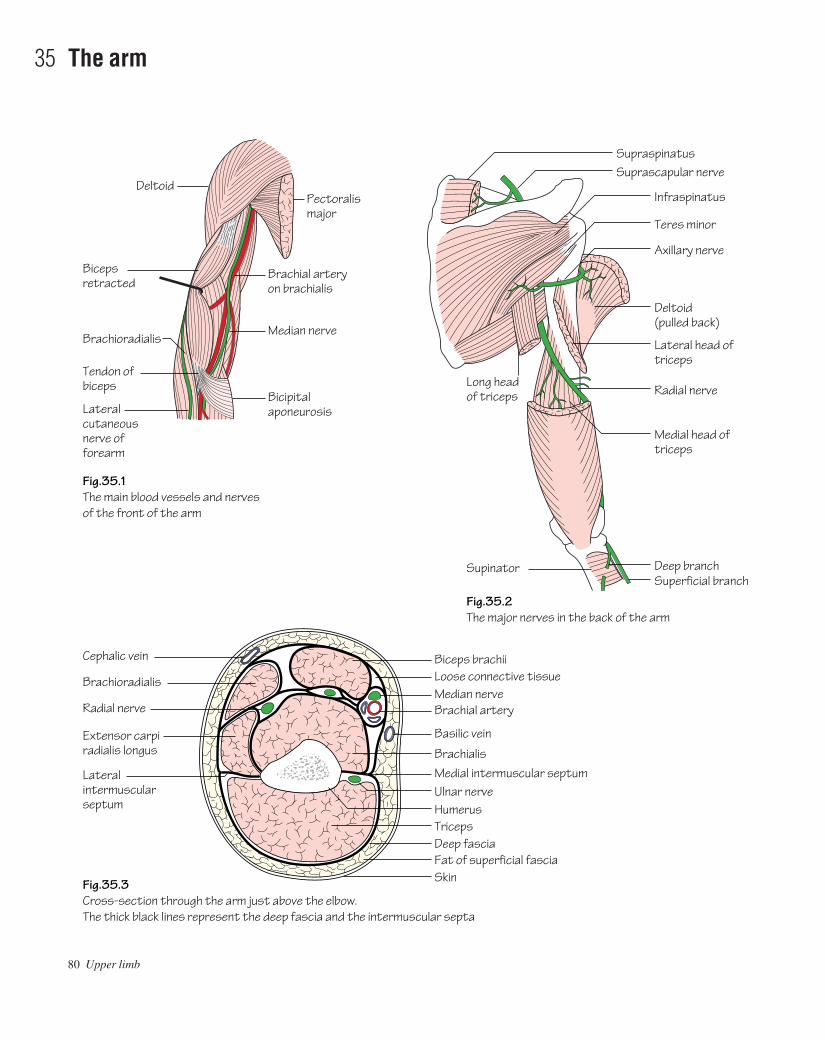
80 Upper limb
35 The arm
Pectoralismajor
Deltoid
Brachial artery on brachialis
Median nerve
Bicepsretracted
Fig.35.1The main blood vessels and nervesof the front of the arm
Fig.35.3Cross-section through the arm just above the elbow.The thick black lines represent the deep fascia and the intermuscular septa
Fig.35.2The major nerves in the back of the arm
Bicipital aponeurosis
Axillary nerve
Teres minor
Infraspinatus
Deltoid (pulled back)
Lateral head of triceps
Radial nerve
Deep branchSuperficial branch
Medial head of triceps
Suprascapular nerve
Long headof triceps
Supinator
Supraspinatus
Loose connective tissueMedian nerveBrachial artery
Basilic vein
BrachialisMedial intermuscular septumUlnar nerveHumerus
Deep fasciaTriceps
Fat of superficial fasciaSkin
Biceps brachiiCephalic vein
Brachioradialis
Radial nerve
Extensor carpiradialis longus
Lateralintermuscular septum
Brachioradialis
Tendon ofbiceps
Lateralcutaneousnerve offorearm
AAAC35 21/5/05 10:49 AM Page 80
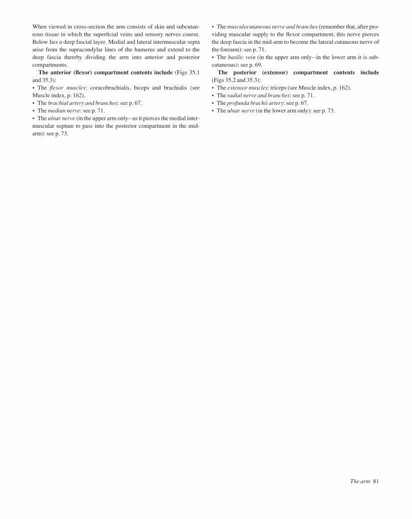
When viewed in cross-section the arm consists of skin and subcutan-eous tissue in which the superficial veins and sensory nerves course.Below lies a deep fascial layer. Medial and lateral intermuscular septaarise from the supracondylar lines of the humerus and extend to thedeep fascia thereby dividing the arm into anterior and posterior compartments.
The anterior (flexor) compartment contents include (Figs 35.1and 35.3):• The flexor muscles: coracobrachialis, biceps and brachialis (seeMuscle index, p. 162).• The brachial artery and branches: see p. 67.• The median nerve: see p. 71.• The ulnar nerve (in the upper arm onlyaas it pierces the medial inter-muscular septum to pass into the posterior compartment in the mid-arm): see p. 73.
• The musculocutaneous nerve and branches (remember that, after pro-viding muscular supply to the flexor compartment, this nerve piercesthe deep fascia in the mid-arm to become the lateral cutaneous nerve ofthe forearm): see p. 71.• The basilic vein (in the upper arm onlyain the lower arm it is sub-cutaneous): see p. 69.
The posterior (extensor) compartment contents include(Figs 35.2 and 35.3):• The extensor muscles: triceps (see Muscle index, p. 162).• The radial nerve and branches: see p. 71.• The profunda brachii artery: see p. 67.• The ulnar nerve (in the lower arm only): see p. 73.
The arm 81
AAAC35 21/5/05 10:49 AM Page 81
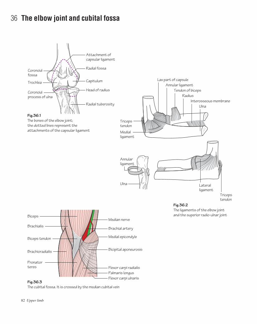
82 Upper limb
36 The elbow joint and cubital fossa
Fig.36.1The bones of the elbow joint; the dotted lines represent the attachments of the capsular ligament
Fig.36.2The ligaments of the elbow joint and the superior radio-ulnar joint
Fig.36.3The cubital fossa. It is crossed by the median cubital vein
Radial fossa
Capitulum
Coronoidfossa
Trochlea
Coronoidprocess of ulna
Head of radius
Radial tuberosity
Tricepstendon
Lax part of capsule
Attachment of capsular ligament
Medialligament
Biceps
Brachialis
Biceps tendon
Brachioradialis
Pronatorteres
Ulna
Annularligament
Annular ligamentTendon of biceps
RadiusInterosseous membrane
Ulna
Triceps tendon
Lateralligament
Median nerve
Brachial artery
Medial epicondyle
Bicipital aponeurosis
Flexor carpi radialisPalmaris longusFlexor carpi ulnaris
AAAC36 21/5/05 10:49 AM Page 82

The elbow joint (Figs 36.1 and 36.2)• Type: synovial hinge joint. At the elbow the humeral capitulumarticulates with the radial head, and the trochlea of the humerus withthe trochlear notch of the ulna. Fossae immediately above the trochleaand capitulum admit the coronoid process of the ulna and the radialhead, respectively, during full flexion. Similarly the olecranon fossaadmits the olecranon process during full elbow extension. The elbowjoint communicates with the superior radio-ulnar joint.• Capsule: the capsule is lax in front and behind to permit full elbowflexion and extension. The non-articular medial and lateral epicondylesare extracapsular.• Ligaments (Fig. 36.2): the capsule is strengthened medially and lat-erally by collateral ligaments.
• The medial collateral ligament is triangular and consists of ant-erior, posterior and middle bands. It extends from the medial epi-condyle of the humerus and the olecranon to the coronoid processof the ulna. The ulnar nerve is adjacent to the medial collateral liga-ment as it passes forwards below the medial epicondyle. Owing tothe close proximity of the ulnar nerve to the humerus it is at risk inmany types of injury, e.g. fracture dislocations, compression andeven during surgical explorations.
• The lateral collateral ligament extends from the lateral epicondyleof the humerus to the annular ligament. The annular ligament isattached medially to the radial notch of the ulna and clasps, butdoes not attach to, the radial head and neck. As the ligament is notattached to the head this is free to rotate within the ligament.
Elbow dislocationThe classical injury is a posterior dislocation caused by a fall on theoutstretched hand. It is commonest in children whilst ossification isincomplete.
The superior radio-ulnar jointThis is a pivot joint. It is formed by the articulation of the radial headand the radial notch of the ulna. The superior radio-ulnar joint commun-icates with the elbow joint.
Movements at the elbowFlexion/extension occur at the elbow joint. Supination/pronation occurmostly at the superior radio-ulnar joint (in conjunction with move-ments at the inferior radio-ulnar joint).• Flexion (140°): biceps, brachialis, brachioradialis and the forearmflexor muscles.• Extension (0°): triceps and to a lesser extent anconeus.• Pronation (90°): pronator teres and pronator quadratus.• Supination (90°): biceps is the most powerful supinator. This move-ment is afforded by the insertion of the muscle on the posterior aspectof the radial tuberosity. Supinator, extensor pollicis longus and brevisare weaker supinators.
The cubital fossa (Fig. 36.3)• This fossa is defined by: a horizontal line joining the two epi-condyles; the medial border of brachioradialis; and the lateral border ofpronator teres. The floor of the fossa consists of brachialis muscle andthe overlying roof of superficial fascia. The median cubital vein runs inthe superficial fascia and connects the basilic to cephalic veins.• Within the fossa the biceps tendon can be palpated. Medial to this liethe brachial artery and the median nerve.• The radial and ulnar nerves lie outside the cubital fossa. The radialnerve passes anterior to the lateral epicondyle between brachialis andbrachioradialis muscles. The ulnar nerve winds behind the medial epicondyle.
The elbow joint and cubital fossa 83
AAAC36 21/5/05 10:49 AM Page 83

84 Upper limb
37 The forearm
Brachio-radialis
Pronatorteres
Flexor carpiradialis
Palmarislongus
Flexor carpiulnaris
Flexor digitorum profundus
Medial epicondyle
Flexor digitorum superficialis
Radial head
Flexor pollicis longus
Pronator quadratus
Ulnar arteryTendon of biceps
Pronator teres
Radial arteryFlexor pollicis longus
Pronator quadratus
First dorsal interosseous
Extensor pollicis longus
Fig.37.1The superficial, intermediate and deep layers of the front of the forearm Fig.37.2
The main arteries and nerves of the front of the forearm
Fig.37.3The muscles of the superficial and deep layers of the back of the forearm and the radial nerve
Common flexor origin
Brachial artery
Supinator
Flexor digitorum superficialis
Median nerveSuperficialradial nerve
Radial nerve
Superficial branch
Supinator
Posteriorinterosseousnerve
Palmaris longus tendon
Dorsal branch of ulnar (cutaneous)
Deep (posteriorinterosseous) branch
Superficial branchDorsal tubercle of radius
Anatomicalsnuffbox
Cutaneous branches to digits
Branches tocarpal joints
Tendons of snuffbox
Extensor retinaculum
Abductor pollicis longusand extensor pollicis brevis
Extensor pollicis longusExtensor pollicis brevis
Abductor pollicis longusPosterior border of ulna
Extensor carpi radialislongus and brevis
Extensor digiti minimiExtensor carpi ulnaris
Extensor digitorumAnconeusFlexor carpi ulnaris
Ulnar nerve
AAAC37 21/5/05 10:48 AM Page 84
The forearm is enclosed in deep fascia which is continuous with that ofthe arm. It is firmly attached to the periosteum of the subcutaneous bor-der of the ulna. Together with the interosseous membrane this dividesthe forearm into anterior and posterior compartments each possessingits own muscles and arterial and nervous supplies. The superficial veinsand cutaneous sensory nerves course in the subcutaneous tissuesuperficial to the deep fascia.
The interosseous membrane• The interosseous membrane unites the interosseous borders of theradius and ulna. The fibres of this tough membrane run obliquely down-wards and medially. A downwards force (e.g. fall on the outstretchedhand) is transmitted from the radius to the ulna and from here to thehumerus and shoulder.• The interosseous membrane provides attachment for neighbouringmuscles.
The contents of the anterior (flexor) compartment of the forearm• Muscles (Fig. 37.1): the muscles within this compartment are con-sidered in superficial, intermediate and deep layers. All of the musclesof the superficial group and part of flexor digitorum superficialis arisefrom the common flexor origin on the medial epicondyle of thehumerus. With the exceptions of flexor carpi ulnaris and the ulnar half
of flexor digitorum profundus, all of the muscles of the anterior com-partment are supplied by the median nerve or its anterior interosseousbranch (see Muscle index, p. 162).• Arteries (Fig. 37.2): ulnar artery and its anterior interosseous branch(via the common interosseous artery); radial artery.• Nerve supply (Fig. 37.2): median nerve and its anterior interosseousbranch; ulnar nerve; superficial radial nerve.
The contents of the posterior fascial (extensor)compartment of the forearm• Muscles (Fig. 37.1): brachioradialis and extensor carpi radialislongus arise separately from the lateral supracondylar ridge of thehumerus. They are innervated by the radial nerve. The remainingextensor muscles are considered in superficial and deep layers whichare innervated by the posterior interosseous branch of the radial nerve.The muscles of the superficial layer arise from the common extensororigin on the lateral epicondyle of the humerus. The muscles of thedeep layer arise from the backs of the radius, ulna and interosseousmembrane (see Muscle index, p. 162).• Arteries: posterior interosseous artery (branch of the commoninterosseous artery).• Nerve supply: posterior interosseous nerve (branch of the radialnerve) (Fig. 37.3).
The forearm 85
AAAC37 21/5/05 10:48 AM Page 85
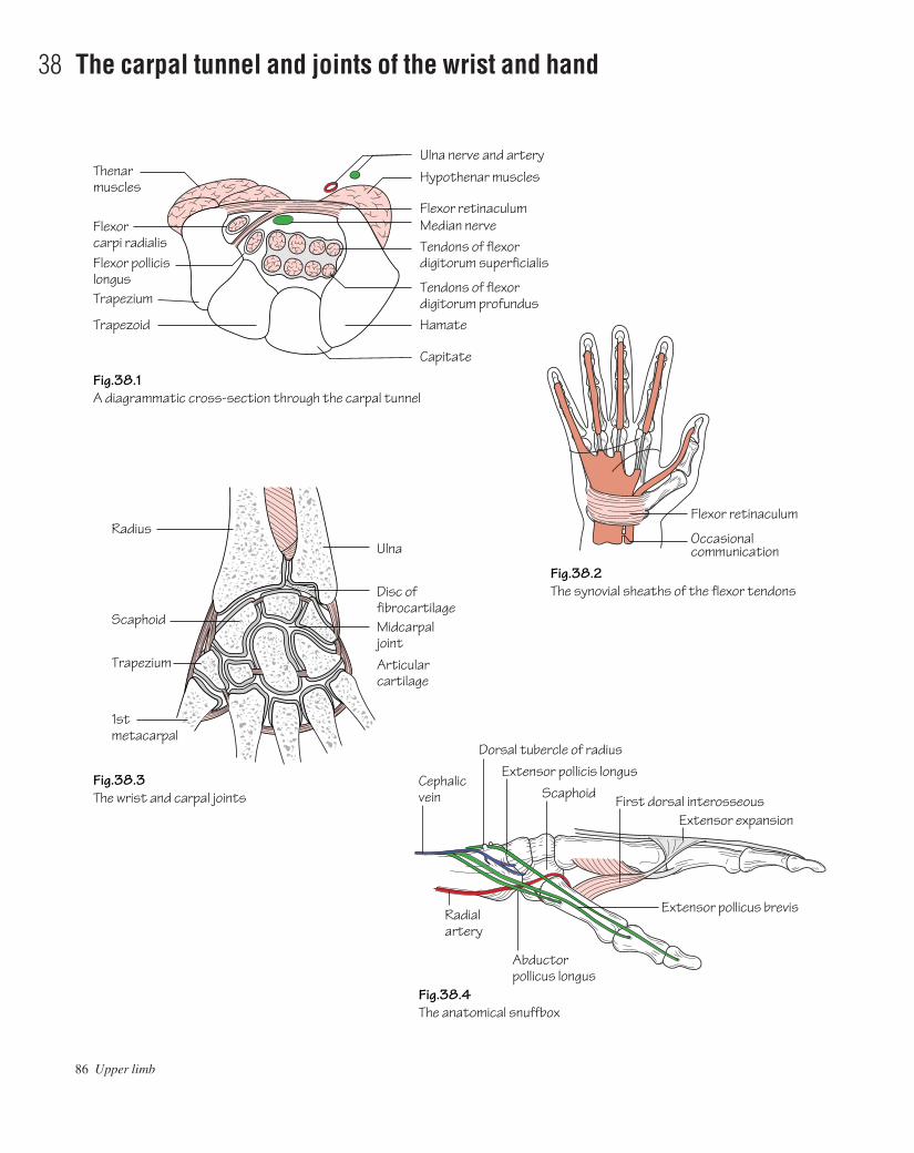
86 Upper limb
38 The carpal tunnel and joints of the wrist and hand
First dorsal interosseousExtensor expansion
Vein
Ulna
Midcarpaljoint
Radius
Scaphoid
Trapezium
1st metacarpal
Cephalicvein
Radialartery
Scaphoid
Dorsal tubercle of radiusExtensor pollicis longus
Extensor pollicus brevis
Abductor pollicus longus
Articular cartilage
Trapezoid
Capitate
Hamate
Median nerveFlexor retinaculum
Hypothenar musclesThenar muscles
Ulna nerve and artery
Fig.38.1A diagrammatic cross-section through the carpal tunnel
Fig.38.2The synovial sheaths of the flexor tendons
Fig.38.3The wrist and carpal joints
Fig.38.4The anatomical snuffbox
Tendons of flexordigitorum superficialis
Tendons of flexordigitorum profundusTrapezium
Flexor carpi radialisFlexor pollicislongus
Flexor retinaculum
Occasional communication
Disc offibrocartilage
AAAC38 21/5/05 10:48 AM Page 86

The flexor retinaculum and carpal tunnel (Fig. 38.1)The carpal tunnel is formed by the carpal bones and the overlying flexorretinaculum. It is through this tunnel that most, but not all, of the fore-arm tendons and the median nerve pass. The flexor retinaculum isattached to four bony pointsathe pisiform, the hook of the hamate, thescaphoid and the trapezium.
The carpal tunnel is narrow and no arteries or veins are transmittedthrough it for risk of potential compression. The median nerve is how-ever at risk of compression when the tunnel is narrowed for any reason.This is the carpal tunnel syndrome and results in signs of median nervemotor and sensory impairment. Note that the ulnar nerve and arterypass over the retinaculum and are thus outside the carpal tunnel.
The synovial sheaths of the flexor tendons (Fig. 38.2)The diagram illustrates the arrangement of the synovial sheaths thatsurround the flexor tendons. It can be seen that flexor pollicis longushas its own sheath and flexor digitorum superficialis and profundusshare one which ends in the palm (except that for the little finger).
The wrist (radiocarpal) joint (Fig. 38.3)• Type: the wrist is a condyloid synovial joint. The distal radius and atriangular disc of fibrocartilage covering the distal ulna form the prox-imal articulating surface. This disc is attached to the edge of the ulnarnotch of the radius and to the base of the styloid process of the ulna andseparates the wrist joint from the inferior radio-ulnar joint. The distalarticulating surface is formed by the scaphoid and lunate bones with thetriquetral participating in adduction.• Capsule: a defined capsule surrounds the joint. It is thickened oneither side by the radial and ulnar collateral ligaments.• Nerve supply: from the anterior interosseous (median) and posteriorinterosseous (radial) nerves.
Wrist movementsFlexion/extension movements occurring at the wrist are accompaniedby movements at the midcarpal joint. Of a total of 80° of wrist flexionthe majority occurs at the midcarpal joint whereas in extension a corres-ponding increased amount occurs at the wrist joint.
The muscles acting on the wrist joint include:• Flexion: all long muscles crossing the joint anteriorly.• Extension: all long muscles crossing the joint posteriorly.• Abduction: flexor carpi radialis and extensors carpi radialis longusand brevis.• Adduction: flexor carpi ulnaris and extensor carpi ulnaris.
The joints of the hand (Fig. 38.3)• Intercarpal joints: the midcarpal joint, located between the prox-imal and distal rows of carpal bones, is the most important of these as itparticipates in wrist movement (see above).• Carpometacarpal joints: the most important of these is the 1st car-pometacarpal (thumb) joint. This is a saddle-shaped joint between thetrapezium and the 1st metacarpal. It is a condyloid synovial joint whichis separate from others in the hand, permitting a range of movementsimilar to that of a ball and socket joint. The most important movementof the thumb is opposition in which the thumb is opposed to the fingersas in holding a pen.• Metacarpophalangeal joints: are synovial condyloid joints.• Interphalangeal joints: are synovial hinge joints.
The anatomical snuffboxFigure 38.4 illustrates the boundaries and contents of the anatomicalsnuffbox.
The carpal tunnel and joints of the wrist and hand 87
AAAC38 21/5/05 10:48 AM Page 87
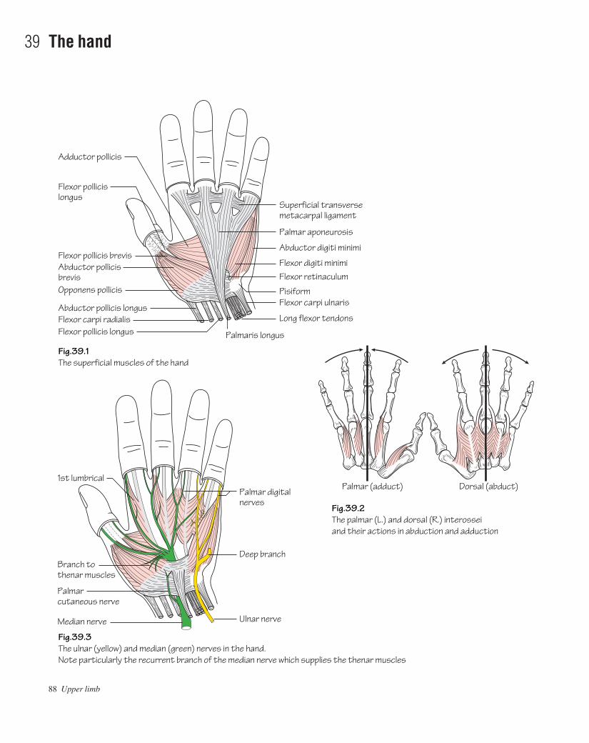
88 Upper limb
39 The hand
Fig.39.1The superficial muscles of the hand
Fig.39.2The palmar (L.) and dorsal (R.) interossei and their actions in abduction and adduction
Fig.39.3The ulnar (yellow) and median (green) nerves in the hand.Note particularly the recurrent branch of the median nerve which supplies the thenar muscles
Abductor pollicis longus
Opponens pollicis
Abductor pollicisbrevis
Flexor pollicis brevis
Flexor pollicislongus
Adductor pollicis
Flexor carpi radialisFlexor pollicis longus
Superficial transversemetacarpal ligament
Palmar aponeurosis
Flexor digiti minimiFlexor retinaculum
PisiformFlexor carpi ulnaris
Abductor digiti minimi
Long flexor tendons
Palmar (adduct) Dorsal (abduct)
Median nerve
1st lumbrical
Ulnar nerve
Deep branch
Palmar digital nerves
Palmar cutaneous nerve
Branch to thenar muscles
Palmaris longus
AAAC39 21/5/05 10:48 AM Page 88

The palm of the hand (Fig. 39.1)• Skin: the skin of the palm is bound to underlying fascia by fibrousbands.• Deep fascia: the palmar aponeurosis is a triangular layer which isattached to the distal border of the flexor retinaculum. Distally theaponeurosis splits into four slips at the bases of the fingers which blendwith the fibrous flexor sheaths (see below). The aponeurosis providesfirm attachment of the overlying skin with protection of the underlyingstructures.• Fibrous flexor sheaths: these are fibrous tunnels in which the flexortendons and their synovial sheaths lie. They arise from the metacarpalheads and pass to the bases of the distal phalanges on the anterior aspectof the digits. They insert into the sides of the phalanges. These sheathsare lax over the joints and thick over the phalanges and hence do notrestrict flexion.• Synovial flexor sheaths: these are sheaths that limit friction betweenthe flexor tendons and the carpal tunnel and fibrous flexor sheaths.• Long flexor tendons: the tendons of flexor digitorum superficialis(FDS) divide into two halves at the level of the proximal phalanx andpass around flexor digitorum profundus (FDP) where they reunite. Atthis point they then split again to insert into the sides of the middle pha-lanx. FDP continues along its path to insert into the distal phalanx.Flexor pollicis longus (FPL) passes through the carpal tunnel in its ownsynovial sheath and inserts into the distal phalanx. The tendons offlexor carpi radialis, palmaris longus and flexor carpi ulnaris passthrough the forearm and also insert in the proximal hand (see Muscleindex, p. 162).
Muscles of the hand (Fig. 39.1)• The thenar muscles: these are the short muscles of the thumb. Theyinclude: abductor pollicis brevis, flexor pollicis brevis, opponens pol-licis and adductor pollicis.• The hypothenar muscles: these are the short muscles of the littlefinger. They include abductor digiti minimi, flexor digiti minimi andopponens digiti minimi.• Lumbricals: these four muscles arise from the tendons of flexor digi-torum profundus. They insert into the radial side of each of the prox-imal phalanges and into the dorsal extensor expansions. The lumbricalsserve to flex the metacarpophalangeal joints without flexing the inter-phalangeal joints.• The interosseous muscles (Fig. 39.2): these comprise eight muscleswhich arise from the shafts of the metacarpals. They are responsible forflexion at the metacarpophalangeal joints and extension of the interpha-langeal joints. They also perform abduction and adduction movements
of the fingers. These movements occur around the middle finger henceadduction is the bringing together of all fingers towards the middlefinger, abduction is moving them away from the middle finger. Thedorsal interossei each arise from two metacarpals and insert into theproximal phalanges so as to provide adduction (P.AD). The dorsalinterossei arise from only one metacarpal and are inserted into the prox-imal phalanges so as to provide abduction (D.AB). Note that the middlefinger cannot be adducted (and hence has no palmar interosseous) butcan be abducted in either direction so it has two dorsal interosseousinsertions.
The dorsum of the hand• Skin: unlike the palm of the hand the skin is thin and freely mobileover the underlying tendons.• Long extensor tendons: the four tendons of extensor digitorum(ED) pass under the extensor retinaculum. On the dorsum of the handthe ED tendon to the index finger is accompanied by the tendon of ex-tensor indicis. The ED tendon to the little finger is accompanied by thedouble tendon of extensor digiti minimi. The ED tendons of the little,ring and middle fingers are connected to each other by fibrous slips. Onthe posterior surface of each finger the extensor tendon spreads to forma dorsal digital expansion. This expansion is triangular shaped and at itsapex splits into three parts: a middle slip which is attached to the base ofthe middle phalanx; and two lateral slips which converge to attach tothe base of the distal phalanx. The base of the expansion receives theappropriate interossei and lumbricals. The tendons of abductor pollicislongus, extensor pollicis brevis and longus form the boundaries of theanatomical snuffbox and proceed to insert into the thumb.
Neurovascular structures of the hand (Fig. 39.3)See chapters on upper limb: arteries, nerves, veins and lymphatics.
Movements of the fingers and thumbThe hand is required to perform a versatile range of movement extend-ing from a firm grasp, such as that needed to carry heavy bags, to preci-sion gripping such as that required to hold a pencil. For precision gripsthe thumb is used to oppose the index finger in which the interpha-langeal joint is extended and the metacarpophalangeal joint is flexed.The thumb opposition is carried out mainly by opponens pollicis whilstthe lumbricals and interossei are responsible for maintaining flexion atthe metacarpophalangeal joints and extension at the interphalangealjoints. Since the thumb is at right angles to the plane of the fingers,abduction of the thumb is a movement away from the plane of the palm.This is used in testing the integrity of the median nerve (abductor pollicis).
The hand 89
AAAC39 21/5/05 10:48 AM Page 89
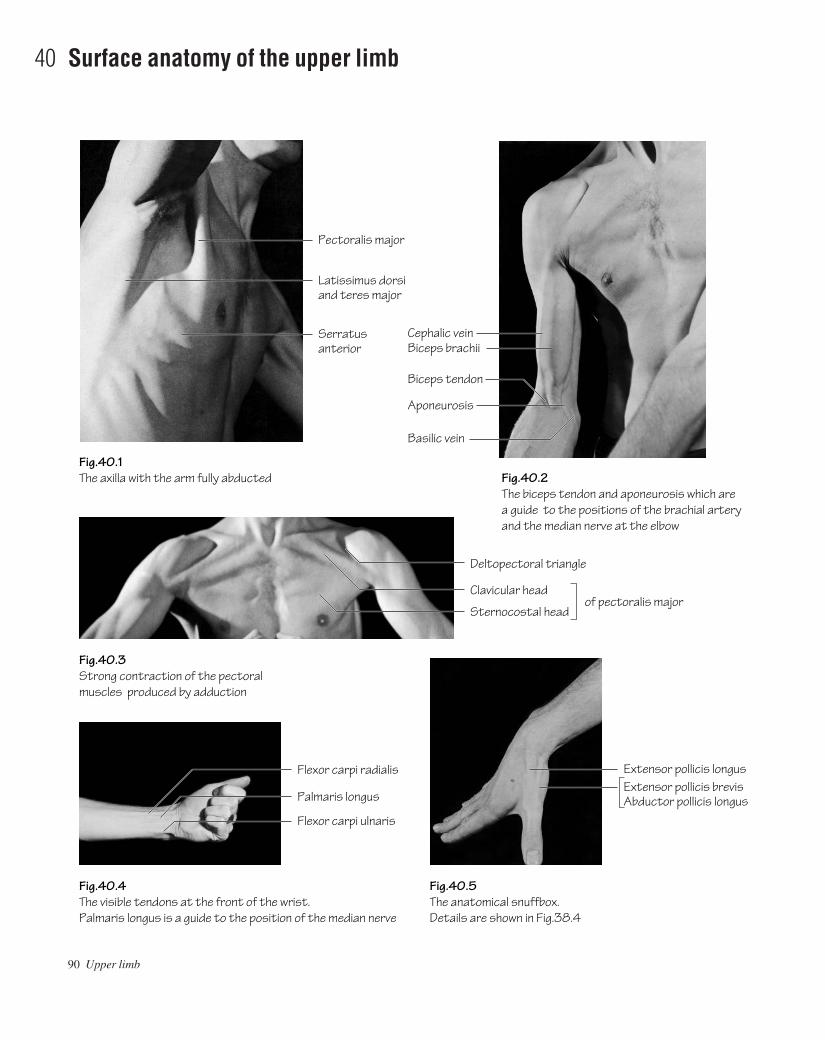
90 Upper limb
40 Surface anatomy of the upper limb
Fig.40.1The axilla with the arm fully abducted Fig.40.2
The biceps tendon and aponeurosis which are a guide to the positions of the brachial artery and the median nerve at the elbow
Fig.40.3Strong contraction of the pectoral muscles produced by adduction
Fig.40.5The anatomical snuffbox. Details are shown in Fig.38.4
Fig.40.4The visible tendons at the front of the wrist. Palmaris longus is a guide to the position of the median nerve
Pectoralis major
Latissimus dorsiand teres major
Clavicular head
Deltopectoral triangle
Sternocostal headof pectoralis major
Extensor pollicis longusExtensor pollicis brevisAbductor pollicis longus
Serratusanterior
Cephalic veinBiceps brachii
Basilic vein
Flexor carpi radialis
Palmaris longus
Flexor carpi ulnaris
Biceps tendon
Aponeurosis
AAAC40 21/5/05 10:47 AM Page 90

Bones and joints• Vertebrae: if a finger is passed down the posterior neck in the mid-line the first bony structure palpated is the spinous process of the 7thcervical vertebra (vertebra prominens)athe first six spinous processesare covered by the ligamentum nuchae.• Scapula: the acromion process can be palpated as a lateral extensionof the spine of the scapula. The spine, superior angle, inferior angleand medial border are palpable posteriorly. The coracoid process canbe palpated below the clavicle anteriorly within the lateral part of thedeltopectoral triangle (Fig 40.1).• Clavicle: is subcutaneous and therefore palpable throughout itslength.• Humerus: the head is palpable in the axilla with the shoulderabducted. The lesser tuberosity can be felt lateral to the coracoid pro-cess. When the arm is externally and internally rotated the lessertuberosity can be felt moving next to the fixed coracoid process.• Elbow: the medial and lateral epicondyles of the humerus and ole-cranon process of the ulna can be palpated in line when the elbow isextended. With the elbow flexed they form a triangle. This assumesimportance clinically in differentiating supracondylar fractures of thehumerus, where the ‘triangle’ is preserved, from elbow dislocationswhere the olecranon comes into line with the epicondyles.• Radius: the radial head can be felt in a hollow distal to the lateralepicondyle on the posterolateral aspect of the extended elbow. Thehead can be felt rotating when the forearm is pronated and supinated.• Ulna: the posterior border is subcutaneous and therefore palpable.• Wrist: the styloid processes of the radius and ulna are palpable. Thedorsal tubercle (of Lister) can be felt on the posterior aspect of the dis-tal radius.• Hand: the pisiform can be palpated at the base of the hypothenareminence. The hook of the hamate can be felt on deep palpation in thehypothenar eminence just distal to the pisiform. The scaphoid bone canbe felt within the anatomical snuffbox (Fig, 40.5).
The soft tissues• Axilla: the anterior axillary fold (formed by the lateral border of pec-toralis major) and the posterior axillary fold (formed by latissimus dorsias it passes around the lower border of teres major) are easily palpable(Fig. 40.1).• Pectoralis major: contracts strongly during arm adduction (Fig.40.3); this is useful in the examination of breast lumps.• Breast: the base of the breast overlaps the 2nd to 6th ribs and extendsfrom the sternum to the mid-axillary line. The nipple (in males) usuallyoverlies the 4th intercostal space.
• Anterior wrist: the proximal transverse crease corresponds to thelevel of the wrist joint. The distal transverse crease lies at the level ofthe proximal border of the flexor retinaculum.• Anatomical snuffbox: the boundaries are formed medially by ex-tensor pollicis longus and laterally by the tendons of abductor pollicislongus and extensor pollicis brevis.
Vessels• The subclavian artery can be felt pulsating as it crosses the 1st rib.• The brachial artery bifurcates into radial and ulnar branches at thelevel of the neck of the radius. The brachial pulse is felt by pressing lat-erally at a point medial to the bicipital tendon (Fig. 40.2). This is thepulse used when taking blood pressure measurements.• At the wrist the radial artery courses on the radial side of flexor carpiradialis (Fig. 40.4) and the ulnar artery and nerve course on the radialside of flexor carpi ulnaris. The pulses of both are easily felt at thesepoints. The radial artery can also be felt in the anatomical snuffbox.• The superficial palmar arch is impalpable and reaches as far as theproximal palmar crease. The deep palmar arch reaches a point approx-imately one fingerbreadth proximal to the superficial arch.• The dorsal venous network (on the dorsum of the hand) drains later-ally into the cephalic vein and medially into the basilic. These veins canbe identified in most lean subjects. The median cubital vein is usuallyvisible in the cubital fossa.
NervesThe ulnar nerve can usually be rolled as it courses behind the medialepicondyleaan important point when considering surgical approachesto the elbow and fractures of the medial epicondyle.
The surface markings of impalpable nerves must be known for safesurgical incisions. These correspond to:• Axillary nerve: winds around behind the surgical neck of thehumerus.• Radial nerve: crosses from medial to lateral behind the humeralshaft at its midpoint.• Posterior interosseous branch (of radial nerve): winds around theradius three fingerbreadths distal to the head of the radius.• Median nerve (at the wrist): lies in the midline, just lateral to the ten-don of palmaris longus.• Ulnar nerve (at the wrist): lies immediately medial to the ulnarartery.
Surface anatomy of the upper limb 91
AAAC40 21/5/05 10:47 AM Page 91
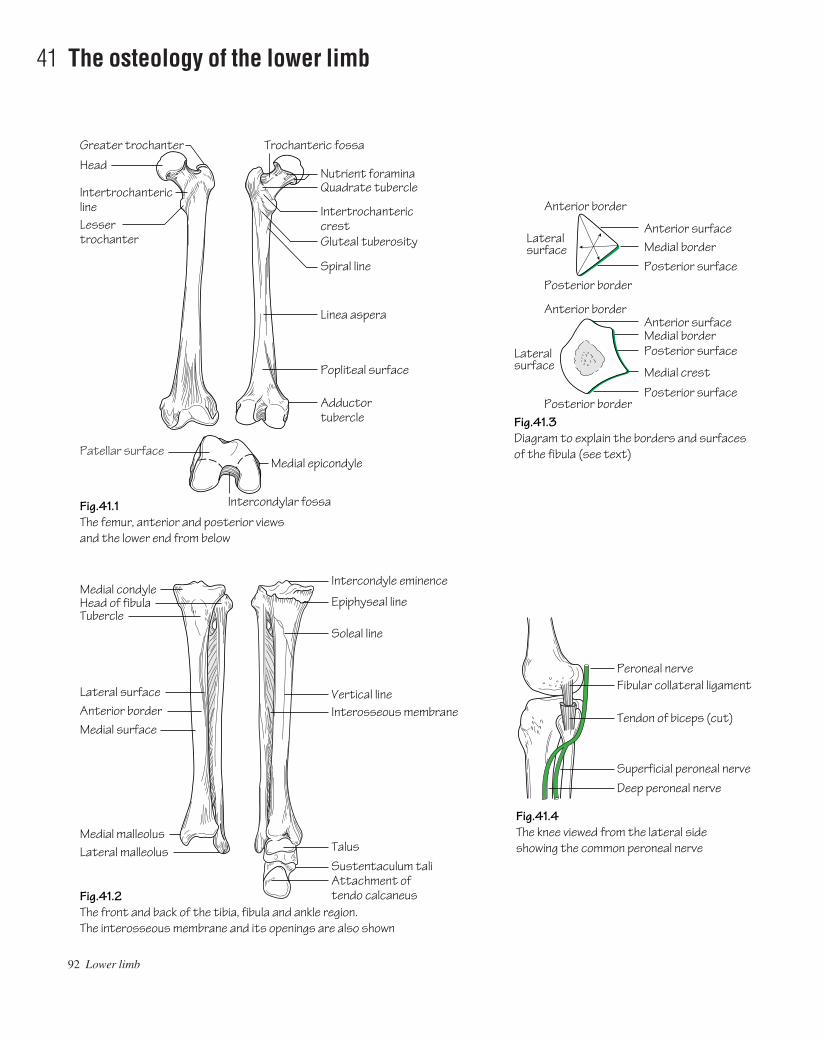
92 Lower limb
41 The osteology of the lower limb
Greater trochanter
Fig.41.1The femur, anterior and posterior viewsand the lower end from below
Fig.41.2The front and back of the tibia, fibula and ankle region.The interosseous membrane and its openings are also shown
Fig.41.4The knee viewed from the lateral side showing the common peroneal nerve
Fig.41.3Diagram to explain the borders and surfacesof the fibula (see text)
Head
Medial condyleHead of fibulaTubercle
Intercondyle eminence
Epiphyseal line
Soleal line
Vertical lineFibular collateral ligament
Tendon of biceps (cut)
Superficial peroneal nerveDeep peroneal nerve
Peroneal nerve
Interosseous membrane
TalusSustentaculum taliAttachment oftendo calcaneus
Lateral surfaceAnterior borderMedial surface
Medial malleolusLateral malleolus
Intertrochantericline
Trochanteric fossa
Quadrate tubercleNutrient foramina
IntertrochantericcrestGluteal tuberosity
Spiral line
Linea aspera
Popliteal surface
Adductor tubercle
Lesser trochanter
Medial epicondyle
Intercondylar fossa
Medial crest
Posterior surface
Posterior surfaceMedial borderAnterior surface
Posterior surface
Lateralsurface
Lateralsurface
Medial borderAnterior surface
Posterior border
Anterior border
Anterior border
Posterior border
Patellar surface
AAAC41 21/5/05 10:55 AM Page 92
The femur (Fig. 41.1)The femur is the longest bone in the body. It has the following charac-teristic features:• The femoral head articulates with the acetabulum of the hip bone atthe hip joint. It extends from the femoral neck and is rounded, smoothand covered with articular cartilage. This configuration permits a widerange of movement. The head faces medially, upwards and forwardsinto the acetabulum. The fovea is the central depression on the head towhich the ligamentum teres is attached.• The femoral neck forms an angle of 125° with the femoral shaft.Pathological lessening or widening of the angle is termed coxa varaand coxa valga deformity, respectively.• The femoral shaft constitutes the length of the bone. At its upper endit carries the greater trochanter and, posteromedially, the lessertrochanter. Anteriorly the rough trochanteric line, and posteriorly thesmooth trochanteric crest, demarcate the junction between the shaftand neck. The linea aspera is the crest seen running longitudinallyalong the posterior surface of the femur splitting in the lower portioninto the supracondylar lines. The medial supracondylar line terminatesat the adductor tubercle.• The lower end of the femur comprises the medial and lateral femoralcondyles. These bear the articular surfaces for articulation with the tibiaat the knee joint. The lateral condyle is more prominent than the medial.This prevents lateral displacement of the patella. The condyles are sep-arated posteriorly by a deep intercondylar notch. Anteriorly the lowerfemoral aspect is smooth for articulation with the posterior surface ofthe patella.
The tibia (Fig. 41.2)The tibia serves to transfer weight from the femur to the talus. It has thefollowing characteristics:• The flattened upper end of the tibiaathe tibial plateauacomprisesmedial and lateral tibial condyles for articulation with the respectivefemoral condyles. In contrast to the femoral condyles, the medial tibialcondyle is the larger of the two.• The intercondylar area is the space between the tibial condyles onwhich can be seen two projectionsathe medial and lateral intercondy-lar tubercles. Together these constitute the intercondylar eminence.The horns of the lateral meniscus are attached close to either side of theeminence.• On the anterior upper shaft the tibial tuberosity is easily identifiable.This is the site of insertion of the ligamentum patellae.• The shaft is triangular in cross-section. It has anterior, medial and lat-eral borders and posterior, lateral and medial surfaces.• The anterior border and medial surface of the shaft are subcutaneousthroughout its length. For this reason the tibial shaft is the commonestsite for open fractures.
• On the posterior surface of the shaft an oblique lineathe soleallineademarcates the tibial origin of soleus. Popliteus inserts into thetriangular area above the soleal line.• The fibula articulates with the tibia superiorly at an articular facet on the postero-inferior aspect of the lateral condyleathe superiortibiofibular joint (synovial).• The fibular notch is situated laterally on the lower end of the tibia forarticulation with the fibula at the inferior tibiofibular joint ( fibrous).• The tibia projects inferiorly as the medial malleolus. It constitutes themedial part of the mortice that stabilizes the talus. The medial malleolusis grooved posteriorly for the passage of the tendon of tibialis posterior.
The fibula (Fig. 41.2)The fibula does not form part of the knee joint and does not participatein weight transmission. The main functions of the fibula are to provideorigin for muscles and to participate in the ankle joint. It has the follow-ing characteristic features:• The styloid process is a prominence on the fibular head onto whichthe tendon of biceps is inserted (around the lateral collateral ligament)(Fig. 41.4).• The fibular neck separates the head from the fibular shaft. The com-mon peroneal nerve winds around the neck prior to dividing intosuperficial and deep branches. This nerve is subject to injury in fibularneck fractures with resulting footdrop (Fig. 41.4).• The fibula is triangular in cross-section. It has anterior, medial(interosseous) and posterior borders with anterior, lateral and posteriorsurfaces. The medial crest is on the posterior surface (Fig. 41.3).• The lower end of the fibula constitutes the lateral malleolus. This isthe lateral part of the mortice that stabilizes the talus. It bears a smoothmedial surface for articulation with the talus. The posterior aspect ofthe malleolus is grooved for the passage of the tendons of peroneuslongus and brevis. The lateral malleolus projects further downwardsthan the medial malleolus.
The patella• The ligamentum patellae, which is attached to the apex of the patellaand the tibial tuberosity, is the true insertion of the quadriceps and thepatella is thus a sesamoid bone (the largest in the body). This arrange-ment constitutes the extensor mechanism. Injuries can occur to anypart of the mechanism due to forcible quadriceps contraction, e.g. rup-tured quadriceps expansion, rupture of the ligamentum patellae oravulsion of the tibial tuberosity.• The posterior surface of the patella is smooth and covered with articu-lar cartilage. It is divided into a large lateral and a smaller medial facetfor articulation with the femoral condyles.
Bones of the footSee p. 114.
The osteology of the lower limb 93
AAAC41 21/5/05 10:55 AM Page 93

94 Lower limb
42 The arteries of the lower limb
Superficial external pudendalDeep external pudendal
Profunda femorisMedial circumflex
Superficial epigastric (to abdominal wall)
Fig.42.1The course and major branches of the femoral artery
Fig.42.2The medial and lateral plantar arteries
Femoral sheathFemoral vein
Gap in adductor magnus
Popliteal
Soleus arch
Posterior tibial
Anterior tibial
Dorsalis pedis
Extensor retinaculum
Passes into sole to formdeep plantar arch
First dorsal metatarsal
Arcuate
Peroneal
Anastomosiswith dorsalartery
Medialplantar artery
Abductor hallucis
Flexor digitorumaccessorius
Lateral plantar artery
Deep plantar arch
Plantar metatarsalartery
Deep peronealnerve
Genicular arteriesto knee joint
Perforating arteries
Lateral circumflex
Femoral nerve
Superficialcircumflexiliac
Femoral
Fig.42.3An angiogram of the lower limbs showing stenosis of the femoral artery on the right side. (The profunda is often known as the deep femoral and the continuation of the femoral artery as thesuperficial femoral)
Stenosis
Right femoral artery
Profunda femoris
AAAC42 21/5/05 10:54 AM Page 94
The femoral artery• Course: the femoral artery commences as a continuation of the ex-ternal iliac artery behind the inguinal ligament at the mid-inguinal point.In the groin the femoral vein lies immediately medial to the artery andboth are enclosed in the femoral sheath. In contrast, the femoral nervelies immediately lateral to the femoral sheath. The femoral arterydescends the thigh to pass under sartorius and then through the adductor(Hunter’s) canal to become the popliteal artery.• Branches:
• Branches in the upper part of the femoral triangleafourbranches are given off which supply the superficial tissues of thelower abdominal wall and perineum (see Fig. 44.1).
• Profunda femorisaarises from the lateral side of the femoralartery 4 cm below the inguinal ligament. Near its origin it gives riseto medial and lateral circumflex femoral branches. These con-tribute to the trochanteric and cruciate anastomoses (see below).The profunda descends deep to adductor longus in the medial com-partment of the thigh and gives rise to four perforating branches.These circle the femur posteriorly perforating, and supplying, allmuscles in their path. The profunda and perforating branches ulti-mately anastomose with the genicular branches of the poplitealartery.
The trochanteric anastomosisThis arterial anastomosis is formed by branches from the medial andlateral circumflex femoral, the superior gluteal and, usually, the in-ferior gluteal arteries. It lies close to the trochanteric fossa and pro-vides branches that ascend the femoral neck beneath the retinacularfibres of the capsule to supply the femoral head.
The cruciate anastomosisThis anastomosis constitutes a collateral supply. It is formed by: thetransverse branches of the medial and lateral circumflex femoral arter-ies, the descending branch of the inferior gluteal artery and the ascend-ing branch of the 1st perforating branch of the profunda.
The popliteal artery• Course: the femoral artery continues as the popliteal artery as itpasses through the hiatus in adductor magnus to enter the poplitealspace. From above, it descends on the posterior surface of the femur,the capsule of the knee joint and then on the fascia overlying popliteusto pass under the fibrous arch of soleus where it bifurcates into anteriorand posterior tibial arteries. In the fossa it is the deepest structure, ren-dering it difficult to feel its pulsations. The popliteal vein crosses theartery superficially and the tibial nerve crosses from lateral to medialover the vein. The peroneal branch of the posterior tibial may ariseearly to form the popliteal trifurcation.• Branches: muscular, sural and five genicular arteries are given off.The last form a rich anastomosis around the knee.
The anterior tibial artery• Course: the anterior tibial artery passes anteriorly from its origin,accompanied by its venae comitantes, over the upper border of the inter-osseous membrane and then descends over the anterior surface of themembrane giving off muscular branches to the extensor compartmentof the leg. The artery crosses the front of the ankle joint midway be-
tween the malleoli where it becomes the dorsalis pedis artery. Tibialisanterior and extensor digitorum longus flank the artery throughout itscourse on its medial and lateral sides, respectively. Extensor hallucislongus commences on the lateral side but crosses the artery to liemedial by the end of its course. The dorsalis pedis artery passes on thedorsum of the foot to the level of the base of the metatarsals and thenbetween the two heads of the first dorsal interosseous muscle to gainaccess to the sole and complete the deep plantar arch. Prior to passing tothe sole it gives off the 1st dorsal metatarsal branch and via an arcuatebranch the three remaining dorsal metatarsal branches (Fig. 42.1).• Branches of the anterior tibial artery include: muscular andmalleolar branches.
The posterior tibial artery• Course: the posterior tibial artery arises as a terminal branch of thepopliteal artery. It is accompanied by its venae comitantes and suppliesthe flexor compartment of the leg. Approximately midway down thecalf the tibial nerve crosses behind the artery from medial to lateral.The artery ultimately passes behind the medial malleolus to divide intomedial and lateral plantar arteries under the flexor retinaculum. Thelatter branches gain access to the sole deep to abductor hallucis.Posterior to the medial malleolus the structures which can beidentifiedafrom front to backaare: tibialis posterior, flexor digitorumlongus, posterior tibial artery and venae comitantes, the tibial nerve andflexor hallucis longus.• Branches:
• Peroneal arteryathis artery usually arises from the posterior tibialartery approximately 2.5 cm along its length. It courses betweentibialis posterior and flexor hallucis longus and supplies the per-oneal (lateral) compartment of the leg. It ends by dividing into aperforating branch that pierces the interosseous membrane and alateral calcaneal branch.
• Other branchesathe posterior tibial artery gives rise to nutrientand muscular branches throughout its course.
• Lateral plantar arteryapasses between flexor accessorius andflexor digitorum brevis to the lateral aspect of the sole where itdivides into superficial and deep branches. The deep branch runsbetween the 3rd and 4th muscle layers of the sole to continue as thedeep plantar arch which is completed by the termination of thedorsalis pedis artery. The arch gives rise to plantar metatarsalbranches which supply the toes (Fig. 42.2).
• Medial plantar arteryaruns on the medial aspect of the sole andsends branches which join with the plantar metatarsal branches ofthe lateral plantar artery to supply the toes.
Peripheral vascular disease (Fig. 42.3)Atheroma causes narrowing of the peripheral arteries with a con-sequent reduction in flow. Whilst flow may be adequate for tissue perfusion at rest, exercise causes pain due to ischaemia (intermittentclaudication). When symptoms are intolerable, pain is present at rest or ischaemic ulceration has occurred, arterial reconstruction isrequired. Reconstruction is performed using either the patient’s ownsaphenous vein or a synthetic graft (Dacron or PTFE) to bypass theocclusion. Disease which is limited in extent may be suitable for inter-ventional procedures such as percutaneous transluminal angioplasty(PTA) or stent insertion.
The arteries of the lower limb 95
AAAC42 21/5/05 10:54 AM Page 95
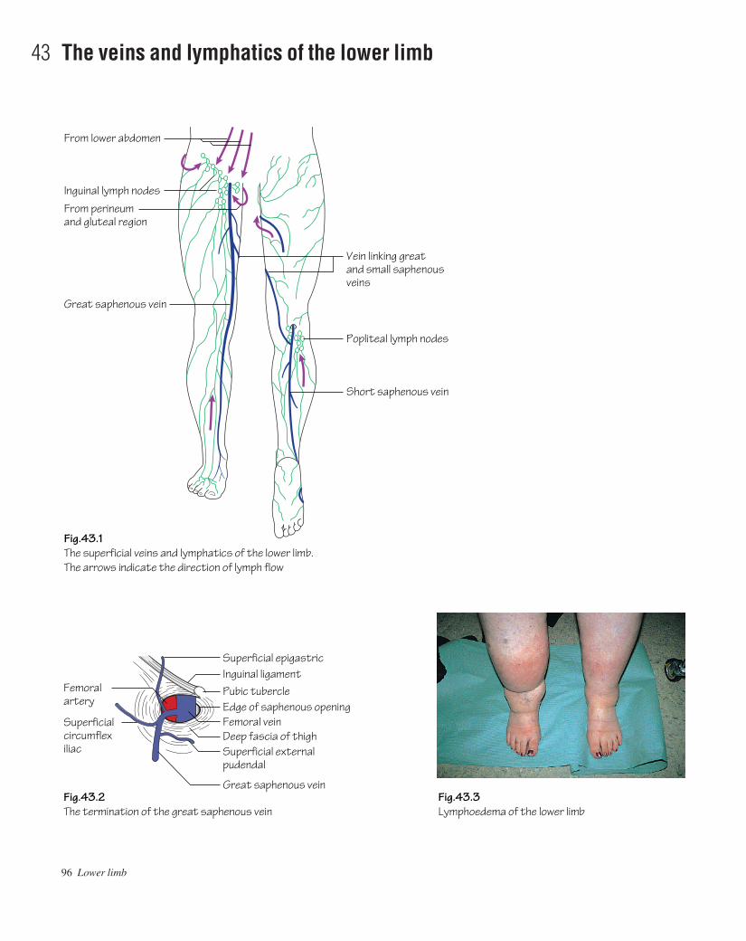
96 Lower limb
43 The veins and lymphatics of the lower limb
Popliteal lymph nodes
From lower abdomen
Inguinal lymph nodesFrom perineumand gluteal region
Great saphenous vein
Short saphenous vein
Fig.43.1The superficial veins and lymphatics of the lower limb.The arrows indicate the direction of lymph flow
Fig.43.3Lymphoedema of the lower limb
Vein linking greatand small saphenousveins
Fig.43.2The termination of the great saphenous vein
Superficial epigastric
Superficial external pudendal
Edge of saphenous opening
Great saphenous vein
Superficialcircumflexiliac
Femoral artery
Pubic tubercle
Femoral veinDeep fascia of thigh
Inguinal ligament
AAAC43 21/5/05 10:54 AM Page 96

The superficial veins of the lower limb (Fig. 43.1)The superficial system comprises the great and small saphenous veins.These are of utmost clinical importance as they are predisposedtowards becoming varicose and consequently often require surgery.They are also the commonly used conduits for coronary artery surgery.• The great saphenous vein arises from the medial end of the dorsalvenous network on the foot. It passes anterior to the medial malleolus,along the anteromedial aspect of the calf (with the saphenous nerve),migrates posteriorly to a handbreadth behind patella at the knee andthen courses forward to ascend the anteromedial thigh. It pierces thecribriform fascia to drain into the femoral vein at the saphenous open-ing. The terminal part of the great saphenous vein usually receivessuperficial tributaries from the external genitalia and the lower abdom-inal wall (Fig. 43.2). At surgery these help to distinguish the saphenousfrom the femoral vein as the only tributary draining into the latter is thesaphenous vein. Anteromedial and posterolateral femoral (lateralaccessory) tributaries, from the medial and lateral aspects of the thigh,also sometimes drain into the great saphenous vein below the saphen-ous opening.
The great saphenous vein is connected to the deep venous system atmultiple levels by perforating veins. These usually occur above andbelow the medial malleolus, in the gaiter area, in the mid-calf region,below the knee and one long connection in the lower thigh. The valvesin the perforators are directed inwards so that blood flows fromsuperficial to deep systems from where it can be pumped upwardsassisted by the muscular contractions of the calf muscles. The deep sys-tem is consequently at higher pressure than the superficial and thus,should the valves in the perforators become incompetent, the increasedpressure is transmitted to the superficial system and these veins becomevaricose.• The small saphenous vein arises from the lateral end of the dorsalvenous network on the foot. It passes behind the lateral malleolus andover the back of the calf to pierce the deep fascia in an inconstant posi-tion to drain into the popliteal vein.
The deep veins of the lower limbThe deep veins of the calf are the venae comitantes of the anterior andposterior tibial arteries which go on to become the popliteal andfemoral veins. The deep veins form an extensive network within the
posterior compartment of the calfathe soleal plexus from which bloodis assisted upwards against gravitational forces by muscular contrac-tion during exercise. Failure of this ‘muscle pump’ to work efficiently,as occurs, for instance, during long flights in cramped conditions, maycause deep venous thrombosis (DVT).
Varicose veinsThese are classified as:• Primary: due to inherent valve dysfunction.• Secondary: due to impedance of flow within the deep venous circula-tion. These can occur in pregnancy or due to obstruction caused bypelvic tumours or previous deep venous thrombosis.
In both cases the superficial veins are subjected to increased pres-sure and become varicose.
The lymphatics of the lower limb (Fig. 43.1)The lymph nodes of the groin are arranged into superficial and deepgroups. The superficial inguinal group lie in the superficial fascia andare arranged in two chains:• Longitudinal chain: these lie along the terminal portion of the saphen-ous vein. They receive lymph from the majority of the superficial tis-sues of the lower limb.• Horizontal chain: these lie parallel to the inguinal ligament. Theyreceive lymph from the superficial tissues of the: lower trunk below thelevel of the umbilicus, the buttock, the external genitalia and the lowerhalf of the anal canal. The superficial nodes drain into the deep nodesthrough the saphenous opening in the deep fascia.
The deep inguinal nodes are situated medial to the femoral vein.They are usually three in number. These nodes receive lymph from allof the tissues deep to the fascia lata of the lower limb. In addition theyalso receive lymph from the skin and superficial tissues of the heel andlateral aspect of the foot by way of the popliteal nodes. The deep nodesconvey lymph to external iliac and thence to the para-aortic nodes.
Obstruction of lymphatics results in lymphoedema (Fig. 43.3). Thiscan be congenital, due to aberrant lymphatic formation, or acquiredsuch as post radiotherapy or following certain infections. In develop-ing countries infection with Filaria bancrofti is a significant cause oflymphoedema that can progress to massive proportions requiring limbreduction or even amputation.
The veins and lymphatics of the lower limb 97
AAAC43 21/5/05 10:54 AM Page 97

98 Lower limb
44 The nerves of the lower limb I
Inguinal ligament
Pubic tubercle
Adductor longusFemoral veinGreat saphenous veinFemoral artery
Gracilis
Saphenous nerveTo vastus medialis
Deep fascia(Skin of medial leg and foot)
Lateral cutaneousnerve of thigh
Tensor fasciae latae
Femoral nerveIliacus
Psoas
Pectineus
Sartorius
Rectus femoris
Nerve to sartorius
Fig.44.1The femoral nerve and its major branches.The upper diagram shows the structuresthat pass under the inguinal ligament
Fig.44.2The anterior and posterior divisions of the obturator nerve
Inguinal ligament
External oblique aponeurosisFemoral nerveFemoral arteryFemoral veinFemoral canal
PectineusPubic tubercleLacunar ligament
To pectineus
Branch toknee joint
Gracilis
Anterior division
Adductorlongus
Adductor brevis
Posteriordivision
Pectineus
Obturatorexternus
Lateral cutaneousnerve of thigh
Anterior superioriliac spine
Iliacus
Psoastendon
To vastus lateralis
To vastus intermediusand rectus femoris
Medial cutaneousnerve of thigh(Skin of medial thigh)
Intermediate cutaneous nerve of thigh(Skin of front of thigh)
AAAC44 21/5/05 10:54 AM Page 98
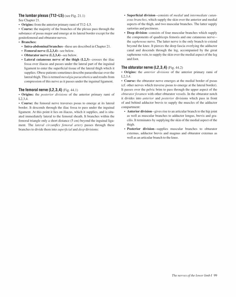
The lumbar plexus (T12–L5) (see Fig. 21.1)See Chapter 21.• Origins: from the anterior primary rami of T12–L5.• Course: the majority of the branches of the plexus pass through thesubstance of psoas major and emerge at its lateral border except for thegenitofemoral and obturator nerves.• Branches:
• Intra-abdominal branchesathese are described in Chapter 21.• Femoral nerve (L2,3,4)asee below.• Obturator nerve (L2,3,4)asee below.• Lateral cutaneous nerve of the thigh (L2,3)acrosses the iliac
fossa over iliacus and passes under the lateral part of the inguinalligament to enter the superficial tissue of the lateral thigh which itsupplies. Obese patients sometimes describe paraesthesiae over thelateral thigh. This is termed meralgia paraesthetica and results fromcompression of this nerve as it passes under the inguinal ligament.
The femoral nerve (L2,3,4) (Fig. 44.1)• Origins: the posterior divisions of the anterior primary rami of L2,3,4.• Course: the femoral nerve traverses psoas to emerge at its lateralborder. It descends through the iliac fossa to pass under the inguinal ligament. At this point it lies on iliacus, which it supplies, and is situ-ated immediately lateral to the femoral sheath. It branches within thefemoral triangle only a short distance (5 cm) beyond the inguinal liga-ment. The lateral circumflex femoral artery passes through thesebranches to divide them into superficial and deep divisions:
• Superficial divisionaconsists of medial and intermediate cutan-eous branches, which supply the skin over the anterior and medialaspects of the thigh, and two muscular branches. The latter supplysartorius and pectineus.
• Deep divisionaconsists of four muscular branches which supplythe components of quadriceps femoris and one cutaneous nervea
the saphenous nerve. The latter nerve is the only branch to extendbeyond the knee. It pierces the deep fascia overlying the adductorcanal and descends through the leg, accompanied by the greatsaphenous vein, to supply the skin over the medial aspect of the legand foot.
The obturator nerve (L2,3,4) (Fig. 44.2)• Origins: the anterior divisions of the anterior primary rami of L2,3,4.• Course: the obturator nerve emerges at the medial border of psoas(cf. other nerves which traverse psoas to emerge at the lateral border). It passes over the pelvic brim to pass through the upper aspect of theobturator foramen with other obturator vessels. In the obturator notch it divides into anterior and posterior divisions which pass in front of and behind adductor brevis to supply the muscles of the adductorcompartment:
• Anterior divisionagives rise to an articular branch to the hip jointas well as muscular branches to adductor longus, brevis and gra-cilis. It terminates by supplying the skin of the medial aspect of thethigh.
• Posterior divisionasupplies muscular branches to obturatorexternus, adductor brevis and magnus and obturator externus aswell as an articular branch to the knee.
The nerves of the lower limb I 99
AAAC44 21/5/05 10:54 AM Page 99
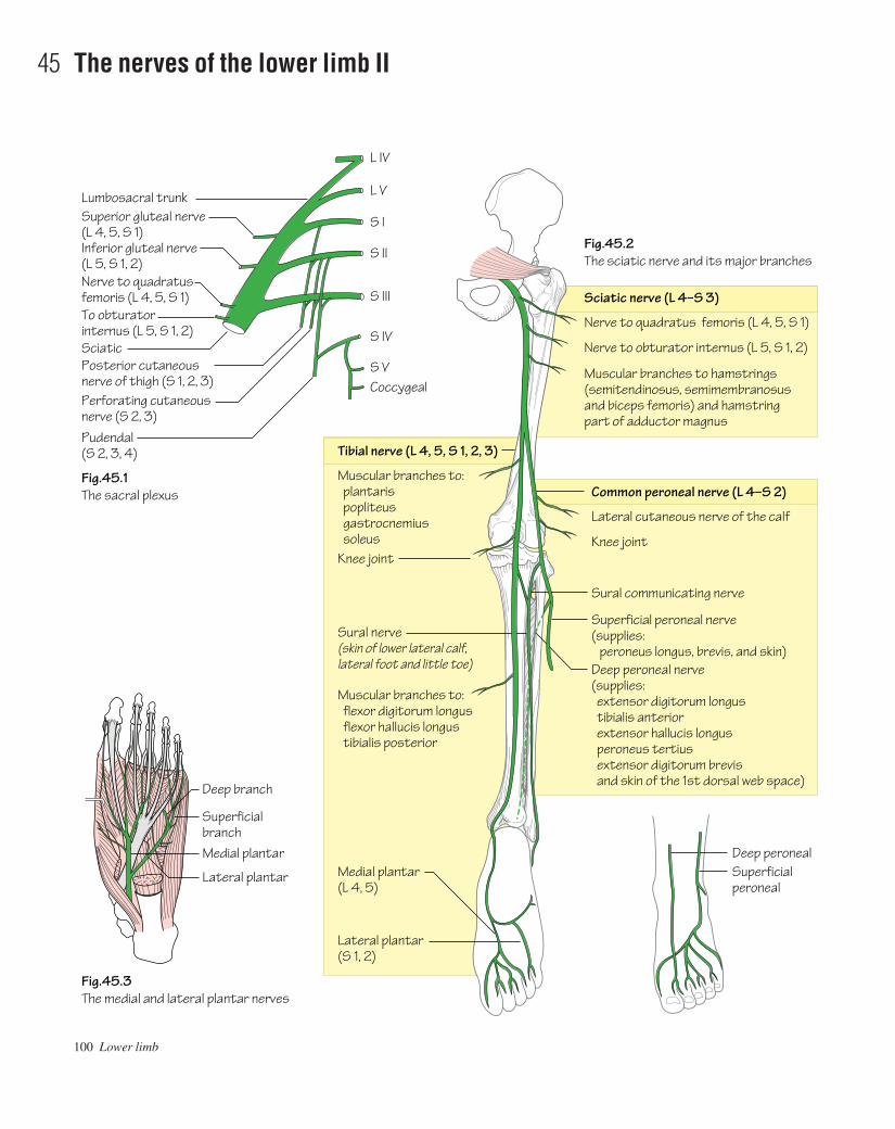
100 Lower limb
45 The nerves of the lower limb II
Deep branch
L IV
L V
S I
S II
S III
S IV
S VCoccygeal
Lumbosacral trunkSuperior gluteal nerve(L 4, 5, S 1)Inferior gluteal nerve(L 5, S 1, 2)Nerve to quadratus femoris (L 4, 5, S 1)To obturator internus (L 5, S 1, 2)SciaticPosterior cutaneous nerve of thigh (S 1, 2, 3)Perforating cutaneousnerve (S 2, 3)Pudendal (S 2, 3, 4)
SuperficialbranchMedial plantar
Lateral plantar
Fig.45.1The sacral plexus
Fig.45.2The sciatic nerve and its major branches
Fig.45.3The medial and lateral plantar nerves
Nerve to quadratus femoris (L 4, 5, S 1)
Nerve to obturator internus (L 5, S 1, 2)
Lateral cutaneous nerve of the calf
Knee joint
Sural communicating nerve
Superficial peroneal nerve(supplies: peroneus longus, brevis, and skin)Deep peroneal nerve(supplies: extensor digitorum longus tibialis anterior extensor hallucis longus peroneus tertius extensor digitorum brevis and skin of the 1st dorsal web space)
Muscular branches to hamstrings(semitendinosus, semimembranosus and biceps femoris) and hamstring part of adductor magnus
Muscular branches to: plantaris popliteus gastrocnemius soleus
Sciatic nerve (L 4–S 3)
Common peroneal nerve (L 4–S 2)
Tibial nerve (L 4, 5, S 1, 2, 3)
Knee joint
Sural nerve(skin of lower lateral calf,lateral foot and little toe)
Muscular branches to: flexor digitorum longus flexor hallucis longus tibialis posterior
Medial plantar(L 4, 5)
Lateral plantar(S 1, 2)
Deep peroneal Superficialperoneal
AAAC45 21/5/05 10:53 AM Page 100

The sacral plexus (L4–S4) (Fig. 45.1)• Origins: from the anterior primary rami of L4–S4.• Course: the sacral nerves emerge through the anterior sacral foram-ina. The nerves unite, and are joined by the lumbosacral trunk (L4,5),anterior to piriformis.• Branches: the branches of the sacral plexus include:
• The superior gluteal nerve (L4,5,S1)aarises from the roots of thesciatic nerve and passes through the greater sciatic foramen abovethe upper border of piriformis. In the gluteal region it runs belowthe middle gluteal line between gluteus medius and minimis (bothof which it supplies) before terminating in the substance of tensorfasciae latae.
• The inferior gluteal nerve (L5,S1,2)aarises from the roots of thesciatic nerve and passes through the greater sciatic foramen belowpiriformis. In the gluteal region it penetrates and supplies gluteusmaximus.
• The posterior cutaneous nerve of the thigh (S1, 2, 3)apassesthrough the greater sciatic foramen below piriformis. Its branchessupply the skin of the scrotum, buttock and back of the thigh up tothe knee.
• The perforating cutaneous nerve (S2, 3)aperforates gluteusmaximus to supply the skin of the buttock.
• The pudendal nerve (S2, 3, 4)apasses briefly into the glutealregion by passing out of the greater sciatic foramen below pirifor-mis over the sacrospinous ligament and passes back into the pelvisthrough the lesser sciatic foramen. It runs forwards in the pudendal(Alcock’s) canal and gives off its inferior rectal branch in theischio-rectal fossa. It continues its course to the perineum anddivides into dorsal and perineal branches that pass deep andsuperficial to the urogenital diaphragm, respectively.
• The sciatic nerveasee below.
The sciatic nerve (L4–S3) (Fig. 45.2)• Origins: anterior primary rami of L4,5,S1,S2,S3.• Course: the sciatic nerve passes through the greater sciatic foramenbelow piriformis under the cover of gluteus maximus. In the glutealregion it passes over the superior gemellus, obturator internus and in-ferior gemellus and then over quadratus femoris and adductor magnusin the thigh as it descends in the midline. The sciatic divides into its terminal branches, the tibial and common peroneal nerves, usually justbelow the mid-thigh, although a higher division is not uncommon.• Branches:
• Muscular branchesato the hamstrings and the ischial part ofadductor magnus.
• Nerve to obturator internus (L5,S1,2)asupplies obturator inter-nus and the superior gemellus.
• Nerve to quadratus femoris (L4,5,S1)asupplies quadratusfemoris and the inferior gemellus.
• Tibial nerveasee below.• Common peroneal nerveasee below.
The tibial nerve (L4–S3) (Fig. 45.2)• Origins: it is a terminal branch of the sciatic nerve.• Course: it traverses the popliteal fossa over the popliteal vein andartery from the lateral to medial side. It leaves the popliteal fossa bypassing under the fibrous arch of soleus and, in the leg, descends withthe posterior tibial artery under the cover of this muscle. The nervecrosses the posterior tibial artery from medial to lateral in the mid-calfand, together with the artery, passes behind the medial malleolus andthen under the flexor retinaculum where it divides into its terminalbranches, the medial and lateral plantar nerves.• Main branches:
• Genicular branchesato the knee joint.• Muscular branchesato plantaris, soleus, gastrocnemius and the
deep muscles at the back of the leg.• Sural nerveaarises in the popliteal fossa and is joined by the sural
communicating branch of the deep peroneal nerve. It pierces thedeep fascia in the calf and descends subcutaneously with the smallsaphenous vein. It passes behind the lateral malleolus and under theflexor retinaculum to divide into its cutaneous terminal brancheswhich supply the skin of the lower lateral calf, foot and little toe.
• Medial plantar nerve (L4,5) (Fig. 45.3)aruns with the medialplantar artery between abductor hallucis and flexor digitorum bre-vis. It sends four motor branches and a cutaneous supply to themedial 31/2 digits.
• Lateral plantar nerve (S1,2) (Fig. 45.3)aruns with the lateralplantar artery to the base of the 5th metatarsal where it divides intosuperficial and deep branches. These collectively supply the skin ofthe lateral 11/2 digits and the remaining muscles of the sole.
The common peroneal nerve (L4–S2) (Fig. 45.2)• Origin: a terminal branch of the sciatic nerve.• Course: it passes along the medial border of the biceps tendon alongthe superolateral margin of the popliteal fossa. The nerve winds aroundthe neck of the fibula and, in the substance of peroneus longus, it dividesinto its terminal branches, the superficial and deep peroneal nerves.• Branches:
• Genicular branches to the knee joint.• Lateral cutaneous nerve of the calf.• A sural communicating branch.• Superficial peroneal nerve (L5,S1,2)athis branch runs in and
supplies the muscles of the lateral (peroneal) compartment of theleg. In addition it supplies the skin over the lateral lower two-thirdsof the leg and the whole of the dorsum of the foot except for thearea between the 1st and 2nd toes, which is supplied by the deepperoneal nerve.
• Deep peroneal nerve (L4,5,S1,2)aruns with the anterior tibialvessels over the interosseous membrane into the anterior compart-ment of the leg and then over the ankle to the dorsum of the foot. Itsupplies all of the muscles of the anterior compartment as well asproviding a cutaneous supply to the area between the 1st and 2nd toes.
The nerves of the lower limb II 101
AAAC45 21/5/05 10:53 AM Page 101

The hip joint (Figs 46.1, 46.2 and 46.5)• Type: the hip is a synovial ball and socket joint. The articulation isbetween the rounded femoral head and the acetabulum which, like theshoulder, is deepened at its margins by a fibrocartilaginous rimathelabrum acetabulare. The central and inferior parts of the acetabulumare devoid of articulating surface. This region is termed the acetabularnotch from which the ligamentum teres passes to the fovea on thefemoral head. The inferior margin below the acetabular notch is com-pleted by the transverse acetabular ligament.• Capsule: the capsule of the hip joint is attached above to the acetabu-lar margin, including the transverse acetabular ligament. The capsuleattaches to the femur anteriorly at the trochanteric line and to the basesof the trochanters. Posteriorly the capsule attaches to the femur at ahigher levelaapproximately 1 cm above the trochanteric crest. Thecapsular fibres are reflected from the lower attachment upwards on the
femoral neck as retinacula. These fibres are of extreme importance asthey carry with them a blood supply to the femoral head.• Stability: the stability of the hip is dependent predominantly on bonyfactors. Ligamentous stability is provided by three ligaments:
(a) Iliofemoral ligament (Bigelow’s ligament)ais inverted, Yshapedand strong. It arises from the anterior inferior iliac spine andinserts at either end of the trochanteric line. This ligament pre-vents hyperextension at the hip.
(b) Pubofemoral ligamentaarises from the iliopubic junction andpasses to the capsule over the trochanteric line where it attaches.
(c) Ischiofemoral ligamentafibres arise from the ischium and someencircle laterally to attach to the base of the greater trochanter.The majority of the fibres, however, spiral and blend with thecapsule around the neck of the femurathe zona orbicularis.
102 Lower limb
46 The hip joint and gluteal region
Fig.46.1The front of the hip joint
Fig.46.2The structures around the acetabulum
Anterior inferior iliac spine
Pubofemoral ligament
Lesser trochanter
Iliofemoral (Bigelow's) ligament
Greater trochanter
Iliofemoral ligament
Pubofemoralligament
Obturatormembrane
Ischiofemoralligament
Transverseligament
Ligamentumteres
Fat padArticular cartilage
Origins of rectus femoris
Labrum acetabulareCut edge ofcapsular ligamentSacrospinousligament
Sacrotuberousligament
AAAC46 21/5/05 10:53 AM Page 102

The hip joint and gluteal region 103
• Synovium: the synovial membrane lines the capsule of the hip jointand is reflected back along the femoral neck. It invests the ligamentumteres as a sleeve and attaches to the articular margins. A psoas bursaoccurs in 10% of the population. This is an outpouching of synovialmembrane through a defect in the anterior capsular wall under thepsoas tendon.• Blood supply (Fig. 46.6): the femoral head derives its blood supplyfrom three main sources:
1 Vessels which pass along the neck with the capsular retinacula andenter the head through large foramina at the base of the head.
These are derived from branches of the circumflex femoral arteriesvia the cruciate and trochanteric anastomoses. This is the mostimportant supply in the adult.
2 Vessels in the ligamentum teres which enter the head throughsmall foramina in the fovea. These are derived from branches ofthe obturator artery.
3 Through the diaphysis via nutrient femoral vessels.• Nerve supply: is from branches of the femoral, sciatic and obturatornerves.
Fig.46.3The superficial muscles of the gluteal region
Gluteus medius
Tensor fasciae latae
Aponeurosis coveringerector spinae
Iliotibial tract
Gracilis
Adductor magnus
Semitendinosus
Biceps femoris
Vastus lateralis
Gluteus maximus
AAAC46 21/5/05 10:53 AM Page 103
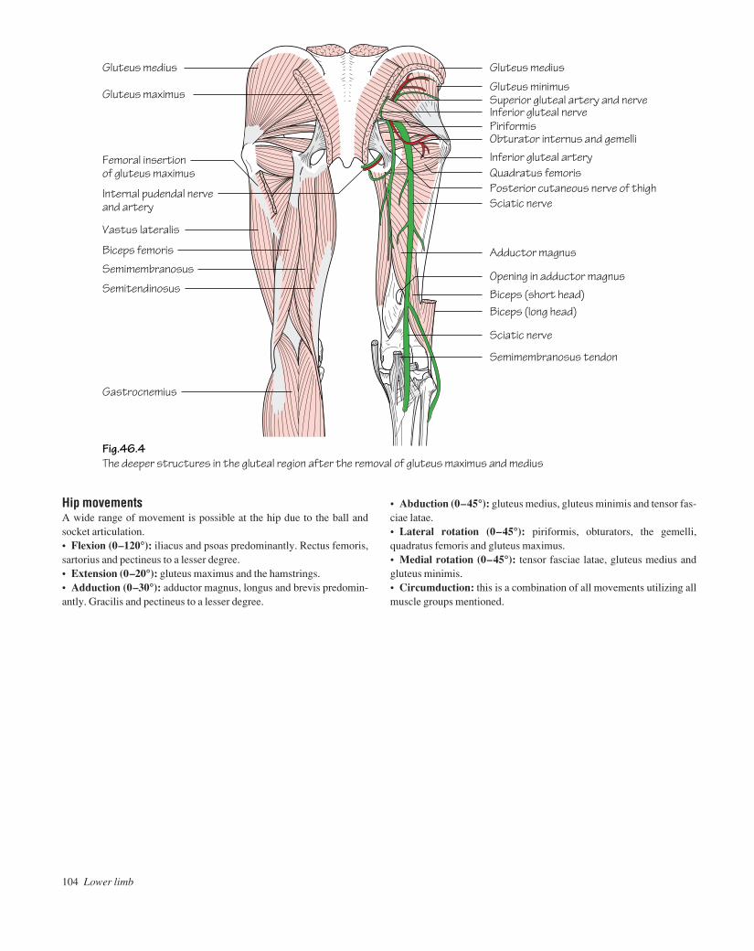
Hip movementsA wide range of movement is possible at the hip due to the ball andsocket articulation.• Flexion (0–120°): iliacus and psoas predominantly. Rectus femoris,sartorius and pectineus to a lesser degree.• Extension (0–20°): gluteus maximus and the hamstrings.• Adduction (0–30°): adductor magnus, longus and brevis predomin-antly. Gracilis and pectineus to a lesser degree.
104 Lower limb
• Abduction (0–45°): gluteus medius, gluteus minimis and tensor fas-ciae latae.• Lateral rotation (0–45°): piriformis, obturators, the gemelli,quadratus femoris and gluteus maximus.• Medial rotation (0–45°): tensor fasciae latae, gluteus medius andgluteus minimis.• Circumduction: this is a combination of all movements utilizing allmuscle groups mentioned.
Fig.46.4The deeper structures in the gluteal region after the removal of gluteus maximus and medius
Gluteus medius Gluteus medius
Gluteus minimus
PiriformisObturator internus and gemelli
Quadratus femoris
Adductor magnus
Inferior gluteal artery
Superior gluteal artery and nerveInferior gluteal nerve
Posterior cutaneous nerve of thigh
Opening in adductor magnus
Biceps (short head)Biceps (long head)
Semimembranosus tendon
Sciatic nerve
Gluteus maximus
Vastus lateralis
Biceps femoris
Semimembranosus
Gastrocnemius
Semitendinosus
Femoral insertion of gluteus maximus
Sciatic nerveInternal pudendal nerve and artery
AAAC46 21/5/05 10:53 AM Page 104
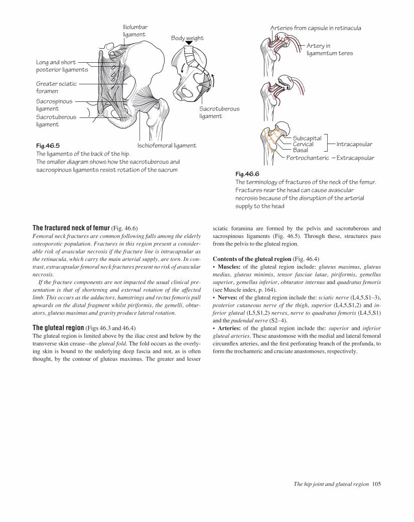
The fractured neck of femur (Fig. 46.6)Femoral neck fractures are common following falls among the elderlyosteoporotic population. Fractures in this region present a consider-able risk of avascular necrosis if the fracture line is intracapsular asthe retinacula, which carry the main arterial supply, are torn. In con-trast, extracapsular femoral neck fractures present no risk of avascularnecrosis.
If the fracture components are not impacted the usual clinical pre-sentation is that of shortening and external rotation of the affectedlimb. This occurs as the adductors, hamstrings and rectus femoris pullupwards on the distal fragment whilst piriformis, the gemelli, obtur-ators, gluteus maximus and gravity produce lateral rotation.
The gluteal region (Figs 46.3 and 46.4)The gluteal region is limited above by the iliac crest and below by thetransverse skin creaseathe gluteal fold. The fold occurs as the overly-ing skin is bound to the underlying deep fascia and not, as is oftenthought, by the contour of gluteus maximus. The greater and lesser
sciatic foramina are formed by the pelvis and sacrotuberous andsacrospinous ligaments (Fig. 46.5). Through these, structures passfrom the pelvis to the gluteal region.
Contents of the gluteal region (Fig. 46.4)• Muscles: of the gluteal region include: gluteus maximus, gluteusmedius, gluteus minimis, tensor fasciae latae, piriformis, gemellussuperior, gemellus inferior, obturator internus and quadratus femoris(see Muscle index, p. 164).• Nerves: of the gluteal region include the: sciatic nerve (L4,5,S1–3),posterior cutaneous nerve of the thigh, superior (L4,5,S1,2) and in-ferior gluteal (L5,S1,2) nerves, nerve to quadratus femoris (L4,5,S1)and the pudendal nerve (S2–4).• Arteries: of the gluteal region include the: superior and inferiorgluteal arteries. These anastomose with the medial and lateral femoralcircumflex arteries, and the first perforating branch of the profunda, toform the trochanteric and cruciate anastomoses, respectively.
The hip joint and gluteal region 105
Fig.46.5The ligaments of the back of the hip.The smaller diagram shows how the sacrotuberous and sacrospinous ligaments resist rotation of the sacrum
Fig.46.6The terminology of fractures of the neck of the femur.Fractures near the head can cause avascularnecrosis because of the disruption of the arterialsupply to the head
Long and shortposterior ligaments
Iliolumbarligament
Ischiofemoral ligament
Sacrotuberous ligament
Arteries from capsule in retinacula
Artery inligamentum teres
Greater sciatic foramen
Sacrospinous ligamentSacrotuberous ligament
Body weight
SubcapitalCervical Intracapsular
ExtracapsularBasal
Pertrochanteric
AAAC46 21/5/05 10:53 AM Page 105

The thigh is divided into flexor, extensor and adductor compartments.The membranous superficial fascia of the abdominal wall fuses to thefascia lata, the deep fascia of the lower limb, at the skin crease of thehip joint just below the inguinal ligament.
The deep fascia of the thigh (fascia lata)This layer of strong fascia covers the thigh. It is attached above to theinguinal ligament and bony margins of the pelvis and below to the tibialcondyles, head of the fibula and patella. Three fascial septa pass fromthe deep surface of the fascia lata to insert onto the linea aspera of thefemur and consequently divide the thigh into three compartments.
On the lateral side the fascia lata is condensed to form the iliotibialtract (Fig. 47.4). The tract is attached above to the iliac crest andreceives the insertions of tensor fasciae latae and three-quarters of glu-teus maximus. These muscles are also enveloped in deep fascia. The ili-otibial tract inserts into the lateral condyle of the tibia.
The saphenous opening is a gap in the deep fascia which is filled withloose connective tissueathe cribriform fascia. The lateral border of theopening, the falciform margin, curves in front of the femoral vesselswhereas on the medial side it curves behind to attach to the iliopectinealline (Fig. 43.1). The great saphenous vein pierces the cribriform fasciato drain into the femoral vein. Superficial branches of the femoral arteryand lymphatics are also transmitted through the saphenous opening.
106 Lower limb
47 The thigh
Fig.47.1The muscles of the front of the thigh.The femoral triangle is outlined
Fig.47.2Psoas, iliacus and the adductorgroup of muscles
Inguinal ligamentTensor fasciae latae
Psoas tendon
Pectineus
Adductor longus
Gracilis
Sartorius
Vastus medialis
Ligamentum patellae
Femoral triangle
IliacusDiaphragm
Right crus
Quadratuslumborum
Rectus femoris
Vastus lateralis
Iliotibial tract
Patellar retinacula
Psoas major
Iliacus
Inguinal ligament
Pectineus
Adductor longus
Adductor magnus
Opening in adductor magnus (for passageof femoral vesselsto popliteal fossa)
AAAC47 21/5/05 10:53 AM Page 106

The thigh 107
Fig.47.3A section through the thigh to show the adductor (subsartorial) canal
Saphenous nerve
Rectus femorisNerve to vastus medialis
Vastus lateralis
SartoriusVastus medialis
Femoral vesselsIliotibial tract
Great saphenous vein
Vastus intermedius
Adductor longus Sciatic nerve
Gracilis ShortProfunda vessels
LongAdductor brevis
heads of biceps
Adductor magnus
Semimembranosus
Semitendinosus
The superficial fascia of the thighContents of the subcutaneous tissue include:• Nerves: the femoral branch of the genitofemoral nerve (p. 51), themedial, intermediate (branches of the femoral nerve, p. 99) and lateralfemoral cutaneous nerves (L2,3, p. 99) and branches of the obturatornerve (p. 99) supply the skin of the anterior thigh. The back of the thighreceives its sensory supply from the posterior cutaneous nerve of thethigh.• Superficial arteries: these include the four superficial branches ofthe femoral artery: the superficial circumflex iliac artery, superficialepigastric artery, superficial external pudendal artery and the deepexternal pudendal artery.• Superficial veins and lymphatics: venous tributaries of the anteriorthigh drain into the great saphenous vein whilst some in the lower pos-terior thigh drain into the popliteal vein. The great saphenous vein isalso accompanied by large lymphatics which pass to the superficialinguinal nodes and, from there, through the cribriform fascia to thedeep inguinal nodes.
The femoral triangle (Figs 44.1 and 47.1)The boundaries of the femoral triangle are: the inguinal ligamentabove, the medial border of sartorius and the medial border of adductorlongus.• The floor consists of: adductor longus, pectineus, psoas tendon andiliacus (see Muscle index, p. 165).
• The roof consists of: fascia lata. The saphenous opening is in theupper part of the triangle.• The contents include (from lateral to medial) the: femoral nerve,artery, vein and their branches and tributaries. The femoral canal is situ-ated medial to the femoral vein. Transversalis fascia and psoas fasciafuse and evaginate to form the femoral sheath below the inguinal liga-ment. The sheath encloses the femoral artery, vein and canal but thefemoral nerve lies outside on its lateral aspect (see Fig. 44.1).
The contents of the anterior compartment of the thigh (Fig. 47.3)• Muscles: these constitute the hip flexors and knee extensors, i.e. sar-torius, iliacus, psoas, pectineus and quadriceps femoris (see Muscleindex, p. 165).• Arteries: the femoral artery and its branches (p. 95).• Veins: the femoral vein is a continuation of the popliteal vein as it passes through the hiatus in adductor magnus. It receives its maintributaryathe great saphenous veinathrough the saphenous opening.• Lymphatics: from the anterior compartment pass to the deep inguinallymph nodes which lie along the terminal part of the femoral vein.• Nerves: the femoral nerve (L2,3,4, p. 99) divides a short distancebelow the inguinal ligament into anterior and posterior divisions. Onlythe saphenous branch passes beyond the knee.
AAAC47 21/5/05 10:53 AM Page 107

The contents of the medial compartment of the thigh (Figs 47.2 and 47.3)• Muscles: these comprise the hip adductors: gracilis, adductorlongus, adductor brevis, adductor magnus and obturator externus (alateral rotator of the thigh at the hip) (see Muscle index, p. 165).• Arteries: profunda femoris (p. 95) as well as its medial circumflexfemoral and perforating branches and the obturator artery.• Veins: profunda femoris and obturator veins.• Nerves: the anterior and posterior divisions of the obturator nerve (p. 99).
The contents of the posterior compartment of the thigh (Fig. 47.3)• Muscles: these are the hamstrings and effect knee flexion and hip ex-tension. They include: biceps femoris, semitendinosus, semimembranosusand the hamstring part of adductor magnus (see Muscle index, p. 165).• Arteries: the perforating branches of profunda femoris.• Veins: the venae comitantes of the small arteries.• Nerves: the sciatic nerve (L4,5,S1–3, p. 101). The muscles of theposterior compartment are supplied by the tibial component of the sci-atic nerve with the exception of the short head of biceps femoris whichis supplied by the common peroneal component.
108 Lower limb
The adductor (subsartorial or Hunter’s) canalThe adductor canal serves to transmit structures from the apex of thefemoral triangle through the hiatus in adductor magnus into thepopliteal fossa. It commences in the mid-portion of the thigh and isformed by the following walls:• The posterior wall: adductor longus, with adductor magnus in thelower part of the thigh.• The lateral wall: vastus medialis.• The roof: thickened fascia underlying sartorius.
The contents of the adductor canalThese include: the femoral artery, the femoral vein which lies deep tothe femoral artery, lymphatics, the saphenous branch of the femoralnerve (which passes behind sartorius to leave the canal and descendsthe lower limb with the great saphenous vein), the nerve to vastus medi-alis (in the upper part) and the subsartorial plexus. This plexus isformed by branches from the saphenous nerve (terminal branch of thefemoral nerve, p. 99), the anterior division of the obturator nerve andthe intermediate cutaneous nerve of the thigh (branch of the femoralnerve, p. 99). It supplies the skin over the medial aspect of the knee.
Fig.47.4The lateral side of the thigh.Note the two muscles insertedinto the iliotibial tract
Iliac crest
Fascia covering gluteus medius
Tensor fasciae latae
Gluteus maximus
Iliotibial tract
Rectus femoris
Vastus lateralis
Biceps femoris (long head)
AAAC47 21/5/05 10:53 AM Page 108
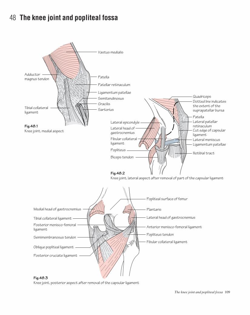
The knee joint and popliteal fossa 109
48 The knee joint and popliteal fossa
Vastus medialis
Fig.48.1Knee joint, medial aspect
Fig.48.2Knee joint, lateral aspect after removal of part of the capsular ligament
Fig.48.3Knee joint, posterior aspect after removal of the capsular ligament
Patella
Patellar retinaculum
Ligamentum patellaeSemitendinosus
Adductormagnus tendon
QuadricepsDotted line indicates the extent of the suprapatellar bursa
PatellaLateral patellarretinaculumCut edge of capsular ligament
Popliteal surface of femur
Ligamentum patellaeLateral meniscus
Iliotibial tract
Tibial collateralligament
Lateral epicondyleLateral head of gastrocnemius
Medial head of gastrocnemius
Tibial collateral ligament
Posterior menisco-femoral ligament
Semimembranosus tendon
Oblique popliteal ligament
Posterior cruciate ligament
Fibular collateral ligament
Popliteus
Biceps tendon
GracilisSartorius
Plantaris
Lateral head of gastrocnemius
Anterior menisco-femoral ligament
Popliteus tendon
Fibular collateral ligament
AAAC48 21/5/05 10:52 AM Page 109

110 Lower limb
• The medial (tibial) collateral ligament (Figs 48.1 and 48.3)acon-sists of superficial and deep parts. The superficial component isattached above to the femoral epicondyle and below to the subcuta-neous surface of the tibia. The deep component is firmly attached tothe medial meniscus.
• The lateral (fibular) collateral ligament (Fig. 48.2)ais attached tothe femoral epicondyle above and, along with biceps femoris, tothe head of the fibula below. Unlike the medial collateral ligamentit lies away from the capsule and meniscus.
The collateral ligaments are taut in full extension and it is in thisposition that they are liable to injury when subjected to extreme val-gus/varus strain.
Behind the knee the oblique popliteal ligament, a reflected exten-sion from the semimembranosus tendon, strengthens the capsule (Fig.48.3). Anteriorly the capsule is reinforced by the ligamentum patellaeand the patellar retinacula. The latter are reflected fibrous expansionsarising from vastus lateralis and medialis muscles which blend with thecapsule anteriorly (Fig. 48.1).
The knee joint (Figs 48.1–48.5)• Type: it is a synovial modified hinge joint which also permits a smalldegree of rotation. In the knee joint the femoral and tibial condylesarticulate as does the patella and patellar surface of the femur. Note: thefibula does not contribute to the knee joint.• Capsule: the articular surfaces are covered by articular cartilage. Thecapsule is attached to the margins of the articular surfaces except anter-iorly where it dips downwards. In the anterior part of the capsule thereis a large opening through which the synovial membrane is continuouswith the suprapatellar bursa (Fig. 48.2). This bursa extends superiorlythree fingerbreadths above the patella between the femur and quad-riceps. When an effusion has collected fluctuation can be elicited.Posteriorly the capsule communicates with another bursa under themedial head of gastrocnemius and often, through it, with the bursa ofsemimembranosus. Posterolaterally another opening in the capsule per-mits the passage of the tendon of popliteus.• Extracapsular ligaments: the capsule of the knee joint is reinforcedby ligaments.
Fig.48.4Anterior view of the flexed knee joint after division of the quadriceps and retraction of the patella
Fig.48.5The upper surface of the tibia and related structures.The dotted line indicates the synovial membrane in the vicinity of the cruciate ligaments.The small diagrams show how the cruciate ligaments resist forward and backward displacement of the femur
Lateral condyle
Fibular collateral ligament
Ligamentum patellae
Medial meniscus
Patella (turned back)
Tibial collateral ligament
FatCoronary ligament
Medial meniscusAnterior cruciate ligamentPosterior cruciate ligamentMedial condyle
Deep infrapatellarbursaCut edge ofcapsular ligament
Lateral meniscus
Fibular collateralligamentPosterior menisco-femoral ligamentTendon of popliteus
Posterior cruciate ligament
Anterior cruciate ligament
Posterior cruciate ligament
Tibial collateralligament
Anterior cruciate ligament
Transverse ligament
AAAC48 21/5/05 10:52 AM Page 110
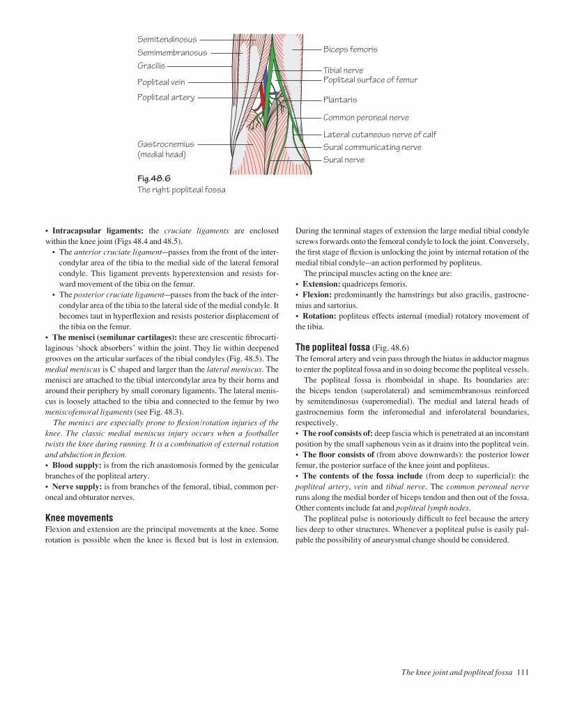
• Intracapsular ligaments: the cruciate ligaments are enclosedwithin the knee joint (Figs 48.4 and 48.5).
• The anterior cruciate ligamentapasses from the front of the inter-condylar area of the tibia to the medial side of the lateral femoralcondyle. This ligament prevents hyperextension and resists for-ward movement of the tibia on the femur.
• The posterior cruciate ligamentapasses from the back of the inter-condylar area of the tibia to the lateral side of the medial condyle. Itbecomes taut in hyperflexion and resists posterior displacement ofthe tibia on the femur.
• The menisci (semilunar cartilages): these are crescentic fibrocarti-laginous ‘shock absorbers’ within the joint. They lie within deepenedgrooves on the articular surfaces of the tibial condyles (Fig. 48.5). Themedial meniscus is C shaped and larger than the lateral meniscus. Themenisci are attached to the tibial intercondylar area by their horns andaround their periphery by small coronary ligaments. The lateral menis-cus is loosely attached to the tibia and connected to the femur by twomeniscofemoral ligaments (see Fig. 48.3).
The menisci are especially prone to flexion/rotation injuries of theknee. The classic medial meniscus injury occurs when a footballertwists the knee during running. It is a combination of external rotationand abduction in flexion.• Blood supply: is from the rich anastomosis formed by the genicularbranches of the popliteal artery.• Nerve supply: is from branches of the femoral, tibial, common per-oneal and obturator nerves.
Knee movementsFlexion and extension are the principal movements at the knee. Somerotation is possible when the knee is flexed but is lost in extension.
During the terminal stages of extension the large medial tibial condylescrews forwards onto the femoral condyle to lock the joint. Conversely,the first stage of flexion is unlocking the joint by internal rotation of themedial tibial condyleaan action performed by popliteus.
The principal muscles acting on the knee are:• Extension: quadriceps femoris.• Flexion: predominantly the hamstrings but also gracilis, gastrocne-mius and sartorius.• Rotation: popliteus effects internal (medial) rotatory movement ofthe tibia.
The popliteal fossa (Fig. 48.6)The femoral artery and vein pass through the hiatus in adductor magnusto enter the popliteal fossa and in so doing become the popliteal vessels.
The popliteal fossa is rhomboidal in shape. Its boundaries are: the biceps tendon (superolateral) and semimembranosus reinforced by semitendinosus (superomedial). The medial and lateral heads of gastrocnemius form the inferomedial and inferolateral boundaries,respectively.• The roof consists of: deep fascia which is penetrated at an inconstantposition by the small saphenous vein as it drains into the popliteal vein.• The floor consists of (from above downwards): the posterior lowerfemur, the posterior surface of the knee joint and popliteus.• The contents of the fossa include (from deep to superficial): thepopliteal artery, vein and tibial nerve. The common peroneal nerveruns along the medial border of biceps tendon and then out of the fossa.Other contents include fat and popliteal lymph nodes.
The popliteal pulse is notoriously difficult to feel because the arterylies deep to other structures. Whenever a popliteal pulse is easily pal-pable the possibility of aneurysmal change should be considered.
The knee joint and popliteal fossa 111
Fig.48.6The right popliteal fossa
Biceps femoris
Tibial nervePopliteal surface of femur
Plantaris
Common peroneal nerve
Lateral cutaneous nerve of calfSural communicating nerveSural nerve
SemitendinosusSemimembranosusGracilis
Popliteal vein
Popliteal artery
Gastrocnemius(medial head)
AAAC48 21/5/05 10:52 AM Page 111

112 Lower limb
49 The leg
Fig.49.1The extensor (dorsiflexor) group of muscles
Fig.49.3The superficial muscles of the calf
Fig.49.4Cross-section through the leg, to be studied in conjunction with Fig.41.4
Fig.49.2The lateral side of the leg and foot
Ligamentumpatellae
Extensor digitorum longus
Vastus medialis
Sartorius
Gastrocnemiusand soleus
Gastrocnemius(medial head)
Medial ligamentof knee joint
Popliteus
Tibialis posterior
Flexor hallucislongus
Extensor digitorum longusExtensor hallucis longus
Peroneus brevisPeroneuslongusFibulaFlexor hallucis longus
Soleus
Gastrocnemius
TibialisanteriorSubcutaneoussurface of tibia
Rectus femoris
Gastrocnemius(lateral head) Tibialis anterior
Tibia
Deep peroneal nerve and anterior tibial vessels
Tibialis posterior
Great saphenous veinFlexor digitorum longus
Tibial vessels and nerve
Small saphenous vein
Plantaris
Head of fibula
Soleus
Gastrocnemius
Peroneus longus
Tendo calcaneus
Vastus lateralis
Peroneus longus and brevis
Extensor hallucis longus
Superior and inferior extensor retinacula
Extensor digitorum brevis
Peroneus tertius
Vastus lateralis
Biceps femoris
Peroneus longus SoleusGastrocnemius
Peroneus brevis
Peroneus retinaculum
Peroneus tertius
Iliotibial tract
Flexor digitorumlongus
AAAC49 21/5/05 10:52 AM Page 112
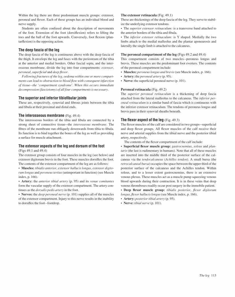
Within the leg there are three predominant muscle groups: extensor,peroneal and flexor. Each of these groups has an individual blood andnerve supply.
Students are often confused about the description of movements of the foot. Extension of the foot (dorsiflexion) refers to lifting the toes and the ball of the foot upwards. Conversely, foot flexion (plan-tarflexion) is the opposing action.
The deep fascia of the legThe deep fascia of the leg is continuous above with the deep fascia ofthe thigh. It envelops the leg and fuses with the periosteum of the tibiaat the anterior and medial borders. Other fascial septa, and the inter-osseous membrane, divide the leg into four compartments: extensor,peroneal, superficial and deep flexor.
Following fractures of the leg, oedema within one or more compart-ments can lead to obstruction to blood flow with consequent infarctionof tissuebthe ‘compartment syndrome’. When this occurs immediatedecompression (fasciotomy) of all four compartments is necessary.
The superior and inferior tibiofibular jointsThese are, respectively, synovial and fibrous joints between the tibiaand fibula at their proximal and distal ends.
The interosseous membrane (Fig. 49.4)The interosseous borders of the tibia and fibula are connected by astrong sheet of connective tissueathe interosseous membrane. Thefibres of the membrane run obliquely downwards from tibia to fibula.Its function is to bind together the bones of the leg as well as providinga surface for muscle attachment.
The extensor aspects of the leg and dorsum of the foot (Figs 49.1 and 49.4)The extensor group consists of four muscles in the leg (see below) andextensor digitorum brevis in the foot. These muscles dorsiflex the foot.The contents of the extensor compartment of the leg are as follows:• Muscles: tibialis anterior, extensor hallucis longus, extensor digito-rum longus and peroneus tertius (unimportant in function) (see Muscleindex, p. 166).• Artery: the anterior tibial artery (p. 95) and its venae comitantesform the vascular supply of the extensor compartment. The artery con-tinues as the dorsalis pedis artery in the foot.• Nerves: the deep peroneal nerve (p. 101) supplies all of the musclesof the extensor compartment. Injury to this nerve results in the inabilityto dorsiflex the footafootdrop.
The extensor retinacula (Fig. 49.1)These are thickenings of the deep fascia of the leg. They serve to stabil-ize the underlying extensor tendons.• The superior extensor retinaculum: is a transverse band attached tothe anterior borders of the tibia and fibula.• The inferior extensor retinaculum: is Y shaped. Medially the twolimbs attach to the medial malleolus and the plantar aponeurosis andlaterally the single limb is attached to the calcaneus.
The peroneal compartment of the leg (Figs 49.2 and 49.4)This compartment consists of two musclesaperoneus longus and brevis. These muscles are the predominant foot everters. The contentsof the peroneal compartment include:• Muscles: peroneus longus and brevis (see Muscle index, p. 166).• Artery: the peroneal artery (p. 95).• Nerve: the superficial peroneal nerve (p. 101).
Peroneal retinacula (Fig. 49.2)The superior peroneal retinaculum is a thickening of deep fasciaattached from the lateral malleolus to the calcaneus. The inferior per-oneal retinaculum is a similar band of fascia which is continuous withthe inferior extensor retinaculum. The tendons of peroneus longus andbrevis pass in their synovial sheaths beneath.
The flexor aspect of the leg (Fig. 49.3)The flexor muscles of the calf are considered in two groupsasuperficialand deep flexor groups. All flexor muscles of the calf receive theirnerve and arterial supplies from the tibial nerve and the posterior tibialartery, respectively.
The contents of the flexor compartment of the calf include:• Superficial flexor muscle group: gastrocnemius, soleus and plan-taris (the last is rudimentary in humans). Note that all of these musclesare inserted into the middle third of the posterior surface of the cal-caneus via the tendocalcaneus (Achilles tendon). A small bursa (theretrocalcaneal bursa) occupies the space between the upper third of theposterior surface of the calcaneus and the Achilles tendon. Withinsoleus, and to a lesser extent gastrocnemius, there is an extensivevenous plexus. These muscles act as a muscle pump squeezing venousblood upwards during their contraction. It is in these veins that deepvenous thromboses readily occur post surgery in the immobile patient.• Deep flexor muscle group: tibialis posterior, flexor digitorumlongus, flexor hallucis longus (see Muscle index, p. 166).• Artery: posterior tibial artery (p. 95).• Nerve: tibial nerve (p. 101).
The leg 113
AAAC49 21/5/05 10:52 AM Page 113

114 Lower limb
50 The ankle and foot I
Fig.50.1The ankle joint from behind, to show how the talus is held in position by ligaments between the tibia and fibula above and the calcaneus below
Fig.50.2The bones of the foot, medial and lateral aspects.The major joints are shown
Fig.50.4The ankle joint, medial aspect
Fig.50.3The ankle joint, lateral aspect after removal of the capsular ligament
Interosseoustibiofibularligament
InterosseoustalocalcanealligamentCalcaneofibularligament
Peroneus longus
Deltoid ligament
Tibialisposterior
Flexor digitorumlongusFlexor hallucis longus
Plantar calcaneonavicular(spring) ligament Long plantar ligament
Tendocalcaneus
Peroneus brevis
Deltoidligament
Sustentaculum tali
Sesamoid bone
Facet for medial malleolus
Talus
1 2
3
4
5
Calcaneus
NavicularTalus
Anterior tibiofibular ligament
Posterior tibiofibular ligament
Positionof bursa
Firstmetatarsal
Tuberosityof navicular
Calcaneofibular ligament
Cervical ligament
Medialcuneiform
CuboidBifurcate ligament
Long plantar ligament
NavicularTuberosity of navicularMedial cuneiform
First metatarsal
Head of talus
Subtalar jointMidtarsal (talonavicular)TarsometatarsalMetatarsophalangealMidtarsal (calcaneocuboid)
1.2.3.4.5.
Joints
CuboidSinus tarsi Facet for
lateral malleolus
Posterior talofibular ligament
AAAC50 21/5/05 10:52 AM Page 114

The ankle joint (Fig. 50.1)• Type: the ankle is a synovial hinge joint involving the tibia, fibulaand talus. The articular surfaces are covered with cartilage and synovialmembrane lines the rest of the joint.• Capsule: the capsule encloses the articular surfaces. The capsule isreinforced on either side by strong collateral ligaments but is lax anter-iorly to permit uninhibited hinged movement.• Ligaments: the medial collateral (deltoid) ligament consists of adeep component which is a vertical band passing from the medialmalleolus to the talus. The superficial component of this ligament is fanshaped and extends from the medial malleolus to (from front to back):the tuberosity of the navicular, the spring ligament (see below), the sus-tentaculum tali and the posterior tubercle of the talus (Figs 50.1 and 50.4).
The lateral collateral ligament consists of three bands: the anteriorand posterior talofibular ligaments and the calcaneofibular ligament(Fig. 50.3). Abduction/adduction forces on the ankle can cause asprainaan incomplete tear of one of the collateral ligaments. Completetears of the ligaments also occur and lead to painful instability at theankle joint on clinical examination. Severe forces on the ankle joint canresult in fracture or fracture dislocation.
The movements at the ankleIt is important to note that the inversion and eversion movements of the foot do not occur at the ankle joint except in full plantarflexion.These occur at the subtalar and midtarsal joints (see below). Onlydorsiflexion (extension) and plantarflexion (flexion) occur at the ankle.The principal muscles are:• Dorsiflexion: tibialis anterior and to a lesser extent extensor hallucislongus and extensor digitorum longus.• Plantarflexion: gastrocnemius and soleus and to a lesser extent tibialis posterior, flexor hallucis longus and flexor digitorum longus.
The foot bones (Fig. 50.2)With the exception of the metatarsals and phalanges the foot bones aretermed collectively the tarsal bones.• Talus: has a body with facets on the superior, medial and lateral sur-faces for articulation with the tibia, medial malleolus and lateral malle-olus, respectively. There is a groove on the posterior surface of thebody for the tendon of flexor hallucis longus. To the groove’s lateralside is the posterior (lateral) tubercle, sometimes known as the ostrigonum, as it ossifies from a separate centre to the talus. A head pro-jects distally which articulates with the navicular. The head is con-nected to the body by a neck.• Calcaneus: has two facets on the superior surface which participatein the subtalar (talocalcaneal and talocalcaneonavicular) joint. The
posterior surface has three areas: a roughened middle part where thetendocalcaneus inserts; a smooth upper part which is separated fromthe tendocalcaneus by a bursa (retrocalcaneal bursa) (Fig. 50.4); and alower part which is covered by a fibro-fatty pad that forms the heel.Medial and lateral tubercles are present on the inferior surface to whichthe plantar aponeurosis is attached. The sustentaculum tali is a distinct-ive projection on the medial surface which forms a shelf for the supportof the talus. The peroneal tubercle, a small projection on the lateral sur-face of the calcaneus, separates the tendons of peroneus longus and brevis. The anterior surface has a facet for articulation with the cuboid.• Cuboid: has a grooved undersurface for the tendon of peroneuslongus.• Navicular: has facets for the articulations with the head of the talusposteriorly and the three cuneiforms anteriorly. It has a tuberosity on itsmedial aspect which provides attachment for tibialis posterior.• Cuneiforms: there are three cuneiforms which articulate anteriorlywith the metatarsals and posteriorly with the navicular. Their wedge-shape helps to maintain the transverse arch of the foot.• Metatarsals and phalanges: these are similar to the metacarpals andphalanges of the hand. Note the articulations of the heads of themetatarsals. The 1st metatarsal is large and is important for balance.The head is grooved on its inferior surface for the two sesamoid boneswithin the tendon of flexor hallucis brevis.
The foot joints• Subtalar joint (Fig. 50.2): this compound joint comprises the talo-calcaneal and the talocalcaneonavicular joints. Inversion and eversionmovements occur at the subtalar joint.
• The talcocalcaneal jointais a synovial plane joint formed by thearticulation of the upper surface of the calcaneus with the lowersurface of the talus.
• The talocalcaneonavicular jointais a synovial ball and socketjoint between the head of the talus and the sustentaculum tali, thespring ligament and the navicular.
• Midtarsal joint (Fig. 50.2): is also a compound joint which con-tributes towards foot inversion/eversion movements. This joint is com-posed of the calcaneocuboid joint and the talonavicular component ofthe talocalcaneonavicular joint.
• The calcaneocuboid jointais a synovial plane joint formedbetween the anterior surface of the calcaneus and the posterior surface of the cuboid.
• Other foot joints (Fig. 50.2): these include other tarsal joints, tar-sometatarsal (synovial plane), intermetatarsal (synovial plane), meta-tarsophalangeal (synovial condyloid) and interphalangeal (synovialhinge) joints.
The ankle and foot I 115
AAAC50 21/5/05 10:52 AM Page 115
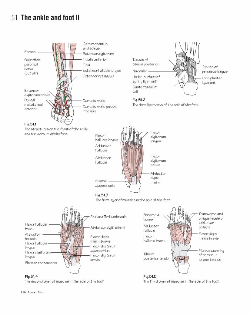
116 Lower limb
51 The ankle and foot II
Fig.51.1The structures on the front of the ankleand the dorsum of the foot
Fig.51.2The deep ligaments of the sole of the foot
Peronei
Superficial peroneal nerve (cut off)
Fig.51.3The first layer of muscles in the sole of the foot
Flexorhallucis longus
Extensor digitorum brevisDorsal metatarsalarteries
Gastrocnemius and soleus
Tibia
Flexordigitorumlongus
Extensor digitorumTibialis anterior
Extensor hallucis longusTendon ofperoneus longus
Long plantarligament
Extensor retinacula Under-surface ofspring ligamentSustentaculumtali
Tendon oftibialis posterior
Navicular
Dorsalis pedis
Dorsalis pedis passes into sole
Adductorhallucis
Abductorhallucis
Plantaraponeurosis
Flexordigitorumbrevis
Abductordigitiminimi
Fig.51.5The third layer of muscles in the sole of the foot
Sesamoidbones
Flexor digitiminimi brevisFlexor
hallucis brevis
Abductorhallucis
Tibialisposterior tendon
Transverse andoblique heads ofadductorpollucis
Fibrous coveringof peroneuslongus tendon
Fig.51.4The second layer of muscles in the sole of the foot
Flexor hallucis brevis
2nd and 3rd lumbricals
Abductorhallucis
Flexor digitorum longus
Flexor hallucis longus
Plantar aponeurosis
Flexor digitorum brevis
Flexor digitorum accessorius
Flexor digiti minimi brevis
Abductor digiti minimi
AAAC51 21/5/05 10:58 AM Page 116
Ligaments of the foot• Spring (plantar calcaneonavicular) ligament (Fig. 51.2): runsfrom the sustentaculum tali to the tuberosity of the navicular forming asupport for the head of the talus.• Bifurcate ligament: is Y shaped and runs from the anterior part ofthe calcaneus to the cuboid and navicular bones. It reinforces the capsule of the talocalcaneonavicular joint.• Long plantar ligament (Figs 51.2 and 50.3): runs from the under-surface of the calcaneus to the cuboid and bases of the lateral meta-tarsals. The ligament runs over the tendon of peroneus longus.• Short plantar ligament: runs from the undersurface of the calca-neus to the cuboid.• Medial and lateral (talocalcaneal) ligaments: strengthen the capsule of the talocalcaneal joint.• Interosseous talocalcaneal ligament: runs in the sinus tarsi, a tunnel formed by deep sulci on the talus and calcaneus.• Deep transverse metacarpal ligaments: join the plantar ligamentsof the metatarsophalangeal joints of the five toes.
The arches of the footThe integrity of the foot is maintained by two longitudinal (medial andlateral) arches and a single transverse arch. The arches are heldtogether by a combination of bony, ligamentous and muscular factorsso that standing weight is taken on the posterior part of the calcaneumand the metatarsal heads as a result of the integrity of the arches.• Medial longitudinal arch (see Fig. 50.2): comprises calcaneus, talus(the apex of the arch), navicular, the three cuneiforms and three medialmetatarsals. The arch is bound together by the spring ligament, musclesand supported from above by tibialis anterior and posterior.• Lateral longitudinal arch (see Fig. 50.2): comprises calcaneus,cuboid and the two lateral metatarsals. The arch is bound together bythe long and short plantar ligaments and supported from above by per-oneus longus and brevis.• Transverse arch: comprises the cuneiforms and bases of the meta-tarsals. The arch is bound together by the deep transverse ligament,plantar ligaments and the interossei. It is supported from above by per-oneus longus and brevis.
The dorsum of the foot (Fig. 51.1)The skin of the dorsum of the foot is supplied by cutaneous branches ofthe superficial peroneal, deep peroneal, saphenous and sural nerves.The dorsal venous arch lies within the subcutaneous tissue overlyingthe metatarsal heads. It receives blood from most of the superficial tis-sues of the foot via digital and communicating branches. The greatsaphenous vein commences from the medial end of the arch and thesmall saphenous vein from the lateral end.
Structures on the dorsum of the foot (Fig. 51.1)• Muscles: extensor digitorum brevis arises from the calcaneus. Othermuscles insert on the dorsum of the foot but arise from the leg. Theseinclude: tibialis anterior, extensor hallucis longus, extensor digitorumlongus, peroneus tertius and peroneus brevis. Each tendon of extensordigitorum longus is joined on its lateral side by a tendon from extensordigitorum brevis. The tendons of extensor digitorum longus and per-oneus tertius share a common synovial sheath whilst the other tendonshave individual sheaths.• Arterial supply: is from the dorsalis pedis arteryathe continuationof the anterior tibial artery. The dorsalis pedis ends by passing to thesole where it completes the plantar arch (p. 95).• Nerve supply: is from the deep peroneal nerve via its medial and lat-eral terminal branches. The latter supplies extensor digitorum breviswhereas the former receives cutaneous branches from the skin.
The sole of the footThe sole is described as consisting of an aponeurosis and four musclelayers. The skin of the sole is supplied by the medial and lateral plantarbranches of the tibial nerve. The medial calcaneal branch of the tibialnerve innervates a small area on the medial aspect of the heel.
The plantar aponeurosisThis aponeurosis lies deep to the superficial fascia of the sole and covers the 1st layer of muscles. It is attached to the calcaneus behindand sends a deep slip to each toe as well as blending superficially withthe skin creases at the base of the toes. The slips that are sent to each toesplit into two parts which pass on either side of the flexor tendons andfuse with the deep transverse metatarsal ligaments.
The muscular layers of the sole• 1st layer consists of: abductor hallucis, flexor digitorum brevis andabductor digiti minimi (Fig. 51.3).• 2nd layer consists of: flexor digitorum accessorius, the lumbricalsand the tendons of flexor digitorum longus and flexor hallucis longus(Fig. 51.4).• 3rd layer consists of: flexor hallucis brevis, adductor hallucis andflexor digiti minimi brevis (Fig. 51.5).• 4th layer consists of: the dorsal and plantar interossei and the ten-dons of peroneus longus and tibialis posterior.
Neurovascular structures of the sole• Arterial supply: is from the posterior tibial artery which divides intomedial and lateral plantar branches. The latter branch contributes themajor part of the deep plantar arch (p. 95).• Nerve supply: is from the tibial nerve which also divides into medialand lateral plantar branches (p. 101).
The ankle and foot II 117
AAAC51 21/5/05 10:58 AM Page 117

118 Lower limb
52 Surface anatomy of the lower limb
Fig.52.1The anatomy of femoral and inguinal herniae.Note the relation of the deep inguinal ring to the inferior epigastric artery and the relation of the two types of hernia to the pubic tubercle
Fig.52.2The surface markings of the sciatic nerve
Fig.52.4Visible structures on the medial sideof the lower limb
Fig.52.5The lateral aspect of the foot to showthe tendons that can be recognised
Iliacus
Position ofdeep ring
Psoas tendonPectineusFemoral hernia
Inguinal hernia
Tendon of adductorlongus
Superficial ring
Linea alba
External oblique aponeurosis
Inferior epigastric artery
1/3
1/3
1/31/2
1/2
Pubic tubercle
Greater trochanter
Ischialtuberosity
Posteriorsuperioriliac spine
Injectionarea
Lacunar (Gimbernat's) ligament
Fig.52.3The structures on the medial side of the ankle
Posteriortibiofibular ligament
Posteriortalofibular ligament
Calcaneofibularligament
Posterior surfaceof calcaneus
Medial malleolusArticular surfaceof talus
Deltoid ligament
Flexor hallucis longus
Posterior tibial nervePosterior tibial artery
Tibialis posterior
Flexor digitorumlongus
Semi membranosus
Semi tendinosus
Short saphenous vein
Extensor digitorumlongus
Peroneus longusand brevis
Tibialis anterior
AAAC52 21/5/05 10:58 AM Page 118
Surface landmarks around the hip and gluteal region• The anterior superior iliac spine is a prominent landmark at theanterior end of the iliac crest.• The greater trochanter of the femur lies approximately a hands-breadth below the iliac crest. It is made more prominent by adductingthe hip.• The ischial tuberosity is covered by gluteus maximus when the hipis extended. It can be palpated in the lower part of the buttock with thehip flexed.• The femoral pulse (Fig. 52.1) is most easily felt halfway betweenthe anterior superior iliac spine and the symphysis pubis (mid-inguinalpoint). The femoral head lies deep to the femoral artery at the mid-inguinal point. The femoral vein lies medial, and the femoral nerve lat-eral, to the artery at this point.• The femoral canal (Fig. 52.1) lies medial to the femoral vein withinthe femoral sheath. The sac of a femoral hernia passes through the canalto expand below the deep fascia. The hernial sac always lies below andlateral to the pubic tubercle (cf. the neck of an inguinal hernia which isalways situated above and medial to the tubercle). The risk of strangula-tion is high in femoral herniae as the femoral canal is narrow and bloodflow to viscera within the hernial sac can easily be impaired.• The great saphenous vein pierces the cribriform fascia in the saph-enous opening of the deep fascia to drain into the femoral vein 4 cmbelow and lateral to the pubic tubercle (Fig. 43.2).• In thin subjects the horizontal chain of superficial inguinal lymphnodes is palpable. It lies below and parallel to the inguinal ligament.• The sciatic nerve has a curved course throughout the gluteal region.Consider two linesaone connects the posterior superior iliac spine andthe ischial tuberosity and the other connects the greater trochanter andthe ischial tuberosity (Fig. 52.2). The nerve descends the thigh in themidline posteriorly. The division of the sciatic nerve into tibial andcommon peroneal components occurs usually at a point a handsbreadthabove the popliteal crease but is highly variable. Sciatic nerve damageis occasionally caused by badly placed intramuscular injections. Thesafest site for intramuscular injection is consequently the upper outerquadrant of the buttock.• The common peroneal nerve winds superficially around the neck ofthe fibula. In thin subjects it can be palpated at this point. Footdrop canresult from fibular neck fractures where damage to this nerve hasoccurred.
Surface landmarks around the knee• The patella and ligamentum patellae are easily palpable with thelimb extended and relaxed. The ligamentum patellae can be traced to itsattachment at the tibial tuberosity.• The adductor tubercle can be felt on the medial aspect of the femurabove the medial condyle.• The femoral and tibial condyles are prominent landmarks. With theknee in flexion the joint line, and outer edges of the menisci within, arepalpable. The medial and lateral collateral ligaments are palpable oneither side of the knee and can be followed to their bony attachments.
• The subcutaneous border of the tibia is palpable throughout itslength.• The fibular head is palpable laterally (Fig. 52.4). The shaft of thefibula is mostly covered but is subcutaneous for the terminal 10 cm.• The popliteal pulse is difficult to feel as it lies deep to the tibial nerveand popliteal vein. It is best felt by palpating in the popliteal fossa withthe patient prone and the knee flexed.
Surface landmarks around the ankle• The medial and lateral malleoli are prominent at the ankle. The lat-eral is more elongated and descends a little further than the medial.• When the foot is dorsiflexed the tendons of tibialis anterior, extensorhallucis longus and extensor digitorum are visible on the anterioraspect of the ankle and the dorsum of the foot.• The tendons of peroneus longus and brevis pass behind the lateralmalleolus.• Passing behind the medial malleolus lie: the tendons of tibialis pos-terior and flexor digitorum longus, the posterior tibial artery and itsvenae comitantes, the tibial nerve and flexor hallucis longus (Fig.52.3).
Surface landmarks around the foot (Fig. 52.5)• The head of the talus is palpable immediately anterior to the distaltibia.• The base of the 5th metatarsal is palpable on the lateral border ofthe foot. The tendon of peroneus brevis inserts onto the tuberosity onthe base.• The heel is formed by the calcaneus. The tendocalcaneus (Achilles)is palpable above the heel. Sudden stretch of this can lead to rupture.When this occurs a gap in the tendon is often palpable.• The tuberosity of the navicular can be palpated 2.5 cm anterior tothe medial malleolus. It receives most of the tendon of tibialis posterior.• The peroneal tubercle of the calcaneum can be felt 2.5 cm belowthe tip of the lateral malleolus.• The sustentaculum tali can be felt 2.5 cm below the medial malleo-lus. The tendon of tibialis posterior lies above the sustentaculum taliand the tendon of flexor hallucis longus winds beneath it.• The dorsalis pedis pulse is located on the dorsum of the foot be-tween the tendons of extensor hallucis longus and extensor digitorum.• The posterior tibial pulse is best felt halfway between the medialmalleolus and the heel.• The dorsal venous arch is visible on the dorsum of the foot. Thesmall saphenous vein drains the lateral end of the arch and passes pos-terior to the lateral malleolus to ascend the calf and drain into thepopliteal vein. The great saphenous vein passes anterior to the medialmalleolus to ascend the length of the lower limb and drain into thefemoral vein. This vein can be accessed consistently by ‘cutting down’anterior to, and above, the medial malleolus following local anaesthe-sia. This is used in emergency situations when intravenous access isdifficult but required urgently.
Surface anatomy of the lower limb 119
AAAC52 21/5/05 10:58 AM Page 119
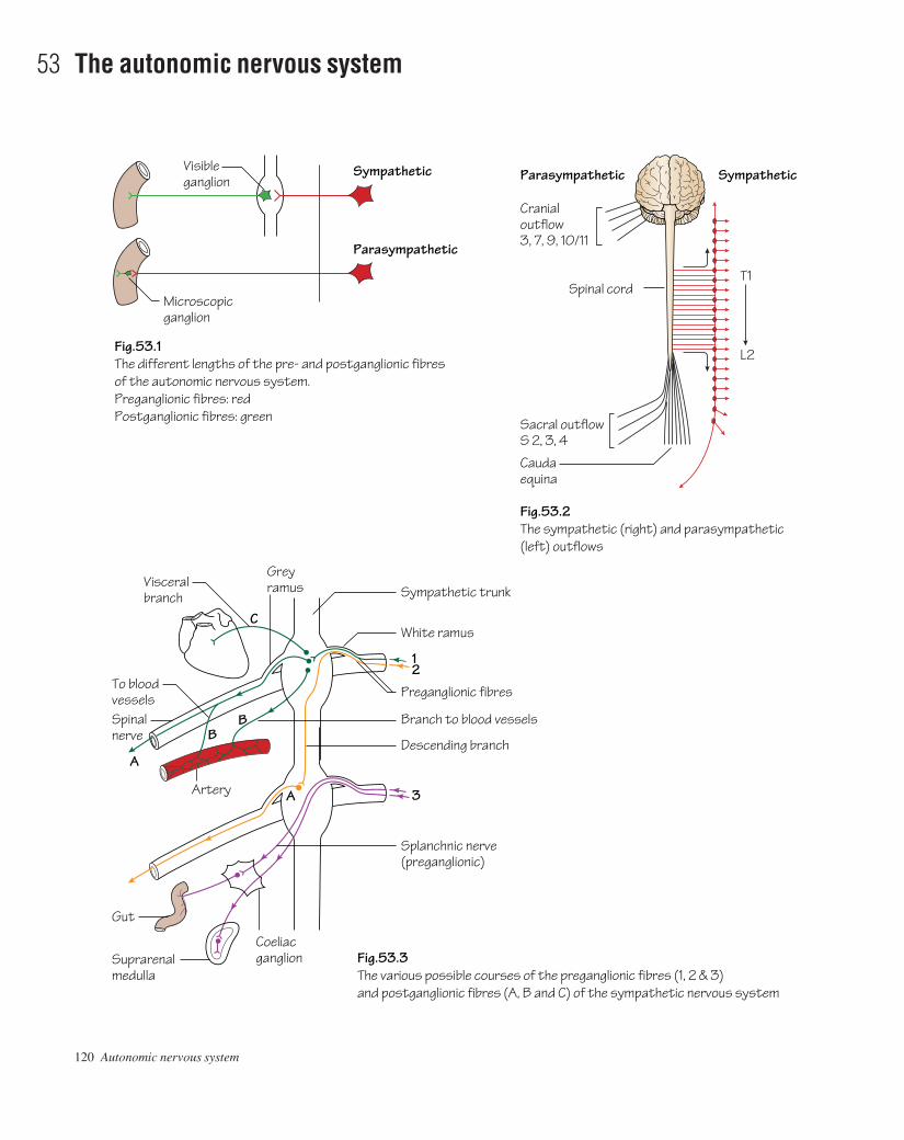
120 Autonomic nervous system
53 The autonomic nervous system
Fig.53.1The different lengths of the pre- and postganglionic fibres of the autonomic nervous system.Preganglionic fibres: redPostganglionic fibres: green
Fig.53.3The various possible courses of the preganglionic fibres (1, 2 & 3)and postganglionic fibres (A, B and C) of the sympathetic nervous system
Fig.53.2The sympathetic (right) and parasympathetic(left) outflows
Visibleganglion
Sympathetic Sympathetic
Parasympathetic
Parasympathetic
Microscopicganglion
Cranialoutflow3, 7, 9, 10/11
Sacral outflowS 2, 3, 4
Spinal cordT1
L2
Caudaequina
BB
C
12
3A
A
To bloodvesselsSpinalnerve
Artery
CoeliacganglionSuprarenal
medulla
Gut
Splanchnic nerve(preganglionic)
Descending branch
Branch to blood vessels
Preganglionic fibres
White ramus
Sympathetic trunkGreyramusVisceral
branch
AAAC53 21/5/05 10:57 AM Page 120

The autonomic nervous system comprises two parts—the sympatheticand the parasympathetic. The former initiates the ‘fight or flight’ reac-tion while the latter controls the body under more relaxed conditions.Both systems have synapses in peripheral ganglia but those of the sym-pathetic system are, for the most part, close to the spinal cord in the gan-glia of the sympathetic trunk whereas those of the parasympatheticsystem are mostly in the walls of the viscera themselves and are micro-scopic (except for the four macroscopic ganglia in the head and neckdescribed below). Thus the sympathetic preganglionic fibres are re-latively short compared to the parasympathetic fibres (Fig. 53.1).• Sympathetic outflow (Fig. 53.2): the anterior rami of spinal nervesT1 to L2 or 3. The fibres leave these spinal nerves as the white ramicommunicantes and synapse in the ganglia of the sympathetic trunk.• Parasympathetic outflow: this comprises:
• Cranial outflowafibres travel as ‘passengers’ in the cranial nerves3, 7, 9 and 10/11 and synapse in one of the four macroscopicperipheral ganglia of the head and neck.
• Sacral outflowatravel in sacral nerves S2, 3 and 4.
The sympathetic system• The sympathetic trunk: from the base of the skull to the tip of thecoccyx where the two trunks join to form the ganglion impar. The trunkcontinues upwards into the carotid canal as the internal carotid nerve.• Superior cervical ganglion: represents the fused ganglia of C1, 2, 3and 4.• Middle cervical ganglion: represents the fused ganglia of C5 and 6.• Inferior cervical ganglion: represents the fused ganglia of C7 and 8.It may be fused with the ganglion of T1 to form the stellate ganglion.
For courses of the pre- and postganglionic fibres see Fig. 53.3.• Preganglionic fibres: when the white (myelinated) rami reach thesympathetic trunk they may follow one of three different routes:
1 They may synapse with a nerve cell in the corresponding ganglion.2 They may pass straight through the corresponding ganglion and travel
up or down the sympathetic trunk, to synapse in another ganglion.3 They may pass straight through their own ganglion, maintaining
their preganglionic status until they synapse in one of the outlyingganglia such as the coeliac ganglion. One exceptional group offibres even pass through the coeliac ganglion and do not synapseuntil they reach the suprarenal medulla.
• Postganglionic fibres: after synapsing, the postganglionic fibresmay follow one of three different routes:
A They may pass back to the spinal nerve as a grey (unmyelinated)ramus and are then distributed with the branches of that nerve.
B They may pass to adjacent arteries to form a plexus around themand are then distributed with the branches of the arteries. Otherfibres leave branches of the spinal nerves later to pass to the arter-ies more distally.
C They may pass directly to the viscera in distinct and sometimesnamed branches such as the cervical cardiac branches of the cer-vical ganglia.
If the sympathetic trunk is divided above T1 or below L2, the headand neck or the lower limb will lose all sympathetic supply.
Loss of the supply to the head and neck will produce Horner’s syn-drome. There will be loss of sweating (anhidrosis), drooping of theupper eyelid (ptosis) and constriction of the pupil (myosis) on that side.
Details of the sympathetic system in the various regions are given inthe appropriate chapters, but Table 53.1 summarizes the autonomicsupply to the most important regions and viscera.
The parasympathetic system• The cranial outflow:III The oculomotor nerve carries parasympathetic fibres to the constrictor pupillae and the ciliary muscle, synapsing in the ciliary ganglion.VII The facial nerve carries fibres for the submandibular and sublin-gual glands (which synapse in the submandibular ganglion) and for thelacrimal gland (which synapse in the sphenopalatine ganglion).IX The glossopharyngeal nerve carries fibres for the parotid glandwhich synapse in the otic ganglion.X/XI The vagus and cranial root of the accessory carry fibres for thethoracic and abdominal viscera down as far as the proximal two-thirdsof the transverse colon, where supply is taken over by the sacral out-flow. Synapses occur in minute ganglia in the cardiac and pulmonaryplexuses and in the walls of the viscera.• The sacral outflow:From the sacral nerves S2, 3 and 4, fibres join the inferior hypogastricplexuses by means of the pelvic splanchnic nerves. They go on to supply the pelvic viscera, synapsing in minute ganglia in the walls ofthe viscera themselves. Some fibres climb out of the pelvis around theinferior mesenteric artery and supply the sigmoid and descending colonand the distal one third of the transverse colon.
The autonomic nervous system 121
Region Origin of connector fibres Site of synapse
SympatheticHead and neck T1–T5 Cervical gangliaUpper limb T2–T6 Inferior cervical and 1st thoracic gangliaLower limb T10–L2 Lumbar and sacral gangliaHeart T1–T5 Cervical and upper thoracic gangliaLungs T2–T4 Upper thoracic gangliaAbdominal and pelvic T6–L2 Coeliac and subsidiary ganglia
viscera
ParasympatheticHead and neck Cranial nerves 3, 7, 9, 10 Various parasympathetic macroscopic gangliaHeart Cranial nerve 10 Ganglia in vicinity of heartLungs Cranial nerve 10 Ganglia in hila of lungsAbdominal and pelvic Cranial nerve 10 Microscopic ganglia in walls of viscera
viscera (down to transverse colon)S2, 3, 4 Microscopic ganglia in walls of viscera
Table 53.1 The autonomic system.
AAAC53 21/5/05 10:57 AM Page 121
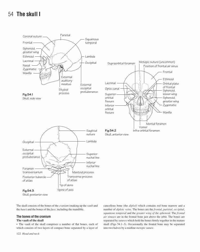
122 Head and neck
54 The skull I
Fig.54.1Skull, side view
Fig.54.3Skull, posterior view
Fig.54.2Skull, anterior view
Styloidprocess
ParietalCoronal suture
Frontal
Sphenoid, greater wingEthmoidLacrimalNasalZygomaticMaxilla
Squamoustemporal
Ethmoid
Frontal
Orbital plateof frontalSphenoid, lesser wingSphenoid, greater wing
Lambda
Occipital
Externaloccipitalprotuberance
Externalauditorymeatus Lacrimal
Optic canal
SuperiororbitalfissureInferiororbitalfissure
Metopic suture (uncommon)Position of frontal air sinus
Infra-orbital foramenVomer
Mental foramen
Supraorbital foramen
Zygomatic
Maxilla
LambdaOccipital
Externaloccipitalprotuberance
Posterior tubercleof atlas
Foramentransversarium
Spine of axisTip of dens
Transverse process of atlas
Mastoid process
Sagittalsuture
Superiornuchal line
Inferiornuchal line
The skull consists of the bones of the cranium (making up the vault andthe base) and the bones of the face, including the mandible.
The bones of the craniumThe vault of the skull• The vault of the skull comprises a number of flat bones, each ofwhich consists of two layers of compact bone separated by a layer of
cancellous bone (the diploë) which contains red bone marrow and anumber of diploic veins. The bones are the frontal, parietal, occipital,squamous temporal and the greater wing of the sphenoid. The frontalair sinuses are in the frontal bone just above the orbit. The bones areseparated by sutures which hold the bones firmly together in the matureskull (Figs 54.1–3). Occasionally the frontal bone may be separatedinto two halves by a midline metopic suture.
AAAC54 21/5/05 10:57 AM Page 122

The skull I 123
Fig.54.4The interior of the skull base.The anterior, middle and posterior cranial fossae are coloured green, red and blue respectively
Orbital plate of frontalDiploë
Lesser wing of sphenoid
Superior orbital fissure
Foramen rotundum
Foramen ovaleForamen spinosum
Foramen lacerum
Internal auditory meatusJugular foramenHypoglossal canal
Groove for transverse sinus
Cribriform plate (ethmoid)
Optic canalSella turcicaGreater wing of sphenoidSquamous temporalPetrous temporal
Arcuate eminence
Foramen magnum
• Foramen rotundum (Maxillary branch of trigeminal nerve)• Foramen ovale (Mandibular branch of trigeminal nerve)• Foramen spinosum (Middle meningeal artery)• Foramen lacerum (Internal carotid artery through upper opening
(p. 133) )• Other features:
• The superior orbital fissure is between the greater and lesser wingsof the sphenoid.
• In the midline is the body of the sphenoid with the sella turcica onits upper aspect. It contains the sphenoidal air sinus.
• The foramen lacerum is the gap between the apex of the petroustemporal and the body of the sphenoid.
• The boundary between the middle and posterior cranial fossae isthe sharp upper border of the petrous temporal bone.
The posterior cranial fossa• Bones:
• Petrous temporal (posterior surface)• Occipital
• Foramina:• Foramen magnum (lower part of medulla, vertebral arteries, spinal
accessory nerve)• Internal auditory meatus (facial and vestibulocochlear nerves,
internal auditory artery)• Jugular foramen (glossopharyngeal, vagus and accessory nerves,
internal jugular vein)• Hypoglossal canal (hypoglossal nerve)• Other features:• The jugular foramen is the gap between the occipital and petrous
temporal bones.• The inner surface of the occipital is marked by deep grooves for the
transverse and sigmoid venous sinuses. They lead down to thejugular foramen.
• There are a number of emissary foramina which transmit emissaryveins. These establish a communication between the intra- and extra-cranial veins.
On an X-ray of the skull there are markings which may be mistakenfor a fracture. These are caused by (1) the middle meningeal artery, (2)diploic veins, or (3) the sutures, including the infrequent metopic suture.
The interior of the base of the skullThe interior of the base of the skull comprises the anterior, middle andposterior cranial fossae (Fig. 54.4).
The anterior cranial fossa• Bones:
• Orbital plate of the frontal bone• Lesser wing of the sphenoid• Cribriform plate of the ethmoid
• Foramina:• In the cribriform plate (Olfactory nerves)• Optic canal (Optic nerve and ophthalmic artery)
• Other features:• The orbital plate of the frontal forms the roof of the orbit.• Lateral to the optic canals are the anterior clinoid processes.• The boundary between the anterior and middle cranial fossae is the
sharp posterior edge of the lesser wing of the sphenoid.
The middle cranial fossa• Bones:
• Greater wing of the sphenoid• Temporal bone
• Foramina:• Superior orbital fissure (Frontal, lacrimal and nasociliary branches
of trigeminal nerve; oculomotor, trochlear and abducent nerves;ophthalmic veins)
AAAC54 21/5/05 10:57 AM Page 123

The outside of the base of the skull (Fig. 55.1)The anterior part of the cranial base is hidden by the bones of the face (p. 125). The remainder consists of the bones that were seen in themiddle and posterior cranial fossae but many of the foramina seen onthe exterior are not visible inside the cranium.• Bones:
• Temporal (Squamous, petrous and tympanic parts and the styloidprocess)
• Sphenoid (body) which carries the medial and lateral pterygoidplates
• Foramina:• Foramen magnum (already described)• Hypoglossal canal (already described)• Stylomastoid foramen (facial nerve)• Jugular foramen (already described)• Foramen lacerum (the internal carotid through its internal opening)
• Carotid canal (internal carotid artery and sympathetic nerves)• Foramen spinosum (already described)• Foramen ovale (already described)
• Other features:• The area between and below the nuchal lines is for the attachment
of the extensor muscles of the neck.• The occipital condyles, for articulation with the atlas, lie on either
side of the foramen magnum.• The mastoid process is part of the petrous temporal and contains
the mastoid air cells (p. 157).• The floor of the external auditory meatus is formed by the tym-
panic plate of the temporal bone.• The carotid canal (see Fig. 59.2) turns inside the temporal bone to
run horizontally forwards. It then opens into the posterior wall of the foramen lacerum before turning upwards again to enter thecranial cavity through the internal opening of the foramen.
124 Head and neck
55 The skull II
Fig.55.1The base of the skull from below
Fig.55.2The internal surface of the mandible
Palatal process of maxillaIncisive fossa
Horizontal plateof palatine
Greater palatine foramen
Jugular foramenCarotid canal
Lateral pterygoid platePterygoid hamulus
Foramen ovaleForamen spinosum andspine of sphenoid
Ramus
Head
Mandibular foramen
Rough area formedial pterygoid
VomerForamen lacerum
Foramen magnum
Mastoid process
Occipital condyle
Alveolarpart of body
Mylohyoid line
Body
Coronoid process
Course of lingual nerve
Tympanic plate
AAAC55 21/5/05 10:57 AM Page 124
The skull II 125
• Behind the foramen spinosum is the spine of the sphenoid whichlies medial to the mandibular fossa for articulation with the head ofthe mandible.
• In front of this is the articular eminence, onto which the head of themandible moves when the mouth is open.
The bones of the face (Figs 54.2 and 55.2)The bones of the face are suspended below the front of the cranium andcomprise the bones of the upper jaw, the bones around the orbit andnasal cavities and the mandible.• Bones:
• Maxilla• Pterygoid plates of the sphenoid• Palatine• Zygomatic• Nasal• Frontal• Lacrimal• Bones of the orbit and nasal cavities (see below)
• Foramina:• Supraorbital (Supraorbital nerve)• Infraorbital (Infraorbital nerve)• Mental (Mental nerve)• Greater and lesser palatine foramina (Greater and lesser palatine
nerves)
• Foramina of the incisive fossa (Nasopalatine nerves and vessels)• Other features:
• The pterygoid plates of the sphenoid support the back of the maxilla.
• Between these two bones is the pterygomaxillary fissure whichleads into the pterygopalatine fossa.
• The hard palate is formed by the palatine process of the maxillaand the horizontal plate of the palatine.
• The teeth are borne in the maxilla.• The maxilla contains the large maxillary air sinus.• The bones of the orbit: the orbital margins are formed by the
frontal, zygomatic and maxillary bones.• The ethmoid lies between the two orbits and contains the ethmoidal
air cells.• The lacrimal has a fossa for the lacrimal sac.• At the back of the orbit are the greater and lesser wings of the sphe-
noid with the superior orbital fissure between them. Also the opticcanal and the infraorbital fissure.
• The bones of the nasal cavity are the maxilla, the inferior concha,the ethmoid, the vomer, the nasal septum and the perpendicularplate of the palatine.
The mandible (Fig. 55.2) consists of the body and two rami. Eachramus divides into a coronoid process and the head, for articulationwith the mandibular fossa. The mandibular foramen transmits the in-ferior alveolar nerve and vessels.
AAAC55 21/5/05 10:57 AM Page 125
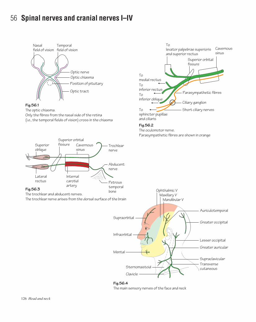
126 Head and neck
56 Spinal nerves and cranial nerves I–IV
Fig.56.1The optic chiasma.Only the fibres from the nasal side of the retina(i.e., the temporal fields of vision) cross in the chiasma
Fig.56.3The trochlear and abducent nerves.The trochlear nerve arises from the dorsal surface of the brain
Fig.56.4The main sensory nerves of the face and neck
Fig.56.2The oculomotor nerve.Parasympathetic fibres are shown in orange
Nasalfield of vision
Optic tract
Position of pituitaryOptic chiasmaOptic nerve
Superioroblique
Superior orbitalfissure Cavernous
sinus
Temporalfield of vision Cavernous
sinus
To levator palpebrae superiorisand superior rectus
To medial rectusTo inferior rectusTo inferior oblique
To sphincter pupillaeand ciliaris
Superior orbitalfissure
Parasympathetic fibres
Ciliary ganglion
Trochlearnerve
Abducentnerve
Petroustemporalbone
Short ciliary nerves
Lateralrectus
Internal carotidartery
Ophthalmic VMaxillary V
Mandibular V
Greater occipital
Lesser occipital
Auriculotemporal
Infraorbital
Supraorbital
Mental
Sternomastoid
Clavicle
Greater auricular
Transverse cutaneous
Supraclavicular
AAAC56 21/5/05 10:56 AM Page 126
The head and neck are supplied by the first four spinal and the 12 cra-nial nerves.
The spinal nerves• C1: supplies the small suboccipital muscles. Its anterior ramus joins thehypoglossal nerve but leaves it later to form the descendens hypoglossi.• C2: The posterior ramus forms the greater occipital nerve which issensory to the scalp.• The posterior rami of C2, 3 and 4 provide muscular and sensorybranches to the back. Their anterior rami provide muscular branches,including the descendens cervicalis (see hypoglossal nerve). They alsosupply sensory branches: the greater auricular, lesser occipital, an-terior cutaneous and the three supraclavicular nerves (Fig. 56.4). Thegreater auricular supplies the skin in the parotid region, the only sens-ory supply to the face which is not derived from the trigeminal. Theothers supply the skin of the neck and the upper part of the thorax.• The remaining cervical nerves (C5–8) join the brachial plexus.
The cranial nerves (Figs 56.1–3)• I. The olfactory nerve: the cell bodies of the olfactory nerve are in
the nasal mucosa. Their axons form the olfactory nerves which ascendthrough the cribriform plate to synapse in the olfactory bulb of thebrain.• II. The optic nerve (Fig. 56.1): the eye and optic nerve develop as anoutgrowth of the embryonic brain and the nerve is therefore envelopedin meninges. The cell bodies are in the retina and the axons pass back inthe optic nerve to the optic chiasma where the axons from the nasalhalves of the retina cross over but those from the temporal side continueon the same side. They then form the optic tract on each side.• III. The oculomotor nerve (Fig. 56.2): arises from the brain just infront of the pons, traverses the cavernous sinus and enters the orbitthrough the superior orbital fissure. Supplies the levator palpebraesuperioris, superior, inferior and medial rectus muscles and the inferioroblique. It also carries parasympathetic fibres to the ciliary ganglionwhere the fibres synapse and then pass in the short ciliary nerves to thesphincter pupillae and the ciliary muscles (see Chapter 69).• IV. The trochlear nerve (Fig. 56.3): arises from the dorsal surfaceof the brain just behind the inferior colliculus, winds round the mid-brain and enters the cavernous sinus. It enters the orbit through thesuperior orbital fissure and supplies the superior oblique.
Spinal nerves and cranial nerves I–IV 127
AAAC56 21/5/05 10:56 AM Page 127

128 Head and neck
57 The trigeminal nerve (V)
Fig.57.1The course and branches of the ophthalmic division of the trigeminal nerve.Parasympathetic fibres are shown in orange
Fig.57.2The course and branches of the maxillary division of the trigeminal nerve.Parasympathetic fibres are shown in orange
Supratrochlear
Infratrochlear
Supraorbital
Skin of face
Parasympathetic fibres fromsphenopalatine ganglionFrontalLacrimalSuperior orbital fissureOphthalmic division
Ethmoidal
Nasociliary
Optic nerve
Long posterior ciliary
Fig.57.3The course and main branches of the mandibular division of the trigeminal nerve.The fibres of the chorda tympani are shown in yellow
Lingual
Foramen ovale
Infra-orbital
Sphenopalatine
Buccal
Deep temporal (to temporalis)
Mandibular divisionSphenopalatine ganglion
AuriculotemporalOtic ganglion
Muscular branches
Chorda tympaniParotid gland
Inferior alveolar
Submandibular ganglionMylohyoid nerveSubmandibular gland
F. rotundumOphthalmic division
Maxillary division
Lacrimal gland
Nasal branches
Greater palatine
Lesser palatinePosterior superior dentalIncisive fossa
AAAC57 21/5/05 10:56 AM Page 128

The trigeminal nerve (Figs 56.4 and 57.1–3) arises from the brain at theside of the pons by a motor and a sensory root. The sensory root carriesthe trigeminal ganglion which consists of the cell bodies of the sensoryaxons and lies in a depression on the petrous temporal bone. It thendivides into ophthalmic, maxillary and mandibular divisions. Themotor root forms part of the mandibular division.
(a) The ophthalmic division (Fig. 57.1)This traverses the cavernous sinus and enters the orbit via the superiororbital fissure where it divides into frontal, lacrimal and nasociliarybranches. The frontal nerve lies just under the roof of the orbit anddivides into supraorbital and supratrochlear nerves which emergefrom the orbit and supply the front of the scalp. The lacrimal nerve lieslaterally and supplies the skin of the eyelids and face. It also carriesparasympathetic secretomotor fibres from the sphenopalatine ganglionto the lacrimal gland. The nasociliary nerve crosses the optic nerve andruns along the medial wall of the orbit to emerge onto the face as theinfratrochlear nerve. It gives off the ethmoidal nerves to the ethmoidalsinuses and the long ciliary nerves to the eye which carry sensory fibresfrom the cornea and sympathetic fibres to the dilator pupillae. Allbranches of the ophthalmic division are sensory.
(b) The maxillary division (Fig. 57.2)This leaves the cranial cavity through the foramen rotundum and entersthe pterygopalatine fossa. It has the sphenopalatine ganglion attachedto it which transmits parasympathetic fibres to the lacrimal gland viacommunications with the lacrimal nerve. The branches of the maxillarynerve are the greater and lesser palatine nerves to the hard and soft
palates, the sphenopalatine nerve to the nasal cavity and thence via thenasal septum, to the incisive fossa to supply the hard palate. The pos-terior superior dental nerve enters the back of the maxilla and suppliesthe teeth. The maxillary nerve leaves the sphenopalatine fossa via theinferior orbital fissure, travels in the floor of the orbit where it gives themiddle and anterior superior dental nerves, and emerges onto the facethrough the infraorbital foramen as the infraorbital nerve. All branchesof the maxillary division are sensory.
(c) The mandibular division (Fig. 57.3)This leaves the cranial cavity through the foramen ovale and immedi-ately breaks up into branches. These are: the mainly sensory inferioralveolar nerve, which enters the mandibular foramen to supply theteeth before emerging onto the face as the mental nerve. This nervedoes have one motor branch, the mylohyoid nerve, which supplies themylohyoid and the anterior belly of the digastric. The lingual nerve liesclose to the mandible just behind the third molar and then passes for-wards to supply the tongue. It is joined by the chorda tympani whichcarries taste fibres from the anterior two-thirds of the tongue andparasympathetic secretomotor fibres to the submandibular and sublin-gual salivary glands. These synapse in the submandibular ganglionwhich is attached to the lingual nerve. The auriculotemporal nerve sup-plies sensory fibres to the side of the scalp. It also carries parasympath-etic secretomotor fibres, which have synapsed in the otic ganglion, tothe parotid gland. The buccal nerve carries sensory fibres from the face.There are muscular branches to the muscles of mastication, includingthe deep temporal nerves which supply temporalis. The mandibulardivision thus contains both motor and sensory branches.
The trigeminal nerve (V) 129
AAAC57 21/5/05 10:56 AM Page 129
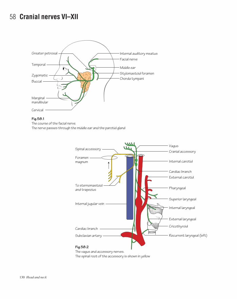
130 Head and neck
58 Cranial nerves VI–XII
Fig.58.1The course of the facial nerve.The nerve passes through the middle ear and the parotid gland
Fig.58.2The vagus and accessory nerves.The spinal root of the accessory is shown in yellow
Greater petrosal
Buccal
Marginal mandibular
Cervical
Temporal
Zygomatic
Internal auditory meatus
Facial nerve
Middle ear
Stylomastoid foramenChorda tympani
Foramenmagnum
To sternomastoidand trapezius
Internal jugular vein
Cardiac branch
Subclavian artery
Spinal accessoryVagus
Pharyngeal
Superior laryngeal
Cranial accessory
Internal carotid
Cardiac branch
Internal laryngeal
External laryngeal
Cricothyroid
Recurrent laryngeal (left)
External carotid
AAAC58 21/5/05 10:56 AM Page 130

• VI. The abducent nerve (see Fig. 56.3): leaves the brain at the pos-terior border of the pons and has a long intracranial course (so is oftenthe first nerve to be affected in raised intracranial pressure) to the cav-ernous sinus, where it is closely applied to the internal carotid artery,and thence to the orbit via the superior orbital fissure. It supplies the lat-eral rectus.• VII. The facial nerve (Fig. 58.1): this leaves the brain near the cere-bellum and passes laterally into the internal auditory meatus. It reachesthe medial wall of the middle ear and turns backwards and down-wards to leave the skull via the stylomastoid foramen. It then traversesthe parotid gland, in which it divides into five branches (temporal,zygomatic, buccal, marginal mandibular and cervical) which are distributed to the muscles of facial expression, the platysma and theposterior belly of the digastric. In the middle ear it gives off the greaterpetrosal branch which carries parasympathetic fibres to thesphenopalatine ganglion and thence to the lacrimal gland. In the middleear it also gives off the chorda tympani which joins the lingual nerveand is distributed with it. Sensory fibres in the chorda tympani havetheir cell bodies in the geniculate ganglion which lies on the facialnerve where it turns downwards.• VIII. The vestibulocochlear (auditory) nerve: this leaves the brainnext to the facial nerve and enters the internal auditory meatus. Itdivides into vestibular and cochlear nerves.• IX. The glossopharyngeal nerve (see Fig. 65.1): leaves the brain atthe side of the medulla and passes through the jugular foramen. It thencurves forwards between the internal and external carotid arteries toenter the pharynx between the superior and middle constrictors. It sup-plies sensory fibres to the posterior one third of the tongue (includingtaste) and the pharynx. It also gives a branch to the carotid body andsinus.• X. The vagus nerve (Fig. 58.2): arises from the side of the medullaand passes through the jugular foramen. It is joined by the accessorynerve but the spinal root of the accessory leaves it again almost imme-diately. The cranial root is distributed with the vagus (hence thenameait is accessory to the vagus). The vagus carries two ganglia forthe cell bodies of its sensory fibres. It descends between the internalcarotid artery and the jugular vein, within the carotid sheath, and enters
the thorax where its further course is described in Chapters 2 and 3. Inthe neck the vagus (and cranial root of the accessory) gives the follow-ing branches:• The pharyngeal branch which runs below and parallel to the glos-
sopharyngeal nerve and supplies the striated muscle of the palateand pharynx.
• Superior and inferior cardiac branches which descend into the thorax to take part in the cardiac plexuses.
• The superior laryngeal nerve which divides into internal and exter-nal laryngeal nerves. The former enters the larynx by piercing thethyrohyoid membrane and is sensory to the larynx above the level ofthe vocal cords, and the latter is motor to the cricothyroid muscle.
• The recurrent laryngeal nerve. On the right side it loops under thesubclavian artery before ascending to the larynx behind the com-mon carotid artery. On the left side it arises from the vagus justbelow the arch of the aorta and ascends to the larynx in the groovebetween the trachea and oesophagus. The recurrent laryngealnerves supply all the muscles of the larynx except for cricopharyn-geus and are sensory to the larynx below the vocal cords.
• XI. The accessory nerve (Fig. 58.2): the cranial root arises from theside of the medulla with the vagus and is distributed with it. The spinalroot arises from the side of the upper five segments of the spinal cord,enters the cranial cavity through the foramen magnum and joins thevagus. It leaves the vagus below the jugular foramen and passes back-wards to enter sternomastoid, which it supplies. It then crosses the pos-terior triangle to supply trapezius (see Fig. 61.3).• XII. The hypoglossal nerve (Figs 59.1 and 65.1): arises from theside of the medulla ventral to the vagus and cranial accessory andpasses through the hypoglossal canal. Below the skull it is joined by theanterior ramus of C1 and it then runs downwards and forward, acrossthe carotid sheath and the upward loop of the lingual artery to enter thetongue. It supplies the intrinsic and extrinsic muscles of the tongue. Itgives off the descendens hypoglossi but this is actually composed offibres from C1. This joins the descendens cervicalis, derived from C2and 3, to form the ansa cervicalis. From this, branches arise to supplythe ‘strap muscles’, i.e. sternothyroid, sternohyoid, thyrohyoid andomohyoid.
Cranial nerves VI–XII 131
AAAC58 21/5/05 10:56 AM Page 131

132 Head and neck
59 The arteries I
Fig.59.1The course and main branches of the external carotid artery.The inferior thyroid artery is also shown
Fig.59.3Carotid angiogram showing internal carotid stenosis
Fig.59.2The intracranial course of the internal carotid artery.The intracranial parts of the two vertebral arteries are also shown diagrammaticallyalthough they are in a different plane
Foramen spinosum
Superficial temporal
Middle meningeal
Inferior thyroid
Recurrent laryngeal nerve
Superior thyroid
Carotid sinus
Lingual
Laryngeal branch
Thyrocervical trunkSubclavian
Thyroid
Dorsallingual
Facial
Internal carotidExternal carotid
Hypoglossal nerveTonsillar branch
MaxillaryOccipital
Anterior cerebral
Striate arteriesMiddle cerebralPosterior communicating
Superior cerebellarCavernous sinusInternal auditory
Posterior cerebral
Anterior inferior cerebellarVertebralPosterior inferior cerebellar
Anterior and posterior spinalCarotid canal
Internalcarotid
Anterior clinoid processCentral arteryof retinaOphthalmic arteryOptic canal
Abducentnerve
Foramenlacerum
AAAC59 21/5/05 10:55 AM Page 132
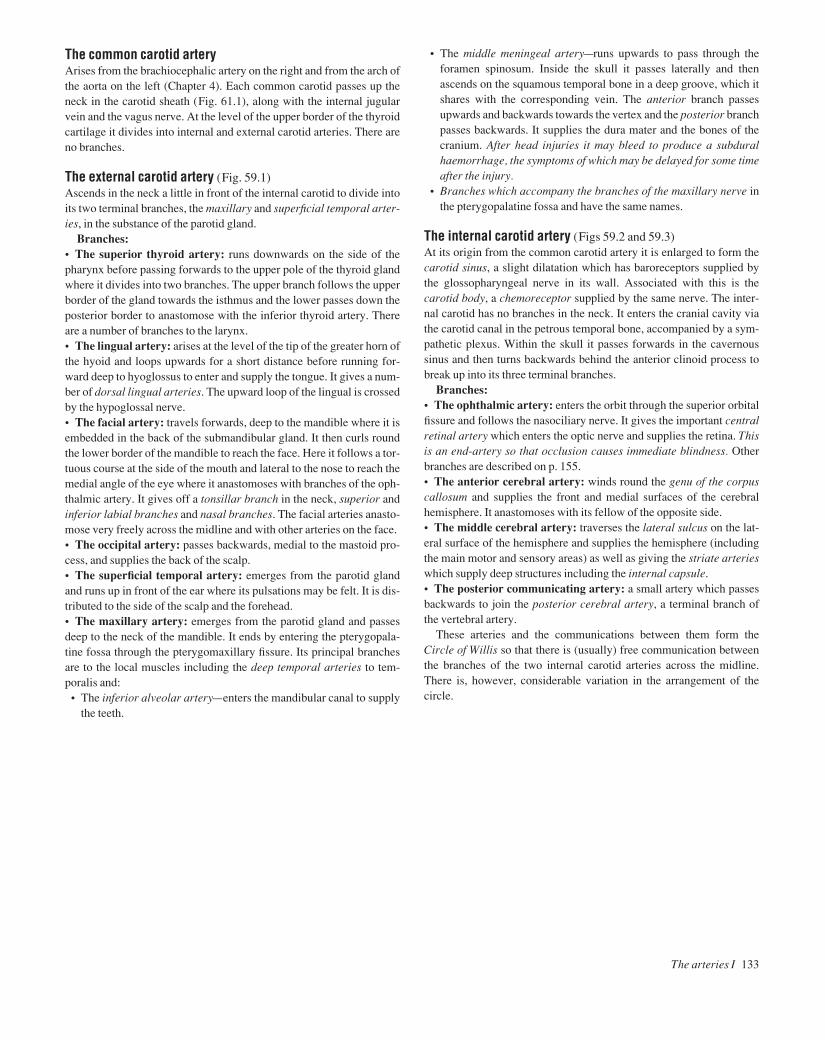
The common carotid arteryArises from the brachiocephalic artery on the right and from the arch ofthe aorta on the left (Chapter 4). Each common carotid passes up theneck in the carotid sheath (Fig. 61.1), along with the internal jugularvein and the vagus nerve. At the level of the upper border of the thyroidcartilage it divides into internal and external carotid arteries. There areno branches.
The external carotid artery (Fig. 59.1)Ascends in the neck a little in front of the internal carotid to divide intoits two terminal branches, the maxillary and superficial temporal arter-ies, in the substance of the parotid gland.
Branches:• The superior thyroid artery: runs downwards on the side of thepharynx before passing forwards to the upper pole of the thyroid glandwhere it divides into two branches. The upper branch follows the upperborder of the gland towards the isthmus and the lower passes down theposterior border to anastomose with the inferior thyroid artery. Thereare a number of branches to the larynx.• The lingual artery: arises at the level of the tip of the greater horn ofthe hyoid and loops upwards for a short distance before running for-ward deep to hyoglossus to enter and supply the tongue. It gives a num-ber of dorsal lingual arteries. The upward loop of the lingual is crossedby the hypoglossal nerve.• The facial artery: travels forwards, deep to the mandible where it isembedded in the back of the submandibular gland. It then curls roundthe lower border of the mandible to reach the face. Here it follows a tor-tuous course at the side of the mouth and lateral to the nose to reach themedial angle of the eye where it anastomoses with branches of the oph-thalmic artery. It gives off a tonsillar branch in the neck, superior andinferior labial branches and nasal branches. The facial arteries anasto-mose very freely across the midline and with other arteries on the face.• The occipital artery: passes backwards, medial to the mastoid pro-cess, and supplies the back of the scalp.• The superficial temporal artery: emerges from the parotid glandand runs up in front of the ear where its pulsations may be felt. It is dis-tributed to the side of the scalp and the forehead.• The maxillary artery: emerges from the parotid gland and passesdeep to the neck of the mandible. It ends by entering the pterygopala-tine fossa through the pterygomaxillary fissure. Its principal branchesare to the local muscles including the deep temporal arteries to tem-poralis and:• The inferior alveolar arteryaenters the mandibular canal to supply
the teeth.
• The middle meningeal arteryaruns upwards to pass through theforamen spinosum. Inside the skull it passes laterally and thenascends on the squamous temporal bone in a deep groove, which itshares with the corresponding vein. The anterior branch passesupwards and backwards towards the vertex and the posterior branchpasses backwards. It supplies the dura mater and the bones of thecranium. After head injuries it may bleed to produce a subduralhaemorrhage, the symptoms of which may be delayed for some timeafter the injury.
• Branches which accompany the branches of the maxillary nerve inthe pterygopalatine fossa and have the same names.
The internal carotid artery (Figs 59.2 and 59.3)At its origin from the common carotid artery it is enlarged to form thecarotid sinus, a slight dilatation which has baroreceptors supplied bythe glossopharyngeal nerve in its wall. Associated with this is thecarotid body, a chemoreceptor supplied by the same nerve. The inter-nal carotid has no branches in the neck. It enters the cranial cavity viathe carotid canal in the petrous temporal bone, accompanied by a sym-pathetic plexus. Within the skull it passes forwards in the cavernoussinus and then turns backwards behind the anterior clinoid process tobreak up into its three terminal branches.
Branches:• The ophthalmic artery: enters the orbit through the superior orbitalfissure and follows the nasociliary nerve. It gives the important centralretinal artery which enters the optic nerve and supplies the retina. Thisis an end-artery so that occlusion causes immediate blindness. Otherbranches are described on p. 155.• The anterior cerebral artery: winds round the genu of the corpuscallosum and supplies the front and medial surfaces of the cerebralhemisphere. It anastomoses with its fellow of the opposite side.• The middle cerebral artery: traverses the lateral sulcus on the lat-eral surface of the hemisphere and supplies the hemisphere (includingthe main motor and sensory areas) as well as giving the striate arterieswhich supply deep structures including the internal capsule.• The posterior communicating artery: a small artery which passesbackwards to join the posterior cerebral artery, a terminal branch ofthe vertebral artery.
These arteries and the communications between them form theCircle of Willis so that there is (usually) free communication betweenthe branches of the two internal carotid arteries across the midline.There is, however, considerable variation in the arrangement of the circle.
The arteries I 133
AAAC59 21/5/05 10:55 AM Page 133

134 Head and neck
60 The arteries II and the veins
Fig.60.2The intracranial venous sinuses
Fig.60.3Vertebral angiogram
Fig.60.1The branches of the subclavian artery
Scalenus anterior
Inferiorthyroid artery
Vertebral artery
Thyrocervical trunk
C6
Axillary artery
Dorsal scapular(usually)
Subclavian artery
Internal thoracic arteryFirst rib
Costocervicaltrunk
Subclavian artery
Beginning of superiorsagittal sinus
Beginning of inferiorsagittal sinus
Superior ophthalmic vein
Costocervicaltrunk
First rib
Superiorintercostal artery
Apex of lung
Intercavernous sinusesCavernous sinus
Superior petrosal sinusInferior petrosal sinusJugular foramenSigmoid sinus
Transverse sinus
Great cerebral veinStraight sinusTentorium cerebelliTransverse sinus
Posterior inferiorcerebellar artery
Posterior cerebralartery
Vertebral artery
AAAC60 21/5/05 10:55 AM Page 134
The subclavian artery (Figs 60.1 and 63.1)Arises from the brachiocephalic artery on the right and the arch of theaorta on the left. It arches across the upper surface of the 1st rib tobecome the axillary artery. It is in close contact with the apex of thelung and lies behind scalenus anterior at the root of the neck.
Branches:• The internal thoracic artery: see p. 13.• The vertebral artery: runs upwards to enter the foramen transver-sarium of the 6th cervical vertebra. It passes through correspondingforamina in the other cervical vertebra to reach the upper surface of theatlas. Here it turns medially in a groove and then enters the cranial cav-ity through the foramen magnum. Here it joins its fellow of the oppositeside to form the basilar artery. It gives off the anterior and posteriorspinal arteries which descend to supply the spinal cord, and the poster-ior inferior cerebellar artery which supplies not only the cerebellumbut also the medulla. The basilar artery passes forwards on the under-surface of the medulla and pons and gives the anterior inferior cerebel-lar artery, branches to the brainstem and to the inner ear (the internalauditory artery) and ends by dividing into the superior cerebellar andposterior cerebral arteries. The latter is joined by the posterior com-municating artery (p. 133).• The costocervical trunk: a small artery that passes backwards tosupply muscles of the back. It also supplies the superior thoracic artery(see Chapter 3).• The thyrocervical trunk: gives off the superficial cervical andsuprascapular arteries and then passes medially as the inferior thyroidartery across the vertebral artery to reach the middle of the posteriorborder of the thyroid. It has a variable relation to the recurrent laryngealnerve, lying in front or behind them, but may branch early with thenerve passing between the branches.• The dorsal scapular artery: usually given off from the third part(Fig. 63.1). It descends along the medial border of the scapula but mayarise in common with the superficial cervical artery.
The veinsThe veins of the brain drain into dural venous sinuses (Fig. 60.2). Themost important of these are:• The superior sagittal sinus: passes backwards in the midline in theattached border of the falx cerebri from just above the cribriform plateto the occipital region, where it communicates with the straight sinus,and then turns to the right to form the right transverse sinus. It thenwinds down on the back of the petrous temporal as the sigmoid sinus
which passes through the right jugular foramen to form the right inter-nal jugular vein.• The inferior sagittal sinus: begins near the origin of the superiorsagittal sinus and runs in the free border of the falx cerebri. It is joinedby the great cerebral vein to form the straight sinus which lies in theattachment of the falx to the tentorium cerebelli. The straight sinusturns to the left to form the left transverse sinus and then the sigmoidsinus. The latter leaves the skull through the left jugular foramen.• The cavernous sinus: this lies at the side of the pituitary fossa andcontains the internal carotid artery. It receives the superior and inferiorophthalmic veins and is connected to some smaller sinusesathe super-ior and inferior petrosal sinuses and the sphenoidal sinus. The two cavernous sinuses are joined in front and behind the pituitary by theintercavernous sinuses.• The emissary veins: see p. 123.• The internal jugular vein: passes down the neck from the jugularforamen, in the carotid sheath along with the internal and commoncarotid arteries and the vagus nerve. It ends by joining the subclavianvein to form the brachiocephalic vein. It receives veins correspondingto the branches of the external carotid artery ( facial, lingual, pharyn-geal, and the superior and middle thyroid veins). The inferior thyroidveins pass downwards in front of the trachea to open into the left bra-chiocephalic vein.
The facial vein communicates around the orbit with tributaries of theophthalmic veins so that infections of the face may spread to the cav-ernous sinus if not properly treated.• The external jugular vein: begins in the parotid gland by the joiningof the retromandibular vein with other small veins. It passes obliquelyacross sternomastoid to open into the subclavian vein. It receives thetransverse cervical, suprascapular and anterior jugular veins near itslower end.• The anterior jugular vein: begins below the chin and runs down theneck near the midline. It then passes deep to sternomastoid to join theexternal jugular vein.• The subclavian vein: lies in a groove on the 1st rib but is separatedfrom the subclavian artery by the scalenus anterior. It receives theexternal jugular vein, veins corresponding to the branches of the sub-clavian artery and, at its junction with the internal jugular vein, the thor-acic duct on the left and the right lymph duct on the right.• The vertebral vein: this is formed at the level of the 6th foramentransversarium from the vertebral plexus of veins that accompany thevertebral artery.
The arteries II and the veins 135
AAAC60 21/5/05 10:55 AM Page 135

136 Head and neck
61 Anterior and posterior triangles
Fig.61.1The basic plan of the neck in cross-section.The arrows indicate the posterior triangle
Fig.61.3The main structures in the posterior triangle Fig.61.4
Subdivisions of the anterior triangle
Pretracheal fasciaInvesting layer of deep fascia
Common carotid artery
Skin and superficial fascia
Sternomastoid
Internal jugular vein
Vagus nervePrevertebral fascia
Trapezius
Fig.61.2A more detailed plan of the neck, based on Fig. 61.1.There are still some structures omitted from the diagramfor the sake of simplicity, for example the strap muscles
Parotid Digastric triangleSemispinalis
Lesser occipital
Splenius capitis
Levator scapulaeTrapezius
Submentaltriangle Carotid
triangle
Hyoid boneBrachial plexus(upper trunk)
Scalenus anterior
External jugular veinTransversecutaneousSupraclavicular nervesAnterior jugular veinOmohyoid
Greater auricularAnterior belly of digastric
Posterior belly of digastric
Sternocleidomastoid
Musculartriangle
Omohyoid
External jugular
TracheaSternomastoid
Thyroid
Pretracheal fascia
Carotid sheath
Sympathetic trunkVagus
TrapeziusSplenius
Scalenus anterior
Scalenus mediusLevator scapulae
Spinal nervePlane of accessorynerve
Vertebral arterySemispinalis
Left recurrent laryngeal nerve
Thoracic ductLongus colliLong thoracicnervePrevertebralfascia
Accessory nerveScalenus medius
AAAC61 21/5/05 11:03 AM Page 136

The neck consists essentially of five blocks of tissue running longitudin-ally (Figs 61.1 and 61.2). These are as follows:1 The cervical vertebrae surrounded by a number of muscles andenclosed in a dense layer of prevertebral fascia.2 The pharynx and larynx, partially enclosed in a thin layer of pre-tracheal fascia. Below the level of C6 these give way to the oesophagusand trachea.3 & 4 Two vascular packets consisting of the common and internalcarotid arteries, the internal jugular vein and the vagus nerve, allenclosed in the fascial carotid sheath.5 An outer enclosing sheath consisting of the sternomastoid andtrapezius and the investing layer of deep fascia of the neck.
The anterior triangleThe anterior triangle (Fig. 61.4) is bounded by:• The lower border of the mandible and its backward continuation.• The anterior border of sternomastoid.• The midline of the neck.
The anterior triangle is subdivided into:(a) The digastric triangle, bounded by:
• The lower border of the mandible.• The two bellies of the digastric.
(b) The carotid triangle, bounded by:• The superior belly of the omohyoid.• The posterior belly of the digastric.• The anterior border of sternomastoid.
(c) The muscular triangle, bounded by:• The superior belly of the omohyoid.• The anterior border of sternomastoid.• The midline of the neck.
The contents of these triangles are mostly structures that are continu-ous, without interruption, from one triangle to another so that it is moreconvenient to describe them individually in other chapters.
The posterior triangle (Fig. 61.3)The posterior triangle is bounded by:• The posterior border of sternomastoid.• The anterior border of trapezius.• The middle part of the clavicle.
Stretching between the two muscles is the investing layer of deepfascia which splits to enclose them and continues to the anterior tri-angle. Embedded in the deep fascia is the spinal part of the accessorynerve which leaves the sternomastoid about halfway down its posteriorborder and passes into trapezius two fingerbreadths above the clavicle.It supplies both muscles. Four cutaneous nerves (transverse cervical,supraclavicular, greater auricular and lesser occipital) also emergenear the accessory nerve and supply the skin of the neck and the upperpart of the chest. The external jugular vein begins near the upper end ofsternomastoid and runs down obliquely across this muscle to enter thesubclavian vein. It is joined by the anterior jugular and other smallveins at its lower end. The inferior belly of the omohyoid musclecrosses the lower part of the triangle.
The floor of the posterior triangle is the prevertebral fascia, deepto which lie, from below upwards: scalenus anterior, scalenus mediusand posterior, levator scapulae, and splenius capitis.
Structures deep to the prevertebral fascia• The upper, middle and lower trunks of the brachial plexus whichemerge between the scalenus anterior and the scalenus medius, thelower trunk resting on the 1st rib.• The supraclavicular branches of the brachial plexus (p. 73).
Anterior and posterior triangles 137
AAAC61 21/5/05 11:03 AM Page 137

138 Head and neck
62 The pharynx and larynx
Fig.62.5The larynx as seen through a laryngoscope
Fig.62.4The muscles causing abduction, adduction and relaxationof the vocal cords. Tightening of the cords is shown in Fig.62.3
Fig.62.3Left, the cricothyroid muscle and right, the posterior crico-arytenoid
Fig.62.2A midline section of the larynx to show thecricovocal membrane and vocal ligaments
Fig.62.1The pharynx and larynx, and some of the related nerves
Pterygomandibularligament
Superior constrictor
Buccinator
Cricoid cartilage
Inferior constrictor
Attachment ofsternohyoid
Attachment ofthyrohyoid
Stylohyoid ligament
Stylopharyngeus
Oesophagus
Vocal foldVestibular fold
EpiglottisEpiglottic tubercle
Glossoepiglotticfold
Base of tongue
Trachea
Falsecord
Aryepiglotticfold
Vallecula
Epiglottis
CricothyroidPosition ofsynovial joint
Vocal ligamentThyro-arytenoid
muscleVocal process
Lateralcrico-arytenoidPosterorcrico-arytenoid
Greater horn of hyoid
Epiglottis
Ala of thyroid cartilage
Arytenoid
Muscular processCrico-arytenoid muscle(posterior)Cricoid
Epiglottis
Vocal process
Thyroid cartilageVocal ligamentCricovocal membrane
Cricoid
First ring of trachea
ArytenoidMuscular processCrico-arytenoidjoint
Facet forinferior hornof thyroid
Glossopharyngeal
Nerves
Superior laryngealInternal laryngealExternal laryngeal
Recurrent laryngeal
Cricothyroid
AAAC62 21/5/05 11:02 AM Page 138
The pharynxThis is an incomplete striated muscular tube, opening in front into thenasal cavity, the mouth and the larynx, thus being made up of thenasopharynx, the oropharynx and the laryngopharynx. The muscularcoat (Fig. 62.1) is formed by:• The superior constrictor: arises from the pterygomandibular liga-ment (which spans between the pterygoid hamulus and the mandiblejust behind the third molar tooth).• The middle constrictor: arises from the stylohyoid ligament and thelesser and greater horns of the hyoid bone.• The inferior constrictor: arises from the thyroid and cricoidcartilages.
The constrictors encircle the pharynx and interdigitate posteriorly.The gaps between the constrictors are filled in by fascia. There is alsoan inner longitudinal layer of muscle. The nasopharynx is lined by cili-ated columnar epithelium and on its posterior wall is situated a mass oflymphatic tissue, the pharyngeal tonsil or adenoid. The pharyngotym-panic (Eustachian) tube opens into the nasopharynx at the level of thefloor of the nose, the cartilage of the tube producing a distinct bulgebehind the opening.• Nerve supply:
• Motorapharyngeal branch of the vagus (p. 131).• Sensoryaglossopharyngeal (p. 131).
The larynxPalpable components• Hyoid bone, level of C3.• Thyroid cartilage, level of C4 and 5.• Cricoid cartilage, level of C6.
Other components• Arytenoid cartilages: attached to upper border of the cricoid by syn-ovial joints so that they can slide and rotate. Each has an anterior vocalprocess and a lateral muscular process (Figs 62.2 and 62.3).• Epiglottis: a leaf-shaped piece of elastic cartilage attached to theback of the thyroid cartilage (Fig. 62.2) and projecting upwards behindthe hyoid.• Thyrohyoid ligament: joins the hyoid and the thyroid (Fig. 62.1).• Cricovocal membrane (cricothyroid ligament): attached to theupper border of the cricoid and passes inside the thyroid to be attachedto the back of the thyroid and to the vocal processes of the arytenoids(Fig. 62.2). The upper border is thickened to form the vocal ligamentwhich, with the mucous membrane that covers it, forms the vocalcords.• Cricothyroid joint: a small synovial joint between the inferior hornof the thyroid cartilage and the cricoid, permitting a hinge-like move-ment (Fig. 62.3).
• Pyriform fossa: the fossa between the posterior border of the thyroidcartilage and the cricoid and arytenoid cartilages. It is important be-cause a carcinoma in this region can remain ‘silent’ until it has spreadto the cervical lymph nodes whereas a tumour of the vocal cords pro-duces early voice changes.• Mucous membrane: mostly respiratory epithelium (ciliated colum-nar) but over the vocal cords it changes to stratified squamous so thatthe cords have a pearly appearance (Fig. 62.5).• Vestibular folds: a pair of additional folds above the vocal folds( false cords). The space between the vocal and vestibular folds is thesinus of the larynx.
The intrinsic muscles of the larynx• Cricothyroid (Figs 62.1 and 62.3): situated on the outside of the larynx. Tenses the vocal cords.• Thyroarytenoid (Fig. 62.4): from the back of the thyroid cartilage tothe vocal process of the arytenoid. Relaxes the vocal cords.• Posterior cricoarytenoid (Figs 62.3 and 62.4): from the back of thecricoid to the muscular process of the arytenoid. Abducts the vocalcords.• Lateral cricoarytenoid (Fig. 62.4): adducts the vocal cords.• Interarytenoids and aryepiglottic muscle: form a ‘sphincter’, to-gether with the epiglottis, in order to close off the entrance to the larynx(glottis) during swallowing.• Nerve supply:
• Motorarecurrent laryngeal nerve, except for the cricothyroidwhich is supplied by the external laryngeal nerve.
• Sensoryaabove the vocal cords, the internal laryngeal nervewhich enters the larynx through the thyrohyoid membrane. Belowthe vocal cords, the recurrent laryngeal nerve (which is therefore amixed nerve) which enters the larynx just behind the cricothyroidjoint.
Laryngoscopy (Fig. 62.5)The vocal cords may be seen by means of a warmed, angled mirrorpassed to the back of the throat. Only the edges of the cords can be seenbecause of the shadow of the false cords. Adduction of the cords may beobserved by asking the patient to say a high-pitched ‘eeeee’ whileabduction occurs when saying ‘Ahhh’. If one of the recurrent laryngealnerves is divided, the cord lies in a position midway between adductionand abduction but this does not produce very severe voice changesbecause the uninjured cord can cross the midline to reach the para-lysed cord. However, if both nerves are cut the cords lie in the cadavericposition, i.e. in the mid-position. If both nerves are damaged but notcompletely divided the cords are adducted since the abductors aremore affected than the adductors (Semon’s law). This may causebreathing difficulties.
The pharynx and larynx 139
AAAC62 21/5/05 11:02 AM Page 139

The area defined by the first thoracic vertebra, the first ribs and themanubrium sterni is called the thoracic inlet (or outlet) (Fig. 63.1).Through this relatively confined space pass the trachea and oesopha-gus, the carotid and subclavian arteries and the corresponding largeveins as well as the apices of the lungs and important nerves.
The scalene muscles• Scalenus anterior: passes down from the transverse processes ofsome of the cervical vertebrae and is inserted by means of a narrow ten-don into the scalene tubercle on the medial border of the 1st rib.
• Scalenus medius: is behind scalenus anterior and is inserted by mus-cular fibres into a large area of the 1st rib. The subclavian artery and thetrunks of the brachial plexus are between the two muscles and the sub-clavian vein is in front of scalenus anterior. The gap between the twomuscles may be narrow and the tough tendon of scalenus anterior maycompress the lower trunk of the brachial plexus and/or the subclavianartery, giving rise to one form of the thoracic outlet syndrome (see alsocervical rib, p. 7).
140 Head and neck
63 The root of the neck
Fig.63.1The deep structures at the root of the neck and in the thoracic outlet.The curved arrow on the right side of the diagram indicates the course of the thoracic duct
Fig.63.2The structures related to the upper surface of the first rib
Vertebral artery
Inferior thyroid artery
Sympathetic trunk
Subclavian arteryVagus
Carotid sheathInferior cervical ganglion
Sympathetic trunkSuperior intercostal artery1st thoracic nerve8th cervical nerve
Scalene tubercle, for attachment of scalenus anterior
Costoclavicular ligament
Subclavian vein
Thoracic duct
Internal thoracic artery
Scalenus posteriorAttachment of scalenus mediusSerratus anterior(first digitation)
Lower trunk of brachial plexusSubclavian arterySubclavian vein
Dorsal scapular arterySuprascapular artery, nerveSuperficial cervical arteryUpper trunk of brachial plexus
Phrenic nerveScalenus anterior
Scalenus medius
Middle cervical ganglion
Subclavius
AAAC63 21/5/05 11:02 AM Page 140
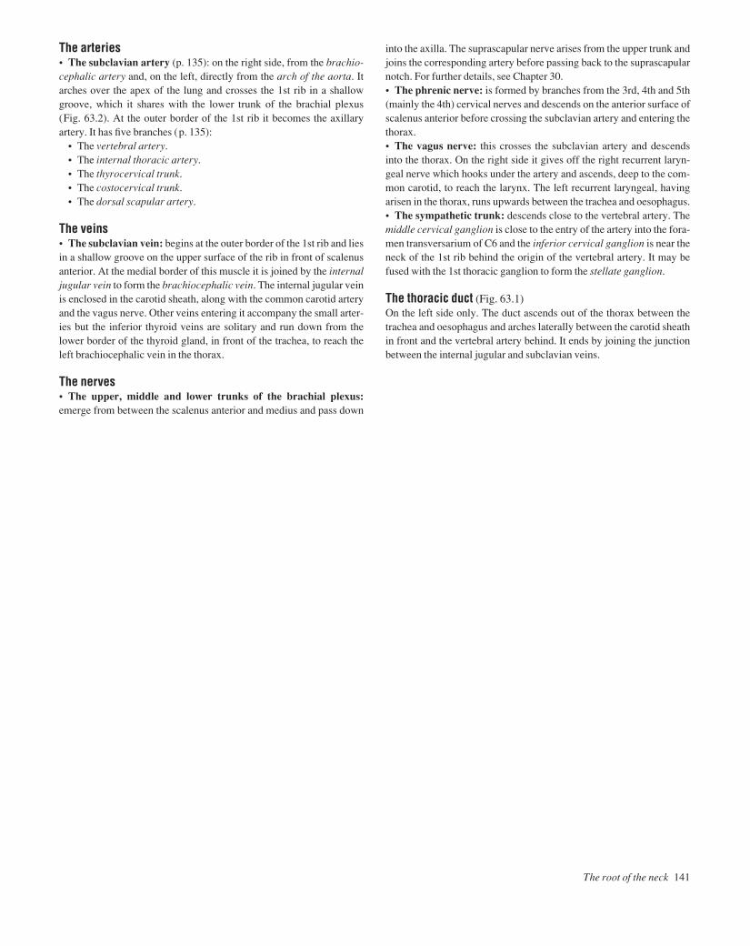
The root of the neck 141
The arteries• The subclavian artery (p. 135): on the right side, from the brachio-cephalic artery and, on the left, directly from the arch of the aorta. Itarches over the apex of the lung and crosses the 1st rib in a shallowgroove, which it shares with the lower trunk of the brachial plexus (Fig. 63.2). At the outer border of the 1st rib it becomes the axillaryartery. It has five branches (p. 135):
• The vertebral artery.• The internal thoracic artery.• The thyrocervical trunk.• The costocervical trunk.• The dorsal scapular artery.
The veins• The subclavian vein: begins at the outer border of the 1st rib and liesin a shallow groove on the upper surface of the rib in front of scalenusanterior. At the medial border of this muscle it is joined by the internaljugular vein to form the brachiocephalic vein. The internal jugular veinis enclosed in the carotid sheath, along with the common carotid arteryand the vagus nerve. Other veins entering it accompany the small arter-ies but the inferior thyroid veins are solitary and run down from thelower border of the thyroid gland, in front of the trachea, to reach theleft brachiocephalic vein in the thorax.
The nerves• The upper, middle and lower trunks of the brachial plexus:emerge from between the scalenus anterior and medius and pass down
into the axilla. The suprascapular nerve arises from the upper trunk andjoins the corresponding artery before passing back to the suprascapularnotch. For further details, see Chapter 30.• The phrenic nerve: is formed by branches from the 3rd, 4th and 5th(mainly the 4th) cervical nerves and descends on the anterior surface ofscalenus anterior before crossing the subclavian artery and entering thethorax.• The vagus nerve: this crosses the subclavian artery and descendsinto the thorax. On the right side it gives off the right recurrent laryn-geal nerve which hooks under the artery and ascends, deep to the com-mon carotid, to reach the larynx. The left recurrent laryngeal, havingarisen in the thorax, runs upwards between the trachea and oesophagus.• The sympathetic trunk: descends close to the vertebral artery. Themiddle cervical ganglion is close to the entry of the artery into the fora-men transversarium of C6 and the inferior cervical ganglion is near theneck of the 1st rib behind the origin of the vertebral artery. It may befused with the 1st thoracic ganglion to form the stellate ganglion.
The thoracic duct (Fig. 63.1)On the left side only. The duct ascends out of the thorax between thetrachea and oesophagus and arches laterally between the carotid sheathin front and the vertebral artery behind. It ends by joining the junctionbetween the internal jugular and subclavian veins.
AAAC63 21/5/05 11:02 AM Page 141

The oesophagusThe oesophagus begins at the level of the cricoid cartilage and runsdown behind and slightly to the left of the trachea. The left recurrentlaryngeal nerve is in the groove between the oesophagus and tracheaand the thoracic duct is to the left of the oesophagus.
The tracheaThe trachea begins at the level of the cricoid cartilage and ends bydividing into left and right bronchi at the level of the manubriosternaljoint. The trachea can be palpated in the midline just above the sup-rasternal notch and can be seen in an X-ray as a dark shadow. The upperpart of the trachea is crossed by the isthmus of the thyroid. The trachea,bronchi and lungs develop from a groove in the floor of the embryonicpharynx which normally separates off except at the upper end. Anomal-ies of the separation process are not uncommon (tracheo-oesophagealfistula), the commonest variety (85%) being that in which the uppersegment of the oesophagus ends blindly while the upper end of the lowersegment communicates with the trachea.
The infrahyoid (‘strap’) muscles• Sternothyroid: arises from the back of the manubrium and ascendsto be attached to the outer surface of the thyroid cartilage.• Thyrohyoid: is a continuation of the latter and is attached to thehyoid bone.• Sternohyoid: is superficial to the other two and runs from themanubrium to the lower border of the hyoid.• Omohyoid: the superior belly is attached to the hyoid and runs downto its intermediate tendon and then continues as the inferior belly acrossthe posterior triangle to be attached to the scapula.
These infrahyoid muscles are all supplied by the ansa cervicalis (C1,2 and 3). Their function is to fix the hyoid bone so that the suprahyoidmuscles have a fixed base. Their main importance lies in their closerelation to the thyroid gland.
The thyroid glandThe thyroid is an endocrine gland with an extremely rich blood supply(Fig. 64.1). Its isthmus lies across the 3rd, 4th and 5th rings of the tra-chea and the lobes lie on either side, reaching up as far as the ‘pocket’under the attachment of sternothyroid to the thyroid cartilage. It isenclosed in the thin pretracheal fascia and also has its own fibrous capsule. When the gland is enlarged, the strap muscles are stretchedtightly over it and the carotid sheath is displaced laterally. An importantdiagnostic feature is that swellings of the thyroid move on swallowing.• Blood supply:
• The superior thyroid arteryacomes from the external carotid andruns down to enter the ‘pocket’ between the sternothyroid and thethyroid cartilage where it is close to the external laryngeal nerve. Itdivides into two branches which run down the posterior border andalong the upper border.
• The inferior thyroid arteryahas been described above (p. 135) and there is a very free anastomosis between the two arteries andbetween their branches and the small arteries of the trachea andoesophagus. It is thus possible to tie all four arteries during subtotalthyroidectomy and still leave an adequate blood supply to theremaining thyroid tissue and the parathyroids.
• Venous drainage: there are three veins on each side: the superiorand middle thyroid veins drain into the internal jugular and the inferiorinto the left brachiocephalic.
142 Head and neck
64 The oesophagus and trachea and the thyroid gland
Right recurrentlaryngeal nerve
Superior thyroid arteryThyrohyoid
Sternothyroid
Cricothyroid
Fig.64.1The thyroid and its blood supply. A large part of the right lobe has been removed
Inferior thyroid artery
Common carotid artery
Inferior thyroid veins
Left brachiocephalic vein
Inferior thyroidartery
AAAC64 21/5/05 11:02 AM Page 142

The oesophagus and trachea and the thyroid gland 143
Developmental anomaliesThe thyroid develops as an outgrowth from the floor of the pharynx(thyroglossal duct) in the position of the future foramen caecum of thetongue. It descends ventral to the larynx before dividing into two lobes.The stem of the outgrowth, the thyroglossal duct, normally disappearsalthough it may remain in part. Aberrant thyroid tissue may occur any-where along the course of the duct or thyroglossal cysts may appear.Such thyroid remnants move upwards when the tongue is protruded.Infection of a persistent thyroglossal duct may occur, when the ductmust be excised.
The parathyroid glandsThere are two parathyroids, superior and inferior, on each side. Theyare about the size of a pea and are embedded in the back of the thyroid(but outside its capsule). They develop from the third (inferior parathy-roid) and fourth (superior parathyroid) pharyngeal pouches of theembryo. The thymus also develops from the third pouch and may dragthe inferior gland down with it so that the adult gland may lie in the thorax.
AAAC64 21/5/05 11:02 AM Page 143

144 Head and neck
65 The upper part of the neck and the submandibular region
Fig.65.2The temporomandibular joint
Fig.65.1The last four cranial nerves and their relation to the large vessels
Fig.65.3The extrinsic muscle of the tongue and the related nerves
Fibrocartilage
External auditorymeatus
Mastoid process
Head ofmandible
Lingual branchof V
Stylohyoid andstyloglossus
XII
Submandibular duct
Lingual artery
Hyoid
Disc
LateralpterygoidStyloidprocess
Hypoglossal nerve
Internal carotid artery
Glossopharyngeal nerve
Superior laryngeal nerve
Hypoglossal nerve
Lingual artery
Internal laryngeal nerve
External laryngeal nerve
Occipital artery
Spinal accessory nerve
C2
C3
Vagus nerve
Sublingual gland
Genioglossus
Geniohyoid
Hyoglossus
Superior ramus ofansa cervicalis
Internal jugular vein
AAAC65 21/5/05 11:01 AM Page 144

The ‘foundation’ for the upper part of the neck consists of the superiorand middle constrictors of the pharynx and on these lie the internal andexternal carotid arteries, the internal jugular vein and the last four cra-nial nerves (Fig. 65.1).• The glossopharyngeal runs forwards, across the internal carotidartery but deep to the external carotid (p. 131).• The vagus, which is joined by the cranial root of the accessory, runsstraight down, between the internal carotid and the internal jugular andwithin the carotid sheath (p. 131).• The spinal root of the accessory runs backwards, crossing the inter-nal jugular vein and the transverse process of the atlas to supply ster-nomastoid and trapezius (p. 131).• The hypoglossal, having left the cranial cavity by means of thehypoglossal canal, is joined by the anterior ramus of the 1st cervicalnerve, winds round the vagus and then runs downwards and forwards,superficial to both carotids, giving off the descendens hypoglossi in theprocess. The branches and distribution of these nerves have alreadybeen described (p. 131).
The infratemporal regionThis is the region deep to the ramus of the mandible.
Contents• The stem of the mandibular division of the trigeminal nervewhich enters through the foramen ovale and immediately breaks upinto branches (Chapter 57).• The otic ganglion, which lies medial to the nerve (Chapter 57).• The medial and lateral pterygoid muscles: the medial pterygoid isinserted into the inner surface of the ramus and thus separates theregion from the structures lying on the superior constrictor, describedabove. The lateral pterygoid runs backwards from the lateral pterygoidplate to the neck of the mandible and the intra-articular disc.• The maxillary artery: enters the region by passing forwards deep tothe neck of the mandible, and its branches correspond to those of thenerve with the addition of the middle meningeal artery which ascendsto pass through the foramen spinosum. The maxillary artery leaves byentering the pterygopalatine fossa.• The temporomandibular joint (Fig. 65.2): this is a synovial jointwith an intra-articular disc but, unlike most other synovial joints, thearticular cartilage and the disc are composed of fibrocartilage or evenfibrous tissue. The lateral pterygoid muscle can pull the disc and thehead of the mandible forwards onto the articular eminence. This occurswhen the mouth is opened so that the joint is not a simple hinge joint.The axis of rotation is through the mandibular foramen so that the in-ferior alveolar nerve and vessels are not stretched when the mouth isopened. The mouth is opened by gravity and the suprahyoid musclessuch as mylohyoid and geniohyoid and closed by the masseter, tem-poralis and medial pterygoid. When the mouth is open and the head ofthe mandible is on the articular eminence the joint is unstable so that ablow on the chin may cause dislocation.
The submandibular regionThis is the region below the mandible and extending upwards deep tothe mandible as far as the attachment of the mylohyoid muscle to themylohyoid line of the mandible. The contents of the submandibularregion include:
Muscles• Mylohyoid: from the hyoid to the mylohyoid line on the mand-ible (Fig. 55.2). On its surface lies the anterior belly of the digastricmuscle, and the two have the same nerve supply (the mylohyoid nerve).• The posterior belly of the digastric runs back to a groove medial tothe mastoid process and the intermediate tendon is attached to the hyoidbone.• Hyoglossus: from the greater horn of the hyoid up to the side of thetongue. Is partly deep to mylohyoid.• The middle constrictor of the pharynx: behind and partly deep tothe hyoglossus.
Nerves and vessels (Fig. 65.3)• The lingual artery: leaves the external carotid with an upward loopand then runs forwards deep to the hyoglossus. It supplies the tongue.• The lingual nerve: enters the region by passing just behind the thirdmolar tooth, directly in contact with the mandible, and then loops for-wards on the hyoglossus to enter the tongue. Suspended from it is thesubmandibular ganglion, in which parasympathetic fibres from thechorda tympani synapse before supplying the submandibular and sub-lingual glands. The lingual nerve carries sensory fibres from the anter-ior two-thirds of the tongue as well as taste fibres which are carried inthe chorda tympani.• The hypoglossal nerve: crosses the loop of the lingual artery andthen runs forwards on the hyoglossus, below the lingual nerve, to enterthe tongue and supply its intrinsic and extrinsic muscles.
Salivary glands• The submandibular gland: lies on the mylohyoid and the anteriorbelly of the digastric, extending up as far as the mylohyoid line. It alsoextends back onto the hyoglossus and has a deep process which passesforwards, deep to the mylohyoid. From this the submandibular(Wharton’s) duct travels forwards to enter the mouth at the sublingualpapilla near the midline. The duct is crossed by the lingual nerve whichthen passes deep to the nerve to enter the tongue. The facial artery isembedded in the posterior part of the gland before turning down,between the gland and the mandible, and then passing over the lowerborder of the mandible to supply the face.• The sublingual gland: lies deep to the mylohyoid near to the mid-line. Its upper surface is covered by the mucous membrane of themouth and its numerous ducts open onto a ridge in the floor of themouth extending back from the sublingual papilla.
The upper part of the neck and the submandibular region 145
AAAC65 21/5/05 11:01 AM Page 145

The hard palate is formed by the palatal process of the maxilla and thehorizontal plate of the palatine bone. The soft palate hangs like a cur-tain behind the mouth cavity.
Muscles• Levator palati: elevates the palate.• Tensor palati: tenses the palate. These two muscles move the softpalate so that it moves towards the back wall of the oropharynx where it meets a part of the superior constrictor which contracts strongly to form a ridgeathe ridge of Passavant. The mouth and nasal cavi-ties are thus separated so that food does not regurgitate into the noseand so as to be able to pronounce the palatal consonants such as ‘g’ and ‘k’.• Palatoglossus and palatopharyngeus: these pass downwards fromthe palate to the side of the tongue and the inside of the pharynx, respect-ively. They raise two ridges, the palatoglossal and palatopharyngealarches, that are also called the anterior and posterior pillars of the
fauces. They separate the mouth from the oropharynx and betweenthem is the tonsillar fossa.
The nerve supply of the pharynxThe pharyngeal plexus is a plexus of nerves formed by:• The pharyngeal branch of the vagus, which includes the cranialroot of the accessory. This provides the motor supply to the musclesexcept for the tensor palati which is supplied by the mandibular divi-sion of the trigeminal.• The glossopharyngeal nerve, which provides the sensory supply tothe pharynx.• Branches from the sympathetic trunk.
The nerve supply of the palateThe palate is supplied by the greater and lesser palatine and thenasopalatine nerves from the maxillary division of the trigeminal (Fig. 57.2). These nerves also supply the inner surface of the gums.
146 Head and neck
66 The mouth, palate and nose
Fig.66.1The upper surface of the tongue
Fig.66.2The lateral wall of the nasal cavity
Fungiform papillae
Filiform papillae
Frontal sinus Opening ofsphenoidal sinus
Openings ofethmoidal sinuses
Opening offrontal sinus
Vallate papillae
Foramen caecumPalatoglossal fold
Lingual lymphatictissue ('Lingual tonsil')
Bulla ethmoidalisHiatussemilunaris
Inferior concha
Cut superior andmiddle conchae
AdenoidTubal elevation
Opening ofauditory tubeOpening of
maxillary sinus
AAAC66 21/5/05 11:01 AM Page 146
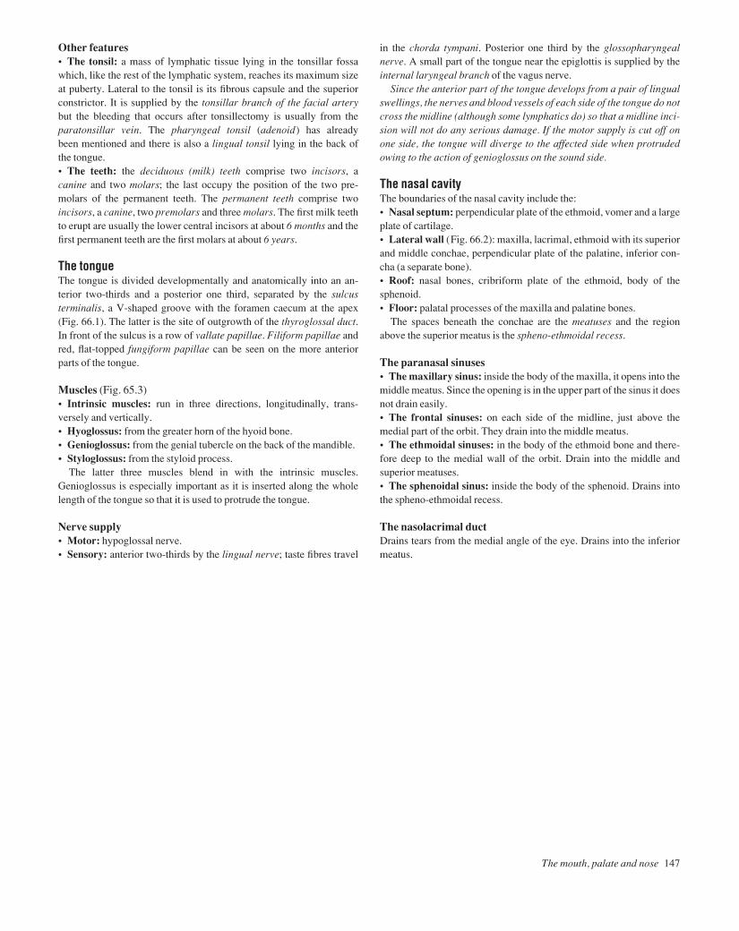
The mouth, palate and nose 147
Other features• The tonsil: a mass of lymphatic tissue lying in the tonsillar fossawhich, like the rest of the lymphatic system, reaches its maximum sizeat puberty. Lateral to the tonsil is its fibrous capsule and the superiorconstrictor. It is supplied by the tonsillar branch of the facial arterybut the bleeding that occurs after tonsillectomy is usually from theparatonsillar vein. The pharyngeal tonsil (adenoid) has already been mentioned and there is also a lingual tonsil lying in the back of the tongue.• The teeth: the deciduous (milk) teeth comprise two incisors, acanine and two molars; the last occupy the position of the two pre-molars of the permanent teeth. The permanent teeth comprise twoincisors, a canine, two premolars and three molars. The first milk teethto erupt are usually the lower central incisors at about 6 months and thefirst permanent teeth are the first molars at about 6 years.
The tongueThe tongue is divided developmentally and anatomically into an an-terior two-thirds and a posterior one third, separated by the sulcus terminalis, a V-shaped groove with the foramen caecum at the apex(Fig. 66.1). The latter is the site of outgrowth of the thyroglossal duct.In front of the sulcus is a row of vallate papillae. Filiform papillae andred, flat-topped fungiform papillae can be seen on the more anteriorparts of the tongue.
Muscles (Fig. 65.3)• Intrinsic muscles: run in three directions, longitudinally, trans-versely and vertically.• Hyoglossus: from the greater horn of the hyoid bone.• Genioglossus: from the genial tubercle on the back of the mandible.• Styloglossus: from the styloid process.
The latter three muscles blend in with the intrinsic muscles.Genioglossus is especially important as it is inserted along the wholelength of the tongue so that it is used to protrude the tongue.
Nerve supply• Motor: hypoglossal nerve.• Sensory: anterior two-thirds by the lingual nerve; taste fibres travel
in the chorda tympani. Posterior one third by the glossopharyngealnerve. A small part of the tongue near the epiglottis is supplied by theinternal laryngeal branch of the vagus nerve.
Since the anterior part of the tongue develops from a pair of lingualswellings, the nerves and blood vessels of each side of the tongue do notcross the midline (although some lymphatics do) so that a midline inci-sion will not do any serious damage. If the motor supply is cut off onone side, the tongue will diverge to the affected side when protrudedowing to the action of genioglossus on the sound side.
The nasal cavityThe boundaries of the nasal cavity include the:• Nasal septum: perpendicular plate of the ethmoid, vomer and a largeplate of cartilage.• Lateral wall (Fig. 66.2): maxilla, lacrimal, ethmoid with its superiorand middle conchae, perpendicular plate of the palatine, inferior con-cha (a separate bone).• Roof: nasal bones, cribriform plate of the ethmoid, body of the sphenoid.• Floor: palatal processes of the maxilla and palatine bones.
The spaces beneath the conchae are the meatuses and the regionabove the superior meatus is the spheno-ethmoidal recess.
The paranasal sinuses• The maxillary sinus: inside the body of the maxilla, it opens into themiddle meatus. Since the opening is in the upper part of the sinus it doesnot drain easily.• The frontal sinuses: on each side of the midline, just above themedial part of the orbit. They drain into the middle meatus.• The ethmoidal sinuses: in the body of the ethmoid bone and there-fore deep to the medial wall of the orbit. Drain into the middle andsuperior meatuses.• The sphenoidal sinus: inside the body of the sphenoid. Drains intothe spheno-ethmoidal recess.
The nasolacrimal ductDrains tears from the medial angle of the eye. Drains into the inferiormeatus.
AAAC66 21/5/05 11:01 AM Page 147
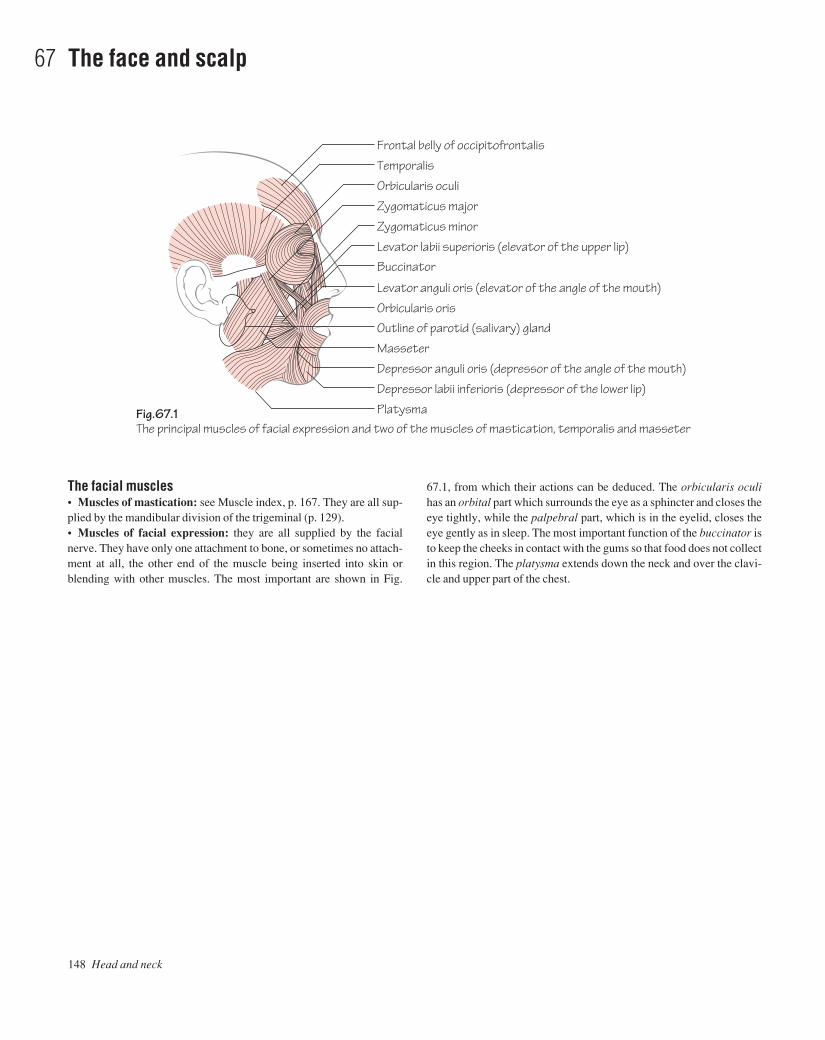
148 Head and neck
67 The face and scalp
Fig.67.1The principal muscles of facial expression and two of the muscles of mastication, temporalis and masseter
Frontal belly of occipitofrontalis
Temporalis
Orbicularis oculi
Zygomaticus major
Zygomaticus minor
Levator labii superioris (elevator of the upper lip)
Levator anguli oris (elevator of the angle of the mouth)
Orbicularis orisOutline of parotid (salivary) gland
Masseter
Depressor anguli oris (depressor of the angle of the mouth)
Depressor labii inferioris (depressor of the lower lip)
Platysma
Buccinator
The facial muscles• Muscles of mastication: see Muscle index, p. 167. They are all sup-plied by the mandibular division of the trigeminal (p. 129).• Muscles of facial expression: they are all supplied by the facialnerve. They have only one attachment to bone, or sometimes no attach-ment at all, the other end of the muscle being inserted into skin orblending with other muscles. The most important are shown in Fig.
67.1, from which their actions can be deduced. The orbicularis oculihas an orbital part which surrounds the eye as a sphincter and closes theeye tightly, while the palpebral part, which is in the eyelid, closes theeye gently as in sleep. The most important function of the buccinator isto keep the cheeks in contact with the gums so that food does not collectin this region. The platysma extends down the neck and over the clavi-cle and upper part of the chest.
AAAC67 21/5/05 11:01 AM Page 148
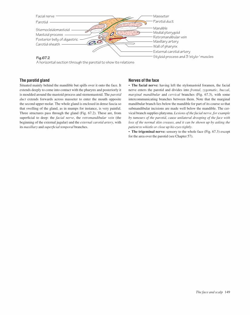
The face and scalp 149
Fig.67.2A horizontal section through the parotid to show its relations
Facial nerveParotid
SternocleidomastoidMastoid processPosterior belly of digastricCarotid sheath
Parotid ductMasseter
MandibleMedial pterygoidRetromandibular veinMaxillary arteryWall of pharynxExternal carotid arteryStyloid process and 3 'stylo-' muscles
The parotid glandSituated mainly behind the mandible but spills over it onto the face. Itextends deeply to come into contact with the pharynx and posteriorly itis moulded around the mastoid process and sternomastoid. The parotidduct extends forwards across masseter to enter the mouth opposite the second upper molar. The whole gland is enclosed in dense fascia sothat swelling of the gland, as in mumps for instance, is very painful.Three structures pass through the gland (Fig. 67.2). These are, fromsuperficial to deep: the facial nerve, the retromandibular vein (thebeginning of the external jugular) and the external carotid artery, withits maxillary and superficial temporal branches.
Nerves of the face• The facial nerve: having left the stylomastoid foramen, the facialnerve enters the parotid and divides into frontal, zygomatic, buccal,marginal mandibular and cervical branches (Fig. 67.3), with someintercommunicating branches between them. Note that the marginalmandibular branch lies below the mandible for part of its course so thatsubmandibular incisions are made well below the mandible. The cer-vical branch supplies platysma. Lesions of the facial nerve, for exampleby tumours of the parotid, cause unilateral drooping of the face withloss of the normal skin creases, and it can be shown up by asking thepatient to whistle or close up his eyes tightly.• The trigeminal nerve: sensory to the whole face (Fig. 67.3) exceptfor the area over the parotid (see Chapter 57).
AAAC67 21/5/05 11:01 AM Page 149
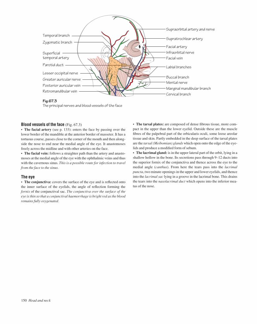
150 Head and neck
Zygomatic branch
Fig.67.3The principal nerves and blood vessels of the face
Temporal branch
Superficial temporal artery
Lesser occipital nerve
Parotid duct
Greater auricular nervePosterior auricular veinRetromandibular vein
Supraorbital artery and nerve
Supratrochlear artery
Facial vein
Facial arteryInfraorbital nerve
Labial branches
Buccal branch
Marginal mandibular branchMental nerve
Cervical branch
Blood vessels of the face (Fig. 67.3)• The facial artery (see p. 133): enters the face by passing over thelower border of the mandible at the anterior border of masseter. It has atortuous course, passes close to the corner of the mouth and then along-side the nose to end near the medial angle of the eye. It anastomosesfreely across the midline and with other arteries on the face.• The facial vein: follows a straighter path than the artery and anasto-moses at the medial angle of the eye with the ophthalmic veins and thuswith the cavernous sinus. This is a possible route for infection to travelfrom the face to the sinus.
The eye• The conjunctiva: covers the surface of the eye and is reflected ontothe inner surface of the eyelids, the angle of reflection forming thefornix of the conjunctival sac. The conjunctiva over the surface of theeye is thin so that a conjunctival haemorrhage is bright red as the bloodremains fully oxygenated.
• The tarsal plates: are composed of dense fibrous tissue, more com-pact in the upper than the lower eyelid. Outside these are the musclefibres of the palpebral part of the orbicularis oculi, some loose areolartissue and skin. Partly embedded in the deep surface of the tarsal platesare the tarsal (Meibomian) glands which open onto the edge of the eye-lids and produce a modified form of sebum.• The lacrimal gland: is in the upper lateral part of the orbit, lying in ashallow hollow in the bone. Its secretions pass through 9–12 ducts intothe superior fornix of the conjunctiva and thence across the eye to themedial angle (canthus). From here the tears pass into the lacrimalpuncta, two minute openings in the upper and lower eyelids, and thenceinto the lacrimal sac lying in a groove in the lacrimal bone. This drainsthe tears into the nasolacrimal duct which opens into the inferior mea-tus of the nose.
AAAC67 21/5/05 11:01 AM Page 150

The face and scalp 151
Fig.67.4The nerves and blood vessels of the scalp.The dotted line shows a temporal 'flap'
Greater occipital
Lesser occipital
Auriculotemporal
SupraorbitalSupratrochear
Nerves Arteries
Supraorbital Supratrochear
Superficial temporal
Posterior auricular
Occipital
The scalpThe scalp is made up of five layers which form a useful mnemonic:• Skin.• Cutaneous fat and connective tissue.• Aponeurosis (epicranial): this is a tough sheet of dense connectivetissue into which are inserted the occipital and frontal bellies of theoccipitofrontalis muscle.• Loose areolar tissue: this forms a plane of cleavage in head injuriesso that, in some cases, ‘scalping’ can occur. It also forms a plane inwhich blood can spread for a long distance.• Pericranium: the periosteum of the outside of the skull.
Blood vessels and nervesAll enter from the periphery and are shown in Fig. 67.4. The vesselsanastomose freely. Incisions can be centred on one artery which cankeep a large flap of skin viable.
Emissary veinsSmall veins that pass through the skull and unite the veins of the scalpwith the intracranial veins. They form a possible route for infection toreach the cranial cavity.
AAAC67 21/5/05 11:01 AM Page 151

The meningesThe meninges comprise the dura, pia and arachnoid mater. The cere-brospinal fluid is produced in the choroid plexuses of the lateral, 3rdand 4th ventricles of the brain and leaves through the three foramina inthe roof of the 3rd ventricle. It passes into the subarachnoid spacebetween the arachnoid and pia and serves to protect the brain and spinalcord.• The dura mater (Fig. 68.1): the fibrous layer is closely adherent tothe bone but the serous layer separates from the fibrous layer in places
to form the dural venous sinuses which are lined by endothelium. Italso forms two large sheetsathe falx cerebri and the tentorium cere-belli (see below).• The arachnoid mater: deep to the dura. Bridges over the sulci andfissures of the brain. The subarachnoid space contains the cerebrospinalfluid.• The pia mater: follows the contours of the brain, dipping into thesulci.
152 Head and neck
68 The cranial cavity
Cerebral veins
Fig.68.1The principal folds of serous dura
Fig.68.3A cross-section through the cavernous sinus
Fig.68.2A cross-section through the superior sagittal sinus to show the arachnoid villiprojecting into the sinus
Falx cerebri
Emissary vein
Fibrous dura
Inferiorsagittal sinus
Arachnoid villus
Cerebral vein draining into lacuna
PiaArachnoid
Brain covered by pia
Tentorium cerebelli
Diaphragma sellae
Oculomotor nerveTrochlear nerveOphthalmic branchMaxillary branch
Mandibular branch
Pituitary
Endothelium of superiorsagittal sinus
Diaphragma sellaeInternal carotid artery
Abducent nerve
Serous dura
Sphenoidal sinusEndothelium of cavernous sinus
Fibrous dura
Emissary vein
AAAC68 21/5/05 11:01 AM Page 152
The cranial cavity 153
The falx cerebriThe falx cerebri projects down in the sagittal plane to separate partiallythe two cerebral hemispheres. It tapers to a point anteriorly but pos-teriorly it is attached to the tentorium. The superior sagittal sinus(Fig. 68.2) is in its attached border and the inferior sagittal sinus is in itsfree border. Veins from the cerebral hemispheres drain into the superiorsagittal sinus or into diverticula from it, the lacunae laterales. In placesthe underlying arachnoid sends small outgrowths through the serousdura to project into the sinus. These are the arachnoid villi and they arethe site of absorption of cerebrospinal fluid into the bloodstream. Inlater life they clump together to form arachnoid villi which makeindentations in the skull.
The tentorium cerebelliThe tentorium cerebelli forms a roof over the posterior cranial fossaand the cerebellum. Its free border is attached to the posterior clinoidprocesses and through the opening passes the brainstem. The trans-verse sinuses are in the attached border of the tentorium.
There are two smaller projections of serous duraathe falx cerebellibetween the two cerebellar hemispheres and the diaphragma sellaewhich forms a roof over the pituitary fossa and the pituitary gland.
The cavernous sinus (Fig. 68.3)The cavernous sinus lies on either side of the pituitary fossa and thebody of the sphenoid. Like the other venous sinuses, it is formed by alayer of serous dura lined by endothelium. In addition, a layer of serousdura from the posterior cranial fossa projects forwards into the side ofthe cavernous sinus to form the cavum trigeminale.
Contents• Internal carotid artery: see p. 133.• Oculomotor nerve: see p. 127.• Trochlear nerve: see p. 127.• Abducent nerve: see p. 131.• Three divisions of the trigeminal nerve: see p. 129.
AAAC68 21/5/05 11:01 AM Page 153
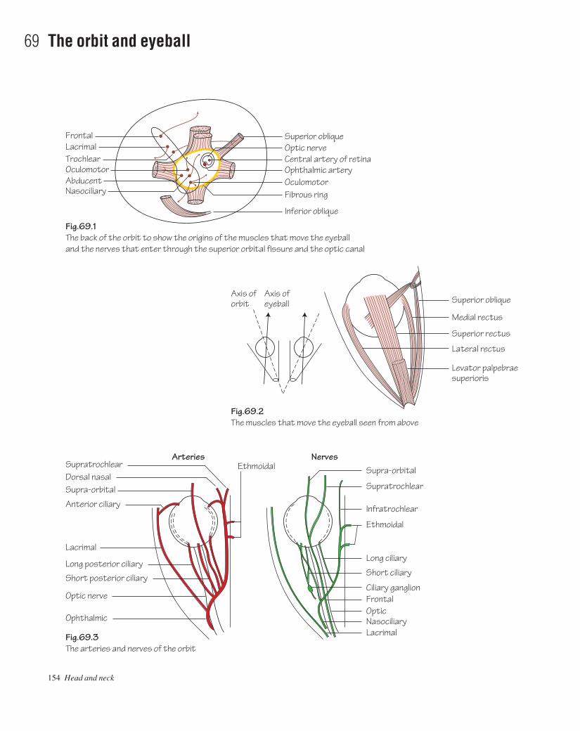
154 Head and neck
69 The orbit and eyeball
Fig.69.1The back of the orbit to show the origins of the muscles that move the eyeballand the nerves that enter through the superior orbital fissure and the optic canal
Fig.69.2The muscles that move the eyeball seen from above
Fig.69.3The arteries and nerves of the orbit
Superior obliqueFrontalLacrimalTrochlear
AbducentNasociliary
Optic nerve
Ophthalmic arteryCentral artery of retina
OculomotorFibrous ring
Inferior oblique
Superior oblique
Supra-orbital
Supratrochlear
Infratrochlear
Ethmoidal
Ethmoidal
Long ciliary
Short ciliary
Ciliary ganglionFrontalOpticNasociliaryLacrimal
Supra-orbitalDorsal nasalSupratrochlear
Anterior ciliary
Lacrimal
Long posterior ciliary
Short posterior ciliary
Optic nerve
Ophthalmic
Medial rectus
Superior rectus
Lateral rectus
Levator palpebraesuperioris
Axis oforbit
Axis ofeyeball
Oculomotor
NervesArteries
AAAC69 21/5/05 11:00 AM Page 154
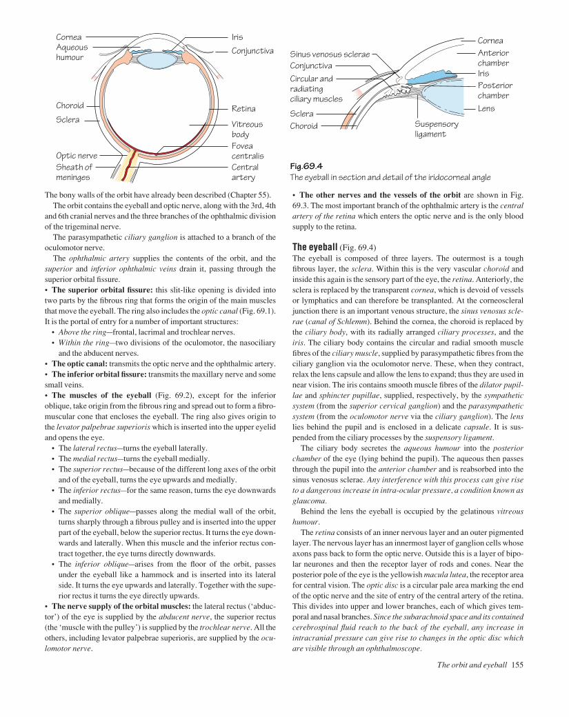
The bony walls of the orbit have already been described (Chapter 55).The orbit contains the eyeball and optic nerve, along with the 3rd, 4th
and 6th cranial nerves and the three branches of the ophthalmic divisionof the trigeminal nerve.
The parasympathetic ciliary ganglion is attached to a branch of theoculomotor nerve.
The ophthalmic artery supplies the contents of the orbit, and thesuperior and inferior ophthalmic veins drain it, passing through thesuperior orbital fissure.• The superior orbital fissure: this slit-like opening is divided intotwo parts by the fibrous ring that forms the origin of the main musclesthat move the eyeball. The ring also includes the optic canal (Fig. 69.1).It is the portal of entry for a number of important structures:
• Above the ringafrontal, lacrimal and trochlear nerves.• Within the ringatwo divisions of the oculomotor, the nasociliary
and the abducent nerves.• The optic canal: transmits the optic nerve and the ophthalmic artery.• The inferior orbital fissure: transmits the maxillary nerve and somesmall veins.• The muscles of the eyeball (Fig. 69.2), except for the inferioroblique, take origin from the fibrous ring and spread out to form a fibro-muscular cone that encloses the eyeball. The ring also gives origin tothe levator palpebrae superioris which is inserted into the upper eyelidand opens the eye.
• The lateral rectusaturns the eyeball laterally.• The medial rectusaturns the eyeball medially.• The superior rectusabecause of the different long axes of the orbit
and of the eyeball, turns the eye upwards and medially.• The inferior rectusafor the same reason, turns the eye downwards
and medially.• The superior obliqueapasses along the medial wall of the orbit,
turns sharply through a fibrous pulley and is inserted into the upperpart of the eyeball, below the superior rectus. It turns the eye down-wards and laterally. When this muscle and the inferior rectus con-tract together, the eye turns directly downwards.
• The inferior obliqueaarises from the floor of the orbit, passesunder the eyeball like a hammock and is inserted into its lateralside. It turns the eye upwards and laterally. Together with the supe-rior rectus it turns the eye directly upwards.
• The nerve supply of the orbital muscles: the lateral rectus (‘abduc-tor’) of the eye is supplied by the abducent nerve, the superior rectus(the ‘muscle with the pulley’) is supplied by the trochlear nerve. All theothers, including levator palpebrae superioris, are supplied by the ocu-lomotor nerve.
• The other nerves and the vessels of the orbit are shown in Fig.69.3. The most important branch of the ophthalmic artery is the centralartery of the retina which enters the optic nerve and is the only bloodsupply to the retina.
The eyeball (Fig. 69.4)The eyeball is composed of three layers. The outermost is a toughfibrous layer, the sclera. Within this is the very vascular choroid andinside this again is the sensory part of the eye, the retina. Anteriorly, thesclera is replaced by the transparent cornea, which is devoid of vesselsor lymphatics and can therefore be transplanted. At the corneoscleraljunction there is an important venous structure, the sinus venosus scle-rae (canal of Schlemm). Behind the cornea, the choroid is replaced bythe ciliary body, with its radially arranged ciliary processes, and theiris. The ciliary body contains the circular and radial smooth musclefibres of the ciliary muscle, supplied by parasympathetic fibres from theciliary ganglion via the oculomotor nerve. These, when they contract,relax the lens capsule and allow the lens to expand; thus they are used innear vision. The iris contains smooth muscle fibres of the dilator pupil-lae and sphincter pupillae, supplied, respectively, by the sympatheticsystem (from the superior cervical ganglion) and the parasympatheticsystem (from the oculomotor nerve via the ciliary ganglion). The lenslies behind the pupil and is enclosed in a delicate capsule. It is sus-pended from the ciliary processes by the suspensory ligament.
The ciliary body secretes the aqueous humour into the posteriorchamber of the eye (lying behind the pupil). The aqueous then passesthrough the pupil into the anterior chamber and is reabsorbed into thesinus venosus sclerae. Any interference with this process can give riseto a dangerous increase in intra-ocular pressure, a condition known asglaucoma.
Behind the lens the eyeball is occupied by the gelatinous vitreoushumour.
The retina consists of an inner nervous layer and an outer pigmentedlayer. The nervous layer has an innermost layer of ganglion cells whoseaxons pass back to form the optic nerve. Outside this is a layer of bipo-lar neurones and then the receptor layer of rods and cones. Near theposterior pole of the eye is the yellowish macula lutea, the receptor areafor central vision. The optic disc is a circular pale area marking the endof the optic nerve and the site of entry of the central artery of the retina.This divides into upper and lower branches, each of which gives tem-poral and nasal branches. Since the subarachnoid space and its containedcerebrospinal fluid reach to the back of the eyeball, any increase inintracranial pressure can give rise to changes in the optic disc whichare visible through an ophthalmoscope.
The orbit and eyeball 155
Fig.69.4The eyeball in section and detail of the iridocorneal angle
CorneaConjunctivaIris
Sclera
Choroid Retina
Vitreous body
Aqueous humour
Central artery
Sheath ofmeninges
Optic nerveFovea centralis
Cornea
Suspensoryligament
LensScleraChoroid
Circular and radiatingciliary muscles
Sinus venosus scleraeConjunctiva
Anterior chamber
Posterior chamber
Iris
AAAC69 21/5/05 11:00 AM Page 155

156 Head and neck
70 The ear, and lymphatics and surface anatomy of the head and neck
Pars flaccida
Fig.70.2The left tympanic membrane, as seen through an auriscope.The 'cone of light' is caused by the reflection of the light of the auriscope
Fig.70.3The principal groups of lymph nodes of the head and neck arranged as a triangle.The inset (right) shows the two major groups into which the others eventually drain
Fig.70.1A diagram representing the middle ear as an opened-out box
Cone of light
Incus
Ridge produced by lateral semicircular canal
Lateral processof malleus
Long process of incus
Anterior Posterior
Handle of malleus
Greater petrosal nerve
Lesser petrosal nerve
Auditory tubeTympanic plexusPromontory
Internal carotid artery
Tympanic branch
Pre-auricular
Tegmen tympaniGeniculate ganglion
Facial nerve
Stapes
Aditus
Malleus
Tympanicmembrane
Chordatympani
Glossopharyngeal nerveInternal jugular vein
Round window
Post-auricular
Buccal
SubmentalSubmandibularUpper deep cervical
Upper deep cervical
Sternocleidomastoid
Lower deep cervicalInfrahyoidParatracheal
Inferior deep cervical
Occipital
AAAC70 21/5/05 11:00 AM Page 156

The earThe ear is subdivided into the outer ear, the middle ear and the inner ear.
The outer earThe outer third of this is cartilaginous and the inner two-thirds is bony.Lined by skin with ceruminous (wax) glands.
The middle earThis has four walls, a roof and a floor. It can therefore be representeddiagrammatically by an opened-out box (Fig. 70.1).• The lateral wall:
• The tympanic membrane (Fig. 70.2)athe handle of the malleusembedded in its middle layer. This is crossed by the chorda tym-pani, above which is the pars flaccida. The ‘cone of light’ is thereflection of the light from the auriscope.
• The epitympanic recess (attic)athe part of the middle ear cavityabove the tympanic membrane.
• The ossicles: the malleus, incus and stapesathe stapes engageswith the oval window. The ossicles transmit vibrations of the mem-brane to the inner ear.
• The medial wall:• The promontoryaa bulge produced by the first turn of the cochlea.• The oval windowaleads into the inner ear.• The facial nervearuns backwards and then downwards in a bony
canal in the medial wall. It bears the geniculate ganglion and givesoff the chorda tympani.
• The anterior wall: the pharyngotympanic (Eustachian) tube opensonto the anterior wall and leads down to the nasopharynx. Its functionis to equalize the pressure between the middle ear and the pharynx.• The posterior wall: the aditus leads backwards into the mastoidantrum, a cavity in the mastoid bone which, in turn, leads into the mas-toid air cells.• The roof: the tegmen tympani, a thin plate of bone that separates themiddle ear from the middle cranial fossa.• The floor: separates the middle ear from the internal carotid arteryand the internal jugular vein.
The inner earThe inner ear is involved in both hearing and balance. It consists of twocomponents:• The osseous labyrinth: comprises the vestibule, the semicircularcanals and the cochlea. The labyrinth itself consists of spaces in thepetrous temporal bone and it contains the membranous labyrinth.• The membranous labyrinth: comprises the utricle and saccule (inthe vestibule), the semicircular ducts (in the semicircular canals) andthe duct of the cochlea (in the cochlea). The utricle and saccule are con-cerned with the sense of position and the semicircular ducts are con-cerned with the sensation of motion. The cochlear duct is the organ ofhearing. All are supplied by the vestibulocochlear (auditory) nerve.
The lymphatics of the head and neck (Fig. 70.3)• Upper deep cervical nodes (jugulodigastric nodes): these are situ-ated between the upper end of sternomastoid and the angle of the
mandible and also deep to sternomastoid. They drain the head and theupper part of the neck, directly or indirectly (but there are no lym-phatics in the cranial cavity).• The lower deep cervical nodes ( jugulo-omohyoid nodes): in theposterior triangle between the lower end of sternomastoid and the clav-icle. They drain the lower part of the neck and also receive lymph fromthe upper deep cervical nodes, from the breast and some of the lymphfrom the thorax and abdomen. The efferents from this group drain intothe thoracic or left lymph duct via the jugular trunk.• Smaller groups of nodes are shown in Fig. 70.3.• The lymph drainage of the tongue: the tip of the tongue drains intothe submental nodes. The rest of the anterior two-thirds drains into thesubmandibular nodes, some crossing the midline and some passingdirectly to the upper deep cervical nodes. The posterior two-thirdsdrains directly into the upper deep cervical nodes.• The lymph drainage of the larynx: above the vocal cords the larynxdrains into the infrahyoid nodes and thence to the upper deep cervicalgroup. Below the cords, drainage is to the paratracheal and inferiordeep cervical nodes.
Surface anatomy of the head and neck• The middle meningeal artery: the anterior branch may be exposedat a point 4 cm above the midpoint of the zygomatic arch. The posteriorbranch may be represented on the surface by a pencil placed behind theear.
The face• The supraorbital, infraorbital and mental nerves: all lie on a ver-tical line passing between the two premolar teeth.• The facial artery can be felt on the mandible at the anterior border ofthe masseter.• The superficial temporal artery is just in front of the tragus of theear.• The parotid duct follows the middle part of a line from the tragus ofthe ear to the middle of the upper lip. It hooks over the anterior borderof the masseter where it can be easily felt.
The neck• The sternomastoid muscle (with the external jugular vein on its sur-face) may be made to contract by asking the patient to turn his head tothe opposite side against resistance.• The trunks of the brachial plexus can be palpated in the anglebetween the sternomastoid and the clavicle.• The subclavian artery is palpable by deep pressure behind the middle of the clavicle. A cervical rib may also be palpable.• The hyoid bone, and the thyroid and cricoid cartilages are easilyfelt. The larynx, and any swellings associated with it, move upwards on swallowing, as does the thyroid gland. Thyroglossal cysts moveupwards when the tongue is protruded.• The trachea is palpable in the suprasternal notch.• The common carotid artery can be felt in front of sternomastoidand can be compressed against the transverse process of the 6th cer-vical vertebra (carotid tubercle).
The ear, and lymphatics and surface anatomy of the head and neck 157
AAAC70 21/5/05 11:00 AM Page 157
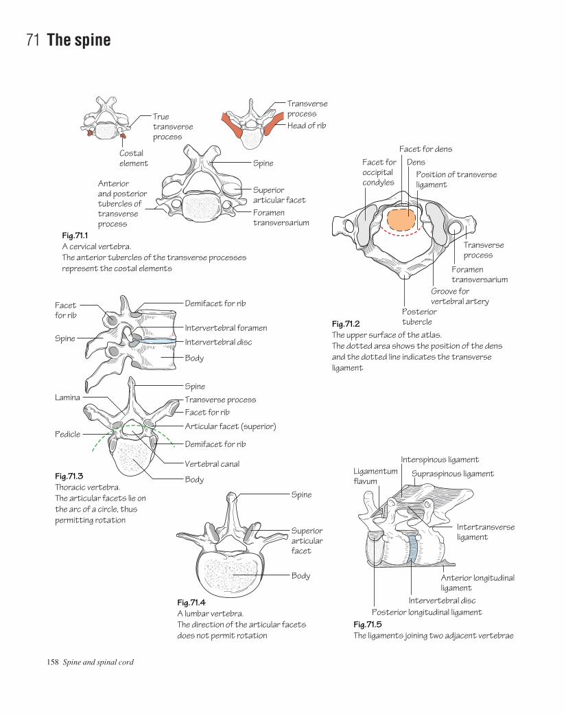
158 Spine and spinal cord
71 The spine
Fig.71.2The upper surface of the atlas.The dotted area shows the position of the densand the dotted line indicates the transverse ligament
Fig.71.5The ligaments joining two adjacent vertebrae
Fig.71.1A cervical vertebra.The anterior tubercles of the transverse processesrepresent the costal elements
Fig.71.3Thoracic vertebra.The articular facets lie onthe arc of a circle, thuspermitting rotation
Anterior and posteriortubercles of transverseprocess
Foramentransversarium
Superiorarticular facet
Spine Facet foroccipitalcondyles
Costalelement
Facet for densDens
Position of transverse ligament
Foramentransversarium
Transverseprocess
Posterior tubercle
Posterior longitudinal ligamentIntervertebral disc
Interspinous ligament
Supraspinous ligamentLigamentum flavum
Anterior longitudinalligament
Intertransverse ligament
Groove forvertebral arteryDemifacet for rib
Transverse processTrue
transverse process
Head of rib
Intervertebral foramen
Intervertebral disc
Body
Fig.71.4A lumbar vertebra.The direction of the articular facetsdoes not permit rotation
Superiorarticular facet
Spine
Body
Spine
Facet for rib
Articular facet (superior)
Transverse process
Demifacet for ribPedicle
Spine
Lamina
Facetfor rib
Vertebral canal
Body
AAAC71 21/5/05 11:00 AM Page 158
There are 12 thoracic, five lumbar, five sacral and 3–5 coccygeal ver-tebrae. They are held together by ligaments, intervertebral discs andsynovial joints between the articular processes. The weight-bearingportion of the vertebra is the body so that the size of the vertebral bodiesincreases from above downwards and when one of the bodies is dis-eased it will eventually collapse. The bodies contain red bone marrowso that the veins that drain them (basivertebral veins) produce largeforamina on the backs of the bodies.• Cervical vertebrae (Fig. 71.1): small bodies, bifid spines, transverseprocesses with anterior and posterior tubercles and a foramentransversarium for the passage of the vertebral artery. The body of thefirst vertebra (atlas) fuses with that of the second (axis) during develop-ment to produce its dens (odontoid process) which is held in place by atransverse ligament (Fig. 71.2).• Thoracic vertebrae (Fig. 71.3): heart-shaped bodies, upper andlower demifacets for the heads of the ribs, long downturned spine, longtransverse processes with a facet for the tubercles of the rib.• Lumbar vertebrae (Fig. 71.4): a massive body, large transverseprocesses, a triangular vertebral canal and large, backwardly project-ing spines so that a needle may be inserted between them in the opera-tion of lumbar puncture. The articular facets face mediolaterally sothat they prevent rotation.• Sacral vertebrae (Fig. 23.3): fused together to form the sacrum.
The intervertebral jointsThe upper and lower surfaces of the bodies are covered with hyalinecartilage and are separated by the fibrocartilaginous intervertebraldiscs. Each disc has a peripheral fibrous ring (annulus fibrosus) and acentral more spongy nucleus pulposus which lies nearer to the backthan to the front of the disc. The nucleus pulposus is rich in gly-cosaminoglycans so that it has a high water content, but this diminisheswith increasing age. The nucleus may then herniate through the annulusfibrosus, passing backwards (compressing the spinal cord), posterolat-erally (compressing a spinal nerve) or upwards (into a vertebralbodyaa Schmorl’s node). The discs are thickest in the lumbar and cer-vical regions, so that these are the regions of most movement.
The vertebrae are also held together by ligaments that join each ofthe components of the vertebrae (Fig. 71.5) except for the pedicles (thespinal nerves have to pass between these in the intervertebral foram-ina). Thus there are:
• Anterior and posterior longitudinal ligaments joining the frontand back of the bodies (the posterior ligaments thus lie within the ver-tebral canal).• Supraspinous and interspinous ligaments.• Intertransverse ligaments.• Ligamenta flava (which contain much elastic tissue and join thelaminae).• Capsular ligaments of the synovial joints between the articular processes.
Curves of the spineIn the fetus, the whole developing spine is curved so that it is concaveforwards (primary curvature). A few months after birth the baby beginsto hold its head up and the cervical spine develops a forward convexity(secondary curvature). Later in the first year the baby begins to standup and another forward convexity develops in the lumbar region (sec-ondary curvature). The primary curves are mainly due to the shape ofthe vertebral bodies but the secondary curves are due to the shape of theintervertebral discs.
Movements of the spine• Cervical spine: movement is free in the cervical region of the spine:flexion and extension, side flexion (associated with rotation to theopposite side) and rotation. In rotation of the head the atlas rotatesaround the dens of the axis, and in flexion of the head the occipitalcondyles move on the articular facets of the atlas.• Thoracic spine: movement is somewhat restricted by the thinnerintervertebral discs and the presence of the ribs. Rotation, however, isfree.• Lumbar spine: flexion and extension are free but rotation is almostabsent because of the direction of the articular facets. Side flexion alsooccurs.
Spina bifidaEach vertebra develops in three partsbthe body and the two halves ofthe neural arch. These ossify in cartilage and, at birth, the three partsare still separate but they soon fuse. Failure of such fusion gives rise tothe condition of spina bifida. This may be symptomless (spina bifidaocculta) although the site of the lesion may be marked by a tuft of hair.If the defect is large, the meninges or even the spinal cord may herniateonto the surface (meningocoele) and this may produce neurologicalsymptoms.
The spine 159
AAAC71 21/5/05 11:00 AM Page 159
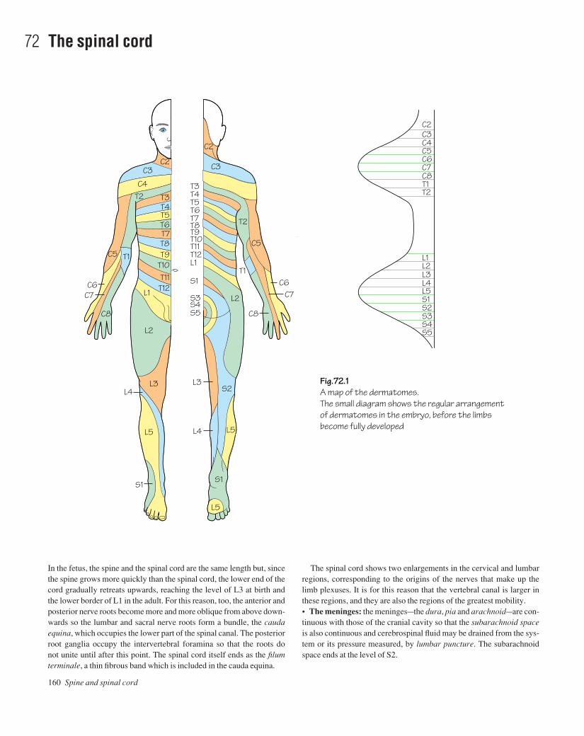
In the fetus, the spine and the spinal cord are the same length but, sincethe spine grows more quickly than the spinal cord, the lower end of thecord gradually retreats upwards, reaching the level of L3 at birth andthe lower border of L1 in the adult. For this reason, too, the anterior andposterior nerve roots become more and more oblique from above down-wards so the lumbar and sacral nerve roots form a bundle, the caudaequina, which occupies the lower part of the spinal canal. The posteriorroot ganglia occupy the intervertebral foramina so that the roots do not unite until after this point. The spinal cord itself ends as the filumterminale, a thin fibrous band which is included in the cauda equina.
The spinal cord shows two enlargements in the cervical and lumbarregions, corresponding to the origins of the nerves that make up thelimb plexuses. It is for this reason that the vertebral canal is larger inthese regions, and they are also the regions of the greatest mobility.• The meninges: the meningesathe dura, pia and arachnoidaare con-tinuous with those of the cranial cavity so that the subarachnoid spaceis also continuous and cerebrospinal fluid may be drained from the sys-tem or its pressure measured, by lumbar puncture. The subarachnoidspace ends at the level of S2.
160 Spine and spinal cord
72 The spinal cord
C3 C3C2
C4 C4
T2 T3T4T5
T7T8T9
T10T11
T12L1
L2
L3
L5
L4
S1
L5
S1
L4 L5
S2L3
S5
S3S4
S1
L1
L2
T12T11T10T9T8T7T6T5T4T3
T2
T1
C2C3C4C5C6C7C8T1T2
L2L1
L3L4L5S1S2S3S4S5
C5
C6C7
C8
C5
C6C7
T1
C8
T6
Fig.72.1A map of the dermatomes.The small diagram shows the regular arrangementof dermatomes in the embryo, before the limbsbecome fully developed
C2
AAAC72 21/5/05 10:59 AM Page 160

The spinal cord 161
• The blood supply of the spinal cord: the spinal cord is suppliedwith blood by spinal arteries that are derived from the vertebral, inter-costal, lumbar and lateral sacral arteries. Each artery divides into dor-sal and ventral branches that follow the corresponding nerve roots tothe spinal cord where they form longitudinally arranged anterior andpaired posterior spinal arteries. These longitudinal anastomoses, how-ever, are incomplete and the spinal arteries themselves vary in size. Thelargest are in the lower thoracic and upper lumbar regions, and theblood supply of the cord may be jeopardized if some of these largerspinal arteries are damaged, for example in resection of the thoracicaorta.
The spinal nervesAs has been described in Chapter 2, the spinal nerves from T2 to L1supply segmental areas of skin and muscles but elsewhere they formplexuses so that the areas supplied by each nerve become more complic-ated. Each named peripheral nerve may contain components of morethan one spinal nerve and each spinal nerve may contribute fibres to
more than one peripheral nerve. However, the area of skin supplied byany one spinal nerve (a dermatome) can be mapped out and these areshown in Fig. 72.1. Knowledge of the dermatome map is of great valuein diagnosing lesions of the spinal nerves but it must be rememberedthat these areas are subject to some variation from one person toanother and there is also a good deal of overlap. Thus a lesion of, say,T4 may cause little or no loss of sensation because of overlap with T3and T5.
Similarly, certain groups of muscles (myotomes) can be recognizedas being supplied by particular spinal nerves. For example, C5 suppliesthe abductors of the shoulder, the flexors of the elbow and the supin-ators so that a lesion of this nerve or of the upper trunk of the brachialplexus will cause the arm to be held in a position of adduction of theshoulder, extension of the elbow and pronation (Erb–Duchenne para-lysis). Similarly, T1 supplies the small muscles of the hand so that alesion of this nerve, due perhaps to the presence of a cervical rib, willcause global wasting of the hand muscles.
AAAC72 21/5/05 10:59 AM Page 161

162 Muscle index
The abdomenAll the muscles of the anterior abdominal wall serve to protect the vis-cera by their contraction, to produce movement and to increase theintra-abdominal pressure, as in defecation, coughing, parturition, etc.All these muscles are supplied by the lower six thoracic and the firstlumbar nerves.• External obliqueOrigin: From the outer surfaces of the lower eight ribs to the iliac crestas far forward as the anterior superior spine.Insertion: The free lower border forms the inguinal ligament betweenthe anterior superior iliac spine and the pubic tubercle. The musclebecomes aponeurotic and reaches the midline where it interdigitateswith the opposite side to form the linea alba. The superficial inguinalring is a gap in the aponeurosis above and medial to the pubic tubercle.The aponeurosis contributes to the anterior rectus sheath.Actions: Flexion of the spine, side flexion and rotation of the trunkatheright external oblique produces rotation to the left.• Internal obliqueOrigin: From the thoracolumbar fascia, the iliac crest and the lateralhalf of the inguinal ligament.Insertion: Into an aponeurosis which is attached to the costal marginand to the linea aspera, after splitting to enclose the rectus abdominis,thus contributing to the rectus sheath. The lower fibres from theinguinal ligament contribute to the conjoint tendon which is attached tothe pubic crest and the pectineal line.Actions: Flexion of the spine, side flexion and rotation of the trunkatheright internal oblique produces rotation to the right.• Transversus abdominisOrigin: From the thoracolumbar fascia, the iliac crest and the lateralone third of the inguinal ligament. Also from the inner surfaces of thelower six ribs, interdigitating with the diaphragm.Insertion: The aponeurosis passes to the linea aspera, contributing tothe rectus sheath. The lower fibres help to form the conjoint tendon.Actions: Most of the fibres are transverse and thus pull in and flatten theabdominal wall.• Rectus abdominisOrigin: From the anterior surfaces of the 5th, 6th and 7th costal cartilages.Insertion: The pubic crest and tubercle and the front of the symphysis.There are three tendinous intersections in the upper part of the musclewhich are adherent to the anterior rectus sheath.Actions: The muscle is enclosed in the rectus sheath. A strong flexor ofthe trunk, it can also tilt the pelvis backwards. In a person lying prone,rectus contracts when the head is lifted from the pillow or when the legis raised from the bed.
For further important details of the muscles of the anterior abdom-inal wall, see the inguinal canal and the rectus sheath (p. 30).• The diaphragmOrigin: From the inner surfaces of the lower six ribs, from the back ofthe xiphisternum, from the right and left crura which are attached,respectively, to the upper three and the upper two lumbar vertebrae, andfrom the medial and lateral arcuate ligaments which bridge over thepsoas major and quadratus lumborum.
Insertion: The fibres (striated muscle) are inserted into the central tendon.Actions: The diaphragm is involved in respiration. When the musclefibres contract, the diaphragm is lowered, thus increasing the verticaldimension of the thorax. In the later stages of contraction, using theliver as a fulcrum it raises the lower ribs, thus increasing the width ofthe lower thorax. At the same time, it increases the intra-abdominalpressure and is thus used in expulsive effortsadefecation, micturition,parturition, etc.Nerve supply: Phrenic nerve (C3, 4 and 5).• Quadratus lumborumOrigin: From the posterior part of the iliac crest.Insertion: To the 12th rib.Action: Side flexion of the trunk.Nerve supply: Adjacent lumbar nerves.• Psoas major: see lower limb (p. 165).
The upper limb• Latissimus dorsiOrigin: From the spines of the lower six thoracic vertebrae, the lumbarspines via the thoracolumbar fascia and the medial part of the iliaccrest.Insertion: To the floor of the intertubercular sulcus, curving round teresmajor.Actions: Adduction and medial rotation of the arm. Can hold up thelower limb girdle as in crutch walking.Nerve supply: Thoracodorsal nerve.• Serratus anteriorOrigin: From the lateral surfaces of the upper eight ribs.Insertion: Into the medial border of the scapula.Actions: Protraction of the scapula and rotation so that the glenoidpoints upwards, thus helping in abduction of the upper limb. Helps tokeep the scapula in contact with the chest wall.Nerve supply: Long thoracic nerve.• Levator scapulaeOrigin: From the transverse processes of the upper cervical vertebrae.Insertion: To the medial border of the scapula above the spine.Action: Elevates the scapula.Nerve supply: C3 and 4.• The rhomboidsOrigin: From spines of thoracic vertebrae.Insertion: To the medial border of the scapula.Action: Bracing back the scapula.Nerve supply: Dorsal scapular nerve.• Trapezius: See p. 167.• Pectoralis majorOrigin: From the sternum and the upper six costal cartilages and fromthe medial half of the clavicle.Insertion: To the lateral lip of the intertubercular sulcus.Actions: Adduction, flexion and medial rotation of the arm.Nerve supply: Medial and lateral pectoral nerves.• Pectoralis minorOrigin: From the 3rd, 4th and 5th ribs.
Muscle index
AAAD01 21/5/05 10:59 AM Page 162

Insertion: To the coracoid process.Action: Depresses the tip of the shoulder.Nerve supply: Medial and lateral pectoral nerves.• DeltoidOrigin: From the lateral third of the clavicle, the acromion and thespine of the scapula.Insertion: To the deltoid tubercle on the lateral surface of the humerus.Actions: Abduction of the upper limb (assisted by supraspinatus andserratus anterior), flexion (anterior fibres) and extension (posteriorfibres) of the arm.Nerve supply: Axillary nerve.• Teres majorOrigin: From the lower angle of the scapula.Insertion: To the medial lip of the intertubercular sulcus.Actions: Adduction and medial rotation of the arm.Nerve supply: Lower subscapular nerve.• The rotator cuffConsists of subscapularis, supraspinatus, infraspinatus and teresminor. Acting together, these muscles maintain the stability of theshoulder joint as well as having their own individual actions, as follows:
• SubscapularisOrigin: From the subscapular fossa.Insertion: Passes in front of the shoulder joint to the lesser tuberos-ity of the humerus.Action: Medial rotation of the arm.Nerve supply: Subscapular nerves.
• SupraspinatusOrigin: From the supraspinous fossa.Insertion: To the top of the greater tuberosity of the humerus.Action: Initiates abduction of the arm.Nerve supply: Suprascapular nerve.
• InfraspinatusOrigin: From the infraspinous fossa.Insertion: To the back of the greater tuberosity.Action: Lateral rotation of the arm.Nerve supply: Suprascapular nerve.
• Teres minorOrigin: From the lateral border of the scapula.Insertion: To the humerus below infraspinatus.Action: Lateral rotation of the arm.Nerve supply: Axillary nerve.
• CoracobrachialisOrigin: From the coracoid process along with the short head of biceps.Insertion: To the medial side of the humerus.Action: Moves the arm upwards and medially.Nerve supply: Musculocutaneous nerve.• Biceps brachiiOrigin: Long head from the supraglenoid tubercle and short head fromthe coracoid process (with coracobrachialis).Insertion: To the radial tuberosity and, via the bicipital aponeurosis,into the deep fascia of the forearm.Actions: Flexion and supination of the forearm.Nerve supply: Musculocutaneous nerve.• BrachialisOrigin: From the front of the lower part of the humerus.Insertion: To the tubercle on the ulna just below the coronoid process.Action: Flexion of the elbow.Nerve supply: Musculocutaneous nerve and radial nerve.
• TricepsOrigin: Three heads: long from the infraglenoid tubercle, lateral fromthe humerus above the spiral line, and medial from the back of thelower part of the humerus.Insertion: Into the olecranon.Action: Extensor of the elbow.Nerve supply: Radial nerve.• Pronator teresOrigin: From the common flexor origin on the medial epicondyle of thehumerus.Insertion: To the lateral surface of the shaft of the radius.Action: Pronation of the forearm.Nerve supply: Median nerve.• Flexor carpi radialisOrigin: From the common flexor origin.Insertion: To the base of metacarpals 2 and 3.Actions: Flexion and abduction of wrist.Nerve supply: Median nerve.• Palmaris longusOrigin: From the common flexor origin.Insertion: To the flexor retinaculum and the palmar aponeurosis.Action: Flexor of the wrist. (Often absent.)Nerve supply: Median nerve.• Flexor carpi ulnarisOrigin: From the common flexor origin and the posterior border of theulna.Insertion: To the pisiform and thence by the pisometacarpal ligamentto the 5th metacarpal.Actions: Flexion and adduction of the wrist.Nerve supply: Ulnar nerve.• Flexor digitorum superficialisOrigin: From the common flexor origin and the shaft of the radius.Insertion: To the sides of the middle phalanges of the four fingers. Thetendons are perforated by the tendons of flexor digitorum profundus.Actions: Flexion of the proximal two phalanges and of the wrist.Nerve supply: Median nerve.• Flexor pollicis longusOrigin: From the front of the shaft of the radius.Insertion: To the distal phalanx of the thumb.Actions: Flexion of all the joints of the thumb.Nerve supply: Median nerve.• Flexor digitorum profundusOrigin: From the front of the shaft of the ulna and its posterior border.Insertion: To the distal phalanges of the four fingers, the tendons pass-ing through those of flexor digitorum superficialis.Actions: Flexion of the fingers and the wrist.Nerve supply: Half by the median nerve and half by the ulnar nerve.• Pronator quadratusOrigin: From the lower end of the front of the radius.Insertion: To the lower end of the ulna.Action: Pronator of the forearm.Nerve supply: Median nerve.• BrachioradialisOrigin: From the lateral supracondylar ridge of the humerus.Insertion: To the lower end of the radius.Action: Flexion of the elbow.Nerve supply: Radial nerve.• Extensor carpi radialis longus and brevisOrigin: From the lateral supracondylar ridge of the humerus.
Muscle index 163
AAAD01 21/5/05 10:59 AM Page 163

Insertion: To the bases of the 2nd and 3rd metacarpals.Actions: Extension and abduction of the wrist.Nerve supply: Radial nerve.• Extensor digitorumOrigin: From the common extensor origin on the lateral epicondyle ofthe humerus.Insertion: To the bases of the middle and distal phalanges of the fourfingers via the extensor expansion.Actions: Extension of the fingers and of the wrist (but see also the lum-bricals and interossei).Nerve supply: Radial (posterior interosseous) nerve.• Extensor digiti minimiOrigin: From the common extensor origin.Insertion: To the extensor expansion of the little finger.Action: Extension of the little finger.Nerve supply: Radial (posterior interosseous) nerve.• Extensor carpi ulnarisOrigin: From the common extensor origin and from the posterior bor-der of the ulna.Insertion: To the base of the 5th metacarpal.Actions: Extension and adduction of the wrist.Nerve supply: Radial (posterior interosseous) nerve.• SupinatorOrigin: From the lateral side of the humerus and the ulna.Insertion: It wraps around the radius from behind to be inserted into theupper part of its shaft.Action: Supination.Nerve supply: Radial (posterior interosseous) nerve.• Abductor pollicis longusOrigin: From the posterior surfaces of the radius and ulna.Insertion: To the base of the 1st metacarpal.Actions: Abduction and extension of the thumb.Nerve supply: Radial (posterior interosseous) nerve.• Extensor pollicis brevisOrigin: From the back of the radius.Insertion: To the base of the proximal phalanx of the thumb.Action: Extension of the proximal phalanx of the thumb.Nerve supply: Radial (posterior interosseous) nerve.• Extensor pollicis longusOrigin: From the back of the ulna.Insertion: Into the base of the distal phalanx of the thumb.Actions: Extension of all the joints of the thumb.Nerve supply: Radial (posterior interosseous) nerve.• Extensor indicisOrigin: From the back of the ulna.Insertion: To the side of the extensor digitorum tendon to the indexfinger.Action: Helps to extend the index finger.Nerve supply: Radial (posterior interosseous) nerve.• Abductor pollicis brevisOrigin: From the flexor retinaculum and adjacent carpal bones.Insertion: To the base of the proximal phalanx of the thumb.Action: Abduction of the thumb.Nerve supply: Median nerve.• Flexor pollicis brevisOrigin: From the flexor retinaculum and adjacent carpal bones.Insertion: To the base of the proximal phalanx of the thumb. Its tendoncontains a sesamoid bone.Action: Flexion of the proximal phalanx of the thumb.
164 Muscle index
Nerve supply: Median nerve.• Opponens pollicisOrigin: From the flexor retinaculum and adjacent carpal bones.Insertion: To the shaft of the 1st metacarpal.Action: Produces opposition of the thumb.Nerve supply: Median nerve.• Adductor pollicisOrigin: Oblique head from the bases of metacarpals; transverse headfrom the shaft of the 3rd metacarpal.Insertion: To the base of the medial side of the proximal phalanx of thethumb. The tendon contains a sesamoid bone.Action: Adduction of the thumb.Nerve supply: Deep branch of the ulnar nerve.• Abductor digiti minimiOrigin: From the pisiform bone.Insertion: To the base of the proximal phalanx of the little finger.Action: Abduction of the little finger.Nerve supply: Ulnar nerve.• Flexor digiti minimiOrigin: From the flexor retinaculum and adjacent carpal bone.Insertion: To the base of the proximal phalanx of the little finger.Action: Flexes the proximal phalanx of the little finger.Nerve supply: Ulnar nerve.• Opponens digiti minimiOrigin: From the flexor retinaculum and adjacent carpal bones.Insertion: To the shaft of the fifth metacarpal.Action: Opposition of the little finger.Nerve supply: Ulnar nerve.• The lumbrical musclesOrigins: The four muscles arise from the sides of the tendons of theflexor digitorum profundus.Insertions: To the lateral sides of the dorsal extensor expansions of theextensor digitorum tendon.Actions: Flexion of the metacarpophalangeal joints and extension ofthe interphalangeal joints.Nerve supply: Medial two muscles by the ulnar nerve and lateral two bythe median nerve.• The interosseiOrigins: Dorsal from adjacent sides of four metacarpals and palmarfrom one side of each metacarpal.Insertions: Both dorsal and palmar are inserted into the sides of theproximal phalanges and the dorsal extensor expansions in such a waythat the dorsal interossei abduct the fingers and the palmar adduct them(Fig. 39.2).Actions: Adduction and abduction as above. Both sets of muscles pro-duce flexion of the metacarpophalangeal joints and extension of theinterphalangeal joints, as in the ‘precision grip’.Nerve supply: All by the deep branch of the ulnar nerve.
The lower limb• Gluteus maximusOrigin: From the posterior part of the gluteal surface of the ilium, theback of the sacrum and its associated ligaments.Insertion: To the gluteal tuberosity of the femur (25%) and the ilio-tibial tract (75%).Actions: Extension and lateral rotation of the thigh.Nerve supply: Inferior gluteal nerve.• Gluteus mediusOrigin: From the gluteal surface of the ilium.
AAAD01 21/5/05 10:59 AM Page 164
Insertion: To the greater trochanter.Actions: Abduction and medial rotation of the thigh.Nerve supply: Superior gluteal nerve.• Gluteus minimusOrigin: From the gluteal surface of the ilium below gluteus medius.Insertion: To the greater trochanter.Actions: Abduction and medial rotation of the thigh. The most import-ant action of medius and minimus is to prevent the pelvis tilting to theunsupported side when taking the weight on one leg, as in walking.Nerve supply: Superior gluteal nerve.• Tensor fasciae lataeOrigin: From the anterior part of the crest of the ilium.Insertion: To the lateral condyle of the tibia via the iliotibial tract.Actions: Extension of the knee joint. Helps the gluteal muscles to pre-vent tilting of the pelvis.Nerve supply: Superior gluteal nerve.• PiriformisOrigin: From the front of the sacrum.Insertion: Into the greater trochanter via the greater sciatic notch.Action: Lateral rotation of the thigh.Nerve supply: From the sacral plexus.• Obturator internusOrigin: From the inner surface of the hip bone and the obturator membrane.Insertion: To the greater trochanter via the lesser sciatic notch.Action: Lateral rotation of the thigh.Nerve supply: From the sacral plexus.• Quadratus femorisOrigin: From the outer surface of the ischial tuberosity.Insertion: To the quadrate tubercle on the intertrochanteric crest of thefemur.Action: Lateral rotation of the thigh.Nerve supply: From the sacral plexus.• Obturator externusOrigin: From the obturator membrane and the surrounding bone.Insertion: To the trochanteric fossa of the femur.Action: Lateral rotation of the thigh.Nerve supply: Obturator nerve.• IliacusOrigin: From the concave inner surface of the ilium.Insertion: Passes under the inguinal ligament to the lesser trochanter, incompany with psoas major.Action: Flexion of the thigh.Nerve supply: Femoral nerve.• Psoas majorOrigin: From the transverse processes and the sides of the bodies andintervertebral discs of the lumbar vertebrae.Insertion: Passes under the inguinal ligament to the lesser trochanter incompany with iliacus. (Joint muscle often called iliopsoas.)Action: Flexion of the thigh.Nerve supply: Femoral nerve.• SartoriusOrigin: From the anterior superior iliac spine.Insertion: To the medial side of the upper end of the tibia just in front ofgracilis and semitendinosus.Actions: Flexion and abduction of the thigh and flexion of the knee (the‘tailor’s position’).Nerve supply: Femoral nerve.• Quadriceps femoris:
• Rectus femorisOrigin: From the anterior inferior iliac spine (straight head) andthe upper lip of the acetabulum (reflected head).
• Vastus medialisOrigin: From the medial lip of the linea aspera.
• Vastus lateralisOrigin: From the lateral lip of the linea aspera.
• Vastus intermediusOrigin: From the lateral and anterior surfaces of the femur.
Insertion of quadriceps: The four parts of quadriceps are inserted intothe patella and, from here, to the tubercle of the tibia. The patella is thusa sesamoid bone in the tendon of quadriceps.Actions: Extension and stabilization of the knee. Rectus is also a weakflexor of the thigh.Nerve supply: Femoral nerve.• PectineusOrigin: From the superior ramus of the pubis.Insertion: To the back of the femur between the lesser trochanter andthe linea aspera.Actions: Adduction and flexion of the thigh.Nerve supply: Femoral and obturator nerves.• Adductor longusOrigin: From the front of the pubis just below the pubic tubercle.Insertion: To the middle third of the linea aspera.Action: Adduction of the thigh.Nerve supply: Obturator nerve.• Adductor brevisOrigin: From the inferior ramus of the pubis.Insertion: To the upper part of the linea aspera.Action: Adduction of the thigh.Nerve supply: Obturator nerve.• Adductor magnusOrigin: From the inferior ramus of the pubis and the ramus of theischium, back as far as the ischial tuberosity.Insertion: To the whole length of the linea aspera and to the adductortubercle of the femur.Actions: Adduction and extension of the thigh (the latter action is car-ried out by the ‘hamstring’ part of the muscle which arises from theischial tuberosity).Nerve supply: Adductor part by the femoral nerve and hamstring partby the sciatic nerve.• GracilisOrigin: From the inferior ramus of the pubis and the ramus of theischium.Insertion: To the medial side of the tibia between sartorius and semitendinosus.Action: Adduction of the thigh.Nerve supply: Obturator nerve.• Biceps femorisOrigin: Long head from the ischial tuberosity and short head from thelinea aspera.Insertion: By a thick tendon into the head of the fibula.Actions: Extension of the hip and flexion of the knee.Nerve supply: Sciatic nerve (both components).• SemitendinosusOrigin: From the ischial tuberosity.Insertion: To the medial side of the front of the tibia, behind sartoriusand gracilis.Actions: Extension of the hip and flexion of the knee joint.
Muscle index 165
AAAD01 21/5/05 10:59 AM Page 165

Nerve supply: Sciatic nerve (tibial component).• SemimembranosusOrigin: From the ischial tuberosity.Insertion: To a groove on the tibial medial condyle.Actions: Extension of the hip and flexion of the knee.Nerve supply: Sciatic nerve (tibial component).• Tibialis anteriorOrigin: From the lateral surface of the tibia.Insertion: To the base of the 1st metatarsal and the medial cuneiform.Actions: Dorsiflexion and inversion of the foot.Nerve supply: Deep peroneal nerve.• Extensor hallucis longusOrigin: From the middle third of the shaft of the fibula.Insertion: To the base of the distal phalanx of the big toe.Actions: Extension of the big toe and dorsiflexion of the foot.Nerve supply: Deep peroneal nerve.• Extensor digitorum longusOrigin: From the shaft of the fibula.Insertion: To the bases of the middle and distal phalanges of the fourlateral toes via the dorsal extensor expansions.Actions: Extension of the toes and dorsiflexion of the foot.Nerve supply: Deep peroneal nerve.• Peroneus tertiusOrigin: Formed by the lower part of extensor digitorum longus.Insertion: Into the base of the 5th metatarsal.Action: Dorsiflexion of the foot.Nerve supply: Deep peroneal nerve.• Extensor digitorum brevisOrigin: From the upper surface of the calcaneus.Insertion: To the proximal phalanx of the big toe and to the extensordigitorum longus tendons of the next three toes.Actions: Dorsiflexion of the foot and extension of the toes.Nerve supply: Deep peroneal nerve.• Peroneus longusOrigin: From the upper two-thirds of the lateral surface of the shaft ofthe fibula.Insertion: To the base of the 1st metatarsal and the medial cuneiform,via the groove on the cuboid.Action: Eversion of the foot.Nerve supply: Superficial peroneal nerve.• Peroneus brevisOrigin: From the lower two-thirds of the shaft of the fibula.Insertion: To the base of the 5th metatarsal.Action: Eversion of the foot.Nerve supply: Superficial peroneal nerve.• GastrocnemiusOrigin: From the femur just above both femoral condyles.Insertion: To the middle third of the back of the calcaneus via the tendocalcaneus.Actions: Plantar flexion of the foot; weak flexion of the knee.Nerve supply: Tibial nerve.• SoleusOrigin: From the soleal line of the tibia and the upper part of the back ofthe fibula.Insertion: To the middle third of the back of the calcaneus via the tendocalcaneus in common with the gastrocnemius.Actions: Plantar flexion of the foot. Is the main factor in the ‘muscle pump’.Nerve supply: Tibial nerve.• Plantaris
166 Muscle index
A detached piece of the lateral head of gastrocnemius with similarproperties.• PopliteusOrigin: From the back of the tibia above the soleal line.Insertion: To the lateral condyle of the femur.Actions: Flexion and medial rotation of the leg (thus ‘unlocking’ theextended knee joint).Nerve supply: Tibial nerve.• Tibialis posteriorOrigin: From the back of the tibia and fibula.Insertion: To the tuberosity of the navicular and other tarsal bones.Actions: Plantar flexion and inversion of the foot.Nerve supply: Tibial nerve.• Flexor hallucis longusOrigin: From the back of the fibula.Insertion: To the base of the distal phalanx of the big toe.Action: Flexion of the big toe.Nerve supply: Tibial nerve.• Flexor digitorum longusOrigin: From the back of the tibia.Insertion: To the bases of the distal phalanges of the four lateral toes viathe openings in the tendons of flexor digitorum brevis.Action: Flexion of the toes.Nerve supply: Tibial nerve.• Abductor hallucisOrigin: From the calcaneal tuberosity.Insertion: To the medial side of the proximal phalanx of the big toe.Action: Abduction of the big toe.Nerve supply: Medial plantar nerve.• Flexor digitorum brevisOrigin: From the calcaneal tuberosity.Insertion: To the sides of the middle phalanges of the lateral four toes,its tendons being perforated by those of flexor digitorum longus.Action: Flexion of the toes.Nerve supply: Medial plantar nerve.• Abductor digiti minimiOrigin: From the calcaneal tuberosity.Insertion: To the proximal phalanx of the little toe.Actions: Flexion and abduction of the little toe.Nerve supply: Lateral plantar nerve.• LumbricalsOrigin: From the tendons of flexor digitorum longus.Insertion: To the dorsal extensor expansions.Actions: Assist the actions of the interossei.Nerve supply: First lumbrical by the medial and the others by the lateralplantar nerves.• Flexor digitorum accessoriusOrigin: From the undersurface of the calcaneus.Insertion: Into the side of the tendon of flexor digitorum longus.Action: Tenses the tendon of this muscle.Nerve supply: Lateral plantar nerve.• Flexor hallucis brevisOrigin: From the underside of the cuboid.Insertion: To the sides of the proximal phalanx of the big toe with asesamoid bone in each tendon.Action: Flexion of the proximal phalanx of the big toe.Nerve supply: Medial plantar nerve.• Adductor hallucisOrigin: From the heads and from the bases of the metatarsals.
AAAD01 21/5/05 10:59 AM Page 166

Insertion: Into the lateral side of the proximal phalanx of the big toe.Action: Adduction of the big toe.Nerve supply: Lateral plantar nerve.• Flexor digiti minimi brevisOrigin: From the base of the 5th metatarsal.Insertion: Into the proximal phalanx of the little toe.Action: Flexion of the little toe.Nerve supply: Lateral plantar nerve.• Interossei:
• DorsalOrigin: From adjacent sides of the metatarsals.Insertion: Into the dorsal extensor expansions and the sides of theproximal phalanges.Actions: Abduction of the toes; flexion of the metatarsophalangealjoints and extension of the interphalangeal joints.Nerve supply: Lateral plantar nerve.
• PlantarOrigin: From the bases of three of the metatarsals.Insertion: Into the dorsal extensor expansions and the sides of theproximal phalanges.Actions: Adduction of the toes and assisting the other actions of thedorsal interossei.Nerve supply: Lateral plantar nerve.
The head and neckThe muscles of mastication• TemporalisOrigin: From the lateral side of the skull below the temporal line.Insertion: To the coronoid process of the mandible, extending down theanterior border as far as the third molar tooth. It passes deep to thezygomatic arch.Actions: Closes the mouth and clenches the teeth. The posterior fibresare horizontal and help to retract the mandible when closing the mouth.• MasseterOrigin: From the lower border of the zygomatic arch.Insertion: To the lateral side of the mandible in the region of the angle.Actions: Closes the mouth and clenches the teeth.• Lateral pterygoidOrigin: From the lateral pterygoid plate.Insertion: To the neck of the mandible and the intra-articular disc of thetemporomandibular joint.Actions: It protrudes the mandible and moves the head of the mandibleonto the articular eminence when the mouth is opened.• Medial pterygoidOrigin: From the lateral pterygoid plate.Insertion: To the medial surface of the mandible near the angle.Action: Helps to close the mouth.Nerve supply of the muscles of mastication: The muscles of masticationare all supplied by the mandibular division of the trigeminal nerve.
The muscles of facial expressionThe principal muscles of facial expression are shown in Fig. 67.1, fromwhich their actions can be deduced. Only a few of them will bedescribed here.• OccipitofrontalisOrigin: The frontal belly extends backwards from the forehead regionand the occipital belly extends forwards from the occipital bone.Insertion: Both parts are inserted into the galea aponeurotica (epicra-nial aponeurosis).
Actions: The frontal belly lifts the eyebrows when looking upwards andboth bellies can move the whole scalp on the underlying loose fascia.• Orbicularis oculiIn two parts. The orbital part surrounds the whole eye, blending withthe frontal belly of occipitofrontalis. It closes the eye tightly. Thepalpebral part is in the eyelid and closes the eye gently as in sleep.• Orbicularis orisSurrounds the whole mouth and blends in with the surrounding musclesthat are shown in Fig. 67.1. Closes the mouth and protrudes the lips.• BuccinatorOrigin: From the pterygomandibular ligament, where it is continuouswith the superior constrictor of the pharynx.Insertion: Blends in with the orbicularis oris. Tightens the cheeks andkeeps them in contact with the gums, thus preventing food collecting inthe vestibule of the mouth.• PlatysmaOrigin: From the skin over the upper part of the chest, crossing themandible to blend in with the orbicularis oris. Pulls down the corners ofthe mouth and has an antisphincteric action on the neck as in looseninga tight collar.Nerve supply of the muscles of facial expression: All the muscles offacial expression are supplied by the facial nerve.• TrapeziusOrigin: From the superior nuchal line, the ligamentum nuchae and thespines of all the thoracic vertebrae.Insertion: Into the spine of the scapula, the lateral edge of the acromionand the lateral third of the clavicle.Actions: Extends the head on looking upwards, raises the tips of theshoulders, braces the shoulders back, and helps serratus anterior torotate the scapula during abduction of the arm.Nerve supply: Spinal accessory nerve.• SternocleidomastoidOrigin: From the front of the manubrium (by a narrow rounded tendon)and the medial third of the clavicle.Insertion: To the mastoid process and the lateral part of the superiornuchal line.Actions: Rotates the head to the opposite side and flexes the cervicalspine to the same side, thus bringing the ear nearer to the shoulder of thesame side. Both muscles acting together can flex the cervical spineagainst resistance. An accessory muscle of respiration.Nerve supply: Spinal accessory nerve.• Scalenus anteriorOrigin: From anterior tubercles of the transverse processes of severalcervical vertebrae.Insertion: To the scalene tubercle on the medial border of the 1st rib, bymeans of a narrow pointed tendon.Actions: Flexes the cervical spine, produces lateral flexion to the same side and rotation to the opposite side. An accessory muscle of respiration.Nerve supply: Cervical spinal nerves.• Scalenus mediusOrigin: From the posterior tubercles of the transverse processes ofmost of the cervical vertebrae.Insertion: Into a large area on the 1st rib behind scalenus anterior.Actions: Similar to those of scalenus anterior.Nerve supply: Cervical spinal nerves.• Other musclesOther muscles of the neck, such as the strap muscles, are describedfully in the text.
Muscle index 167
AAAD01 21/5/05 10:59 AM Page 167

168 Index
The index is arranged in word-by-word sequence; page numbers in italics refer to figures.
annular ligament, 83annulus fibrosus, 159anorectal ring, 59ansa cervicalis, 131anterior
abdominal wall, 30, 37, 162cerebral artery, 133chest wall, 9clinoid processes, 123cranial fossae, 123cruciate ligament, 111cutaneous nerve, 127gluteal line, 55inferior cerebellar artery, 135inferior iliac spine, 102intercostal artery, 9intercostal spaces, 9intercostal veins, 9interosseous artery, 67interosseous branch, 71, 85jugular vein, 135longitudinal ligaments, 159mediastinum, 11notch (spleen), 53pillars of the fauces, 146rectus sheath, 162sacral foramina, 55sacro-iliac ligament, 55shoulder dislocation, 79spinal artery, 135, 161superior dental nerve, 129superior iliac spine, 55, 119thorax, 27tibial artery, 95
venae comitantes, 97, 113triangle of neck, 136–7trunk, branches, 57tubercle, 159wrist, 91
aorta, 9, 53aortic
arch, 13, 135, 141valve, 20, 21
apical lymph nodes, 69aponeurosis, 90, 151appendicectomy, 43appendices epiploicae, 43appendicitis, 43appendix, 42, 43
blood supply, 32epididymis, 59testis, 59
aqueous humour, 155arachnoid
mater, 152, 160villi, 152, 153
arcuatebranch, 49ligaments, 9
areola, 69areolar tissue, 151arm, 80–1
see also forearmarteria pancreatica magna, 47arterial reconstruction, 95arteries, 132–5
abdomen, 31–3abdominal wall, 29, 30epigastric, 30foot, 117forearm, 85leg, 113lower limb, 94–5
neck, 141orbit, 154pelvis, 57thigh, 107, 108upper limb, 66–7
articulareminence, 125facets, 158, 159
aryepiglottic muscle, 139arytenoid cartilages, 139ascending aorta, 13atheroma, 95atlas, 158, 159atrioventricular
bundle, 23node, 23
attic, 157atypical synovial joint, 75auditory nerve, 131, 157auricular surface of the ilium, 55auriculotemporal nerve, 129auriscope, 156, 157auscultation, valvular, 27autodigestion, 47autonomic nervous system, 120–1avascular necrosis, 65, 105axilla, 68, 69, 75, 76–7, 90, 91axillary
artery, 67, 76, 77, 135clearance, 77lymph nodes, 69, 77nerve, 71, 79, 91tail, 69vein, 67, 69, 77
axons, 127azygos vein, 13, 17
back, muscles, 74Bartholin’s glands, 59base (skull), 123, 124–5basilar artery, 135basilic vein, 69, 81basivertebral veins, 159biceps, 73, 81, 83
brachii, 163femoris, 108, 165tendon, 64, 90
bifid spines, 159bifurcate ligament, 117Bigelow’s ligament, 102bile
canaliculi, 44ducts, 44
biliarycolic, 45system, 46tree, 44–5
bladder, 53, 60–1blindness, 133blood vessels, arm, 80bloodless fold of Treves, 43body of the sphenoid, 123brachial
artery, 67, 69, 81, 90, 91plexus, 6, 7, 70, 71
branches, 71, 73, 137injuries, 71, 73trunks, 141, 157
brachialis, 71, 73, 81, 83, 163brachiocephalic
artery, 13, 135, 141vein, 13, 135, 141
brachioradialis, 71, 85, 163
Index
abdomenarteries, 31–3lymphatics, 34–5muscles, 162nerves, 50–1surface anatomy, 52–3veins, 34–5
abdominalaorta, 31, 32oesophagus, 39viscera, surface markings, 53
abdominal wall, 28–30lymphatic drainage, 30, 35surface markings, 53
abducent nerve, 126, 131, 155abductor
digiti minimi, 89, 117, 166hallucis, 117, 166pollicis, 89pollicis brevis, 89, 164pollicis longus, 164
accessoryduct (of Santorini), 47hemiazygos vein, 13muscles of respiration, 17nerve, 130, 131
cranial root, 121spinal root, 137, 145
acetabular notch, 102acetabulum, 93, 102Achilles tendon, 113, 115, 119acromioclavicular joint, 62, 75acromion process, 91action potential, heart, 22acute cholecystitis, 45acute pancreatitis, 47adductor
brevis, 108, 165canal, 95, 107, 108digiti minimi, 164hallucis, 166–7longus, 108, 165magnus, 108, 165muscles, 105, 106pollicis, 164tubercle, 93, 119
adenocarcinomas, oesophagus, 11adenoid, 139, 147aditus (middle ear), 157adrenal glands, 49airways, 14–15Alcock’s canal, 59ampulla
of rectum, 43of Vater, 47
analcanal, 43, 59sphincter, 42, 59
anastomoses, porto-systemic, 34, 35anatomical snuffbox, 65, 67, 86, 87, 90, 91anatomical spaces, 75anconeus, 83angina, 23angiograms
carotid, 132vertebral, 134
angioplasty, 23angle of Louis, 27angular incisure, 39anhidrosis, 121ankle, 92, 114–17, 118
joint, 114, 115surface anatomy, 119
AAAD02-Index 21/5/05 10:59 AM Page 168
Index 169
breast, 91cancer, 77carcinomas, 69lymphatic drainage, 68–9surface markings, 27venous drainage, 68–9
broad ligament, 56, 57, 61bronchi, 14, 15, 142bronchial
arteries, 13, 17carcinomas, 15veins, 17
bronchopulmonary segments, 15, 16buccal nerve, 129, 131buccinator, 148, 167bulbospongiosus, 59bundle of His, 23
caecum, 43calcaneocuboid joint, 115calcaneus, 114, 115, 119calf, 97
muscles, 112, 113Camper’s fascia, 30canal of Schlemm, 155canine teeth, 147canthus, 150capsular ligaments, 82, 109, 159capsule (eyeball), 155carcinomas
breast, 69bronchial, 15larynx, 139oesophagus, 11
cardiacorifice, 39plexus, 25tamponade, 19veins, 24, 25
cardinal ligaments, 57cardiomegaly, 17carotid
body, 133canal, 124sheath, 137, 142sinus, 133triangle, 137
carpal tunnel, 65, 86–7syndrome, 71, 87
carpometacarpal joints, 87carrying angle, 63cauda equina, 160cave of Retzius, 61cavernous sinus, 127, 135, 152, 153cavum trigeminale, 153central
mass (sacrum), 55retinal artery, 133, 155tendon, 9
centrilobular veins, 44cephalic vein, 69, 75cerebrospinal fluid, 152, 155cervical
cardiac branches, 121nerve, 127, 131, 149rib, 6, 7spine, 159sympathetic chain, 73vertebrae, 137, 158, 159
cervix, 61chemoreceptors, 133chest wall, 9, 69
muscles, 75chest X-ray (CXR), 16, 17cholangitis, 45cholelithiasis, 45cholesterol stones, 45chorda tympani, 128, 129, 131, 145, 147, 157choroid, 155ciliary
body, 155ganglion, 127, 155muscle, 121, 155processes, 155
Circle of Willis, 133circumflex scapular artery, 67cisterna chyli, 11, 35clavicle, 62, 91, 137clavipectoral fascia, 75clitoris, 59coccygeus, 55coccyx, 55cochlea, 157cochlear nerve, 131coeliac
ganglion, 51, 121trunk, 32, 33
colic arteries, 33collateral ligaments, 83, 115Colles’ fascia, 30Colles’ fracture, 64colon, 43common
carotid artery, 133, 157extensor origin, 85flexor origin, 85iliac arteries, 57interosseous artery, 67palmar digital arteries, 67peroneal nerve, 92, 100, 101, 111, 119
compartment syndrome, 113condyloid synovial joints, 87cone of light, 156, 157conjoint tendon, 162conjunctiva, 150conjunctival
fornix, 150haemorrhage, 150
conoidligament, 62tubercle, 62
constrictor pupillae, 121coracoacromial ligament, 79coracobrachialis, 79, 81, 163coracoclavicular ligaments, 62, 75coracohumeral ligament, 79coracoid process, 91cornea, 155coronary
arteries, 22, 23bypass grafting, 23surgery, 97
ligament, 36, 37corpus
cavernosum, 59spongiosum, 59
cortical radial branch, 49costal
cartilages, 7margin, 27, 45, 53
costocervical trunk, 13, 135, 141costochondral joint, 7costoclavicular ligaments, 62, 75costophrenic angle, 17costotransverse joint, 7costovertebral joint, 7Cowper’s glands, 59coxa
valga, 93vara, 93
cranialcavity, 152–3nerves, 126–7, 130–1, 144, 145outflow, 121root, 131
cranium, 123cribriform
fascia, 106, 107plate of ethmoid, 123
cricoid cartilage, 139, 157cricothyroid
joint, 139ligament, 139muscle, 138, 139
cricovocal membrane, 138, 139cruciate
anastomosis, 95ligaments, 110, 111
cubital fossa, 82–3cuboid bone, 115cuneiform bone, 115cutaneous nerves, 68, 127, 137CXR (chest X-ray), 16, 17
dartos, 59deciduous teeth, 147deep
circumflex iliac artery, 30external pudendal artery, 107fascia of neck, 137flexor muscles, 113inguinal ring, 53, 118palmar arch, 67, 91palmar branch, 67perineal pouch, 59peroneal nerve, 101, 113plantar arch, 95, 117temporal arteries, 133temporal nerves, 129terminal branch, 73transverse metacarpal ligaments, 117transverse metatarsal ligaments, 117veins, 69, 97venous thrombosis, 97, 113
deltoid, 71, 75, 79, 115, 163injuries, 73
deltopectoral triangle, 75, 91demifacets, 159Denonvilliers’ fascia, 43, 61dens, 158, 159dentate line, 43dermatomes, 160, 161descendens
cervicalis, 127, 131hypoglossi, 127, 131, 145
descending thoracic aorta, 12, 13detrusor, 61diaphragm, 8, 9, 162
chest X-rays, 17surface markings, 27
diaphragma selli, 153digastric
muscle, 145triangle, 137
dilator pupillae, 155diploë, 123diploic veins, 123direct inguinal hernia, 53distal transverse crease, 91dorsal
carpal arch, 67carpal branch, 67cutaneous branch, 73interosseous muscle, 88, 89, 95, 117, 167lingual arteries, 133metatarsal branch, 95scapular artery, 13, 135, 141tubercle (of Lister), 64, 91venous arch, 119venous network, 69, 91
dorsalispedis artery, 95, 113pedis pulse, 119
dorsiflexor muscles, 112ductus deferens see vas deferensduodenal ulcer, perforated, 25duodenum, 39
blood supply, 39dura mater, 160
fibrous layer, 152serous layer, 152
dural venous sinuses, 135, 152DVT (deep venous thrombosis), 97, 113
ear, 156–7ED (extensor digitorum), 89, 119, 164efferent ducts, 59ejaculatory ducts, 61elbow, 91
joint, 82–3electrocardiograms, 22embryonic pharynx, 142
AAAD02-Index 21/5/05 10:59 AM Page 169
170 Index
emissaryforamina, 123veins, 123, 135, 151
empyema, 15endometrium, 61endopelvic fascia, 57epidymis, 58, 59epigastric arteries, 30epiglottis, 139epitympanic recess, 157Erb–Duchenne paralysis, 73, 161ethmoid, 125ethmoidal
nerves, 129sinuses, 147
Eustachian tube, 139, 157expiration, mechanisms, 17extensor
carpi radialis brevis, 87, 163–4carpi radialis longus, 85, 87, 163–4carpi ulnaris, 164digiti minimi, 164digitorum, 89, 119, 164digitorum brevis, 113, 166digitorum longus, 113, 115, 166hallucis longus, 113, 115, 119, 166indicis, 164muscles, 81, 112pollicis brevis, 83, 164pollicis longus, 83, 164
externalanal sphincter, 59auditory meatus, 124carotid artery, 132, 133, 145, 149iliac artery, 57, 95intercostal muscles, 9jugular vein, 135, 137, 157laryngeal nerve, 131, 139oblique muscles, 28, 30, 162urethral meatus, 59
extrinsic muscle (tongue), 144eye, 129, 148, 150
anterior chamber, 155posterior chamber, 155
eyeball, 154–5
face, 123, 148–50bones, 125nerves, 126, 149
facialartery, 133, 145, 150, 157muscles, 148nerve, 121, 130, 131, 149, 157vein, 135, 150
facial expression, muscles, 148, 167falciform
ligament, 36, 37, 45margin, 106
fallopian tubes, 61false
cords, 139pelvis, 55ribs, 7
falxcerebelli, 153cerebri, 152, 153
fasciaof Denonvilliers, 43, 61lata, 106
fasciotomy, 113FDP (flexor digitorum profundus), 85, 87, 89, 163FDS (flexor digitorum superficialis), 85, 87, 89, 163femoral
artery, 94, 95, 107canal, 107, 119condyles, 93, 119head, 93, 103hernia, 118neck, 93, 103
fractures, 105nerve, 51, 98, 99, 107pulse, 119shaft, 93sheath, 107
triangle, 95, 106, 107vein, 97, 106, 107
femur, 92, 93, 110fibrocartilage, 75fibrous flexor sheaths, 89fibula, 92, 93, 114fibular
head, 93, 119neck, 93notch, 93
fight or flight reaction, 121Filaria bancrofti, 97filiform papillae, 147filum terminale, 160fingers, movements, 89flexor
carpi radialis, 87, 163carpi ulnaris, 85, 87, 163digiti minimi, 89, 164digiti minimi brevis, 117, 167digitorum accessorius, 117, 166digitorum brevis, 117, 166digitorum longus, 113, 115, 117, 119, 166digitorum profundus, 85, 87, 89, 163digitorum superficialis, 85, 87, 89, 163hallucis brevis, 117, 166hallucis longus, 113, 115, 117, 119, 166muscles, 81, 83, 113pollicis brevis, 89, 164pollicis longus, 87, 89, 163retinaculum, 65, 87tendons, synovial sheaths, 86, 87
foot, 112, 114–17dorsum, 116, 117joints, 115movements, 113muscles, 116, 117sole, 116, 117surface anatomy, 119tendons, 118
foramencaecum (tongue), 143epiploic, 36, 37lacerum, 123, 124magnum, 123, 124, 135ovale, 123, 124rotundum, 123spinosum, 123, 124, 125, 145transversarium, 159of Winslow, 37
forearm, 84–5lateral cutaneous nerve, 71
fourchette, 59fovea, 93, 102FPL (flexor pollicis longus), 87, 89, 163frenulum of glans penis, 59frontal
air sinuses, 123bone, 123, 125nerve, 129, 149sinuses, 147
fundus of uterus, 61fungiform papillae, 147
gall-bladder, 44–5, 53fossa, 45
gallstones, 45, 47ganglia, 121ganglion impar, 121gastric arteries, 31, 33gastrocnemius, 113, 115, 166gastrosplenic ligament, 36, 37, 47gemellus
inferior, 105superior, 105
genicularartery, 95branches, 101
geniculate ganglion, 131genioglossus muscle, 147geniohyoid muscle, 145genitofemoral nerve, 51, 107genu of the corpus callosum, 133glans penis, 59
glaucoma, 155glenohumeral
joint, 78–9ligaments, 79
glenoidcavity, 78fossa, 63labrum, 79
glossopharyngeal nerve, 121, 131, 145, 146, 147
glottis, 139gluteal
fold, 105surface (ilium), 55
gluteal region, 105deep structures, 104superficial muscles, 103surface anatomy, 119
gluteusmaximus, 105, 164medius, 105, 164–5minimus, 165
gonadal arteries, 33gracilis, 108, 165great
saphenous vein, 97, 106, 107, 119vessels, 18
surface markings, 27greater
auricular nerve, 127, 137occipital nerve, 127palatine foramen, 125palatine nerves, 129petrosal branch, 131sciatic foramina, 55sciatic notch, 55trochanter, 93, 119tubercle (humerus), 63wings of sphenoid, 123
groin, lymph nodes, 97
haematuria, 49haemopericardium, 19haemorrhage, 11hamstrings, 105, 108hand, 65, 88–9, 91
‘clawed’, 73nerves, 72–3, 88
hard palate, 146haustra, 43head
injuries, 133lymphatics, 156–7muscles, 167surface anatomy, 156–7
heart, 18–23borders, 17surface markings, 26surfaces, 19
heel, 119hemiazygos vein, 13, 17hepatic
arteries, 31, 33, 45veins, 45
hernia, 52hip
bone, 54, 55joint, 93, 102–5surface anatomy, 119
hook of the hamate, 91Horner’s syndrome, 73, 121humeral
capitulum, 63, 83head, 63, 79shaft, fracture, 71
humerus, 63, 91anatomical neck, 63surgical neck, 63
Hunter’s canal, 95, 107, 108hyaline cartilage, 79hydatid of Morgagni, 59hydronephrosis, 49hyoglossus muscle, 145, 147hyoid bone, 139, 157
AAAD02-Index 21/5/05 10:59 AM Page 170

Index 171
hypercalcaemia, 49hyperhidrosis, 25hypogastric plexuses, 121hypoglossal
canal, 123, 124nerve, 131, 133, 145
hypothenar muscles, 89
ileal branches, 33ileocaecal fold, 43ileocolic artery, 31, 33ileum, 40, 41iliac
crest, 55fossa, 55
iliacus, 106, 107, 165iliococcygeus, 55iliofemoral ligament, 102iliohypogastric nerve, 51ilioinguinal nerve, 51iliolumbar artery, 57iliopectineal line, 55iliotibial tract, 106, 108ilium, 55incisive fossa, foramina, 125incisors, 147incisura angularis, 39incus, 157indirect inguinal hernia, 53inferior
alveolar artery, 133alveolar nerve, 129cardiac branch, 131cervical ganglion, 121, 141colliculus, 127concha, 125constrictor, 139deep cervical lymph nodes, 69, 157epigastric artery, 53, 118extensor retinaculum, 113gluteal artery, 57, 95, 105gluteal line, 55gluteal nerve, 101, 105hypogastric plexus, 51labial branch, 133mediastinum, 11mesenteric artery, 32, 33oblique muscle, 155ophthalmic vein, 155orbital fissure, 155parathyroid gland, 143peroneal retinaculum, 113petrosal sinus, 135pubic ramus, 55rectal branches, 59rectus muscle, 155sagittal sinus, 135, 153thyroid artery, 132, 141, 142thyroid vein, 135, 142tibiofibular joint, 93, 113ulnar collateral branch, 67vena cava, 9, 34, 35, 44, 45, 49vesical artery, 57, 61
infraclavicular lymph nodes, 69, 75infrahyoid
lymph nodes, 157muscles, 131, 142
infraorbitalforamen, 125nerve, 129, 157
infraspinatus, 75, 79, 163infratemporal region, 145infratrochlear nerve, 129inguinal
canal, 29, 30, 52, 162hernia, 53, 118ligament, 53, 98, 107, 162
inner ear, 157innermost intercostals, 9insect bites, 69inspiration, mechanisms, 17interarytenoid muscles, 139intercavernous sinuses, 135interchondral joint, 7
intercondylararea, 93eminence, 93notch, 93
intercostalartery, 161nerves, 9space, 8, 9
intercostales intimi, 9interlobar branch, 49intermediate cutaneous nerve, 99intermittent claudication, 95internal
anal sphincter, 59auditory artery, 135auditory meatus, 123capsule, 133carotid artery, 132, 133, 145carotid nerve, 121carotid stenosis, 132iliac artery, 57intercostal muscle, 9jugular vein, 13, 135, 137, 141, 145laryngeal branch, 147laryngeal nerve, 131, 139oblique muscles, 28, 30, 162pudendal artery, 57thoracic artery, 13, 135, 141urethral meatus, 61
interosseousborders, 64membrane, 85, 92, 113muscles, 88, 89, 164, 167sacro-iliac ligament, 55talocalcaneal ligament, 117
interphalangeal joints, 73, 87interspinous ligaments, 159intertransverse ligaments, 159intertubercular sulcus, 63interureteric ridge, 61intervertebral
discs, 159joints, 159
intestinal tumours, 45intra-abdominal branches, 99intracranial venous sinuses, 134intramuscular injections, 119intrinsic muscles of tongue, 147ipsilateral anhidrosis, 73iridocorneal angle, 155iris, 155ischaemia, 95ischaemic necrosis, 67ischial
ramus, 55tuberosity, 55, 119
ischiocavernosus, 59ischiofemoral ligament, 102ischiorectal fossae, 42, 59ischium, 55IVC (inferior vena cava), 9, 34, 35, 44, 45, 49
jejunal branches, 33jejunum, 40jugular
foramen, 123, 124trunk, 157
jugulo-omohyoid lymph nodes, 157jugulodigastric lymph nodes, 157
kidney, 48, 49, 53Klumpke’s paralysis, 73knee, 92
injuries, 111joint, 109–11nerves, 101
labiamajora, 59minora, 59
labrum acetabulare, 102lacrimal
bone, 125gland, 121, 129, 150nerve, 129
puncta, 150sac, 125, 150
lactiferousducts, 68sinus, 69
lacunae laterales, 153laryngopharynx, 139laryngoscopy, 138, 139larynx, 137, 138–9
carcinomas, 139lymph drainage, 157
lateralcalcaneal branch, 95circumflex femoral artery, 95, 99collateral ligament, 83, 93, 110, 115, 119cricoarytenoid, 139cutaneous nerve, 71, 99
calf, 101thigh, 51
epicondyle of humerus, 85, 91femoral cutaneous nerve, 107head of triceps, 63intercondylar tubercles, 93ligament of foot, 117longitudinal arch of foot, 117malleolus, 93mass (sacrum), 55meniscus, 111pectoral nerve, 73, 75plantar artery, 94, 95, 117plantar nerve, 100, 101, 117pterygoid muscle, 145, 167pterygoid plate, 124rectus muscle, 155sacral artery, 57, 161sulcus, 133supracondylar ridge, 85thoracic artery, 67
latissimus dorsi, 75, 79, 162paralysis, 77
leftatrium, 20, 21common carotid artery, 13coronary artery, 13crus, 9phrenic nerve, 25recurrent laryngeal nerve, 25, 142subclavian artery, 13vagus nerve, 24, 25ventricle, 20, 21
leg, 112–13lens, 155lesser
occipital nerve, 127, 137palatine foramen, 125palatine nerves, 129sciatic foramina, 55sciatic notch, 55trochanter, 93tubercle (humerus), 63tuberosity, 91wing of sphenoid, 123
levatorani, 54, 55palati, 146palpebrae superioris, 155prostatae, 55scapulae, 75, 162
lienorenal ligament, 36, 37, 47ligamenta flava, 159ligamentum
nuchae, 91patellae, 93, 110, 119teres, 36, 37, 45, 93, 102, 103venosum, 45
lineaalba, 28, 30, 53, 162aspera, 93semilunaris, 28, 30, 53
lingualartery, 133, 145nerve, 129, 145, 147tonsil, 147vein, 135
AAAD02-Index 21/5/05 10:59 AM Page 171
172 Index
lingular segment, 17liver, 44–5, 53
cirrhosis, 11hilum, 45lobules, 44, 45
lobar branch, 49long
ciliary nerves, 129extensor tendons, 89flexor tendons, 89plantar ligament, 117thoracic nerve (of Bell), 73, 77
longitudinal arches of foot, 117lower
deep cervical lymph nodes, 157gastrointestinal tract, 42–3subscapular nerve, 73ureter, 49
lower limbarteries, 94–5leg, 112–13lymphatics, 96–7lymphoedema, 96, 97muscles, 164–7nerves, 98–101osteology, 92–3surface anatomy, 118–19veins, 96–7
lumbarartery, 161plexus, 50, 51, 99puncture, 159, 160spine, 159sympathectomy, 51sympathetic chain, 51vertebrae, 158, 159
lumbosacral angle, 55lumbrical muscles, 71, 89, 117, 164,
166lunate bone, 64lung, 16–17, 142
collapse, 15hilum, 14, 15surface markings, 26, 27
lymph, 69lower limb, 96
lymph nodes, 35, 68, 69head and neck, 156lower limb, 97
lymphadenopathy, 69lymphatics, 75
abdomen, 34–5breast, 68chest, 9, 10, 11, 68, 69head, 156–7lower limb, 96–7neck, 156–7thigh, 107upper limb, 69
lymphoedemalower limb, 96, 97upper limb, 69
McBurney’s point, 52, 53Mackenrodt’s ligaments, 57macula lutea, 155malleus, 157mandible, 124, 125, 145mandibular
division, 129foramen, 125fossa, 125nerve, 149
manubriosternal joint, 7manubrium, 7marginal artery (of Drummond), 32, 33marginal mandibular nerve, 131masseter, 145, 148, 167mastication, muscles, 148, 167mastoid
air cells, 124, 157antrum, 157process, 124
maxilla, 125
maxillaryair sinus, 125artery, 133, 145, 149division, 129nerve, 133sinus, 147
meatuses of nose, 147median cubital vein, 91medial
circumflex femoral branch, 95collateral ligament, 83, 110, 115, 119crest, 93cutaneous branch, 99cutaneous nerve, 73epicondyle of humerus, 63, 91
fracture, 73head of gastrocnemius, 110head of triceps, 63intercondylar tubercles, 93ligament, 117longitudinal arch of foot, 117malleolus, 93, 119meniscus, 111pectoral nerve, 73plantar artery, 94, 95, 117plantar nerve, 100, 101, 117pterygoid muscle, 145, 167pterygoid plate, 124rectus muscle, 155
mediancubital vein, 69, 82nerve, 70, 71, 81, 85, 88, 90, 91raphe (scrotum), 59umbilical ligament, 37, 60
mediastinalartery, 13structures, chest X-rays, 17
mediastinum, 10–13, 24middle, 19
Meibomian glands, 150membranous
labyrinth, 157urethra, 61
meninges, 152, 159, 160meningococcus, 47meningocoele, 159menisci, 111, 119meniscofemoral ligaments, 111mental
foramen, 125nerve, 129, 157
meralgia paraesthetica, 99mesenteric artery, 32mesenteries, 36, 37mesocolons, 37metacarpals, 89metacarpophalangeal joints, 73, 87metatarsals, 115, 119metopic suture, 123midcarpal joint, 87middle
cerebral artery, 133cervical ganglion, 121, 141constrictor, 139, 145cranial fossae, 123ear, 130, 156, 157mediastinum, 11meningeal artery, 123, 133, 145, 157rectal artery, 57superior dental nerve, 129thyroid vein, 135, 142
midtarsal joint, 115milk teeth, 147mitral (bicuspid) valve, 20, 21molars, 147mons pubis, 59Montgomery’s glands, 69mouth, 145, 146–7mucous membranes, 15, 139muscles
abdomen, 162abdominal wall, 30calf, 112, 113chest wall, 75
eyeball, 155foot, 116, 117forearm, 84, 85head, 167leg, 113lower limb, 164–7neck, 167nerves, 101pectoral region, 74scapular region, 74thigh, 106upper limb, 74, 75, 162–4triangle, 137
musculocutaneous nerve, 71, 81mylohyoid
muscle, 145nerve, 129
myocardial infarction, 23myometrium, 61myosis, 121myotomes, 161
nasalbone, 125branch of facial artery, 133cavity, 125, 146, 147septum, 125, 147
nasociliary nerve, 129nasolacrimal duct, 147, 150nasopalatine nerves, 125nasopharynx, 139navicular bone of foot, 115, 119neck, 136, 137
arteries, 141lymphatics, 156–7muscles, 167nerves, 126, 141root, 140–1surface anatomy, 156–7upper, 144–5veins, 141
nervesabdomen, 50–1arm, 80face, 126, 149foot, 117forearm, 85leg, 113lower limb, 98–101muscles, 101neck, 126, 141orbit, 154palsies, 72thigh, 107, 108thorax, 24–5upper limb, 70–3, 91
nose, 146–7nucleus pulposus, 159nutrient artery, 67, 95
oblique popliteal ligament, 110obturator
artery, 57, 103externus, 108, 165fascia, 54foramen, 53, 99internus, 105, 165
nerve, 101membrane, 55nerve, 51, 98, 99, 107
occipitalartery, 133bone, 123condyles, 124
occipitofrontalis muscle, 151, 167oculomotor nerve, 121, 126, 127,
155odontoid process, 159oedema, 113oesophageal
artery, 13varices, 11
oesophagus, 9, 10, 11, 137, 142carcinomas, 11
AAAD02-Index 21/5/05 10:59 AM Page 172

Index 173
olecranon process, 91olfactory nerve, 123, 127omenta, 36, 37, 39omohyoid muscle, 142ophthalmic
artery, 123, 133, 155division, 129veins, 135
opponensdigiti minimi, 89, 164pollicis, 89, 164
opticcanal, 123, 154, 155chiasma, 126, 127disc, 155nerve, 123, 127, 129tract, 127
orbicularisoculi, 148, 167oris, 167
orbit, 125, 135, 154–5orbital muscle, 148oropharynx, 139os
innominatum, 54, 55trigonum, 115
osseous labyrinth, 157ossicles, 157osteoarthritis, 65otic ganglion, 121, 145outer ear, 157oval window, 157ovarian ligament, 61ovary, 61
palate, 146–7palatine
bone, 125horizontal plate, 125, 146perpendicular plate, 125
process of maxilla, 125, 146palatoglossal arch, 146palatoglossus muscle, 146palatopharyngeal arch, 146palatopharyngeus muscle, 146palm (of hand), 89palmar
aponeurosis, 89carpal arch, 67carpal branch, 67cutaneous branch of median nerve, 71cutaneous branch of ulnar nerve, 73interosseous muscle, 88metacarpal arteries, 67skin, cutaneous supply, 71
palmaris longus, 90, 163palpebral muscle, 148pancreas, 46–7, 53pancreatic duct (of Santorini), 39pancreatic duct (of Wirsung), 39, 45,
47pancreaticoduodenal artery, 31, 33pancreatitis, 45, 47paranasal sinuses, 147parasympathetic
fibres, 126, 128nervous system, 51, 120, 121, 155outflow, 121
parathyroid glands, 143paratonsillar vein, 147paratracheal lymph nodes, 157parietal bone, 123parotid
duct, 149, 157gland, 121, 130, 149
tumours, 149pars flaccida, 157patella, 93, 110, 119
injuries, 93patellar retinacula, 110pectineus, 107, 165pectoral
muscles, 90region, 74–5
pectoralismajor, 75, 79, 91, 162minor, 67, 75, 162–3
pudendal canal, 59pelvic
arteries, 56brim, 55cavity, 55
contents, 60fascia, 57floor, 54, 55outlet, 55splanchnic nerves, 51, 121tumours, 97viscera, 60–1
pelvis, 50, 54–7, 60, 105lymphatic drainage, 35sex differences, 55
penile urethra, 61penis, 59peptic ulcer disease, 39percutaneous transluminal angioplasty (PTA), 95perforating
arteries, 95cutaneous nerve, 101veins, 97
pericardium, 18, 19pericranium, 151perineal
body, 55membrane, 59
perinephric fat, 49perineum, 58–9peripheral nerve, 161peripheral vascular disease, 51, 95peritoneal cavity, 37peritoneum, 36–7permanent teeth, 147peroneal
artery, 95, 113compartment, 113retinacula, 113tubercle, 115, 119
peroneusbrevis, 113, 119, 166longus, 113, 119, 166tertius, 113, 166
petrous temporal, 123phalanges, 115pharyngeal
branch of vagus, 131, 146plexus, 146tonsil, 139, 147vein, 135
pharyngotympanic tube, 139, 157pharynx, 137, 138–9, 143, 145, 146phrenic nerve, 25, 141pia mater, 152, 160piriformis muscle, 105, 165pisiform bone, 91pivot joints, 83plantar
aponeurosis, 117calcaneonavicular, 117interosseous, 167metatarsal branch, 95
plantaris, 113, 166platysma, 148, 167pleura, 9, 14–15
surface markings, 27pleural aspiration, 9pleural cavity, 15
surface markings, 26pleurisy, 15pneumococcus, 47pneumonia, 15pneumothorax, 15popliteal
artery, 95, 111fossa, 111, 111lymph nodes, 111pulse, 119trifurcation, 95vein, 95, 97, 107, 111
popliteus, 93, 110, 166porta hepatis, 45portal
system, 44vein, 34, 35, 45
posteriorabdominal wall, 48–9cerebral artery, 133, 135chest wall, 9communicating artery, 133, 135cranial fossa, 123crico-arytenoid, 138, 139cruciate ligament, 111cutaneous nerve of thigh, 101, 105gluteal line, 55inferior cerebellar artery, 135intercostal artery, 9, 13intercostal spaces, 9interosseous artery, 67interosseous branch, 85, 91interosseous nerve, 71longitudinal ligaments, 159mediastinum, 11pillar of fauces, 146rami, 127sacro-iliac ligament, 55spinal artery, 135, 161superior dental nerve, 129superior iliac spine, 55tibial artery, 95, 113
venae comitantes, 97, 119tibial pulse, 119triangle, 136–7trunk, branches, 57tubercle, 115, 159
postganglionic fibres, 120, 121pouch of Douglas, 43preganglionic fibres, 120, 121premolars, 147prepuce, 59presacral nerves, 51pretracheal fascia, 137, 142prevertebral fascia, 137princeps pollicis, 67processus vaginalis, 53profunda
brachii artery, 67, 71, 81femoris artery, 95, 108
promontory of tympanic cavity, 157pronation, mechanisms, 64pronator
quadratus, 83, 163teres, 83, 163
prostate, 60, 61prostatic
sinus, 61urethra, 61utricle, 61
proximalphalanges, 89transverse crease, 91
psoas, 106, 107bursa, 103major, 162, 165
PTA (percutaneous transluminal angioplasty),95
pterygoid plates of the sphenoid, 125pterygomandibular ligament, 139pterygomaxillary fissure, 125pterygopalatine fossa, 125, 129, 133, 145ptosis, 73, 121pubic
arch, 55crest, 55tubercle, 53, 55, 118
pubis, 55pubocervical ligaments, 57pubofemoral ligament, 102puboprostatic ligaments, 61puborectalis, 55pubovesical ligaments, 57, 61pudendal
canal, 59nerve, 101, 105
AAAD02-Index 21/5/05 10:59 AM Page 173
174 Index
pulmonaryligament, 15plexus, 17valve, 20, 21
pupil, 155pupillary constriction, 73pyloric sphincter, 38, 39pyramidalis, 30pyriform fossa, 139
quadrangular space, 74, 75quadratus
femoris, 105, 165nerve, 101, 105
lumborum, 162quadriceps femoris, 93, 107, 110, 165
radialartery, 67, 85, 91collateral ligament, 87head, 63, 64, 91nerve, 63, 70, 71, 81, 84, 85, 91tuberosity, 64
radialis indicis, 67radiocarpal joint, 87radius, 64, 91Raynaud’s syndrome, 25recto-uterine pouch (of Douglas), 43, 61rectovesical fascia, 43, 61rectum, 42, 43rectus
abdominis, 28, 30, 162femoris, 105, 165sheath, 28, 30, 162
recurrent branchlaryngeal nerve, 131, 139median nerve, 71, 88
red pulp, 47renal
arteries, 32, 33, 49columns, 48cortex, 49fascia, 49medulla, 49pelvis, 49veins, 49
respiration, mechanisms, 17rete testis, 59retina, 126, 127, 155retinacula of hip joint, 102, 105retrocalcaneal bursa, 113, 115retromandibular vein, 135, 149rhomboids, 162
major, 75minor, 75
ribs, 6, 7, 140ridge of Passavant, 146right
atrium, 20, 21common carotid, 13coronary artery, 13crus of diaphragm, 9, 10internal jugular vein, 135lymph duct, 10, 11, 135lymphatic trunks, 69phrenic nerve, 24, 25subclavian, 13transverse sinus, 135vagus nerve, 24, 25ventricle, 21
rotator cuff muscles, 63, 78, 79, 163round ligament, 57rugae of stomach, 39
saccule of labyrinth, 157sacral
canal, 55cornua, 55hiatus, 55outflow, 121plexus, 57, 100, 101promontory, 55vertebrae, 159
sacro-iliac joint, 55sacrospinous ligament, 54, 55, 105sacrotuberous ligament, 54, 55, 105sacrum, 54, 55, 159salivary glands, 145saphenous
nerve, 99opening, 106
sartorius, 107, 165SBO (small bowel obstruction), 40, 41scalene
muscles, 140tubercle, 7
scalenusanterior, 140, 167medius, 140, 167
scalp, 151scaphoid bone, 64, 65, 91scapula, 27, 63, 91scapular
anastomosis, 77inferior angle, 91region, 74–5
Scarpa’s fascia, 30Schmorl’s node, 159sciatic
foramina, 105nerve, 100, 101, 105, 107, 118
damage, 119sclera, 155scrotum, 53, 59segmental
arteries, 49bronchus, 16
sella turcica, 123semicircular
canals, 157ducts, 157
semilunar cartilages, 111semimembranosus, 108, 166
bursa, 110seminal vesicles, 61seminiferous tubules, 59semitendinosus, 108, 165–6sensory
fibres, 145nerves, 126
serous dura, 152serratus anterior, 75, 79, 162
paralysis, 77sex hormones, 61short plantar ligament, 117shoulder, 78–9
dislocations, 71, 78, 79sigmoid
branches, 32, 33sinus, 135
sinu-atrial (SA) node, 22, 23sinus
of larynx, 139venosus sclerae, 155
skinabdominal wall, 30hand, 89
skull, 122–5small
bowel obstruction, 40, 41intestine, 41
mesentery of, 36, 37saphenous vein, 97, 119
soft palate, 146soleal
line, 93plexus, 97
soleus, 113, 115, 166spermatic cord, 29, 30, 53spermatogenesis, 59spheno-ethmoidal recess, 147sphenoid, 124sphenoidal
air sinus, 123, 135, 147sphenopalatine
ganglion, 121, 129nerve, 129
sphincterof Oddi, 39pupillae, 155urethrae, 61vaginae, 55vesicae, 61
spina bifida, 159spinal
arteries, 161cord, 159, 160–1nerves, 121, 126–7, 161root, 131
spine, 158–9of scapula, 91
splanchnic nerves, 24, 25spleen, 46–7, 53splenectomy, 47splenic
artery, 31, 33lymphoid follicles, 47pedicles, 47
spring ligament, 117squamous carcinomas, oesophagus, 11squamous temporal, 123stapes, 157stellate ganglion, 121, 141stent insertion, 95sternal angle, 27sternebrae, 7sternoclavicular
joint, 62, 74, 75ligaments, 62
sternocleidomastoid, 167sternocostal joint, 7sternohyoid muscle, 142sternomastoid, 137, 157sternothyroid, 142sternum, 7steroid hormones, 49stomach, 38, 39
lymphatic drainage, 35straight sinus, 135strap muscles, 131, 142striate arteries, 133styloglossus muscle, 147stylohyoid ligament, 139styloid process, 91, 93stylomastoid foramen, 124subacromial bursa, 79subarachnoid space, 152, 155, 160subclavian
artery, 7, 13, 91, 134, 135, 140, 141, 157vein, 13, 135, 141
subcostalartery, 13muscle, 9nerve, 51plane, 53
subdural haemorrhage, 133sublingual
gland, 121, 145papilla, 145
submandibularduct, 145ganglion, 121, 145gland, 121, 145lymph nodes, 157region, 144–5
submental lymph nodes, 157subsartorial canal, 95, 107, 108subsartorial plexus, 108subscapular
artery, 67bursa, 79
subscapularis, 75, 79, 163subtalar joint, 115sulcus terminalis, 147superficial
cervical artery, 135cervical ganglion, 155circumflex iliac artery, 107epigastric artery, 107external pudendal artery, 107inguinal lymph nodes, 119
AAAD02-Index 21/5/05 10:59 AM Page 174

Index 175
inguinal ring, 53, 162palmar arch, 67, 91palmar branch (radial artery), 67perineal pouch, 59peroneal nerve, 101, 113radial nerve, 71, 85temporal artery, 133, 149, 157transverse perineal muscles, 59
superiorangle of scapula, 91cardiac branch, 131cerebellar artery, 135cervical ganglion, 121constrictor muscle, 139, 145, 146extensor retinaculum, 113flexor muscles, 113gluteal artery, 57, 95, 105gluteal nerve, 101, 105hypogastric plexus, 51labial branch, 133laryngeal nerve, 131mediastinum, 11mesenteric artery, 31, 33oblique muscle, 155ophthalmic vein, 155orbital fissure, 123, 127, 129, 154, 155parathyroid gland, 143peroneal retinaculum, 113petrosal sinus, 135radio-ulnar joint, 82, 83rectal artery, 32, 33rectus muscle, 155sagittal sinus, 135, 152, 153thoracic artery, 135thyroid artery, 133, 142thyroid vein, 135, 142tibiofibular joint, 93, 113ulnar collateral branch, 67vena cava, 13vesical artery, 57
supination, mechanism, 64supinator, 83, 164supraclavicular nerves, 127, 137supracondylar lines, 93suprahyoid muscles, 145supraorbital
foramen, 125nerve, 129, 157
suprapatellar bursa, 110suprarenal
glands, 49medulla, 121
suprascapularartery, 135nerve, 73vein, 135
supraspinatus, 75, 79, 163supraspinous ligaments, 159suprasternal notch, 27supratrochlear
lymph nodes, 69nerve, 129
supravaginal cervix, 61sural
artery, 95communicating branch, 101nerve, 101
suspensory ligamentsof axilla, 69, 75of eye, 155of Treitz, 39
sustentaculum tali, 115, 119sutures, 123sympathectomy, upper limb, 25sympathetic
nervous system, 50, 51, 120, 121, 155
outflow, 121trunk, 121, 141
symphysis pubis, 53, 55synapses, 121synovial
ball and socket joints, 79, 102flexor sheaths, 89
hinge joints, 83, 115, 145modified, 110
membrane, 79, 110synovium, 103synthetic grafts, 95
talocalcanealjoint, 115ligament, 117
talocalcaneonavicular joint, 115talus, 114, 115, 119tarsal
bones, 115glands, 150joints, 115plates, 150
taste fibres, 145teeth, 147tegmen tympani, 157temporal
bone, 123, 124tympanic plate, 124
nerve, 131temporalis, 145, 148, 167temporomandibular joint, 144, 145tendo calcaneus, 113, 115, 119tendons, wrist, 90teniae coli, 42, 43tensor
fasciae latae, 105, 165palati muscle, 146
tentorium cerebelli, 152, 153teres
major, 75, 79, 163inferior border, 67
minor, 71, 75, 79, 163testicular artery, 59testis, 58, 59
lymphatic drainage, 35thenar muscles, 88, 89thigh, 106–8thoracic
aorta, 12, 13cage, 6, 7duct, 10, 11, 69, 135, 140, 141, 142inlet, 6, 7outlet, 6, 140
syndrome, 140spine, 159sympathetic trunk, 25vertebrae, 158, 159wall, 6–9
thoracoacromial artery, 67, 75thoracodorsal nerve, 73
injuries, 77thorax
lines of orientation, 27nerves, 24–5surface anatomy, 26–7vessels, 12–13
thumb, movements, 89thymus gland, 11
development, 143thyroarytenoid muscle, 139thyrocervical trunk, 13, 135, 141thyroglossal
cysts, 143, 157duct, 143, 147
thyrohyoidligament, 139muscle, 142
thyroidcartilage, 139, 157gland, 142–3, 157
thyroidea ima artery, 13tibia, 92, 93, 110, 114, 119tibial
condyles, 93, 119nerve, 95, 100, 101, 111, 113, 119plateau, 93tuberosity, 93, 119
tibialisanterior, 113, 115, 119, 166posterior, 113, 115, 119, 166
toes, 95tongue, 143, 144, 146
lymph drainage, 157tonsil, 147tonsillar
branch of facial artery, 133, 147fossa, 146
tonsillectomy, 147trachea, 14, 15, 137, 142, 157
surface markings, 27tracheo-oesophageal fistula, 142transpyloric plane (of Addison), 53transtubercular plane, 53transverse
acetabular ligament, 102arch, 117cervical nerve, 137cervical vein, 135ligament, 158process, 159
anterior tubercles, 158sinuses, 153
transversusabdominis, 28, 30, 162thoracis, 9
trapezius, 75, 137, 162, 167trapezoid
ligament, 62line, 62
triangularligaments, 36, 37space, 74, 75
triceps, 71, 81, 83, 163tricuspid valves, 20, 21trigeminal
ganglion, 129nerve, 128–9, 149
mandibular division, 145trigone, 61trochanteric
anastomosis, 95crest, 93, 102line, 93, 102
trochlea, 63trochlear
nerve, 126, 127, 155notch of ulna, 63
truepelvis, 55ribs, 7
trunks, 35tumours
intestinal, 45oesophagus, 11parotid gland, 149vocal cords, 139
tunicaalbuginea, 59, 61vaginalis, 59
tympanic membrane, 156, 157
ulna, 64, 91olecranon process, 91
ulnarartery, 67, 85, 91collateral ligament, 87head, 64nerve, 63, 72, 73, 81, 88, 91
umbilical artery, 57uncinate process, 47upper
deep cervical lymph nodes, 157gastrointestinal tract, 38–41subscapular nerve, 73ureter, 49
upper limbarm, 80–1arteries, 66–7bones, 91joints, 91lymphatic drainage, 68–9lymphoedema, 69muscles, 74, 75, 162–4nerves, 70–3, 91
AAAD02-Index 21/5/05 10:59 AM Page 175

176 Index
upper limb (cont.)osteology, 62–5soft tissues, 91surface anatomy, 90–1venous drainage, 68–9vessels, 91
urachus, 60ureter, 49, 60ureteric
colic, 49stones, 49
urethra, 59, 61urethral
crest, 61sphincter, 59
urogenital region, 59uterine artery, 57, 60utero-sacral ligaments, 57uterus, 56, 60, 61utricle, 157
vagina, 59, 60, 61vaginal
artery, 57, 59branch of uterine artery, 59cervix, 61fornices, 59
vagus nerve, 121, 130, 131, 141, 145vallate papilla, 147valves of Houston, 43varicose veins, 97
high saphenous ligation, 96vas (ductus) deferens, 57, 61vastus medialis, 108, 165vault (skull), 123veins, 134–5
abdomen, 34–5abdominal wall, 29, 30neck, 141pelvis, 57
venousplexus, 113sinuses, 134, 135, 152
vertebrae, 91, 158vertebral
arteries, 13, 132, 135, 141, 161bodies, 159canal, 159vein, 135
verumontanum, 61vestibular
folds, 139nerve, 131
vestibule, 59, 157
vestibulocochlear nerve, 131, 157vitreous humour, 155vocal
cords, 138, 139tumours, 139
ligaments, 138, 139Volkman’s ischaemic contracture, 67vomer, 125vulva, 59
weakened arm abduction, 77Wharton’s duct, 145white pulp, 47white rami communicantes, 121wrist, 91
injuries, 64joints, 86–7nerves, 71tendons, 90
xiphisternal joint, 7, 27xiphoid, 7
process, 53
zona orbicularis, 102zygomatic
bone, 125nerve, 131, 149
AAAD02-Index 21/5/05 10:59 AM Page 176