and. - bjo.bmj.com · thebritish journal of ophthalmology thpexperiments seven dogs were madethe...
TRANSCRIPT
1 IIE BRITISH. JOURNAL OF OPHTHALTMOLOGY
of fibro-lipoma of the cornea of the size of a hen's egg. At firstsight this appears similar to the present case, but it shows theimportant morphological diffefence that rudiments of the lens werepresent, thus retarding the date of origin until after the 10mm. stageat least. Stargardt places this case in the first group but this isdebatable in view of the presence of the lens.The present case resembles that of Stargardt most closely though
it is not possible on the material at hand to prove the persistence ofthe hyaloid artery. The tumour was probably not as large asStargardt's and. only shows a slight constriction round its base,which was embraced by the lids. The condition appears, however,to be of sufficient raritv to warrant its publication.
REFERENCES
1. Stargardt. Ueber eine seltene Missbildung am Auge. Zeitschr. f. A lgenheilk.,Vol. XXXVII, p. '5, 1917.
2. Swanzy and Leber.-A case of dermoid tumour of the cornea. Dublin Quart.Journ. Med. Sci., 1871.
3, Hanke. - Dermoid der Cornea. Mikrophthalmus, usw. Archiv. f. Ophthal.,Bd. LVII, S. 38, 1904.
4. Wagenmann.--Ueber einen merkwiurdigen Fall von Dermoidgeschwulst usw.Archiv.f. Oththal., Bd. XXXV, Abt. 3, S 112, 1889.
THE SURGERY OF THE VITREOUS:A preliminary paper dealing with an experimental Investigation into theSurgery of the Vitreous Body with special reference to its application incases of Detached Retina, Primary Vitreous Disease, Haemorrhage or
Traumatic loss of Vitreous.
BY
P. C. LIVINGSTONSQUADRONI LEADER, R.A.F.M.S.
HistoryThe vitreous body, the very origin of which is yet open to
speculationl 2 may in common with all living matter undergochanges both physiological and pathological that profoundlyinfluence the vitality and function of its neighbouring structures.Between those delicate alterations of substance that produce thesubjective symptoms associated with " muscae volitantes " on theone hand, to the sweeping havoc of a panophthalmitis on the other,lie conditions the ultimate termination of which are largely con-trolled by the state of the vitreous. A diseased vitreous body isa source of potential danger to sight both directly and indirectly,for in the first instance visual acuity must weaken when a hazy
copyright. on D
ecember 29, 2019 by guest. P
rotected byhttp://bjo.bm
j.com/
Br J O
phthalmol: first published as 10.1136/bjo.14.7.330 on 1 July 1930. D
ownloaded from
SURGERY OF THE VITREOUS
medium has to be negotiated, while in the second place the dis-integration of the supporting " gel " into a substance of little morethan water makes the occurrence of such ophthalmic disasters asretinal detachment or haemorrhage mechanically more possible.Regarding retinal detachment itself from a surgical standpoint
no one operation at the present time can be said to hold out muchhelp of permanent recovery although tenmporary benefit frequentlyresults. The fact that so many surgical procedures are practisedhelps to strengthen the contention that none is reasonablvsuccessful.De Schweinitz emnploys scleral puncture and salt injection.
McMullen, repeated scleral puncture. Galezowski, a permanentdrain with gold wire. Deutschmann performs a more drasticoperation for di-minishing the size of the eye globe itself; Mullerexcises some 20 mm. of sclera via Kronlein's approach, whileParker trephines and punctures and Meyer Weiner uses a horse-hair drain. Nor is this list exhaustive, and each records successes:but no one form of technique stands out prominently as theoperation of election unless perhaps it be cautery and puncture, ortrephine and puncture. Regarding the use of cautery one writer3.working experimentally, produces detachments by this means witha view to proving that the condition itself is the result of somechange in the choroid whereby a post-retinal exudate is formed.On such grounds it would appear that the cautery as a means ofcure is wrongly applied. Published results of the operative treat-ment of retinal detachment are confusing and variable. Theyrange from something like S0 per cent. calculated successes, to allbut complete failures and all in the hands of operators of repute.One authority, Vail (U.S.A.) in summarising the end results of aconsiderable number of cases studied by himself, comes to theunhappy conclusion that of these only some 20 may be maintainedas representing permanent cures.
Object of this PaperApproaching this discussion from anotlher angle, we find studies
of the vitreous body under normal and abnormal states dating sofar back as 1741"; and coming nearer to the points at* issue,researches concerning the effect on the eye of injections of foreignsubstances into the vitreous chamber5. The proposition at presentunder annunciation reveals an attempt to examine the subject ofretinal detachment by dealing not only with the detachment assuch, but also with the whole vitreous body should its state, fromclinical observation, demand such procedure. The generalprinciple of this vitreous surgery may prove of value also, wherenon-resolving haemorrhage, traumatic loss, or extensive primarydisease makes such measures justifiable.
copyright. on D
ecember 29, 2019 by guest. P
rotected byhttp://bjo.bm
j.com/
Br J O
phthalmol: first published as 10.1136/bjo.14.7.330 on 1 July 1930. D
ownloaded from
THE BRITISH JOURNAL OF OPHTHALMOLOGY
Thp ExperimentsSeven dogs were made the subject of experiment in the investi-
gation, which was designed to prove whether or not a tolerance.existed, or could be without undue reaction established, betweenthe vitreous of animals of the same species, and of animals ofdifferent species; and further, if such compatibility could be madeout, whether vitreous retained in cold storage (for practical reasons)made any difference to the issue. The dogs were kept for varyingperiods after operation and the resulting series of ocular and con-stitutional changes are noted. At the conclusion of such time aswas considered necessary for reactions to have reached their naturaltermination, the animals were destroyed. The eyes operated uponand in the case of the first four dogs, the unoperated eyes as well,were removed and prepared for section.
TechniqueThe technique employed was relatively simple and was not varied
throughout the experiments. Vitreous was collected with adequateprecaution against infection and injected into sterile test tubeswhich were immersed in a water bath at body heat. The period oftimes between obtaining the eyes (in the case of pigs and sheep)from the slaughter house and the extraction of the vitreous in thelaboratory was on the average an hour-usually six eyes were usedat a time so that sufficient vitreous might be to hand. Each eyein the case of pigs yielded 1P5 c.c. of vitreous without difficulty orcomplications suchi as the suction of lens or retinal substance intothe syringe. The syringe was of the 5 c.c. " record " pattern, andhad a needle of 2 mm. inside bore attached to it. A dog wasanaesthetised (with chloroform and ether), the conjunctival sacwashed out and a large speculum placed in position. Canthotomywas performed, and a long conjunctival incision was made parallelto the cornea and as near the equator of the globe as possible. Awide area of sclerotic was thus exposed and cleared.Three fine silk sutures were taken and each in turn was made to
enter and pass through the outer fibres of the sclerotic, twice, witha space of about 2 mm. intervening between the first and secondinsertion. 'I'his procedure gave a narrow strip of sclerotic con-trolled on either side by sutures. About 3 mm. separated one suchsuture from the next in vertical line. Owing to the extreme thin-ness of the dog's sclerotic (05 or 0 75 mm.), some difficulty wasexperienced in preventing complete perforation of the globe withthe needles. Next, an incision was made down the line of guardedsclerotic. When the thickness of the sclerotic had been incisedwith a fine knife, a puncture opened choroid and retina and vitreousbegan to present. Next the needle with the syringe attached was
332
copyright. on D
ecember 29, 2019 by guest. P
rotected byhttp://bjo.bm
j.com/
Br J O
phthalmol: first published as 10.1136/bjo.14.7.330 on 1 July 1930. D
ownloaded from

SURGERY OF THE VITREOUS
pushed wvell into the substance of the vitreous and 1 c.c. of itremoved. After the sclerotic sutures had been knotted once and theedges of the wound drawn close about the needle, the syringe wasnow detached and another substituted which contained about 2 c.c.of foreign (e.g., dog, pig, or sheep) vitreous, and this was thenpassed into the eye, digital pressure assuring the operator that theintra-ocular pressure did not rise too high.
It may be said here that sclerotic incision was found necessary,and no needle of any design could be made to perforate sclerotictissue without undue pressure (7 and 9 oz.). When sufficient vit-reous had been injected to leave the eye under normal conditionsof pressure, the sutures were taken by an assistant and as the needlewas withdrawn, tightened. By this means only a very slightvitreous loss was encountered, and the risk of losing an eye from" leakage " made exceedingly small. The wound of the conjunc-tiva was closed over in the ordinary manner.
Clinical and Pathological ResultsOf the seven dogs subjected to operation, three were treated with
pigs' vitreous, one with sheeps' vitreous, one as a control with itsown vitreous removed as described and replaced, and two withvitreous taken from other dogs. The attached table gives in briefthe general reactions taking place from the time of the operationtill the dogs were destroyed and the eyes removed. It will beobserved that of the seven dogs operated upon, two eyes were lost,two retained useful sight, and three suffered visual loss (exceptfor perception of light) without, however, experiencing markedreaction. These results would appear on the surface discouraging,and if representing the terminal product of experiment, shouldundoubtedly be so regarded. It must be appreciated, however,that these issues express in themselves merely the elementary stagein the process of this investigation; the work thus far being con-cerned solely with the question of tissue tolerance. Confined tosuch grounds we see that so far as tolerance is concerned, theseveral cases show up in a satisfactory light, in so much as twoonly of seven operations failed, and one of these from immediatehaemorrhage which is beside the issue. Of the six rqjnaining theonly gross lesion appeared in the shape of one case of keratoconus,from protracted deep keratitis (Case 1), and one case of absoluteglaucoma of secondary form (Case 5), which resulted from theintermingling of sheep vitreous with that of dog. Pig vitreousseemed reasonably inert while intermixing dog vitreous could beachieved without fear. The question of vision at this stage mustbe taken in a very open sense for it should be remembered thatvitreous injected through a 2 mm. bore needle is broken by itspassage into globules which reform imperfectly and will therefore
333
copyright. on D
ecember 29, 2019 by guest. P
rotected byhttp://bjo.bm
j.com/
Br J O
phthalmol: first published as 10.1136/bjo.14.7.330 on 1 July 1930. D
ownloaded from

HE BRITISH JOURNAL OF OPHTHALMOLOGY
DOG 1. (Operated eye) 1 c.c. pigs' vitreous.
DOG. 3. (i cc. pigs' vitreous.) Vision satisfactory aftertwelve weeks.
334
copyright. on D
ecember 29, 2019 by guest. P
rotected byhttp://bjo.bm
j.com/
Br J O
phthalmol: first published as 10.1136/bjo.14.7.330 on 1 July 1930. D
ownloaded from
SURGERY OF THE VITREOUS
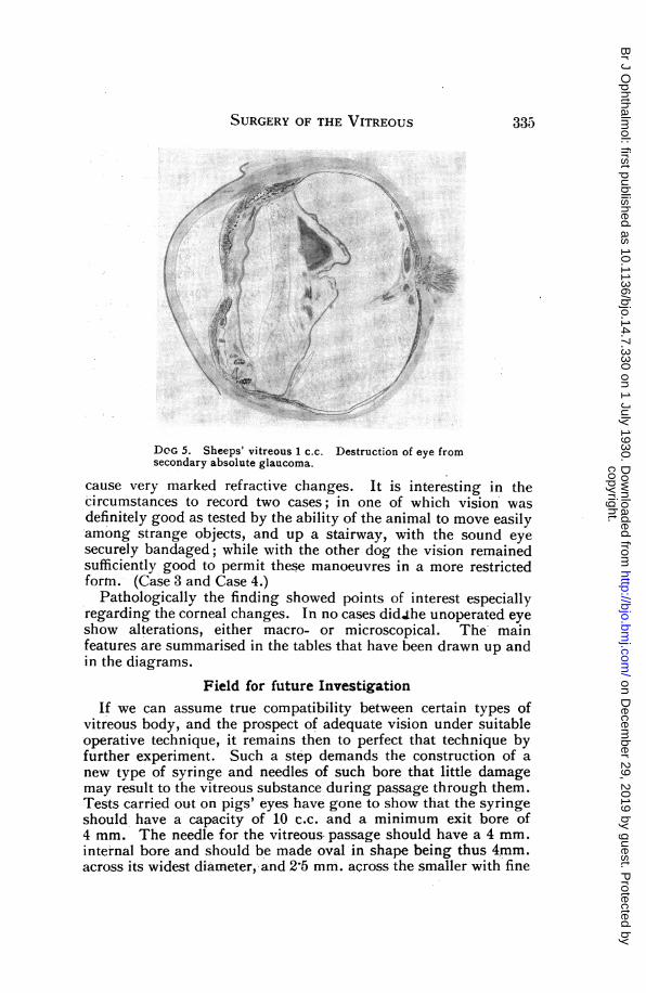
DOG 5. Sheeps' vitreous 1 c.c. Destruction of eye fromsecondary absolute glaucoma.
cause very marked refractive changes. It is interesting in thecircumstances to record two cases; in one of which vision wasdefinitely good as tested by the ability of the animal to move easilyamong strange objects, and up a stairway, with the sound eyesecurely bandaged; while with the other dog the vision remainedsufficiently good to permit these manoeuvres in a more restrictedform. (Case 3 and Case 4.)
Pathologically the finding showed points of interest especiallyregarding the corneal changes. In no cases didJ;he unoperated eyeshow alterations, either macro- or microscopical. The mainfeatures are summarised in the tables that have been drawn up andin the diagrams.
Field for future InvestigationIf we can assume true compatibility between certain types of
vitreous body, and the prospect of adequate vision under suitableoperative technique, it remains then to perfect that technique byfurther experiment. Such a step demands the construction of anew type of syringe and needles of such bore that little damagemay result to the vitreous substance during passage through them.Tests carried out on pigs' eyes have gone to show that the syringeshould have a capacity of 10 c.c. and a minimum exit bore of4 mm. The needle for the vitreous passage should have a 4 mm.internal bore and should be made oval in shape being thus 4mm.across its widest diameter, and 2-5 mm. across the smaller with fine
335
copyright. on D
ecember 29, 2019 by guest. P
rotected byhttp://bjo.bm
j.com/
Br J O
phthalmol: first published as 10.1136/bjo.14.7.330 on 1 July 1930. D
ownloaded from

3HE BRITISH JOURNAL OF OPHTHALMOLOGY
rubber tubing between it and the syringe. The operation sugges-ted demands the simultaneous insertion and withdrawal of vitreous,the point of " exit " being the internal peri-equatorial area, andthe point of " inlet," the external peri-equatorial area. The needleto be employed for the " exit " would be bent at right angles sothat it lies flat against the globe and does not hinder the internalocular movements. Two fine eyelets welded to it would be avail-able so that its position might be firmly fixed on the sclerotic bymeans of sutures. The needle to be employed at the " inlet "would have a similar design. TI wo syringes are necessary, bracedin a common frame with a cross bar between them in order thatthey may be used either together or separately. With such anapparatus an outline of the operation suggested would be asfollows:
(1) Attack first the detachment itself preferably by simplepuncture or narrow bore trephine-and when the result is estab-lished (say three weeks) proceed with the next step.
(2) With the eye turned as far outwards as possible open theconjunctiva, retract it, and open the sclerotic between sutures (asdescribed) adding two sutures for fixation through the eyelets inthe needle. Pass and fasten the needle and attach rubbertubing.
(3) With the eye turned in as far as possible proceed as before,and fix the outer needle and tubing in place.
(4) Start by extracting vitreous up to 1 c.c. and then commencepassing in the fresh vitreous from the outside syringe and continueuntil 5-6 c.c. have been inserted and removed.
(5) Close the wounds as described starting with the internalone.Owing to other work the practical application of this technique
has not been possible, but in view of the obvious stability foundbetween certain types of vitreous it would seem a justifiableprinciple and one that might well be followed to its experimentalconclusion.
I wish to thank Professor E. B. Verney, of University College,for his interest and ever ready help with this investigation; mycolleague, Wing-Commander H. E. Whittingham, director ofpathology R.A.F.M.S. for so kindly drawing the diagrams; andMessrs. Walls, of Chiswick, whose staff helped me considerably byproviding at all times sufficient pigs' eyes to make the work smoothand easy.
REFERENCES.1. Mann, Ida.-Development of the human eye.2. Howard.-Origin of vitreous. Amer. Ji. of Ophthal., 589, 3/1920.3. Weekers, L.-Ddcollement de la Ret. exp. Arch. Obhthal., January, 1926.4. Beaugrand. Etude de corps vitrd, 1880.5. Nakashima, C.-Arch. f. Ophthal., 116, 403-437, 1926.
336
copyright. on D
ecember 29, 2019 by guest. P
rotected byhttp://bjo.bm
j.com/
Br J O
phthalmol: first published as 10.1136/bjo.14.7.330 on 1 July 1930. D
ownloaded from

SURGERY OF THE VITREOUS
Pathological FindingsDOG 1. Pigs' vitreous 1 c.c. normal.Cornea.-About double its thickness in parts. Epithelium
intact. Substantia propria thickened and thrown into irregularwaves chiefly the superficial layers directly behind Bowman'smembrane. The fibres in this area are ill defined. Increase innumber of corneal corpuscles. Increase of all elements immediatelyin front of Descemet's membrane. Endothelium in parts infiltratedby round and spindle-shaped cells.
Iris, lens, ligamentum pectinatum, clear.Schlemm's Canal, clear.Ciliary Body.-Some round-celled infiltration with disturbance
of pigment cells. Migration of pigment cells.Vitreous Body.-Areas of faintly staining material containing
here and there scattered round cells. Migrated pigment cells. Noleucocytes. No evidence (by oil immersion) of bacterial infection.
Optic Nerve and Retina.-Retina widely detached.. Appearsnormal until in region of nerve head. Here are again massed roundcells and a band of tissue similar in appearance to the optic nerveprojects well into the vitreous. It does not have the appearance oforganised tissue. The unoperated eye shows no abnormalchanges.DOG 3. Pig 1 c.c.Cornea.-One or two limited areas where the substantia propria
is thrown into folds, and its continuity thereby broken. No definitecell invasion here and no thickening or alteration of the deeperlayers. This condition can be seen to a very slight extent in sec-tions of the normal eye and therefore may be explained as due insmall part to the processes to which the eyes have been subjectedin sectioning.
Iris, ciliary body, lens, vitreous and retina show no micro-scopical alterations.The optic nerve is clear. (See diagram.)DOG 4. Dog into Dog.(Vitreous extracted and returned into same eye as control).In the section of this eye and in those of the unoperated eye no
abnormal changes can be noted.DOG 5. Sheep into Dog.Sections here show very profound reactions. Anterior chamber
full of exudate and corneal distortion. Extensive iridic and ciliarybody infiltration. Iris bomb6. Destruction of lens. Markedvitreous reaction. Peculiar infiltration localised around nerve head,choroid and retina and lateral area. (See diagram.)
337
copyright. on D
ecember 29, 2019 by guest. P
rotected byhttp://bjo.bm
j.com/
Br J O
phthalmol: first published as 10.1136/bjo.14.7.330 on 1 July 1930. D
ownloaded from

THE BRITISH JOURNAL OF OPHTHALMOLOGY0o X C
xa Ca- CXS a->
_4 U toVIC3 ar a)a) a)~~~~~~~~~0n r
> 'Cl0 00
o 0. w20b 0
co
4.. )o
o~~~~~~~0>~a.
X30 a0x 0OaC Ca3 ca
Ca .0 0Cd (
U ~ ~ ~ Ca~~Ca nzmU
4) w ~ ~ ~
a 4.0 cd~'0a Ca Ca Ca
0C
Ca) 0 Ca 0
0nv. a) Ca
0~~~~~~~0 0.0~
Ca~~~~~~~~~~~~C
0 I-~~~~~~E dcdz 0~~~~~~.000o0 Cd 0
0 04.0U0 -. Caa.
4) 0 b0e o.oCaC
--4co co (L) 4)~~~~~~~~a b
o bO>~~~~~~~~~~~~. 0 00 0z t= 0 Z'-~~~C I... .0 O
Cd Cd~ ~ ~ ~~a bCaa
Cd~~~~~~~a
J J6 Li
b,O biO
0 0
0 0bD bD:
cq c
0 0
C.)
r.-4
0 '-
0
I
0(I)
o 00 0
*ba
ocwU
o o (A
0 0
-
338
0I'w
o)ay CaEcd 0 0w ~4404
(J)0zSz
¢I-
¢
z
1-4
04
0En(f
0Da)
-W
Cd5r.
*.)C-)
bOD
04
0
copyright. on D
ecember 29, 2019 by guest. P
rotected byhttp://bjo.bm
j.com/
Br J O
phthalmol: first published as 10.1136/bjo.14.7.330 on 1 July 1930. D
ownloaded from