complications of pterygium treatment - bjo.bmj.com · late complications ofpterygium treatment...
TRANSCRIPT

British Journal of Ophthalmology, 1980, 64, 496-505
Late complications of pterygium treatmentK. H. TARR AND I. J. CONSTABLEFrom the University Department of Ophthalmology, Royal Perth Hospital, Perth, Australia
SUMMARY Long-term complications of pterygium excision and beta irradiation in 63 eyes of 57patients are described. The age of the patients at treatment ranged from 27 to 69 years (mean48 ±1 1), and complications were assessed 3 to 20 years later (mean 12 ±3). The pterygia were excised,leaving bare sclera, and beta irradiation of total dose 750 to 5200 rads (mean 3475 ±916) was given,except in 7 patients who had repeated courses or overlapping fields of beta irradiation. Scleralulceration was present in 51 eyes and sectorial lens opacities with normal visual acuity (VA) in 19eyes. Radiation induced cataract occurred in 3 eyes, with reduced vision. Ptosis, symblepharon,and iris atrophy were also seen. Pseudomonas endophthalmitis occurred in 4 patients with scleralulceration. Beta irradiation to prevent recurrence of pterygia is a significant cause of iatrogenicocular disease. There is a need to modify the beta irradiation dosimetry at present in use.
Pterygium excision is a common ophthalmic opera-tion that is often considered trivial, but withoutconjunctival radiotherapy recurrence rates may beas high as 69%, especially in the hot, dry, sunnyenvironments of Australia1 and the Middle East.2Numerous surgical modifications to simple excisionhave been reported, but none are notably advanta-geous.2 Beta irradiation after excision reduced therecurrence rate to a range of 05%3 to 16%.1 Apartfrom local steroids, artificial tears, and dark glassespostoperatively, the only alternative treatment istopical thiotepa. Although reported to be as effectiveas radiotherapy,4 thiotepa has not been widelyaccepted.
Pterygium recurrence rates have often beenreported, but late complications of pterygiumtreatment have received little emphasis. Fifty-sevenpatients with late complications are presented andtheir relationship to beta irradiation discussed.
Patients and methods
Patients with conjunctival, corneal, or scleralcomplications from previous pterygium treatmentwere obtained on request from other ophthal-mologists and by a search of the records of 3 teach-ing hospitals. Four new cases presented at hospitalclinics during the study. All but 5 patients were
Correspondence to Dr K. H. Tarr, University Departmentof Ophthalmology, Royal Perth Hospital, GPO Box X2213,Perth 6001, Western Australia.
examined by one of us (K.H.T.). Adequate recordsand photographs for inclusion in parts of the studywere available on these 5.
Details of pterygium treatment, the course ofocular symptomatology, corrected visual acuities(VA), biomicroscopic measurement of avascularconjunctival patches, size of scleral ulceration anddistance from the limbus, the position and assess-ment of ulcer depth, colour, and presence of over-lying tissue were recorded. In addition telangiectaticvessels were noted, and after pupillary dilatationlens opacities were drawn and graded. The tearmeniscus, break-up time, and results of an unstimu-lated Schirmer's test were recorded together withfluorescein and rose Bengal staining. Any otherocular pathology was noted and an external photo-graph was taken. Radiotherapy records with com-plete details of treatment were available in all but2 patients. Of the 57 patients examined, 46 (51 eyes)had scleral ulceration, 2 had corneal ulceration, and9 (10 eyes) had pathology confined mainly to theconjunctiva. The age of initial pterygium treatmentwith excision and beta irradiation ranged from 27to 69 (mean 48 ±(SD) 11). Examination took place3 to 20 years later (mean 12±3). There were 32males and 25 females, of whom 1 was aboriginaland the rest were European.
Pterygium excision leaving bare sclera was usedin all patients. In 15 eyes recurrent pterygia had beenre-excised, and 8 of these had previously had betairradiation (Table 1). Beta irradiation was repeatedin 5 eyes and involved overlapping fields in 2 other
496
on 4 May 2019 by guest. P
rotected by copyright.http://bjo.bm
j.com/
Br J O
phthalmol: first published as 10.1136/bjo.64.7.496 on 1 July 1980. D
ownloaded from

Late complications ofpterygium treatment
Table 1 Summary of pterygium treatment in 57 patients with late complications: treatment is outlined Jbr eyeswith corneal or sckeral ulceration, with surface pathology without ulceration, andfbr fellow eyes
Treatment
E1, E. E1±+1 E,E, E3 E1+5 E.+-/2 E.,+2 EE1+51
E1+51 E,+(3EE4-3 E. E3+52 E2+52 E only
Ulcerated 45* 2 1 0 1 2 2 0
Surface pathology only 7 2 0 1 0 0 0 0
Fellow eyes 9 1 1 1 0 0 1 5
E=excision; ,3=beta irradiation; subscript indicates sequence of E and 53 respectively. *Includes 2 corneal ulcers.
Table 2 Late complications ojfpterygium treatment: summary ofpatients with repeated or overlappingfields of beta irradiation
Beta irradiation Total dose Age at initial Years of Visual Lens External eyetreatment (rads) irradiation* follow-up acuity opacity appearance
1. 3600 r over 2 weeks repeated 2 7200 57 13 3/60 Dense cataract Deep scleral ulceryears later
Fellow eye - - 6/6 Minimal Normal
2. 4500 r over 2 weeks, 5 yr later 6800 28 15 6/6 Sectorial Deep scleral ulcer2300 r over I week
Fellow eye - - 6/5 Nil Normal
3. 3000 r over 2 weeks repeated 6000 44 19 6/6 Sectorial Deep scleral ulcer7 years later
Fellow eye, only initial 3000 r 3000 44 19 6/6 Nil Normal
4. 3600 r over 2 weeks, 1 yr later 5900 45 13 6/36 Sectorial Moderately deep scleral2300 r ulcer
Fellow eye, only initial 3600 r 3600 45 13 6/5 Nil Recurrent pterygium
5. To both eyes: 4000 r over 5 6400 48 15 6/6 Nil Full thickness verticalweeks linear defect, uveal
prolapse, covered byconjunctiva (Fig. 5)
10 months later, 2400 r to each (8800) 6/6 Nil Normalof 2 overlapping fields
6. 4000 r over I week, then at 1 5300 30 12 6,'5 Sectorial Moderately deep scleralmonth, 1300 r to each of 2 (6600) ulceroverlapping fields
Fellow eye, 2400 r over 2 weeks 2400 37 5 614 Nil Normal
7. 3200 r over 3 weeks to 2 3200 56 14 6/4 Nil Deep scleral ulceroverlapping fields (6400)
Fellow eye - - 6/4 Nil Normal
*In years; r=rads.
eyes (Table 2). Beta irradiation was started withinthe first week after excision in 55 eyes (87%). Twiceas many were treated during winter as duringsummer. All but 2 eyes had fractionated treatment(Table 3). Two strontium-90 applicators were used.One was a 1-5 cm circular plaque with an emissionrate of 8-7 rads s-1 when supplied in 1962. The otherwas an ellipse with active surface dimensions of13x 18 mm and an emission rate of 15 3 rads s-1 in1966. For the 3-year periods of 1970-2, 1973-5,1976-8, 1470,4525 and 4864 patients respectively hadbeta irradiation after pterygium excision in Perth.
Results
SYMPTOMATOLOGYOn examination ocular pain or irritation, epiphora,and photophobia were severe in 15 (26%) of thepatients with ulceration and in 2 of 9 patientsreferred with mainly conjunctival abnormalities.Symptoms were absent in 12 patients (21%). Thesesymptoms had been present from the time of initialpterygium treatment in 24 patients (42%). Severeocular pain and photophobia during or immediatelyfollowing beta irradiation was present in 18 (32%).
497
on 4 May 2019 by guest. P
rotected by copyright.http://bjo.bm
j.com/
Br J O
phthalmol: first published as 10.1136/bjo.64.7.496 on 1 July 1980. D
ownloaded from

K. H. Tarr and I. J. Constable
Table 3 Late complications ofpterygium treatment: fractionation and dosage* oj initial beta irradiation
Single dose at 3 doses over 3 doses over 4 doses over 4 or 5 doses overoperation <1 week 2 weeks 3 weeks 4 weeks
Ulceratedtnumber 1 15 27 3 4dose range 750 2100-5100 2400-5100 3200-5200 4000-5300mean - 3135 3350 4400 4475
Surface pathologynumber 1 2 7 0 0dose range 2200 4500 2400-4800mean - each 2700
Fellow eyesnumber 0 3 8 1 1dose range 2600-4500 2400-4000 4800 4000mean 3433 3125 -
*In rads; t2 unknown.
Table 4 Patients with severe visual loss or aphakia
Age at Years of Visual Radiationtreatment follow-up acuity dose* Ocular pathology
1. 45 13 6/36 3600+2300 Marked posterior sectorial and anterior crescentic opacity
6/5
2. 52 1 1 6/6
6/5
3. 57 1 3 3/60
6/6
4. 60 13 6/9
Nil
5. 55 11 3/CF
6/12
6. 65 12 6/12
6/12
1 1 6/36
6/24
12 3/CF6/18
9 6/HM
6/18
3 Nil
1 yr later3600 only
4500
Nil
3600+ 36002 yrs laterNil
3600
Nil
3900
3900
2700
Nil
3000
Nil
45004500
2400
2400
2400
6/12 2400
11 Nil 2800
6/6 Nil
Moderately deep scleral ulcerNormal
Aphakic (8 yr after treatment)Deep scleral ulcerNormal
Marked posterior sectorial and anterior cortical opacitiesDeep scleral ulcerMinimal wedge opacities
Aphakic (12 yr after treatment)Deep scleral ulcerEnucleated (trauma)
Aphakic (8 yr after treatment)Deep scleral ulcerAphakic and deep scleral ulcer
Aphakic (8 yr after treatment)Deep scleral ulcerAphakic (9 yr after treatment)No ulceration
Aphakic (5 yr after treatment)Deep scleral ulcerAphakic (6 yr after treatment)No ulceration
Dense cataract, deep scleral ulcerAphakic (8 yr after treatment)
Posterior polar cataractModerately deep scleral ulcerPosterior polar cataract
Deep scleral ulcerPseudomonas endophthalmitisEvisceratedNo lens opacityDeep scleral ulcer
Deep scleral ulcerPseudomonas endophthalmitisEvisceratedNormal
In rads given as 3 doses over 2 weeks except the 2300 given as a single dose. CF=count fingers; HM=hand movements.
498
7. 69
8. 69
9. 65
10. 55
11. 53
on 4 May 2019 by guest. P
rotected by copyright.http://bjo.bm
j.com/
Br J O
phthalmol: first published as 10.1136/bjo.64.7.496 on 1 July 1980. D
ownloaded from

Late complications ofpterygium treatment
Table 5 Clinical details ofpatients with scleral or conjunctival complications after pterygium treatmentexcluding repeated or overlapping fields of beta irradiation*
Age at initial Years of Total dose of Ulcer sizetExternal pathology treatment (no.) follow-up irradiation (no.)
Superficial ulceration 40+11 (6) 12+3 3710±1415 5-1 +4-7 (5)
Moderately deep ulcer 52+10 (11) 11 ±3 3183±771 5-4±3-4 (10)
Deep ulceration 49±10 (26) 12+3 3507+891 63+3 6(25)
Surface pathology only 42±11 (9) 9 5±4 3111±1121 -
*In rads; t6 unknown.
Reduced vision was a major complaint in 6 patients(10%) (Table 4). Four patients (7%) presented withan acute red eye of which two had pseudomonasendophthalmitis.
SCLERAL ULCERATIONScleral ulceration occurred in 51 eyes of 46 patients.Five patients had bilateral ulceration and 1 had anasal and a temporal ulcer on the one eye. Clinicallyulceration was classified into 3 grades. Superficialulceration was characterised by exposed sclera withan irregular white surface and shallow ulcerationless than one-third scleral thickness (6 eyes) (Fig.1). Moderate ulceration was characterised by adepth of one-third to two-thirds scleral thicknessand a white base (14 eyes) (Fig. 2). Deep ulcerationwas characterised by a depth of two-thirds orgreater with a dark base (31 eyes with 32 ulcers)(Fig. 3). Often only a thin layer of scleral fibrescovered uveal tissue. In all 3 groups frequency andseverity of symptoms were similar. There was nosignificant difference between the groups withrespect to age at treatment (P<0-1), follow-uptime (P<0 3), or total irradiation dosage whenretreated cases were excluded (P<0-2) (Table 5).The typical ulcer was 2-3 mm in diameter, had a
corneal edge 1-2 mm from the limbus and wascentred within half a clock hour of the horizontalmeridian (Fig. 3). The ulcer usually had steep edgessurrounded by very white avascular sclera, whichwas usually bare, and avascular conjunctiva withtelangiectasia. The area of ulceration varied from2 5 mm2 to 16 mm2 (mean 6-2±3-7 mm2). There wasno correlation between area and ulcer depth (Table5). The centre of ulceration was more than 1 clockhour from the horizontal meridian in 9 eyes (17%).Seven (13%) were inferiorly situated (Figs. 2 and 4).It was often difficult to ascertain whether thesclera was completely bare of any tissue. A thintransparent layer of tissue was present in 11 eyes(21%) on careful biomicroscopy with cobalt bluelight. Four (7%) had a documented history ofpersistent bare sciera with punctate rose Bengal
staining and failure of conjunctival regrowth afterinitial treatment for 1 to 7 years before ulcerationdeveloped. In 3 eyes a row of 2-4 translucent smallspots were present. These were separate spotsequidistant from the limbus by 1-5 to 2 mm, in linewith the scleral ulcer. From 2 of these a small(0 5 mm) vascular tuft arose. This was not connectedto surrounding surface vessels. Similar tufts wereseen within the bare area unassociated with trans-lucent spots in a few other eyes. A small encrustationoccurred on the adjacent conjunctiva or in the ulcerbed in 10 eyes (20%). This was rough, hard, andstained with fluorescein and rose Bengal. An analysisrevealed calcium and other crystals which werebirefringent negative to a red plate compensator,suggesting urate. Episcleral vessels partially coveredthe ulcer base in 13 eyes (25%). In 4 eyes previousphotographs revealed minimal vessel extension over2 to 6 years. Telangiectasia surrounded the baresclera in 48 eyes (92%) and was severe and cos-metically unacceptable in 18 (34%).Two scleral ulcers were atypical. One was a small
1-5 mm diameter punched-out moderately deepulcer covered by mildly abnormal conjunctiva. Itdeveloped 10 years after excision and a single doseof 750 rads. The other was a vertical curvilinearfull-thickness scleral defect with a knuckle of uvealtissue covered by avascular and telangiectaticconjunctiva (Fig. 5). It was noted on routine exami-nation 12 years after repeated excision and betairradiation. Both eyes were treated, initially with4000 rads over 2 weeks, then 10 months later afurther 2400 rads was given to 2 overlapping fields.The scleral slit was perpendicular to the axis of theoverlap. The fellow eye had white avascular scleracovered by avascular conjunctiva with surroundingtelangiectatic vessels.
SURFACE PATHOLOGY IN EYES WITHOUTULCERATIONTen eyes of 9 patients had no ulceration and path-ology mainly confined to the conjunctiva. Thisgroup did not significantly differ from those with
499
on 4 May 2019 by guest. P
rotected by copyright.http://bjo.bm
j.com/
Br J O
phthalmol: first published as 10.1136/bjo.64.7.496 on 1 July 1980. D
ownloaded from
K. H. Tarr and I. J. Constable
Fig. I Fig. 2
Fig. 3 Fig. 4
Fig. 5 Fig. 6
Fig. 7
500
F'ig. 8
Y!li
on 4 May 2019 by guest. P
rotected by copyright.http://bjo.bm
j.com/
Br J O
phthalmol: first published as 10.1136/bjo.64.7.496 on 1 July 1980. D
ownloaded from

Late complications ofpterygium treatment
ulceration with respect to age (P<0 1), follow-up(P<0 1), or total dose of beta irradiation (P<0-2)(Table 5). When analysed in combination with thesuperficial scleral group, the lesser affected patientswere significantly younger than patients with deeperulceration (P<0-01). Conjunctival pathology con-
sisted of severe telangiectasia in 6 eyes (60%) andthinned avascular conjunctiva of area 3 to 18 mm(mean 9 mm) with an adherent encrustation in 3(30%). Three (30%) had recurrent pterygia. Theunderlying sclera had a very white appearance in8 (80%).
FELLOW EYESThirteen patients with unilateral ulceration hadfellow eyes with previous pterygium excision andbeta irradiation (Table 1). None were symptomatic.Four had recurrent pterygia present, to make a
total of 12 eyes (16%) with recurrent pterygia afterexcision and beta irradiation. The conjunctiva was
mildly abnormal in 6 fellow eyes (46%). Thirty-eight fellow eyes had no previous beta irradiation.Six eyes (16%) had untreated pterygia. One of 5pterygia previously treated by simple excision alonehad recurred.
CORNEAL ULCERATION
Indolent corneal ulcers occurred in 2 patients. Onewas aged 34 years at the time of initial unilateralpterygium treatment with simple excision and betairradiation with 5000 rads in 3 doses over 2 weeks.An elliptical strontium applicator, active area of13x 18 mm and emission rate of 14 8 rads s-1, wasused. The eye was persistently sore and irritableafter treatment until 10 years later, when a diagnosisof corneal ulceration was made (Fig. 6).The ulceration of half corneal thickness was
vertically oval and measured 2x 3 mm. The edgewas 1 mm from the limbus and situated at the siteof the previous pterygium head. The ulcer edge andbase were irregular, and the bulbar conjunctiva hada large avascular patch surrounded by prominenttelangiectatic vessels. The tear film was normal andthere was no corneal staining elsewhere.The other patient who developed corneal ulcera-
tion was aged 36 years at the time of treatment 12years before examination. Bilateral pterygia wereexcised and beta irradiation with 3600 rads in 3doses over 2 weeks given.A round, flat strontium-90 plaque of emission
rate 7-8 rads s-1 as well as the elliptical plaquementioned above was used. The eye settled until 3years prior to examination, when a sore left eyedeveloped after irritation from sawdust. Nasally a4x 4 mm patch of bare sclera surrounded by a fewtelangiectatic vessels were present. Mild rose Bengalstaining was found. A half-thickness oval cornealulcer measuring 1.5x2 5 mm was situated at thesite of the previous pterygium head and was sepa-rated from the limbus by I mm of almost normalcornea. The tear appearance was normal, but aSchirmer's test was 18 mm in the affected eyecompared to 5 mm in the fellow normal eye.
SYMBLEPHARON AND PTOSISThree eyes (6%) with scleral ulceration had symble-pharon. One patient treated with simple excisionand 5200 rads over 3 weeks (12 years later) hadmarked symblepharon to the upper canalicular area
and punctum. Another patient had simple excision3 times to both eyes before excision and beta irra-diation with 4800 rads over 4 weeks. Extensivesymblepharon to both canalicular areas with punctalocclusion and tethering of the globe was present 16years later. A third patient had recurrent bilateralpterygia treated by excision and beta irradiationwith 3600 rads over 2 weeks. The right pterygiumrecurred within 3 months and was treated by exci-sion and 2300 rads as a single dose. 3 mm of ptosishad persisted since treatment, and 11 years latersymblepharon to the upper canalicular area wasalso present.
ASSESSMENT OF TEAR FUNCTIONA poor tear meniscus, decreased fluorescein break-up time, a Schirmer's test less than 5 mm in 5minutes, and punctate corneal staining occurredalone or in combination in 17 patients (30%).Thirty-five patients (70%) had a normal tear assess-ment. In 14 patients (26%) the tear assessment wasabnormal bilaterally despite the presence of anuntreated fellow eye in 12 of these patients. Fivepatients were not examined. Ulcerated eyes had aSchirmer's range of 10 to 35 mm (mean 18±10) in5 minutes, while fellow eyes measured 2 to 35 mm(mean 14j10). In none was tear production lessand in 13 (25%) it was 6 mm more in ulceratedthan in fellow eyes. Rose Bengal staining of theulcer base was a constant feature. Adjacent baresclera stained in 34 ulcerated eyes (63%).
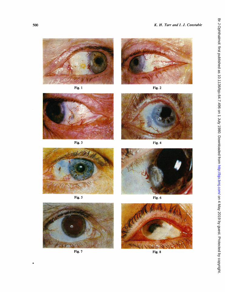
Figs. 1-8 Late complications ofpterygium treatment. (1) Superficial scleral ulceration with avascular conjunctiva,mild telangiectasia, and encrustation. (2) Moderately deep scleral ulceration inferiorly situated. (3) Deep scleralulceration. (4) Inferiorly situated scleral ulceration with aphakia. (5) An atypical scleral ulcer with overlying conjunctiva.(6) Indolent corneal ulceration and a large area of bare sclera. (7) Sectorial posterior lens opacity, scleral ulceration,and an encrustation. (8) Pseudomonas endophthalmilis with hypopyon and ulceration within an area of bare sclera.
501
on 4 May 2019 by guest. P
rotected by copyright.http://bjo.bm
j.com/
Br J O
phthalmol: first published as 10.1136/bjo.64.7.496 on 1 July 1980. D
ownloaded from

Table 6 Late complications ofpterygium treatment: correlation of lens complications with patient details,treatment, and external pathology
Patients (no.) Eyes (no.)Lens Visualstate acuity Treated Surface
Age Follow-up Total dose Ulceration fellows pathology Untreated
Normal 6/9 or better 44+10 (30) 10±3-2 3101 ± 1004 (34) 27 8 8 32
Significant opacity 6/9 or better 48±12 (13) 13±+29 4179±+730 (14) 14 3 2 3t
Cataract 6/18 or worse 621±67 (7) II 1-2 3540t858 (10) 4 1 0 0
Aphakic 6 1 0 2
Excluded* 12 patients 18 eyes 2 0 3 0
*Repeated and overlapping fields of irradiation (7) and unknown (5). tNone sectorial.
IRIS PATHOLOGY
Localised nasal iris atrophy was present in both
eyes of I patient 9 years after bilateral pterygium
excision with 4500 rads over 3 consecutive days.
The atrophy involved the to 5 clock hours central
to the collarette in one eye and a patch extending
from the nasal pupillary margin to the iris root in
the other eye. Another patient had 2 abnormal iris
vessels on the nasal side in association with a
scleral ulcer.
CATARACT FORMATION
Nine patients (16%) had 14 eyes with reduced vision
from cataract (Table 4). Ten of these eyes also had
scleral ulceration. This group represented 190o of all
ulcerated eyes. In 2 of the 5 cataracts marked
sectorial opacities extended from the nasal side and
reduced vision. Patients with reduced vision or
aphakia were significantly older at age of treament
(P<0-001) but had not been followed longer
(P<~015) than patients with normal vision. Total
dose of beta irradiation was not significantlydifferent from that given to eyes without cataract
(P<0-1) (Table 6).
Sectorial cortical opacities with normal visual
acuity were found in 19 treated eyes (250o) (Table 6).
Minimal opacities limited to the equator or only
faintly visible were excluded. Sixteen had a single
course of beta irradiation with a total dose of
2900 to 5500 rads, while 3 eyes had additional beta
irradiation (Table 2). The opacity was present
mainly in the posterior cortex and in the same
quadrant as the scleral ulceration (Fig. 7). The
anterior cortex was involved in more severe catar-
acts. Thirteen eyes (25%) with scleral ulceration
and I with corneal ulceration had such opacities.No equivalent opacities were seen in these eyes on
the temporal side or in untreated fellow eyes. Three
untreated eyes had diffuse equatorial or posterior
polar cataracts. There was no significant difference
in age (P<0-1 5) between patients with and without
sectorial opacities, but those with opacities hadbeen followed up significantly longer (P<0.01). Thetotal dose of beta irradiation was significantlygreater (P<0-00l) for eyes with sectorial opacitiesand normal vision than for those without opacities(Table 6).
PSEUDOMONAS ENDOPHTHALMITISThis has occurred in 4 patients. All had deep scieralulceration. Three have been reported elsewhere bythe authors.-' The fourth developed endophthalmitis3 months after inclusion in this survey. The patient,a 71-year-old female, had a pterygium excision and4500 rads over 2 weeks 9 years before the suddenonset of ocular symptoms and the diagnosis of ascleral ulcer. Two years later, on inclusion in thissurvey, a 2 X 3 mm clearly defined deep scleralulcer 2 mm from the limbus in the horizontalmeridian was present associated with an encrustationand severe telangiectatic vessels. The tear film andSchirmer's test were normal. A nasal sectorial lensopacity was present. Three months later the eyespontaneously became red and painful. A largeavascular area of sclera surrounded the ulceration.Peripheral corneal infiltration and a large hypopyonwere present together with a mucopurulent dis-charge (Fig. 8). A moderate growth of Pseudomonasaeruginosa was cultured from the ulcer surface.Six days of treatment with gentamicin drops hourlyand subconjunctival injection daily with oralamoxycillin was ineffective. This treatment wasreplaced by a continuous infusion of tobramycininto the upper fornix and intravenous ticarcillin aspreviously reported.5 An initial rapid reduction inthe hypopyon was followed by a slow improvement.During the following 3 weeks she developed peri-pheral corneal vascularisation, mild iritis, posteriorsynechiae, and a dense cataract. Visual acuity wasperception of light. Treatment was then continuedwith Chloromyxin ointment (chloramphenicol andpolymyxin B) 5 times a day for a further three
K. H. Tarr and L J. Constable502
on 4 May 2019 by guest. P
rotected by copyright.http://bjo.bm
j.com/
Br J O
phthalmol: first published as 10.1136/bjo.64.7.496 on 1 July 1980. D
ownloaded from

Late complications ofpterygium treatment
weeks. Two of the eyes with Pseudomonas aeruginosainfection were eviscerated and 2 salvaged after 6 to7 weeks' hospitalisation. One retained normal visualacuity and the other suffered profound visual lossdue to complicated cataract.
RELATIONSHIP OF RADIOTHERAPY TOSCLERAL ULCERATIONBilateral deep scleral ulceration occurred in 5 patientsafter identical treatment to each eye. Each eye hada single pterygium excision and beta irradiation with2400 to 4400 rads (mean 3460±780).
Unilateral ulceration occurred in the presence ofan untreated fellow eye in 29 patients. Five weresuperficial, 7 moderately deep, and 18 deep ulcers.Three eyes had had repeated courses or overlappingfields of beta irradiation (Table 2). Apart from these3 irradiation dosage ranged from 750 to 5200 rads(mean 3386±1079). The remaining 12 patients hadunilateral scleral ulceration despite bilateral ptery-gium treatment. Nine patients had similar treatmentgiven to each eye. Eight had a single course of betairradiation (range 2400 to 4800 rads, mean 3437±944). One ulcer was superficial, 1 moderately deep,and 6 deep. One patient had bilateral repeated betairradiation with unilateral atypical ulceration (Table2, no. 5). Three patients developed ulceration onlyin the eye given additional beta irradiation (Table 2).Apart from retreated and overlapping fields of betairradiation 1 eye had a single dose of 750 rads andthe remainder had fractionated treatment (Table 3)with a total dose of 2000 to 2400 rads in 10 eyes,2500 to 3400 in 9 eyes, 3500 to 4400 in 25 eyes, and4500 to 5200 in 7 eyes. Six patients with ulcerationwere initially treated before 1964, 12 during 1964-6,22 during 1967-9, 5 during 1970-2, and I in 1974.
Discussion
Cosmesis, irritation, or threatened vision are stan-dard indications for pterygium treatment, yet mostdo not cause marked discomfort or limit vision.1 6Reduced visual acuity from untreated pterygia waspresent in only 0-2% of 1352 patients7 in one seriesand other reports are of single cases.' 8 The efficacyof pterygium treatment is limited by a high recur-rence rate without beta irradiation'7 and othercomplications. Apart from recurrence rates, latecomplications have received little attention.
Patient acceptance of treatment and subsequentocular discomfort should be important considera-tions in pterygium treatment. Treatment was apainful experience in 32% of the population westudied, and this was universally attributed to betairradiation. Pain may arise from repeated mechanicaldamage during application or from acute punctate
corneal erosion.9 Ocular discomfort and photo-phobia persisted for many years in 42% of oursubjects despite protective measures and topicaltreatment. The severity of symptoms did not corre-late with the size or depth of scleral ulceration.
Scleral ulceration after pterygium excision andbeta irradiation has been previously reported.10-'6In our 46 patients with 52 scleral ulcers the depth ofulceration did not correlate with patient age attreatment, follow-up time, or total dose of radiation.The ulcer was inferiorly situated in 7 eyes (13%).This would be consistent with elevation of the eyeduring radiation treatment if ulceration occurs atthe point of contact of the applicator on the eye.The nature and timing of ulceration, the appearanceof the sclera and adjacent tissues, and the develop-ment of ulceration only after additional beta irradia-tion clearly implicate radiation induced pathologyin the pathogenesis of scleral ulceration. However,sclera is generally known to be radioresistant. Veryhigh doses of beta irradiation to rabbit eyes haveproduced no scleral ulceration.'6
In man beta irradiation to the posterior sclerawith up to 100 000 rads over 8-14 days did not causenecrosis in 62 patients followed up for 1 to 9 years(mean 4 years).'7 Reports of 'partial scleral slough-ing"8 and scleral necrosis'920 following very highdoses of cobalt-60 plaque treatment do exist butare rare. At least 1 such case occurred anteriorly inassociation with the conjunctiva.'9 On the otherhand high doses of radiation are not necessary forscleral ulceration to develop anteriorly.'6 Even withthe currently used dose of 2400 rads or less, 11scleral ulcers were seen in our series. It wouldappear that an additional factor contributes to'radionecrosis' of the sclera anteriorly. Possiblefactors include surgical damage, scleral exposure,the influence of damaged conjunctiva, or the tears.
Faulty surgical technique, particularly cautery,has been implicated.610'2 An irregular distributionof translucent spots within the bare sclera followedthe use of a heavy tipped cautery in 1 reported case.12In our survey 3 eyes had translucent spots, but inan area equidistant from the limbus. A smallvascular tuft arose from some spots. The appearancesuggested that these spots developed at the penetra-tion site of anterior ciliary vessels. The locationwould be more random if due to surgical damage.The appearance of I atypical case (Table 2, no. 5)did suggest a scleral incision.The possible role of exposure has been the subject
of a short-term experimental study in rabbits bythe authors (to be published). It has been found thatexposure alone may cause rapid and indolentscleral necrosis. Bare sclera persisting after ptery-gium treatment has been previously noted and may
503
on 4 May 2019 by guest. P
rotected by copyright.http://bjo.bm
j.com/
Br J O
phthalmol: first published as 10.1136/bjo.64.7.496 on 1 July 1980. D
ownloaded from

K. H. Tarr and L J. Constable
occur without symptoms in many patients."101213Four of our patients (61%) were noted to havepersistent bare sclera for 1 to 7 years before develop-ing ulceration. In eyes that do regain conjunctivalcover late conjunctival necrosis could precedescleral ulceration. The presence of bare sclera inalmost all ulcerated eyes and the occurrence ofulceration after low doses of radiation suggeststhat conjunctival breakdown is the first event.
In this survey ulcerated eyes had a slightly greatertear production than nonulcerated fellow eyes(P<005). Therefore it is felt that lack of tears isnot a significant factor.Pseudomonas endophthalmitis occurred in 4
patients with deep scleral ulceration. Three havebeen reported by the authors., A spontaneous onsetoccurred in 3, and the relationship to scleral ulcera-tion was undeniable. Two eyes came to eviscerationand 2 were salvaged only after intensive and pro-longed hospital treatment. The occurrence ofendophthalmitis emphasises the importance ofsurgical repair of radiation induced deep scleralulceration.
Corneal ulceration has been reported9 afterpterygium excision and very high doses of radium-Dbeta irradiation or in widespread corneal disease orfrom external beam irradiation. Two cases ofcorneal ulceration are described here. Both wereindolent with necrotic margins and no inflammationor vascularisation. They occurred at the site of theprevious pterygium head. Corneal erosions maypersist for up to 1 year after beta irradiation, and1 case of corneal dystrophy has been describedpreviously.21
Less important complications of treatment werealso seen. A thinned vascular area of conjunctivasurrounded by telangiectatic vessels was commonlyseen. It was not more severe in eyes with scleralulceration. Symblepharon was present in 3 patientsassociated with a tethered globe in one and ptosisin another. This is a recognised complication ofpterygium excision but can also occur after radia-tion treatment.'9 Localised iris atrophy occurredbilaterally in one patient who also had lens opacitiesbut normal visual acuity.Beta irradiation-induced cataract is well docu-
mented. In one series 38 of 115 patients had non-progressive lens opacities when examined a minimumof 8 years after beta irradiation.22 20% of patientswho received less than 3600 reps developed opacities(1 rep= 1-08 rads tissue). Hilgers23 found no lensopacities 5 years after a fractionated dose of 3000reps of strontium-90 beta irradiation, but 3 of 46eyes (6%) developed lens opacities after 3000-5000reps. This incidence was considered acceptable tomaintain a low rate of pterygium recurrence. In
addition a single dose of 2200 rads after pterygiumexcision has been considered safe even when repeatedtwice for subsequent recurrences.' Typical sectorialposterior cortical lens opacities, with a visual acuityof 6/9 or better, were present in 19 eyes (25%) inthis survey. The total dose of beta irradiation rangedfrom 2900 to 5200 rads (Table 6). The anteriorcortex was involved only in the more severe cataracts,an observation previously noted.24 Compared topatients without cataract our patients showed nosignificant difference in age or length of follow-up,but a strong correlation was found with respect tothe total dose of radiation (P<0 001). This surveyconfirms that some opacities will develop after afractionated dose of 3000-5000 rads.Reduced vision from beta irradiation-induced
cataract is not common. Seven cases occurred 3 to6-5 years after radium beta irradiation.25 None hasbeen reported after pterygium excision and stron-tium-90 beta irradiation. In this study radiation-induced cataract caused reduced vision in 3 patients.(Table 4, nos. 1, 2, 3). The influence of beta irradia-tion on cataract formation in the other 6 patientsin Table 4 cannot be determined. The fact that thesepatients were older at treatment than those withnormal vision (P<0 001) probably reflects thedistribution of senile cataract rather than anysusceptibility of the older lens to radiation. It is theyounger growing lens that is particularly susceptible.
This survey would support a more cautiousattitude both to the management of pterygium andto the use of beta irradiation. The complications oftreatment, especially scleral necrosis and cataract,may lead to visual loss. With scleral ulceration,surgical treatment may be indicated to relievesymptoms alone and should certainly be consideredfor deep scleral ulceration to prevent pseudomonasendophthalmitis. The development of cataract aftertreatment of a benign lesion is not acceptable. Thereis a need to reconsider the indications for surgicaltreatment and the application of beta irradiation.Lower doses of radiation may reduce the complica-tion rate but may also be less effective in preventingrecurrence.
We are most grateful to all the Perth ophthalmologists whokindly referred their patients for examination and providedbackground information. We are also indebted to Dr A. J. M.Nelson and his staff for providing detailed radiotherapyrecords and for advice. We are grateful for the untiringefforts of Miss H. M. Deady in her typing of the manuscript.
References
'Cameron ME. Pterygiumn throughout the World. Springfield,Illinois: Thomas, 1965.2Youngson RM, Pterygium in Israel. Am J Ophthalmol1972; 74: 954-9.
504
on 4 May 2019 by guest. P
rotected by copyright.http://bjo.bm
j.com/
Br J O
phthalmol: first published as 10.1136/bjo.64.7.496 on 1 July 1980. D
ownloaded from

Late complications oj pterygium treatment
3Ozarda AT, Evaluation of postexcisional strontium-90beta ray therapy for pterygium. South Med J 1977; 70:1304.4Harrison M, Kelly A, Ohlrich J, Pterygium: 'Thiotepa'versus beta radiation, a double blind trial. Trans AustColl Ophthalmol Ophthalmol Soc NZ 1969; 22: 64-6.5Tarr KH, Constable IJ. Pseudomonas endophthalmitisassociated with scleral necrosis. In press.6Paton D. Pterygium management based oni a theory ofpathogenesis. Trans Am Acad Ophthalmol Otolaryngol1975; 79: OP-603-OP-612.'Wilson B. Beta irradiation of pterygia. Trans OphthalmolSoc Aust 1963; 23: 96-100.8Taylor HR, Hollows FC. Pterygium leading to blindness:a case report. Aust J Ophthalmol 1978; 6: 155-6.9Leahey BD. Beta radiation in ophthalmology. Am JOphthalmol 1960; 49: 7-29.
°0Brosnan JD. Pterygium: resection plus beta irradiation.Trans Ophthalmol Soc NZ 1969; 21: 107-13.
1Hellein R. RaQio necrosis of the sclera-treated withauricula cartilage graft. Aust J Ophthalmol 1976; 4: 104.
12Cameron ME. Preventable complications of pterygiumexcision with beta-irradiation. Br J Ophthalmol 1972; 56:52-6.
'3Cameron ME. The treatment of beta irradiation necrosisof the sclera. Aust J Ophthalmol 1978; 6: 86-9.
14Cappin JM. Radiation scleral necrosis simulating earlyscleromalacia perforans. Br J Ophthalnol 1973; 57: 425-8.
15Talbot AN. Complications of beta ray treatment of pterygia.Trans Ophthalmol Soc NZ 1979; 31: 62-3.
'Lommatzsch PK. Morphologische und funktionelleVeranderungen des Kaninchenauges nach Einwirkung vonBetastrahlen auf den dorsalen Bulbusabschnitt. Albrechtvon Graefes Arch Klin Exp Ophthalmol 1968; 176: 100-25.
17Lommatzsch PK. Experiences in the treatment of malignantmelanoma of the choroid with 106Ru/'06Rh beta ray appli-cators. Trans Ophthalmol Soc UK 1973; 93: 119-32.'Stallard HB. Radiotherapy for malignant melanoma of thechoroid. Br J Ophthalmol 1966; 50: 147-55.
'9MacFaul PA, Bedford MA. Ocular complications aftertherapeutic irradiation. Br J Ophthalmol 1970; 54: 237-47.
20Rotman M, Long R, Packer S, Moroson H, Galin M,Chan B. Radiation therapy of choroidal melanoma. TransOphthalmol Soc UK 1977; 97: 431-4.
2'Bedford MA. The corneal and conjunctival complicationsfollowing radiotherapy. Proc R Soc Med 1966; 59: 529-30.
22Thomas CI, Storaasli JP, Friedell HL. Lenticular changesassociated with beta radiation of the eye and their signifi-cance. Radiology 1972; 79: 588-95.
23Hilgers JHC. Strontium-90 Di-irradiation, cataractogenicityand pterygium recurrence. Arch Ophthalmol 1966; 76: 329-33.
24Duke-Elder S. System of Ophthalmology. London: Kimp-ton, 1972: 14: 994-6.
25Merriam GR. The effects of beta radiation on the eye.Radiology 1956; 66: 240-4.
505
on 4 May 2019 by guest. P
rotected by copyright.http://bjo.bm
j.com/
Br J O
phthalmol: first published as 10.1136/bjo.64.7.496 on 1 July 1980. D
ownloaded from