and review of literature lymphangioleiomyomatosis: a case ... filedecreased breath sound on the...
TRANSCRIPT

Received 11/18/2018 Review began 12/23/2018 Review ended 01/10/2019 Published 01/22/2019
© Copyright 2019Rhee et al. This is an open accessarticle distributed under the terms ofthe Creative Commons AttributionLicense CC-BY 3.0., which permitsunrestricted use, distribution, andreproduction in any medium, providedthe original author and source arecredited.
Lymphangioleiomyomatosis: A Case Reportand Review of LiteratureJee Ah Rhee , Ajay Adial , Rammohan Gumpeni , Asma Iftikhar
1. Internal Medicine, New York - Presbyterian Hospital Queens, Flushing, USA
Corresponding author: Asma Iftikhar, [email protected] Disclosures can be found in Additional Information at the end of the article
AbstractPulmonary lymphangioleiomyomatosis (LAM) is a disease, which is most commonly seen inwomen of childbearing age. The objective of this article was to provide education about thetypical clinical presentation, radiologic findings, histology, treatment approaches, anddifferential diagnosis. Pulmonary LAM is a cystic lung disease, usually generalized andprogressive and extremely difficult to treat and is considered to have a poor prognosis. Patientswith LAM often present with an insidious onset of dyspnea; this could be secondary topneumothorax. However, it could also be present as chylothorax and hemoptysis. We discusseda case who presented with chest pain and shortness of breath due to pneumothorax andretrospectively diagnosed with LAM.
Categories: PulmonologyKeywords: cystic lung disease, pneumothorax, lymphangioleiomyomatosis
IntroductionLymphangioleiomyomatosis (LAM) is a multisystem disorder characterized by the proliferationof smooth muscle cells that result in cystic lung disease as well as extrapulmonarymanifestations such as angiomyolipomas and lymphatic tumors. It predominantly occursin pre-menopausal women but could be present in postmenopausal women, presenting mostcommonly with progressive dyspnea and spontaneous pneumothorax. Once thought to be afatal disease for young women, with the only treatment option being lung transplant, thedefinition of classical LAM is actively evolving, thanks to a growing accumulation of data onLAM with international registries, which may help catalyze the advancement in diagnosticstudies and therapeutic options.
Case PresentationA 55-year-old woman presented three days after a sudden onset of right-sided chest pain,pleuritic and positional in nature, associated with an acute onset of shortness of breath. Shehad gone to her primary care physician, who performed a chest X-ray and urged her to come tothe hospital. Upon presentation at the emergency department, her oxygen saturation was above95% on room air, and she was not in any respiratory distress, but her exam was significant fordecreased breath sound on the right. A chest X-ray confirmed a large right-sided pneumothoraxwith small pleural effusion. A chest tube was inserted on the right side for the resolution of thepneumothorax, and subsequent computed tomography (CT) scan of the chest revealed bilateraldiffuse bullous disease of the lung with multiple cysts (Figure 1-2). The patient underwentvideo-assisted thoracoscopic surgery for right thoracoscopic wedge resection of a lung bleb andtalc pleurodesis. Gross examination of the specimen revealed several dilated air-like spacesranging from 0.2 cm to 0.4 cm in size. The hospital course was complicated by postsurgical
1 1 1 1
Open Access CaseReport DOI: 10.7759/cureus.3938
How to cite this articleRhee J, Adial A, Gumpeni R, et al. (January 22, 2019) Lymphangioleiomyomatosis: A Case Report andReview of Literature. Cureus 11(1): e3938. DOI 10.7759/cureus.3938

pneumonia, but she recovered fully and was discharged to home with only minimal symptomsof dyspnea on exertion. Upon further investigations, she was found to have multiple smalllesions of angiomyolipoma on the right kidney with diffuse retroperitoneal lymphadenopathy.One of the lymph nodes was biopsied, and pathology revealed predominantly spindle cellspositive for HHF35 and smooth muscle actin, consistent with the diagnosis of leiomyoma. Atthe eight-month follow-up at the pulmonology clinic, her pulmonary function test (PFT)showed normal vital capacity and forced expiratory volume in one second (FEV1), butmoderately reduced diffusion capacity, which may also be related to LAM. At her 12-month and24-month follow-up visits, her PFT results showed improvements in peak flow and diffusioncapacity, and the patient continues to report no symptoms other than minimal dyspnea onexertion.
FIGURE 1: Coronal section: Chest computed tomography scanshowing multiple cysts
2019 Rhee et al. Cureus 11(1): e3938. DOI 10.7759/cureus.3938 2 of 6

FIGURE 2: Sagittal section: Chest computed tomography scanshowing multiple cysts
DiscussionOur patient represents a classic presentation of LAM in a woman of menopausal age; LAM ishistorically considered a rare and fatal disease of women of childbearing age. Pathology of thisrare disease is characterized by abnormal proliferation of smooth muscle cells, most notably inthe lung parenchyma and airway walls as well as the lymphatics [1-2]. As the disease progresses,it leads to narrowing and obstruction of the airway that presents similarly to obstructive lungdisease, but ultimately resulting in alveolar damage and the development of cystic disease ofthe lungs as well as the lymphatic system. Patients with severe LAM in whom diffusing capacityof the lungs for carbon monoxide (DLCO) and/or FEV1 have decreased to less than 40% ofpredicted who require continuous oxygen therapy should refer for lung transplantation [3].
The estimated prevalence of LAM is thought to be around one to 2.6 patients in 1,000,000 inthe general female population [4]. However, due to the insidious nature of the diseaseprogression in LAM and the lack of specific laboratory tests for the diagnosis, the incidence ofdisease is probably underestimated.
The clinical presentation of LAM greatly varies but most commonly includes dyspnea onexertion. Other common physical findings of LAM include spontaneous pneumothorax (57%),hemoptysis (32%), abdominal angiolipomas (32%), lymphangioleiomyoma (29%), and pleuraleffusions (12%), and less commonly, chylothorax, chylous ascites, chyluria, chyloptysis, andabdominal hemorrhage caused by renal angiolipomas [5]. These symptoms typically present inthe later stages of the disease, and therefore, the initial presentation of LAM is often mistakenfor symptoms of reactive airway disease. Therefore, patients are often misdiagnosed andusually treated with bronchodilators [1]. Patients with LAM are at a higher risk of spontaneouspneumothorax due to the proliferation of smooth muscles in the bronchioles and subsequentnarrowing of the airway and air trapping that leads to diffuse cystic lung disease [3]. One study
2019 Rhee et al. Cureus 11(1): e3938. DOI 10.7759/cureus.3938 3 of 6

reported 40% to 80% of LAM patients can have recurrent pneumothoraces [2].
The involvement of the smooth muscle cells of the pulmonary venous vasculature may lead tohemosiderin deposition in the lung parenchyma due to recurrent hemorrhage, but clinicallysignificant hemoptysis in LAM is present only in about 32% of patients [2,5]. Renalangiolipomas could be incidental findings in up to 50% of patients [6]. Radiographic findingsoften vary depending on disease severity and progression. Typically, chest X-rays are notablefor hyperinflated lungs due to the obstructive nature of LAM, and CT shows the diffuse cysticlung parenchyma. It may also show a ground-glass appearance from hemosiderosis [7]. Areticular pattern on X-ray may be present in later stages of LAM from the coalescence of thecysts, but this must be differentiated from Langerhans’ cell histiocytosis, which may alsopresent with similar symptoms of the pulmonary and lymphatic disease and the reticularpattern on X-rays [8].
Concomitant with the chest X-ray findings of hyperinflated lungs, PFT in patients with LAM isoften obstructive or mixed pattern [1,7]. As in obstructive lung diseases, total lung capacity(TLC) is often increased due to air-trapping, and residual volume (RV), as well as the RV/TLCratio, is also increased. Airflow is limited with reduced FEV1, and this may be due to not onlythe increased airway resistance but also the decreased lung elasticity [8]. About 20% of patientswith LAM demonstrate improvement with bronchodilators. LAM patients may have near-normal PFTs at rest, and only with exercise testing, they would reveal abnormalities inventilation and gas exchange with hypoxemia. However, disease progression can be bestmonitored with diffusion capacity and FEV1 [9].
LAM should be in the differential diagnosis of any female patients with these symptoms orfindings. The diagnosis can be made with high-resolution CT scans, but in most cases, a tissuebiopsy is obtained by various means, and the diagnosis is confirmed with the characteristicimmunochemical stains that are specific for smooth muscle cells (e.g., actin, desmin, or HMB-45). Among these stains, HMB-45 is the gold standard that is specific for the atypical smoothmuscle cells of LAM [10].
The fact that LAM presents predominantly in premenopausal women and never beforemenarche has led to numerous studies to determine the role of estrogens in the pathogenesis ofLAM [11]. Estrogen and progesterone receptors were found in the smooth muscle cells of thelungs and angiomyolipomas, and worsening pulmonary function was noted during pregnancyor estrogen therapy [12-13]. Moreover, the disease progression is shown to subside afteroophorectomy or menopause. These studies highlight the significant association in the diseaseprogression with progesterone, and no association has been established, and hence, there isyet no definite therapeutic strategies targeting the hormonal receptors [14].
Bronchodilators are part of the supportive measures in LAM patients with dyspnea andsometimes are the only treatment LAM patients require. Depending on the disease severity,some patients are started on sirolimus or everolimus, immune-modulating therapies that targetthe mammalian rapamycin (mTOR) signaling pathway by inhibiting the mTOR complex, whichprovides a median transplant-free survival of approximately 29 years from the onset ofsymptoms and 10-year transplant-free survival of 86% [15-16]. It should be noted that thesetherapeutic options are only stabilizing and not curative, and lung transplantation remains thelast treatment option for patients with advanced LAM for improvement in their quality of life.Diagnostic criteria have been ESR as outlined below (Figure 3) [17].
2019 Rhee et al. Cureus 11(1): e3938. DOI 10.7759/cureus.3938 4 of 6
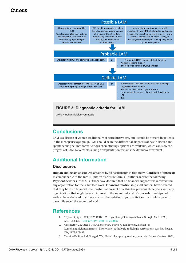
FIGURE 3: Diagnostic criteria for LAMLAM: lymphangioleiomyomatosis
ConclusionsLAM is a disease of women traditionally of reproductive age, but it could be present in patientsin the menopause age group. LAM should be in the differential diagnosis of cystic disease andspontaneous pneumothorax. Various chemotherapy options are available, which can slow theprogress of LAM. Nevertheless, lung transplantation remains the definitive treatment.
Additional InformationDisclosuresHuman subjects: Consent was obtained by all participants in this study. Conflicts of interest:In compliance with the ICMJE uniform disclosure form, all authors declare the following:Payment/services info: All authors have declared that no financial support was received fromany organization for the submitted work. Financial relationships: All authors have declaredthat they have no financial relationships at present or within the previous three years with anyorganizations that might have an interest in the submitted work. Other relationships: Allauthors have declared that there are no other relationships or activities that could appear tohave influenced the submitted work.
References1. Taylor JR, Ryu J, Colby TV, Raffin TA: Lymphangioleiomyomatosis. N Engl J Med. 1990,
323:1254-60. 10.1056/NEJM1990110132318072. Carrington CB, Cugell DW, Gaensler EA, Marks A, Redding RA, Schaaf JT:
Lymphangioleiomyomatosis. Physiologic-pathologic-radiologic correlations. Am Rev Respir.Dis, 1977:977-95.
3. Taveira-DaSilva AM, Steagall WK, Moss J: Lymphangioleiomyomatosis. Cancer Control. 2006,
2019 Rhee et al. Cureus 11(1): e3938. DOI 10.7759/cureus.3938 5 of 6
13:276-85. 10.1177/1073274806013004054. Yamazaki A, Miyamoto H, Futagawa T, et al.: An early case of pulmonary
lymphangioleiomyomatosis diagnosed by video-assisted thoracoscopic surgery. Ann ThoracCardiovasc Surg. 2005, 11:405-7.
5. Ryu JH, Moss J, Beck GJ, et al.: The NHLBI lymphangioleiomyomatosis registry: characteristicsof 230 patients at enrollment. Am J Respir Crit Care Med. 2006, 173:105-11.10.1164/rccm.200409-1298OC
6. Johnson SR: Lymphangioleiomyomatosis. Eur Respir J. 2006, 27:1056-65.10.1183/09031936.06.00113303
7. Kitaichi M, Nishimura K, Itoh H Izumi T: Pulmonary lymphangioleiomyomatosis: a report of46 patients including a clinicopathologic study of prognostic factors. Am J Respir Crit CareMed. 1995, 151:527-33. 10.1164/ajrccm.151.2.7842216
8. Chu SC, Horiba K, Usuki J, et al.: Comprehensive evaluation of 35 patients withlymphangioleiomyomatosis. Chest. 1999, 115:1041-52.
9. Steagall WK, Taveira-DaSilva AM, Moss J: Clinical and molecular insights intolymphangioleiomyomatosis. Sarcoidosis Vasc Diffuse Lung Dis. 2005, 22:49-66.
10. Bonetti F, Chiodera P.L, Pea M, Martignoni G, Bosi F, Zamboni G: Transbronchial biopsy inlymphangiomyomatosis of the lung. HMB45 for diagnosis. Am J Surg Pathol. 1993, 17:1092-102.
11. McCormack FX: Lymphangioleiomyomatosis: a clinical update . Chest. 2008, 133:507-16.10.1378/chest.07-0898
12. Logginidou H, Ao X, Russo I, Henske EP: Frequent estrogen and progesterone receptorimmunoreactivity in renal angiomyolipomas from women with pulmonarylymphangioleiomyomatosis. Chest. 2000, 117:25-30.
13. Brunelli A, Catalini G, Fianchini A: Pregnancy exacerbating unsuspected mediastinallymphangioleiomyomatosis and chylothorax. Int J Gynaecol Obstet. 1996, 52:289-90.
14. Taveira-DaSilva AM, Stylianou MP, Hedin CJ, Hathaway O, Moss J: Decline in lung function inpatients with lymphangioleiomyomatosis treated with or without progesterone. Chest. 2004,126:1867-74. 10.1378/chest.126.6.1867
15. Oprescu N, McCormack FX, Byrnes S, Kinder BW: Clinical predictors of mortality and cause ofdeath in lymphangioleiomyomatosis: a population-based registry. Lung. 2013, 191:35-42.10.1007/s00408-012-9419-3
16. Huang J, Manning BD: A complex interplay between Akt, TSC2 and the two mTOR complexes .Biochem Soc Trans. 2009, 37:217-22. 10.1042/BST0370217
17. Johnson SR: Lymphangioleiomyomatosis. Eur Respir J. 2006, 27:1056-65.10.1183/09031936.06.00113303
2019 Rhee et al. Cureus 11(1): e3938. DOI 10.7759/cureus.3938 6 of 6