andrew sarkodie appiah - university of tasmania€¦ · andrew s. appiah, frederick l. sossah,...
TRANSCRIPT

Studies on Groundnut rosette disease in Ghana and genomic
analysis of a novel Phasey bean virus in Australia
Andrew Sarkodie Appiah B. Sc. (Hons), M. Phil
Submitted in fulfilment of the requirements
For the degree of
Doctor of Philosophy
UNIVERSITY
OF TASMANIA
February, 2017

i
DECLARATTION OF ORIGINALITY AND AUTHORITY OF ACCESS
This is to certify that:
This thesis contains no material which has been accepted for a degree or diploma by the
University or any other institution, except by way of background information and duly
acknowledged in the thesis. To the best of my knowledge and belief, no material previously
published or written by another person, except where due acknowledgement is made in the
text of the thesis, nor does the thesis contain any material that infringes copyright.
The publishers of the papers (as indicated in next section) hold the copyright for that content,
and access to the material should be sought from the respective journals. The remaining non-
published content of the thesis is not to be made available for loan or copying for two years
following the date this statement was signed. Following that time, the remaining non-
published content of the thesis may be made available for loan and limited copying and
communication in accordance with the Copyright Act 1968.
The research does not contain any studies with human participants or animals performed by
any of the authors
Andrew Sarkodie Appiah
University of Tasmania
February, 2017

ii
ACKNOWLEDGEMENTS
I would like to express my sincere gratitude to my primary supervisor, A/Prof. Calum Wilson
(University of Tasmania) and Co-supervisors Dr Robert Tegg (University of Tasmania) and
Prof. Samuel Kwame Offei (University of Ghana) for their support, patience and insightful
comments which led to significant improvements of this thesis. I am so grateful to you for
steering me in the right direction to make this thesis a reality. I could not have imagined
having better advisors and mentors for my PhD study.
My sincere thanks also go to Mrs Annabel Wilson, Dr Tamil Thangavel (Tasmanian Institute
of Agriculture) Dr Alison Dann, Mr Shane Hossel and Mr Peter Cross (Department of
Primary Industries, Parks, Water and Environment) for their immense contribution towards
the execution of the research. I am also grateful to Dr Samuel Amiteye, Mr Jonathan
Amponsah and Mr Iddriss Mohammed (BNARI, Ghana Atomic Energy Commission) for
their assistance with field work conducted in Ghana. I thank my fellow lab mates at the New
Town research Laboratories (Mark Balendres, Kritika Krishnamorthy and Sabine Tanois) for
the stimulating discussions, and for sharing our coffee breaks and lunch times together.
I am grateful to the Australian Government for the AusAID scholarship. This has been a great
opportunity for me to further my education. The generous offer has adequately empowered
me to contribute my quota to the development of Agriculture in Ghana. I am also thankful to
the Ghana Atomic Energy Commission for granting me study leave with pay and for
supporting my field work which was conducted in Ghana.
I would like to express my profound gratitude to my wife (Vivian), children (Carlis, Thelma
and Caitlyn) for their unfailing support, continuous encouragement and prayers throughout
my years of study. My sincere thanks also go to my parents, brothers and sisters for their
support and prayers.
Finally, I would like to thank the Almighty God for His protection and guidance. Without
Him, this accomplishment would not have been possible.

iii
STATEMENT OF CO-AUTHORSHIP AND CONTRIBUTION TO
PUBLICATIONS
The following people and institutions contributed to the publication of work undertaken as
part of this thesis:
1. Andrew Sarkodie Appiah Tasmanian Institute of Agriculture, UTAS, Australia
BNARI, Ghana Atomic Energy Commission, Accra
2. Samuel Kwame Offei College of Basic and Applied Sci., University of Ghana, Accra
3. Robert Stevee Tegg Tasmanian Institute of Agriculture, UTAS, Australia
4. Frederick Leo Sossah Jilin Agricultural University, College of Agronomy, China
5. Murray Sharman Department of Agriculture & Fisheries, Brisbane, QLD,
Australia
6. Calum Rae Wilson Tasmanian Institute of Agriculture, UTAS, Australia
Paper 1: Appiah, A. S., Offei, S. K., Tegg, R. S., and Wilson, C. R. (2016). Impact of
groundnut rosette disease on nutritive value and elemental composition of four varieties of
peanut (Arachis hypogaea). Annals of Applied Biology 168: 400 – 408 (Chapter 3).
Paper 2: Appiah, A. S., Offei, S. K., Tegg, R. S., and Wilson, C. R. (2016). Varietal response
to groundnut rosette disease and the first report of Groundnut ringspot virus in Ghana. Plant
Disease 100:946-952 (Chapter 4)
Paper 3: Andrew S. Appiah, Frederick L. Sossah, Robert S. Tegg, Samuel K. Offei, Calum R.
Wilson (2017). Assessing sequence diversity of Groundnut rosette disease agents and the
distribution of Groundnut rosette assistor virus in major groundnut-producing regions of
Ghana. Tropical Plant Pathology 42:109–120 (Chapter 5)

iv
Paper 4: A.S. Appiah, R.S. Tegg, M. Sharman and C.R. Wilson (2017). Host range,
complete genome sequencing and molecular phylogeny of a novel Polerovirus from
Australian legumes. Manuscript under preparation. (Chapter 6)
Conference presentations
Appiah, A.S., Tegg, R.S., Wilson, C.R. Studies on Groundnut rosette disease and other
legume viruses. Africa Australia Research Forum on Mining, agriculture and development:
Bread from Stones? A joint conference of The Crawford Fund and the Africa Australia
Research Forum. 25th - 28th August 2013, Perth, Australia.
Appiah, A.S., Tegg, R.S., Wilson, C.R. Host range, genome sequencing and molecular
phylogeny of a novel Polerovirus from Australian legumes. Australasian Plant Pathology
Society (Tasmanian Division) 2016 Symposium, 29th April, 2016, Ross, Tasmania, Australia.
Andrew S. Appiah, Robert Tegg, Samuel K. Offei, Calum R. Wilson. Studies on Groundnut
rosette disease and implications of the newly reported Groundnut ringspot virus for
groundnut production in Ghana. American Phytopathological Society’s Annual Conference
‘Science to practice’, 30th July to 3rd August, 2016, Tampa, Florida, USA
Candidate was the primary author of all papers, contributed to the conception and design of
the study and executed field and laboratory work. Candidate also contributed to the analysis
and interpretation of data and drafted significant portions of the paper, revision and final
approval of all articles.
Robert Tegg contributed to the design of the experiment, guided statistical analysis and
interpretation of data and revision and final approval of the manuscripts.
Samuel Offei contributed to the conception of of the research, revision and final approval of
the manuscript.

v
Calum Wilson contributed to the conception and design of the research project, guided
statistical analysis and interpretation of results, provided technical support, revision and final
approval of all manuscripts.
Frederick Leo Sossah assisted with data collection and analyis and the final approval of the
manuscript for Chapter five.
Murray Sharman provided the Queensland isolate of the PhBMYV and the Aphis craccivora
vector used in the study (Chapter six) and a contributed to the revision and final approval of
the manuscript.

vi
Declaration of Agreement
We the undersigned agree with the above stated “proportion of work undertaken” for each of
the above published (or submitted) peer-reviewed manuscripts contributing to this thesis:
___________________ ______________________
Andrew Sarkodie Appiah Calum Rae Wilson
Candidate Supervisor
School of Land and Food School of Land and Food
University of Tasmania University of Tasmania
Date: February 24, 2017 Date: February 24, 2017
__________________ _____________________
Robert Steven Tegg Samuel Kwame Offei
Co-Supervisor Co-Supervisor
School of Land and Food Biotechnology Centre
University of Tasmania College of Basic and Applied Sciences
Date: February 24, 2017 University of Ghana, Legon
Date: February 24, 2017

vii
ABSTRACT
Viruses present a major challenge to the production of major food crops worldwide, including
legumes. The diseases they cause have profound effects on both plant growth and the quality
of produce, resulting in significant losses. The current study investigated the proximate and
elemental composition of four groundnut (peanut, Arachis hypogaea L.) cultivars infected
with groundnut rosette disease (GRD), screened local cultivars of groundnut for resistance to
GRD, detected Groundnut ringspot virus for the first time in Ghana and assessed the genetic
diversity within Ghanaian isolates of Groundnut rosette assistor virus (GRAV), Groundnut
rosette virus (GRV) and satellite RNA of GRV and compare those with known isolates from
other African countires. In a related study, the complete genome of an isolate of novel virus
infecting Phasey bean (Macroptilium lathyroides L.) in Australia; Phasey bean mild yellows
virus (PhBMYV) was sequenced with evidence of genomic recombination found and its
transmission to other legumes demonstrated.
Proximate analysis of seeds from GRD-infected groundnuts showed a decrease in
moisture and ash content, while fat and energy content increased. Protein and carbohydrate
content varied inconsistently between seeds of diseased and healthy plants of the different
cultivars. Instrumental neutron activation analysis (INAA) of ten elements within leaves,
stems and seeds revealed elevated levels of K, Al and Cl in leaves, stems and seeds in at least
three of the four GRD-infected cultivars while Na was decreased in stems but increased in
seeds. Despite significant differences, Mg, Mn, Ca and Zn did not show any consistent
change with respect to plant part or genotype, between diseased and healthy plants. V and Fe
were not detected in seeds but were found at low levels in leaves and stems. This work has
been published in Annals of Applied Biology (2015) and represents the first report on the
effect of GRD on the nutritive quality of groundnuts.

viii
Twelve cultivars of groundnut were screened in field trials for resistance to GRD in
the coastal savannah agro-ecological zone of Ghana. Cultivar ‘Oboshie’ was rated as highly
resistant; ‘Bremaowuo’, ‘Nkatefufuo’, and ‘Behenase’ as resistant; and ‘Nkosuor’,
‘Kumawu’, and ‘Otuhia’ as moderately resistant. GRAV infection rates of 11.8 to 61.8% and
13.9 to 100% were found within the field trial for dry and wet seasons respectively. These
included symptomless plants suggesting that some lacked co-infection with GRV and its
satellite RNA which are responsible for symptom induction. Some plants exhibited chlorotic
and line pattern symptoms suggestive of Groundnut ringspot virus (GRSV) infection, which
was confirmed by enzyme-linked immunosorbent assay, reverse-transcription polymerase
chain reaction, and amplicon sequencing. This represents the first report of GRSV in Ghana.
GRSV infection rates within the field trial were 0.0 to 69.5% (dry season) and 26.1 to 69.5%
(wet season) and was commonly found in mixed infections with GRAV in all cultivars except
Nkosuor and Bremaowuo in the dry season. Graft-inoculated groundnut cultivars showed
significantly reduced height, leaf area, chlorophyll content, dry haulm weight, and seed yield
compared to healthy plants. The sources of resistance to GRV and possibly GRAV and
GRSV identified in this study could be exploited in groundnut breeding programs. This work
has been published in Plant Disease (2015).
GRAV incidence in farmers’ fields was assessed through crop surveys in the three
northern groundnut-producing regions of Ghana. High (69.5 to 75.0%) but insignificantly
different incidences were found between the regions. Isolates of GRAV, GRV and sat RNA
collected during the survey were sequenced. There were no obvious isolate diversity patterns
among the Ghanaian isolates of all three agents of GRD based on the regions from where
they were collected. Nucleotide sequences of the coat protein gene of GRAV showed 99-100%
identity among the Ghanaian isolates and 97-100% similarity to GRAV sequences from
Nigeria and Malawi for both nucleotide and predicted amino acids. Ghanaian GRV isolates

ix
were closer in nucleotide sequence identity to Nigerian isolates (95 - 98%) than Malawian
isolates (88 - 90%). Similarly, Ghanaian satRNA isolates shared close nucleotide identities
(94-100%), but were distinct from Nigerian (82 - 87%) and Malawian (82 - 86%) isolates.
This work has been accepted for publication in Tropical Plant Pathology (2017) and presents
the first report on the distribution and genetic diversity of GRD agents in Ghana.
The complete genome of a QLD isolate of the novel PhBMYV was determined. The
genome consisted of six open reading frames (ORFs) typical of Poleroviruses, with their
respective putative proteins closely related to two previously reported PhBMYV isolates
from New South Wales (NSW) and Western Australia (WA), except within the RNA-
dependent-RNA-polymerase (RdRp) and Coat protein-Read through (CP-RT). The RdRp
only shared ~63% amino acid identity with the NSW and WA isolates and the CP-RT was
distinct (33 – 34% amino acid identity with other PhBMYV isolates) and shared 53% identity
with Chickpea chlorotic stunt virus (CpCSV). Recombination analysis using RDP4 suggested
the QLD isolate was an evolutionary product of recombination between the NSW (minor
parent) and WA (major parent) isolates. The virus was successfully transmitted from Phasey
bean to pea (Pisum sativum), chickpea (Cicer arietinum) and to Phasey bean plants using
both vector and graft transmission methods. Based on the results of this study, PhBMYV
QLDCL 16 is suggested as a genetic variant of PhBMYV and perhaps represents a distinct
species.

x
TABLE OF CONTENTS
DECLARATTION OF ORIGINALITY AND AUTHORITY OF ACCESS ................................... i
ACKNOWLEDGEMENTS ................................................................................................................. ii
STATEMENT OF CO-AUTHORSHIP AND CONTRIBUTION TO PUBLICATIONS ............ iii
ABSTRACT ......................................................................................................................................... vii
TABLE OF CONTENTS ..................................................................................................................... x
LIST OF FIGURES ........................................................................................................................... xiv
LIST OF TABLES ............................................................................................................................ xvii
1 CHAPTER ONE: GENERAL INTRODUCTION .................................................................... 1
1.1.1 Groundnut significance and production .......................................................................... 1
1.2 Groundnut rosette disease ....................................................................................................... 2
1.3 Viral diseases of Legumes in Australia ................................................................................... 3
1.3.1 Phasey bean mild yellows viral disease .......................................................................... 4
1.4 Research Objectives ................................................................................................................ 5
1.5 Overview of thesis content ...................................................................................................... 6
2 CHAPTER TWO: LITERATURE REVIEW ............................................................................ 9
2.1 Groundnut: origin, distribution, production and uses ............................................................. 9
2.2 Diseases and Pests of groundnut ........................................................................................... 11
2.2.1 Groundnut rosette .......................................................................................................... 12
2.3 Other viruses of Groundnut .................................................................................................. 20
2.4 Host resistance to GRD ......................................................................................................... 21
2.5 Current management strategies for GRD and the way forward ............................................ 22
2.6 Family Luteoviridae .............................................................................................................. 24
2.6.1 Host range Luteoviruses ................................................................................................ 26
2.6.2 Vector Association and Transmission ........................................................................... 26
2.7 Genome Organization of Luteoviruses ................................................................................. 27
2.7.1 The Polerovirus Genome .............................................................................................. 28
2.7.2 Poleroviruses of legumes .............................................................................................. 30
3 CHAPTER THREE: Impact of groundnut rosette disease on nutritive value and elemental
composition of four varieties of peanut (Arachis hypogaea) ............................................................ 32
3.1 Abstract ................................................................................................................................. 32
3.2 Introduction ........................................................................................................................... 33
3.3 Materials and methods .......................................................................................................... 34

xi
3.3.1 Plant material ................................................................................................................ 34
3.3.2 Graft inoculation ........................................................................................................... 35
3.3.3 Proximate analysis ........................................................................................................ 36
3.3.4 Elemental composition analysis using instrumental neutron activation analysis .......... 36
3.4 Results ................................................................................................................................... 38
3.5 Discussion ............................................................................................................................. 46
3.6 Acknowledgements ............................................................................................................... 50
4 CHAPTER FOUR: Varietal Response to Groundnut Rosette Disease and the First Report
of Groundnut ringspot virus in Ghana ............................................................................................. 51
4.1 Abstract ................................................................................................................................. 51
4.2 Introduction ........................................................................................................................... 52
4.3 Materials and Methods .......................................................................................................... 54
4.3.1 Study site ....................................................................................................................... 54
4.3.2 Groundnut cultivars ...................................................................................................... 54
4.3.3 Trial design ................................................................................................................... 55
4.3.4 Estimation of disease incidence and cultivar rating ...................................................... 55
4.3.5 Serological detection of viruses .................................................................................... 56
4.3.6 RNA isolation, RT-PCR and sequencing of the GRSV nucleocapsid gene .................. 56
4.3.7 Effect of GRD on agronomic performance ................................................................... 57
4.3.8 Data analysis ................................................................................................................. 58
4.4 Results ................................................................................................................................... 59
4.4.1 Disease incidence and cultivar rating to GRD .............................................................. 59
4.4.2 Serological detection of viruses .................................................................................... 61
4.4.3 Effect of GRD on agronomic performance ................................................................... 63
4.5 Discussion ............................................................................................................................. 69
4.6 Acknowledgments ................................................................................................................. 73
5 CHAPTER FIVE: Assessing sequence diversity of Groundnut rosette disease agents and
the distribution of Groundnut rosette assistor virus in major groundnut-producing regions of
Ghana ................................................................................................................................................... 74
5.1 Abstract ................................................................................................................................. 74
5.2 Introduction ........................................................................................................................... 75
5.3 Materials and Methods .......................................................................................................... 77
5.3.1 Field survey and collection of viral isolates ......................................................................... 77
5.3.1 Nitrocellulose membrane enzyme-linked immunosorbent assay (NCM-ELISA) ........ 78
5.3.2 RNA isolation ............................................................................................................... 78

xii
5.4 Results ................................................................................................................................... 81
5.4.1 Field survey ................................................................................................................... 81
5.4.2 Serological detection of GRAV .................................................................................... 83
5.4.3 RT-PCR detection of GRAV, GRV and GRV-satRNA ................................................ 85
5.5 Discussion ............................................................................................................................. 95
5.6 Acknowledgements ............................................................................................................... 99
5.7 Compliance with ethical Standards ....................................................................................... 99
6 CHAPTER SIX: Genomic analysis and transmission of recombinant Phasey bean mild
yellows virus isolates from Queensland and Tasmania ................................................................. 100
6.1 Abstract ............................................................................................................................... 101
6.2 Introduction ......................................................................................................................... 102
6.3 Materials and Methods ........................................................................................................ 104
6.3.1 Transmission studies using Queensland isolate of PhMYV ....................................... 104
6.3.2 RNA isolation and Reverse-transcription polymerase chain reaction (RT-PCR). ...... 105
6.3.3 Next-generation sequencing (NGS) of QLD isolate of PhBMYV .............................. 105
6.4 Genome sequencing of Tasmanian isolate of Phasey bean mild yellows virus .................. 107
6.5 Genome analysis ................................................................................................................. 107
6.5.1 Phylogenetic analysis .................................................................................................. 108
6.6 Results ................................................................................................................................. 110
6.6.1 Virus transmission studies .......................................................................................... 110
6.6.2 Full length genome analyses of PhBMYV isolate QLD CL 16 .................................. 111
6.6.3 Analysis of partial sequence of Tasmanian isolate of PhBMYV ................................ 113
6.6.4 Phylogenetic analysis .................................................................................................. 114
6.7 Recombination analysis ...................................................................................................... 119
6.8 Discussion ........................................................................................................................... 120
6.9 Acknowledgement .............................................................................................................. 124
7 CHAPTER SEVEN: Discussion, conclusion and future studies ........................................... 125
7.1 General discussion .................................................................................................................... 125
7.1.1 Effect of GRD on proximate and elemental composition groundnuts .............................. 126
7.1.2 Screening for resistance to GRD and first report of GRSV in Ghana ................................ 127
7.1.3 GRAV incidence and genetic diversity within Ghanaian GRD agents .............................. 128
7.1. 4 Genomic analysis and molecular phylogeny of Queensland and Tasmanian isolatyes of
Phasey bean mild yellows virus .................................................................................................. 129
7.2 Conclusion ................................................................................................................................ 131
7.3 Future research .......................................................................................................................... 132

xiii
References .......................................................................................................................................... 134

xiv
LIST OF FIGURES
Figure 1.1 Regional distribution of groundnut production in Ghana (Angelucci and
Bazzucchi, 2013) ................................................................................................................ 2
Figure 1.2 Australian pulse production (Pulse Market Insight, 2016). ..................................... 4
Figure 2.1 Worldwide distribution of groundnut, the groundnut rosette disease and its aphid
vector (Naidu et al., 1999b). ............................................................................................. 13
Figure 2.2 Peanut plants showing typical symptoms of GRD A. chlorotic rosette and B.
green rosette ...................................................................................................................... 14
Figure 2.3 Interaction of GRV, GRAV and sGRV in groundnut rosette disease development
and vector transmission. ................................................................................................... 15
Figure 2.4 The genome of GRV showing the different expression strategies: production of
subgenomic RNA(s) (sgRNA(s), initiation of translation at the first optimal AUG on the
genomic RNA (gRNA; ORF1) or sgRNA(s) (ORF3 and ORF4)), and initiation of
translation as for ORF1 and frameshift (ORF1+ORF2). Lines represent RNA molecules,
grey boxes represent open reading frames and black boxes represent translation products
(Taliansky et al., 2003a). ................................................................................................. 18
Figure 2.5 Genomes of family Luteoviridae compared A. PLRV (genus Polerovirus) and B.
BYDV-PAV (genus Luteovirus) (Viral Zone, Swiss Institute of Bioinformatics). ......... 29
Figure 3.1 (A) Healthy and Groundnut rosette diseased peanut plants of the variety
Sinkapoporigo. (B) Close-up of Groundnut rosette diseased peanut plant ...................... 39
Figure 4.1 Diseased groundnut plants of variety Sinkapoporigo within the field eight weeks
after exposure to sources of inoculum A: green rosette symptoms of GRD B: chlorotic
ringspots, leaf deformation and line pattern symptoms of GRSV infection C: healthy
plant .................................................................................................................................. 59
Figure 4.2 Extent of co-infection of groundnut varieties within trial plots with Groundnut
rosette assistor virus (GRAV) and Groundnut ringspot virus (GRSV). Vertical bars
indicate standard errors. Plants were tested 10 weeks after exposure to sources of
inoculum. .......................................................................................................................... 63
Figure 4.3 Example of the effect of GRD on yield of groundnut pods in variety
Sinkapoporigo ................................................................................................................... 65

xv
Figure 5.1 A - GRD-affected field plant showing green rosette symptoms B - GRD-affected
plant showing chlorotic rosette symptoms and C – Diseased plant with numerous pegs
and unfulfilled pods D: Healthy plant with several pods. ................................................ 82
Figure 5.2 Map of Ghana showing sampling sites (highlighted in red). ................................. 83
Figure 5.3 Amplification of GRAV, GRV and GRV-sat RNA using specific primers; Lane M
– 50 bp DNA ladder (Bioline), Lanes 1 – 4 (GRAV5/GRAV3), lanes 5 – 8 (S3/C3 for
GRV) and lanes 9 - 12 (Sat505F/Sat505R). ..................................................................... 86
Figure 5.4 Phylogenetic tree (Neighbor-joining) of ~586 nt fragment of Groundnut rosette
assistor virus CP gene from Ghana (GhN = Northern region, GhE = Upper East region,
GhW = Upper West region), Nigeria (N) and Malawi (M). Numbers at the nodes denotes
the percentage of 1000 bootstraps iterations supporting the branches. Nodes with <70 %
were collapsed. Potato leafroll virus sequence was used as outgroup.............................. 87
Figure 5.5 Phylogenetic tree (Neighbor-joining) of ~693 nt fragment of Groundnut rosette
virus overlapping ORF3 and 4 gene from Ghana (GhN = Northern region, GhE = Upper
East region, GhW = Upper West region), Nigeria (N) and Malawi (M). Numbers at the
nodes denotes the percentage of 1000 bootstraps iterations supporting the branches.
Nodes with <70 % were collapsed. GVRP is a Malawian isolate of Taliansky et al.
(1996). Carrot mottle virus sequence was used as outgroup. ........................................... 89
Figure 5.6 Phylogenetic tree (Neighbor-joining) of deduced amino acid (aa) sequences of
Groundnut rosette virus ORF3 (262 aa) (Top) and ORF 4 (241 aa) (Bottom) from
Ghanaian (GhN = Northern region, GhE = Upper East region, GhW = Upper West
region), Nigerian (N) and Malawian (M) isolates. Numbers at the nodes denotes the
percentage of 1000 bootstraps iterations supporting the branches. Nodes with <70 %
were collapsed. Carrot mottle virus sequence was used as outgroup. .............................. 92
Figure 5.7 Phylogenetic analysis of the nucleotide sequences of GRV-sat RNA isolates from
Ghana (GhN = Northern region, GhE = Upper East region, GhW = Upper West region),
Nigeria (N) and Malawi (M). The tree was generated by Neighbor-Joining method in
Geneious. Numbers at the nodes represent boostrap values determined from 1000
replicates. Nodes with <70 % were collapsed. Pea enation mosaic virus (PEMV)-sat
RNA sequence was used as outgroup. .............................................................................. 94
Figure 6.1 Phasey bean plants; A: PhBMYV-inoculated plant showing stunting, yellowing
and reduced leaf size and B: Uninoculated Phasey bean plant....................................... 110

xvi
Figure 6.2 Phasey bean mild yellows virus-inoculated plants: A. Healthy pea B. Aphid-
inoculated pea C. Graft-inoculated pea D. Healthy chickpea E. Aphid-inoculated
chickpea. ......................................................................................................................... 111
Figure 6.3 Schematic representation of the genome organisation of PhBMYV QLD isolate.
The sizes of the deduced gene products are written either above or below the name of the
ORF. ............................................................................................................................... 112
Figure 6.4 Phylogenetic relationship between A. PhBMYV QLDCL 16 and other members
of the family Luteoviridae. Trees were based on the complete genome sequences and
distances constructed using the neighbour-joining method. B. PhBMYV isolates from
Australia. Figures at the nodes indicate the frequency of the cluster after bootstrap
analysis (1000 replicates). Nodes with values <50% were collapsed. ........................... 116
Figure 6.5 Phylogenetic analysis based on the predicted amino acid sequences A. P0, B.
P1+P2, C. P3, D. P4 and E. P3 + P5. Distance trees were constructed using the
neighbour-joining method. Figures at the nodes indicate the frequency of the cluster after
bootstrap analysis (100 replicates; shown only when >50%)......................................... 119
Figure 6.6 A graph showing the result of recombination analysis of aligned Polerovirus
genome sequences using the RDP method. The complete nucleotide sequence of the
Queensland isolate of Phasey bean mild yellows virus (potential recombinant) was
compared with those of Phasey bean mild yellows virus isolates ESPCL 15 (major parent)
and NSWCP 15 (minor parent). ..................................................................................... 120
xvii
LIST OF TABLES
Table 2.1 Established members of Family Luteoviridae (ICTV, 2013).................................. 24
Table 3.1 Proximate analysis of GRD-infected peanut seeds ................................................. 40
Table 3.2 Elemental compositions of leaves of four varieties of Groundnut rosette disease-
infected and healthy peanuts............................................................................................. 42
Table 3.3 Elemental composition of stems of four varieties of Groundnut rosette disease-
infected and healthy peanuts............................................................................................. 43
Table 3.4 Elemental composition of seeds of four varieties of Groundnut rosette disease-
infected and healthy peanuts............................................................................................. 44
Table 4.1 Groundnut rosette disease incidence and resistance rating of 12 groundnut varieties
based on visual symptoms. ............................................................................................... 60
Table 4.2 Infection of 12 groundnut varieties by Groundnut rosette assistor virus (GRAV)
and Groundnut ringspot virus (GRSV) ............................................................................ 61
Table 4.3 Effect of GRD on plant height, leaf area, chlorophyll content, haulm dry weight
and yield of groundnut cultivars ....................................................................................... 66
Table 5.1 Oligonucleotide primers used for the amplification of GRAV, GRV and the
SatRNA ............................................................................................................................. 80
Table 5.2 GRAV incidence in major groundnut-growing regions of Ghana .......................... 84
Table 6.1 Oligonucleotide primers used for detection and sequencing of the virus genome 107
Table 6.2 List of Luteovirus sequences retrieved from the GenBank for this study ............. 108
Table 6.3 Comparison of deduced amino acid sequences for major open reading frames for
the Queensland isolate of PhBMYV and some members of the family Luteoviridae ... 114

1
1 CHAPTER ONE: GENERAL INTRODUCTION
1.1.1 Groundnut significance and production
Groundnut (Peanut, Arachis hypogaea) is a crop of global importance, widely grown in
tropical and subtropical regions, by small holder and commercial producers, and is a food
staple and valuable cash crop for millions of households. The seeds are a rich source of
protein (22–30%), oil (44–56%), total carbohydrates (9.5–19.0%), minerals (phosphorus,
calcium, magnesium and potassium) and vitamins (E, K and B group). It is ranked as the
world’s 13th
most important food crop, 6th most important source of edible oil and 3rd
most
important source of vegetable protein (Waliyar et al., 2007). In Ghana, groundnuts play an
important dietary role where they provide high-quality cooking oil and are an important
source of protein for both humans and animals. World groundnut production is approximately
42 million tonnes per year, with China, India Nigeria and the USA being the world’s largest
producers (FAO, 2014). Developing countries account for 97% of the global groundnut area
and 94% of the global production with production concentrated in Asia and Africa (Waliyar
et al., 2007). Sub-Saharan Africa (SSA) cultivates 40% of the world’s groundnut harvested
area, yet contributes only 26% of the world’s groundnut production (Angelucci and
Bazzucchi, 2013). This could be attributed partly to diseases of which Groundnut rosette
disease is important. Ghana is ranked as the 8th
largest producer of groundnuts in the world
with an annual production of 0.4 million metric tonnes (USDA, 2013). The crop is grown in
almost all agro-ecological zones of the country but the three northern regions (Northern,
Upper East and Upper West) account for over 70% of production (Fig. 1.1) (Tsigbey et al.,
2003). Despite the enormous potential of the crop to meet the nutritional needs of millions of
people in SSA, its production has been constrained by several biotic and abiotic factors of
which diseases are of significant importance.

2
Figure 1.1 Regional distribution of groundnut production in Ghana (Angelucci and
Bazzucchi, 2013)
1.2 Groundnut rosette disease
Although several diseases have been reported in groundnut in SSA, groundnut rosette disease
(GRD) has been identified as the most devastating, responsible for annual yield losses of over
US$150 million (Waliyar et al., 2007). The disease is caused by a multi-infection complex of
viral agents; Groundnut rosette assistor virus (GRAV): genus Luteovirus, Groundnut rosette
virus (GRV): genus Umbravirus and its satellite RNA (SatRNA). The disease is transmitted
from plant to plant by the aphid vector Aphis craccivora Koch (Okusanya and Watson, 1966)
and induces either green rosette, chlorotic rosette (Gibbons, 1977; Murant and Kumar, 1990)
or mosaic rosette symptoms in infected plants (Storey and Ryland, 1957). The major
countries where the disease has been identified include Burkina Faso, Ghana, Nigeria,
Malawi, Mozambique, Uganda (Olorunju et al., 2001), Angola, Cˆote d’Ivoire, Gambia,
Kenya, Madagascar, Niger, Senegal, South Africa, Sudan, Swaziland, Tanzania and Zaire
(Gibbons 1977; Naidu et al., 1999a). In Ghana, the disease occurs across all growing regions
often resulting in total crop failure (Chapter 4 Appiah et al., 2016). GRD epidemics in
northern Nigeria in 1975 (Yayock et al., 1976) and Zambia resulted in the destruction of

3
about 0.75 million ha and 43,000 ha of groundnut respectively while groundnut production
was reduced by 23% in the central region of Malawi (Anon., 1996).
In Ghana, the average yields of 500 to 800 kg/ha are far below the potential yields of 1,800 to
2,800 kg/ha (Adu-Dapaah et al., 2004). This is at least partially due to GRD, which is not
adequately managed and other unidentified viral diseases. In a recent study, Groundnut
ringspot virus (GRSV) which is regarded as an emerging threat to crop production in several
important crops, was identified as infecting groundnuts in Ghana (Chapter 4, Appiah et al.,
2016). Viral diseases, besides reducing yield, may also affect the nutritive quality of the
produce. The mosaic symptoms, and/or necrotic lesions induced by systemic viral infection
may indicate structural changes in the chloroplasts, altered carbon metabolism and the
accumulation of starch grains. Plant diseases also have the potential of altering membrane
permeability in plants resulting in impaired mineral uptake (Huber and Graham, 1999).
1.3 Viral diseases of Legumes in Australia
Australia is among the world’s largest exporters of legumes (pulses), including lentils (Lens
culinaris), chickpeas (Cicer arietinum), faba beans (Vicia faba L.) and field peas (Fig. 1.2).
The legume industry currently contributes about AUD$1.6 billion per year to the Australian
economy (Australian Food News, 2016). Diseases caused by viruses have been identified as
one of the major constraints to legume crops and pastures production worldwide (Bos et al.,
1988; Edwardson and Christie, 1991). In Australia, several different virus species have been
reported as infecting legumes (Latham and Jones, 2001a; Schwinghamer and Schilg, 2003;
Thomas et al., 2004), the most common being Alfalfa mosaic virus (Bromoviridae, genus
Alfamovirus) (AMV), Cucumber mosaic virus, (Bromoviridae, genus Cucumovirus) (CMV),
Bean yellow mosaic virus and Pea seedborne mosaic virus (Potyviridae, genus Potyvirus)
(BYMV and PSbMV) (Jones and Coutts, 1996). Viruses belonging to the Luteovirus family

4
have also been reported as infecting legumes in Australia. These include Bean leaf roll virus
(genus Luteovirus) (BLRV), Beet western yellows virus (genus Polerovirus) (BWYV)
(Schwinghamer et al., 1999; Latham and Jones, 2001a), Soybean dwarf virus (Wright and
Jones, 2003) and more recently Phasey bean mild yellows virus (PhBMYV) (Sharman et al.,
2016).
1.3.1 Phasey bean mild yellows viral disease
Phasey bean mild yellows virus (PhBMYV) is a novel Polerovirus that infect legumes in
Australia. The virus was first identified as infecting peas in Tasmania (Wilson et al., 2012)
and Phasey bean in Queensland (QLD) and subsequently in Western Australia (WA) and
New South Wales (NSW) as infecting Trifolium subterraneum (subterranean clover) and
Cicer arietinum (chickpea) respectively (Sharman et al., 2016). The virus induces mild
yellowing symptoms in the infected Phasey bean (Sharman et al., 2016) and symptomless
infection in peas (Wilson et al., 2012). Poleroviruses have high genetic diversity and
Figure 1.2 Australian pulse production (Pulse Market Insight, 2016).

5
evidence for genomic recombination is common. They are particularly associated with the
emergence of new viral diseases worldwide (Lotos et al., 2016), and are responsible for
major yield losses in vegetable and arable crops. Thus, the identification of this novel virus in
Australian legumes should be of great concern. Other new Poleroviruses have also been
recently reported in legumes elsewhere (Abraham et al., 2006; Zhou et al., 2012; Abraham et
al., 2008).
1.4 Research Objectives
The low yields of groundnut usually obtained in Ghana and other countries in SSA could be
partly attributed to GRD (Naidu et al., 1999a; Waliyar et al., 2007). Cultural methods
involving delaying planting to avoid the insect vectors, removal of volunteer plants, rogueing,
and chemical spray have been used over the years (Naidu et al., 1999b) but these have not
provided adequate control as the disease still persists causing severe losses. Although several
rosette-resistant varieties have been released, these are only partially resistant to GRV and the
satRNA and not GRAV (Olorunju et al., 2001). Thus more effort is needed to screen
additional sources of germplasm especially in Ghana to identify more sources of resistance.
Information such as virus distribution and isolate variability are of paramount importance to
disease management and efficient breeding programmes. Additionally, information on the
effect of the disease on the quality of groundnuts is lacking. Although several researchers
have reported GRD (Adu-Dapaah et al., 2004; Olorunju et al., 2001; Gibbons, 1977; Naidu et
al., 1999a) there is limited or no information on the effect of the disease on the quality of the
produce.
In Australia, legumes play an important role as food for humans, feed for animals and in the
export sector (Australia Food News, 2016). Farmers are increasingly incorporating pulse
crops into their farming systems due to their ability to fix nitrogen and improve soil fertility.

6
In the year 2015, a total of 2.2 million tonnes of grain worth A$1.2 billion in exports was
produced from 1.8 million ha of pulse crops. This level of production is envisaged to increase
to 4.2 million tonnes with a commodity value of A$1.504 billion and a farm benefit of A$538
million assuming all constraints are overcome (Pulse Australia, 2016). The identification of a
novel Polerovirus infecting diverse Australian legume crops and weeds is therefore a concern
and necessitates studies which will enable informed decisions to be made regarding control
strategies for the management of the disease.
This thesis will:
(i) Determine the distribution of Groundnut rosette assistor virus in peanut growing
regions of Ghana through surveys.
(ii) Screen local germplasm for additional sources of resistance to GRD.
(iii) Determine the impact of the GRD on nutritive value of groundnuts.
(iv) Determine the genetic diversity of the three Groundnut rosette disease viral agents
in Ghana.
(v) Attempt to identify the host range of the novel Phasey bean mild yellows
(PhBMYV) virus.
(vi) Generate genomic sequences of the PhBMYV isolate from Queensland and
Tasmania using Next Generation and Sanger sequencing techniques.
(vii) Develop virus-specific primers for its detection.
The outcome of this research is believed to serve as a guide for formulating and
implementing control measures for the management of both viral diseases.
1.5 Overview of thesis content
This thesis has been prepared using the PhD thesis by publication option. As such, it is a
collection of published, accepted or ready-to-be-ubmitted papers. It is important to note that

7
some of the content, including literature review, general introduction and methods may
partially overlap between chapters.
Chapter 1: provides a brief background, the extent of the problem, research area with
specific objectives and aims of the research. It also identifies some research gaps that
need attention and shows an outline of the thesis structure.
Chapter 2: reviews important literature on Groundnut rosette disease in sub-Saharan
Africa and viruses of legumes in Australia. It highlights current production levels of
groundnut and pulses in SSA and Australia respectively, and identifies viral diseases as
one of major constraints to production. It identifies efforts made in combating viral
diseases of peanuts and research gaps that need to be addressed. It also highlights the
emergence of a novel Polerovirus as a threat to legume production in Australia and the
need to devise control measures.
Chapter 3: is an experimental chapter that focusses on the impact of groundnut rosette
disease on the nutritive value and elemental composition of peanuts (Arachis hypogaea)
Reference: Appiah, A. S., Offei, S. K., Tegg, R. S., and Wilson, C. R. (2016). Impact of
groundnut rosette disease on nutritive value and elemental composition of four varieties
of peanut (Arachis hypogaea). Annals of Applied Biology 168: 400 – 408.
Chapter 4: is an experimental chapter that focusses on screening of local Ghanaian
groundnut cultivars for resistance to GRD. This chapter also reports the presence of
Groundnut ringspot virus (GRSV) for the first time in Ghanaian groundnuts.
8
Reference: Appiah, A. S., Offei, S. K., Tegg, R. S., and Wilson, C. R. (2016). Varietal
response to groundnut rosette disease and the first report of Groundnut ringspot virus in
Ghana. Plant Disease 100:946-952.
Chapter 5: is an experimental chapter focusses on assessing the genetic diversity of
Groundnut rosette disease agents and the distribution of Groundnut rosette assistor virus
in major groundnut-producing regions of Ghana
Reference: Andrew S. Appiah, Frederick L. Sossah, Robert S. Tegg, Samuel K. Offei,
Calum R. Wilson (2017).Assessing sequence diversity of Groundnut rosette disease
agents and the distribution of Groundnut rosette assistor virus in major groundnut-
producing regions of Ghana. Tropical Plant Pathology, 42:109–120.
Chapter 6: an experimental chapter that focusses on studies on transmission, complete
genome sequencing and molecular phylogeny of a novel Polerovirus from Australian
legumes.
Reference: Appiah, A.S., Tegg, R.S., Sharman, M., Wilson, C.R. Genomic analysis and
transmission of Phasey bean mild yellows virus isolates from Queensland and Tasmania.
Manuscript under preparation
Chapter 7: provides a summary and conclusion for this study. It identifies gap in this
research that needs further attention and provides recommendations for disease
management.

9
2 CHAPTER TWO: LITERATURE REVIEW
2.1 Groundnut: origin, distribution, production and uses
Groundnut, also called Peanut (Arachis hypogaea L.) is an allo-tetraploid: 2n=40, x =10
(Ravi, 2011) and one of the world’s most economically-important legumes (Dang et al.,
2010). The crop is believed to have originated in southern Bolivia and north-western
Argentina on the eastern slopes of the Andes (Rao, 1987), from where it has spread to other
areas and is now cultivated in tropical and subtropical regions (Naidu et al., 1999a) as well as
the warmer areas of temperate regions (Hammons, 1994). The crop is grown on 26.4 million
ha of land area in over 100 countries (Ntare et al., 2008) with more than half of the
production area occurring in arid and semi-arid regions (Reddy et al., 2003). The crop is
cultivated on six continents between 40°N and 40°S (Naidu et al., 1999a) with its
geographical classification delineated into six regions namely, the Americas, Asia, Africa,
New East Asia, Europe and Oceania (Gregory et al., 1980).
Peanut is the world’s 13th
most important food crop, sixth most important source of edible oil
and third most important source of vegetable protein (Waliyar et al., 2007). Developing
countries account for 97% of the global groundnut area, contributing about 94% to the global
production. Groundnut production occurs most in Asia and Africa with Asia accounting for
56% of the global area and 68% of the total global production while Africa boasts 40% of the
global area and 26% of the total global production (Waliyar et al., 2007). On a global basis,
China is the leading producer with 15.7 million metric tonnes followed by India, Nigeria and
United States of America with 6.6, 3.4 and 2.4 million metric tonnes respectively (FAO,
2014). Ghana is currently ranked eighth globally and fourth in Africa with 0.44 million
metric tonnes (USDA, 2013). In sub-Saharan Africa (SSA), groundnut is grown
predominantly by small-land holders under rainfed conditions (Waliyar et al., 2007) with the

10
largest producers being Nigeria, Senegal and Sudan with 1.55, 1.0 and 0.85 million metric
tonnes respectively (USDA, 2013).
Although groundnut is grown in all agro-ecological zones of Ghana, the Guinea and Sudan
savanna agro-ecological zones have about 85% of the total area under cultivation and account
for the bulk of the nation’s groundnut production (Atuahene-Amankwa et al., 1990). During
the 2003 cropping season, 439,030 metric tonnes of groundnut were produced in Ghana from
a land area of 464,710 ha with the northern sector (Northern, Upper West and Upper East
regions) producing 91.4% of the national output (MOFA-SRID, 2004). In 2010, the northern
sector recorded an average annual production of 498,134 metric tonnes from an average land
area of 327,550 hectares (MOFA-SRID, 2011).
Peanut is a major source of protein and vegetable oil as well as providing dietary protein and
vitamins (thiamine, riboflavin, and niacin) for people in many developing countries (Savage
et al., 1994). Two thirds of the world’s peanut production is processed into oil while the
remaining is consumed by humans as food (Dang et al., 2010). The seeds of peanut contain
44-52% high quality edible oil, 26-28%, easily digestible protein and 20% carbohydrates
(Waliyar et al., 2007), and are an important source of vitamins E, K, B1 and B3, minerals
and dietary fibre (Ntare et al., 2008). According to Higgs (2003), peanut has high lipid
content (ca. 46%) that is rich in monounsaturated fatty acids, with no cholesterol. Several
researchers have shown that frequent consumption of peanuts lowers serum low density
lipoprotein (LDL)-cholesterol levels, thus promoting cardiovascular health and reducing the
risk of development of type II diabetes (Fraser et al., 1992; Hu et al., 1998; Alper and Mattes,
2003). Furthermore, it has been shown to promote weight management when consumed as
part of a moderate fat diet because of its satiating effect (Higgs, 2005). After harvesting, the
leaves and stalk (haulms) are utilized as fodder for livestock (Marfo et al., 1999) and the
‘cake’ that is formed after oil extraction is a rich source of protein for animal feed. The shells

11
are used as fuel, as filler in animal feed and in making cardboard (Waliyar et al., 2007). As a
legume, it improves soil fertility by fixing atmospheric nitrogen (Adu-Dapaah et al., 2004).
In Ghana, peanut is used extensively as a source of cooking oil and in confectionery products
for human consumption. Peanut hay (vine) is an important fodder resource for livestock
production in northern Ghana especially, in the dry season when green forage is rarely
available (Tsigbey et al., 2003; Naab et al., 2005). Despite its numerous benefits, the
production of the crop has been constrained by several diseases of which viral diseases are of
economic importance.
2.2 Diseases and Pests of groundnut
Diseases and pests are a major constraint to the production of groundnut throughout the world.
Several diseases caused by bacteria, fungi, nematodes, parasitic flowering plants, viruses and
mycoplasmas have been identified in groundnuts (Subrahmanyam et al., 1992) and are
responsible for low yields (McDonald et al., 1998). Bacterial wilt of groundnut, caused by
infection with Pseudomonas solanacearum is the only important bacterial disease of
groundnut, occurring in groundnut-producing areas of Africa and Asia (Mehan et al., 1985).
Fungi cause seed rots and seedling diseases such as root rot, stem rot, wilts, blight, pod rot
and foliar diseases. Early and late spot diseases caused by Cercospora arachidicola Hori and
Cercosporidium personatum (Berk and Curt.) respectively have been identified as the most
important foliar diseases of groundnut in the world (Ogwulumba et al., 2008; Smith, 1984).
Rust (Puccinia arachidis Speg.) has also been found to infect groundnut worldwide causing
serious losses (Subrahrnanyam et a1., 1985). Several viral diseases are known to be
constraints to production in all groundnut-growing areas of the world (Reddy, 1988). In SSA,
GRD has been found to be the most devastating (Waliyar et al., 2007).

12
Globally, the most important insect pests found on groundnut are aphids (Aphis craccivora),
thrips (Frankliniella spp.), jassids (Empoasca dolichi), white grubs (larvae of various beetles),
termites (mainly Microtermes spp.), millipedes, ants and the red tea bug Hilda patruelis.
Aphids are particularly harmful because they are vectors of GRD. Root-knot nematodes
including Meloidogyne arenaria, Meloidogyne hapla and Meloidogyne javanica may also
cause considerable yield loss in groundnut (Starr et al., 2002).
Storage pests include Bruchid beetles (Caryedon serratus, Callosobruchus spp., and
Acanthoscelides spp.) and flour beetles (Tribolium spp.). Parasitic plants (Alectra vogelii
Benth. and Striga spp.) have also been reported as causing damage to groundnut in various
African countries (Ntare, 2007).
2.2.1 Groundnut rosette
Groundnut rosette has been reported as the most destructive virus disease of groundnut
(Arachis hypogaea L.) in sub-Saharan Africa (Reddy, 1991) occurring wherever the crop is
grown. The disease was first reported in 1907 from Tanganyika, now called Tanzania
(Zimmermann, 1907). It is restricted to the African continent and its off-shore islands,
including Madagascar (Naidu et al., 1999a) (Fig. 2.1), and is responsible for devastating
losses to groundnut production (Naidu et al., 1998). The major areas of disease occurrence
include Burkina Faso, Ghana, Nigeria, Malawi, Mozambique and Uganda (Olorunju et al.,
2001). The disease has also been reported in Angola, Côte d’Ivoire, Gambia, Kenya,
Madagascar, Niger, Senegal, South Africa, Sudan, Swaziland, Tanzania and Zaire (Gibbons,
1977; Naidu et al., 1999a).

13
Figure 2.1 Worldwide distribution of groundnut, the groundnut rosette disease and its aphid
vector (Naidu et al., 1999b).
According to Subrahmanyam et al. (1997) GRD outbreaks are sporadic and
unpredictable, but can result in yield losses of up to 100%. According to Yayock (1976), an
epidemic of GRD in northern Nigeria in 1975 resulted in the destruction of approximately
0.75 million hectares of groundnut, with an estimated loss of about US$250 million in
regional trade. A similar epidemic in eastern Zambia in 1995 affected approximately 43, 000
ha of groundnut, with an estimated loss of US$4.89 million. In 1996, groundnut production
was reduced by 23% in the central region of Malawi as a result of groundnut rosette (Anon.,
1996).
Losses in yield of groundnut GRD depend largely on the plant growth stage at which
infection occurs. According to Naidu et al. (1999b), a 100% loss in pod yield may result if
infection occurs before flowering with variable losses when infection occurs between
flowering and pod maturing stage. After this stage, any infections normally cause negligible
effects.

14
2.2.1.1 Etiology and vector of GRD
Groundnut rosette is caused by infection with a complex of two viruses; Groundnut rosette
virus (GRV) genus Umbravirus (Murant et al., 1995), and its satellite ribonucleic acids
(satRNA) (Blok et al., 1995), and the Groundnut rosette assistor virus (GRAV) genus
Luteovirus (Reddy et al., 1985). GRV does not encode a coat protein, and its genomic RNA
is encapsidated in the coat protein of GRAV (Casper et al., 1983). The satGRV is essential
for this encapsidation and symptom development by GRV. The GRV in turn potentiates the
replication of satGRV (Fig. 2.3). On their own, infection with either GRAV or GRV results
in symptomless or transient mild mottle symptoms. Murant et al. (1988) showed that GRV
cultures lacking satellite RNA induce no symptoms, or only transient chlorotic leaf mottling
in groundnut, suggesting that the satellite RNA is essential for the rosette symptom induction.
According to Murant and Kumar (1990), variants of the satellite RNA are responsible for the
different forms of symptoms; chlorotic rosette (Fig. 2.2) and green rosette as reported by
Waliyar et al. (2007) and Gibbons (1977). Findings by Murant (1990) revealed that aphid
transmission of GRV depends not only on GRAV but also on the satGRV, explaining why
satellite-free isolates of GRV have not been found in nature.
Figure 2.2 Peanut plants showing typical symptoms of GRD A. chlorotic rosette and B.
green rosette

15
Figure 2.3 Interaction of GRV, GRAV and sGRV in groundnut rosette disease development
and vector transmission.
Groundnut rosette is transmitted by the cowpea aphid (Aphis craccivora Koch) in a persistent,
circulative manner (Okusanya and Watson, 1966). Even though a single aphid vector acquires
all three disease agents during feeding on infected plants, it can transmit either GRAV or
GRV and its satellite separately (Naidu and Kimmins, 2007). This implies that symptomatic
plants do not necessarily contain the aphid-transmissible GRAV. According to Misari et al.
(1988a), A. cracivora requires 4 hrs and 8 hrs to acquire GRV from chlorotic and green
rosette plants respectively. The latent period varied from 1 to 11 days with median latent
periods of 26.4 hr and 38.4 hr respectively for chlorotic and green rosette. Inoculation access
period for aphids fed on both symptom forms were 10 min after 24 hr latency. The maximum
retention time was found to be the lifetime of the aphids which was approximately 14 days.
On the contrary, Marion et al. (1967) revealed that most A. craccivora required longer than
24 h acquisition feeding on infected groundnuts to acquire the virus, and many needed
inoculation access period of 2-3 days to cause infection, even after several days of feeding on
infected plants. The delay partly reflects the slow uptake of virus and possibly a period
GRV
GRAV sGRV
Coat protein and aphid transmission

16
needed for virus multiplication in aphid tissue but some is lost through resistance of the test
plants to infection.
2.2.1.2 The Groundnut rosette assistor virus (GRAV)
GRAV was first recognised and named by Hull and Adams (1968) as one of the agents of
Groundnut rosette, acting as a helper virus for aphid transmission of GRV and sat RNA
(Murant, 1990). It was identified as a member of the Luteoviridae (Casper et al., 1983; Reddy
et al., 1985) and found to be serologically related to several Luteoviruses, reacting with
polyclonal antisera raised against Bean leafroll virus (BLRV), Beet western yellows virus
(BWYV) and Potato leafroll virus (PLRV) in immunosorbent electron microscopy tests, and
with antiserum to BWYV in ELISA. Furthermore, Rajeshwari et al. (1987) found that three
out of ten anti-PLRV monoclonal antibodies (MAbs) raised by Massalski and Harrison in
1987 reacted with GRAV particles in triple-antibody sandwich ELISA using PLRV or
BWYV polyclonal antisera as capture antibodies.
The virus is transmitted by A. craccivora and the melon or cotton aphid; A. gossypii in a
persistent manner. It is retained when the vector moults, does not replicate in the vector and is
not transmitted congenitally to the progeny of the vector (ICTVdB Management, 2006).
GRAV alone causes no obvious symptoms in infected groundnut plants, although it may be
responsible for minor yield reductions (Scott et al., 1996). Because GRV RNA and sat RNA
are encapsidated within the CP of GRAV, the vector transmission characteristics of
groundnut rosette are influenced by GRAV and not the other two agents. Therefore, GRAV
plays a significant role in the epidemiology and perpetuation of groundnut rosette with
infected plants lacking GRAV regarded as ‘terminal’ for the spread of the disease (Naidu and
Kimmins, 2007).

17
Groundnut is the only known natural host of GRAV, but experimentally the virus has been
found to infect seven other species within the family Leguminoceae (Pisum sativum,
Stylosanthes gracilis, S. hamata, S. mucronata, S. sundaica, Trifolium incarnatum and T.
pratense) and four species from other families (Capsella bursa-pastoris, Gomphrena globosa,
Montia perfoliata and Spinacia oleracea) (Okusanya and Watson, 1966; Rajeshwari and
Murrant, 1988; Hull and Adams, 1968). In all of these hosts, virus infection resulted in no
obvious symptoms except in C. bursa-pastoris, in which chlorosis may occur.
The genome of GRAV consists of a linear, single-stranded RNA, 6.9 kb in size. Replication
of GRAV occurs in the cytoplasm and does not depend on a helper virus (Waliyar et al.,
2007).
2.2.1.3 Groundnut rosette virus
Groundnut rosette virus (GRV) is a self-replicating ssRNA which does not produce a coat
protein and therefore has no conventional particles. Virions are associated with its helper
virus (GRAV) and are dependent on co-infection of this helper virus during replication. It
requires GRAV for its transmission, and the satellite RNA for symptom induction. GRV is
required for the replication of the satellite RNA (2.7kb) which is also encapsidated within the
GRAV virion. GRV is transmitted by the aphid A. cracivora in a persistent manner when co-
infected by GRAV. The virus is retained when the vector moults, does not multiply in the
vector, and is not transmitted congenitally to the progeny of the vector. It is transmitted by
mechanical inoculation and grafting but not by contact between plants or by seed (Brunt et al.,
1996).
Eleven plant species, including A. hypogaea, have been shown to be susceptible hosts of
GRV within families Chenopodiaceae, Leguminosae-Papilionoideae and Solanaceae under
experimental conditions. These are Arachis hypogaea, Chenopodium amaranticolor, C.

18
murale, C. quinoa, Glycine max, Nicotiana benthamiana, N. clevelandii, N. rustica,
Nicotiana x edwardsonii, Trifolium incarnatum, T. repens. The most suitable diagnostic
plants are A. hypogaea, N. clevelandii, expressing necrotic rings, systemic curling and
malformation and Chenopodium amaranticolor, showing chlorotic local lesions (Brunt et al.,
1996).
The single-stranded RNA of GRV comprises 4019 nucleotides and contains four large open
reading frames (ORFs) (Fig. 2.4). The second ORF from the 5' end contains sequences that
encode motifs characteristic of a viral RNA-dependent RNA polymerase and is probably
expressed by a -1 frameshift mechanism as a fusion protein with the product of the 5'-most
ORF. The other two ORFs that are probably expressed from subgenomic RNAs, are almost
completely overlapping in different reading frames. One of the putative products shares
sequence similarity with viral movement proteins. None of the proteins encoded by GRV
RNA are structural proteins (Taliansky et al., 1996)
Figure 2.4 The genome of GRV showing the different expression strategies: production of
subgenomic RNA(s) (sgRNA(s), initiation of translation at the first optimal AUG on the
genomic RNA (gRNA; ORF1) or sgRNA(s) (ORF3 and ORF4)), and initiation of translation
as for ORF1 and frameshift (ORF1+ORF2). Lines represent RNA molecules, grey boxes

19
represent open reading frames and black boxes represent translation products (Taliansky et
al., 2003a).
2.2.1.4 Satellite RNA of GRV
The GRV-satRNA is 895 to 903 nt long (Murant, 1990) and its variants have been shown to
be responsible for the different forms of symptoms in GRD (Murant and Kumar, 1990). It has
also been established that aphid transmission of GRV depends not only on GRAV, but also
on the GRV satellite RNA (Murrant, 1990). Although no virus-like particles have been seen
in plants infected with GRV alone, such plants have been found to contain abundant dsRNA
forming a characteristic electrophoretic band pattern with three major species; 4.6 kbp
(dsRNA-I), 1.3 kbp (dsRNA-2) and 900 bp (dsRNA-3) (Murant et al., 1988). Preparations
containing the dsRNA species become infective only when heat-denatured, indicating that the
infective RNA molecules are single-stranded. It has been shown that dsRNA-3 is a double-
stranded form of a satellite RNA which depends on RNA-1 for replication in plants while
dsRNA-2 seems to represent a double-stranded form of a sub-genomic fragment of RNA-1
(Murant et al., 1988).
2.2.1.5 Role of Satellite RNAs in symptom expression by plant
viruses
Viral satellites are viruses or nucleic acids that depend on a helper virus for replication but
are not essential for the replication of the helper virus and lack appreciable sequence
homology with the helper virus genome (Murant and Mayo, 1982). Unlike satellite viruses,
SatRNAs do not encode their own coat proteins. They are either separately encapsidated
within the coat protein of their helper viruses, or in association with the viral RNA(s)

20
depending on the particular satRNA (Collmer and Howell, 1992). Satellite RNAs are
relatively short molecules, usually <1,500 nt.
A unique feature of satellite RNAs that interests plant virologists is the ability to alter
symptoms produced by plant viruses. While most viral satellites such as the satellites of
Tobacco ringspot virus (TobRV) attenuate disease, others may exacerbate symptoms of the
disease produced by the virus alone or the virus in association with another, avirulent satellite
(Li and Simon, 1990). Furthermore, some satellites produce new symptoms that are not
associated with the helper virus alone, examples being Lethal tomato necrosis, Brilliant
yellowing of tobacco, and chlorosis of tomato caused by satRNAs of Cucumber mosaic virus
(CMV), hop nettlehead caused by a satRNA of Arabis mosaic virus (ArMV); and Groundnut
rosette caused by the satellite-like RNAs of GRV (Collmer and Howell, 1992). According to
Kumar et al. (1991) some Malawian cultures of GRV induced brilliant yellow blotch mosaic
symptoms in N. benthamiana, instead of the characteristic veinal chlorosis and mild mottle.
Nevertheless, the usual chlorotic type of rosette symptoms in groundnut was still evident.
2.3 Other viruses of Groundnut
About 31 viruses have been reported to naturally infect groundnut worldwide. Those of
global or regional economic importance include Tomato spotted wilt virus (TSWV),
Groundnut bud necrosis virus (GBNV), Tobacco streak virus (TSV), Peanut clump virus
(PCV), Peanut stripe virus (PStV), a strain of Bean common mosaic virus (BCMV), Peanut
mottle virus (PeMoV) and CMV, GRV and GRAV (Sreenivasulu et al., 2008). In addition to
GRAV and GRV (Naidu et al., 1999a), the following viruses have been reported as naturally
infecting groundnuts in West Africa: Cowpea mild mottle virus (CPMMV) (Iizuka et al.,
1984), Groundnut chlorotic spotting virus (GCSV) (Fauquet et al., 1985; Dollet et al., 1987),
Groundnut eyespot virus (GEV) (Dubern and Dollet, 1980), Peanut clump virus (PCV)
21
(Thouvenel et al., 1976), Peanut yellow mottle virus (PeYMV) (Lana, 1980), TSWV
(Culbreath et al., 2003) and more recently GRSV (Appiah et al., 2016). GRSV belongs to the
genus Tospovirus in the family Bunyaviridae, is not seed borne, and is naturally vectored by
several species of thrips from the genus Franklinella (Pappu et al., 2009). GRSV is regarded
as an emerging threat to crop production in several crops of economic importance, including
groundnut. The virus was first identified in groundnuts from South Africa (de A´ vila et al.,
1993) and subsequently in Argentina (de Breuil et al., 2007), Brazil (Camelo-Garc´ıa et al.,
2014) and Ghana (Appiah et al., 2016). The virus has also been found as infecting other crops
such as tomato (Webster et al., 2010), cubiu (Boari et al., 2002), cucumber (Spadotti et al.,
2014) and watermelon (Leão et al., 2014).
2.4 Host resistance to GRD
Sources of resistance to GRD in groundnut were first discovered in 1952 in landraces of the
late-maturing Virginia (A. hypogaea L. subsp. hypogaea var. hypogaea ) from Burkina Faso
and Cote d.Ivoire (Catherinet et al., 1954) and has since formed the basis for breeding
programmes throughout Africa (Olorunju et al., 2001). The resistance was found to be
controlled by two recessive genes and was effective against the GRV and its sat RNA (Bock
et al., 1990) and might not be inherited (Misari et al., 1988b). Furthermore, GRV resistant
lines are not immune to the virus and individual plants can succumb to the disease under
heavy inoculum pressure (Wynne et al., 1991). Resistance to GRAV on the other hand, has
not been identified (Chiyembekeza et al., 1997) and all rosette-resistant lines and genotypes
are susceptible to the virus (Subrahmanyam et al., 1998). Evaluation of groundnut germplasm
resulted in the identification of several GRV-resistant sources (Subrahmanyam et al., 1998;
Olorunju et al., 2001). Resistance to the aphid vector has been identified in some groundnut
breeding lines and is controlled by a single recessive gene (Herselman et al., 2004). However,

22
aphid-resistant sources have been shown to be susceptible to GRAV and GRV as well as the
sat RNA (Minja et al., 1999).
2.5 Current management strategies for GRD and the way
forward
The devastating nature of GRD necessitates the use of several methods by small holder
farmers to manage the disease. Removal of volunteer groundnut plants that serve as sources
of inoculum to perpetuate the disease, rogueing, cultural practices that disrupt vector
movement, the control of aphid vectors with insecticides and use of rosette disease resistant
cultivars are among the control strategies being used currently (Naidu et al., 1999b). GRD is
a polycyclic disease and each infected plant serves as a source of inoculum for disease spread
in the field (Naidu et al., 1998). Thus, the removal of volunteer groundnut plants helps in the
control of the disease by either eliminating or reducing sources of inoculum.
The long feeding period required by the vector to acquire the virus provides an opportunity
for their control with insecticide sprays before they spread the disease to healthy plants in the
field. However, factors such as timing of spray, dosage and the type of insecticide are critical
in achieving the desired results. According to Davies (1975), Menazon, used at a rate of 294
g a.i./ha and applied four times at ten day intervals starting ten days after plant emergence,
was effective in reducing groundnut rosette incidence. The high cost and unavailability of
insecticides however, make their use unattractive to farmers (Waliyar et al., 2007). Moreover,
regular insecticide application increases the risk of development of aphid resistance and poses
risks to the environment and human health.
Early sowing in the rainy season to take advantage of the low aphid population and the
removal and destruction of early infected plants may also help to reduce the source of
inoculum and help curtail the spread of the disease (Waliyar et al., 2007). However, this may
23
not be very effective since plants normally become infected before becoming symptomatic
and removed; and virus spread may occur from the asymptomatic plants. Intercropping
peanut with fast growing cereals such as maize, sorghum, pearl millet and cowpea have been
shown to interfere with the movement and colonization of the aphid vectors hence reducing
GRD incidence (Alegbejo, 1997). Identification and destruction of alternative hosts would
also help to control the disease by breaking the disease cycle.
The use of multiple control measures in an integrated manner may help to minimise the
impact of the disease and boost production. According to (Waliyar et al., 2007), there is a
need to develop models to forecast epidemics in order to prevent future occurrence of GRD
epidemics. This may however, require data on risk factors such as aphid population dynamics,
inoculum source and its abundance, cropping patterns and weather parameters that favour
vectors so as to maximize the effectiveness of various GRD management strategies .
Control of the disease has been difficult despite the availability of numerous control measures.
Resistant varieties mostly succumb to the virus either due to excessive inoculum build-up or
by a more virulent strain. Breeding for robust resistance to virus infection is the most
effective control strategy available for managing the disease. A prerequisite for a more
efficient breeding programme for virus resistance is to identify virus distribution and
characterise viral isolates.

24
2.6 Family Luteoviridae
Luteoviridae is a family of single-stranded RNA plant viruses. The name is derived from a
Latin word ‘luteus’ meaning yellow, since all original members of the group are known to
induce yellowing or chlorotic symptoms in their hosts (Martin et al., 1990). The taxonomy
within the family is determined by the arrangement of the genome. According to ICTVdB
(2013), there are 28 established members, 15 of which are classified under the three main
genera; Luteovirus (six species), Polerovirus (13 species) and Enamovirus (one species)
(Table 2.1). The other eight species are currently unassigned to any genus within the
Luteoviridae since their genomes have not been fully characterized. Nevertheless, they
resemble luteovirids in their biological properties and coat-protein (CP) sequences.
Table 2.1 Established members of Family Luteoviridae (ICTV, 2013)
Nr Species Name Abbreviation
Genus Luteovirus
1 Barley yellow dwarf virus- MAV BYDV-MAV
2 Barley yellow dwarf virus-PAV BYDV-PAV
3 Barley yellow dwarf virus-PAS BYDV-PAS
4 Bean leafroll virus BLRV
5 Rose spring dwarf-associated virus RSDaV
6 Soybean dwarf virus SbDV
Genus Polerovirus
1 Beet chlorosis virus BChV
2 Beet mild yellowing virus BMYV
3 Beet western yellows virus BWYV
4 Carrot red leaf virus CtRLV
5 Cereal yellow dwarf virus-RPS CYDV-RPS
6 Cereal yellow dwarf virus-RPV CYDV-RPV
7 Chickpea chlorotic stunt virus CpCSV

25
8 Cucurbit aphid-borne yellow virus CABYV
9 Melon aphid-borne yellows virus MABYV
10 Potato leafroll virus PLRV
11 Sugarcane yellow leaf virus ScYLV
12 Tobacco vein distorting virus TVDV
13 Turnip yellows virus TuYV
Genus Enamovirus
1 Pea enation mosaic virus – 1 PEMV-1
Unassigned Luteoviridae
1 Barley yellow dwarf virus-GPV BYDV-GPV
2 Barley yellow dwarf virus-RMV BYDV-RMV
3 Barley yellow dwarf virus-SGV BYDV-SGV
4 Chickpea stunt disease associated virus CpSDaV
5 Groundnut rosette assistor virus GRAV
6 Indonesian soybean dwarf virus ISDV
7 Sweet potato leaf speckling virus SPLSV
8 Tobacco necrotic dwarf virus TNDV
http://ictvonline.org/virusTaxonomy.asp?taxnode_id=20111141
Additional members of Luteoviridae have been discovered and characterised but as yet
formally remain unclassified (Chomič, 2011). These proposed new Luteoviruses include
Barley yellow dwarf virus-ORV (oat redleaf virus); BYDV-ORV (Robertson and French,
2007), Chickpea yellows virus; CpYV (Abraham et al., 2008), Cotton leafroll dwarf virus;
CLRDV (Corrêa et al., 2005), Lentil stunt virus; LStV (Abraham et al., 2008), Pepper vein
yellows virus; PeVYV (Murakami et al., 2011), Pepper yellow leaf curl virus; PYLCV
(Dombrovsky et al., 2010), Pepper yellows virus; PYV (NCBI* Nr FN600344), Suakwa
aphid-borne yellows virus; SABYV (Shang et al., 2009) and Wheat yellow dwarf virus-GPV;
WYDV-GPV (Zhang et al., 2009). Apart from the BYDV-ORV (Luteovirus), all the others
belong to the genus Polerovirus.

26
2.6.1 Host range Luteoviruses
Most Luteoviruses have a limited host range (e.g. Potato leafroll virus, PLRV; carrot redleaf
virus, CRLV) whereas a few like Beet western yellows virus (BWYV) have a very broad host
range (Martin et al., 1990). Members of Luteoviridae are of serious economic concern to
global agriculture since their preferred hosts include the world’s most important agricultural
crops. These include cereals (barley, oat, wheat, maize, rice, etc.), legumes (chickpea, faba
bean, groundnut, soybean, pea, etc.), potato (and sweet potato), various cucurbit crops
(watermelon, melon, squash, zucchini, etc.), sugarcane and sugar beet. These crops are the
main source of proteins and carbohydrates to billions of people worldwide. According to
Stevens et al. (2008), Turnip yellows virus (TuYV) was responsible for losses worth 60-90
million pounds in oil seed rape production in the UK. In Australia and New Zealand alone, an
average annual loss of NZ$ 70m has been reported for Barley yellow dwarf viruses
(Johnstone, 1995).
2.6.2 Vector Association and Transmission
Luteoviruses are transmitted in a persistent, circulative and non-propagative manner by their
aphid vectors (Duffus, 1977), but not congenitally to the progeny of the vector and are
retained when the insect moults (Eskandari et al., 1979). Each Luteovirus is transmitted by a
limited number of aphid species, indicating a high level of vector-specificity. They are
generally phloem-limited in their hosts and aphids acquire them for transmission while
feeding on phloem sieve tube elements of host plants. Acquisition by aphids requires specific
virus recognition at, and transport through the epithelial tissues of the aphid gut before
release into the hemocoel. The virus is transported through the insect hemocoel, possibly
associated with a chaperone-like protein and enters the insect’s salivary glands again
following specific recognition at the organ membrane. Transmission may then occur with the

27
virus moving from the salivary glands into the common salivary duct that extends the length
of the stylets (Gray and Gildow, 2003). Acquisition and inoculation access feeding times by
aphids have been reported to range from 0.1-4.0 h and 0.2-1.0 h respectively (Thottappilly et
al., 1977). On the contrary, Elnagar and Murant (1978) observed that most Luteoviruses
require 24 hrs of feeding access time for both acquisition and inoculation to achieve efficient
transmission with the minimum latent period between 12 and 24 hrs. According to Gray and
Wildow (2003), this minimal latent period may extend to as much as 3–4 days. Median latent
periods are 35-50 h at 15-20°C for BYDV-RPV, 44-65 h at 15-20°C for BYDV-MAV (Van
der Broek and Gill, 1980) and 30-49 h at 20-25°C for PLRV (Eskandari et al., 1979). The
length of the latent period is influenced by many factors such as the concentration of virus in
the plant tissue, inherent transmission efficiency of each aphid population for a specific virus
isolate as well as environmental factors such as temperature (Power and Gray, 1995; Van der
Broek and Gill, 1980).
Members of the Luteoviridae are not transmitted by contact between hosts, through the seeds
nor by pollen and mechanical sap inoculation (Brunt et al., 1996). However, a recent study by
Mayo et al. (2000) has shown that co-infection of Nicotiana benthamiana with GRV and
PLRV or BMYV (genus Polerovirus) resulted in the polerovirus becoming mechanically
transmissible. Furthermore, PLRV was transmissible from mixtures with the CMV (ORF4)
recombinant, in which the CMV movement protein gene had been replaced with that of GRV
(Ryabov et al., 2001). Luteoviruses can also be transmitted by grafting.
2.7 Genome Organization of Luteoviruses
All members of the Luteoviridae share common genomic structural and organizational
features. Their genomes are positive-sense ssRNA of about 1.8-2 x 10⁶ Mr (5.5-6.0 kb) with
a genome-linked protein (VPg) covalently attached at the 5' end of the RNA molecule (Mayo

28
et al., 1982; Murphy et al., 1989) which is not polyadenylated at the 3'-end (Mayo et al.,
1982; Murphy et al., 1987). Furthermore, the 3' ends do not appear to possess tRNA-like
structures such as those associated with other plant viral groups (Miller et al., 1988).
Comparison of the BYDV-PAV, PLRV, and BWYV nucleotide sequences suggests the
existence of at least two major Luteovirus genome organizations (Martin et al., 1990) which
separate the Luteoviruses (e.g. BYDV-PAV) and Polerovirus (e.g. PLRV).
2.7.1 The Polerovirus Genome
The genome is monopartite, linear, positive sense ssRNA (ssRNA(+)) of approximately 6 kb
with a VPg bound at the 5’ end with no poly(A) tail or tRNA-like structure at the 3’ end. The
virion RNA is infectious, serving as both genome and viral messenger RNA. The genome
codes for six open reading frames (ORFs) designated ORF0, ORF1, ORF2, ORF3, ORF4 and
ORF5 (Domier, 2012). When the genomes of the three virus genera in the family
Luteoviridae are compared, the Poleroviruses can be distinguished from Luteoviruses by the
presence of ORF 0, while the absence of ORF4 differentiates Enamoviruses from
Poleroviruses (Domier, 2012) (Fig. 2.5) ORF0 is believed to be a suppressor of post-
transcriptional gene silencing (Pfeffer et al., 2002). ORFs 1 and 2 encode the replication-
related proteins through a ribosomal frameshift near the end of ORF1 (D’Arcy et al., 2000).
In Polero- and Enamoviruses, ORF 1 overlaps ORF 2 by more than 400 nt while in
Luteoviruses it overlaps ORF 2 by less than 20 nt. The intergenic region between ORFs 2 and
3 is about 200 nt in Polero- and Enamoviruses and 100 nt in Luteoviruses (Domier et al.,
2002). ORFs 3 and 5 encode the coat and readthrough proteins of the viruses (Domier et al.,
2002).
PLRV sgRNA2 may code for two viral proteins of 7.1 kDa (ORF6) and 14 kDa (ORF7) with
the latter displaying nucleic acid binding activity. In CABYV, the proteins are of 8.7 and 8.3

29
kDa in size for ORF6 and ORF7 respectively (Ashoub et al., 1998). Comparisons of the
nucleotide and predicted amino acid sequences by Miller et al. (1995) of luteoviruses with
other members of the family and with different virus families identified RNA recombination
as an important factor in the generation of new species within the Luteoviridae.
Figure 2.5 Genomes of family Luteoviridae compared A. PLRV (genus Polerovirus) and B.
BYDV-PAV (genus Luteovirus) (Viral Zone, Swiss Institute of Bioinformatics).
A
B

30
2.7.2 Poleroviruses of legumes
Several cool- and warm-season pulse crops (grain legumes) including chickpea, field pea,
faba bean, lentil, cowpea and pigeon pea are grown in rotation with cereals and pasture,
forming sustainable farming systems in Australia (Siddique and Sykes, 1997). Australia’s
legume industry currently contributes AUD$1.6 billion per year to the Australian economy,
making the country one of the world’s largest exporters of legumes (Australian Food News,
2016). Virus diseases present major biotic constraints to the production of legumes,
especially in the tropical and subtropical regions (Sastry and Zitter, 2014). At least 150
viruses belonging to different genera are known to naturally infect cultivated food legumes
worldwide (ICTV, 2012) and several different virus species have been identified as infecting
legumes in Australia (Latham and Jones, 2001a; Schwinghamer and Schilg, 2003; Thomas et
al., 2004).
Two important viruses belonging to the family Luteoviridae namely Bean leaf roll virus
(genus Luteovirus) (BLRV) and Beet western yellows virus (genus Polerovirus) (BWYV)
(Schwinghamer et al., 1999; Latham and Jones, 2001a; Freeman and Aftab, 2001; Van Leur
et al., 2003; Aftab et al., 2005) have been found in pulse crops in Australia. Members of this
family are considered to some of the most destructive viral diseases of crops worldwide (Bos
et al., 1988). Of the three virus genera of family Luteoviridae, members of the Polerovirus
genus are emerging as a threat to food production worldwide (Lotos et al., 2016). New
Poleroviruses have been identified as infecting crops including cotton in Australia and
Argentina (Ellis et al., 2013; Distefano et al., 2010), strawberry in Canada (Xiang et al.,
2015), cabbage, maize and brassicas in China (Zhang et al., 2014; Wang et al., 2016; Xiang
et al., 2011) and cucurbits in Taiwan (Knierim et al., 2010). New Poleroviruses have been
reported as infecting chickpea and faba beans in Ethiopia (Abraham et al., 2006), peas and

31
Faba bean in China (Zhou et al., 2012), chickpeas in Sudan and Lentils in Ethiopia (Abraham
et al., 2008).
More recently, a novel polerovirus, tentatively named Phasey bean mild yellows virus
(PhBMYV) was identified as infecting peas (Wilson et al., 2012), Trifolium subterraneum
(subterranean clover), Cicer arietinum (chickpea) and Macroptilium lathyroides (phasey bean)
in Australia (Sharman et al., 2016). The virus is transmitted by the aphid vector, A.
craccivora and induces stunting and mild yellowing symptoms in infected plants. Being a
new virus, it is imperative that studies relating to its host range and full genome identification
and the development of specific primers for its identification be carried out. This current
research sought to identify these to enable informed decisions to be made regarding
mitigation strategies for the management of the disease.

32
3 CHAPTER THREE: Impact of groundnut rosette
disease on nutritive value and elemental composition of
four varieties of peanut (Arachis hypogaea)
A.S. Appiah1,2, R.S. Tegg
1, S.K. Offei
3 and C.R. Wilson
1
1 Tasmanian Institute of Agriculture, University of Tasmania, New Town, Tasmania,
Australia
2 Biotechnology and Nuclear Agriculture Research Institute, Ghana Atomic Energy
Commission, Legon, Accra, Ghana
3 Biotechnology Centre, College of Basic and Applied Sciences, University of Ghana, Legon,
Accra, Ghana
Published in: Annals of Applied Biology 168: 400 – 408
3.1 Abstract
Proximate and elemental composition of four peanut genotypes infected with groundnut
rosette disease (GRD) was examined. Moisture and ash content generally decreased while fat
and energy content increased in seeds from diseased plants. Protein and carbohydrate varied
between seeds of diseased and healthy plants of the different varieties with no consistent
pattern. Instrumental neutron activation analysis of 10 elements within leaves, stems and
seeds showed elevated levels of K, Al and Cl in leaves, stems and seeds in at least three of
the four varieties infected with GRD while Na was decreased in stems but increased in seeds.
While significant differences were found, Mg, Mn, Ca and Zn did not show any consistent
change with respect to plant part or genotype, between diseased and healthy plants. V and Fe
were found at low levels in leaves and stems and not detected in seeds. This represents the
first report on the effect of GRD on the nutritive quality of peanuts.

33
3.2 Introduction
Peanut (Arachis hypogaea L.) also known as groundnut is a leguminous, nitrogen-fixing plant
that is well suited to tropical, subtropical and warm temperate regions (Hammons, 1994). It is
the world’s 13th most important food crop, 6th most important source of edible oil and 3rd
most important source of vegetable protein (Waliyar et al., 2007). In Ghana, the crop is
widely grown in the three northern regions for both home consumption and sale, with women
actively involved in the harvest, processing and marketing (Masters et al., 2013). Peanut
seeds contain 44–56% oil, 22–30% protein (on a dry seed basis), 9.5–19.0% total
carbohydrates (soluble and insoluble) and are a rich source of minerals (phosphorus, calcium,
magnesium and potassium) and vitamins (E, K and B group). Relative to other staple crops,
peanut is less expensive to grow and more nutritious making available nutrients that are
insufficiently consumed by those most vulnerable to malnutrition (Savage and Keenan, 1994).
It is also used as an ingredient in many traditional dishes and snacks as a major source of
energy, protein, vitamins and minerals. Furthermore, the haulms (leaves and stock) are
utilised as fodder and the cake (formed after the oil extraction) is a high-protein animal feed.
Despite the great potential of the crop to meet the nutritional requirements of the poor and
vulnerable, its production has been constrained by several factors of which diseases are the
most significant. Groundnut rosette disease (GRD), caused by synergistic interaction between
Groundnut rosette assistor virus (GRAV): genus Luteovirus, Groundnut rosette virus (GRV):
genus Umbravirus and its satellite RNA (SatRNA), is the most devastating disease of peanut
in sub-Saharan Africa and is largely responsible for annual peanut yield loss worth over
US$150 million (Waliyar et al., 2007). Disease, besides reducing yield, may also affect the
nutritive quality of the produce. Mosaic symptoms and/or necrotic lesions caused by systemic
viral infection are indicative of structural changes in the chloroplasts, altered carbon
metabolism and the accumulation of starch grains. In GRD, two predominant symptom types

34
are observed; ‘chlorotic’ and ‘green’ rosette. The expression of either form of the symptoms
in young plants results in severe stunting due to shortened internodes and reduced leaf size
(Naidu et al., 1999a). In Ghana, both forms are present, but green rosette is more common.
Changes in nutrient and mineral content of plant tissues following virus infection have been
reported for a wide range of crops and pathogens. These are briefly reviewed in this article.
Such changes may mean that a particular mineral could be absorbed in excess, leading to
toxicity or may be absorbed in quantities lower than required as a result of viral infection.
Although several studies have reported on GRD, most attention has been given to the
quantitative effect regarding yield losses. The impact of the disease on the nutritive quality of
peanuts has received little or no attention. However, it is important to understand the effect
the disease has on quality of peanuts as this determines the value of the crop on both local
and international markets and the capacity to meet nutritional needs. This study focuses on
the effect of GRD ‘green rosette’ on the nutritive as well as the elemental composition of four
varieties of peanut grown under greenhouse conditions.
3.3 Materials and methods
3.3.1 Plant material
Four peanut genotypes obtained from the Crop Research Institute of the Council for
Scientific and Industrial Research, Kumasi, Ghana, were used in this study. Three genotypes
were commercial varieties with both Nkosuor and Otuhia having putative resistance to GRD
and Yenyawoso regarded as susceptible. The fourth genotype was Sinkapoporigo; an
unofficial variety commonly grown by Ghanaian farmers. This study was conducted at the
Biotechnology and Nuclear Agriculture Research Institute of the Ghana Atomic Energy
Commission, Accra.

35
3.3.2 Graft inoculation
Twenty seedlings of each genotype were raised in 2 L polythene bags filled with a steam
sterilised loam soil in an insect-proof screen house. They were side-grafted 3weeks after
emergence with GRD-infected peanut scions collected from a single field-grown plant.
GRAV infection of the plant from which the scions were taken, and the absence of infection
with Groundnut ringspot virus, Peanut mottle virus, Groundnut bud necrosis virus (GBNV)
and Cowpea mild mottle virus was confirmed by enzyme-linked immunosorbent assay
(ELISA). Control plants were grafted with healthy scions obtained from plants raised in an
insect-proof screen house. The grafted seedlings were covered with polythene bags for 1
week and then grown within an insect-proof screen house and observed for disease symptoms.
Daily temperature and humidity were measured using a thermo-hygrometer.
Infection of the inoculated plants was confirmed by ELISA testing for the presence of GRAV
at 30 days after inoculation when disease symptoms had fully developed. Fully expanded
leaves at the same positions (topmost, second and third topmost) on the plants were sampled.
The test format used was the triple antibody sandwich (TAS) ELISA using a polyclonal
antibody to a Beet western yellows virus isolate obtained from the DSMZ Plant Virus
Collection as a capture antibody and the monoclonal antibody, SCRI 6 obtained from the
James Hutton Institute, Scotland as the detecting antibody. TAS-ELISA was carried out as
previously described by Rajeshwari et al. (1987). Leaf samples (0.5 g) were ground in 5mL
of extraction buffer containing 20 g of polyvinylpyrrolidone, 2 g of ovalbumin, 1.3 g of
sodium sulphite (anhydrous), 0.5 mL of Tween-20, 20.8 g of sodium chloride, 0.2 g of
potassium dihydrogen orthophosphate, 1.15 g of disodium hydrogen orthophosphate and 0.2
g of potassium chloride per litre. A known GRD sample and healthy peanut were used as
positive and negative controls, respectively. Absorbance values were measured at 405nm

36
using a spectrophotometer (Multiskan Ascent VI.25-Version 1.3.1). Samples with absorbance
values more than twice that of the healthy controls were considered positive.
3.3.3 Proximate analysis
Analyses were carried out on the seeds harvested from GRD plants of each genotype and
their respective controls to determine the ash, moisture, fat, protein, carbohydrate and energy
content. Ash content was determined by weighing the remainder of a 5 g sample heated in a
furnace at 550∘C for 16 h (AOAC, 2000). Moisture content was determined by drying 3 g of
the samples in a hot-air oven at 105∘C for 3 h until a constant weight was achieved (AOAC,
2000). The crude fat content was determined following a Soxhlet Extraction procedure
(AOAC, 2000). Fats were extracted from approximately 3 g samples with diethyl ether in a
Soxhlet Apparatus at 80∘C for 14 h. Total protein was determined using a Kjedahl method
(AOAC, 2000), with a nitrogen conversion factor of 5.46 (protein-nitrogen conversion factor
for peanut). The carbohydrate content of the seeds was determined by difference, from the
following equation: carbohydrate (%) =solids – (protein+fat+ash), where solids=100 –
moisture content. The energy content of the seeds was estimated using the Atwater specific
factor system (Food and Agricultural Organization, 2002) from the following equation:
energy (kcal)=8.37 (fat)+3.47(protein)+4.07 (carbohydrates).
3.3.4 Elemental composition analysis using instrumental neutron
activation analysis
Infected plants of the four peanut genotypes and their healthy controls were harvested and
separated into leaves, stems and pods. The pods were sun-dried for 24 h and then shelled.
Samples were rinsed with sterilised distilled water, freeze-dried and pulverised and the
powdered samples stored in plastic bags at 4∘C prior to analysis. Samples (200 mg) and
37
control (National Bureau of Standards, Standard Reference Material 1572 Citrus leaves) were
wrapped in a transparent polythene film, placed in irradiation capsules and heat sealed for
neutron activation. Each powdered sample was assayed three times in separate plastic vials.
The samples and controls were irradiated at the Ghana Research Reactor-1 (GHARR-1; a
miniature neutron source reactor) at the Ghana Atomic Energy Commission. GHARR-1 is
operated at 15KW which generates a thermal flux of 5 × 10¹¹n-cm−2s−1. The irradiation
scheme was based on elements of interest and the half-life of the radionuclide. Short-lived
radionuclides (Al, Ca, Cl, Mg, Mn and V) were irradiated for 2 min and the activated samples
delayed for 2–5min and then counted for 10min. Medium-lived radionuclides (K, Na) were
irradiated for 1 h, delayed for 24 h and then counted for 10 min. Long-lived radionuclides (Fe,
Zn) were irradiated for 6 h and the activated samples delayed for 2weeks and then counted
for 10 h. Radioactivity measurements of induced radionuclides were performed by a PC-
based 𝛾-ray spectrometry set-up consisting of an N-type High purity Germanium detector
Model GR 2518 (Canberra Industries Inc. Meriden, CT, USA) coupled to a computer based
multi-channel analyser via electronic modules. The relative efficiency of the detector is 40%
and its energy resolution is 1.8 keV at a -ray energy of 1332 keV of 60Co source. Data
acquisition and identification of 𝛾-rays of product radionuclides were identified by their 𝛾-ray
energies via ORTEC MAESTRO-32 (Maestro for Windows model A65-B32, Version 6.05,
Advanced Measurement Technology, Inc., Oak Ridge, TN, USA). Gamma spectrum
evaluation and quantitative analysis of each sample and its control were done with
multipurpose 𝛾-ray spectrum analysis software; WinSPAN-2010 version 2.10 using relative
comparator methodology.

38
3.4 Results
All four peanut genotypes used in this study succumbed to GRD and expressed typical green
rosette symptoms (Waliyar et al., 2007) these being shortening of the internodes leading to
severe stunting, reduced leaf size, leaf distortion and mosaic (Fig. 3.1). Symptoms appeared
14 days after grafting and persisted throughout growth of the plants. The seeds obtained from
the infected plants at harvest were shrivelled and smaller than those from healthy plants.
TAS-ELISA confirmed the presence of GRAV in all inoculated plants while the controls
tested negative. Nkosuor had the highest GRAV titre (0.628 ELISA absorbance value) which
was significantly greater than Otuhia (0.451) and Yenyawoso (0.442). The differences in the
virus titre among the genotypes did not influence symptom severity with all showing severe
stunting and reduced leaf size. The proximate analysis of seeds from diseased plants of the
four peanut genotypes and their respective controls is shown in Table 1. Moisture content
was significantly reduced (P <0.01) in seeds from diseased plants of Nkosuor, Sinkapoporigo
and Yenyawoso with the highest reduction observed in Sinkapoporigo (37.8%). In Otuhia,
however, moisture increased (P <0.01) in seeds from diseased plants. Similarly, decreased
ash content was observed in seeds of Sinkapoporigo and Nkosuor while an increase was seen
in Otuhia. Protein content significantly increased (P <0.01) in the seeds of GRD-infected
plants of Nkosuor (25.55%) and Otuhia (26.6%) but decreased in Sinkapoporigo. No
significant differences in protein content were found in the seeds of Yenyawoso. Fat content
increased significantly (P >0.01) in seeds of infected plants of all the genotypes except
Nkosuor. Carbohydrate content decreased significantly in seeds of virus-infected plants of
Otuhia (25.72%) and Yenyawoso (38.03%) but increased (P <0.01) in that of Nkosuor
(41.07%) and Sinkapoporigo (28.51%) when compared with healthy seeds. With the
exception of Nkosuor, all the seeds from virus-infected plants of the four varieties had

39
significantly higher energy content (P <0.01) than their respective controls, with the highest
energy content associated with Sinkapoporigo (483.46 kcal).
Figure 3.1 (A) Healthy and Groundnut rosette diseased peanut plants of the variety
Sinkapoporigo. (B) Close-up of Groundnut rosette diseased peanut plant

40
Table 3.1 Proximate analysis of GRD-infected peanut seeds
D = diseased; H = healthy
Values in columns followed by the same letter are not significantly different (P >0.05) from each other according to the least significant
difference test.
Variety
Treatment
Mean ELISA
absorbance values
Moisture (%)
Ash (%)
Protein (%)
Fat (%)
Carbohydrate (%)
Energy
(kcal/100g)
Nkosuor H 0.099 c 15.40 c 2.17 b 23.63 c 27.75 b 31.01 d 468.34 c
D 0.628 a 12.20 e 1.95 d 25.55 b 19.23 d 41.07 b 439.60 e
Otuhia H 0.135 c 20.39 b 1.78 e 21.70 d 18.00 e 38.13 c 401.29 g
D 0.451 b 21.55 a 1.96 cd 26.60 ab 24.17 c 25.72 f 426.83 f
Sinkapoporigo H 0.128 c 21.89 a 2.53 a 26.99 a 26.89 b 22.12 g 438.40 e
D 0.493 ab 13.62 d 2.12 bc 25.73 b 29.61 a 28.51 e 483.46 b
Yenyawoso H 0.115 c 9.67 f 2.53 a 23.64 c 20.23 d 43.92 a 452.37 d
D 0.442 b 8.62 g 2.53 a 23.61 c 27.21 b 38.03 c 491.43 a
Pr value
LSD
<0.001
0.152
<0.001
0.921
<0.001
0.161
<0.001
1.066
<0.001
1.226
<0.001
1.921
0.001
7.399

41
Pronounced changes in the mineral content of leaves, stems and seeds of virus-infected plants
were observed (Tables 3.2, 3.3, and 3.4). For the macronutrients, potassium content increased
significantly (P <0.01) in the infected tissues of all genotypes. In general, the concentration
of K was lower in the seeds compared with the stems and leaves. The highest concentration
of K was observed in the infected leaves of Sinkapoporigo (5640 mg/100 g). Calcium levels
significantly increased in Nkosuor (leaves and seeds only), Yenyawoso (leaves and stems),
Otuhia (seeds) and Sinkapoporigo (leaves and stems) and significantly decreased in Nkosuor
(stems) and Otuhia (leaves and stems). Magnesium content increased significantly (P <0.01)
in all infected tissues except for leaves of Otuhia and Yenyawoso and seeds of Otuhia which
showed significant decreases, and seeds of Sinkapoporigo and Yenyawoso which showed no
differences. The highest concentration of Mg was found in infected leaf samples
of Sinkapoporigo (1090 mg/100 g).
With the micronutrients, chlorine content increased significantly (P <0.01) in the leaves,
stems and seeds of virus-infected plants of the four varieties; however, a much lower Cl
content was recorded in the seeds than in the leaves and stems. The highest concentration of
Cl was found in infected stem of Otuhia (3450 mg/100 g). Aluminium content increased
significantly (P <0.01) in the tissues of all varieties except within leaf and seed samples of
Yenyawoso. The concentration of Al in the seeds was again, much lower than those found in
the stems and leaves and ranged from 4.41 to 6.06 mg/100 g. Manganese content
significantly increased (P <0.01) in the infected leaf samples of Sinkapoporigo and Nkosuor
but decreased (P <0.01) in Otuhia and Yenyawoso. A significant decrease in Mn content was
also found in infected stems of Nkosuor.
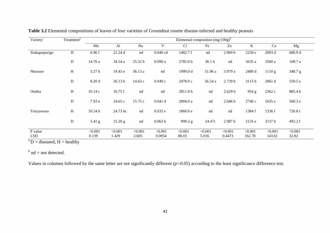
42
Table 3.2 Elemental compositions of leaves of four varieties of Groundnut rosette disease-infected and healthy peanuts
Variety/ Treatmenta Elemental composition (mg/100g)
b
Mn Al Na V Cl Fe Zn K Ca Mg
Sinkapoporigo H 6.96 f 21.24 d nd 0.046 cd 1482.7 f nd 2.969 b 2258 e 2093 d 686.9 d
D 14.76 a 34.54 a 25.52 b 0.090 a 2785.0 b 36.1 b nd 5635 a 3560 a 108.7 a
Nkosuor H 3.27 h 19.43 e 36.13 a nd 1999.0 d 51.96 a 3.979 a 2489 d 1110 g 348.7 g
D 8.20 d 26.13 b 14.63 c 0.049 c 2478.0 c 56.54 a 2.739 b 3115 b 2061 d 559.5 e
Otuhia H 10.14 c 16.75 f nd nd 2811.0 b nd 2.629 b 954 g 2362 c 885.4 b
D 7.93 e 24.65 c 15.75 c 0.041 d 2904.0 a nd 2.646 b 2746 c 1635 e 560.3 e
Yenyawoso H 10.54 b 24.73 bc nd 0.035 e 1860.0 e nd nd 1384 f 1336 f 720.8 c
D 5.41 g 15.20 g nd 0.063 b 999.2 g 24.47c 2.987 b 2135 e 3137 b 492.2 f
P value
LSD
<0.001
0.139
<0.001
1.429
<0.001
2.605
<0.001
0.0054
<0.001
88.03
<0.001
5.016
<0.001
0.4473
<0.001
162.78
<0.001
143.61
<0.001
32.82
a D = diseased, H = healthy
b nd = not detected.
Values in columns followed by the same letter are not significantly different (p>0.05) according to the least significance difference test.
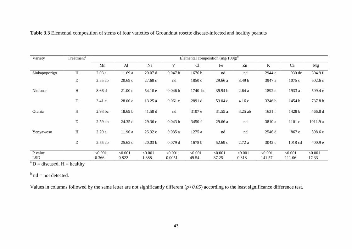
43
Table 3.3 Elemental composition of stems of four varieties of Groundnut rosette disease-infected and healthy peanuts
Variety Treatmenta Elemental composition (mg/100g)
b
Mn Al Na V Cl Fe Zn K Ca Mg
Sinkapoporigo H 2.03 a 11.69 a 29.07 d 0.047 b 1676 b nd nd 2944 c 930 de 304.9 f
D 2.55 ab 20.69 c 27.68 c nd 1850 c 29.66 a 3.49 b 3947 a 1075 c 602.6 c
Nkosuor H 8.66 d 21.00 c 54.10 e 0.046 b 1740 bc 39.94 b 2.64 a 1892 e 1933 a 599.4 c
D 3.41 c 28.00 e 13.25 a 0.061 c 2891 d 53.04 c 4.16 c 3246 b 1454 b 737.8 b
Otuhia H 2.98 bc 18.69 b 41.58 d nd 3107 e 31.55 a 3.25 ab 1631 f 1428 b 466.8 d
D 2.59 ab 24.35 d 29.36 c 0.043 b 3450 f 29.66 a nd 3810 a 1101 c 1011.9 a
Yenyawoso H 2.20 a 11.90 a 25.32 c 0.035 a 1275 a nd nd 2546 d 867 e 398.6 e
D 2.55 ab 25.62 d 20.03 b 0.079 d 1678 b 52.69 c 2.72 a 3042 c 1018 cd 400.9 e
P value
LSD
<0.001
0.366
<0.001
0.822
<0.001
1.388
<0.001
0.0051
<0.001
49.54
<0.001
37.25
<0.001
0.318
<0.001
141.57
<0.001
111.06
<0.001
17.33 a D = diseased, H = healthy
b nd = not detected.
Values in columns followed by the same letter are not significantly different (p>0.05) according to the least significance difference test.

44
Table 3.4 Elemental composition of seeds of four varieties of Groundnut rosette disease-infected and healthy peanuts
Variety Treatmenta Elemental composition (mg/100g)
b
Mn Al Na V Cl Fe Zn K Ca Mg
Sinkapoporigo H 1.51 c 4.96 a 34.47 a nd 23.93 a nd 5.02 c 806.5 cd 300.5 a 206.6 cd
D 2.01 e 5.85 b 42.08 a nd 38.88 c nd 4.36 ab 991.1 a 288.4 ab 215.4 bc
Nkosuor H 1.43 bc 4.76 a 40.96 a nd 26.27 a nd 3.09 a 781.9 d 220.8 e 219.8 b
D 1.33 ab 5.73 b 66.98 b nd 53.25 d nd 5.38 c 960.4 b 276.2 bc 248.5 a
Otuhia H 1.36 abc 4.48 a 40.38 a nd 36.40 bc nd 4.61 c 693.2 f 181.4 f 201.2 d
D 1.32 ab 4.87 a 69.42 b nd 73.06 e nd 4.41 ab 812.0 c 247.7 d 183.8 e
Yenyawoso H 1.83 d 6.06 b 35.13 a nd 25.65 a nd 5.48 c 724.7 e 257.8 cd 243.2 a
D 1.19 a 4.41 a 93.27 c nd 31.87 b nd nd 812.2 c 238.3 de 241.1 a
P value
LSD
<0.001
0.098
<0.001
0.261
<0.001
5.041
<0.001
1.402
<0.001
0.433
<0.001
25.23
<0.001
22.92
<0.001
9.05
a D = diseased, H = healthy
b nd = not detected.
Values in columns followed by the same letter are not significantly different

45
In the seeds, Mn content significantly (P <0.01) increased in virus-infected samples of
Sinkapoporigo but decreased in Yenyawoso. The highest content of Mn (14.76 mg/100 g)
was found in the infected leaf sample of Sinkapoporigo. Sodium was not detected in the
healthy leaf samples of Sinkapoporigo and Otuhia or in healthy and infected leaf samples of
Yenyawoso. In Nkosuor, Na content was significantly reduced (P <0.01) in the infected
sample (14.63 mg/100 g) compared with the healthy sample (36.13 mg/100 g). Sodium
content significantly (P <0.01) decreased in stems of infected plants but increased (P <0.01)
in the seeds of infected plants of all the genotypes, with the highest content (93.27 mg/100 g)
associated with the seeds of Yenyawoso. Detection of Zn in this study was inconsistent with
failure to detect this element in some leaves, stems and seed samples from both infected and
healthy plants. The concentration of Zn decreased (P <0.01) in the infected leaf sample of
Nkosuor (2.73 mg/100 g) but increased significantly (P <0.05) in the stems (4.16mg/100 g)
compared with the healthy samples (2.64mg/100 g). The concentration of Zn was
significantly increased (P <0.01) in infected seeds of Nkosuor (5.38 mg/100 g) but decreased
in that of Sinkapoporigo (4.36 mg/100 g) and Otuhia (4.41 mg/100 g) while it was undetected
in infected seeds of Yenyawoso. The concentration of vanadium significantly increased (P
<0.01) in leaves of infected plants of Sinkapoporigo (0.090 mg/100 g) and Yenyawoso
(0.063 mg/100 g), and stems of Nkosuor (0.061 mg/100 g) and Yenyawoso (0.079 mg/100 g).
It was however, neither detected in some leaf and stem samples nor in any seed samples of
the four varieties. Similarly, Fe was not detected in any of the seed samples or in some leaf
and stem samples of both infected and healthy plants. Notwithstanding, an increased (P
<0.01) Fe content was found in infected stem samples of Nkosuor (53.04 mg/100 g). Other
samples showed no significant differences in Fe content.

46
3.5 Discussion
All four genotypes showed typical GRD symptoms with no marked differences in the
symptoms expressed. Variability between varieties in GRD symptom expression has been
previously reported (Naidu et al., 1998). While ELISA testing only confirmed infection with
GRAV, the presence of GRV and Sat RNA within symptomatic plants could be inferred as
GRV and SatRNA are required for GRD symptoms (Murant et al., 1988) and satellite-free
isolates of GRV have not been found in nature (Murant, 1990). GRAV or GRV alone results
in symptomless infection in peanuts. Proximate analysis of peanut seeds showed significant
changes in the measured factors between infected and healthy plants, which often varied with
genotype. The reduction in moisture content seen for three of the four genotypes may be due
to premature loss of water from the hulls as a result of the disease, culminating in accelerated
seed desiccation and shrivelling. A reduction in relative water content of pepper infected with
Tobacco mosaic virus has also been reported (Pazarlar et al., 2013). In contrast an increased
moisture content of banana leaves associated with Banana bunchy top virus (BBTV)
infection was shown (Hooks et al., 2009) as was found for the infected Otuhia seeds.
Diminished ash content as seen in two peanut genotypes has also been reported in virus-
infected cowpea (Taiwo and Akinjogunla, 2006). The elevation of protein content of the
seeds of virus-infected plants of Nkosuor and Otuhia may be attributed to the synthesis of
virus coat protein and other virus-associated non-structural proteins. A marked increase in
amino acid concentration has been reported previously in GRD-infected peanut (Singh and
Srivastava, 1974). Several other reports note increased protein content as a result of virus
infection, including within leaves of Cucurbita pepo infected with Zucchini yellow mosaic
virus (Radwan et al., 2007), soybean infected with Soybean mosaic virus (Cheema et al.,
2003) and tomato with a tospovirus infection (Sutha et al., 1998). In contrast, similar
reductions of protein content as seen in seeds from diseased plants of Sinkapoporigo have

47
been found in seeds from virus-infected cowpea plants (Taiwo and Akinjogunla, 2006), and
in seed from peanut plants infected with Peanut stripe virus (PStV; Ross et al., 1989).
Fat content increased in three of the four genotypes which contrasts with reports of reduced
oil in peanut seeds following GBNV infection (Sunkad and Naik, 2013) and in oil content of
soybean infected either singly or in double combinations with PMoV, Cowpea chlorotic
mottle virus, Soybean mosaic virus and Tobacco ringspot virus (Demski and Jellum, 1975).
The altered carbohydrate content observed in the seeds of diseased plants in this study could
be due to alterations in photosynthetic metabolism and disorders in plant carbohydrate
metabolism as a result of the infection (Gonçalves et al., 2005). The disparities in
carbohydrate content observed among the genotypes could be due to differences in their
response to the disease. The reduction in carbohydrate content of seeds of virus-infected
plants could be considered a secondary effect of the viral infection. Infection may have
inhibited phloem transport, culminating in the accumulation of photosynthetic products in
chloroplasts and a subsequent reduction of their supply from source leaves to seeds. The
decrease in carbohydrate content could also be due to increased respiration that characterises
virus-infected plants and the possible conversion of carbohydrates into amino acids, which
are used for protein synthesis (Gupta et al., 2010). Reduction in carbohydrate content in Bean
yellow mosaic virus-infected bean has also been shown (Mali et al., 2000). In contrast,
increased carbohydrate content of peanut seeds following PStV infection (Ross et al., 1989),
cowpea seeds following virus infection (Taiwo and Akinjogunla, 2006) and sugarcane leaves
as a consequence of Sugarcane yellow leaf virus infection (Gonçalves et al., 2005) have been
reported. The accumulation of carbohydrates in the virus-infected seeds observed in this
study could also be due to more efficient translocation of assimilate such as was observed
with Cocoa swollen shoot virus-infected cocoa (Adomako and Hutcheon, 1974) or the
inactivation of carbohydrate-utilising enzymes due to the infection, resulting in reduced

48
breakdown of carbohydrates. The higher energy content associated with the seeds from the
virus-infected plants in this study could be due to the increased fat content.
The mineral content of stems and seeds of the tested peanut genotypes also varied with GRD
infection. Plant disease impairs physiology, including nutrient uptake, assimilation,
translocation from the root to the shoot and utilisation. GRD affected plants may have altered
membrane permeability (Huber and Graham, 1999), resulting in increased or decreased
uptake of particular elements. Furthermore, the impaired uptake of elements by plant tissues
may result in nutrient interactions leading to changes in their concentrations (Fageria, 1983).
Within the measured macronutrients, increased potassium content observed in all genotypes
is consistent with a prior study comparing sugar beet infected with Beet yellows virus (Clover
et al., 1999) but contrasts with reports of decreased K in peanut seed infected with PStV
(Ross et al., 1989) and virus-infected millet (Kandhasamy et al., 2010). Calcium and
magnesium content both increased and decreased in the peanut genotypes tested. There are
similar contrasting prior reports of changes in Ca content in virus-infected plants. Decreased
Ca has been found in tiger millet infected with Ragi mottle streak virus (Kandhasamy et al.,
2010), while peanut seed showed no change in Ca following PStV infection (Ross et al.,
1989). Reported changes in Mg concentration of virus-infected plants are also inconsistent
with increased Mg in virus-infected peanut (Ross et al., 1989) and tropical soda apple
(Overholt et al., 2009) while finger millet (Kandhasamy et al., 2010), and Solanum nigrum
(Takács et al., 2001) showed decreased Mg content. Potassium has an antagonistic effect on
the absorption of Ca2+ and Mg2+ at higher concentration and this depends on plant species
and environmental conditions. It is thus possible that the higher concentration of K observed
in this studymay have led to the relatively lower concentrations of Ca and Mg in the plant
tissues (Fageria, 1983). The increase in chlorine and aluminium content following GRD

49
infection agrees with Ikram-ul-Haq et al. (2014) whom observed a trend for greater Cl
content in leaves of banana infected with BBTV. Variability in the response of virus infection
on micronutrient content of plant tissues has been previously reported. Virus infection
resulted in increased (Overholt et al., 2009) or decreased (Takács et al., 2001) Mn content in
Solanum spp. or no effect in grapevine (Moutinho-Pereira et al., 2012). Similarly, both
reduction (Takács et al., 2001; Ashfaq et al., 2014) and increase in Na (Clover et al., 1999;
Ikram-ul-Haq et al. (2014)) and reduction (Takács et al., 2001), increase (Overholt et al.,
2009; Ashfaq et al., 2014) or no effect in Zn content (Moutinho-Pereira et al., 2012) of virus-
infected plants have been reported. Similarly, reduced (Kandhasamy et al., 2010) or increased
(Ashfaq et al., 2014) Fe content of virus-infected plants has been reported. Vanadium
increased in foliar parts of some of the varieties. Acute vanadium poisoning was found in
sheep following experimental daily feeding of diets containing 400–800mgkg−1 (40–80mg
100 g−1) ammonium metavanadate (Hansard et al., 1978). Furthermore, higher accumulation
of V in soils, water and vegetables poses potential effects to human and animal health
(Vachirapatama et al., 2011). However, V was not detected in the nuts which are the parts
consumed by humans and the concentration of V observed in the stems and leaves which are
used as fodder for livestock, are too low (0.035–0.090 mg/100 g) to have any deleterious
effects when fed to animals. Despite the importance of peanut as a source of energy and
nutrients for people and livestock of sub-Saharan Africa, the impact of disease on the
nutritive quality of peanuts has received little or no attention. This study has detailed changes
in nutritive qualities of peanut leaf, stem and seed tissues when plants succumb to GRD.
Significant changes as a result of infection were found which varied with variety. In the
majority of varieties, energy content increased with virus infection. While disease results in
profound yield loss, such changes in quality can affect the nutritive benefits of the produce.

50
Increased energy content may compensate to some extent for reduced yield by providing
nutritional boost. Concerns over changes in elemental composition that may render stock or
human feed toxic following virus infection are allayed.
3.6 Acknowledgements
The authors wish to thank Australian Agency for International Development (AusAID) for
the funding. We are also grateful to the Tasmanian Institute of Agriculture and the
Biotechnology and Nuclear Agriculture Research Institute of the Ghana Atomic Energy
Commission (GAEC) for their support in terms of facilities.
This chapter has been removed for copyright or proprietary reasons.
Appiah, A. S., Offei, S. K., Tegg, R. S., Wilson, C. R., 2016. Varietal response to groundnut rosette disease and the first report of Groundnut ringspot virus in Ghana, Plant disease, 100(5),946-952 https://doi.org/10.1094/PDIS-07-15-0838-RE

This chapter has been removed for copyright or proprietary reasons.
Appiah, A. S., Sossah, F. L., Tegg, R. S., Offei, S. K., Wilson, C. R., 2017. Assessing sequence diversity of Groundnut rosette disease agents and the distribution of Groundnut rosette assistor virus in major groundnut-producing regions of Ghana, Tropical plant pathology, 42(2), 109–120http://dx.doi.org/10.1007/s40858-017-0140-x

100
6 CHAPTER SIX: Genomic analysis and transmission of
recombinant Phasey bean mild yellows virus isolates
from Queensland and Tasmania
A.S. Appiah1,2
, R.S. Tegg,1 M. Sharman
3 and C.R. Wilson
1
1 Tasmanian Institute of Agriculture, University of Tasmania, New Town, Tasmania,
Australia
2 Biotechnology and Nuclear Agriculture Research Institute, Ghana Atomic Energy
Commission, Legon, Accra, Ghana
3Department of Agriculture and Fisheries, Brisbane, Queensland, Australia
Correspondence
C.R. Wilson, Tasmanian Institute of Agriculture, University of Tasmania, 13 St. Johns
Avenue, New Town, TAS 7008, Australia.
Email: [email protected]

101
6.1 Abstract
The Poleroviruses include many species of economic importance, affecting many plant
species and causing significant losses. Furthermore, the relatively frequent discovery of new
Poleroviruses and a propensity for genomic recombination has identified this genus as one
providing numerous emerging viruses of importance. In this study, the full genome a
Queensland isolate of the novel Polerovirus, tentatively named Phasey bean mild yellows
virus (PhBMYV) was determined. The full genome consisted of six open reading frames
(ORFs), with their respective nucleotide and putative protein sequences sharing close identity
to two previously reported isolates from New South Wales (NSW) and Western Australia
(WA), except within the RNA-dependent-RNA-polymerase (RdRp) and Coat protein-Read
through (CP-RT) genomic regions. The RdRp shared ~63% amino acid identity with the
NSW and WA isolates while the CP-RT was highly divergent sharing only 33-34%, being
more closely related to that of Chickpea chlorotic stunt virus (53%). Recombination analysis
suggested this novel virus isolate is likely to have been derived from a recombination event
with an exchange point identified at the boundary of the CP and CP-RT genomic regions. In
an experimental host range study, the virus was successfully transmitted to Phasey bean, pea
and chickpea plants using both aphid vector and graft transmission methods. Symptoms
observed were severe and included yellowing, stunting and reduced leaf size. Based on the
results of this study, PhBMYV QLDCL 16 is suggested as a genetic variant of PhBMYV.

102
6.2 Introduction
Legume production is an important venture in Australia with the industry currently
contributing about AUD$1.6 billion per year to the Australian economy (Australian Food
News, 2016). These include lentils, chickpeas, faba beans, field peas, lupins and mungbeans
(Pulse Australia, 2015). Virus diseases pose a serious risk to legume production. Several
virus species have been identified from legume crops in Australia (Latham and Jones, 2001a;
Schwinghamer and Schilg, 2003; Thomas et al., 2004). Of these, Alfalfa mosaic virus
(Bromoviridae, genus Alfamovirus) (AMV), Cucumber mosaic virus, (Bromoviridae, genus
Cucumovirus) (CMV), Bean yellow mosaic virus and Pea seedborne mosaic virus
(Potyviridae, genus Potyvirus) (BYMV and PSbMV), (Jones and Coutts, 1996) and
Subterranean clover stunt virus (SCSV) (Nanovirus) (Wilson et al., 2012) are the most
commonly found. However, many crop surveys across Australia have confirmed the presence
of viruses belonging to the the family Luteoviridae in pulse crops, whilst sometimes less
abundant, often resulting in severe disease. These include Turnip yellows virus (TYV) (genus
Polerovirus) (Wilson et al., 2012), Bean leaf roll virus (genus Luteovirus) (BLRV) and Beet
western yellows virus (genus Polerovirus) (BWYV) (Schwinghamer et al., 1999; Latham and
Jones, 2001a; Freeman and Aftab, 2001; Van Leur et al., 2003; Aftab et al., 2005). The
Luteoviridae is made up of three genera; Luteoviruses, Enamoviruses and Poleroviruses.
They are all transmitted in persistent (circulative, non-propagative) manner by aphid vectors
(Duffus, 1977). Their genomes are positive-sense ssRNA of about 1.8-2 x 10⁶ Mr (5.5-6.0 kb)
with a genome-linked protein (VPg) covalently attached at the 5' end of the RNA molecule
which is not polyadenylated at the 3'-end (Mayo et al., 1982; Murphy et al., 1987). The 3'
ends do not appear to possess tRNA-like structures such as those associated with other plant
viral groups (Miller et al., 1988). The genomes of the Luteoviridae contain five to six open
reading frames (ORFs) (Pfeffer et al., 2002).

103
The Polerovirus genus is particularly associated with the emergence of new viral diseases
worldwide due to the existence of high genetic diversity and putative propensity for genetic
recombinations (Moonan et al., 2000; Stevens et al., 2005; Lotos et al., 2016), and are
known to cause major yield losses in vegetable and arable crops. Recently, novel
Poleroviruses have been reported in chickpea and faba bean in Ethiopia (Abraham et al.,
2006), cotton in Australia and Argentina (Ellis et al., 2013; Distefano et al., 2010),
strawberry in Canada (Xiang et al., 2015), cabbage, maize and brassicas in China (Zhang et
al., 2014; Wang et al., 2016; Xiang et al., 2011) and cucurbits in Taiwan (Knierim et al.,
2010). In a recent survey of vegetable crops in Tasmania, a novel polerovirus was identified
as infecting peas (Wilson et al., 2012). The virus has subsequently been found in Trifolium
subterraneum (subterranean clover) in Western Australia (WA), Cicer arietinum (chickpea)
in New South Wales (NSW) and Macroptilium lathyroides (phasey bean) in Queensland
(QLD) and has been tentatively been named Phasey bean mild yellows virus, PhBMYV
(Sharman et al., 2016). The emergence of novel viruses and the diseases they cause; many of
which are yet to be identified, present a major constraint to the production of food crops
worldwide. The identification of a novel Polerovirus in Australian legumes thus necessitates
studies on its epidemiology and genome characteristics. Here we describe virus hosts,
confirm aphid transmission, obtain full and partial genomic sequences of new isolates, and
compare genome sequences for evolutional recombination and to develop virus- specific
primers for detection and diagnosis of PhBMYV; a pre-requisite for making informed
decisions regarding mitigation strategies for the management of the disease.

104
6.3 Materials and Methods
6.3.1 Transmission studies using Queensland isolate of PhMYV
An isolate of PhBMYV from a naturally infected Phasey bean plant and a colony of cowpea
aphids (Aphid craccivora), its putative vector were obtained from the Department of
Agriculture and Fisheries, Brisbane, Queensland, Australia. The virus-infected Phasey bean
plant was maintained under glasshouse conditions (20±2oC) while the aphids were
maintained on cowpea plants in a controlled environment cabinet at 28oC with a 16 h
photoperiod.
All transmission tests were carried out in an insect-proofed glasshouse. Host plants tested
belonged to the Leguminaceae (pea; cultivars Greenfeast, Snow pea, Sugar snap, Dwarf pea
and TIA – a locally sourced cultivar), chickpea, cowpea, peanut, Faba bean and bean),
Solanaceae (tomato, eggplant and capsicum), Asteraceae (lettuce) and Cucurbitaceae
(zucchini and cucumber). Adult aphids were given a 24 hr acquisition access period (AAP)
on leaves from a PhBMYV-infected plant and then individually transferred to two-week old
receipient plants. About 10 – 15 adult aphids were used per plant. Eight to 15 plants of each
test plant were aphid inoculated. A further ten plants were graft-inoculated with a scion from
infected Phasey bean plant. Aphids fed on healthy Phasey bean plants were also transferred
to ten test plants and healthy scions were grafted onto five healthy plants to serve as controls.
The aphids were allowed 48 hr inoculation access period (IAP) and then killed with a
pyrethrum-based insecticide. Inoculated plants were grown in an aphid-proof glasshouse for 4
– 8 weeks and observed for symptoms. Virus infection was tested 2-4 weeks post-inoculation
by reverse-transcription polymerase chain reaction (RT-PCR).

105
6.3.2 RNA isolation and Reverse-transcription polymerase chain
reaction (RT-PCR).
Total RNA was extracted from leaf tissues of both inoculated and non-inoculated plants using
a PowerPlant RNA Isolation Kit (MO BIO Laboratories Inc. California, USA) following the
manufacturer’s instructions, 50mg of leave samples initially homogeniseed at 6.5 rpm for 45
sec in a FastPrep-24 Tissue and Cell Homogenizer (MP Biomedicals, Model 6004-500, USA).
The isolated RNA was kept at -80oC used.
Complementary DNA (cDNA) was synthesized in a 25 μl reaction mix using the iScript
Reverse Transcription Supermix (BIO RAD, California, USA). PCR was carried out in a 20
μl reaction volume containing 10 μl of HotStarTaq Plus Master mix comprising 1 unit
HotStartTaq Plus DNA polymerase, 1x PCR buffer and 200μM of each dNTP), 0.75μl each
of forward and reverse primers (10 μM), and 1 μl of template cDNA. PCR was performed in
Eppendorf Mastercycler, Hamburg, Germany, using a thermocycle of 30 cycles of 30 s at
94°C, 60 s at 58°C, and 3 min at 72°C, preceded by an initial denaturation for 1 min at 94°C
and a final extension at 72°C for 10 min. Amplified products were electrophoresed on 1%
agarose gel stained with SYBR Safe DNA gel stain (Invitrogen, CA, USA), and viewed with
the Safe Imager Transluminator (Invitrogen, CA, USA).
6.3.3 Next-generation sequencing (NGS) of QLD isolate of PhBMYV
RNA was isolated from 6g of infected leaves and precipitated by addition of 0.1 volume 3M
sodium acetate and 2 volumes 100% ethanol followed by centrifugation at 13,000 rpm for 1
minute. Pelleted RNA was sent to Macrogen (Seoul, South Korea) for library preparation and
100-bp paired-end sequencing on an Illumina HiSeq2000.

106
6.3.3.1 Analysis of NGS data
The reads were first trimmed and then assembled into contigs using the de novo assembly
function in Geneious 8.1.8 (Biomatters). Contigs were sorted by length (minimum length 500)
and the longest subjected to BLAST searches (Altschul et al., 1990). In addition, the reads
were also mapped to reference genomes obtained from GenBank (KT962999 for NSWCP15
and KT963000 for ESPCL15); as variants of the same virus. Mapping was performed with
minimum overlap of 10%, minimum overlap identity of 80%, allow gaps of 10% and fine
tuning set to iterate up to 10 times (Kehoe et al., 2014). A consensus between the contigs of
interest and mapping was created in Geneious by alignment with Clustal W. The resulting
sequences were compared with the reference sequences used in the mapping process.
The complete sequence obtained was aligned with the two reference genomes using ClustalW
(Thompson et al., 1994). Construction and confirmation of the genome sequence determined
by NGS was done by PCR amplification of overlapping genome sequences and Sanger
sequencing. PhBMYV primers were designed on the consensus sequence using the Primer3
function implemented in Geneious 8.1.8 (Biomatters) (Table 6.1). Complementary DNA
(cDNA) was synthesized from a total RNA extract in a 25 µl reaction mix using the iScript
Reverse Transcription Supermix (BIO RAD, California, USA) following the manufacturer’s
protocol. The cDNA was then used for PCR using reaction mixes and thermocycle profile as
previously described, and amplicons viewed as before. DNA bands of interest were excised
from the gel and purified using QIAGEN gel extraction kit (QIAGEN GmbH, Hilden,
Germany). The purified products were then sequenced in both directions at the Australian
Genome Research Facility, Melbourne, Australia. The resulting sequences were edited in
Geneious 8.1.8 (Biomatters) and then mapped to the sequence obtained by the NGS. The
sequences were then edited to ensure complete homology and the consensus sequence
extracted.

107
Table 6.1 Oligonucleotide primers used for detection and sequencing of the virus genome
Primer
Sequence
Amplicon
product size
PhBV1F 5’-CAACTACCGCCGCTCCCAGT- 3’
PhBV842R 5’-GTTACCCCTACCTTGTGGGC-3’ 842bp
PhBV197F 5’-GGCGCAAAAGCTGAACTGAA-3’
PhBV1679R 5’-ATTGCTCTCACGTGGCTTGA-3’ 1472bp
PhBV739F 5’-ATACAGCTCTGGAGGGTCGT-3’
PhBV2032R 5’-AGAGGCCTCGGAGATGAACT-3’ 1293bp
PhBV1660F 5’-TCAAGCCACGTGAGAGCAAT-3’
PhBV2760R 5’-TGACCCAGCACAAAGTTGGT-3’ 1100bp
PhBV2741F 5’-ACCAACTTTGTGCTGGGTCA-3’
PhBV3919R 5’-GCCCGGTTGTCAACCCGGAT-3’ 1178bp
PhB7F 5’-GATCCTTGTGCAAGTTTGTT-3’
PhB455R 5’-GAATGAGACCTTTGTAAGTA-3’ 467bp
PhB477F 5’-GATCTGTGGGGGCTCGCAT- 3’
PhB830R 5’-TTCCTGGAAAATTTTCCCCAG -3’ 373bp
PhB972F 5’-GTCCGCCTGTTTAATGGAAC-3’
PhB1994R 5’-TTGGCAATCCATTGTTGCCGC-3’ 1042bp
PhB4354F 5’-TTCGTTCACCTCTTTGACAAAG-3’
PhB5789R 5’-ACTTGCCCATCAGTAGACAGG-3’ 1455bp
6.4 Genome sequencing of Tasmanian isolate of Phasey
bean mild yellows virus
The primer pairs (Table 6.1) were then used to amplify genomic sequences from a Tasmanian
isolate of PhBMYV collected from an infected field pea sample in 2012 (Wilson et al. 2012).
Total RNA extraction, cDNA synthesis, PCR and Sanger sequencing were done as previously
described.
6.5 Genome analysis
Prediction of putative open reading frames (ORFs) using ORF finder and genome annotation
were done in Geneious 8.1.8 (Biomatters, Auckland, NZ). To investigate potential

108
recombination events that might have occurred within the genome, the aligned sequences
were subjected to recombination detection analysis using the Recombination Detection
Program v.4.16 (RDP4) (Martin et al., 2015) with default settings. Potential recombination
events (PREs) detected by the RDP were further checked with other recombination detection
methods such as GENECONV (Padidam et al., 1999), BOOTSCAN (Martin et al., 2005),
MaxChi (Maynard Smith, 1992) and CHIMAERA (Martin et al., 2005); implemented in
RDP4. A breakpoint map showing the positions of all clearly identified breakpoints for a set
of unique PREs was constructed.
6.5.1 Phylogenetic analysis
The complete genomic sequence of the QLD isolate of PhBMYV was aligned with other
Luteovirus genomes retrieved from the Genbank (Table 6.2) including the PhBMYV isolates
from NSW, NSWCP15 (KT962999) and WA, ESPCL15 (KT963000) using ClustalW
(Thompson et al., 1994). The complete genome, and derived amino acid sequence data for
individual open reading frames were subjected to phylogenetic analysis using the Neighbor-
joining method using the Tamura-Nei model with a bootstrap value of 1000. Phylograms
were visualised using TreeView (Page, 1996). Additionally, the PhBMYV isolates from QLD,
NSW, WA and TAS were aligned, trimmed and used in phylogenetic analysis as described.
Table 6.2 List of Luteovirus sequences retrieved from the GenBank for this study
Virus Acronym Accession
Number
Reference
Polerovirus
Beet chlorosis virus BChV NC_002766.1 Hauser et al., 2002
Beet mild yellowing virus BMYV NC_003491.1 Guilley et al., 1995
Beet western yellows virus isolate Rouen 1 BWYV KU521325.1 Hehn et al.,
unpublished
Carrot red leaf virus CtRLV NC_006265.1 Huang et al., 2005
Cereal yellow dwarf virus-RPS RPV Mex-1 CYDV- AF235168.2| Beckett & Miller

109
RPS unpublished
Cereal yellow dwarf virus - RPV CYDV-
RPV
L25299.1 Vincent et al., 1991
Chickpea chlorotic stunt virus CpCSV NC_008249.1 Abraham et al.,
2006
Cucurbit aphid-borne yellows virus CABYV LC082306.1 Yoon et al.,
unpublished
Melon aphid-borne yellows virus MABYV NC_010809.1 Xiang et al., 2008
Potato leafroll virus isolate PLRV-IM PLRV KC456052.1 Li et al.,
unpublished
Sugarcane yellow leaf virus ScYLV NC_000874.1 Moonan et al.,
2000
Tobacco vein distorting virus TVDV NC_010732.1 Mo et al., 2010
Turnip yellows virus TuYV NC_003743.1 Garcia et al., 1993
Phasey bean mild yellows virus isolate
ESPCL15
PhBMYV KT963000.1 Sharman et al.
2016
Phasey bean mild yellows virus isolate
NSWCP15
PhBMYV KT962999.1 Sharman et al.
2016
Luteovirus
Barley yellow dwarf virus - PAV BYDV-
PAV
NC_004750.1
Miller et al., 1988
Bean leafroll virus BLRV NC_003369.1 Domier et al., 2002
Soybean dwarf virus SbDV NC_003056.1 Terauchi et al.,
2001
Enamovirus
Pea enation mosaic virus-1 PEMV-1 NC_003629.1 Demler et al., 1991

110
6.6 Results
6.6.1 Virus transmission studies
Successful transmissions of PhBMYV from infected Phasey plant using with both Aphis
craccivora as a vector or by stem grafting were achieved into healthy Phasey bean, pea and
chickpea receipient plants. Phasey bean (Fig. 6.1), pea and chickpea (Fig. 6.2) plants showed
stunting with severe yellowing and reduced leaf size. Tomato, eggplant, capsicum, lettuce,
bean, cowpea, cucumber, zucchini, peanut and faba bean plants inoculated with the virus by
either aphid vector or grafting remained symptomless and the virus was was not detected in
the receipient plants. Infections of the inoculated Phasey bean, pea and chickpea plants were
confirmed by RT-PCR and sequencing of the resultant amplicons.
Figure 6.1 Phasey bean plants; A: PhBMYV-inoculated plant showing stunting, yellowing
and reduced leaf size and B: Uninoculated Phasey bean plant.

111
Figure 6.2 Phasey bean mild yellows virus-inoculated plants: A. Healthy pea B. Aphid-
inoculated pea C. Graft-inoculated pea D. Healthy chickpea E. Aphid-inoculated chickpea.
6.6.2 Full length genome analyses of PhBMYV isolate QLD CL 16
From the single PhBMYV sample, 9,871,832 raw reads were obtained following NGS after
trimming. Following de novo assembly in Geneious, 20,000 contigs ranging from 150 to 3,
000 nt were obtained. Contigs of interest were those between 500 and 3000 nt. After mapping
to reference genomes in Geneious, the lengths of the consensus sequences were 5,867 nt for
NSWCP15 (49,335 reads assembled) and 5,909 nt for ESPCL15 48,283 reads assembled). A
full genome sequence of QLD isolate was derived form the consensus mapped to the
ESPCL15 sequence. Oligonucleotides primers designed on the QLD PhBMYV consensus
sequence were used in RT-PCR and are given in Table 6.1. Sanger sequencing of PCR
amplicons was used to confirm the genomic sequence derived from NGS. The final sequence
was the consensus of the full sequence from the NGS and the Sanger sequencing. The 5,868

112
nt complete genomic RNA of the QLDCL16 is shown in Fig. 6.3. Thirty (30) ORFs were
predicted from the genome of which six (ORF0, ORF1, ORF2, ORF3, ORF4 and ORF5 were
identified as typical of Poleroviruses. The overlapping region of ORF1 and ORF2 is ~578 nt
long, while the intergenic region between ORF2 and ORF3 is ~195 nt.
Figure 6.3 Schematic representation of the genome organisation of PhBMYV QLD isolate.
The sizes of the deduced gene products are written either above or below the name of the
ORF.
The predicted amino acid (aa) sequences of the six ORFs showed significant variation
between the PhBMYV QLDCL 16 and the other members of the family Luteoviridae (Table
6.3). Sequence identities for the various proteins are as follows; P0 (09 – 97%), P1 (07 –
90%), P1 + P2 (06 – 61%), P3 (29 – 97%), P4 (26 – 93%), and P3+P5 (29 – 53%). The QLD
isolate shared close aa identity to the NSW, WA and TAS isolates for all the proteins except
P1 + P2 (52 - 61%) and P3+P5 (33-34%). The closest to the P3 + P5 protein of the PhBMYV
QLDCL 16 was that of CpCSV sharing identity of 53%. The P4 protein shared aa sequence

113
identity of 67%, 68% and 69% with CABYV, PhBMYV TAS 12 and MABYV respectively.
With the exception of PEMV-1, the P3 proteins appear less variable.
6.6.3 Analysis of partial sequence of Tasmanian isolate of PhBMYV
A 4300 nt partial sequence spanning the entire ORF0, ORF1, ORF2 and segments of ORF3
and ORF4 were obtained by using RT-PCR and Sanger sequencing. Nucleotide alignment of
this segment with other PhBMYV isolates showed nucleotide identity of 96%, 94% and 90%
with NSWCP 15, QLDCL 16 and ESPCL 16 respectively. The predicted amino acids
sequences for ORF0, ORF1, ORF2, ORF3 and ORF4 of the Tasmanian isolate showed
significant variation compared to the NSW, WA and the QLD isolates. For the P0 aa
sequence, the TAS isolate shared 84% (WA), 74% (NSW), and 97% (QLD). The P1 aa
sequences were less variable with the TAS isolate sharing 95%, 85% and 89% identities with
WA, NSW and QLD isolates respectively. The P1 + P2 aa sequence showed greater variation,
with the TAS isolate sharing 22% identity with the WA and NSW isolates and 52% identity
with the QLD isolate. Amino acid sequence identity for the ORF3 and ORF4 were in the
range of 68 – 71% and 64% - 68% respectively.

114
Table 6.3 Comparison of deduced amino acid sequences for major open reading frames for
the Queensland isolate of PhBMYV and some members of the family Luteoviridae
Virus
ORF0 ORF1 ORF1+ORF2 ORF3 ORF4 OFR3+ORF5
P0
(261 aa)
%
P1
(581aa)
%
P1 + P2
(1061 aa)
%
P3
(195 aa)
%
P4
(143 aa)
%
P3+P5
(656 aa)
%
BChV 17 22 16 67 47 32
BMYV 18 27 20 67 49 32
BWYV 20 32 21 68 49 31
CtRLV 13 27 18 54 37 29
CYDV-RPV 14 26 18 66 48 32
CpCSV 18 27 19 69 55 53
CABYV 18 31 20 77 67 48
MABYV 21 29 21 76 70 48
PLRV 14 28 20 65 48 31
ScYLV 11 22 16 39 26 28
TVDV 20 29 20 57 42 30
TuYV 17 29 21 67 48 33
PhBMYV ESPCL15 85 86 60 97 93 34
PhBMYV NSWCP15 76 90 61 95 89 33
PhBMYV TAS 12 97 89 52 71 68 NP
PEMV-1 09 16 08 29 NP 30
BYDV-PAV NP 07 07 47 28 26
BLRV NP 09 06 57 44 31
SbDV NP 08 08 59 46 30
*NP – not present
P1+P2 = RNA-dependent-RNA- polymerase (RdRp)
P3 = Coat protein (CP)
P4 = Movement protein (MP)
P3+P5 = Coat protein + read-through protein (CP-RT)
6.6.4 Phylogenetic analysis
Nucleotide alignment of the complete genome of the new Queensland isolate (QLDCL 16)
with the genomes of other Luteoviruses obtained from the Genbank (Table 6.2) showed
nucleotide identities in the range of 31 to 88%. PhBMYV isolate QLDCL 16 shared
nucleotide identity of 90% and 91% with NSWCP15 and ESPCL15 respectively.
Phylogenetic analysis showed the three PhBMYV isolates forming a distinct group (Fig. 4)
and appeared closer to MABYV and CABYV (59%), and CpCSV (53%) than the other

115
Poleroviruses. Phylogenetic analysis of the four PhBMYV (Fig. 6.4B) showed PhBMYV
ESPCL 15 and PhBMYV TAS 12 grouping separately from the other isolates.
A
B

116
Figure 6.4 Phylogenetic relationship between A. PhBMYV QLDCL 16 and other members
of the family Luteoviridae. Trees were based on the complete genome sequences and
distances constructed using the neighbour-joining method. B. PhBMYV isolates from
Australia. Figures at the nodes indicate the frequency of the cluster after bootstrap analysis
(1000 replicates). Nodes with values <50% were collapsed.
Phylogenetic analysis of the predicted amino acid sequences of the P0, P1 + P2, P3, P4 and
P3+P5 are shown in figure 6.5. The topologies of the phylogenetic trees for the P0, P1 + P2
(RdRp), P3 (CP) and P4 (MP) show close relationship of the PhBMYV QLD CL16 to those
of NSW, WA and TAS, usually clustering together, separate from the other Poleroviruses.
The CP-RT protein of PhBMYV QLD CL16 was however, more closely related to CpCSV.
A

117
B
C

118
D
E

119
Figure 6.5 Phylogenetic analysis based on the predicted amino acid sequences A. P0, B.
P1+P2, C. P3, D. P4 and E. P3 + P5. Distance trees were constructed using the neighbour-
joining method. Figures at the nodes indicate the frequency of the cluster after bootstrap
analysis (100 replicates; shown only when >50%).
6.7 Recombination analysis
Using default settings, the results from RDP suggested PhBMYV QLDCL 16 as a potential
evolutionary product of a recombination. (Fig. 6.6). The RDP located the recombination
event between positions 5284 and 5975 with a P value of 1.825 x10⁻²⁶
. The same event was
detected by GENECONV (P=2.838 x 10-53)
), BootScan, (P= 3.924 x 10⁻³⁵
), MaxChi
(P=6.752 x10⁻¹⁵
), Chimaera (P= 6.941 x 10⁻¹⁹
), Siscan (P= 5.304 x 10-52
) and 3Seq (P=4.884
x10-26
).

120
Figure 6.6 A graph showing the result of recombination analysis of aligned Polerovirus
genome sequences using the RDP method. The complete nucleotide sequence of the
Queensland isolate of Phasey bean mild yellows virus (potential recombinant) was compared
to those of Phasey bean mild yellows virus isolates ESPCL 15 (potential major parent) and
NSWCP 15 (potential minor parent).
6.8 Discussion
This study reports the full genome sequence of Queenland and a partial genome sequence of
Tasmanian isolates of PhBMYV; a novel polerovirus infecting legumes in Australia. The
QLD isolate was originally found in Phasey bean; a perennial weed while the TAS isolate
was found in peas. Two prior genomes of this virus have previously been reported infecting
chickpea in New South Wales and subterranean clover in Western Australia (Sharman et al.,
121
2016). An additional partial genome was reported from field peas in Tasmania (Wilson et al.,
2012). In this study, successful transmission of the Queensland isolate was achieved to
healthy Phasey bean, chickpea and pea plants using both the cowpea aphid vector and
grafting. Symptoms expressed in infected Phasey bean plants were severe, contrary to the
mild symptoms observed in the field (Sharman et al., 2016) and included yellowing, stunting
and reduced plant leaf size (Fig. 6.1). Similar severe symptoms were expressed in infected
peas and chickpeas. The severe symptom manifestation in the glasshouse could be attributed
to the modified environmental conditions especially light and temperature. Reduced
illumination has been shown to enhance virus content and increase the susceptibility of plants
to infection (Bawden and Roberts, 1947). Furthermore, Manfre et al. (2011) demonstrated
increased susceptibility of Nicotiana banthamiana grown in low light intensity to Turnip
mosaic virus. In addition, the relatively low average day temperature (20±2oC) in the
glasshouse may have influenced virus replication resulting in high virus concentration in
infected plants, leading to severe symptoms as has been reported by Vela´zqueza et al., 2010.
Attempts to infect other legumes such as cowpea, peanut, Faba beans and members of the
Solanaceae (tomato, pepper, and eggplant), Cucurbitaceae (cucumber, zucchini) and
Asteraceae (Lettuce) were unsuccessful. The plants remained symptomless and the virus was
not detected suggesting they may not be hosts of this new virus. Nonetheless, there is the
need to test different varieties of these plants to ascertain differences in varietal response to
the virus.
The genome organisation of the PhBMYV QLDCL 16 and PhBMYV TAS 12 were similar to
other Poleroviruses, containing six ORFs (except for ORF5 which is yet to be determined for
TAS 12 isolate). Genome comparisons revealed close nucleotide identity to PhBMYV
ESPCL 15, PhBMYV TAS 12 and PhBMYV NSWCP 15 compared to the other members of
the genus, sharing nucleotide identities of >90%. Other Poleroviruses of close identity to this

122
virus were CABYV and CpCSV. Phylogenetic analysis of the full genomes saw isolate
QLDCL 16 clustering with isolates ESPCL 15 and NSWCP 15, forming a distinct clade, but
appeared closer to CABYV, MABYV and CpCSV than the other Poleroviruses. This reflects
the results of Sharman et al. (2016) whom using partial sequences suggested isolates
ESPCL15, NSWCP15 and the CABYV-like isolate from Tasmania (HQ543091) are all
members of the same PhBMYV, a novel polerovirus. In a separate phylogeny involving the
four PhBMYV isolates, isolate TAS 12 appeared closer to ESPCL 15 than NSWCP 15 and
QLDCL 16.
With the exception of PhBMYV isolates ESPCL 15, NSWCP 15 and TAS 12, the sequence
identity of the predicted amino acids between PhBMYV QLDCL 16 and other Poleroviruses
was relatively low and significantly different for all the virus encoded proteins. Although the
predicted P3 and P4 proteins of QLDCL 16 differed significantly from those of other
Poleroviruses, they appear less variable within the genus. According to Guyader and Ducray
(2002) and Plchova et al. (2009), ORF 3 (P3) and ORF 4 (P4) are the most conserved regions
in PLRV; the type member of the genus. Isolates ESPCL 15, NSWCP 15 and TAS 12 were
much closer to QLDCL 16 in amino acid identity for the P0, P1, P3 and P4 proteins, with
TAS 12 sharing aa identity of 97% for the P0. Contrary, the predicted amino acid for the coat
protein-read through (CP-RT) segment (P3-P5) of the genome was distinct from isolates
ESPCL 15 and NSWCP 15, showed closer identity to CpCSV, which we suggest may
indicate a recombination event. The P5 protein is involved in aphid transmission of
Poleroviruses (Brault et al., 2003) therefore, the high variability in the P5 amino acid
sequences between the QLD and the NSWCP 15 and ESPCL 15 isolates may suggest that
these viruses have different aphid vectors. The predicted P1+P2 fusion protein (RdRp) also
differs from isolates ESPCL 15 (60%), NSWCP 15 (61%) and TAS 12 (52%). Phylogenetic

123
analysis of all virus-encoded proteins saw all three PhBMYV isolates clustering together,
except the CP-RT where isolate QLDCL 16 grouped with CpCSV in a separate clade.
With the exception of P1, P3 and P4 proteins, all the other virus-encoded proteins of QLDCL
16 were significantly different from those of ESPCL 15 and NSWCP 15 by more than 10% in
amino acid sequence identity. Likewise, isolate TAS 12 differed significantly from isolates
ESPCL 15, NSWCP 15 and QLDCL 16 for all virus-encoded proteins by more than 10%
except for the P1 and P0 (which showed 97% identity to QLDCL 16). Using the Polerovirus
species demarcation criterion of Domier (2011), PhBMYV QLDCL 16 and TAS 12 should be
considered distinct strains of the novel PhBMYV, which may have arisen through
recombination. Recombination analysis by RDP4 identifies isolate QLDCL 16 as a
recombinant; with isolates ESPCL 15 as the major parent but the minor parent is unknown
(potentially isolate NSWCP 15). Recombination events have been shown to play important
role in generating diversity among members of family Luteoviridae. RNA recombination
event(s) may have been responsible for the divergence observed between the
genera Luteovirus and Polerovirus (D'Arcy and Mayo, 1997). Subsequent recombination
events between polerovirus-polerovirus (Gibba and Cooper, 1995) and luteovirus-polerovirus
(Rathjen et al., 1994; Moonan et al., 2000) have been reported.
The mapping of the complete and partial genomic sequences of the QLD and TAS isolates of
this novel virus, development of virus specific primers for its detection and the identification
of some potential hosts for isolate QLDCL16 are important steps towards the control and
management of the disease caused by this virus. It is however, imperative to ascertain the
natural host range by conducting field surveys of legumes of economic importance as well as
other plant families known to be infected by Poleroviruses. Experimental host range studies
should be expanded to include the vast majority of legumes including pasture legumes which
are of economic importance in Australia. Given the wide geographic range of PhBMYV

124
including tropical, mediteranean and cool temperate climate regions, it is likely that distinct
aphid species are involved natural transmission. Further studies on identification of aphid
vectors of PhBMYV isolates from around Australia should be conducted. This information is
useful especially in management strategies targeting the insect vector. Losses due the disease
should be assessed and quantified in field experiments while efforts should be made towards
breeding for resistance.
6.9 Acknowledgement
The authors wish to acknowledge the contributions of Dr Alison Dann, Mr Peter Cross, Mr
Shane Rossel and Dr Tamilarasan Thangavel. We are also grateful to the Department of
Primary Industries, Parks, Water and Environment (DPIPWE) for the use of the TASAG
ELISA Lab.

125
7 CHAPTER SEVEN: Discussion, conclusion and future
studies
7.1 General discussion
Plant viruses and viruses-like agents cause disease in most crop plants with effects ranging
from latent infections to severe disease including in rare occasions plant death (Gergerich and
Dolya, 2006). All major staple crops suffer from at least one debilitating disease induced by
virus infection which worsens the current deficit of global food supply where currently at
least 800 million people are inadequately fed (Strange and Scott, 2005). Virus diseases are
particularly problematic as once infections occur, there are no treatments to mitigate disease
and thus control must focus on prevention of infection or genetic tolerance (Strange and Scott,
2005). This study has shown the effect of GRD on nutritive and elemental composition when
groundnut plants succumb to the disease. The results indicate drastic changes in nutritive
qualities of peanut leaf, stem and seed tissues due to GRD infection. Elevated levels of 3
elements were found in virus infected plants while the levels of 4 elements were inconsistent
(Chapter 3). It has identified putatively GRD-resistant groundnut cultivars which could be
used in the interim management of the disease and also utilised in breeding programmes
(Chapter 4). The study also identified Groundnut ringspot virus (GRSV) in Ghanaian
groundnuts for the first time (Chapter 4) and assessed the diversity of Ghanaian isolates of
Groundnut rosette assistor virus (GRAV), Groundnut rosette virus (GRV) and the satellite
RNA, which are the agents of GRD (Chapter 5). Possible hosts and complete genome
sequence of a novel Polerovirus infecting legumes in Australia were also determined
(Chapter 6). The work reported herein are not the total solutions to these viral diseases but
offer the prerequisite knowledge on which control measures would be formulated and
monitored.

126
7.1.1 Effect of GRD on proximate and elemental composition groundnuts
Several studies have focused on the effect of viral diseases on plant growth with minimal
attention on the potential effect on the nutritive value. Although peanuts are a good source of
protein for humans and peanut cake serves as a livestock feed, the impact of GRD on the
nutritive value of peanuts has not been examined so far. This study (Chapter 3) reports
changes in nutrient levels of leaf, stem and seed tissues and the proximate (moisture, ash,
toatal carbohydrate, crude protein and fat) content of seeds from GRD-infected groundnut
plants. Significant changes due to the disease were observed but these varied with variety.
Consistent with the findings of Pazarlar et al. (2013), the current study observed reduction in
water content from seeds of GRD-infected plants in three of four varieties resulting in
shrivelling of seeds. This makes the seeds less attractive and could potentially reduce its
market value. Protein content was elevated in seeds of virus-infected plants of cultivars
‘Nkosuor’ and ‘Otuhia’ while a reduction was seen in seeds from Sinkapoporigo. Several
other reports also note elevation (Singh and Srivastava, 1974; Radwan et al., 2007; Cheema
et al., 2003; Sutha et al., 1998) and reduction (Taiwo and Akinjogunla, 2006; Ross et al.,
1989) of protein content due to virus infection. Carbohydrate content of virus-infected seeds
was inconsistent among the cultivars and this could be due to differential responses to the
disease as reported by Kuria et al. (2017) and Mandal et al. (2002). Moreover, disparities in
the carbohydrate content of virus-infected seeds have been reported by different authors
(Ross et al., 1989; Mali et al., 2000). Energy content increased in three of the four cultivars,
probably due to the increased fat content and it is assumed that this may provide nutritional
boost to compensate for any loss in yield resulting from the disease.
Elevated levels of K, Al and Cl were found in all plant parts tested in at least three of the
GRD-infected cultivars. Mg, Mn, Ca and Zn did not show any consistent change while Na
was decreased in stems but increased in seeds. Increase (Yardinci et al., 2007; Shattuck, 1987)

127
and decrease (Shattuck, 1987; Mofunanya et al., 2015; Yardinci et al., 2007) in K, Mg and
Zn have been associated with virus-infected plant parts. Furthermore, decreased Mn
(Yardinci et al., 2007), Ca, Na, and Fe (Mofunanya et al., 2015) content of virus-infected
plant parts have been reported. V and Fe were not detected in seeds but were found at low
levels in leaves and stems. Despite these findings, the study did determine the vitamin content
and the amino acid profile of the GRD-infected groundnuts. It also failed to determine the
level of some important elements such as nickel, cobalt, phosphorus, molybdenum, boron and
especially sulphur which plays vital role in disease resistance (Höller et al., 2010).
Nevertheless, the uncertainty regarding mineral toxicity of GRD-infected groundnuts has
been ascertained in at least some of the important minerals.
7.1.2 Screening for resistance to GRD and first report of GRSV in Ghana
Due to the lack of therapeutic measures to cure virus-infected plants, indirect measures are
often deployed to control viral diseases. Although aphid-control using insecticide-sprays can
be used to minimize the spread of GRD, it is not economically feasible for smallholder
farmers in Ghana. Furthermore, improper use of insecticides could lead to the development of
insecticide-resistant aphid biotypes, besides human health and environmental risks. Other
management strategies such as rogueing, manipulating planting dates and plant density have
also been used previously to control GRD but with limited success (Subrahmayam et al.,
1998), making host plant resistance the best option for disease control. In this study (Chapter
4), field resistance screening led to the identification of four putative GRD-resistant cultivars
in four local cultivars while resistance was confirmed in three improved cultivars. These
putatively resistant varieties could be used alongside already released GRD-resistant cultivars
(Olorunju et al., 2001; Subrahmanyam et al., 1998) in the management of the disease and
also utilised in breeding programmes. The break down of resistance of the improved cultivars
in this study re-emphasises the need to characterise isolates of GRD agents (Chapter 5) and

128
used in virus challenge during breeding. Perhaps these cultivars may have succumbed to
different strains of the disease agents not present at where they were bred (Appiah et al., 2016;
Chapter 4). The study has also identified GRSV in Ghanaian groundnuts, commonly in
mixed infection with GRD agents. The virus is a relatively recently described one, regarded
as an emerging threat to food crop production (Pappu et al., 2009). It is possible that GRSV is
a recent introduction or may have been present in Ghanaian groundnuts but has escaped
detection due to symptom masking by GRD (Appiah et al., 2016; Chapter 4). GRSV in
Florida has been identified as resulting from the reassortment of TCSV M RNA (Webster et
al., 2011). Despite the significant outcomes, this study did not attempt to monitor aphid or
thrips vector populations in field trials and also dealt with limited number of groundnut
cultivars. The study did not clearly associate seasonal influences in Ghana with greatest GRD
incidence, although high incidence was observed in the wet season (Chapter 4).
7.1.3 GRAV incidence and genetic diversity within Ghanaian GRD agents
A high percentage of crop loss is caused by viruses because of their abundant presence in
most environments (Adriaenssens and Cowan, 2014). Therefore, early identification of plant
viruses is vital to preventing virus spread and formulating control measusres (Akinyemi et al.,
2016). Additionally, viruses with RNA genomes are known to maintain heterogeneous
(quasi-species) and divergent populations (Domingo et al., 2012) in susceptible plants, with
individual isolates capable of exhibiting distinct epidemiological characteristics. Therefore,
with a complex viral disease such as GRD, the likelihood of viral isolates overcoming host
plant resistance is higher. Chapter 5 of this thesis looks at the distribution of GRAV in the
three northern groundnut-producing regions of Ghana (Northern region, Upper east and
Upper west) as well as virus isolate characterization of all GRD agents (GRAV, GRV and Sat
RNA). GRD was prevalent in all three regions with two major symptom types; green rosette
and isolated cases of chlorotic rosette. GRAV incidence was high in all the three regions,

129
with no significant regional differences in its distrubtion. Nucleotide and amino acid
sequences of Ghanaian GRAV isolates did not show any major variation, and shared close
nucleotide identity with previously characterised isolates from Nigeria and Malawi. This
presents a good opportunity for the development of transgenic groundnut plants (Deom et al.,
2000) which could be used across the entire SSA region. With the exception of isolates
GhE3GRV- KX607020 and GhE9GRV- KX607024, the Ghanaian GRV isolates were not
different from each other. However, the Ghanaian, Nigerian and Malawian GRV isolates
formed different clusters in phylogenetic analysis indicating they are probably genetically
distinct. This re-echoes the importance of isolate characterization to ensure that inbred lines
are challenged with all possible variants of the viral agents, leading to the development of
groundnut cultivars with broad spectrum resistance to GRD. Likewise, the Ghanaian isolates
of sat RNA were close in nucleotide sequence identity to each other but were distinct from
the Nigerian and Malawian isolates (Deom et al., 2000). Sat RNAs poses some limitations in
its use in PDR development (Palukaitis et al., 1996), nevertheless, the high degree of
variability within the sat RNA could be exploited to determine which nucleotide differences
are responsible for the different types of symptoms and the degree of pathogenicity (Deom et
al., 2000). One important aspect of GRD epidemiology, which this thesis could not address,
is the contribution of alternative hosts in the perprtuation of the disease. The study did not
also consider the distribution of GRV and Sat RNA, largely because these could not be tested
by ELISA or immuno dot blot, the method of choice for the survey.
7.1. 4 Genomic analysis and molecular phylogeny of Queensland and
Tasmanian isolatyes of Phasey bean mild yellows virus
Emerging and re-emerging plant viral diseases pose a serious risk to modern agriculture and
food security. However, the development of a plethora of modern techniques has the capacity
to diminish the impact of emerging plant diseases by allowing their early detection and

130
characterization. For effective initiation of strategic control measures, detailed knowledge of
emerging plant viral disease outbreaks is of paramount importance. The current (Chapter 6)
study sequenced and analysed the complete genome of an isolate of the PhBMYV infecting
Phasey bean in Queensland (QLDCL 16); a perennial weed and partial sequence of an isolate
infecting peas in Tasmania (TAS 12), Australia. It also achieved the transmission of the
isolate QLDCL 16 to peas and chickpeas; important legume crops in Australia and developed
specific primers for its detection. Contrary to the mild yellowing symptoms observed on
Phasey bean plants in the field, severe yellowing and stunting was associated with
experimentally inoculated Phasey bean, pea and chickpea plants. Sequence analysis of isolate
QLDCL 16 revealed close nucleotide identities with the the previously reported isolates from
New South Wales and Western Australia. However, the amino acid sequence for the RNA-
dependent RNA polymerase (RdRP) and the coat protein-read through (CP-RT) segment of
the genome were substancially distinct, suggesting that the QLD isolate of the virus may be a
genetic recombinant. Variability in the CP-RT region may reflect variation in vector
specificity. Recombination analysis identified the QLD isolate as recombinant. Likewise,
isolate TAS 12 differed from ESPCL 15, NSWCP 15 and QLDCL 16 in all virus-encoded
proteins suggestiong it is also a recombinant. The existence of high genetic diversity within
the genus Polerovirus, has been attributed to emergence of new viral diseases in various crops
worldwide (Lotos et al., 2016) and several new Poleroviruses, arising presumably from
recombination have been reported (Abraham et al., 2006; Lotos et al., 2016; Ellis et al., 2013;
Distefano et al., 2010; Xiang et al., 2015; Zhang et al., 2014; Wang et al., 2016; Xiang et al.,
2011; Knierim et al., 2010; Wilson et al., 2012). The outcomes of this thesis warrant further
studies on the virus and its strains, natural host range, potential aphid vectors and vector
characteristics which were not addressed in this study. Furthermore, the impact of the virus
on pea and other legumes is currently unknown. Although no obvious symptoms were

131
observed from the original field samples, experimentally inoculated plants showed substantial
yellowing and reduced growth (Chapter six). Therefore, further study of the impact of this
virus and its variants on the various legume hosts is warranted.
7.2 Conclusion
This thesis s provides significant outcomes which could be utilised in the management
strategies for GRD (Chapters 3, 4 and 5) in SSA and the emerging PhBMYV disease in
Australia (Chapter 6). The most important outcomes include but not limited to:
1. GRD infected groundnut cultivars had significant changes in proximate and elemental
composition for nuts, stems and leaves. Protein content was significantly increased in
seeds of virus-infected plants while changes in mineral composition were rather
inconsistent, except for K, Al and Cl whose contents were elevated in virtually all
virus-infected plant parts. Fears regarding mineral toxicity of GRD-infected have
been allayed (Chapter 3).
2. Four local groundnut varieties were resistant to GRD during field screening trials.
These putatively resistant cultivars could be used in the management of the disease
and and also utilised in breeding programmes (Chapter 4). Groundnut ringspot virus
was identitied in Ghanaian groundnuts for the first time. This presents a new
challenge to the management of viral diseases in groundnut in Ghana and necessitates
its inclusion in current breeding programmes (Chapter 4).
3. GRD was prevalent in all three northern regions of Ghana. GRAV incidence in the
three regions were not significantly different, an information which will be useful in
multilocation trials of inbred lines. Nitrocellulose membrane ELISA was successfully
used in GRAV surveys, offering a more rapid, efficient and less expensive method of
conducting disease surveys (Chapter 5).

132
4. Ghanaian isolates of GRAV, GRV and sat RNA were not distinct from each other in
terms of nucleotide and amino acid sequences. Ghanaian isolates of GRV and sat
RNA were however distinct from those reported in Nigeria and Malawi (Chapter 5).
5. The QLD isolate of PhBMYV is a genetic recombinant of WA and NSW isolates. The
virus infected peas and chickpeas in experimental aphid and graft transmission studies
and could therefore be regarded as potential hosts. The Tasmanian isolate may also be
regarded as a recombinant.
7.3 Future research
This study has detailed the effect of GRD on groundnut nutritional composition (chapter 3).
However, the content of other important nutrients such as vitamins was not addressed.
Vitamin B1 especially has been shown to induce systemic acquired resistance (SAR) against
plant diseases (Ahn et al., 2005) and it would important to investigate its content in GRD-
infected and resistant groundnut cultivars. The determination of the amino acid profile of the
GRD-infected groundnuts is recommended in order to ascertain which amino acids are
reduced or elevated. The content of some important elements such as nickel, cobalt,
phosphorus, molybdenum, boron and especially sulphur which plays vital role in disease
resistance should be determined in GRD-infected plants.
Chapter four resulted in the identification of putative resistance to GRD in four Ghanaian
local cultivars of groundnut. It must be noted that the current study screened only a limited
number of groundnut cultivars and there is the need to screen a wider number of cultivars to
identify additional sources of resistance. Other areas of relevance which need further research
are the determination of whether the putative resistance found in the four local cultivars is
related to the viral agents or the aphid vector. Resistance to A. craccivora has been identified
in some groundnut breeding lines (Herselman et al., 2004). The determination of the natural

133
host range of the newly reported GRSV in Ghanaian vegetables as well as the identification
of its thrips vector(s) is of paramount importance to the control of the disease. Genetic
diversity analysis of GRSV isolates from groundnuts and other possible hosts and the
inclusion of the virus in current breeding programmes is warranted.
The current study (Chapter 5) addressed GRD in only the major groundnut-growing regions
of Ghana and it is suggested that future studies should include other minor growing areas.
These minor growing areas occur in different agro-ecological zones and there is the
possibility of existence of different strains of the viral agents. It is also necessary to
characterize the diversity of GRD agents in all groundnut-growing regions of SSA in order to
enhance resistance breeding. Future studies should include the determination of the complete
genome sequences of the GRD agents in Ghana, especially the GRAV which has yet to have
its complete genome sequenced. The complete genome sequences of GRV (Taliansky et al.,
2003a) and sat RNA (Deom et al., 2000) have been reported.
Although this study has generated the full genome of the QLD isolate and a partial genomic
sequence of TAS isolate of the novel PhBMYV, future studies are required to assess and
quantify losses due the disease. Average annual loss of about NZ$ 70m has been reported for
Barley yellow dwarf viruses (Polerovirus) in Australia and New Zealand alone (Johnstone,
1995). Identification of the aphid vector(s) and natural host range of these novel viruses is of
paramount importance. This will contribute significantly towards the management of the
disease through vector control while efforts should be made towards breeding for resistance.
A live isolate of the TAS isolate is also required to undertake host range studies.

134
References
Abraham, A. D., Varrelmann, M., Vetten, H. J. (2008). Molecular evidence for the
occurrence of two new luteoviruses in cool season food legumes in Northeast Africa.
African Journal of Biotechnology 7:414-420.
Abraham, A. D., Menzel, W., Lesemann, D. E., Varrelmann, M., Vetten, H. J. (2006).
Chickpea chlorotic stunt virus: A New Polerovirus Infecting Cool-Season Food
Legumes in Ethiopia. Phytopathology 96:437-446.
Abraham, A. D., Varrelmann, M., Vetten, H. J. (2008). Molecular evidence for the
occurrence of two new luteoviruses in cool season food legumes in Northeast Africa.
African Journal of Biotechnology 7:414-420.
Abraham, A., Makkouk, K. M. (2002). The incidence and distribution of seed-transmitted
viruses in pea and lentil seed lots in Ethiopia. Seed Science and Technology 30:567-
574
Adam, G., Peters, D., Goldbach, R. W. (1996). Serological comparison of Tospovirus isolates
using polyclonal and monoclonal antibodies. Acta Horticuturae 431:135-158.
Adomako, D., Hutcheon, W. V. (1974). Carbohydrate metabolism and translocation in
healthy and Cocoa swollen shoot virus-infected cocoa plants. Physiologia Plantarum
30:90–96.
Adriaenssens, E. M., Cowan, D. A. (2014). Using signature genes as tools to assess
environmental viral ecology and diversity. Applied Environmental
Microbiology 80:4470–4480.
Adu-Dapaah, H. K., Asibuo, J. Y., Danquah, O. A., Asumadu, H., Haleegoah, J., Agyei, B. A.
(2004). Farmer participation in groundnut rosette resistant varietal selection in Ghana.
Research report, Crops Research Institute, Kumasi, Ghana.
Aftab, M., Freeman, A., Davidson, J. (2005). Pulse virus surveys in south eastern Australia
(2003–2004). In: “Proceedings of the Australasian Plant Pathology Society 15th
Biennial Conference, 24–29 September 2005, Geelong, Victoria”, p. 338.
135
Ahn, II-P., Kim, S., Lee, Y-H. (2005). Vitamin B1 Functions as an Activator of Plant
Disease Resistance. Plant Physiology 138:1505–1515.
Akinyemi, I. A. , Wang, F. , Zhou, B. , Qi, S. , Wu, Q. (2016). Ecogenomic survey of plant
viruses infecting Tobacco by Next generation sequencing. Virology Journal 13:181.
Alegbejo, M. D. (1997). Survey of the effect of intercropping of groundnut with cereals on
the incidence of Groundnut rosette virus disease in Northern Nigeria. International
Arachis Newsletter 17:39-40.
Alper, C. M., Mattes, R. D. (2003). Peanut consumption improves indices of cardiovascular
disease risk in healthy adults. Journal of American College of Nutrition 22:133-141.
Altschul, S. F., Gish, W., Miller, W., Myers, E. W., Lipman, D. J. (1990). Basic local
alignment search tool. Journal of Molecular Biology 215:403-410.
Angelucci, F., Bazzucchi, A. (2013). Analysis of incentives and disincentives for groundnuts
in Ghana. Technical notes series, MAFAP, FAO, Rome. http://www.fao.org/3/a-
at549e.pdf
Anonymous (1996). Groundnut Project Annual Progress Report for 1996. SADC/ICRISAT,
Chitedze Research Station, Lilongwe, Malawi.
AOAC (2000). Official Method of Analysis. 17 Edn. Volume 1, Gaithersburg, ML, USA:
Association of Official Analytical Chemists, Inc. pp. 20877-22417.
Appiah, A. S., Offei, S. K., Tegg, R. S., Wilson, C. R. (2016). Varietal response to groundnut
rosette disease and the first report of Groundnut ringspot virus in Ghana. Plant
Disease 100:946-952.
Ashfaq, M., Khan, M. A., Mukhtar, T., Sahi, S. T. (2014). Role of mineral metabolism and
some physiological factors in resistance against urdbean leaf crinkle virus in
blackgram genotypes. International Journal of Agriculture and Biology 16:189-194.
Ashoub, A., Rohde, W., Prüfer, D. (1998). In planta transcription of a second subgenomic
RNA increases the complexity of the subgroup 2 Luteovirus genome. Nucleic Acids
Research 26:420-426.

136
Atuahene-Amankwa, G., Hossain, M. A., Assibi, M. A. (1990). Groundnut Production and
Improvement in Ghana. First ICRISAT Regional Groundnut Meeting for West Africa,
13-16 Sept. 1988. Niamey, Niger, pp. 45-47.
Australian Food News (2016). http://www.ausfoodnews.com.au/2015/11/18/australia-
prepares-for-2016-as-year-of-legumes-or-year-of-pulses.html
Bawden, F. C., Roberts, F. M. (1947). The influence of light intensity on the susceptibility of
plants to certain viruses. Annals of Applied Biology 34:286-296.
Blok, V. C., Ziegler, A., Scott, K., Dangora, D. B., Robinson, D. J., Murant, A. F. (1995).
Detection of groundnut rosette umbravirus infections with radioactive and non-
radioactive probes to its satellite RNA. Annals of Applied Biology 127:321-328.
Boari, A. J., Maciel-Zambolim, E., Lau, D. D., Lima, G. S. A., Kitajima, E. W.,
Brommonschenkel, S. H., Zerbini, F. M. (2002). Detection and partial
characterization of an isolate of Groundnut ringspot virus in Solanum sessiliflorum.
Fitopatologia Brasileira 27:249-253.
Bock, K., Murant, A., Rajeshwari, R. (1990). The nature of the resistance in groundnut to
rosette disease. Annals of Applied Biology 117:379-384.
Bock, K. R., Nigam, S. N. (1988). Methodology of groundnut rosette screening and vector-
ecology studies in Malawi, In: Coordinated research on groundnut rosette virus
disease. McDonald, D., Reddy, D. V. R, Wightman, J. A. and Nigam, S. N. Eds.
International Crops Research Institute for the Semi-Arid Tropics. (ICRISAT), Andhra
Pradesh, India. pp 6-10.
Bos, L., Hampton, R. O., Makkouk, K. M. (1988). Viruses and virus diseases of pea, lentil,
faba bean and chickpea. In: RJ Summerfield (ed.). World Crops; Cool Season Food
Legumes. Kluwer Academic Publishers, Dordrecht/Boston/London. pp 591-615.
Brault, V., Bergdoll, M., Mutterer, J., Prasad, V., Pfeffer, S., Erdinger, M., Richards, K. E.,
Ziegler-Graff, V. (2003). Effects of point mutations in the major capsid protein of
Beet western yellows virus on capsid formation, virus accumulation, and aphid
transmission. Journal of Virology 77:3247-3256.

137
Brunt, A. A., Crabtree, K., Dallwitz, M. J, Gibbs, A. J., Watson, L., Zurcher, E. J., (Eds.)
Plant Viruses Online: Descriptions and Lists from the VIDE Database. Version: 20th
August. ; 1996. http://biology.anu.edu.au/Groups/MES/vide
Camelo-Garc´ıa, V. M., Lima, E. F. B., Mansilla-C ´ordova, P. J., Rezende, J. A. M.,
Kitajima, E. W., Barreto, M. (2014). Occurrence of Groundnut ringspot virus on
Brazilian peanut crops. Journal of General Plant Pathology 80:282-286.
Casper, R., Meyer, S., Lesemann, D. E., Reddy, D. V. R., Rajeshwari, R., Misari, S. M.,
Subbarayadu, S. S. (1983). Detection of a Luteovirus in groundnut rosette diseased
groundnuts (Arachis hypogaea) by enzyme-linked immonosorbent assay and
immunoelectron microscopy. Phytopathology Z 108:12-17.
Catherinet, M., Sauger, L., Durand, Y. (1954). Contribution a 1’etude de la rosette
chlorotique de l’arachide. Bull. Agron. Ministere France Outremer 13:163-180.
Cheema, S. S., Thiara, S. K.., Kang, S. S. (2003). Biochemical changes induced by Soybean
yellow mosaic virus in two soybean varieties. Plant Disease Research (Ludhiana)
18:159-161.
Chiyembekeza, A. J., Subrahmanyam, P., Hildebrand, G. L. (1997). Identification and farm
evaluation of rosette-resistant groundnut genotypes in Malawi. In: Reddy DVR,
Delfosse P, Lenne JM, Subrahmanyam P (Eds.) Groundnut virus diseases in Africa:
summary and recommendations. Sixth meeting of the international working group,
Agricultural Research Council, Plant protection Research Institute, Pretoria, South
Africa. International Crops Research Institute for the Semi-Arid Tropics, Andhra
Pradesh, India and Belgian Administration for Development Co-operation, Belgium,
pp 20–21.
Chomič, A. (2011). Molecular Detection of the Luteoviridae. A PhD Thesis, Lincoln
University, Christchurch, New Zealand.
Chukwumah, Y., Walker, L., Vogler, B., Verghese, M. (2007). Changes in the phytochemical
composition and profile of raw, boiled, and roasted peanuts. Journal of Agriculture
and Food Chemistry 55:9266-9273.

138
Clover, G. R. G., Azam-Alib, S. N., Jaggard, K. W., Smith, H. G. (1999). The effects of
beet yellows virus on the growth and physiology of sugar beet (Beta vulgaris). Plant
Pathology 48:129-138.
Collmer, C. W., Howell, H. S. (1992). Role of satellite RNA in the expression of symptoms
caused by plant viruses. Annual Review of Phytopathology 30:419-442.
Corrêa, R. L., Silva, T. F., Simões-Araújo, J. L, Barroso, P. A., Vidal, M. S., Vaslin, M. F.
(2005). Molecular characterization of a virus from the family Luteoviridae associated
with cotton blue disease. Archives of Virology 150:1357-1367.
Culbreath, A. K., Todd, J. W., Brown, S. L. (2003). Epidemiology and management of
Tomato spotted wilt virus in peanut. Annual Review of Phytopathology 41:53-75.
D’Arcy, C. J., Domier, L. L., Mayo, M. A. (2000). Family Luteoviridae. In: Virus Taxonomy:
Seventh Report of the International Committee on Taxonomy of Viruses, pp. 775-784.
(Eds.) M. H. V. van Regenmortel, C. M. Fauquet, D. H. L. Bishop, E. B. Carstens, M.
K. Estes, S. M. Lemon, J. Maniloff, M. A. Mayo, D. J. McGeoch, C. R. Pringle & R.
B. Wickner. San Diego:Academic Press.
Dang, P. M., Scully, B. T., Lamb, M. C., Guo, B. Z. (2010). Analysis and RT-PCR
identification of viral sequences in peanut (Arachis hypogaea L.) expressed sequence
tags from different peanut tissues. Plant Pathology Journal 9:14-22.
D'Arcy, C. J., Mayo, M. (1997). Proposals for changes in luteovirus taxonomy and
nomenclature. Archives of Virology 142:1285-1287.
Davies, J. C. (1975). Use of Menazon insecticide for control of rosette disease of groundnuts
in Uganda. Tropical Agriculture (Trinidad) 52:359-367.
de A´ vila, A. C., de Haan, P., Kormelink, R., Resende, R. O., Goldbach, R.W., Peters, D.
(1993). Classification of Tospoviruses based on phylogeny of nucleoprotein gene
sequences. Journal of General Virology 74:153-159.
de Breuil, S., Abad, J. A., Nome, C. F., Giolitti, F. J., Lambertini, P. L., Lenardon, S. (2007).
Groundnut ringspot virus: An emerging Tospoviruses inducing disease in peanut
crops. Journal of Phytopathology 155:251-254.

139
Demler, S. A., de Zoeten, G. A. (1991).The nucleotide sequence and Luteovirus-like nature
of RNA 1 of an aphid non-transmissible strain of pea enation mosaic virus. Journal
of General Virology 72:1819-1834.
Demski, J. W., Jellum, M. D. (1975). Single and double virus infection of soybean: plant
characteristics and chemical composition. Phytopathology 65:1154–1156.
Deom, C. M., Naidu, R. A., Chiyembekeza, A. J., Ntare, B. R., Subrahmanyam, P. (2000).
Sequence diversity within the three agents of groundnut rosette disease.
Phytopathology 90:214-219.
Diste´fano, A. J., Kresic, I. B., Hopp, H. E. (2010). The complete genome sequence of a
virus associated with cotton blue disease, cotton leafroll dwarf virus, confirms that it
is a new member of the genus Polerovirus. Archives of Virology DOI 10.1007/s00705-
010-0764-3
Dollet, M., Dubern, J., Fauquet, C., Thouvenel, J. C., Bockelee-Morvan, A. (1987). Les
viruses de l’arachide en Afrque de l’Quest. Oleagineux 42:291-297.
Dombrovsky, A., Glanz, E., Pearlsman, M., Lachman, O., Antignus, Y. (2010).
Characterization of pepper yellow leaf curl virus, a tentative new Polerovirus species
causing a yellowing disease of pepper. Phytoparasitica 38:477-486.
Domier, L. L. (2011). Family Luteoviridae In: King, A. M. Q, Adams, M .J., Carstens, E. B.,
Lefkowitz, E. J. (Eds), Virus Taxonomy, Ninth Report of the International Committee
on Taxonomy of Viruses. Elsevier, Oxford, pp. 1045-1053.
Domier, L. L., McCoppin, N. K., Larsen, R. C., D’Arcy, C. J. (2002). Nucleotide sequence
shows that Bean leafroll virus has a Luteovirus-like genome organization. Journal of
General Virology 83:1791-1798.
Domingo, E., Sheldon, J., Perales, C. (2012). Viral Quasispecies Evolution. Microbiology
and Molecular Biology Reviews, 76:159-216.
Dubern, J., Dollet, M. (1980). Groundnut eyespot virus, a new member of the potyvirus
group. Annals of Applied Biology 96:193-200.

140
Duffus. J . E . (1977). Aphids, viruses and the yellow plague. In: Aphids as Virus Vectors,
Harris, K . F., Maramorosch, K . (eds.), New York: Academic. pp. 361-83.
Edwardson, J. R., Christie, R. (Eds) (1991) ‘CRC Handbook of viruses infecting legumes.’
(CRC Press: Boca Raton, FL).
Ellis, M. H., Silva, T. F., Stiller, W. N., Wilson, L. J., Vaslin, M. F. S., Sharman, M.,
Llewellyn, D. J. (2013). Identification of a new Polerovirus (family Luteoviridae)
associated with cotton bunchy top disease in Australia. Australasian Plant Pathology
42:261–269.
Elnagar, S., Murant, A. F. (1978). Relations of carrot red leaf and carrot mottle viruses with
their aphid vector, Cavariella aegopodii. Annals of Applied Biology 89:237-24.
Eskandari, F., Sylvester, E. S., Richardson, J. (1979). Evidence for lack of propagation of
potato leafroll virus in its aphid vector, Myzus persicae. Phytopathology 69:45-47.
Fageria, N. K. (1983). Ionic interactions in rice plants from dilute solutions. Plant and Soil 70:
309–316.
FAOSTAT, Food and Agricultural Organization of the United Nations, Statistics Division
(2014). Retrieved 23 November 2016. Production data for from FAOSTAT, choose
(Item: 'Groundnuts, with shell', Area: World, Year: 2014)"
Fauquet, C., Thouvenel, J. C., Fargette, D. (1985). Dne nouvelle mala die virale de l’arachide
en cote d’Ivoire, la maladie des taches chlorotiques de l’arachide. C. R. Acad. Sci.
Paris.
Fraser, G. E., Sabate, J., Beeson, W. L, Strahan, T. M. A. (1992). Possible protective effect of
nut consumption on risk of coronary heart disease. Archives of Internal Medicine
152:1416- 1424.
Freeman, A. J., Aftab, M. (2001). Surveying for and mapping of viruses in pulse crops in
south-eastern Australia. In: “Proceedings of the Australasian Plant Pathology Society
13th Biennial Conference, 24–27 September 2001. Cairns, Queensland”, p.149.

141
Food and Agricultural Organization (2002). Food energy – methods of analysis and
conversion factors. FAO Food and Nutrition Paper 77. Report of technical workshop,
Rome, 3–6 December.
Funayama, S., Sonoike, K., Terashima, I. (1997). Photosynthetic properties of leaves of
Eupatorium makinoi infected by a geminivirus. Photosynthesis Research 53:253-261.
Garcia, A., van Duin, J., Pleij, C. W. (1993). Differential response to frameshift signals in
eukaryotic and prokaryotic translational systems. Nucleic Acids Research 21:401-
406.
Gergerich, R .C. Dolya, V. V. (2006). Introduction to plant viruses, the invisible foe. The
Plant Health Instructor DOI: 10.1094/PHI-I-2006-0414-01
Gerlach, W. L., Llewellyn, D., Haseloff, J. (1987). Construction of a plant disease resistance
gene from the satellite RNA of tobacco ringspot virus. Nature 328:802-805.
Gibbons, R. W. (1977) Groundnut rosette virus. In: Diseases of Tropical Crops Kranz J,
Schutter J, Koch W (Eds), Verlag Paul Parey, Berlin, pp 19-21.
Gibbs, A. J., Gower, J. C. (1960). The use of multiple-transfer method in plant virus
transmission studies-some statistical points arising in the analysis of results. Annals of
Applied Biology 48:75-83.
Gonçalves, M. C., Vega, J., Oliveira, J. G., Gomes, M. M. A. (2005). Sugarcane yellow leaf
virus infection leads to alterations in photosynthetic efficiency and carbohydrate
accumulation in sugarcane leaves. Fitopatologia Brasileira 30:10–16.
Gray, S., Gildow, F. E. (2003). Luteovirus-aphid interactions. Annual Review
Phytopathology 41:539–566.
Gregory, N. C., Krapovickas, A., Gregory. M. I. J. (1980). Structural variation, evolution and
classification in Arachis. Advances in Legume Science Royal Botanical Garden, Kew,
UK, pp. 469-481.

142
Guilley, H., Richards, K. E., Jonard, G. (1995). Nucleotide sequence of beet mild yellowing
virus RNA. Archives of Virology 140:1109-1118.
Gupta, U. P., Srivastava, M., Gupta, U. (2010). Influence of Soybean mosaic virus infection
on carbohydrate content in nodule of soybean (Glycine max L. Merr.) International
Journal of virology 6:240-245.
Guyader. S., Ducray, D. G. (2002). Sequence analysis of Potato leafroll virus isolates reveals
genetic stability, major evolutionary events and differential selection pressure
between overlapping reading frame products. Journal of General Virology, 83:1799-
1807.
Hammons R. O. (1994). The origin and history of the groundnut. In: The groundnut crop: a
scientific basis for improvement. Smartt, J. (ed.), Chapman Hall, London, UK. pp 24-
39.
Hansard, S. L., Ammerman, C. B., Fick, K. R., Miller, S. M. (1978). Performance and
vanadium content of tissues in sheep as influenced by dietary vanadium. Journal of
Animal Science 46:1091-1095.
Harrison, B. D., Mayo, M. A., Baulcombe, D. C. (1987). Virus resistance in transgenic plants
that express Cucumber mosaic virus satellite RNA. Nature 328:799-801.
Hauser, S., Stevens, M., Beuve, M., Lemaire, O. (2002). Biological properties and molecular
characterization of beet chlorosis virus (BChV). Archives of Virology 147:745-762.
Herselman, L., Thwaites, R., Kimmins, F. M., Courtois, B., van der Merwe, P. J. A., Seal, S.
E. (2004). Identification and mapping of AFLP markers linked to peanut (Arachis
hypogaea L.) resistance to the aphid vector of groundnut rosette disease.
Theoretical and Applied Genetics 109:1426-1433.
Higgs, J. (2003). The beneficial role of peanuts in the diet-Part 2. Nutrition and Food Science
23:56-64.
Higgs, J. (2005). The potential role of peanuts in the prevention of obesity,
Nutrition and Food Science 35:353-358.
143
Höller, K., Király, L., Künstler, A., Müller, M., Gullner, G., Fattinger, M., Zechmann, B.
(2010). Enhanced glutathione metabolism is correlated with sulfur-induced resistance
in Tobacco mosaic virus–infected genetically susceptible Nicotiana tabacum
plants. Molecular Plant-Microbe Interactions 23:1448–1459.
Hooks, C. R. R., Manandhar, R., Perez, E. P., Wang, K.-H., Almeida, R. P. P. (2009).
Comparative susceptibility of two banana cultivars to Banana bunchy top virus under
laboratory and field environments. Journal of Economic Entomology 102:897–904.
Hu, F. B., Stampfer, M. J., Manson, J. E., Rim, J. E., Colditz, G. A., Rosner, B. A., Speizer,
F. E., Hennekens, C. H., Wilett, W. C. (1998). Frequent nut consumption and risk of
coronary heart disease in women: prospective cohort study. British Medical Journal
317:1341-1345.
Huang, L. F., Naylor, M., Pallett, D. W., Reeves, J., Cooper, J. I., Wang, H. (2005). The
complete genome sequence, organization and affinities of carrot red leaf virus.
Archives of Virology 150:1845-1855.
Huber, D. M., Graham, R. D. (1999). The role of nutrition in crop resistance and tolerance to
disease. In: Mineral Nutrition of Crops, Fundamental Mechanisms and Implications,
Rengel, Z. (Ed.), Food Product Press. New York, NY, USA. pp. 205–226.
Hull, R. (1964). Spread of Groundnut rosette virus by Aphis craccivora (Koch). Nature
202:213-214.
Hull R. (2014) Chapter 15, Ecology, epidemiology, and control of plant viruses. In Mathews’
Plant Virology. 5th edn, pp. 809–876. Academic Press: London, UK.
DOI:10.1016/B978-0-12-384871-0.00014-5
Hull, R., Adams, A. N. (1968). Groundnut rosette and its assistor virus. Annals of Applied
Biology 62: 139-145.
Ibrahim, M., Florkowski, W. J. (2015). Analysis of Farmers’ Willingness to Adopt Improved
Peanut Varieties in Northern Ghana with the use of Baseline Survey Data. Southern
Agricultural Economics Association 47th
Annual Meeting Program February 1-4,
2015 Westin Peachtree Plaza Hotel Atlanta, Georgia.

144
International Committee on Taxonomy of Viruses (2012). The viruses. In: Virus taxonomy—
Ninth report of the international committee on taxonomy of viruses. King, A. M. Q.,
Adams, M. J., Carstens, E. B., Lefkowitz, E. J. (Eds.), United Kingdom:
Elsevier/Academic Press. pp. 21–1197.
ICTVdB Management (2006). 00.039.0.00.010. Groundnut rosette assistor virus. In: ICTVdB
- The Universal Virus Database, version 4. Büchen-Osmond, C. (Ed), Columbia
University, New York, USA.
ICTV (2013). Virus Taxonomy: 2011 Release
http://ictvonline.org/virusTaxonomy.asp?taxnode_id=20111141
Ikram-ul-Haq, Shahrukh, Um-E-Aiman, Khan, S., Parveen, N., Fatima, M., Dahot, M. U.
(2014). Certain growth related attributes of bunchy top virus infected banana under
ex-vitro conditions. African Journal of Biotechnology 13:1876–1882.
Johnstone, G. R. (1995). Epidemiology of Barley yellow dwarf in Australasia. In D'Arcy, C.
J., Burnett, P. A. (Eds.), Barley yellow dwarf 40 years of progress. St. Paul, Minnesota:
APS Press. pp. 129-139.
Jones, R. A. C., Coutts, B. A. (1996). Alfalfa mosaic and cucumber mosaic virus infection in
chickpea and lentil: incidence and seed transmission. Annals of Applied Biology
129:491-506.
Kabbia, M. K. (2013). Studies on the inheritance of seed quality traits in groundnut (Arachis
hypogaea L.) M. Sc. thesis, Kwame Nkrumah University of Science and Technology,
Kumasi, Ghana.
Kandhasamy, S., Ambalavanan, S., Palanisamy, M. (2010). Changes in physiology and
biochemistry of mottle streak virus infected finger millet plants. Archives of
Phytopathology and Plant Protection 43:1273–1285.
145
Kawchuk, L. M., Lynch, D. R., Martin, R. R., Kozub, G. C., Farries, B. (1997). Field
resistance to the Potato leafroll Luteovirus in transgenic and somaclone potato plants
reduces tuber disease symptoms. Canadian Journal of Plant Pathology 19:260-266.
Kehoe, M. A., Coutts, B. A., Buirchell, B. J., Jones, R. A. C. (2014). Plant Virology and Next
Generation Sequencing: Experiences with a Potyvirus. PLoS ONE 9 (8):e104580.
doi:10.1371/journal.pone.0104580.
Kim, S. J., Lee, S. J., Kim, B-D., Paek, K-H. (1997). Satellite-RNA-mediated resistance to
cucumber mosaic virus in transgenic plants of hot pepper (Capsicum annuum cv.
Golden Tower). Plant Cell Reports 16:825-830.
Knierim, D., Deng, T. C., Tsai, W. S., Green, S. K., Kenyon, L. (2010). Molecular
identification of three distinct Polerovirus species and a recombinant Cucurbit aphid-
borne yellows virus strain infecting cucurbit crops in Taiwan. Plant Pathology
59:991–1002.
Kumar, I. K., Murant, A. F., Robinson, D. J. (1991). A variant of the satellite RNA of
groundnut rosette virus that induces brilliant yellow blotch mosaic symptoms in
Nicotiana benthamiana. Annals of Applied Biology 118:555–564.
Kuria, P., Ilyas, M., Ateka, E., Miano, D., Onguso, J., Carrington, J. C., Taylor, N. J. (2017).
Differential response of cassava genotypes to infection by Cassava mosaic
geminiviruses. Virus Research 2:69-81.
Lotos, L., Maliogka, V. I., Katis, N. I. (2016). New Poleroviruses associated with yellowing
symptoms in different vegetable crops in Greece. Archives of Virology 161:431–436.
Lana, F. A. (1980). Properties of a virus occurring in Arachis hypogaea in Nigeria.
Phytopathology Z. 97:169-178.
Latham, L. J., Jones, R. A. C. (2001). Incidence of virus infection in experimental plots,
commercial crops, and seed stocks of cool season crop legumes. Australian
Journal of Agricultural Research 52:397–413.

146
Leão, E. U., Spadotti, D. M. A., Rocha, K. C. G., Pantoja, K. F.C., Rezende, J. A. M., Pavan,
M. A., Krause-Sakate, R. (2014). Citrullus lanatus is a new natural host of Groundnut
ringspot virus in Brazil. Journal of Phytopathology 163:1014-1018.
Li, X. H., Simon, A. E. (1990). Symptom intensification on cruciferous hosts by the virulent
satellite RNA of turnip crinkle virus. Phytopathology 80:238-242.
Lizarrag, C., Fernandez-Northcote, E. N. (1989). Detection of potato viruses X and Y in sap
extracts by a modified indirect enzyme-linked immunosorbent assay on nitrocellulose
membranes (NCM-ELISA). Plant Disease 73:11-14.
Lomonossoff, G. P. (1995) Pathogen-derived resistance to plant viruses. Annual Review
Phytopathology 33:323-343.
Mali, P. C., Burman, U., Lodha, S. (2000). Effect of planting dates and development of
yellow mosaic virus on biochemical constituents of moth bean genotypes. Indian
Phytopathology 53:379–383.
Mandal, B., Pappu, H. R., Culbreath, A. K., Holbrook, C. C., Gorbet, D. W., Todd, J. W.
(2002). Differential response of selected peanut (Arachis hypogaea) genotypes to
mechanical inoculation by Tomato spotted wilt virus. Plant Disease 86:939-944.
Manfre A., Glenn M., Nunez A., Moreau R., Dardick C. (2011). Light quantity and
photosystem function mediate host susceptibility to turnip mosaic virus via a salicylic
acid-independent mechanism. Molecular Plant-Microbe Interactions 24:315–327.
Marfo, K. O., Denwar, N. N., Adu-Dapaah, H. K., Asafo Agyei, B, Marfo, K. A., Adjei, J.,
Haleegoah, J. (1999). Groundnut Production in Ghana. Proceedings of a national
workshop on groundmised groundnut aflatoxins, Kumasi Ghana. pp 11-16.
Marion, A, Watson, A., Okusanya, B. A. M. (1967). Studies on the transmission of
Groundnut rosette virus by Aphis craccivora Koch. Annals of Applied Biology
60:199-208.
Martin, D. P., Murrell, B., Golden, M., Khoosal, A., Muhire, B. (2015). RDP4: Detection
and analysis of recombination patterns in virus genomes. Virus Evolution 1(1):
vev003, doi: 10.1093/ve/vev003

147
Martin, D. P., Posada, D., Crandall, K. A., Williamson, C. (2005). A modified bootscan
algorithm for automated identification of recombinant sequences and recombination
breakpoints. AIDS Research and Human Retroviruses 21:98-102.
Martin, R. R., Keese, P. K., Young, M. J., Waterhouse, P. M., Gerlach, W. L. (1990).
Evolution and Molecular biology of Luteoviruses. Annual Review of Phytopathology
28:341-63.
Masters, W. A., Ghosh, S., Daniels, J. A., Sarpong, D.B. (2013). Comprehensive Assessment
of the Peanut Value Chain for Nutrition Improvement in Ghana. Final Report. Global
Alliance for Improved Nutrition: Tufts University, Medford, MA, USA.
Maynard Smith, J. (1992). Analysing the mosaic structure of genes. Journal of Molecular
Evolution 34:126–129.
Mayo, M. A., Barker, H., Robinson, D. J., Tamada, T., Harrison, B. D. (1982). Evidence that
potato leafroll virus RNA is positive-stranded, is linked to a small protein and does
not contain polyadenylate. Journal of General Virology 59:163–167.
McDonald, D., Reddy, D. V. R., Sharma, S. B., Mehan, V. K., Subrahmanyam, P. (1998).
Diseases of groundnut. In: The Pathology of Food and Pasture Legumes. CAB
International in association with the International Crops Research Institute for the
Semi-Arid Tropics (ICRISAT), 63-124.
Mehan, V. K., McDonald, D., Subrahmanyam, P. (1985). Bacterial Wilt of Groundnut:
Control with Emphasis on Host Plant Resistance. In: Proceedings of an International
Workshop on Bacterial Wilt Disease in Asia and the South Pacific, 8-10 Oct 1985,
Los Banos, Laguna, Philippines.
Miller, W. A., Dineshkumar, S. P., Paul, C. P. (1995). Luteovirus gene expression. Critical
Reviews in Plant Science 14:179-211.
Miller, W. A., Waterhouse, P. M., Gerlach, W. L. (1988). Sequence and organization of
Barley yellow dwarf virus genomic RNA. Nucleic Acids Research 16:6097-6111.
Ministry of Food and Agriculture (2011) - Statistics, Research and Info. Directorate (SRID)
(Agriculture in Ghana: Facts and Figures http://mofa.gov.gh/site/page id=1031

148
Ministry of Food and Agriculture-MOFA (2004). Statistics, Research and Information
Directorate. Production and cropped area for major crops in Ghana-2003.
Minja, E. M., van der Merwe, P. J. A., Kimmins, F. M., Subrahmanyam, P. (1999). Screening
groundnut breeding lines for resistance to aphids, Aphid craccivora Koch.
International Arachis Newsletter 19:21-23.
Misari, S. M., Abraham, J. M., Demski, J. W., Ansa O. A., Kuhn, C. W., Casper, R., Breyel,
E. (1988a). Aphid transmission of the viruses causing chlorotic and green rosette of
peanut in Nigeria. Plant Disease 72:250-253.
Misari, S. M., Ansa, O. A., Demski, J. W., Kuhn, C. W., Casper, O . F. R., Breyel, E. (1988b).
Groundnut rosette: Epidemiology and management in Nigeria. In Coordinated
Research on Groundnut Rosette Virus Disease: Summary Proc. Consult. Group Meet.
Discuss. CoUab. Res. Groundnut Rosette Virus Disease, 8-10 March, Lilongwe,
Malawi, pp. 1 5-16. Patancheru, India: ICRISAT.
Mo, X. H., Chen, Z. B., Chen, J. P. (2010). Complete nucleotide sequence and genome
organization of a Chinese isolate of Tobacco vein distorting virus. Virus Genes
41:425-431.
Mofunanya, A. A. J., Owolabi, A. T., Nkang, A. (2015). Reaction of Amaranthus hybridus L.
(Green) to Telfairia Mosaic Virus (TeMV) Infection. International Journal of
Virology 11:87-95.
Moonan, F., Molina, J., Mirkov, T. E. (2000). Sugarcane yellow leaf virus: an emerging virus
that has evolved by a recombination between luteoviral and poleroviral ancestors.
Virology 269:156-171.
Moutinho-Pereira, J., Correia, C. M., Gonçalves, B., Bacelar, E. A., Coutinho, J. F., Ferreira,
H. F., Lousada, J. L., Cortez, M. I. (2012). Impacts of leafroll-associated viruses
(GLRaV-1 and -3) on the physiology of the Portuguese grapevine cultivar ‘Touriga
Nacional’ growing under field conditions. Annals of Applied Biology 160:237–249.

149
Murakami, R., Nakashima, N., Hinomoto, N., Kawano, S., Toyosato, T. (2011). The
genome sequence of pepper vein yellows virus (family Luteoviridae,
genus Polerovirus Archives of Virology 156:921-923.
Murant, A. F. (1990). Dependence of groundnut rosette virus on its satellite RNA as well as
on groundnut rosette assistor luteovirus for transmission by Aphis craccivora. Journal
of General Virology 71:2163-2166.
Murant A. F., Rajeshwari R., Robinson D. J., Raschké J. H. (1988). A satellite RNA of
groundnut rosette virus that is largely responsible for symptoms of groundnut rosette
disease. Journal of General Virology 69:1479–1486.
Murant, A. F., Kumar, I. K. (1990). Different variants of the satellite RNA of groundnut
rosette virus are responsible for the chlorotic and green forms of groundnut rosette
disease. Annals of Applied Biology 117:85-92.
Murant, A. F., Robinson, D. J., Gibbs, M. J. (1995). Genus Umbravirus. In: Murphy, F. A.,
Fauquet, C. M., Bishop, D. H. L., Ghabrial, S. A., Jarvis, A. W., Martelli, G. P., Mayo,
M. A., Summers, M. D. (Eds.) Virus taxonomy classification and nomenclature of
viruses. Sixth Report of the International Committee on the Taxonomy of Viruses,
Springeer- Verlag, Vienna, pp 388-391.
Murphy, J. F., Clark, J. M., D'Arcy, C. J. (1987). Barley yellow dwarf virus-RPV-IL RNA
has a protein covalently linked to its 5' terminus. Phytopathology 77:1705.
Murrant, A. F., Mayo, M. A. (1982). Satellites of plant viruses Annual Review of
Phytopathology 20:49-70.
Naab, J. B., Tsigbey, F. K., Prasad, P. V. V., Boote, K. J., Bailey, J. E., Brandenburg, R.L.
(2005). Effects of sowing date and fungicide application on yield of early and late
maturing peanut cultivars grown under rainfed conditions in Ghana. Crop Protection
24:325-332.

150
Naidu, R. A., Bottenberg, H., Subrahmanyam, P., Kimmins, F. M., Robinson, D. J., Thresh, J.
M. (1998). Epidemiology of groundnut rosette virus disease: current status and future
research needs. Annals of Applied Biology 132:525-548.
Naidu R. A., Kimmins F. M., Deom C. M., Subrahmanyam, P., Chiyembekeza, A. J., van der
Merwe, P. J. A. (1999a). Groundnut rosette: a virus disease affecting groundnut
production in Sub Saharan Africa. Plant Disease 83:700-709.
Naidu, R. A., Kimmins, F. M., Holt, J., Robinson, D. J., Deom, C. M., Subrahmanyam, P.
(1999b). Spatiotemporal separation of groundnut rosette disease agents.
Phytopathology 89:934-941.
Naidu, R. A., Kimmins, F. M. (2007). The Effect of Groundnut rosette assistor virus on the
Agronomic Performance of Four Groundnut (Arachis hypogaea L.) Genotypes.
Journal of Phytopathology 155:350-356.
Ntare B. R., Diallo, A. T., Ndjeunga, J., Waliyar, F. (2008). Groundnut Seed Production
Manual. Patancheru 502 324, Andhra Pradesh, India: International Crops Research
Institute for the Semi-Arid Tropics (ICRISAT). 20 pp
Ntare, B. R., (2007). Arachis hypogaea L. In: van der Vossen, H. A. M., Mkamilo, G. S.
(Eds). PROTA 14: Vegetable oils/Oléagineux. [CD-Rom]. PROTA, Wageningen,
Netherlands.
Ntare, B. R., Olorunju, P. E. (2001). Variation in yield and resistance to groundnut rosette
disease in early- and medium-maturing groundnut genotypes in Nigeria. African
Crop Science Journal 9:451-461.
Ogwulumba, S. I., Ugwuoke, K. I., Iloba, C. (2008). Prophylactic effect of paw-paw leaf and
bitter leaf extracts on the incidence of foliar myco-pathogens of groundnut (Arachis
hypogaea L.) in Ishiagu, Nigeria. African Journal of Biotechnology 7:2878-2880.
Okusanya, B. A. M., Watson, M. A. (1966). Host range and some properties of Groundnut
rosette virus. Annals of Applied Biology 58:377–553.

151
Olorunju, P. E., Ntare, B. R., Pande, S., Reddy, S. V. (2001). Additional sources of resistance
to groundnut rosette disease in groundnut germplasm and breeding lines. Annals of
Applied Biology 139:259-268.
Olorunju, P. E., Kuhn, C. W., Demski, K. W., Misari, S. M., Ansa, O. A. (1991). Disease
reactions and yield performance of peanut genotypes grown under groundnut rosette
and rosette-free field environments. Plant Disease 75:1269-1273.
Overholt, W. A., Markle L., Rosskopf, E., Manrique, V., Albano, J., Cave, E., Adkins, S.
(2009). The interactions of tropical soda apple mosaic tobamovirus and Gratiana
boliviana (Coleoptera: Chrysomelidae), an introduced biological control agent of
tropical soda apple (Solanum viarum). Biological Control 48:294-300.
Owusu-Akyaw, M., Mochiah, M. B., Gyasi-Boakye, S., Asafu-Agyei, J. N. (2014). Integrated
Practices to Manage Diseases, Nematodes, Weeds and Arthropod Pests of Groundnut
in Ghana. Council for Scientific and Industrial Research-Crops Research Institute,
Kumasi, Ghana, and North Carolina State University, Raleigh, NC.
Padidam, M., Sawyer, S., Fauquet, C. M. (1999). Possible emergence of new geminiviruses
by frequent recombination. Virology 265:218–225.
Page, R. D. (1996). TreeView: An application to display phylogenetic trees on personal
computers. Computer Applications in the Biosciences 12:357-358.
Palukaitis, P., Roossinck, M. J. (1996). Spontaneous change of a benign satellite RNA of
Cucumber mosaic virus to a pathogenic variant. Nature Biotechnology 14:1264-1268.
Pappu, H., Jones, R., Jain, R. (2009). Global status of Tospovirus epidemics in diverse
cropping systems: Successes achieved and challenges ahead. Virus Research 141:219-
236.
Pazarlar, S., Gümüs, M., Öztekin, G. B. (2013). The effects of Tobacco mosaic virus
infection on growth and physiological parameters in some pepper varieties (Capsicum
annuum). Notulae Botanicae Horti Agrobotanici 41:427-433.

152
Pfeffer, S., Dunoyer, P., Heim,F., Richards, K. E., Jonard, G., Ziegler-Graff, V. (2002). P0
of Beet Western Yellows Virus Is a Suppressor of Posttranscriptional Gene Silencing.
Journal of Virology 76:6815–6824.
Pietersen, G., Morris, J. (2002). Natural Occurrence of Groundnut ringspot virus on Soybean
in South Africa. Plant Disease 86:1271.
Plchova, H., Cerovska, N., Moravec, T., Dedic, P. (2009). Short communication: molecular
analysis of Potato leafroll virus isolates from the Czech Republic. Virus Genes
39:153–155.
Power, A. G., Gray, S. M. (1995). Aphid transmission of Barley yellow dwarf viruses:
interactions between viruses, vectors, and host plants. In Barley Yellow Dwarf: 40
Years of Progress. Burnett, P. A. (Ed.), St. Paul, MN: APS Press. pp. 259–91.
Pulse Australia (2016). Australian pulse industry.
http://www.pulseaus.com.au/about/australian-pulse-industry
Pulse Market Insight (2016). How Will Australian Pulse Crops Affect the Markets?
http://www.pulse.ab.ca/news/article/fall-meetings-and-events
Qu, J., Ye, J., Fang, R. (2007). Artificial microRNA-mediated virus resistance in plants
Journal of Virology 81:6690–6699.
Radwan, D. E. M., Fayez, K. A., Mahmoud, S. Y., Hamad, A., Lu, G. Q. (2007).
Physiological and metabolic changes of Cucurbita pepo leaves in response to zucchini
yellow mosaic virus (ZYMV) infection and salicylic acid treatments. Plant
Physiology and Biochemistry 45:480-489.
Rajeshwari, R., Murant, A. F., Massalski, P. R. (1987). Use of monoclonal antibody to potato
leafroll virus for detecting groundnut rosette assistor virus by ELISA. Annals of
Applied Biology 111:353-358.
Rajeshwari, R., Murant, A. F. (1988). Purification and particle properties of groundnut rosette
assistor virus and production of a specific antiserum. Annals of Applied Biology
112:403-414.

153
Rao, V. R. (1987) Origin, Distribution, and Taxonomy of Arachis and Sources of Resistance
to Groundnut Rust (Puccinia arachidis Speg.). In: Groundnut Disease Proceedings of
a Discussion Group Meeting, 24 - 28 September 1984, ICRISAT Patanceru, India.
Rathjen, J. P., Karageorgos, L. E., Habili, N., Waterhouse, P. M., Symons, R. H. (1994).
Soybean dwarf Luteovirus contains the third variant genome type in the Luteovirus
group. Virology 198:671-679.
Ravi, K., Vadez, V., Isobe, S., Mir, R. R., Guo, Y, Nigam, S. N., Gowda, M. V. C.,
Radhakrishnan, T., Bertioli, D. J., Knapp, S. J., Varshney, R. K. (2011). Identification
of several small main-effect QTLs and a large number of epistatic QTLs for drought
tolerance related traits in groundnut (Arachis hypogaea L.), Theoretical and Applied
Genetics 122:1119–1132.
Reddy, D. V. R., Murant, A. F., Duncan, G. H., Ansa, O. A., Demski, J. W., Kuhn, C. W.
(1985). Viruses associated with chlorotic rosette and green rosette diseases of
groundnut in Nigeria. Annals of Applied Biology 107:57-64.
Reddy, D. V. R. (1991). Groundnut viruses and virus diseases: distribution, identification and
control. Review of Plant Pathology 70:665-679.
Reddy, D. V. R. (1988). Groundnut virus diseases in India. Pages 509-525 in: Groundnut.
Indian Council of Agricultural Research, New Delhi.
Reddy, E. Y., Reddy, V. R., Anbumozhi, V. (2003). Physiological responses of groundnut
(Arachis hypogaea L.) to drought stress and its amelioration: a critical review. Plant
Growth Regulation 41:75-88.
Riley, D. G., Joseph, S. V., Srinivasan, R., Diffie, S. (2011). Thrips vectors of tospoviruses.
Journal of Integrated Pest Management 1:1-10.
Ross, L. F., Lynch, R. E., Conkerton, E. J., Demski ,J. W., Daigle, D. J., McCombs, C.
(1989). The effect of peanut stripe virus infection on peanut composition. Peanut
Science 16: 43–45.

154
Ryabov, E. V., Robinson, D. J., Taliansky, M. (2001). Umbravirus encoded proteins that both
stabilise heterologous viral RNA in vivo and mediate its systemic movement in some
plant species. Virology 288:391-400.
Sastry, K. S., Zitter, T. A. (2014). Management of virus and viroid diseases of crops in the
tropics. In: Plant virus and viroid diseases in the tropics: Vol. 1. (pp. 149–480).
Springer Science+Business Media B.V. (Chapter 2). http://dx.doi.org/10.1007/978-
94-007- 7820-7_2
Savage, G. P., Keenan, J. I. (1994). The composition and nutritive value of groundnut kernels.
In: The Groundnut Crop: A Scientific Basis for Improvement. J Smartt, ed. Chapman
& Hall, London, pp. 173-213.
Schwinghamer, M. W., Johnstone, G. R., Johnstone-Lord, C. F. (1999). First record of Bean
leafroll luteovirus in Australia. Australasian Plant Pathology 28:260.
Schwinghamer, M., Schilg, M. (2003). Virus situation in chickpeas, faba beans and canola.
GRDC Research Update for Advisors, Northern Region, February 2003.
http://www.grdc.com.au/director/events/researchupdates?item_id=AC8FC87A029D7
8063C26FE770A709990&pageNumber=1
Scott, K. P., Farmer, M-J., Robinson, D. J., Torrance, L., Murrant, A. F. (1996). Comparison
of the coat protein of groundnut rosette assistor virus with those of other Luteoviruses.
Annals of Applied Biology 128:77-83.
Shang, Q-X., Xiang, H-Y., Han, C-G., Li, D-W., Yu, J-L. (2009). Distribution and molecular
diversity of three cucurbit- infecting Poleroviruses in China. Virus Research 145:341-
346.
Sharman, M., Kehoe, M., Coutts, B., van Leur, J., Filardo, F., Thomas, J. (2016). Two
complete genome sequences of Phasey bean mild yellows virus, a novel member of
the Luteoviridae from Australia. Genome Announcement 4(1):e01569-15.
doi:10.1128/genomeA.01569-15.
Shattuck, V. I. (1987). Effect of Turnip mosaic virus infection on the mineral composition of
rutabaga. Communications in Soil Science and Plant Analysis 18:1269-1279.

155
Siddique, K. H. M., Sykes, J. (1997). Pulse production in Australia past, present and future.
Australian Journal of Experimental Agriculture 37:103-111.
Singh, R., Srivastava, R. P. (1974). Studies on rosette virus infected groundnut (Arachis
hypogaea). III. Effect on carbohydrate and phosphorus metabolism. Acta Botanica
Indica 2: 33–36.
Singh, V., Shukla, K. (2009). Effect of PRSV infection on pigment content and assimilation
of carbohydrate in Carica papaya L. Annals of Plant Protection Sciences 17:152-156.
Sinha, A., Srivastava, M. (2010). Biochemical Changes in Mungbean Plants Infected
by Mungbean yellow mosaic virus. International Journal of Virology 6:150-157.
Smith, D. H. (1984). Foliar disease-Early and late leaf spots, In Porter, D. M., Smith, D. H.,
Rodriguez-Kabana, R. (Eds.) Compendium of Peanut Diseases. American
Phytopathology Society, St. Paul, MN. pp. 5-7.
Spadotti, D. M. A., Leão, E. U., Rocha, K. C. G., Pavan, M. A., Krause-Sakate, R. (2014).
First report of Groundnut ringspot virus in cucumber fruits in Brazil. New Disease
Reports 29:25.
Stevens, M., Freeman, B., Liu, H-Y., Herrbach, E., Lemaire, O. (2005). Beet polerovirus:
close friends or distant relatives? Molecular Plant Pathology 6:1–9.
Stevens, M., McGrann, G., Clark, B. (2008). Turnip yellows virus (syn Beet western
yellows virus): an emerging threat to European oilseed rape production? Research
Review 69 HGCA
http://www.hgca.com/document.aspx?fn=load&media_id=4473&publicationId=4579.
Storey, H. H., Ryland, A. K. (1955). Transmission of Groundnut rosette virus. Annals of
Applied Biology 43:423-432.
Storey, H. H., Ryland, A. K. (1957). Viruses causing rosette and other diseases in groundnuts.
Annals of Applied Biology 45:318-326.

156
Starr, J. L., Morgan, E. R. and Simpson, C. E. (2002). Management of the peanut root-knot
nematode, Meloidogyne arenaria, with host resistance. Online. Plant Health Progress.
http://www.plantmanagementnetwork.org/pub/php/management/rootknot/
Strange, R.N., Scott, P.R. (2005). Plant Disease: A Threat to Global Food Security Annual
Review of Phytopathology 43:83–116.
Subrahmanyam, P., Hildebrand, G. L., Naidu, R. A., Reddy, L. J., Singh, A. K. (1998).
Sources of resistance to groundnut rosette disease in global groundnut germplasm.
Annals of Applied Biology 132:473-485.
Subrahmanyam, P., Reddy, L. J., Gibbons, R. W., McDonald, D. (1985). Peanut rust: a major
threat to peanut production in the semiarid tropics. Plant Disease 69:813-819.
Subrahmanyam, P., van Wyk, P. S., Kisyombe, C. T., Cole, D. L., Hildebrand, G. L.,
Chiyembekeza, A. J., van der Merwe, P. J. A. (1997). Diseases of groundnut in the
Southern African Development Community Region and their management.
International Journal of Pest Management 43:261-273.
Subrahmanyam, P., Wongkaew, S., Reddy, D.V.R., Demsk i, J.W., McDonald, D., Sharma,
S.B., Smith, D. H. (1992). Field diagnosis of groundnut diseases. Information Bulletin
no. 36. (In En, Fr. Summaries in En, Fr, Es.) Patancheru, A. P. 502 324, India:
International Crops Research Institute for the Semi -Ar id Tropics. 84 p p . ISBN 92-
9066-255-7. Order code IBE 036.
Sunkad, G., Naik, M. K. (2013). Effect of Peanut bud necrosis disease (PBND) on growth
and yield parameters of groundnut at different stages of infection. Indian
Phytopathology 66: 406-407.
Sutha, R., Remaiah, M., Rajappan, K. (1998) .Changes in protein and amino acid
composition of tomato due to a tospovirus infection. Indian Phytopathology 51:136-
139.
Sutha, R. M. R., Rajappan, K. (1998). Effect of tomato spotted wilt virus infection on
photosynthetic pigments in tomato. Plant Disease Research 13:138-140.
Swiss Institute of Bioinformatics (2010). Viral Zone: Umbravirus.

157
http://viralzone.expasy.org/all_by_species/610.html
Taiwo, M. A., Akinjogunla, O. J. (2006). Cowpea viruses: quantitative and qualitative effects
of single and mixed viral infections. African Journal of Biotechnology 5:1749-1756.
Takács, A., Kazinczi, G., Pribék, D. (2001). Biological decline of Solanum nigrum L. due to
tobacco mosaic tobamovirus (TMV) infection in Growth and nutrient uptake. Acta
Phytopathologica et Entomologica Hungarica 36:9–14.
Taliansky, M., Roberts, I. M., Kalanina, N., Ryabov, E. V., Raj, S. K., Robinson, D. J.,
Oparka, K. J. (2003b). An Umbraviral protein, involved in long-distance RNA
movement binds viral RNA and forms unique, protective ribonucleoprotein
complexes. Journal of Virology 77:3031-3040.
Taliansky, M. E., Robinson, D. J. (1997). Trans-acting untranslated elements of groundnut
rosette virus satellite RNA are involved in symptom production. Journal of General
Virology 78:1277-1285.
Taliansky, M. E., Robinson, D. J. (2003a). Molecular biology of umbraviruses: phantom
warriors. Journal of General Virology 84:1951-1960.
Taliansky, M. E., Robinson, D. J., Murant, A. F. (1996). Complete nucleotide sequence and
organization of the RNA genome of groundnut rosette umbravirus. Journal of
General Virology 77:2335-2345.
Tengey, T. K., Kena, A. W., Kwoseh, C., Akromah, R. (2013). Evidence for the production
and accumulation of phytoalexins in the cotyledons of selected legumes following
treatments with biotic elicitors. International Journal of Plant Pathology 4:9-22.
Terauchi, H., Kanematsu, S., Honda, K., Mikoshiba, Y., Ishiguro, K., Hidaka, S. (2001).
Comparison of complete nucleotide sequences of genomic RNAs of four Soybean
dwarf virus strains that differ in their vector specificity and symptom production.
Archives of Virology 146:1885-1898.

158
Thomas, J. E., Schwinghamer, M. W., Parry, J. N., Sharman, M., Schilig, M. A., Dann, E. K.
(2004). First report of Tomato spotted wilt virus in chickpea (Cicer arietnum) in
Australia. Australasian Plant Pathology 33:597-599.
Thompson, J. D., Higgins, D. G., Gibson, T. J. (1994). CLUSTAL W: Improving the
sensitivity of progressive multiple sequence alignment through sequence weighting,
position- specific gap penalties and weight matrix choice. Nucleic Acids Research
22:4673-4680.
Thottappilly, G., Kao, Y., Hooper, G. R., Bath, J. E. (1977). Host range, symptomatology and
electron microscopy of a persistent, aphid-transmitted virus from alfalfa in Michigan.
Phytopathology 67:1451-1459.
Thouvenel, J. C., Dollet, M., Fauquet, C. (1976). Some properties of peanut clump, a newly
discovered virus. Annals of Applied Biology 84:311-320.
Tien, P., Zhang, X., Qiu, B., Qin, B., Wu, G. (1987). Satellite RNA for the control of plant
diseases caused by cucumber mosaic virus. Annals of Applied Biology 111:143-152.
Tsigbey, F. K., Brandenburg, R. L., Clottey, V. A. (2003). Peanut production methods in
northern Ghana and some disease perspectives. Online Journal of Agronomy 34:36-47.
Tweneboah, C. K. (2000). Modern Agric in the tropics with special reference to Ghana. Cp-
Wood Publisher, pp. 189 – 190.
Ullman, D. E., Sherwood, J. L., German, T. L. (1997). Thrips as vectors of plant pathogens In:
Thrips as Crop Pests. Lewis, T. (Ed.), CAB International, New York, pp. 539–565.
USDA (2013). Foreign Agricultural Service. Circular Series WAP 13-03
http://www.fas.usda.gov/wap/current/
Vachirapatama, N., Jirakiattikul, Y., Dicinoski, G., Townsend, A.T., Haadad, P.R. (2011).
Effect of vanadium on plant growth and its accumulation in plant tissues.
Songklanakarin Journal of Science and Technology 33:255–261.
159
Van der Broek, L. J., Gill, C. C. (1980). The median latent periods for three isolates of barley
yellow dwarf virus in aphid vectors. Phytopathology 70:644-646.
van der Wilk, F., Willink, D.P-L., Huisman, M. J., Huttinga, H., Goldbach, R. (1991).
Expression of the Potato leafroll luteovirus coat protein gene in transgenic potato
plants inhibits viral infection. Plant Molecular Biology 17:431-439.
Van Leur, J. A. G., Makkouk, K. M., Freeman, A. J., Schilg, M. A. (2003). Occurrence of
viruses in faba bean on the Liverpool Plains, northern New South Wales. In:
“Proceedings of the 8th
International Congress of Plant Pathology: solving problems
in the real world (incorporating 14th biennial Australasian Plant Pathology
Conference) 2–7 February 2003, Christchurch, New Zealand”. p.265.
Vela´zqueza, K., Renovella, A., Comellasa, M., Serraa, P., Garcı´ab, M. L., Pinaa, J. A.,
Navarroa, L., Morenoa, P., Guerria, J. (2010). Effect of temperature on RNA
silencing of a negative stranded RNA plant virus: Citrus psorosis virus. Plant
Pathology 59:982-990.
Vincent, J. R., Lister, R. M., Larkins, B. A. (1991). Nucleotide sequence analysis and
genomic organization of the NY-RPV isolate of barley yellow dwarf virus. Journal of
General Virology 72:2347-2355.
Waliyar, F., Kumar, P. L., Ntare, B. R., Monyo, E., Nigam, S. N., Reddy, A. S., Osiru, M.,
Diallo, A. T. (2007). A century of research on groundnut rosette disease and its
management. Inf. Bull. No. 75. International Crops Research Institute for the Semi-
Arid Tropics, Andhra Pradesh, India.
Wang, F., Zhou, B. G., Gao, Z. L., Xu, D. F. (2016). A New Species of the Genus
Polerovirus Causing Symptoms Similar to Maize yellow dwarf virus-RMV of Maize
in China. Plant Disease 100:1508.
Wangai, A. W., Pappu, S. S., Pappu, H. R., Deom, C. M., Naidu, R. A. (2001). Distribution
and characteristics of groundnut rosette disease in Kenya. Plant Disease 85:470-474.
Webster, C. G., Frantz, G., Reitz, S. R., Funderburk, J. E., Mellinger, H. C., McAvoy, E.,
Turechek, W. W., Marshall, S. H., Tantiwanich, Y., McGrath, M. T., Daughtrey, M.
L., Adkins, S. (2015). Emergence of Groundnut ringspot virus and Tomato chlorotic

160
spot virus in vegetables in Florida and the Southeastern United States. Phytopathology
105:388-398.
Webster, C. G., Perry, K. L., Lu, X., Horsman, L., Frantz, G., Mellinger, C., Adkins, S.
(2010). First report of Groundnut ringspot virus infecting tomato in south Florida.
Online publication. Plant Health Progress, doi: 10.1094/PHP-2010-0707-01-BR
Webster, C. G., Reitz, S. R., Perry, K. L., and Adkins, S. (2011). A natural M RNA
reassortant arising from two species of plant- and insect-infecting bunyaviruses and
comparison of its sequence and biological properties to parental
species. Virology 413:216-225.
Wilson, C. R, Lambert, S. J., Dann, A. L., Cross, P., Hay, F. S. (2012). Occurrence of viruses
within Tasmanian vegetable crops and identification of a novel Polerovirus infecting
pea. Australasian Plant Pathology 41:311–319.
Wilson, C. R. (2014). Applied Plant Virology. CABI Press, Wallingford, UK.
Wright, D., Jones, R. (2003). Disease survey in three-year-old lucerne pastures. Grain
Research and Development Corporation, Ground Cover, Issue 43SA.
https://grdc.com.au/Media-Centre/Ground-Cover/Ground-Cover-Issue-43-
SA/Disease-survey-in-threeyearold-lucerne-pastures-by-Dominie-Wright-and-Roger-
Jones-Department-of-Agriculture-Western-Australia
Wynne, J. C., Beute, M. K., Nigam, S. N. (1991). Breeding for resistance in peanut (Arachis
hypogaea L.). Annual Review of Phytopathology 29:279-303.
Xiang, Y., Bernardy, M., Bhagwat, B., Wiersma, P. A., DeYoung, R., Bouthillier, M. (2015).
The complete genome sequence of a new polerovirus in strawberry plants from
eastern Canada showing strawberry decline symptoms. Archives of Virology 160:553–
556.
Xiang, H. Y., Shang, Q. X., Han, C. G., Li, D. W., Yu, J. L. (2008). Complete sequence
analysis reveals two distinct Poleroviruses infecting cucurbits in China. Archives of
Virology 153:1155-1160.

161
Xiang, Hai-Ying; Dong, Shu-Wei; Shang, Qiao-Xia; Zhou, Cui-Ji; Li, Da-Wei; Yu, Jia-Lin;
Han, Cheng-Gui (2011). Molecular characterization of two genotypes of a new
polerovirus infecting brassicas in China. Archives of Virology 156:2251.
Yardımcı, N., Eryiğit, H., Erdal, I. (2007). Effect of Alfalfa mosaic virus (AMV) on the
content of some macro- and micro nutrients in Alfalfa. Journal of Culture Collections
5:90-93.
Yayock, J. Y, Rossel, H. W. and Harkness, C. (1976). A review of the 1975 groundnut rosette
epidemic in Nigeria. Samaru Conf. Pap. 9. Zaria, Nigeria: Institute of Agricultural
Research, Ahmadu Bello University.
Zhang, X. Y., Xiang, H. Y., Zhou, C. J., Li, D. W., Yu, J. L., Han, C. G. (2014). Complete
genome sequence analysis identifies a new genotype of brassica yellows virus that
infects cabbage and radish in China. Archives of Virology 159:2177–2180.
Zhang, W., Cheng, Z., Xu, L., Wu, M., Waterhouse, P., Zhou, G. (2009). The complete
nucleotide sequence of the Barley yellow dwarf GPV isolate from China shows that it
is a new member of the genus Polerovirus. Archives of Virology 154:1125-1128.
Zhang, X. Y., Xiang, H. Y., Zhou, C. J., Li, D. W., Yu, J. L., Han, C. G. (2014). Complete
genome sequence analysis identifies a new genotype of brassica yellows virus that
infects cabbage and radish in China. Archives of Virology 159:2177–2180.
Zhou, C. J., Xiang, H. Y., Zhuo, T, Li, D-W, Yu, J-L, Han, C-G. (2012). Nucleotide sequence
of a chickpea chlorotic stunt virus relative that infects pea and faba bean in China.
Archives of Virology 157:1393–1396.
Zimmermann, G. (1907). Über eine Krankheit der Erdnüsse (Arachis hypogaea). Der
Pflanzer 3:129-133.