anterior mediastinal masses: the 4 t's
TRANSCRIPT
Anterior Mediastinal Masses: The 4 T’s
Rachel Van Sambeek, Harvard Medical School, Year IIIGillian Lieberman, MD
Rachel Van SambeekGillian Lieberman, MD May 2001
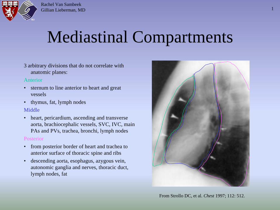
Mediastinal Compartments3 arbitrary divisions that do not correlate with
anatomic planes:Anterior• sternum to line anterior to heart and great
vessels• thymus, fat, lymph nodesMiddle• heart, pericardium, ascending and transverse
aorta, brachiocephalic vessels, SVC, IVC, main PAs and PVs, trachea, bronchi, lymph nodes
Posterior• from posterior border of heart and trachea to
anterior surface of thoracic spine and ribs• descending aorta, esophagus, azygous vein,
autonomic ganglia and nerves, thoracic duct, lymph nodes, fat
From Strollo DC, et al. Chest 1997; 112: 512.
Rachel Van SambeekGillian Lieberman, MD 1
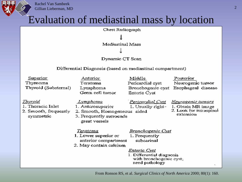
Evaluation of mediastinal mass by location
Rachel Van SambeekGillian Lieberman, MD
From Ronson RS, et al. Surgical Clinics of North America 2000; 80(1): 160.
2
Differential Diagnosis of Anterior Mediastinal Masses
1. Thymusa. Thymomab. Thymic Cystc. Thymic Hyperplasiad. Thymolipomae. Thymic Carcinomaf. Thymic Carcinoid
2. Teratoma & other germ cell tumors3. Thyroid (intrathoracic goiter) 4. “Terrible” Lymphoma (can be middle or posterior mediastinal)**These are often difficult to differentiate radiographically!**
(the presence of fat or fluid or the use of nuclear scanning can help in some situations)
Rachel Van SambeekGillian Lieberman, MD 3
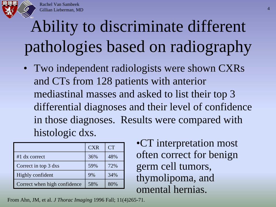
Ability to discriminate different pathologies based on radiography• Two independent radiologists were shown CXRs
and CTs from 128 patients with anterior mediastinal masses and asked to list their top 3 differential diagnoses and their level of confidence in those diagnoses. Results were compared with histologic dxs.
CXR CT
#1 dx correct 36% 48%
Correct in top 3 dxs 59% 72%
Highly confident 9% 34%
Correct when high confidence 58% 80%
•CT interpretation most often correct for benign germ cell tumors, thymolipoma, and omental hernias.
From Ahn, JM, et al. J Thorac Imaging 1996 Fall; 11(4)265-71.
Rachel Van SambeekGillian Lieberman, MD 4
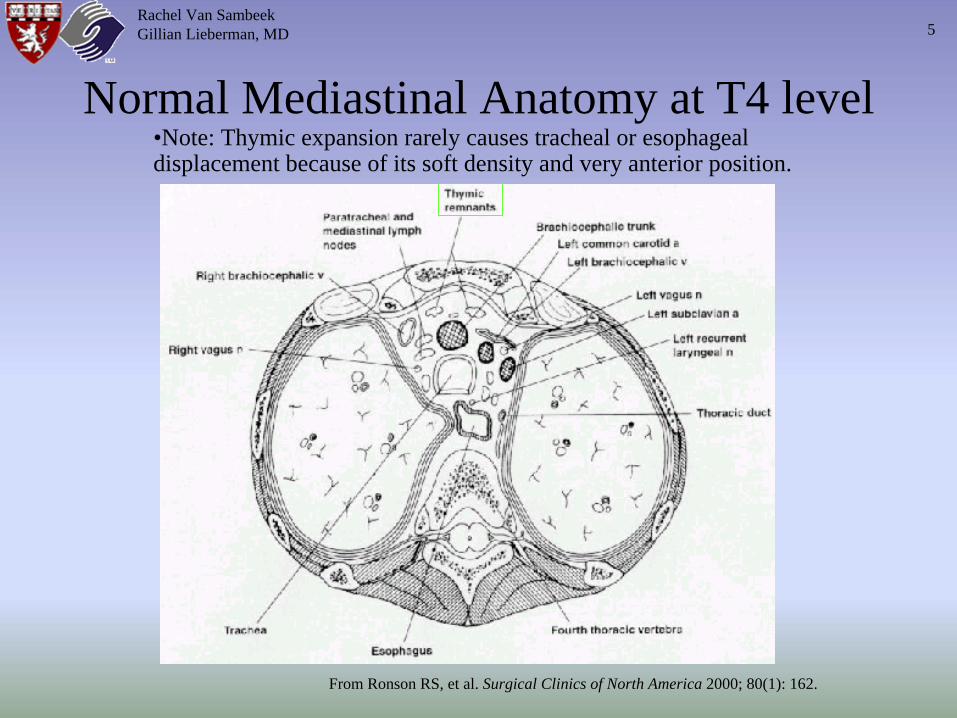
Normal Mediastinal Anatomy at T4 level
Rachel Van SambeekGillian Lieberman, MD
From Ronson RS, et al. Surgical Clinics of North America 2000; 80(1): 162.
5
•Note: Thymic expansion rarely causes tracheal or esophageal displacement because of its soft density and very anterior position.
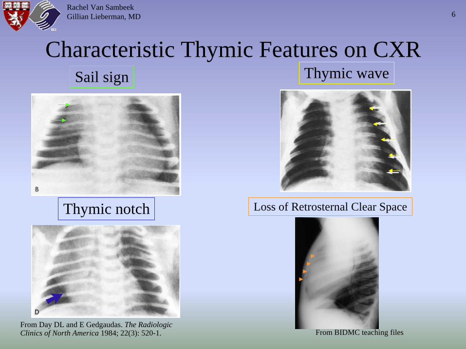
Characteristic Thymic Features on CXRSail sign
Thymic notch
Thymic wave
Loss of Retrosternal Clear Space
Rachel Van SambeekGillian Lieberman, MD
From BIDMC teaching files
6
From Day DL and E Gedgaudas. The Radiologic Clinics of North America 1984; 22(3): 520-1.
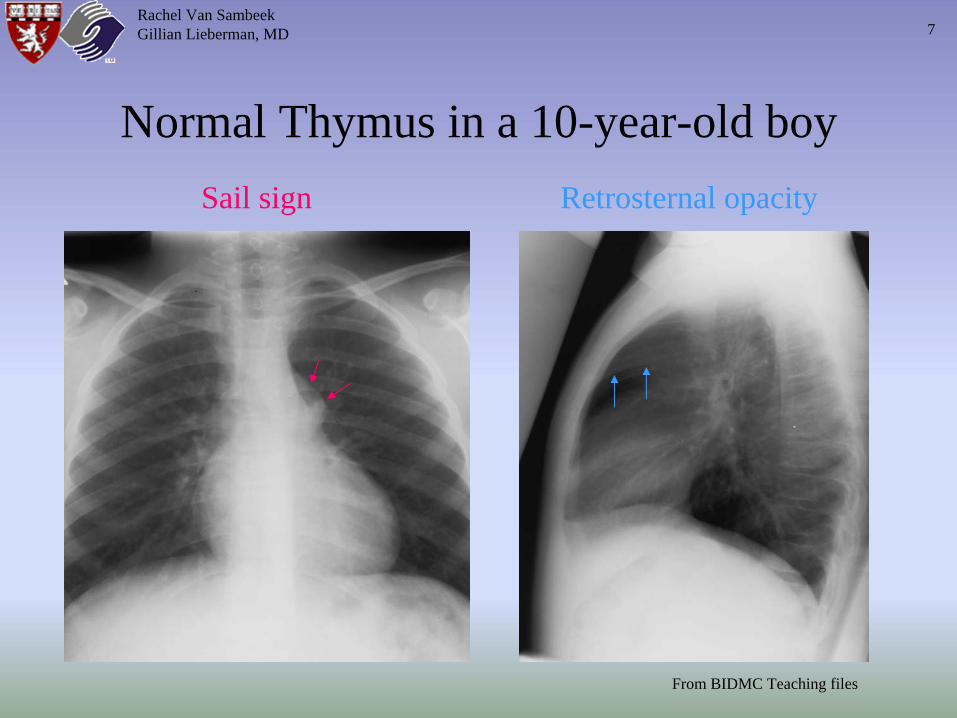
Normal Thymus in a 10-year-old boySail sign Retrosternal opacity
Rachel Van SambeekGillian Lieberman, MD
From BIDMC Teaching files
7
Thymoma• Most common anterior mediastinal primary tumor; 20% of adult mediastinal
neoplasms• Presentation between ages 30-50 (most patients are >40 years old)• 50% are asymptomatic• Symptoms secondary to compression: chest pain, cough, dyspnea, SVC
syndrome (obstructed SVC → head and neck venous congestion, facial edema)• Parathymic syndromes (approx. 40% of patients)
– Myasthenia Gravis: seen in 30-50% of thymoma patients; 8-20% of MG patients have a thymoma (although 90% have some sort of thymic abnormality); may develop post thymectomy
– Pure Red Cell Aplasia: seen in 5% of thymoma patients, but 50% of patients with red cell aplasia have a thymoma
– Others: hypogammaglobulinemia (10%), endocrine disorders, connective tissue disorders
• Usually arise in the midline and extend unilaterally• Grading: invasive vs. non-invasive (histologically identical)• Staging
– I: within intact capsule– II: extension through capsule into surrounding fat, pleura, or pericardium– III: intrathoracic metastasis (including pleural seeding)– IV: extrathoracic metastasis
Rachel Van SambeekGillian Lieberman, MD 8
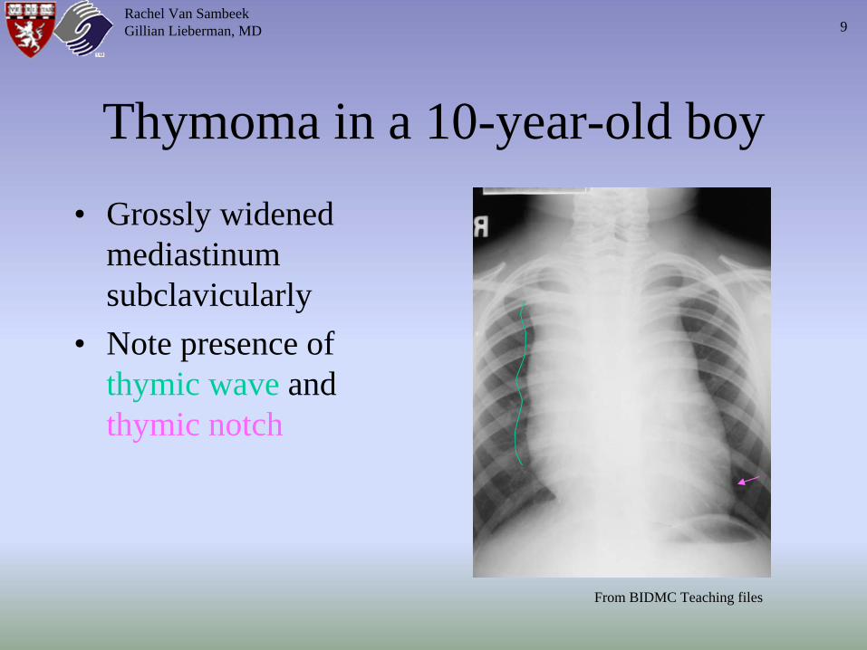
Thymoma in a 10-year-old boy• Grossly widened
mediastinum subclavicularly
• Note presence of thymic wave and thymic notch
Rachel Van SambeekGillian Lieberman, MD
From BIDMC Teaching files
9
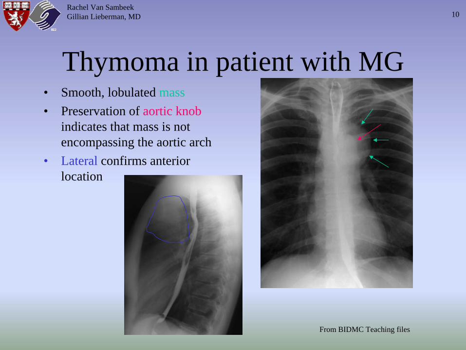
Thymoma in patient with MG• Smooth, lobulated mass• Preservation of aortic knob
indicates that mass is not encompassing the aortic arch
• Lateral confirms anterior location
Rachel Van SambeekGillian Lieberman, MD
From BIDMC Teaching files
10
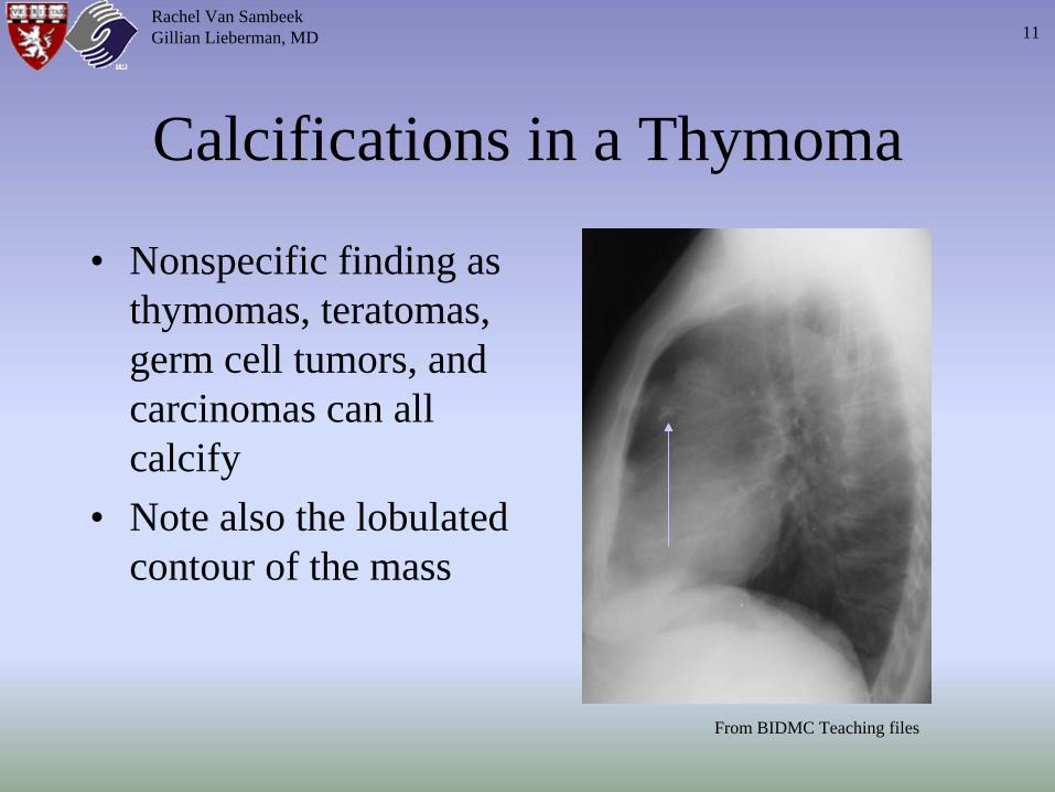
Calcifications in a Thymoma
• Nonspecific finding as thymomas, teratomas, germ cell tumors, and carcinomas can all calcify
• Note also the lobulated contour of the mass
Rachel Van SambeekGillian Lieberman, MD
From BIDMC Teaching files
11
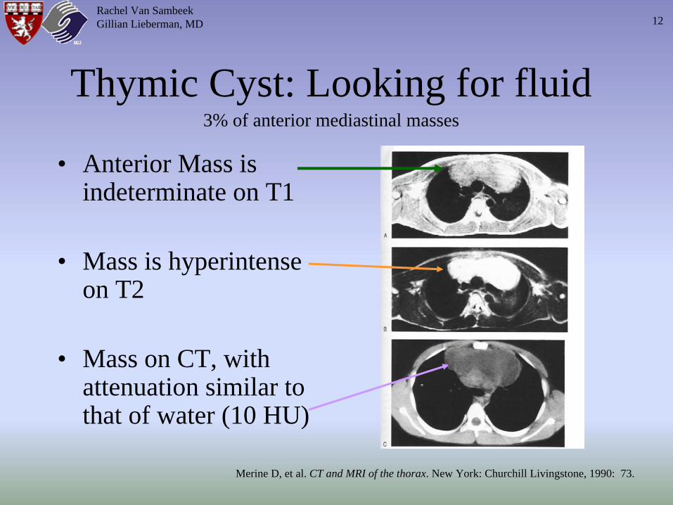
Thymic Cyst: Looking for fluid
• Anterior Mass is indeterminate on T1
• Mass is hyperintense on T2
• Mass on CT, with attenuation similar to that of water (10 HU)
Rachel Van SambeekGillian Lieberman, MD
Merine D, et al. CT and MRI of the thorax. New York: Churchill Livingstone, 1990: 73.
12
3% of anterior mediastinal masses
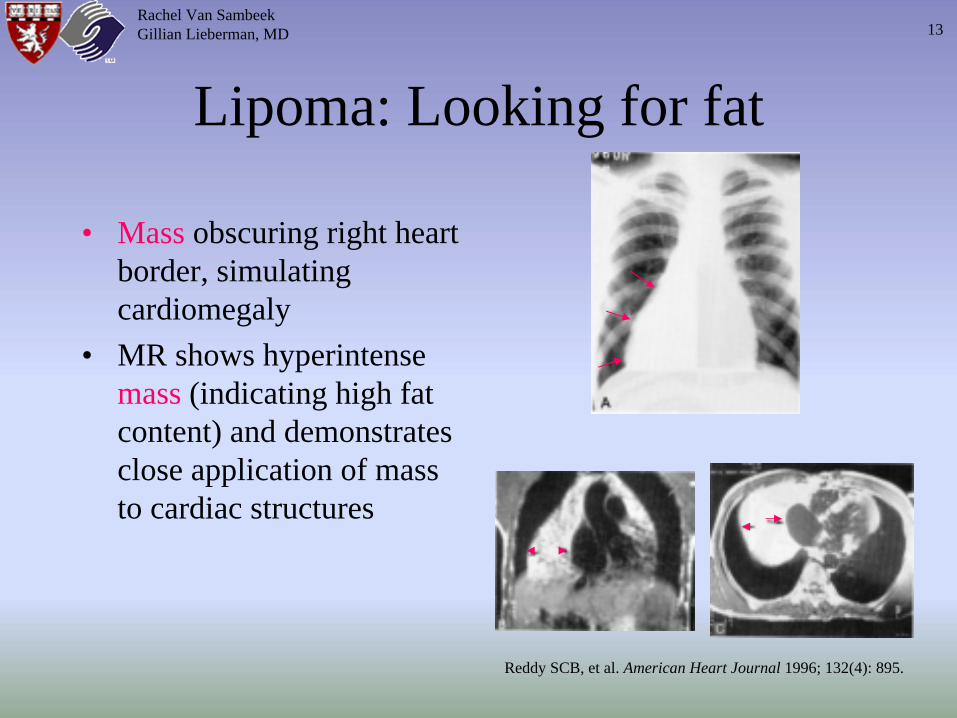
Lipoma: Looking for fat
• Mass obscuring right heart border, simulating cardiomegaly
• MR shows hyperintense mass (indicating high fat content) and demonstrates close application of mass to cardiac structures
Rachel Van SambeekGillian Lieberman, MD
Reddy SCB, et al. American Heart Journal 1996; 132(4): 895.
13
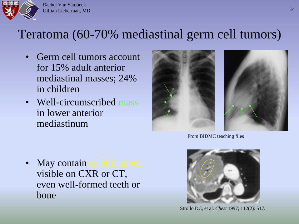
Teratoma (60-70% mediastinal germ cell tumors)• Germ cell tumors account
for 15% adult anterior mediastinal masses; 24% in children
• Well-circumscribed mass in lower anterior mediastinum
• May contain calcifications visible on CXR or CT, even well-formed teeth or bone
From BIDMC teaching files
Rachel Van SambeekGillian Lieberman, MD
Strollo DC, et al. Chest 1997; 112(2): 517.
14
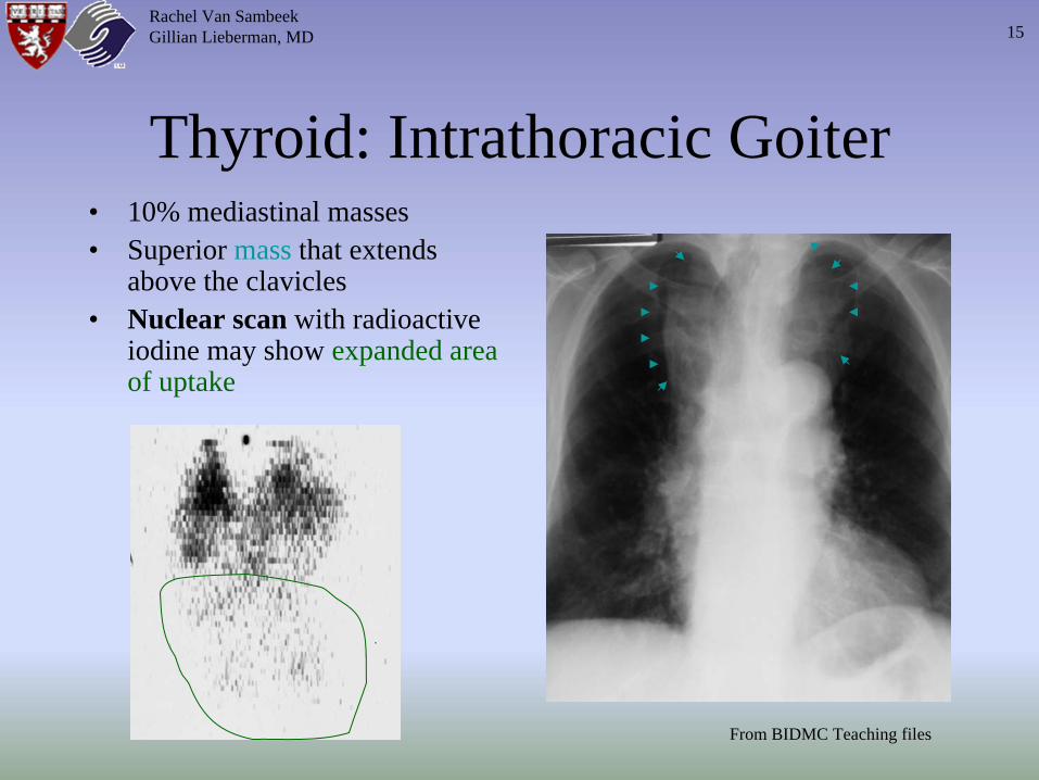
Thyroid: Intrathoracic Goiter• 10% mediastinal masses• Superior mass that extends
above the clavicles• Nuclear scan with radioactive
iodine may show expanded area of uptake
Rachel Van SambeekGillian Lieberman, MD
From BIDMC Teaching files
15
The Patient: KL
• CC: A 56-year old man with fever, cough, and fatigue for about 1 month, presents 4/21/01 with dizziness and ↑
weakness
• ER: Hct of 22.7 (1/02 baseline: 40); CXR notes unusual R heart border contour, but report “doubts any significance” to this finding
• In hospital, hematologic evaluation was obtained given inappropriately low reticulocyte count (3.5%)
Rachel Van SambeekGillian Lieberman, MD 16
The Consult: Hem/Onc Fellow•Bone marrow biopsy showed only deficiency of erythroid precursors.
Viral serologies were negative.•Suggested thymoma in differential of
red cell aplasia in the absence of bone marrow pathology.
•Recommended chest CT evaluation
Rachel Van SambeekGillian Lieberman, MD 17
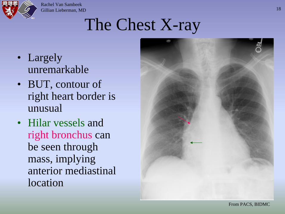
The Chest X-ray
• Largely unremarkable
• BUT, contour of right heart border is unusual
• Hilar vessels and right bronchus can be seen through mass, implying anterior mediastinal location
From PACS, BIDMC
Rachel Van SambeekGillian Lieberman, MD 18
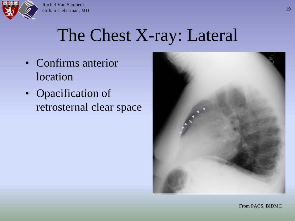
The Chest X-ray: Lateral• Confirms anterior
location• Opacification of
retrosternal clear space
From PACS, BIDMC
Rachel Van SambeekGillian Lieberman, MD 19
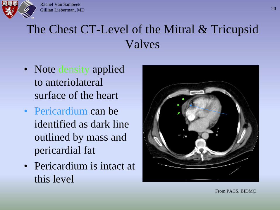
The Chest CT-Level of the Mitral & Tricupsid Valves
• Note density applied to anteriolateral surface of the heart
• Pericardium can be identified as dark line outlined by mass and pericardial fat
• Pericardium is intact at this level
From PACS, BIDMC
Rachel Van SambeekGillian Lieberman, MD 20
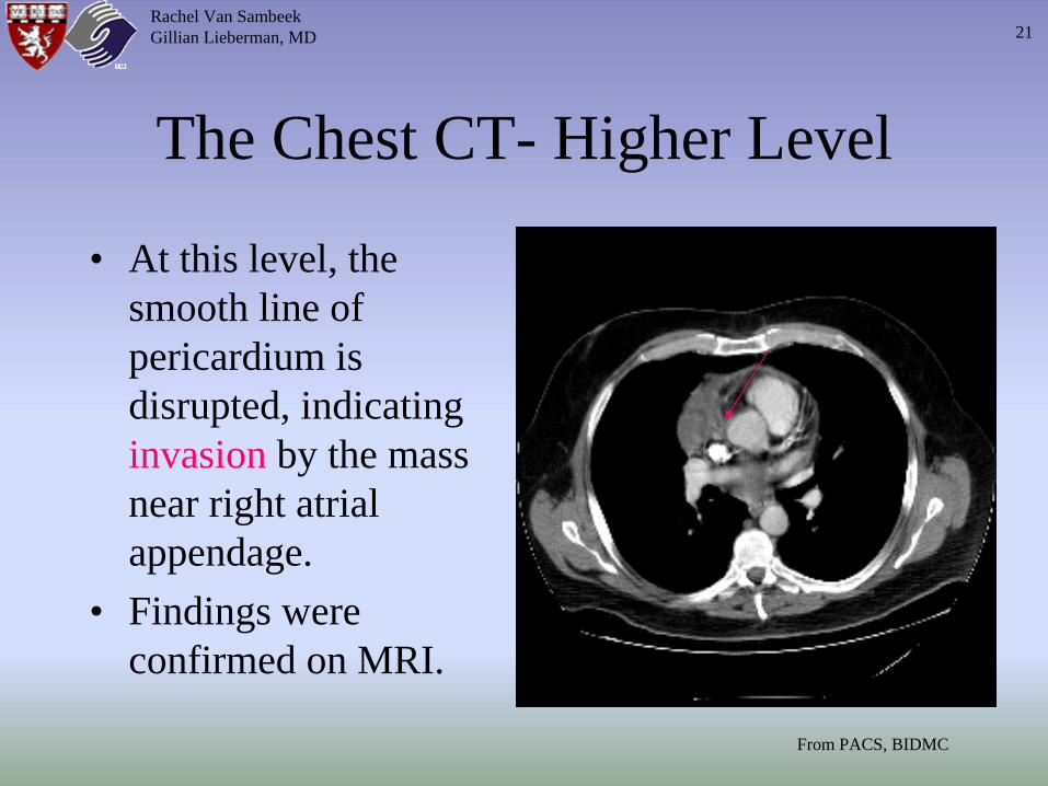
The Chest CT- Higher Level
• At this level, the smooth line of pericardium is disrupted, indicating invasion by the mass near right atrial appendage.
• Findings were confirmed on MRI.
From PACS, BIDMC
Rachel Van SambeekGillian Lieberman, MD 21
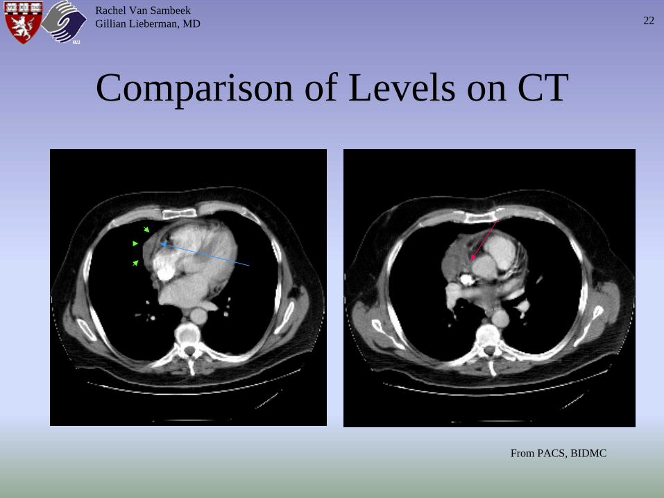
Comparison of Levels on CT
Rachel Van SambeekGillian Lieberman, MD
From PACS, BIDMC
22
The Surgery• Complete thymectomy was performed two weeks later.• Involvement of pericardium was noted at surgery, but
gross tissue planes were still identifiable• Histologically, tumor cells were identified to within 2-3
cell layers from the pericardial specimen margin.• Pathologic analysis of the surgical specimen was
somewhat controversial. There was some disagreement as to the etiology of the tumor: primary thymoma vs. metastatic lymphoma. CT imaging of head, abdomen, and pelvis did not reveal any other tumor sites.
• Pure red cell aplasia supports the diagnosis of thymoma.
Rachel Van SambeekGillian Lieberman, MD 23
References• Ahn JM, Lee KS, Goo JM, Song KS, Kim SJ, Im JG. Predicting the histology of anterior
mediastinal masses: comparison of chest radiography and CT. Journal of Thoracic Imaging 1996; 11(4): 265-71.
• Cohn WE. Anterior mediastinal mass lesions. UpToDate, Inc., 2001.• Day DL and E Gedgaudas. The thymus. The Radiologic Clinics of North America 1984;
22(3): 519-37.• Reddy SCB, Taneja K, Kothari SS, Goel P, Sharma S, Rajani M, Wasir HS. Mediastinal
lipoma mimicking dextrocardia and cardiac enlargement: Diagnosis by magnetic resonance imaging. American Heart Journal 1996; 132(4): 894-6.
• Ronson RS, Duarte I, Miller JI. Embryology and surgical anatomy of the mediastinum with clinical implications. Surgical Clinics of North America 2000; 80(1): 157-69.
• Rosado de Christenson ML, Galobardes J, Moran CA. Thymoma: radiologic-pathologic correlation. RadioGraphics 1992; 12(1): 151-68.
• Strollo DC, Rosado de Christenson ML, Jett JR. Primary mediastinal tumors, part 1: tumors of the anterior mediastinum. Chest 1997; 112(2): 511-22.
• Merine D, Pessar ML, Zerhouni EA, Fishman EK, Soulen RL. CT and MRI assessment of the mediastinum. CT and MRI of the thorax. New York: Churchill Livingstone, 1990: 67-91.
Rachel Van SambeekGillian Lieberman, MD 24
Acknowledgements
• Dr. James Busch• Larry Barbaras and CaraLyn D’amour, Webmasters
Rachel Van SambeekGillian Lieberman, MD 25