anti-filarial activity of antibiotic therapy is due to...
TRANSCRIPT

Anti-filarial Activity of Antibiotic Therapy Is Due toExtensive Apoptosis after Wolbachia Depletion fromFilarial NematodesFrederic Landmann1., Denis Voronin2., William Sullivan1, Mark J. Taylor2*
1 Department of Molecular, Cell and Developmental Biology, Sinsheimer Labs, University of California, Santa Cruz, California, United States of America, 2 Molecular and
Biochemical Parasitology, Liverpool School of Tropical Medicine, Pembroke Place, Liverpool, United Kingdom
Abstract
Filarial nematodes maintain a mutualistic relationship with the endosymbiont Wolbachia. Depletion of Wolbachia producesprofound defects in nematode development, fertility and viability and thus has great promise as a novel approach fortreating filarial diseases. However, little is known concerning the basis for this mutualistic relationship. Here we demonstrateusing whole mount confocal microscopy that an immediate response to Wolbachia depletion is extensive apoptosis in theadult germline, and in the somatic cells of the embryos, microfilariae and fourth-stage larvae (L4). Surprisingly, apoptosisoccurs in the majority of embryonic cells that had not been infected prior to antibiotic treatment. In addition, no apoptosisoccurs in the hypodermal chords, which are populated with large numbers of Wolbachia, although disruption of thehypodermal cytoskeleton occurs following their depletion. Thus, the induction of apoptosis upon Wolbachia depletion isnon-cell autonomous and suggests the involvement of factors originating from Wolbachia in the hypodermal chords. Thepattern of apoptosis correlates closely with the nematode tissues and processes initially perturbed following depletion ofWolbachia, embryogenesis and long-term sterilization, which are sustained for several months until the premature death ofthe adult worms. Our observations provide a cellular mechanism to account for the sustained reductions in microfilarialloads and interruption of transmission that occurs prior to macrofilaricidal activity following antibiotic therapy of filarialnematodes.
Citation: Landmann F, Voronin D, Sullivan W, Taylor MJ (2011) Anti-filarial Activity of Antibiotic Therapy Is Due to Extensive Apoptosis after Wolbachia Depletionfrom Filarial Nematodes. PLoS Pathog 7(11): e1002351. doi:10.1371/journal.ppat.1002351
Editor: David S. Schneider, Stanford University, United States of America
Received June 19, 2011; Accepted September 19, 2011; Published November 3, 2011
Copyright: � 2011 Landmann et al. This is an open-access article distributed under the terms of the Creative Commons Attribution License, which permitsunrestricted use, distribution, and reproduction in any medium, provided the original author and source are credited.
Funding: We thank the Bill and Melinda Gates Foundation for financial support of the ANWOL consortium through a grant awarded to the Liverpool School OfTropical Medicine. The funders had no role in study design, data collection and analysis, decision to publish, or preparation of the manuscript.
Competing Interests: The authors have declared that no competing interests exist.
* E-mail: [email protected]
. These authors contributed equally to this work.
Introduction
The majority of filarial nematodes host Wolbachia bacteria in a
mutualistic symbiotic association. In adult worms, the endosym-
bionts are situated in the hypodermal lateral chord cells, located
within host-derived vacuoles. In females, Wolbachia are also found
in the ovaries, oocytes and developing embryos within the uteri
[1–4].
The mutualistic association of Wolbachia in filarial nematodes
has been exploited as a novel approach to the treatment of
lymphatic filariasis caused by Wuchereria bancrofti and Brugia malayi
and onchocerciasis caused by Onchocerca volvulus [5]. The use of
tetracyclines or rifamycins to deplete Wolbachia leads to an arrested
development of larval and embryonic stages resulting in permanent
sterilization of adult female worms [6]. The adult parasites die
prematurely after 1–2 years following depletion of Wolbachia,
compared to their typical lifespan of 10–14 years, delivering for
the first time a safe and potent macrofilaricidal treatment for
filariasis [5].
Although the effects of Wolbachia depletion on the development,
fertility and viability of filarial nematodes has been documented
(reviewed in [7]), the reason why depletion of Wolbachia leads to
these anti-filarial outcomes is unknown. Here we have used whole
mount confocal microscopy to observe the consequences of
Wolbachia depletion on host nematode cellular and nuclear
structure. Our observations reveal an extensive and profound
development of apoptosis in germline cells and embryos following
antibiotic depletion of Wolbachia, which occurs soon after bacterial
depletion in B. malayi and is sustained for at least 21 months in O.
volvulus. We find extensive apoptosis even in cells that had not been
infected with Wolbachia prior to antibiotic treatment. Nuclear
structure in most somatic tissues remains intact, although
disruption of cytoskeleton arrangement occurs in the lateral chord
cells, where the vast majority of the bacteria reside.
Results
Morphological alteration of in vivo treated Brugia malayiadult worms
To investigate the contribution of Wolbachia to its filarial host
fitness and fertility, jirds infected with B. malayi were treated with
tetracycline (2.5 mg/ml in drinking water), for a period of 6 weeks.
Parasites were recovered from the peritoneal cavity at 8 weeks
post-treatment. Female worms from treated and non-treated jirds
were collected and stained for the presence of Wolbachia in the
lateral chords, the somatic tissue that they populate in the adult. As
PLoS Pathogens | www.plospathogens.org 1 November 2011 | Volume 7 | Issue 11 | e1002351
expected a dramatic reduction of the bacterial population was
observed in this tissue after the antibiotic treatment (Figure 1A, B).
Wolbachia depletion was confirmed and quantified by quantitative
PCR using a ratio of single copy genes: wsp (for Wolbachia) and gst
(for B. malayi) [8], which showed an 99% reduction of bacterial
load in treated adult female worms and in 14 day old L4 larvae.
Morphological defects in somatic tissues and in the germline,
based on DNA and actin staining were investigated using confocal
microscopy on whole mount nematodes. Numerous pyknotic
nuclei were observed throughout the ovaries and uteri in the
female germline through to the later stages of embryogenesis and
‘stretched’ microfilariae in treated worms (Figure 2). For example,
condensed and fragmented oogoniae nuclei surrounded by an
intense actin staining were observed in the treated females,
suggesting a reduction of the cytoplasmic volume (Figure 2 A, B).
Most of the intrauterine ‘stretched’ microfilariae that resulted from
a completed embryogenesis showed morphological defects such as
abnormal muscle quadrants, associated with pyknotic nuclei
(Figure 2 C, D).
Since pyknosis is a hallmark of cell death, the next step was to
determine whether the depletion of Wolbachia induced apoptosis as
detected using the TUNEL assay [9]. TUNEL allows detection of
apoptosis-caused DNA fragmentation by incorporation of fluores-
cent dUTP to DNA 3’ OH free ends. The germline in ovaries and
the embryos in the uterus were examined and the proportion of
each stage undergoing apoptosis was quantified (Figure 3 A to J).
In non-treated females, apoptosis at the level of germline nuclei is
a rare event (0.4%, n = 724), while apoptotic nuclei were widely
detected as patches in the ovaries of treated females (22%,
n = 2000 nuclei from a total of 4 treated females) (Figure 3 B, C, J).
Apoptotic nuclei became more numerous as the uteri filled with
embryos in treated females, while no abnormal apoptosis was
detected in the control non-treated females (Figure 3 D, E and J).
In embryos, a few cells were TUNEL positive in the untreated
control samples (Figure 3 F, J). This apoptosis is likely to be
developmentally programmed. In Caenorhabditis elegans, where it is
well characterized, about 12% of the total adult somatic cells
undergo programmed cell death during development [10]. In
contrast, the majority of the blastomeres of TUNEL-positive
embryos from treated females were apoptotic (Figure 3 G, J, 53%,
n = 550).
We finally observed the intrauterine ‘stretched’ microfilariae
extracted from the proximal uteri. No apoptosis was detected in
the control samples, whereas most of the ‘stretched’ microfilariae
from treated females were entirely undergoing apoptosis (Figure 3
H, I, J 83%, n = 45). Apoptosis appeared to be cumulative, with
more and more embryos affected as development progresses.
To better understand the contribution of the Wolbachia in the
lateral chord versus the few Wolbachia present in oocytes, and
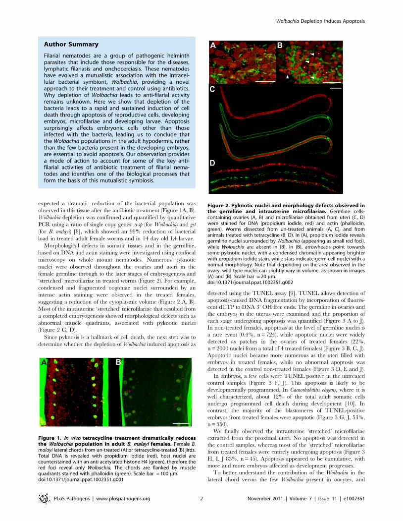
Figure 1. In vivo tetracycline treatment dramatically reducesthe Wolbachia population in adult B. malayi females. Female B.malayi lateral chords from un-treated (A) or tetracycline-treated (B) jirds.Total DNA is revealed with propidium iodide (red), host nuclei arecounterstained with an anti acetylated histone H4 (green), therefore thered foci reveal only Wolbachia. The chords are flanked by musclequadrants stained with phalloidin (green). Scale bar = 100 mm.doi:10.1371/journal.ppat.1002351.g001
Figure 2. Pyknotic nuclei and morphology defects observed inthe germline and intrauterine microfilariae. Germline cells-containing ovaries (A, B) and microfilariae obtained from uteri (C, D)were stained for DNA (propidium iodide, red) and actin (phalloidin,green). Worms dissected from un-treated animals (A, C), and fromanimals treated with tetracycline (B, D). In (A), propidium iodide revealsgermline nuclei surrounded by Wolbachia (appearing as small red foci),while Wolbachia are absent in (B). In (B), arrowheads point towardssome pyknotic nuclei, with a condensed chromatin appearing brighterwith propidium iodide stain, while stars indicate germ cell nuclei with anormal morphology. Note that depending on the area observed in theovary, wild type nuclei can slightly vary in volume, as shown in images(A) and (B). Scale bar = 20 mm.doi:10.1371/journal.ppat.1002351.g002
Author Summary
Filarial nematodes are a group of pathogenic helminthparasites that include those responsible for the diseases,lymphatic filariasis and onchocerciasis. These nematodeshave evolved a mutualistic association with the intracel-lular bacterial symbiont, Wolbachia, providing a novelapproach to their treatment and control using antibiotics.Why depletion of Wolbachia leads to anti-filarial activityremains unknown. Here we show that depletion of thebacteria leads to a rapid and sustained induction of celldeath through apoptosis of reproductive cells, developingembryos, microfilariae and developing larvae. Apoptosissurprisingly affects embryonic cells other than thoseinfected with the bacteria, leading us to conclude thatthe Wolbachia populations in the adult hypodermis, ratherthan the few bacteria present in the developing embryos,are essential to avoid apoptosis. Our observation providesa mode of action to account for some of the key anti-filarial activities of antibiotic treatment of filarial nema-todes and identifies one of the biological processes thatform the basis of this mutualistic symbiosis.
Wolbachia Depletion Induces Apoptosis
PLoS Pathogens | www.plospathogens.org 2 November 2011 | Volume 7 | Issue 11 | e1002351

subsequently present in the embryo by maternal transmission
(from early to mid-embryogenesis we found an average of 70
Wolbachia +/212 (n = 10) per embryo), we examined B. malayi
males, which are devoid of Wolbachia in the germline. We
performed a TUNEL assay in males obtained from the same
jirds. While no apoptosis was detected in control males, we
observed a small number of apoptotic events in irregular patches
in the germline of treated males, suggesting that the Wolbachia in
the chords play a role in preventing apoptosis in the male germline
(Figure 4 A to F).
In females, the chords are larger than in males and contain ,10
fold more Wolbachia in worms 6 months or older, although they
begin their adult lives with equivalent numbers and ratios [11].
The chords are closely apposed to the uteri, and this adjacency
possibly facilitates the supply of nutrients or critical metabolites to
the growing embryos [12]. The Wolbachia present in the female
lateral chords may therefore have a more important role than in
males, and their contribution may be crucial to avoid apoptosis
during female germline and embryonic development.
The effect of Wolbachia depletion on the nuclei and cytoskeleton
morphologies in the lateral chords in treated and non-treated
females was investigated next (Figures 1 and 5). Lateral chord cells
are syncytial and the prominent rows of nematode nuclei are easily
observed. The lateral chord nuclei showed no evidence of pyknosis
or any difference in TUNEL staining in either treated or untreated
worms (Figure 5 A, B). This suggests that that the loss of Wolbachia
in somatic tissues, does not lead to apoptosis of the lateral chord
cells. However, the cortical microtubule network, circumferentially
oriented in loose bundles in control samples, was disrupted in
treated females (Figure 5 C, D). These cytoskeleton defects may
impair the chords function in supplying nutrients/metabolites to
the germline and the developing embryos.
In order to determine whether the observed increase in
apoptosis in germline cells and embryos was dependent on any
mammalian host factors, we cultured untreated female and male
worms in vitro with doxycycline (8 mM) for a period of five days [8].
Worms were TUNEL-assayed at day 1, 2, 4 and 5 post-treatment.
No differences were seen between control and treated worms until
day 4. On days 4 and 5, doxycycline-treated worms showed
abnormal apoptosis in germ cells, during fertilization, and in
young embryos (Figure 6). Based on this in vitro doxycycline
treatment, we concluded that the in vivo tetracycline-induced
Wolbachia depletion could cause apoptosis independently of any
mammalian host derived factors.
Apoptosis in Brugia malayi microfilaria followingtetracycline treatment
Following intra-peritoneal infection of jirds with B. malayi, the
adults mate and release microfilariae, which remain confined to
the peritoneal cavity. Because microfilariae accumulate in the
peritoneal cavity, a proportion of those recovered will include
Figure 3. In vivo tetracycline treatment leads to apoptosis inadult worm reproductive tissues. (A) Schematic drawing of onefemale reproductive tract, representing the approximate localization ofthe different sections observed in the TUNEL assay. TUNEL experimentsshowing the DNA (PI in red) and incorporated fluorescein-dUTP (green),in samples from non-treated (B, D, F, H) or tetracycline-treated (C, E, G, I)B. malayi females. Mitotic proliferation zone in the distal ovary (B, C).Arrowheads indicate mitotic nuclei, arrows point to somatic gonad
nuclei. (D) Uterus filled with elongating embryos. (E) Fertilization area inthe distal uterus in the top left corner, and proximal uterus filled (indiagonal) with developing embryos. (F, G) Single developing embryos.(H, I) Intrauterine microfilariae extracted from proximal uteri. (J) TUNELquantification. For each un-treated (NT) or tetracycline-treated (TET)sample, TUNEL-positive nuclei or embryos were counted and expressedas a percentage of total nuclei or embryos (based on DNA staining withPI). For the embryonic count (‘‘embryos NT’’, ‘‘embryos TET’’), embryoswith a number of TUNEL positive nuclei equal or less than 2 positivenuclei were considered as negative embryos. All the intrauterinemicrofilariae found TUNEL-positive in the TET sample had every nucleiTUNEL-positive (I). Scale bar = 15 mm.doi:10.1371/journal.ppat.1002351.g003
Wolbachia Depletion Induces Apoptosis
PLoS Pathogens | www.plospathogens.org 3 November 2011 | Volume 7 | Issue 11 | e1002351
moribund or dead parasites. Therefore, we first determined the
basal level of apoptosis using the TUNEL assay in microfilaria
from untreated jirds using the following criteria: high (more than
20 nuclei), medium (5–20 nuclei) and low (less than 5 nuclei) levels
of apoptotic-positive nuclei per single microfilariae. One hundred
microfilariae from each treatment group were analyzed. Eighty
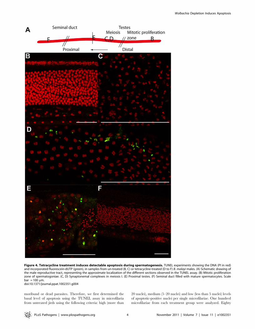
Figure 4. Tetracycline treatment induces detectable apoptosis during spermatogenesis. TUNEL experiments showing the DNA (PI in red)and incorporated fluorescein-dUTP (green), in samples from un-treated (B, C) or tetracycline-treated (D to F) B. malayi males. (A) Schematic drawing ofthe male reproductive tract, representing the approximate localization of the different sections observed in the TUNEL assay. (B) Mitotic proliferationzone of spermatogoniae. (C, D) Synaptonemal complexes in meiosis I. (E) Proximal testes. (F) Seminal duct filled with mature spermatocytes. Scalebar = 100 mm.doi:10.1371/journal.ppat.1002351.g004
Wolbachia Depletion Induces Apoptosis
PLoS Pathogens | www.plospathogens.org 4 November 2011 | Volume 7 | Issue 11 | e1002351

Figure 5. Cytoskeleton defects are revealed in somatic tissues, without apoptotic phenotypes. Female B. malayi lateral chords from un-treated (A, C) or tetracycline-treated (B, D) jirds. No pyknotic nuclei were detected, and the TUNEL levels were similar in both samples (A, B). (C, D)Apical microtubule network. Scale bar = 100 mm.doi:10.1371/journal.ppat.1002351.g005
Figure 6. Doxycycline treatment leads to apoptosis in vitro. (A, C) control worms and treated worms (B, D) were TUNEL assayed (green) andstained for DNA (PI in red). (A, B) Germ cells in mitotic proliferation in the ovaries. (C) Proximal uteri filled with developing embryos. (D) Apoptoticoocytes and early embryos (arrows) in a distal uterus, surrounded by sperm cells (arrowheads). Scale bar = 15 mm.doi:10.1371/journal.ppat.1002351.g006
Wolbachia Depletion Induces Apoptosis
PLoS Pathogens | www.plospathogens.org 5 November 2011 | Volume 7 | Issue 11 | e1002351
three percent of microfilaria from untreated control groups had
none or less than 5 apoptotic positive nuclei, 14% had a medium
number of apoptotic positive nuclei (5–20) and 3% contained the
highest level (.20). In the tetracycline treated group there was a
2.6 fold increase in the proportion of microfilaria with a medium
number of apoptotic nuclei (34%) and a 7.3 fold increase in the
proportion of microfilariae with the highest level of apoptotic
nuclei (22%) (Table 1). So, although we observed an increase in
the levels of apoptosis in released microfilariae, a significant
proportion (44%) only show minimal or no induction of apoptosis,
unlike intrauterine ‘stretched’ microfilariae where the evidence of
apoptosis is extensive and widespread affecting the vast majority of
the terminal development stage in the uterus.
Increases in cell death protein-3 (ced-3) gene expressionand levels of activated CED-3 protein are observed inWolbachia-depleted parasites
To further investigate the induction of apoptosis following
depletion of Wolbachia, we analyzed gene expression and protein
profiles of cell death protein-3 (ced-3), a homologue of human
Caspase-3. The relative level of cell death protein-3 (ced-3) gene
expression was significantly increased in tetracycline treated
females (p,0.01, n = 6 worms) compared with untreated controls
(Figure 7 A). We performed a western blot analysis of CED-3
protein extracted from microfilariae and 14 day old L4 larvae
obtained ex vivo. Caspase-3 was detected in the inactive form
(,50 kDa) and as cleaved activated forms (from 47 to 19 kDa).
Figure 7B shows an increased amount of inactive and cleaved
CED-3 forms in the tetracycline treated samples compared with
controls, demonstrating the activation of CED-3 and it’s over
expression in microfilaria and L4 larvae following depletion of
Wolbachia.
Apoptosis is observed by TUNEL in Onchocerca volvulusfrom nodule biopsies of doxycycline-treated humanpatients
One of the outcomes of doxycycline therapy of filarial parasites
is the long-term sterilization through blockage of embryogenesis
leading to sustained reductions in microfilarial loads post treatment.
In order to investigate whether this long-term sterility was a direct
result of sustained embryonic apoptosis, we analyzed adult O.
volvulus obtained from a field trial of doxycycline in Cameroon [13].
Paraffin sections of O. volvulus nodules collected from 6 different
patients of each treatment group (doxycycline treated and placebo
treated, see Materials and Methods) were investigated using the
TUNEL assay. In all samples obtained from the doxycycline treated
group, numerous apoptotic-positive cells were observed in germline or
early embryonic cells and in somatic nuclei of the surrounding uterus
within adult female parasites (Figure 8). Other uterine embryonic
stages were absent due to the blockage of embryogenesis observed
following doxycycline therapy [14]. In contrast, samples collected
from placebo treated patients, showed only very occasional evidence
of apoptotic-cells in the different embryonic developmental stages in
utero and no evidence of apoptosis in the uterine wall (Figure 8 A, B).
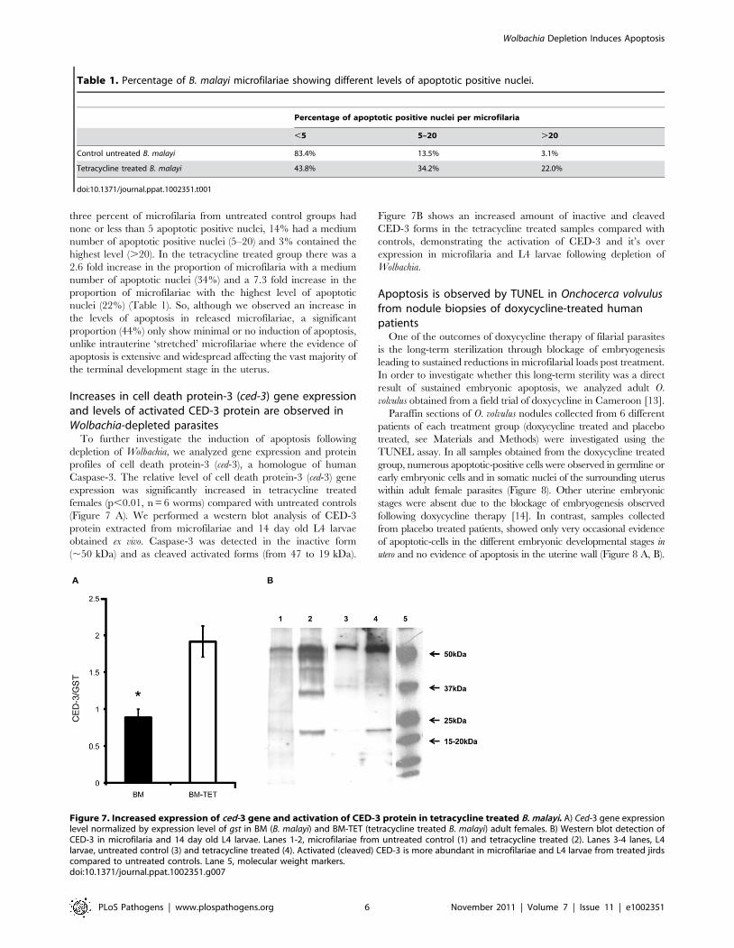
Table 1. Percentage of B. malayi microfilariae showing different levels of apoptotic positive nuclei.
Percentage of apoptotic positive nuclei per microfilaria
,5 5–20 .20
Control untreated B. malayi 83.4% 13.5% 3.1%
Tetracycline treated B. malayi 43.8% 34.2% 22.0%
doi:10.1371/journal.ppat.1002351.t001
Figure 7. Increased expression of ced-3 gene and activation of CED-3 protein in tetracycline treated B. malayi. A) Ced-3 gene expressionlevel normalized by expression level of gst in BM (B. malayi) and BM-TET (tetracycline treated B. malayi) adult females. B) Western blot detection ofCED-3 in microfilaria and 14 day old L4 larvae. Lanes 1-2, microfilariae from untreated control (1) and tetracycline treated (2). Lanes 3-4 lanes, L4larvae, untreated control (3) and tetracycline treated (4). Activated (cleaved) CED-3 is more abundant in microfilariae and L4 larvae from treated jirdscompared to untreated controls. Lane 5, molecular weight markers.doi:10.1371/journal.ppat.1002351.g007
Wolbachia Depletion Induces Apoptosis
PLoS Pathogens | www.plospathogens.org 6 November 2011 | Volume 7 | Issue 11 | e1002351

Discussion
Wolbachia depletion induces apoptosis in germline andsomatic tissues
Our observations show that an extensive apoptosis of adult
germline cells, embryos and somatic cells of microfilariae occurs
following the depletion of Wolbachia from filarial nematodes. The
development of apoptosis occurs soon after the depletion of
bacteria with tetracycline in experimental infections of B. malayi in
animals and in vitro. These observations were confirmed by
evidence of activation of Cell death protein-3 in treated adult
females, microfilaria and L4 larvae. Furthermore, apoptosis is
observed in the germline cells and uterine tissues of O. volvulus at
least 21 months following antibiotic treatment of people with
onchocerciasis. These observations are consistent with the known
anti-filarial effects of Wolbachia depletion on the rapid and
sustained blockage of embryogenesis, the decline of microfilarial
loads and the interruption of transmission to vectors and the
arrested development of larvae to adults in the mammalian host
[7,14–16]. Previous studies showing that antibiotic treatment of
the Wolbachia-free filarial nematode, Acanthocheilonema viteae has no
effect on the viability or biological processes of this species [17],
supports our conclusion that the observed apoptosis is due to the
loss of Wolbachia rather than a direct effect of tetracycline
treatment. The lack of apoptosis in lateral chord cells and other
somatic tissues suggests the event is not a global consequence of
Wolbachia depletion, which is consistent with the long and gradual
decline in the viability of adult worms.
In the case of onchocerciasis, permanent sterilization of adult
females is a therapeutically attractive outcome, as this blocks the
Figure 8. Apoptosis and apoptotic bodies are detected in O. volvulus tissues from human nodules of doxycycline treated patients. A,B. Cross-sections of adult female worm showing absence of apoptosis and intact embryonic inter-uterine stages (oocytes, pretzel stages, coiledembryonic microfilariae). C-G. Cross-sections of adult female worms depleted of Wolbachia showing extensive apoptosis of germline and earlyembryonic cells and uterine epithelial cells. Stars label inter-uterine content, black arrowheads label apoptotic germline and early embryonic cells aswell as human cells surrounding the worm, white arrowheads point to somatic cells, such as epithelial cells surrounded uteri. Scale bar = 20 mm.doi:10.1371/journal.ppat.1002351.g008
Wolbachia Depletion Induces Apoptosis
PLoS Pathogens | www.plospathogens.org 7 November 2011 | Volume 7 | Issue 11 | e1002351
release of the skin dwelling microfilariae, the developmental stage
that gives rise to skin and eye disease [3,7,14]. In lymphatic
filariasis, where the adult stage is responsible for disease
pathogenesis of damage to the lymphatics, the removal of adult
worms is required. In both cases the permanent sterilization of
adult worms will deliver important benefits for the interruption of
parasite transmission via blood feeding insect vectors [5,18].
Non cell-autonomous effects of Wolbachia depletionRegulation of host cell apoptosis is common to many
intracellular bacteria [19–21]. In many cases the bacterial
pathogens commandeer conserved apoptotic regulating cascades.
This requires the pathogen to reside within or be directly associated
with the affected cell. A unique feature of Wolbachia’s anti-apoptotic
effect is that there is no correlation between Wolbachia-populated
cells and cells that undergo apoptosis upon Wolbachia depletion
(Figure 9). For example, Wolbachia-laden hypodermal chords do not
undergo apoptosis upon Wolbachia depletion. In contrast, while only
a few cells in the early embryo (the C linage, [12]) possess Wolbachia,
the majority of cells in the embryo undergo apoptosis. Similarly,
while only the middle third of intrauterine ‘stretched’ microfilariae
are Wolbachia-infected, apoptosis occurs throughout the entire
organism upon Wolbachia depletion (Figure 9). These data clearly
demonstrate that the effects of Wolbachia-depletion on apoptosis are
non-cell autonomous.
The non-cell autonomous behaviour implies that Wolbachia-
infected cells suppress apoptosis in distant non-neighbouring
uninfected cells. The source of the Wolbachia-infected cells, which
are capable of this action-at-distance apoptosis suppression and
when in development this occurs remains unclear. Given the
abundant Wolbachia populations in the female lateral chords, it is
likely that these are primarily responsible for apoptotic rates in the
oocytes, embryos and intrauterine ‘stretched’ microfilariae. This
view is supported by the anatomy of the female in which the lateral
chords reside in close proximity to the ovaries and uteri [1,12] and
the greater needs of embryo requirements compared to sperma-
tozoa. Further evidence to support the predominant role of
depleted chord cell Wolbachia populations in inducing apoptosis in
intrauterine developmental stages comes from our observations on
released microfilariae. Although microfilariae recovered from
tetracycline treated animals did show an increase in the proportion
of parasites with medium and high levels of apoptosis, this was not
as extensive as we observed with intrauterine ‘stretched’
microfilariae. We were unable, however, to distinguish between
microfilariae that were released prior to or after treatment and
further experiments using isolated microfilariae only infections are
planned to confirm this observation. Our results are, however,
consistent with previous observations on the effect of Wolbachia
depletion on microfilarial viability and transmission. The majority
of microfilariae depleted of Wolbachia retain their motility and
viability in the mammalian host but their ability to develop in the
vector is compromised [15].
Mechanism of apoptosis inductionOur studies raise the issue of the mechanism by which Wolbachia
suppresses apoptosis. Studies of the influence of other intracellular
bacteria on host cell apoptosis demonstrate this may be achieved
either by direct targeting of the apoptotic signaling cascade or
indirectly by influencing host metabolism [19,22,23]. An example
of the former occurs in Chlamydia, which produces a protein that
targets the host pro-apoptotic protein Bim for proteasome-based
destruction [24]. Anaplasma phagocytophilium, a close relative of
Wolbachia, inhibits host cell apoptosis through targeting multiple
apoptotic pathways [21] and releases a protein, Ats1, via the type
IV secretion system that reduces the sensitivity of mitochondria to
apoptotic signals [25]. Potential molecular candidates from
Wolbachia, which have been shown to inhibit apoptosis, include
Wolbachia surface protein (WSP) [26] or lipoproteins such as
wBmPAL, which drive innate and adaptive inflammatory immunity
associated with disease pathogenesis [27] and can inhibit apoptosis
of human neutrophils [28] (Taylor et al. unpublished observation).
Bacterial pathogens subvert apoptosis to enhance their ability to
replicate and survive in host cells, whereas, Wolbachia, which has a
mutualistic rather than a parasitic or pathogenic association with its
host nematode, clearly benefits both itself and it’s host by preventing
apoptosis during larval, germline and embryonic development to
ensure its transmission to the next generation. The only other
example of a mutualistic association with Wolbachia and arthropods
is the parasitic wasp Asobara tabida in which depletion of Wolbachia
results in increased apoptosis in the nurse cells of the female
germline [29], suggesting these mutualistic Wolbachia associations
may share common pathways.
Wolbachia’s effect on apoptosis may be through more in-
direct mechanisms such as influencing the provisioning of essential
metabolites, a process likely to be compromised by the hypodermal
cytoskeletal defects following Wolbachia depletion. In this context it is
notable that the nematode biological process most sensitive to
Wolbachia depletion, embryogenesis and larval development, are
processes with high metabolic demands for tissue development,
growth and organogenesis. Examples of metabolite provision
dependent on Wolbachia under nutritional stress have been described
in Drosophila showing that Wolbachia up-regulates bacterioferritin, an
iron chelator, and may buffer the host from dramatic changes in
iron concentration in the environment [30,31]. Thus, Wolbachia
may provide protection from oxidative stress produced by high
internal concentrations of iron. Support for a similar protective
mechanism operating in filarial nematodes comes from genomic
studies demonstrating that B. malayi lacks the heme biosynthetic
pathway while the Wolbachia genome retains most of the entire heme
and riboflavin pathways [32]. Heme and riboflavin are efficient iron
chelators. Thus the abundance of Wolbachia in the chords has a
tremendous iron buffering potential.
Together our observations provide a cellular mechanism to
explain some of the anti-filarial effects, which occur following
antibiotic depletion of Wolbachia from filarial nematodes. The
pattern of apoptosis we observe closely correlates with the outcomes
previously observed in antibiotic therapy in laboratory models of
filarial nematodes and in human field trials with doxycycline [5].
The first event, which occurs soon after bacterial depletion from
adult female worms, is the blockage of embryogenesis and cessation
of microfilarial production, which is consistent with the extensive
and profound apoptosis observed in uterine embryonic stages of B.
malayi. This results in long-term sterilization of adult female worms
and the slow decline in microfilarial levels, until patients reach a
sustained amicrofilarial state, with benefits to disease reduction in
onchocerciasis and interruption of transmission in both onchocer-
ciasis and lymphatic filariasis. This state of sterilization persists for at
least 21 months, which is reflected in the retention of widespread
apoptotic cells in adult female germline and uterine cells of O.
volvulus obtained from patients treated with doxycycline, which
suggests this is a permanent status. The increase in the proportion of
released microfilariae with medium to high levels of apoptotic cells,
but less extensive than that observed in intrauterine ‘stretched’
microfilarial stages, is consistent with the observation that following
Wolbachia depletion, a proportion of microfilariae retain their
motility and viability, yet their capacity to develop within the vector
host is reduced. A similar pattern of activation of apoptosis in
Wolbachia-depleted L4 larvae suggests that this process also accounts
Wolbachia Depletion Induces Apoptosis
PLoS Pathogens | www.plospathogens.org 8 November 2011 | Volume 7 | Issue 11 | e1002351
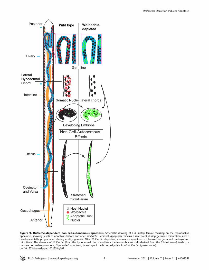
Figure 9. Wolbachia-dependent non cell-autonomous apoptosis. Schematic drawing of a B. malayi female focusing on the reproductiveapparatus, showing levels of apoptosis before and after Wolbachia removal. Apoptosis remains a rare event during germline maturation, and isdevelopmentally programmed during embryogenesis. After Wolbachia depletion, cumulative apoptosis is observed in germ cell, embryo andmicrofilaria. The absence of Wolbachia (from the hypodermal chords and from the few embryonic cells derived from the C blastomere) leads to amassive non cell-autonomous, ‘‘bystander’’ apoptosis, in embryonic cells normally devoid of Wolbachia (green nuclei).doi:10.1371/journal.ppat.1002351.g009
Wolbachia Depletion Induces Apoptosis
PLoS Pathogens | www.plospathogens.org 9 November 2011 | Volume 7 | Issue 11 | e1002351

for the arrested development of L4 larvae to adult worms. Finally,
we also observed that apoptosis is not widely induced in the nuclei of
somatic tissues and the syncytial chord cells of adult worms, where
the majority of the bacterial population exist, which is again
consistent with the long-term macrofilaricidal effects seen in both
lymphatic filariasis and onchocerciasis, which only manifest after 12
months or 18–27 months respectively. Further studies to define the
process by which Wolbachia regulates host nematode apoptosis may
provide alternative targets to screen for drugs or biomarkers of anti-
Wolbachia activity, which could yield alternative and improved
therapeutic options for the control and elimination of lymphatic
filariasis and onchocerciasis.
Materials and Methods
Ethics statementHuman parasite material was obtained from patients enrolled in
a double-blind placebo-controlled randomized clinical trial
conducted in Cameroon. The experimental protocol for this study
was designed in accordance with the general ethical principles
outlined in the Declaration of Helsinki. The trial was approved by
ethics committees of the Tropical Medicine Research Station,
Kumba and the Research Ethics Committee of The Liverpool
School of Tropical Medicine. Written informed consent was
obtained from all participants, with the exception of those who
were illiterate, where a literate witness signed on behalf of the
participant and the participant added a thumbprint. The trial is
registered with the current controlled trials registry, no:
ISRCTN48118452.
The animal experiments were carried out in strict accordance
with the Animals Scientific (Procedures Act) 1986 (UK) under a
license granted by the Home Office (London, UK). Experimental
procedures were reviewed and approved by the Animal Welfare
Committee, Liverpool School of Tropical Medicine and the Home
Office (London, UK).
Parasite materialAdult B. malayi cultivated in the peritoneal cavity of jirds
(Meriones unguiculatus) for were obtained from TRS Laboratories
(Athens Georgia). In Liverpool, infected animals received
tetracycline at 2.5 mg/ml in drinking water for a period of 6
weeks. Control infected jirds were maintained in a similar fashion
but without the tetracycline. Two weeks after the end of the
treatment adult worms and microfilariae were collected from the
peritoneal cavities using preheated (37uC) culture medium RPMI-
1640 supplemented with 100 U/ml penicillin, 100 mg/ml strep-
tomycin, 2 mM L-glutamine, 2.5 mg/ml amphotericin B, and
25 mM HEPES (GIBCO). A further two groups of jirds were
infected with 500 infective third-stage larvae (L3) and L4 larvae
were collected 14 days after the jirds were treated with tetracycline
(2.5 mg/ml) in drinking water or as untreated controls. All
parasites were washed in PBS and either fixed for confocal
microscopy or processed for RNA and protein. Ethanol-fixed
paraffin-embedded onchocercomas were obtained from patients
infected with O. volvulus enrolled in a double-blind placebo-
controlled randomized clinical trial conducted in Cameroon [13]
(trial registration number ISRCTN48118452). Patients received
doxycycline for 6 weeks (200 mg/day) or placebo. Nodules were
removed surgically after 21 months from the start of the trial.
Immunofluorescent microscopyB. malayi material was stained for actin and DNA with a
fluorescent phalloidin and propidium iodide respectively. An anti-
acetylated histone H4 (1:300, Upstate) was used to stain the host
chromatin (B. malayi chord nuclei). These fluorescent and
immunostaining techniques are described in detail elsewhere [12].
Tunnel assayB. malayi adult worms were fixed by 4% formaldehyde (Sigma)
in PBS and stored until used. The adult worms were cut into
several fragments in PBS with 0.1% Triton-X100 (PBST), and
transferred to 500 mL eppendorf tubes, with heptane (2/3 V), and
5 mL of NP40. Tubes were vortexed, and rotated for 10 minutes
before centrifugation for 1 minute at 4,000 RPM. The superna-
tant was removed and worm fragments washed in PBST. RNAse
treatment was performed with the US biological RNAse at
100 mg/ml overnight at 4uC in PBST. After one wash in PBST, an
additional permeabilization was performed, with a DNAse free
proteinase K (Roche Applied Sciences) at 20 mg/ml in 10 mM
Tris HCl pH7.5, for 30 minutes at 37uC. After a wash in PBST,
we followed the manufacturer’s protocol for TUNEL staining at
37uC for 1 hour (In Situ Cell Death Detection Kit, Fluorescein,
Roche), mounted in Vectashield with propidium iodide and
observed 24 hours later with a Leica SP2 confocal microscope.
For O. volvulus nodules, tissue sections of 4 mm were cut by
microtome, mounted by electrothermal bath at 45uC on Poly-L-
lysine. Paraffin sections were deparaffinised by xylene (Fisher
Scientific) and rehydrated in a series of ethanol with PBS. Then
the material was stained with the TUNEL assay kit (apoTACS In
Situ Apoptosis Detection Kit, Trevigen) following manufacturer’s
instruction. Nuclei containing fragmented DNA and/or denatured
cytoplasm of the cells are stained in blue. Cells that are condensed
(pyknotic, apoptotic nuclei or apoptotic bodies) exhibit increased
Nuclear Fast Red uptake that results in a darker colour of nuclei
(Trevigen). Samples obtained from 6 patients from placebo and 6
patients from doxycycline treated groups were analysed. Stained
sections were observed on brightfield (light) Olympus BX 60
microscope.
Gene expressionTotal RNA was extracted from adult females by a Trizol-based
method [33]. Purified RNA was treated with 1 U DNase I
(Epicentre) at 37uC for 30 min followed by inactivation by EDTA.
Approximately 5 mg of treated RNA was used as a template for
cDNA synthesis performed by SuperScript III (Invitrogen).
Synthesised cDNA was treated by RNase H for 20 min at 37uCand stored at 280uC to be used in quantitative reverse
transcription PCR (qRT-PCR) analysis. Specific primers for
detection of ced-3 gene expression level were designed by
PrimerPrimier 4.0 program using cDNA of ced-3 B. malayi (Bm1
42735) as a template: forward primer 5’- tgtgtgcaaaggagatgctta
and reverse 5’- caggcttgcaggaaaaagag. The gst gene of B. malayi is
routinely used for an internal control of qRT-PCR [8]. All
amplification and fluorescence quantification were performed by a
Bio-Rad Chromo 4 real-time PCR Detector (Bio-Rad). Standard
curves were generated using 10-fold serial dilutions of measured
PCR products of each gene. All qRT-PCR reactions were
performed in total volumes of 20 ml containing 10 ml of SYBR
Green I PCR Master Mix (QIAGene), 300 nmol of each gene-
specific primer, 30 ng of equilibrated cDNA template, and
nuclease free water in the following conditions: 95uC for 15 min
followed by 30 cycles of denaturation at 95uC for 15 s, annealing
at 60uC (ced-3) or 55uC (gst), and extension at 72uC for 30 s. The
abundance of ced-3 gene product in a cDNA sample was estimated
from its standard curve and normalized against the gst transcript
abundance in the same cDNA sample. All comparisons were
replicated on two biological samples with three technical replicates
for each.
Wolbachia Depletion Induces Apoptosis
PLoS Pathogens | www.plospathogens.org 10 November 2011 | Volume 7 | Issue 11 | e1002351
Western blotMicrofilariae and 14 day old L4 larvae collected from treated
and un-treated jirds were washed three times in PBS and then
lysed with 50 ml of Tissue Extraction Reagent (Invitrogen). Protein
concentration was determined by BCA Protein Assay Kit (Thermo
Scientific) using BSA for the standard curve. Equal amounts of
protein (40 ng) were mixed with LDS sample buffer (NuPAGE,
Invitrogen), boiled for 10 min and chilled on ice for 1 min. Then
samples were separated via 4–20% gradient SDS - polyacrylamide
gel electrophoresis and transferred to PVDF membranes (Milli-
pore). Western blotting of Cell Death Protein-3 was performed
using anti-Caspase-3 monoclonal antibody (NEB), as the primary
antibody. The secondary anti-rabbit immunoglobulin G labeled
by HRP was applied to the membranes and was developed and
detected using SuperSignal chemiluminescence (Thermo Scientific)
according to the instructions. Developed signals were exposed on
film (Fuji). All protein samples from both treated and control groups
were placed on the same gel and then transferred on the same
membrane. Three samples per experimental group were used to
confirm the same pattern of activated CED-3 protein.
Acknowledgments
We thank Dr. D. Cook (LSTM, UK) for assistance with animal treatment.
Author Contributions
Conceived and designed the experiments: FL DV WS MJT. Performed the
experiments: FL DV. Analyzed the data: FL DV WS MJT. Contributed
reagents/materials/analysis tools: WS MJT. Wrote the paper: FL DV WS
MT.
References
1. Kozek WJ (1977) Transovarially-transmitted intracellular microorganisms in
adult and larval stages of Brugia malayi. J Parasitol 63: 992–1000.2. Kozek WJ, Marroquin HF (1977) Intracytoplasmic bacteria in Onchocerca
volvulus. Am J Trop Med Hyg 26: 663–678.
3. Taylor MJ, Hoerauf A (1999) Wolbachia bacteria of filarial nematodes. ParasitolToday 15: 437–442.
4. Taylor MJ, Bilo K, Cross HF, Archer JP, Underwood AP (1999) 16S rDNAphylogeny and ultrastructural characterization of Wolbachia intracellular
bacteria of the filarial nematodes Brugia malayi, B. pahangi, and Wuchereriabancrofti. Exp Parasitol 91: 356–361.
5. Taylor MJ, Hoerauf A, Bockarie M (2010) Lymphatic filariasis and
onchocerciasis. Lancet 376: 1175–1185.6. Townson S, Hutton D, Siemienska J, Hollick L, Scanlon T, et al. (2000)
Antibiotics and Wolbachia in filarial nematodes: antifilarial activity ofrifampicin, oxytetracycline and chloramphenicol against Onchocerca gutturosa,
Onchocerca lienalis and Brugia pahangi. Ann Trop Med Parasit 94: 801–816.
7. Taylor MJ, Bandi C, Hoerauf A (2005) Wolbachia bacterial endosymbionts offilarial nematodes. Adv Parasit 60: 245–284.
8. Johnston KL, Wu B, Guimaraes A, Ford L, Slatko BE, et al. (2010) Lipoproteinbiosynthesis as a target for anti-Wolbachia treatment of filarial nematodes.
Parasit Vectors 3: 99.9. Gavrieli Y, Sherman Y, Ben-Sasson SA (1992) Identification of programmed cell
death in situ via specific labeling of nuclear DNA fragmentation. J Cell Biol 119:
493–501.10. Sulston JE (1988) The Nematode Caenorhabditis elegans. New York: Cold
Spring Harbor Lab. Press. NY.11. McGarry HF, Egerton GL, Taylor MJ (2004) Population dynamics of
Wolbachia bacterial endosymbionts in Brugia malayi. Mol Biochem Parasit
135: 57–67.12. Landmann F, Foster JM, Slatko B, Sullivan W (2010) Asymmetric Wolbachia
segregation during early Brugia malayi embryogenesis determines its distributionin adult host tissues. PLoS NTD 4: e758.
13. Turner JD, Tendongfor N, Esum M, Johnston KL, Langley RS, et al. (2010)Macrofilaricidal activity after doxycycline only treatment of Onchocerca
volvulus in an area of Loa loa co-endemicity: a randomized controlled trial.
PLoS NTD 4: e660.14. Hoerauf A, Mand S, Volkmann L, Buttner M, Marfo-Debrekyei Y, et al. (2003)
Doxycycline in the treatment of human onchocerciasis: Kinetics of Wolbachiaendobacteria reduction and of inhibition of embryogenesis in female
Onchocerca worms. Microbes Infect 5: 261–273.
15. Arumugam S, Pfarr KM, Hoerauf A (2008) Infection of the intermediate mitehost with Wolbachia-depleted Litomosoides sigmodontis microfilariae: impaired
L1 to L3 development and subsequent sex-ratio distortion in adult worms.Int J Parasitol 38: 981–987.
16. Srivastava K, Misra-Bhattacharya S (2003) Tetracycline, a tool for transmissionblocking of Brugia malayi in Mastomys coucha. Curr Sci 85: 588–589.
17. Hoerauf A, Nissen-Pahle K, Henkle-Duhrsen K, Blaxter ML, Buttner DW, et al.
(1999) Tetracycline therapy targets intracellular bacteria in the filarial nematode
Litomosoides sigmodontis and results in filarial infertility. J Clin Invest 103:
11–18.
18. Slatko BE, Taylor MJ, Foster JM (2010) The Wolbachia endosymbiont as an
anti-filarial nematode target. Symbiosis 51: 55–65.
19. Faherty CS, Maurelli AT (2008) Staying alive: bacterial inhibition of apoptosis
during infection. Trends Microbiol 16: 173–180.
20. Rudel T, Kepp O, Kozjak-Pavlovic V (2010) Interactions between bacterial
pathogens and mitochondrial cell death pathways. Nat Rev Microbiol 8:
693–705.
21. Rikihisa Y (2010) Anaplasma phagocytophilum and Ehrlichia chaffeensis:
subversive manipulators of host cells. Nat Rev Microbiol 8: 328–339.
22. Hacker G, Fischer SF (2002) Bacterial anti-apoptotic activities. FEMS Microbiol
Lett 211: 1–6.
23. James ER, Green DR (2004) Manipulation of apoptosis in the host-parasite
interaction. Trends Parasitol 20: 280–287.
24. Fischer SF, Vier J, Kirschnek S, Klos A, Hess S, et al. (2004) Chlamydia inhibit
host cell apoptosis by degradation of proapoptotic BH3-only proteins. J Exp
Med 200: 905–916.
25. Niu H, Kozjak-Pavlovic V, Rudel T, Rikihisa Y (2010) Anaplasma phagocy-
tophilum Ats-1 is imported into host cell mitochondria and interferes with
apoptosis induction. PLoS Path 6: e1000774.
26. Bazzocchi C, Comazzi S, Santoni R, Bandi C, Genchi C, et al. (2007)
Wolbachia surface protein (WSP) inhibits apoptosis in human neutrophils.
Parasite Immunol 29: 73–79.
27. Turner JD, Langley RS, Johnston KL, Gentil K, Ford L, et al. (2009) Wolbachia
lipoprotein stimulates innate and adaptive immunity through Toll-like receptors
2 and 6 to induce disease manifestations of filariasis. J Biol Chem 284:
22364–22378.
28. Power CP, Wang JH, Manning B, Kell MR, Aherne NJ, et al. (2004) Bacterial
lipoprotein delays apoptosis in human neutrophils through inhibition of caspase-
3 activity: regulatory roles for CD14 and TLR-2. J Immunol 173: 5229–5237.
29. Pannebakker BA, Loppin B, Elemans CP, Humblot L, Vavre F (2007) Parasitic
inhibition of cell death facilitates symbiosis. Proc Natl Acad Sci U S A 104:
213–215.
30. Brownlie JC, Cass BN, Riegler M, Witsenburg JJ, Iturbe-Ormaetxe I, et al.
(2009) Evidence for metabolic provisioning by a common invertebrate
endosymbiont, Wolbachia pipientis, during periods of nutritional stress. PLoS
Path 5: e1000368.
31. Kremer N, Voronin D, Charif D, Mavingui P, Mollereau B, et al. (2009)
Wolbachia interferes with ferritin expression and iron metabolism in insects.
PLoS Path 5: e1000630.
32. Wu B, Novelli J, Foster J, Vaisvila R, Conway L, et al. (2009) The heme
biosynthetic pathway of the obligate Wolbachia endosymbiont of Brugia malayi
as a potential anti-filarial drug target. PLoS NTD 3: e475.
33. Ford L, Zhang J, Liu J, Hashmi S, Fuhrman JA, et al. (2009) Functional analysis
of the cathepsin-like cysteine protease genes in adult Brugia malayi using RNA
interference. PLoS NTD 3: e377.
Wolbachia Depletion Induces Apoptosis
PLoS Pathogens | www.plospathogens.org 11 November 2011 | Volume 7 | Issue 11 | e1002351