human filarial wolbachia lipopeptide directly activates human
TRANSCRIPT
Human filarial Wolbachia lipopeptide directly activates human
neutrophils in vitro
F. TAMAROZZI,1,† H. L. WRIGHT,2 K. L. JOHNSTON,1 S. W. EDWARDS,2 J. D. TURNER1 & M. J. TAYLOR1
1Department of Parasitology, Liverpool School of Tropical Medicine, Liverpool, UK 2Institute of Integrative Biology, University ofLiverpool, Liverpool, UK
SUMMARY
The host inflammatory response to the Onchocerca volvulusendosymbiont, Wolbachia, is a major contributing factor inthe development of chronic pathology in humans (onchocerci-asis/river blindness). Recently, the toll-like pattern recogni-tion receptor motif of the major inflammatory ligands offilarial Wolbachia, membrane-associated diacylated lipopro-teins, was functionally defined in murine models of pathol-ogy, including mediation of neutrophil recruitment to thecornea. However, the extent to which human neutrophils canbe activated in response to this Wolbachia pattern recogni-tion motif is not known. Therefore, the responses of purifiedperipheral blood human neutrophils to a synthetic N-terminaldiacylated lipopeptide (WoLP) of filarial Wolbachia pepti-doglycan-associated lipoprotein (PAL) were characterized.WoLP exposure led to a dose-dependent activation ofhealthy, human neutrophils that included gross morphologicalalterations and modulation of surface expressed integrinsinvolved in tethering, rolling and extravasation. WoLP expo-sure induced chemotaxis but not chemokinesis of neutrophils,and secretion of the major neutrophil chemokine, interleukin8. WoLP also induced and primed the respiratory burst, andenhanced neutrophil survival by delay of apoptosis. Theseresults indicate that the major inflammatory motif of filarialWolbachia lipoproteins directly activates human neutrophilsin vitro and promotes a molecular pathway by which humanneutrophils are recruited to sites of Onchocerca parasitism.
Keywords filariasis, human neutrophils activation, Oncho-cerca volvulus, river blindness, Wolbachia, Wolbachia lipo-proteins
INTRODUCTION
Onchocerciasis (river blindness) is a parasitic diseaseaffecting 37 million people worldwide. An estimated270 000 people have been blinded by infection with anadditional 500 000 suffering visual impairment (1, 2). Thecausative agent, the filarial worm Onchocerca volvulus,resides in subcutaneous nodules, also known as onchocer-comas. Disease is caused by the migration into and subse-quent death of numerous microscopic larval progeny(microfilariae; mf) in dermal and ocular tissues. Inflamma-tory responses invoked by the release of somatic antigensfrom dead mf are important in the initiation of diseasepathogenesis. In particular, liberated Wolbachia, endosym-biotic bacteria found in many filarial species, are majorinnate inflammatory stimuli (3).Granulocytes are the principal component of the early
inflammatory infiltrate around damaged and dying mf inthe cornea. Neutrophils contribute to the Wolbachia-medi-ated pathogenesis of onchocerciasis (4–7) and their infil-tration into ocular tissues following mf exposure isdependent on the presence of Wolbachia. In mouse modelsof ocular Onchocerca keratitis, neutrophils surround mf inthe cornea and are recruited early after injection of iso-lated Wolbachia bacteria and Wolbachia-containing filarialextracts but not of extracts from worms Wolbachia-depleted or naturally devoid of the symbiont, resulting incorneal haze and opacity (4, 8, 9). After injection of mfinto the cornea, Wolbachia are found in neutrophil phago-somes fusing with granules (4).In dermal and subcutaneous tissues, recruitment and
maintenance of neutrophils at the site of Onchocercainfection and inflammation appears to mirror that occur-ring in the cornea (5, 10). In both human and bovine
Correspondence: Mark J. Taylor, Department of Parasitology,Liverpool School of Tropical Medicine, Liverpool, UK (e-mail:[email protected]).†Present address: Department of Clinical, Surgical, Diagnosticand Paediatric Sciences, University of Pavia, Pavia, ItalyPotential conflict of interest: none.Disclosures: none.Received: 13 March 2014Accepted for publication: 3 June 2014
© 2014 The Authors. Parasite Immunology Published by John Wiley & Sons Ltd.This is an open access article under the terms of the Creative Commons Attribution-NonCommercial-NoDerivs License,
which permits use and distribution in any medium, provided the original work is properly cited, the use is non-commercial andno modifications or adaptations are made.
494
Parasite Immunology, 2014, 36, 494–502 DOI: 10.1111/pim.12122
onchocerciasis, neutrophil recruitment and maintenancewithin adult worm nodules is sensitive to anti-Wolbachiatetracycline-based chemotherapy (11–14). In the latterinfection system, neutrophil depletion occurs at a pointfollowing sterilisation of the adult Onchocerca tissues ofendosymbionts but prior to significant decline in adultworm viability (14). Further evidence to support a centralrole of nematode Wolbachia in promoting neutrophilicresponses to filariae comes from observations that neu-trophils accumulate around Wolbachia-containing Oncho-cerca spp but not around Onchocerca spp. naturally devoidof the endosymbiont (11, 15).Systemically, neutrophil activation is also observed dur-
ing the adverse reactions following microfilaricidal treat-ment of filariasis patients, which correlate withpretreatment mf loads and the presence of liberated Wol-bachia DNA or whole bacterial cells in the circulation(16–18). Systemic adverse events and levels of liberatedcirculating Wolbachia also positively correlate with neutro-philia, circulating levels of pro-inflammatory cytokines,including the neutrophil CXC chemokine IL-8 (CXCL8)and neutrophil-derived molecules, such as calprotectin andcalgranulin B (16–19).Despite being pivotal effector cells in the pathogenesis of
onchocercal disease, few studies have addressed the mecha-nisms of neutrophil activation and recruitment by filarialWolbachia. In mice, it has been established that productionof neutrophil-specific chemokines, neutrophil recruitmentand subsequent development of corneal opacity is Toll-likeReceptor (TLR)2 and MyD88-dependent, implicating Wol-bachia TLR ligands in the initiation of neutrophil responses(8, 9). Recently, the diacylated membrane-associated lipo-proteins of filarial Wolbachia have been promoted as themajor pro-inflammatory TLR ligands expressed by filarialendosymbionts and their biosynthetic pathway have beensuggested as a potential target for anti-Wolbachia filarialtherapy (20, 21). Bioinformatic and database searches con-sistently predict the presence of two lipoproteins in Wolba-chia: peptidoglycan-associated lipoprotein (PAL), and atype IV secretion system protein (VirB6), which share anN-terminal motif (20). Wolbachia PAL of Brugia malayi(wBmPAL) had a predicted outer membrane location (20)and has been identified as an abundant Wolbachia proteinin the secretome and proteome of B. malayi (22, 23).Depletion of lipid or protein from total B. malayi femaleworm extracts nullifies innate immune activation, support-ing a role of native Wolbachia lipoproteins as the ligands ofTLR2 innate immune reactivity (20). A synthetic, lipolatedversion of the N-terminus of Wolbachia PAL, WoLP, hasbeen identified as important in the mediation of filarialinflammation and modulation of antifilarial adaptiveimmune responses via the ligation of (TLR)2/6 and to a
minor extent of TLR2/1 on myeloid immune cells and stro-mal cells (20). Most importantly, WoLP can replicate theeffects of Wolbachia bacteria in mediating experimentalOnchocerca keratitis in a TLR2/6 dependent manner (20).The responses of purified human neutrophils to Wolba-
chia lipoproteins have not been elucidated, but these likelyrepresent responses to both PAL and VirB6, due to theshared N-terminal motif (20). In this study, we investi-gated the interaction between WoLP and humanneutrophils in vitro and determined that this Wolbachia-specific lipoprotein motif is sufficient to activate a rangeof activation phenotypes. Our data therefore demonstratesa direct functional role of the major Wolbachia inflamma-tory ligand in promoting human neutrophil responses. Wehypothesise that diacylated Wolbachia lipoproteins are crit-ical in the induction and maintenance of neutrophilrecruitment during human onchocerciasis.
MATERIALS AND METHODS
Human neutrophil isolation
The use of blood neutrophils from adult healthy volunteerswas approved by the Research Ethics Committee of theUniversity of Liverpool, UK. Peripheral blood was col-lected by venepuncture in lithium heparin vacutainers, andneutrophils were isolated using Polymorphprep (Axis ShieldDundee, Scotland) following manufacturers instructions.Contaminating red blood cells were lysed with 9 : 1 ammo-nium chloride lysis buffer (13�4 mM KHCO3, 155 mM
NH4Cl, 96�7 lM EDTA) in RPMI 1640 culture media (Gib-co, Life Technologies, Carlsbad, CA, USA). Cell viabilitywas assessed by 0�2% trypan blue staining (Sigma AldrichGillingham, UK) and was always ≥98%. The purity of iso-lated neutrophils was assessed by Rapid Romanowsky stain(HD Supplies, TCS Biosciences, Buckingham, UK) of cyto-spins (Cytospins3, Shandon, Thermo Scientific, Loughbor-ough, UK) followed by differential count of ≥700 cells byoptical microscopy. The purity of isolated neutrophils wasalways ≥97% with ≤0�14% monocyte contamination. Neu-trophils were incubated at 37°C in a humidified incubatorin RPMI 1640 (with 25 mM HEPES and 2 mM L-gluta-mine) culture media (Gibco).
Neutrophil stimuli
Stimuli for neutrophil cultures were synthetic 20-mers ofthe N-terminal region of wBmPAL (CSKRGVNAINKMNFVVKQMK) di-palmtoylated at the N-terminalcysteine residue (WoLP) (20) (EMC MicrocollectionsTubingen, Germany); recombinant human TNFa (Calbio-chem); recombinant, Merk Millipore, Darmstadt, Germany
© 2014 The Authors. Parasite Immunology Published by John Wiley & Sons Ltd., Parasite Immunology, 36, 494–502 495
Volume 36, Number 10, October 2014 Wolbachia WoLP activates human neutrophils
human GM-CSF (Roche Welwyn Garden City, UK); andN-formyl-methionine-leucine-phenylalanine (fMLP) andphorbol 12-myristate 13-acetate (PMA, both from SigmaAldrich) at the indicated concentrations. TLR2/6 specific-ity of WoLP was confirmed by lack of TNFa productionin response to WoLP stimulation in TLR2�/� and TLR6�/� mouse macrophage cultures as previously performed(data not shown). Similarly, lack of LPS contaminationwas confirmed by determining equivalent TNFa releasein WT and TLR4�/�-derived macrophages (data notshown). In all assays, vehicle Dimethyl Sulfoxide (DMSO,Sigma Aldrich) was included as control stimulus dissolvedin culture media when stimuli dissolved in DMSO wereused.
Cell culture
For incubations <8 h, neutrophils were cultured at 5 9 106
cells/mL in 1�5 mL screw-top tubes (Eppendorf Stevenage,UK) with gentle rotation. For incubations ≥8 h, neutroph-ils were cultured at 1 9 106 cells/mL in 24-well cultureplates in culture media supplemented with 10% heat-inacti-vated human AB serum (Sigma Aldrich) in the presence of5% CO2.
Morphological assessment of neutrophil activation
After 1�5 h culture with WoLP (1 lg/mL) and controlstimuli DMSO and fMLP (0�01 lM), the morphology ofneutrophils was visualized using a Zeiss Axiovert S100TVmicroscope (Carl Zeiss, Cambridge, UK) supporting aHamamatsu multiformat CCD camera (Hamamatsu Cor-poration, Welwyn Garden City, UK) with AQM ADVANCE 6software (Kinetic Imaging, Liverpool, UK).
Chemotactic and chemokinetic assays
Chemotactic and chemokinetic assays were performedusing a transwell system (Millicell 24-wells Cell CultureHanging Inserts, Millipore, Darmstadt, Germany) in 24-well culture plates precoated with sterile Poly-Hema(Sigma Aldrich), according to the manufacturers instruc-tions. For the chemotaxis assay, WoLP (0�5–5 lg/mL), andcontrol stimuli DMSO and fMLP (0�01 lM) were added inthe culture wells in RPMI 1640 culture media, while neu-trophils were added at 5 9 106 cells/mL in RPMI 1640culture media in the upper hanging insert. To differentiatebetween chemotaxis (migration towards a chemotactic gra-dient) and chemokinesis (increased random movementsupon exposure to a stimulus in the absence of a gradient),the assay was then carried out with equal concentrationsof stimuli WoLP (1 lg/mL) and control DMSO and fMLP
(0�01 lM) in both the upper and lower chambers of thetranswell system. The chambers were incubated for 1�5 hat 37°C and the cells migrating into the lower chamberwere counted using a Beckman Coulter cell counter sup-porting MULTISIZER 3 software (Beckman Coulter, HighWycombe, UK), counting only particles between 8 and12 lM diameter.
Assessment of expression of surface adhesion moleculesand apoptosis by flow cytometry
After 1 h culture in the presence of WoLP (0�1 lg/mL)and control stimuli DMSO and GM-CSF (5 ng/mL), thesurface expression of b2-integrins CD11b and CD18, andof L-Selectin by neutrophils was assessed by flow cytome-try (FC). Briefly, neutrophils were stained for 30 min onice in FC buffer (0�2% Bovine Serum Albumin (SigmaAldrich) in PBS) with FITC-conjugated rat antihumanCD11b IgG2b (Miltenyi Biotec, Bisley, UK), mouse anti-human CD18 IgG1 (R&D Systems, Minneapolis, MN,USA), mouse antihuman L-Selectin IgG1 (R&D Systems)and mouse IgG1 isotype control (Santa Cruz Biotechnol-ogy Dallas, TX, USA). After fixation in 2% paraformalde-hyde in FC buffer, cells (2 9 105/mL in FC buffer) wereanalysed with a Guava EasyCyte Plus (Millipore) flowcytometer and CYTOSOFT 5.3 software.For the assessment of apoptosis, neutrophils were cul-
tured with WoLP (1 ng–5 lg/mL) and control stimuliDMSO and GM-CSF (5 ng/mL) for 15 and 20 h. Apopto-tic cells were labelled for 15 min at room temperature withAnnexin-V-FITC (Biosource Life Technologies, Carlsbad,CA, USA) 1 : 100 in HBSS (Gibco). Propidium iodide(PI, from Sigma Aldrich) was added at 1 lg/mL in HBSSto wells to label late apoptotic and necrotic cells.Unstained cells in HBSS were included as control forbackground fluorescence. Neutrophils (1 9 105/mL) wereanalysed as above.
Respiratory burst assay
Production of total intra- and extra-cellular reactive oxygenspecies (ROS) was measured using a luminol-enhancedchemiluminescence assay. Neutrophils were primed for30 min with WoLP (1 ng–5 lg/mL) and control stimuliDMSO and TNFa (10 ng/mL). Cells were then added towhite, low-adhesion 96-well plates and stimulated withfMLP (1 lM), PMA (100 ng/mL) or DMSO (unstimulatedcells) in the presence of luminol (10 lM in HBSS, fromSigma Aldrich). Chemiluminescence was read every 30 sfor 30 min in a Wallac VictorTM Light 1420 LuminescenceCounter (Perkin Elmer, Waltham, MA, USA) at 37°C.Background chemiluminescence was assessed by inclusion
496 © 2014 The Authors. Parasite Immunology Published by John Wiley & Sons Ltd., Parasite Immunology, 36, 494–502
F. Tamarozzi et al. Parasite Immunology

of one well without cells per each stimulus. Total chemilu-minescence was calculated using the area under the curve(AUC) method. Cell viability at the time of peak ROS pro-duction was assessed by 0�2% trypan blue staining and wasalways ≥93%.
Assessment of cytokine production by ELISA
Levels of IL-1b, IL12-p70, IL-8, GM-CSF and TNFa inneutrophil culture supernatants were analysed using Duo-Set ELISA Development kits (R&D Systems) as per themanufacturers instruction. Absorbance was read in aFLUOstar Omega plate reader supporting MARS dataanalysis software 1.20 (BMG Labtech, Ortemberg, Ger-many). The best-fit curve method was used to calculatethe cytokine concentration in each sample.
Statistical analysis
Samples from ≥3 donors were used for cell cultures andneutrophil functional assays. Means were compared usingindependent samples t-test. For neutrophil surface adhe-sion molecule expression, mean percentage changes inmean fluorescence intensity compared with control wereanalysed using one-sample t-test. A P ≤ 0�05 was consid-ered significant. All analyses were carried out using SPSS
STATISTICS 20.0 (IBM Postsmouth, UK).
RESULTS
Neutrophil morphology
Activation of neutrophils resulted in a change in cell mor-phology, with resting neutrophils having a typical roundshape, but an elongated aspect after activation. After 1�5 hexposure to WoLP, neutrophils from peripheral blood ofhuman volunteers showed an evident activated cell shape(Figure 1a), similar to that obtained by exposure tofMLP (Figure 1b), while cells exposed to culturemedia � DMSO had a resting morphology (Figure 1c, d).
Chemotaxis and chemokinesis
Migration of neutrophils in response to WoLP was analy-sed in a transwell system using fMLP as a positive control.In the presence of a concentration gradient, WoLP andfMLP induced the migration of cells in higher numberscompared with control stimuli at all concentration used(P ≤ 0�004, Figure 2a). It was then assessed whether thisenhanced migration was due to chemotaxis or to increasedrandom movement (chemokinesis). As shown in Fig-ure 2b, the WoLP- and fMLP-induced migration of neu-trophils into the lower chamber was significantly impairedwhen the stimulus was present in both chambers, that is,in the absence of a concentration gradient (P ≤ 0�007),
(a) (b)
(c) (d)
Figure 1 Isolated neutrophils acquire activated cell morphology upon exposure for 1�5 h to WoLP (a), similar to that induced by N-formyl-methionine-leucine-phenylalanine (fMLP) (b). Exposure to media alone (c) and Dimethyl Sulfoxide (DMSO) control (d) did not result inany evident change in cell morphology. Original magnification 329. Scale bar, 50 lm. Images are representative of n = 3 independentexperiments.
© 2014 The Authors. Parasite Immunology Published by John Wiley & Sons Ltd., Parasite Immunology, 36, 494–502 497
Volume 36, Number 10, October 2014 Wolbachia WoLP activates human neutrophils

indicating that WoLP exerts a chemotactic rather thanchemokinetic effect on human neutrophils in vitro.
Surface adhesion molecule expression
L-Selectin is the major selectin expressed on neutrophils,contributing to leucocyte rolling on endothelial cells ofblood capillaries, and is rapidly shed from the cell surfaceupon activation. CD11b/CD18 is one of the major neutro-phil integrins, involved in strong binding of activatedneutrophils to the endothelium, and its surface expressionis upregulated upon activation. One hour incubation withWoLP led to significant downregulation of surface expres-sion of L-Selectin (P < 0�001) and upregulation of CD11b(P = 0�001) and CD18 (P = 0�025) compared with incuba-tion with DMSO control, mirroring the effects of stimula-tion with the positive control, GM-CSF (Figure 3).
Apoptosis
While resting neutrophils are short-lived cells undergoingapoptosis after ~12 h, their activation leads to an increasein their lifespan. A marker of apoptosis is the re-arrange-ment of molecules present on the inner and outer leafletof the cell membrane. In particular, phosphatidylserineappears on the outer surface of the cell membrane ofapoptotic cells, where it can be labelled with Annexin-V-FITC. Cells in late apoptosis and necrosis show anincreased cell membrane permeability which can bedetected by PI staining. After 20 h culture in media alone(Figure 4), 56�96% (�8�11%) neutrophils underwent apop-tosis. At this time point, WoLP induced a decrease in thepercentage of neutrophils undergoing apoptosis (Annexin-V+ PI�) in a concentration-dependent manner (P = 0�004upon stimulation with WoLP 0�1 lg/mL and P = 0�047upon stimulation with WoLP 1 lg/mL) (Figure 4). Com-parable findings were observed after 15-h incubation (data
(a) (b)
Figure 2 (a) In a transwell chemotaxis assay isolated neutrophils migrate towards a concentration gradient of WoLP (0�5–5 lg/mL) andcontrol stimulus N-formyl-methionine-leucine-phenylalanine (fMLP), but not Dimethyl Sulfoxide (DMSO) and media alone. **P = 0�004;***P = 0�001. (b) Chemokinesis assay: isolated neutrophils migrate only towards (chemotaxis, black bars) but not in the absence of(chemokinesis, white bars) concentration gradient of WoLP and fMLP. **P = 0�007; ***P < 0�001. Bar graphs mean � SD of cellsmigrated transwell of n = 3 donors each tested in duplicate for chemotaxis and of n = 6 donors for chemochinesis experiment.
Figure 3 WoLP (0�1 lg/mL) induces upregulation of integrins(CD11b and CD18) and shedding of L-Selectin on isolatedneutrophils, similar to the effects of GM-CSF. *P = 0�025 forWoLP and P = 0�033 for GM-CSF; **P = 0�003; ***P ≤ 0�001.Bar graph represents percentage change (mean � SD) in meanfluorescence intensity compared with stimulation with controlDimethyl Sulfoxide (DMSO) (for WoLP) and media alone (forGM-CSF) of neutrophils from n = 5 donors.
Figure 4 Exposure to WoLP (0�001–5 lg/mL) for 20 h delaysapoptosis of isolated neutrophils similar to exposure to thepositive control stimulus, GM-CSF. *P = 0�040 WoLP 0�1 lg/mLand P = 0�047 WoLP 1 lg/mL; P = 0�023 GM-CSF. Bar graphsrepresent mean � SD percentage of early apoptotic neutrophilsfrom n = 3 donors each tested in duplicate.
498 © 2014 The Authors. Parasite Immunology Published by John Wiley & Sons Ltd., Parasite Immunology, 36, 494–502
F. Tamarozzi et al. Parasite Immunology
not shown). No differences were found in the percentageof cells in late apoptosis (Annexin-V+ PI+, range 0�06%-4�98%) or necrosis (Annexin-V� PI+, range 0�1–2�84%)(data not shown).
Respiratory burst
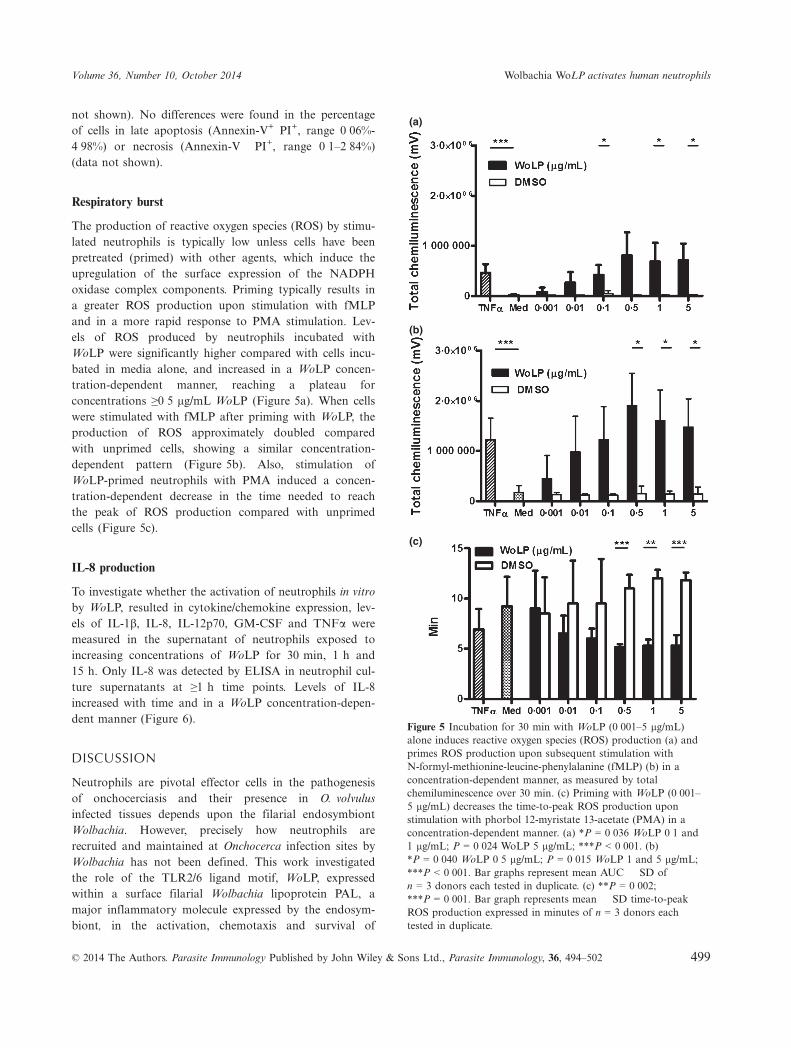
The production of reactive oxygen species (ROS) by stimu-lated neutrophils is typically low unless cells have beenpretreated (primed) with other agents, which induce theupregulation of the surface expression of the NADPHoxidase complex components. Priming typically results ina greater ROS production upon stimulation with fMLPand in a more rapid response to PMA stimulation. Lev-els of ROS produced by neutrophils incubated withWoLP were significantly higher compared with cells incu-bated in media alone, and increased in a WoLP concen-tration-dependent manner, reaching a plateau forconcentrations ≥0�5 lg/mL WoLP (Figure 5a). When cellswere stimulated with fMLP after priming with WoLP, theproduction of ROS approximately doubled comparedwith unprimed cells, showing a similar concentration-dependent pattern (Figure 5b). Also, stimulation ofWoLP-primed neutrophils with PMA induced a concen-tration-dependent decrease in the time needed to reachthe peak of ROS production compared with unprimedcells (Figure 5c).
IL-8 production
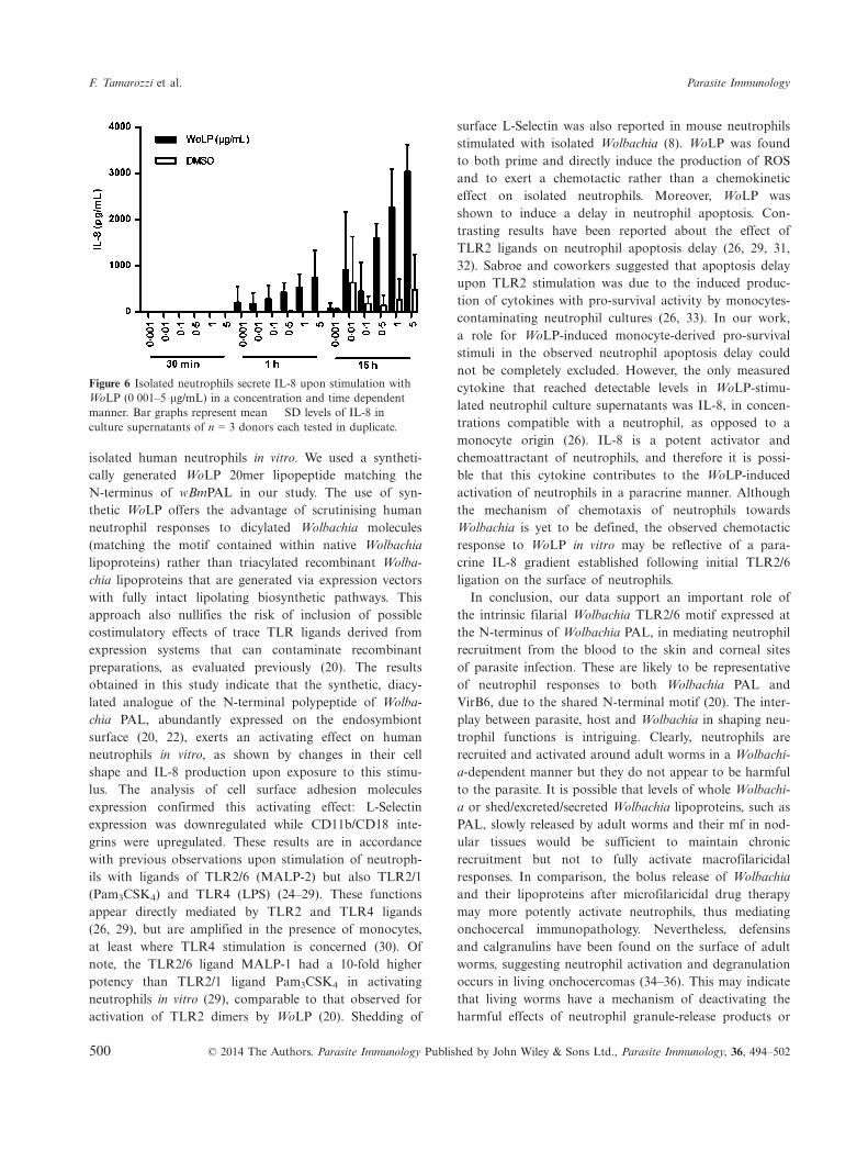
To investigate whether the activation of neutrophils in vitroby WoLP, resulted in cytokine/chemokine expression, lev-els of IL-1b, IL-8, IL-12p70, GM-CSF and TNFa weremeasured in the supernatant of neutrophils exposed toincreasing concentrations of WoLP for 30 min, 1 h and15 h. Only IL-8 was detected by ELISA in neutrophil cul-ture supernatants at ≥1 h time points. Levels of IL-8increased with time and in a WoLP concentration-depen-dent manner (Figure 6).
DISCUSSION
Neutrophils are pivotal effector cells in the pathogenesisof onchocerciasis and their presence in O. volvulusinfected tissues depends upon the filarial endosymbiontWolbachia. However, precisely how neutrophils arerecruited and maintained at Onchocerca infection sites byWolbachia has not been defined. This work investigatedthe role of the TLR2/6 ligand motif, WoLP, expressedwithin a surface filarial Wolbachia lipoprotein PAL, amajor inflammatory molecule expressed by the endosym-biont, in the activation, chemotaxis and survival of
(a)
(b)
(c)
Figure 5 Incubation for 30 min with WoLP (0�001–5 lg/mL)alone induces reactive oxygen species (ROS) production (a) andprimes ROS production upon subsequent stimulation withN-formyl-methionine-leucine-phenylalanine (fMLP) (b) in aconcentration-dependent manner, as measured by totalchemiluminescence over 30 min. (c) Priming with WoLP (0�001–5 lg/mL) decreases the time-to-peak ROS production uponstimulation with phorbol 12-myristate 13-acetate (PMA) in aconcentration-dependent manner. (a) *P = 0�036 WoLP 0�1 and1 lg/mL; P = 0�024 WoLP 5 lg/mL; ***P < 0�001. (b)*P = 0�040 WoLP 0�5 lg/mL; P = 0�015 WoLP 1 and 5 lg/mL;***P < 0�001. Bar graphs represent mean AUC � SD ofn = 3 donors each tested in duplicate. (c) **P = 0�002;***P = 0�001. Bar graph represents mean � SD time-to-peakROS production expressed in minutes of n = 3 donors eachtested in duplicate.
© 2014 The Authors. Parasite Immunology Published by John Wiley & Sons Ltd., Parasite Immunology, 36, 494–502 499
Volume 36, Number 10, October 2014 Wolbachia WoLP activates human neutrophils
isolated human neutrophils in vitro. We used a syntheti-cally generated WoLP 20mer lipopeptide matching theN-terminus of wBmPAL in our study. The use of syn-thetic WoLP offers the advantage of scrutinising humanneutrophil responses to dicylated Wolbachia molecules(matching the motif contained within native Wolbachialipoproteins) rather than triacylated recombinant Wolba-chia lipoproteins that are generated via expression vectorswith fully intact lipolating biosynthetic pathways. Thisapproach also nullifies the risk of inclusion of possiblecostimulatory effects of trace TLR ligands derived fromexpression systems that can contaminate recombinantpreparations, as evaluated previously (20). The resultsobtained in this study indicate that the synthetic, diacy-lated analogue of the N-terminal polypeptide of Wolba-chia PAL, abundantly expressed on the endosymbiontsurface (20, 22), exerts an activating effect on humanneutrophils in vitro, as shown by changes in their cellshape and IL-8 production upon exposure to this stimu-lus. The analysis of cell surface adhesion moleculesexpression confirmed this activating effect: L-Selectinexpression was downregulated while CD11b/CD18 inte-grins were upregulated. These results are in accordancewith previous observations upon stimulation of neutroph-ils with ligands of TLR2/6 (MALP-2) but also TLR2/1(Pam3CSK4) and TLR4 (LPS) (24–29). These functionsappear directly mediated by TLR2 and TLR4 ligands(26, 29), but are amplified in the presence of monocytes,at least where TLR4 stimulation is concerned (30). Ofnote, the TLR2/6 ligand MALP-1 had a 10-fold higherpotency than TLR2/1 ligand Pam3CSK4 in activatingneutrophils in vitro (29), comparable to that observed foractivation of TLR2 dimers by WoLP (20). Shedding of
surface L-Selectin was also reported in mouse neutrophilsstimulated with isolated Wolbachia (8). WoLP was foundto both prime and directly induce the production of ROSand to exert a chemotactic rather than a chemokineticeffect on isolated neutrophils. Moreover, WoLP wasshown to induce a delay in neutrophil apoptosis. Con-trasting results have been reported about the effect ofTLR2 ligands on neutrophil apoptosis delay (26, 29, 31,32). Sabroe and coworkers suggested that apoptosis delayupon TLR2 stimulation was due to the induced produc-tion of cytokines with pro-survival activity by monocytes-contaminating neutrophil cultures (26, 33). In our work,a role for WoLP-induced monocyte-derived pro-survivalstimuli in the observed neutrophil apoptosis delay couldnot be completely excluded. However, the only measuredcytokine that reached detectable levels in WoLP-stimu-lated neutrophil culture supernatants was IL-8, in concen-trations compatible with a neutrophil, as opposed to amonocyte origin (26). IL-8 is a potent activator andchemoattractant of neutrophils, and therefore it is possi-ble that this cytokine contributes to the WoLP-inducedactivation of neutrophils in a paracrine manner. Althoughthe mechanism of chemotaxis of neutrophils towardsWolbachia is yet to be defined, the observed chemotacticresponse to WoLP in vitro may be reflective of a para-crine IL-8 gradient established following initial TLR2/6ligation on the surface of neutrophils.In conclusion, our data support an important role of
the intrinsic filarial Wolbachia TLR2/6 motif expressed atthe N-terminus of Wolbachia PAL, in mediating neutrophilrecruitment from the blood to the skin and corneal sitesof parasite infection. These are likely to be representativeof neutrophil responses to both Wolbachia PAL andVirB6, due to the shared N-terminal motif (20). The inter-play between parasite, host and Wolbachia in shaping neu-trophil functions is intriguing. Clearly, neutrophils arerecruited and activated around adult worms in a Wolbachi-a-dependent manner but they do not appear to be harmfulto the parasite. It is possible that levels of whole Wolbachi-a or shed/excreted/secreted Wolbachia lipoproteins, such asPAL, slowly released by adult worms and their mf in nod-ular tissues would be sufficient to maintain chronicrecruitment but not to fully activate macrofilaricidalresponses. In comparison, the bolus release of Wolbachiaand their lipoproteins after microfilaricidal drug therapymay more potently activate neutrophils, thus mediatingonchocercal immunopathology. Nevertheless, defensinsand calgranulins have been found on the surface of adultworms, suggesting neutrophil activation and degranulationoccurs in living onchocercomas (34–36). This may indicatethat living worms have a mechanism of deactivating theharmful effects of neutrophil granule-release products or
Figure 6 Isolated neutrophils secrete IL-8 upon stimulation withWoLP (0�001–5 lg/mL) in a concentration and time dependentmanner. Bar graphs represent mean � SD levels of IL-8 inculture supernatants of n = 3 donors each tested in duplicate.
500 © 2014 The Authors. Parasite Immunology Published by John Wiley & Sons Ltd., Parasite Immunology, 36, 494–502
F. Tamarozzi et al. Parasite Immunology
are else inherently resistant to the toxic effects of neutro-phil products. Indeed, several parasite molecules that neu-tralize host reactants, such as antioxidants and proteaseinhibitors, have been characterized (12). Far from beingdamaging to adult Onchocerca, a recent hypothesis hasbeen proposed that the large infiltrate of neutrophils prox-imal to the nematode cuticle in onchocercomas may actu-ally be beneficial to parasite survival. As such, neutrophilrecruitment and release of neutrophil products adjacent tothe nematode surface may form a bio-physical barrierblocking infiltration of more macrofilaricidal immuneeffector cells, such as eosinophils (13, 14). Interestingly, ithas been reported that binding of eosinophil peroxidase tohuman neutrophils in vitro leads to reversible inhibition ofits peroxidase activity (37), and, in this regard, the recentlydiscovered process of neutrophil death via extrusion of cel-lular DNA, called NETosis, deserves investigation (38, 39).Finally, recent advances in the understanding of neutrophilfunctions illustrate that these cells are able to shape theadaptive immune response (40, 41). Thus, Wolbachia and its
surface lipoproteins may also influence the development ofadaptive immune responses to different Onchocerca lifecycle stages in humans via the activation and recruitment ofneutrophils, a proposal that deserves further investigation.
ACKNOWLEDGEMENTS
This work was supported by the A•WOL Consortiumthrough a grant of the Bill and Melinda Gates Foundationto Professor Mark J Taylor. Dr Helen L Wright was sup-ported by Arthritis Research UK.
AUTHOR’S CONTRIBUTION
FT and HLW performed the experiments; FT, HLW,SWE, JDT and MJT designed the study; JDT and KLJprovided essential reagents; FT and HLW analysed thedata; FT wrote the paper; all Authors revised the criticallythe data and the manuscript and approved the submittedfinal version of the manuscript.
REFERENCES
1 Basanez MG, Pion SD, Churcher TS, Brei-tling LP, Little MP & Boussinesq M. Riverblindness: a success story under threat?PLoS Med 2006; 3: e371.
2 WHO. Onchocerciasis and its control.Report of a WHO Expert Committee onOnchocerciasis Control. World Health OrganTech Rep Ser 1995; 852: 1–104.
3 Tamarozzi F, Halliday A, Gentil K, HoeraufA, Pearlman E & Taylor MJ. Onchocerciasis:the role of Wolbachia bacterial endos-ymbionts in parasite biology, disease patho-genesis, and treatment. Clin Microbiol Rev2011; 24: 459–468.
4 Gillette-Ferguson I, Hise AG, McGarry HF,et al. Wolbachia-induced neutrophil activa-tion in a mouse model of ocular onchocerci-asis (river blindness). Infect Immun 2004; 72:5687–5692.
5 Pearlman E, Garhart CA, Grand DJ, Dia-conu E, Strine ER & Hall LR. Temporalrecruitment of neutrophils and eosinophilsto the skin in a murine model for onchocer-cal dermatitis. Am J Trop Med Hyg 1999;61: 14–18.
6 Pearlman E, Hall LR, Higgins AW, et al.The role of eosinophils and neutrophils inhelminth-induced keratitis. Invest OphthalmolVis Sci 1998; 39: 1176–1182.
7 Saint Andre A, Blackwell NM, Hall LR,et al. The role of endosymbiotic Wolbachiabacteria in the pathogenesis of river blind-ness. Science 2002; 295: 1892–1895.
8 Gillette-Ferguson I, Daehnel K, Hise AG,et al. Toll-like receptor 2 regulates CXCchemokine production and neutrophilrecruitment to the cornea in Onchocerca
volvulus/Wolbachia-induced keratitis. InfectImmun 2007; 75: 5908–5915.
9 Gillette-Ferguson I, Hise AG, Sun Y, et al.Wolbachia- and Onchocerca volvulus-inducedkeratitis (river blindness) is dependent onmyeloid differentiation factor 88. Infect Im-mun 2006; 74: 2442–2445.
10 Gutierrez-Pena EJ, Knab J & Buttner DW.Neutrophil granule proteins: evidence for theparticipation in the host reaction to skin mi-crofilariae of Onchocerca volvulus after dieth-ylcarbamazine administration. Parasitology1996; 113(Pt 4): 403–414.
11 Brattig NW, Buttner DW & Hoerauf A.Neutrophil accumulation around Onchocercaworms and chemotaxis of neutrophils aredependent on Wolbachia endobacteria.Microbes Infect 2001; 3: 439–446.
12 Brattig NW. Pathogenesis and host responsesin human onchocerciasis: impact of Oncho-cerca filariae and Wolbachia endobacteria.Microbes Infect 2004; 6: 113–128.
13 Nfon CK, Makepeace BL, Njongmeta LM,Tanya VN, Bain O & Trees AJ. Eosinophilscontribute to killing of adult Onchocercaochengi within onchocercomata followingelimination of Wolbachia. Microbes Infect2006; 8: 2698–2705.
14 Hansen RD, Trees AJ, Bah GS, et al. Aworms best friend: recruitment of neutroph-ils by Wolbachia confounds eosinophildegranulation against the filarial nematodeOnchocerca ochengi. Proc Biol Sci 2011; 278:2293–2302.
15 Wildenburg G, Plenge-Bonig A, Renz A, Fi-scher P & Buttner DW. Distribution of mastcells and their correlation with inflammatory
cells around Onchocerca gutturosa, O. tarsi-cola, O. ochengi, and O. flexuosa. ParasitolRes 1997; 83: 109–120.
16 Cross HF, Haarbrink M, Egerton G, Ya-zdanbakhsh M & Taylor MJ. Severe reac-tions to filarial chemotherapy and release ofWolbachia endosymbionts into blood. Lancet2001; 358: 1873–1875.
17 Keiser PB, Reynolds SM, Awadzi K, OttesenEA, Taylor MJ & Nutman TB. Bacterial en-dosymbionts of Onchocerca volvulus in thepathogenesis of posttreatment reactions. JInfect Dis 2002; 185: 805–811.
18 Turner JD, Mand S, Debrah AY, et al. Arandomized, double-blind clinical trial of a3-week course of doxycycline plus albenda-zole and ivermectin for the treatment ofWuchereria bancrofti infection. Clin InfectDis 2006; 42: 1081–1089.
19 Njoo FL, Hack CE, Oosting J, Stilma JS &Kijlstra A. Neutrophil activation in ivermec-tin-treated onchocerciasis patients. Clin ExpImmunol 1993; 94: 330–333.
20 Turner JD, Langley RS, Johnston KL, et al.Wolbachia lipoprotein stimulates innate andadaptive immunity through Toll-like recep-tors 2 and 6 to induce disease manifestationsof filariasis. J Biol Chem 2009; 284: 22364–22378.
21 Johnston KL, Wu B, Guimaraes A, Ford L,Slatko BE & Taylor MJ. Lipoprotein biosyn-thesis as a target for anti-Wolbachia treat-ment of filarial nematodes. Parasit Vectors2010; 3: 99.
22 Bennuru S, Meng Z, Ribeiro JM, et al.Stage-specific proteomic expression pat-terns of the human filarial parasite Brugia
© 2014 The Authors. Parasite Immunology Published by John Wiley & Sons Ltd., Parasite Immunology, 36, 494–502 501
Volume 36, Number 10, October 2014 Wolbachia WoLP activates human neutrophils
malayi and its endosymbiont Wolbachia.Proc Natl Acad Sci USA 2011; 108: 9649–9654.
23 Bennuru S, Semnani R, Meng Z, RibeiroJM, Veenstra TD & Nutman TB. Brugiamalayi excreted/secreted proteins at the host/parasite interface: stage- and gender-specificproteomic profiling. PLoS Negl Trop Dis2009; 3: e410.
24 Hayashi F, Means TK & Luster AD. Toll-like receptors stimulate human neutrophilfunction. Blood 2003; 102: 2660–2669.
25 Neufert C, Pai RK, Noss EH, Berger M,Boom WH & Harding CV. Mycobacteriumtuberculosis 19-kDa lipoprotein promotesneutrophil activation. J Immunol 2001; 167:1542–1549.
26 Sabroe I, Prince LR, Jones EC, et al. Selec-tive roles for Toll-like receptor (TLR)2 andTLR4 in the regulation of neutrophil activa-tion and life span. J Immunol 2003; 170:5268–5275.
27 Seifert R, Schultz G, Richter-Freund M,et al. Activation of superoxide formationand lysozyme release in human neutrophilsby the synthetic lipopeptide Pam3Cys-Ser-(Lys)4. Involvement of guanine-nucleotide-binding proteins and synergism with chemo-tactic peptides. Biochem J 1990; 267: 795–802.
28 Soler-Rodriguez AM, Zhang H, LichensteinHS, et al. Neutrophil activation by bacteriallipoprotein versus lipopolysaccharide: differ-ential requirements for serum and CD14. JImmunol 2000; 164: 2674–2683.
29 Wilde I, Lotz S, Engelmann D, et al. Directstimulatory effects of the TLR2/6 ligandbacterial lipopeptide MALP-2 on neutrophilgranulocytes. Med Microbiol Immunol 2007;196: 61–71.
30 Sabroe I, Jones EC, Usher LR, Whyte MK& Dower SK. Toll-like receptor (TLR)2 andTLR4 in human peripheral blood granulo-cytes: a critical role for monocytes in leuko-cyte lipopolysaccharide responses. J Immunol2002; 168: 4701–4710.
31 Francois S, El Benna J, Dang PM, PedruzziE, Gougerot-Pocidalo MA & Elbim C. Inhi-bition of neutrophil apoptosis by TLR agon-ists in whole blood: involvement of thephosphoinositide 3-kinase/Akt and NF-kap-paB signaling pathways, leading to increasedlevels of Mcl-1, A1, and phosphorylatedBad. J Immunol 2005; 174: 3633–3642.
32 Power CP, Wang JH, Manning B, et al. Bac-terial lipoprotein delays apoptosis in humanneutrophils through inhibition of caspase-3activity: regulatory roles for CD14 andTLR-2. J Immunol 2004; 173: 5229–5237.
33 Prince LR, Allen L, Jones EC, et al. Therole of interleukin-1beta in direct and toll-like receptor 4-mediated neutrophil activa-tion and survival. Am J Pathol 2004; 165:1819–1826.
34 Gallin MY, Jacobi AB, Buttner DW, Schon-berger O, Marti T & Erttmann KD. Humanautoantibody to defensin: disease associationwith hyperreactive onchocerciasis (sowda). JExp Med 1995; 182: 41–47.
35 Edgeworth JD, Abiose A & Jones BR. Animmunohistochemical analysis of onchocer-cal nodules: evidence for an interactionbetween macrophage MRP8/MRP14 andadult Onchocerca volvulus. Clin Exp Immunol1993; 92: 84–92.
36 Gottsch JD, Eisinger SW, Liu SH & ScottAL. Calgranulin C has filariacidal and filari-astatic activity. Infect Immun 1999; 67: 6631–6636.
37 Zabucchi G, Menegazzi R, Cramer R, Nar-don E & Patriarca P. Mutual influencebetween eosinophil peroxidase (EPO) andneutrophils: neutrophils reversibly inhibitEPO enzymatic activity and EPO increasesneutrophil adhesiveness. Immunology 1990;69: 580–587.
38 Fuchs TA, Abed U, Goosmann C, et al.Novel cell death program leads to neutrophilextracellular traps. J Cell Biol 2007; 176:231–241.
39 Guimaraes-Costa AB, Nascimento MT, War-dini AB, Pinto-da-Silva LH & Saraiva EM.ETosis: a microbicidal mechanism beyond celldeath. J Parasitol Res 2012; 2012: 929743.
40 Mantovani A, Cassatella MA, Costantini C& Jaillon S. Neutrophils in the activationand regulation of innate and adaptive immu-nity. Nat Rev Immunol 2011; 11: 519–531.
41 Abi Abdallah DS, Egan CE, Butcher BA &Denkers EY. Mouse neutrophils are profes-sional antigen-presenting cells programmedto instruct Th1 and Th17 T-cell differentia-tion. Int Immunol 2011; 23: 317–326.
502 © 2014 The Authors. Parasite Immunology Published by John Wiley & Sons Ltd., Parasite Immunology, 36, 494–502
F. Tamarozzi et al. Parasite Immunology