anti-oxidative and anti-adipogenic
TRANSCRIPT
©The Japan Endocrine Society
2020, 67 (4), 439-447
Original
Anti-oxidative and anti-adipogenic effects of caffeine inan in vitro model of Graves’ orbitopathyJaeSang Ko1), Ji-Young Kim1), Jae-woo Kim2), 3) and Jin Sook Yoon1)
1) Department of Ophthalmology, Severance Hospital, Institute of Vision Research, Yonsei University College of Medicine, Seoul,Korea
2) Department of Biochemistry and Molecular Biology, Yonsei University College of Medicine, Seoul, Korea3) Brain Korea 21 PLUS Project for Medical Science, Yonsei University, Seoul, Korea
Abstract. Oxidative stress and adipogenesis play key roles in the pathogenesis of Graves’ orbitopathy (GO). In this study, thetherapeutic effects of caffeine on the reduction of oxidative stress and adipogenesis were evaluated in primary cultured GOorbital fibroblasts in vitro. Orbital fibroblasts were cultured from orbital connective tissues obtained from individuals withGO. Intracellular reactive oxygen species (ROS) levels induced by hydrogen peroxide or cigarette smoke extract and theexpression of anti-oxidative enzymes were measured after caffeine treatment. After adipogenic differentiation and caffeinetreatment, cells were stained with Oil Red O and the levels of peroxisome proliferator activator γ (PPARγ), C/EBPα, and C/EBPβ were determined by western blot analysis. Hydrogen peroxide and cigarette smoke extract increased the levels ofintracellular ROS and anti-oxidative enzymes, which decreased in a dose-dependent manner upon pretreatment with caffeinein GO orbital fibroblasts. Oil Red-O staining results revealed a decrease in lipid droplets; furthermore, PPARγ, C/EBPα, andC/EBPβ protein expression levels were inhibited upon treatment with caffeine during adipocyte differentiation. In conclusion,caffeine decreased oxidative stress and adipogenesis in GO orbital fibroblasts in vitro. These findings may contribute to thedevelopment of new types of caffeine-containing pharmacological agents for use in the management of GO.
Key words: Graves’ orbitopathy, Thyroid eye disease, Oxidative stress, Adipogenesis, Caffeine
GRAVES’ ORBITOPATHY (GO) is an autoimmunedisorder of the orbit involving the infiltration of T cells,B cells, plasma cells, and macrophages. The pathogene‐sis of GO involves inflammation of orbital connectivetissue that leads to the enlargement of extraocular mus‐cles and orbital adipose tissue. Such tissue expansionwithin the bony orbits is thought to be responsible for theprogression and complications of GO.
At the center of this pathogenesis lies inflammationand oxidative stress. Infiltration of inflammatory cells tothe orbit triggers and propagates tissue expansion. Oxy‐gen free radicals trigger fibroblast proliferation [1] andthe abundance of thyroid hormones in Graves’ diseaserenders orbital fibroblasts more vulnerable to oxidativedamage [2]. Clinically, cigarette smoking is the mostimportant environmental risk factor for the development
Submitted Nov. 7, 2019; Accepted Dec. 12, 2019 as EJ19-0521Released online in J-STAGE as advance publication Jan. 16, 2020Correspondence to: Jin Sook Yoon, MD, PhD, Department of Oph‐thalmology, Severance Hospital, Institute of Vision Research, Yon‐sei University College of Medicine, 50-1 Yonsei-ro, Seodaemun-gu, Seoul 03722, Korea.E-mail: [email protected]
and progression of GO [3, 4], suggesting that oxidativestress is one of the key effectors in its pathogenesis.
Caffeine (1,3,7-trimethylxanthine) is a plant alkaloidfound in coffee, tea, chocolate, cola, and other softdrinks commonly consumed around the world [5]. It isabsorbed rapidly and completely from the gastrointesti‐nal tract and is detectable in all body fluids including inumbilical cord blood [6, 7]. Numerous studies havedetermined that it acts as a free radical scavenger, takingpart in antioxidant activities in vitro and in vivo in bothanimals and humans [8-10]. Furthermore, its anti-adipogenic properties in various cell types in vitro haveshown that caffeine inhibited the expression ofadipogenesis-related cytokines and transcription factors,resulting in a decrease in adipocyte differentiation andintracellular lipid accumulation [5, 11]. Given the funda‐mental role of oxidative stress and adipogenesis in thepathogenesis of GO, we investigated the therapeuticeffect of caffeine in an in vitro model of GO.

Materials and Methods
Reagents and chemicalsDulbecco’s modified Eagle’s medium (DMEM), fetal
bovine serum (FBS), penicillin, and gentamicin werepurchased from Hyclone Laboratories, Inc. (Logan, UT,USA). Caffeine was purchased from Sigma-AldrichCorp. (St. Louis, MO, USA). The 3-(4,5-dimethylthiazol-2-yl)-5-(3-carboxymethoxyphenyl)-2-(4-sulfophenyl)-2H-tetrazolium, inner salt (MTS) assay solution waspurchased from Promega Corporation (Madison, WI,USA). Cigarette smoke extract (CSE) was freshly pre‐pared within an hour of each experiment from commer‐cially available filtered cigarettes [Marlboro 20 class Acigarettes (8.0 mg tar; 0.7 mg nicotine); Philip MorrisKorea, Inc., Seoul, Korea] as described in our previousstudy [12]. Recombinant human IL-1β was purchasedfrom R&D Systems (Minneapolis, MN, USA) and OilRed O was purchased from Sigma-Aldrich Corp.
Cell culture and differentiation protocolsOrbital adipose/connective tissue explants were
obtained as surgical waste during decompression surgeryin five patients with GO (three females, two males; 29–51 years of age). Normal control tissues were harvestedduring upper-lid blepharoplasty from the post-septal areaof five individuals with no history or clinical evidence ofthyroid disease or GO (three females, two males; 31–58years of age). All five patients with GO achieved stableeuthyroidism at the time of surgery, at which point theirclinical activity scores were less than three. Furthermore,none of the patients with GO received steroid treatmentor radiotherapy for at least three months before surgery.The Institutional Review Board of Severance Hospital,Yonsei University College of Medicine (Seoul, Korea)approved the study and written informed consent wasobtained from all participants after explanation of thenature and possible consequences of the study. Thisstudy followed the tenets of the Declaration of Helsinki.
Primary cultures of orbital fibroblasts were establishedas described in our previous study [13]. Briefly, mincedtissue was placed directly in 1:1 DMEM:F12 mediumwith 20% FBS and antibiotics. When the growth offibroblasts was observed, monolayers were passagedserially with trypsin/ethylenediaminetetraacetic acid so‐lution and cultures were maintained in DMEM with 10%FBS and antibiotics. The strains were stored in liquidnitrogen until further analysis; cells between the secondand fifth passages were used.
The anti-adipogenic effect of caffeine was evaluatedusing our previously reported adipocyte differentiationprotocol for GO orbital fibroblasts involving dexametha‐sone, rosiglitazone (10 μM; Cayman Chemical, Ann
Arbor, MI, USA), and IL-1β (10 ng/mL) treatment [13,14]. Briefly, after 10 days of adipogenic differentiationco-treated with serial concentrations of caffeine (0.1–5mM), differentiated adipocytes were visualized with OilRed O staining and expression of adipogenic transcrip‐tion factors were evaluated with western blotting.
Cell viability assayTo evaluate the effect of caffeine on cell viability of
orbital fibroblasts, primary cultured orbital fibroblastsobtained from patients with GO were seeded on 24-wellculture plates (1 × 10⁴ cells/well) and treated with vari‐ous concentrations of caffeine (0.01 mM, 0.1 mM, 0.5mM, 1 μM, 2 mM, and 5 mM) for 48 h and 72 h. There‐after, MTS solution was added and the plate was incu‐bated again for 4 h under the same conditions. Theabsorbance of the dye was measured at 490 nm using anELISA plate reader. Cell viability is expressed as a per‐centage relative to untreated control cells.
Intracellular ROS measurementROS release was determined with 5-(and 6)-
carboxy-20,70-dichlorodihydrofluorescein diacetate(H2DCFDA; Invitrogen/Thermo Fisher Scientific, Carls‐bad, CA, USA), an oxidant-sensitive fluorescent probe,as we described previously [12]. Briefly, primary cul‐tured orbital fibroblasts were pretreated with variousconcentrations of caffeine for 24 h. The culture mediumwas then removed, and the cells were washed with PBS,incubated with 10 mM H2DCFDA at 37°C for 30 min,then stimulated with CSE (2%) or H2O2 (10 μM) for 30min [12, 15, 16]. The fluorescently stained cells wereexamined microscopically at 40× and were quantified viaflow-cytometric analysis.
Western blottingWestern blot analysis was performed as described pre‐
viously [17]. To assess the induction of oxidative stressby CSE and H2O2 and its inhibition by caffeine, conflu‐ent orbital fibroblasts were pre-exposed to serial concen‐trations of caffeine for 24 h, followed by stimulationwith CSE or H2O2 for 24 h. Expression levels of manga‐nese superoxide dismutase (Mn-SOD), copper-zincsuperoxide dismutase (Cu/Zn-SOD), thioredoxin (Trx),and heme oxygenase-1 (HO-1) were assessed to evaluateoxidative stress levels. To evaluate the expression of adi‐pogenic transcription factors after adipogenic differentia‐tion, the expression levels of peroxisome proliferatoractivator gamma (PPARγ), C/EBPα, and C/EBPβ wereevaluated in GO cells after ten days of adipocyte differ‐entiation. Activation of intracellular signalling proteinswas assayed via western blot analysis of phosphorylatedand total AKT, extracellular signal-regulated kinase
440 Ko et al.
(ERK), nuclear factor kappa-light-chain-enhancer ofactivated B cells (NF-κB), c-Jun NH(2)-terminal kinase(JNK), and p38 protein. The relative amount of proteinin each immunoreactive band was quantified by densi‐tometry and normalised to the concentration of β-actin inthe same sample.
Anti-Mn-SOD, anti-Zn/Cu-SOD, anti-Trx, anti-HO-1,anti-peroxisome proliferator activator gamma (PPARγ),anti-C/EBPα, anti-C/EBPβ, anti-NF-κB, p38, and anti–β-actin antibodies were all obtained from Santa Cruz Bio‐technology (Dallas, TX, USA) and antibodies againstAKT, ERK, and JNK were produced by Cell SignalingTechnology (Danvers, MA, USA).
Oil Red O stainingAfter ten days of adipocyte differentiation of orbital
fibroblasts, cells were stained with an Oil Red O workingsolution as described previously [13, 14]. Stained cellswere visualised and photographed at 40×, then quantifiedusing a spectrophotometer at 490 nm according to themanufacturer’s instructions. Experiments for the quanti‐tative assessment of adipogenic differentiation were per‐formed in duplicate using cells from different donors; theresults were normalised to the absorbance of untreateddifferentiated control cells.
Statistical analysisAll experiments were performed using cells from at
least three different samples and the samples wereassayed in duplicate each time. Differences in parameterestimates between the experimental and control groupswere assessed by the Student’s t-test or Wilcoxon rank-sum test using R version 3.1.2 (R Foundation, Vienna,Austria). Values of p < 0.05 were considered significant.
Results
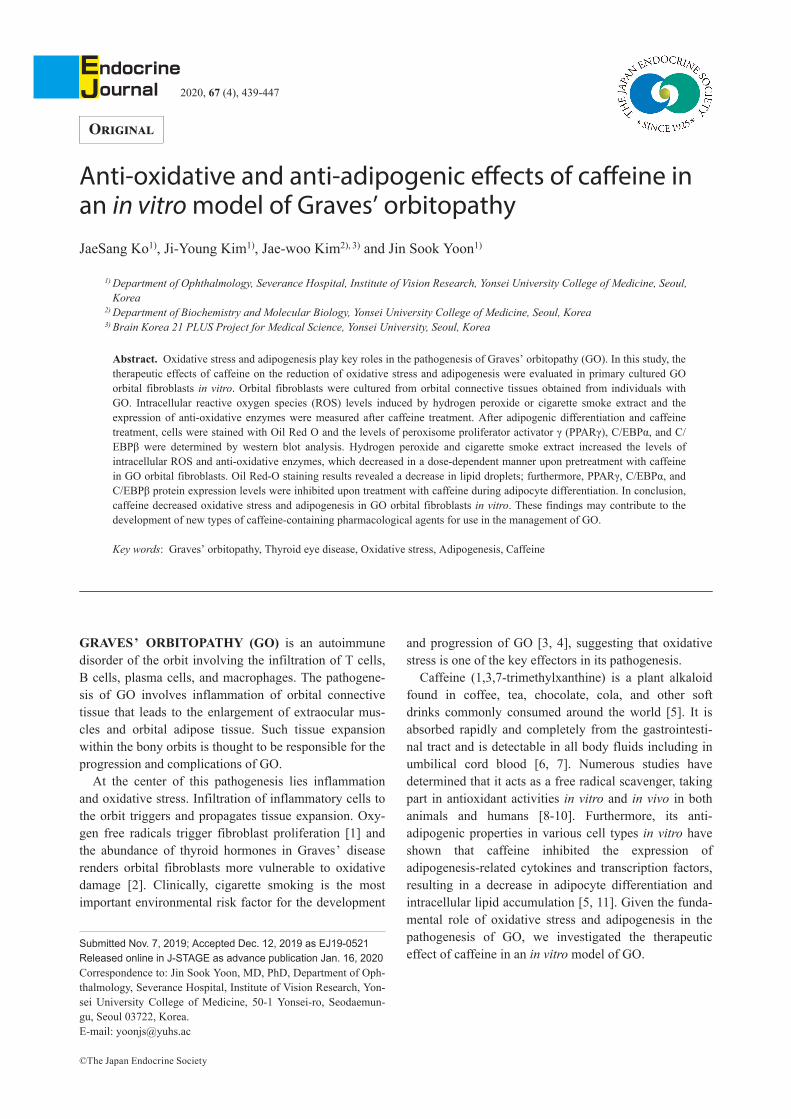
Cell viability using MTS analysisTo determine non-toxic concentrations of caffeine in
orbital fibroblasts, an MTS assay was performed. Orbitalfibroblasts from patients with and without GO weretreated with caffeine at concentrations ≤5 mM for 48 hand 72 h. The 0.01 to 5 mM range of caffeine did notdecrease cell viability below 95% in both normal andGO orbital fibroblasts in the 48 h treatment (Fig. 1, datafor non-GO orbital fibroblasts are not shown).
Caffeine decreased ROS levels induced by H2O2 orCSE
To demonstrate the anti-oxidative effect of caffeine inGO orbital fibroblasts, oxidative stress was induced by10 μM H2O2 or 2% CSE with or without 0.01–5 mM caf‐feine pretreatment for 24 h. Treatment with 10 μM H2O2
or 2% CSE for 30 min significantly increased ROSproduction relative to that in untreated control cells,which was reduced by caffeine pretreatment in a dose-dependent manner (Fig. 2A). Similarly, upon fluorescentstaining of cellular ROS, a significant reduction in fluo‐rescently stained cells was observed after treatment withcaffeine (40×; Fig. 2B).
Effect of caffeine on SOD, Trx, and HO-1The relative expression levels of Mn-SOD, Cu/Zn-
SOD, and HO-1 increased upon treatment with 10 μMH2O2 or 2% CSE in GO orbital fibroblasts for 24 h,while the expression of Trx increased only upon treat‐ment with CSE, not H2O2. Upon pretreatment with aserial concentration of caffeine, we found that caffeineconcentrations >1 mM decreased the expression of Mn-SOD, Cu/Zn-SOD, Trx, and HO-1 (Fig. 3).
Fig. 1 Effect of caffeine on the viability of orbital fibroblastsOrbital fibroblasts from individuals with GO were seeded in 24-well culture plates and treated with various concentrations (0–5mM) of caffeine for 48 h (A) and 72 h (B). After treatment, MTS assays were used to evaluate viability. Assays were performed induplicate with cells from three different donors. Results are expressed as percentages of untreated control values and are presentedas means ± standard deviation.
Therapeutic potential of caffeine in GO 441

Caffeine reduced adipogenesis in GO orbitalfibroblasts
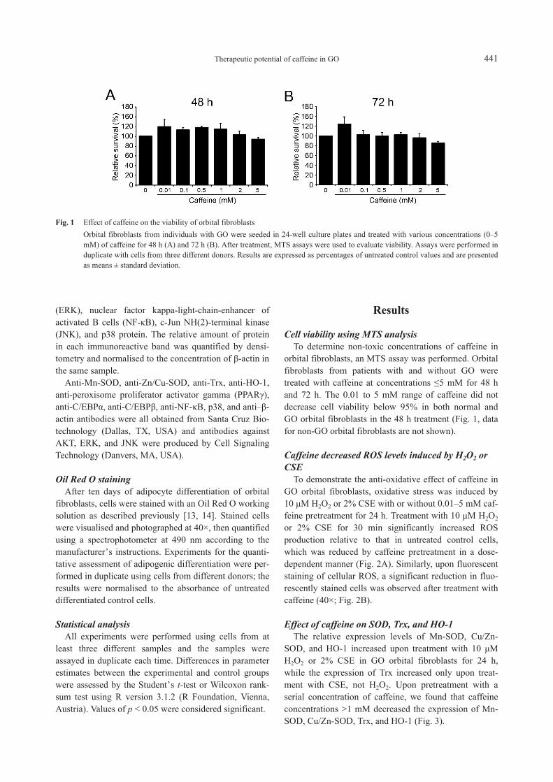
To determine the effect of caffeine on adipogenesis inorbital fibroblasts, GO cells were treated with caffeineduring adipocyte differentiation. When caffeine (0.5–5mM) was added to the adipogenic medium, it reducedthe number of adipocytes and suppressed the accumula‐tion of lipid droplets. The optical density of Oil Red O-stained cell lysates showed that caffeine-treated cellsexhibited decreased absorbance at 490 nm (Fig. 4A).Western blot analysis was then performed to investigatewhether caffeine affects the production of the adipogenictranscription factors PPARγ, C/EBPα, and C/EBPβ dur‐ing adipogenesis. Caffeine had dose-dependent inhibi‐tory effects on the expression of adipogenic transcriptionfactors (Fig. 4B). Each experiment was performed inthree GO cells from different patient samples and sam‐ples were assayed in duplicate.
Effect of caffeine on intracellular signallingpathways
To investigate the signalling pathways through whichcaffeine mediates its effects in GO orbital fibroblasts, theexpression levels of multiple transcription factors wereassessed after caffeine treatment. Both GO and non-GOcells were treated with 1 mM caffeine for various dura‐tions. Western blot analysis (Fig. 5) showed that caffeineinhibited the phosphorylation of ERK and p38 protein ina time-dependent manner. The phosphorylation of AKT,JNK, and NF-κB remained unchanged with 1 mM caf‐feine treatment (data not shown).
Discussion
In this study, we found that caffeine curtailed oxida‐tive stress and adipogenesis, the main pathogenic mecha‐nisms of GO. Based on our results, caffeine appears tohave an inhibitory effect on ROS generation in responseto oxidative stress, as well as an anti-adipogenic effect.
A state of oxidative stress has been described inGraves’ disease and GO [18-20]. In general, an increasein ROS or the reduced elimination of radicals by anti-oxidative enzymes will result in oxidative damage,inflammation, and loss of function. In the in vitro patho‐genic mechanism of GO, superoxide radicals stimulateorbital fibroblasts to proliferate and differentiate intomature adipocytes [20]. Based on clinical reports, ciga‐rette smoking—likely the most important environmentalfactor associated with GO occurrence and progression—may act, among other mechanisms, by stimulating thegeneration of ROS and reducing antioxidant production[3, 20, 21]. In this regard, several in vitro studies demon‐strate the therapeutic effectiveness of drugs with antioxi‐dant potential in primary cultured orbital fibroblasts fromGO patients [12, 15-17]. Clinical studies on the use ofantioxidants for the management of GO have also shownthem to be clinically effective in patients experiencingmild severity [22-24].
Although caffeine is known to act as a free radicalscavenger—the basis for its antioxidant activities—itshould be noted that it exhibits both anti-oxidant andpro-oxidant properties depending on dose: at low con‐centrations, caffeine shows antioxidant effects, while athigh concentrations, it may increase cytotoxicity, leading
Fig. 2 Effect of caffeine on H2O2- and CSE-induced generation of intracellular ROS in orbital fibroblasts from GO patientsOrbital fibroblasts from GO patients (n = 3) were treated with 10 μM H2O2 or 2% CSE for 30 min with or without pretreatmentwith caffeine for 24 h. ROS levels were measured via flow cytometry using H2DCFDA (A) and microscopically visualised at 40×(B). The results are expressed as percentages of the untreated control values and presented as means ± standard deviation. Assayswere performed in duplicate with cells from three different GO samples; data from a representative experiment are shown (*p <0.05 and **p < 0.01 versus H2O2- or CSE-treated cells without caffeine pretreatment).
442 Ko et al.

Fig. 3 Effect of caffeine on H2O2- and CSE-induced expression of SOD, Trx, and HO-1 in orbital fibroblasts from GO patientsOrbital fibroblasts from GO patients (n = 3) were treated with 10 μM H2O2 (A) or 2% CSE (B) for 24 h with or withoutpretreatment with 0.1–5 mM caffeine for 24 h. The cell lysates were subjected to western blot analysis. Experiments wereperformed three times using different strains and samples were assayed in duplicate. Results are expressed as the relative densityof each protein normalised to the level of β-actin and presented as means ± standard deviation. GO cells treated with neither CSEnor H2O2 were used as controls (*p < 0.05 and **p < 0.01 versus H2O2- or CSE- treated cells without pretreatment).
Therapeutic potential of caffeine in GO 443
to the subsequent generation of reactive oxygen species(ROS) [5, 25, 26]. In this study, however, orbital fibro‐blasts were resistant to the generation of ROS at highconcentrations of caffeine, which acted as an antioxidantin H2O2- or CSE-stimulated orbital fibroblasts even atthe highest concentration (5 mM) without causing celldamage.
Adipogenesis, the process by which orbital fibroblastsdevelop into mature adipocytes, is one of the key patho‐genic mechanisms in GO. We found that drugs withantioxidant effects significantly suppressed adipocytedifferentiation in primary Graves’ orbital fibroblasts [12,15-17]. Recently, groups studying obesity and metabo‐lism have reported that caffeine inhibits adipogenic dif‐ferentiation and reduces lipid accumulation [5, 11]. Theyshowed that caffeine may prohibit the activation of C/EBPβ, the first response to adipogenic inducers, blockingthe adipogenic cascade involving PPARγ, aP2, or leptinprotein. In this study, we showed that ≥0.5 mM caffeineinhibited adipogenic differentiation and lipid accumula‐tion, as well as the expression of adipogenic transcriptionfactors (PPARγ, C/EBPα, and C/EBPβ).
The differentiation of preadipocytes is triggered byhormonal agents and proceeds by the coordination andprecise control of transcriptional cascades. The first stageof adipocyte differentiation is growth arrest at a conflu‐
ent state; proliferating cells will not accumulate lipiddroplets in their cytoplasm until they become confluent.Upon induction of differentiation, growth-arrested cellsundergo additional rounds of cell division, known asmitotic clonal expansion [27]. A recent study showedthat ROS are important in regulating mitotic clonalexpansion during adipogenesis and that they facilitateadipocyte differentiation by accelerating mitotic clonalexpansion [28]. Moreover, it has been demonstrated thatcaffeine inhibits adipogenesis through the modulation ofmitotic clonal expansion in 3T3-L1 adipocytes [11].
Mitogen-activated protein kinases (MAPKs) constitutea family of protein kinases (ERK, JNK, and the p38-MAPKs in mammals) that play an essential role in relay‐ing extracellular signals from the cell membrane to thenucleus via a cascade of phosphorylation events [29].MAPKs are key participants in signal transduction path‐ways activated by mitogenic stimuli, environmentalstress, and inflammatory agents [30]. Various studieshave documented the involvement of the MAPK signal‐ling pathways in redox-stressed cells and tissues [31].Moreover, it has been shown that the pathogenesis of GOinvolves activation of the MAPK signalling pathways,especially the p38 and ERK pathways [32, 33]. Ourresults showed that the levels of phosphorylated p38 andERK decreased when they were cotreated at increasing
Fig. 4 Effect of caffeine on adipogenesis in GO orbital fibroblasts(A) Ten days of adipocyte differentiation was induced in primary cultured orbital fibroblasts from GO patients. The addition ofcaffeine to the adipogenic medium attenuated adipocyte differentiation, as evidenced by microscopic examinations (40×) after OilRed O staining. The optical density at 490 nm of stained cell lysates also yielded quantitative evidence of the inhibitory effect ofcaffeine on the accumulation of lipid droplets. (B) Western blot analysis of PPARγ, C/EBPα, and C/EBPβ protein was performedafter ten days of adipogenic differentiation. The experiments were performed in duplicate with cells from three different donors.PPARγ, C/EBPα, and C/EBPβ levels determined by densitometry were normalised to the level of β-actin in the same sample. Theresults are presented as the mean relative density ratios (%) ± standard deviation (*p < 0.05 and **p < 0.01 versus cells withoutcaffeine treatment).
444 Ko et al.

timeframes of caffeine exposure.The results of the present study show that caffeine has
anti-oxidative and anti-adipogenic properties; however, itshould be noted that the concentration of caffeine used inthis in vitro study is somewhat higher than the normalphysiological range. A caffeine concentration of 0.1 mM(19.4 μg/mL) is approximately equivalent to that in a cupof coffee containing 150 mg caffeine (Starbucks© latte)[5]; it has been reported that caffeine causes adverseeffects at excessively high blood concentrations (>80μg/mL) in humans [34]. In this study, caffeine exhibitedanti-adipogenic effects beyond concentrations of 0.5 mMand anti-oxidative effects at 0.1 mM. As GO is a hetero‐geneous autoimmune disease affected by multiplegenetic and environmental factors, in vitro results maynot correlate precisely with in vivo conditions. Addition‐ally, we induced pathologic conditions in cells by stimu‐lation using concentrations of chemicals, hormones, andgrowth factors beyond physiological ranges—for exam‐
ple, our use of oxidants such as H2O2 or CSE.In conclusion, to the best of our knowledge, this is the
first study to investigate the effects of caffeine on oxida‐tive stress and adipogenesis in orbital fibroblasts fromGO patients. Until now, no clinical or epidemiologicstudies have emerged that assess the relationshipbetween caffeine and GD or GO. Our in vitro results pro‐vide evidence to support further investigation regardingthe influence of caffeine on GO progression in clinicalsettings.
Acknowledgements
Funding: This research was supported by the BasicScience Research Program through the National ResearchFoundation of Korea (NRF) funded by the Ministry ofScience and ICT (NRF-2017R1A2B4009565).
Fig. 5 Effect of caffeine on intracellular signalling pathwaysOrbital fibroblasts from GO patients (n = 3) and non-GO individuals (n = 3) were treated with 1 mM caffeine for 0–180 min. Thecell lysates were subjected to western blot analysis. Experiments were performed three times using different strains and sampleswere assayed in duplicate. Results are expressed as the relative density of each protein normalised to the level of β-actin andpresented as means ± standard deviation (*p < 0.05 and **p < 0.01 versus cells without caffeine treatment).
Therapeutic potential of caffeine in GO 445

Disclosure
The authors have no conflicts of interest to declare.
References
1. Lehmann GM, Feldon SE, Smith TJ, Phipps RP (2008)Immune mechanisms in thyroid eye disease. Thyroid 18:959–965.
2. Burch HB, Lahiri S, Bahn RS, Barnes S (1997) Superox‐ide radical production stimulates retroocular fibroblastproliferation in Graves’ ophthalmopathy. Exp Eye Res 65:311–316.
3. Pfeilschifter J, Ziegler R (1996) Smoking and endocrineophthalmopathy: impact of smoking severity and currentvs. lifetime cigarette consumption. Clin Endocrinol (Oxf)45: 477–481.
4. Stan MN, Bahn RS (2010) Risk factors for developmentor deterioration of Graves’ ophthalmopathy. Thyroid 20:777–783.
5. Su SH, Shyu HW, Yeh YT, Chen KM, Yeh H, et al.(2013) Caffeine inhibits adipogenic differentiation of pri‐mary adipose-derived stem cells and bone marrow stromalcells. Toxicol In Vitro 27: 1830–1837.
6. Arnaud MJ (1993) Metabolism of caffeine and other com‐ponents of coffee. In: Garattini S. (ed.), Caffeine, Coffee,and Health. Raven Press, New York, USA: 43–96.
7. Dempsey DA, Partridge JC, Jones RT, Rowbotham MC(1998) Cocaine, nicotine, caffeine, and metabolite plasmaconcentrations in neonates. J Anal Toxicol 22: 220–224.
8. Lv X, Chen Z, Li J, Zhang L, Liu H, et al. (2010) Caffeineprotects against alcoholic liver injury by attenuatinginflammatory response and oxidative stress. Inflamm Res59: 635–645.
9. Jagdeo J, Brody N (2011) Complementary antioxidantfunction of caffeine and green tea polyphenols in normalhuman skin fibroblasts. J Drugs Dermatol 10: 753–761.
10. Varma SD, Hegde KR, Kovtun S (2010) Oxidative stressin lens in vivo: inhibitory effect of caffeine. A preliminaryreport. Mol Vis 16: 501–505.
11. Kim AR, Yoon BK, Park H, Seok JW, Choi H, et al.(2016) Caffeine inhibits adipogenesis through modulationof mitotic clonal expansion and the AKT/GSK3 pathwayin 3T3-L1 adipocytes. BMB Rep 49: 111–115.
12. Yoon JS, Lee HJ, Chae MK, Lee SY, Lee EJ (2013)Cigarette smoke extract-induced adipogenesis in Graves’orbital fibroblasts is inhibited by quercetin via reductionin oxidative stress. J Endocrinol 216: 145–156.
13. Kim SE, Lee JH, Chae MK, Lee EJ, Yoon JS (2016) Therole of sphingosine-1-phosphate in adipogenesis ofGraves’ orbitopathy. Invest Ophthalmol Vis Sci 57: 301–311.
14. Ko J, Kim JY, Lee EJ, Yoon JS (2018) Inhibitory effect ofidelalisib, a selective phosphatidylinositol 3-kinase deltainhibitor, on adipogenesis in an in vitro model of Graves’
orbitopathy. Invest Ophthalmol Vis Sci 59: 4477–4485.15. Kim CY, Lee HJ, Chae MK, Byun JW, Lee EJ, et al.
(2015) Therapeutic effect of resveratrol on oxidative stressin Graves’ orbitopathy orbital fibroblasts. Invest Ophthal‐mol Vis Sci 56: 6352–6361.
16. Rhiu S, Chae MK, Lee EJ, Lee JB, Yoon JS (2014) Effectof tanshinone IIA in an in vitro model of Graves’ orbitop‐athy. Invest Ophthalmol Vis Sci 55: 5900–5910.
17. Byun JW, Hwang S, Kang CW, Kim JH, Chae MK, et al.(2016) Therapeutic effect of protocatechuic aldehyde in anin vitro model of Graves’ orbitopathy. Invest OphthalmolVis Sci 57: 4055–4062.
18. Hondur A, Konuk O, Dincel AS, Bilgihan A, Unal M, etal. (2008) Oxidative stress and antioxidant activity inorbital fibroadipose tissue in Graves ’ ophthalmopathy.Curr Eye Res 33: 421–427.
19. Komosinska-Vassev K, Olczyk K, Kucharz EJ, Marcisz C,Winsz-Szczotka K, et al. (2000) Free radical activity andantioxidant defense mechanisms in patients with hyper‐thyroidism due to Graves’ disease during therapy. ClinicaChimica Acta 300: 107–117.
20. Bartalena L, Tanda ML, Piantanida E, Lai A (2003) Oxi‐dative stress and Graves’ ophthalmopathy: in vitro studiesand therapeutic implications. Biofactors 19: 155–163.
21. Shine B, Fells P, Edwards OM, Weetman AP (1990)Association between Graves’ ophthalmopathy and smok‐ing. Lancet 335: 1261–1263.
22. Marcocci C, Kahaly GJ, Krassas GE, Bartalena L,Prummel M, et al. (2011) Selenium and the course of mildGraves’ orbitopathy. N Engl J Med 364: 1920–1931.
23. Bednarek J, Wysocki H, Sowinski J (2005) Oxidativestress peripheral parameters in Graves’ disease: the effectof methimazole treatment in patients with and withoutinfiltrative ophthalmopathy. Clin Biochem 38: 13–18.
24. Bouzas EA, Karadimas P, Mastorakos G, Koutras DA(2000) Antioxidant agents in the treatment of Graves’ophthalmopathy. Am J Ophthalmol 129: 618–622.
25. Azam S, Hadi N, Khan NU, Hadi SM (2003) Antioxidantand prooxidant properties of caffeine, theobromine andxanthine. Med Sci Monit 9: BR325–BR330.
26. Tiwari KK, Chu C, Couroucli X, Moorthy B, Lingappan K(2014) Differential concentration-specific effects of caf‐feine on cell viability, oxidative stress, and cell cycle inpulmonary oxygen toxicity in vitro. Biochem Biophys ResCommun 450: 1345–1350.
27. MacDougald OA, Lane MD (1995) Transcriptional regu‐lation of gene expression during adipocyte differentiation.Annu Rev Biochem 64: 345–373.
28. Lee H, Lee YJ, Choi H, Ko EH, Kim JW (2009) Reactive
446 Ko et al.

oxygen species facilitate adipocyte differentiation byaccelerating mitotic clonal expansion. J Biol Chem 284:10601–10609.
29. Son Y, Cheong YK, Kim NH, Chung HT, Kang DG, et al.(2011) Mitogen-activated protein kinases and reactiveoxygen species: how can ROS activate MAPK pathways?J Signal Transduct 2011: 792639.
30. Kyriakis JM, Avruch J (2012) Mammalian MAPK signaltransduction pathways activated by stress and inflamma‐tion: a 10-year update. Physiol Rev 92: 689–737.
31. Gaitanaki C, Konstantina S, Chrysa S, Beis I (2003) Oxi‐dative stress stimulates multiple MAPK signalling path‐ways and phosphorylation of the small HSP27 in the
perfused amphibian heart. J Exp Biol 206: 2759–2769.32. Chen B, Tsui S, Smith TJ (2005) IL-1 beta induces IL-6
expression in human orbital fibroblasts: identification ofan anatomic-site specific phenotypic attribute relevant tothyroid-associated ophthalmopathy. J Immunol 175: 1310–1319.
33. Kazim M, Goldberg RA, Smith TJ (2002) Insights into thepathogenesis of thyroid-associated orbitopathy: evolvingrationale for therapy. Arch Ophthalmol 120: 380–386.
34. Nomura M, Ichimatsu D, Moritani S, Koyama I, Dong Z,et al. (2005) Inhibition of epidermal growth factor-induced cell transformation and Akt activation by caf‐feine. Mol Carcinog 44: 67–76.
Therapeutic potential of caffeine in GO 447