soft substrate maintains proliferative and adipogenic ... · on tissue culture plastic (tcp) lose...
TRANSCRIPT
RESEARCH ARTICLE
Soft substrate maintains proliferative and adipogenicdifferentiation potential of human mesenchymal stem cellson long-term expansion by delaying senescenceSanjay Kumar Kureel*, Pankaj Mogha*, Akshada Khadpekar, Vardhman Kumar, Rohit Joshi, Siddhartha Das,Jayesh Bellare and Abhijit Majumder‡
ABSTRACTHuman mesenchymal stem cells (hMSCs), during in vitro expansion,gradually lose their distinct spindle morphology, self-renewalability, multi-lineage differentiation potential and enter replicativesenescence. This loss of cellular function is a major roadblock forclinical applications which demand cells in large numbers. Here, wedemonstrate a novel role of substrate stiffness in the maintenance ofhMSCs over long-term expansion. When serially passaged for45 days from passage 3 to passage 18 on polyacrylamide gel ofYoung’s modulus E=5 kPa, hMSCs maintained their proliferation rateand showed nine times higher population doubling in comparison totheir counterparts cultured on plastic Petri-plates. They did notexpress markers of senescence, maintained their morphology andother mechanical properties such as cell stiffness and cellulartraction, and were significantly superior in adipogenic differentiationpotential. These results were demonstrated in hMSCs from twodifferent sources, umbilical cord and bone marrow. In summary,our result shows that a soft gel is a suitable substrate to maintainthe stemness of mesenchymal stem cells. As preparation ofpolyacrylamide gel is a well-established, and well-standardizedprotocol, we propose that this novel system of cell expansion will beuseful in therapeutic and research applications of hMSCs.
KEY WORDS: Cell mechanics, Mesenchymal stem cells,Senescence, Stemness, Substrate rigidity
INTRODUCTIONHuman mesenchymal stem cells (hMSCs), due to their multi-lineage differentiation potential, immuno-suppressive capability,and immuno-modulatory effects have been used with varyingdegree of success to treat cardiovascular, musculoskeletal, immune-related and hemopoietic diseases (Pittenger et al., 1999; Ranganathet al., 2012; Tuan, 2013; Wang et al., 2016). Though MSCs areavailable from multiple adult tissues (Caplan and Bruder, 2001),critically low availability of MSCs in the isolated sample is a majorroadblock for clinical trials. For example, in bone marrow aspirates,
only 0.001–0.01% of the nucleated cells are MSCs, while a dose ofroughly about 100 million cells are required to treat a person 70 kgin weight (Ren et al., 2012). As a result, a long-term in vitroexpansion is essential to reach a significant number of cells forautologous treatment.
However, on in vitro expansion, MSCs lose their proliferativeability and multilineage differentiation potential (Banfi et al., 2000;Wagner et al., 2008). Like any other primary somatic cells, after acertain number of cell divisions, they enter a senescence state, whichis morphologically characterized by enhanced spreading area andshape irregularity (Bonab et al., 2006; Wagner et al., 2010b). Moreimportantly, they lose their multilineage potential, migration andhoming ability (De Becker and Van Riet, 2016; Honczarenko et al.,2006), making them unsuitable for clinical use (Kassem, 2006; Ullahet al., 2015). Thoughmultiple approaches have been tried to maintainMSC stemness over prolonged expansion (Saei Arezoumand et al.,2017), finding an easy-to-use culture system to achieve the same isstill an unmet need. In this context, it might be noted that the NIH ontheir website has listed six points that need to be addressedto realize the potential of stem cell-based therapies. The first one inthat list is “Stem cells must be reproducibly made to proliferateextensively and generate sufficient quantities of cells for makingtissue” (Stem Cell Basics IV. | stemcells.nih.gov, 2017, https://stemcells.nih.gov/info/basics/1.htm). A culture system that can fulfillthis need may help to progress regenerative medicine significantly.
Controlling the physical microenvironment of the cell culturesystem might offer a solution in this context. In the past 15 years, ithas been shown that mechanical cues such as stiffness of cell culturesubstrate, shear stress, mechanical strain, cell morphology, substratetopology, etc., influence a wide array of cell behavior and cell fateincluding survival, proliferation and differentiation (Anderson et al.,2016; Engler et al., 2006; Gilbert et al., 2010; Lutolf et al., 2009;Murphy et al., 2014; Winer et al., 2009; Yeung et al., 2005). It hasalso been shown that such mechanical cues may play an importantrole in maintaining MSCs stemness. For example, MSCs culturedon micro-contact printed islands as spheroids and on nano-patternswere shown to retain multipotency and proliferative capacity(Cesarz and Tamama, 2016; Lee et al., 2015; McMurray et al.,2011; Zhang and Kilian, 2013). However, both micro-contactprinting and spheroid culture restrict the proliferation of MSCsleading to limited or no expansion in cell number. Moreover,creating micro-patterns or nano-patterns for a large area is adaunting task and demands huge infrastructure and cost.
In this work, we have shown that hMSCs maintain their stemnessover long passages when cultured on an optimally soft polyacrylamide(PAA) gel. The soft substrate also preserves cellular morphology.Staining for β-gal and BrdU respectively showed that in these cellsonset of senescence is delayed and proliferative potential isReceived 17 October 2018; Accepted 22 March 2019
Department of Chemical Engineering, Indian Institute of Technology Bombay(IITB), Mumbai 400076, India.*These authors contributed equally to this work
‡Author for correspondence ([email protected])
S.K.K., 0000-0003-0562-0316; P.M., 0000-0003-0144-0107; A.K., 0000-0002-0786-2098; R.J., 0000-0001-8875-4576; A.M., 0000-0002-0695-3967
This is an Open Access article distributed under the terms of the Creative Commons AttributionLicense (https://creativecommons.org/licenses/by/4.0), which permits unrestricted use,distribution and reproduction in any medium provided that the original work is properly attributed.
1
© 2019. Published by The Company of Biologists Ltd | Biology Open (2019) 8, bio039453. doi:10.1242/bio.039453
BiologyOpen
by guest on December 29, 2019http://bio.biologists.org/Downloaded from
maintained. Staining for other senescence-related changes such as lossof Lamin B and gain of Lamin A confirmed this observation. Not onlythe proliferative potential but the cells cultured on gel coulddifferentiate into the adipo lineage, as shown by the expression ofPPAR-gamma and accumulation of oil droplets, while cells culturedon tissue culture plastic (TCP) lose their adipogenic differentiationpotential. Finally, we have shown that surface markers, used tocharacterize MSCs, remain unaltered in the cells cultured on softsubstrate ensuring the maintenance of cellular identity.
RESULTS AND DISCUSSIONLoss of cell morphology and induction of senescence duringlong-term in vitro expansionTo study the effect of substrate stiffness on maintenance ofstemness, we cultured umbilical cord-derived hMSCs (UC-hMSCs)on polyacrylamide gel and on TCP, both coated with collagen I,from passage 3 (P3) to passage 13 (P13) (Fig. 1). These cells werewell characterized (SI appendix, Fig. S1) and applicable bio-safetyand ethical guidelines were followed. For better understanding ofthe long-term effect of passaging on cellular behavior, we groupedour results as ‘early passage’ (EP), ‘mid passage’ (MP), and‘late passage’ (LP), which were defined as passage number (P≤6),(P=7–10), and (P>10), respectively. This classification, thougharbitrary, was done based on the prevalent practice that MSCs aregenerally used till maximum P6 for research and clinicalapplications (Binato et al., 2013; Bonab et al., 2006).To measure spread area, cells were imaged after 24 h of cell
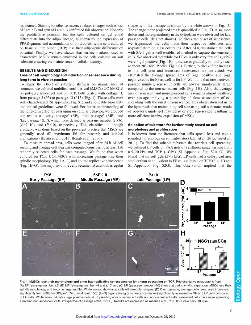
seeding and average cell area was computed considering at least 150randomly selected cells for each passage. We found that whencultured on TCP, UC-hMSCs with increasing passage lose theirspindle morphology (Fig. 1A–C) and go into replicative senescence(Fig. 1E–G). The majority of the cells became flat and took irregular
shapes with the passage as shown by the white arrows in Fig. 1C.The change in the projected area is quantified in Fig. 1D. Also, moredebris and more granularity in the cytoplasm were observed for laterpassage cells (data not shown). To check the onset of senescence,we trypsinized the cells from their respective substrates andre-plated them on glass coverslips. After 24 h, we stained the cellswith SA-β-gal, a well-established method to capture the senescentcells. We observed that while for EP cells only very few cells (<5%)were β-gal positive (Fig. 1E), it increases gradually to finally reachat about 20% for LP cells (Fig. 1G). Further, to check if the increasein the cell area and increased senescence are correlated, weestimated the average spread area of β-gal positive and β-galnegative cells for EP as well as for LP. We found that irrespective ofpassage number, senescent cells are always significantly largercompared to the non-senescent cells (Fig. 1H). Also, the averagearea of senescent and non-senescent cells remains almost unalteredover passage implying a possibility of close association of cellspreading with the onset of senescence. This observation led us tothe hypothesis that maintaining cell size using soft substrates madeof polyacrylamide gel may delay or stop senescence resulting inmore efficient in vitro expansion of MSCs.
Selection of substrate for further study based on cellmorphology and proliferationIt is known from the literature that cells spread less and take arounded morphology on soft substrates (Jalali et al., 2015; Tee et al.,2011). To find the suitable substrate that restricts cell spreading,we cultured LP cells on PAA gels of a stiffness range varying from0.5–20 kPa and TCP (∼GPa) (SI Appendix, Fig. S2A–G). Wefound that on soft gels (E≤5 kPa), LP cells had a cell-spread areasmaller than or equivalent to EP cells cultured on TCP (Fig. 1D andSI Appendix, Fig. S2G). This observation implied that the
Fig. 1. hMSCs lose their morphology and enter into replicative senescence on long-term passaging on TCP. Representative micrographs from(A) EP (passage number ≤6) (B) MP (passage number >6 and ≤10) and (C) LP (passage number >10) show that during in vitro expansion, MSCs lose theirspindle morphology and become large and flat. White arrows show large cells with irregular shapes. (D) Over passage, average cell-spread area increasessignificantly from ∼3000–4500 µm2. (N=3, n=at least 150). (E–G) β-gal staining (a senescence marker) significantly increased in MP and LP cells comparedto EP cells. White arrow indicates β-gal positive cells. (H) Spreading area of senescent cells and non-senescent cells: senescent cells have more spreadingarea than non-senescent cells, irrespective of passage (N=3, n>100). Results are expressed as mean±s.e.m., *P<0.05. Scale bars: 100 µm.
2
RESEARCH ARTICLE Biology Open (2019) 8, bio039453. doi:10.1242/bio.039453
BiologyOpen
by guest on December 29, 2019http://bio.biologists.org/Downloaded from
cell-spread area can be kept restricted over passages if cultured on softgels (E≤5 kPa). However the question remains, how soft can we go?To answer this question, we need to consider another parameter, i.e.the effect of substrate stiffness on cell proliferation. It is known thatsoft gel induces reversible cell cycle arrest or quiescence in hMSCs(Rumman et al., 2018; Winer et al., 2009). As a result, the very softgel cannot be used for cell number expansion. To find the optimumrange of stiffness, we cultured cells on substrates of different stiffness.After 48 h of culture on these gels, which is sufficient to inducequiescence (Rumman et al., 2018), we gave a 4 h pulse of BrdU thattags the replicating DNA.We found that while cells on 1 or 2 kPa gelshowed critically less replicating DNA, cells on gels of 5 kPa andhigher stiffness had more than 30% of dividing cells which isequivalent to that on TCP (SI Appendix, Fig. S2H–L).Putting these two observations together, we selected 5 kPa gel for
all our ongoing studies to compare the effect of substrate stiffness onlong-term in vitro culture.
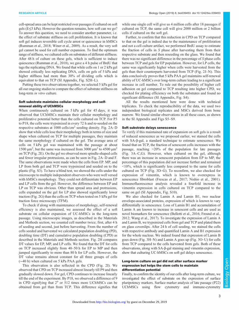
Soft substrate maintains cellular morphology and self-renewal ability of UChMSCsWhen continuously cultured on 5 kPa gel for 45 days, it wasobserved that UChMSCs maintain their cellular morphology andproliferative potential better than the cells cultured on TCP. For P3to P18, the cells were trypsinized in every 72 h and re-seeded on therespective substrates at 1000 cells/cm2 seeding density. Fig. 2A–Dshow that while cells lose their morphology both in terms of size andshape when cultured on TCP for multiple passages, they maintainthe same morphology if cultured on the gel. The average area ofcells on 5 kPa gel was maintained with the passage at about1500 µm2, but the same was increased from 3000 µm2 to 4500 µm2
on TCP (Fig. 2E). On the gel we observedmore spindle-shaped cellsand fewer irregular protrusions, as can be seen in Fig. 2A–D and F.The same observations were made when the cells from EP. MP, andLP from both gel and TCP were trypsinized and seeded on to theplastic (Fig. S3). To have a blind test, we showed the cells under themicroscope to multiple independent observers whowerewell versedwith hMSCs morphology. They could not differentiate between EPand LP cells from the gel. However, the difference between EP andLP on TCP was obvious. Other than spread area and protrusions,cells expanded on the gel for LP also showed significantly lowertraction (Fig. 2G) than the cells on TCP when tested on 5 kPa gel fortraction force microscopy (TFM).To check if along with maintenance of morphology, self-renewal
efficiency is also maintained, we assessed the effect of a softsubstrate on cellular expansion of UC-hMSCs in the long-termpassage. Using microscopic images, as described in the Materialsand Methods section, we counted cell number twice; first, after 4 hof seeding and second, just before harvesting. From the number ofcells seeded and harvested we calculated population doubling (PD),doubling time (DT) and cumulative population doubling (CPD) asdescribed in the Materials and Methods section. Fig. 2H comparesDT values for EP, MP, and LP cells. We found that the DT for cellson TCP increased slightly from 40–50 h for EP to MP and thenjumped significantly to more than 80 h for LP cells. However, theDT value remains almost constant for all three groups of cells(∼40 h) when cultured on 5 kPa PAA gels.This observation is also reflected in the CPD (Fig. 2I). We
observed that CPD on TCP increased almost linearly till P9 and thengradually slowed down. For gel, CPD continues to increase linearlytill the end of the experiment. By P18, we observed a difference of 9in CPD signifying that 29 or 512 times more UChMSCs can beobtained from gel than from TCP. This difference signifies that
while one single cell will give us 4 million cells after 18 passages ifcultured on TCP, the same cell will give 2000 million or 2 billioncells if cultured on the soft gel.
Further, to confirm that this reduction in CPD on TCP comparedto that on the gel is indeed due to the maintenance of proliferationand not a cell culture artifact, we performed BrdU assay to estimatethe fraction of cells in S phase after harvesting them from theirrespective substrate and then reseeding on the glass. We found thatthere was no significant difference in the percentage of S phase cellsbetween TCP and gels for EP population. However, for LP cells, thesame was significantly higher when cells were harvested from thegels than their counterparts harvested from TCP (Fig. 2J–N). Thisdata conclusively proves that 5 kPa PAA gel maintains self-renewalability of UC-hMSCs over long-term culture leading to a significantincrease in cell number. To rule out the possibility of higher celladhesion on gel compared to TCP resulting into higher CPD, wechecked for plating efficiency on both the substrates and found nosignificant difference (SI Appendix, Fig. S4).
All the results mentioned here were done with technicaltriplicates. To check the reproducibility of the data, we used twoindependent biological replicates and MSCs derived from bonemarrow. We found similar observations in all these cases, as shownin the SI Appendix and Figs S5–S9.
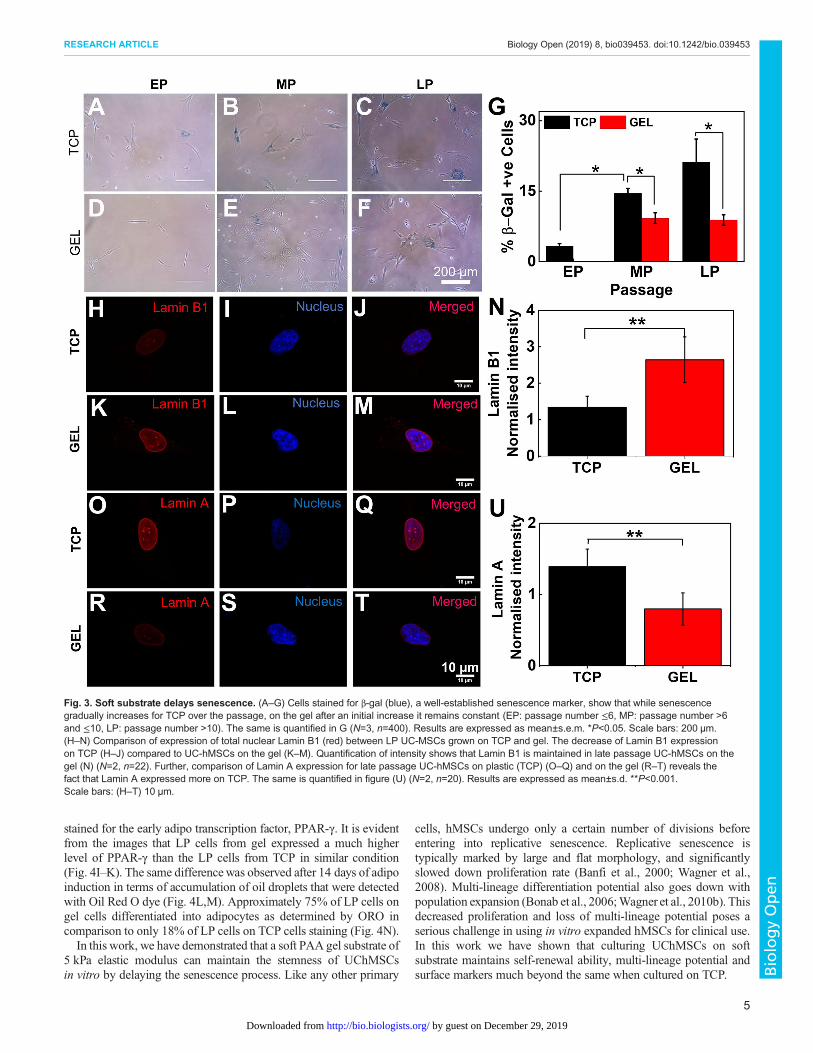
Soft substrate delays senescenceTo verify if this maintained rate of expansion on soft gels is a resultof reduced senescence as we proposed earlier, we stained the cellswith SA-β-gal, a standard technique to estimate senescence. Wefound that on TCP, the fraction of senescent cells increases with thepassage, reaching >20% of the population for late passages(Fig. 3A–C,G). However, when cultured on the gel, althoughthere was an increase in senescent population from EP to MP, thepercentage of this population did not increase further and remained<10%, which is significantly less compared to the same for cellscultured on TCP (Fig. 3D–G). To reconfirm, we also checked forexpression of vimentin, which is known to overexpress insenescence fibroblast (Frescas et al., 2017; Nishio et al., 2001).Immunofluorescence analysis revealed a fourfold increase invimentin expression in cells cultured on TCP compared to thesame on gel (SI Appendix, Fig. S10).
We also checked for Lamin A and Lamin B1, two nuclearenvelope-associated proteins, expression of which is known to varydifferentially in senescence. Loss of Lamin B1 and accumulation ofLamin A are known to increase in senescent cells and are used asnovel biomarkers for senescence (Bellotti et al., 2016; Freund et al.,2012; Wang et al., 2017). To investigate the expression of Lamin Aand Lamin B, we trypsinized cells from gel and TCP and plated themon glass coverslips. After 24 h of cell seeding, we stained the cellswith respective antibody and quantified Lamin A and B1 expressionfor the whole nucleus. We indeed found that expression of Lamin Bgoes down (Fig. 3H–N) and Lamin A goes up (Fig. 3O–U) for cellsfrom TCP compared to the cells harvested from gels. Both of theseobservations, along with SA-β-gal staining and vimentin expression,show that culturing UC-hMSCs on soft gel delays senescence.
Long-term culture on gel did not alter surface markerexpression but helps the stem cells to maintaindifferentiation potentialFinally, to confirm the identity of our cells after long-term culture, weinvestigated the effect of substrate on the expression of surfacepluripotency markers. Surface marker analysis of late passage (P22)UChMSCs using flow cytometry and immune-cytometry
3
RESEARCH ARTICLE Biology Open (2019) 8, bio039453. doi:10.1242/bio.039453
BiologyOpen
by guest on December 29, 2019http://bio.biologists.org/Downloaded from
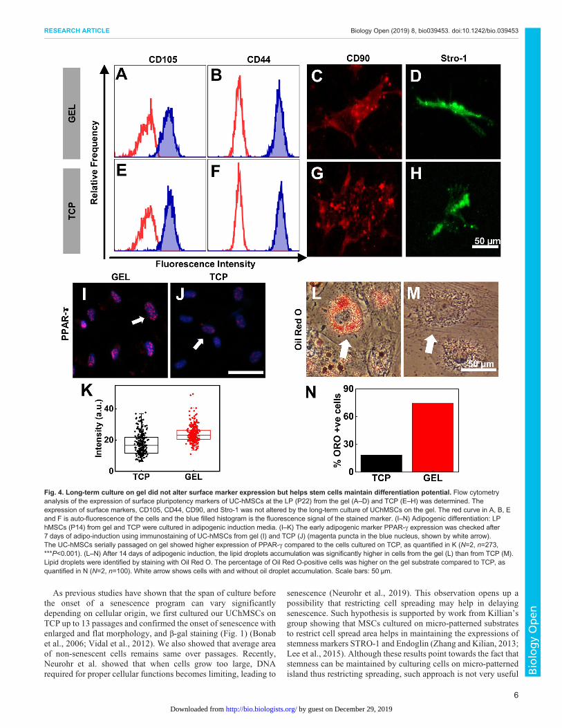
demonstrated that cells cultured either on gel or on TCP expresscharacteristic positive surface markers, CD105 (Fig. 4A,E), CD44(Fig. 4B,F), CD90 (Fig. 4C,G) and Stro-1 (Fig. 4D,H). This is inaccordance with the previous studies which showed that the MSCsurface marker expression remains same with increasing passage
(Kundrotas et al., 2016). However, the difference appears in theirdifferentiation potential. While LP cells on TCP lose their adipogenicpotential, LP cells on gel did not (Fig. 4I–N). We harvested the cellsfrom their respective substrates and cultured them on TCP inadipogenic induction media for 7 days. Then the cells were fixed and
Fig. 2. Soft substrate maintains cellular morphology, rate of expansion and proliferation during serial passage. Representative phase contrastimages of (A) EP (passage number ≤6) and (B) LP (passage number >10) UC-hMSCs on collagen-coated TCP and (C) EP and (D) LP on soft gel. (E) Cellspread area increases with passage on TCP; however, spreading area of cells on gel showed no significant difference across passages. (N=3, n=150).(F) The number of protrusions, as shown by white arrows in B, are significantly increased in LP cells cultured on TCP (N=3, n=150). (G) LP UChMSCs ongel showed significantly lower traction than that on TCP. The experiment was performed using two biological samples (N=3, n=20). (H) DT of UC-hMSCscultured on gel and TCP over the passage. While DT increases with passage when cultured on TCP, it remains unaffected when expanded on 5 kPa gel.The difference in DT for EP and MP are negligible while for LP the difference is significant (N=3, n=1). (I) CPD of UC-hMSCs cultured on gel and TCP overthe passage. CPD increases linearly for UChMSCs on the gel but approaches a plateau for cells cultured on TCP (N=1, n=11). (J) Immunofluorescenceimages of nuclei co-stained with DAPI (blue) and BrdU (red) capturing a relative percentage of cycling cells. (K) Percentage BrdU positive cell for P14 (LP)from gels are significantly higher than the cells from TCP, though for EP (P6) cells there was no significant difference between gel and TCP. This datareconfirms that cells maintain their proliferative potential if cultured on the soft gel. Results are expressed as mean±s.e.m. *P<0.05, **P<0.001, ***P<0.0001.Scale bars: 100 µm.
4
RESEARCH ARTICLE Biology Open (2019) 8, bio039453. doi:10.1242/bio.039453
BiologyOpen
by guest on December 29, 2019http://bio.biologists.org/Downloaded from
stained for the early adipo transcription factor, PPAR-γ. It is evidentfrom the images that LP cells from gel expressed a much higherlevel of PPAR-γ than the LP cells from TCP in similar condition(Fig. 4I–K). The same differencewas observed after 14 days of adipoinduction in terms of accumulation of oil droplets that were detectedwith Oil Red O dye (Fig. 4L,M). Approximately 75% of LP cells ongel cells differentiated into adipocytes as determined by ORO incomparison to only 18% of LP cells on TCP cells staining (Fig. 4N).In this work, we have demonstrated that a soft PAA gel substrate of
5 kPa elastic modulus can maintain the stemness of UChMSCsin vitro by delaying the senescence process. Like any other primary
cells, hMSCs undergo only a certain number of divisions beforeentering into replicative senescence. Replicative senescence istypically marked by large and flat morphology, and significantlyslowed down proliferation rate (Banfi et al., 2000; Wagner et al.,2008). Multi-lineage differentiation potential also goes down withpopulation expansion (Bonab et al., 2006;Wagner et al., 2010b). Thisdecreased proliferation and loss of multi-lineage potential poses aserious challenge in using in vitro expanded hMSCs for clinical use.In this work we have shown that culturing UChMSCs on softsubstrate maintains self-renewal ability, multi-lineage potential andsurface markers much beyond the same when cultured on TCP.
Fig. 3. Soft substrate delays senescence. (A–G) Cells stained for β-gal (blue), a well-established senescence marker, show that while senescencegradually increases for TCP over the passage, on the gel after an initial increase it remains constant (EP: passage number ≤6, MP: passage number >6and ≤10, LP: passage number >10). The same is quantified in G (N=3, n=400). Results are expressed as mean±s.e.m. *P<0.05. Scale bars: 200 µm.(H–N) Comparison of expression of total nuclear Lamin B1 (red) between LP UC-MSCs grown on TCP and gel. The decrease of Lamin B1 expressionon TCP (H–J) compared to UC-hMSCs on the gel (K–M). Quantification of intensity shows that Lamin B1 is maintained in late passage UC-hMSCs on thegel (N) (N=2, n=22). Further, comparison of Lamin A expression for late passage UC-hMSCs on plastic (TCP) (O–Q) and on the gel (R–T) reveals thefact that Lamin A expressed more on TCP. The same is quantified in figure (U) (N=2, n=20). Results are expressed as mean±s.d. **P<0.001.Scale bars: (H–T) 10 µm.
5
RESEARCH ARTICLE Biology Open (2019) 8, bio039453. doi:10.1242/bio.039453
BiologyOpen
by guest on December 29, 2019http://bio.biologists.org/Downloaded from
As previous studies have shown that the span of culture beforethe onset of a senescence program can vary significantlydepending on cellular origin, we first cultured our UChMSCs onTCP up to 13 passages and confirmed the onset of senescence withenlarged and flat morphology, and β-gal staining (Fig. 1) (Bonabet al., 2006; Vidal et al., 2012). We also showed that average areaof non-senescent cells remains same over passages. Recently,Neurohr et al. showed that when cells grow too large, DNArequired for proper cellular functions becomes limiting, leading to
senescence (Neurohr et al., 2019). This observation opens up apossibility that restricting cell spreading may help in delayingsenescence. Such hypothesis is supported by work from Killian’sgroup showing that MSCs cultured on micro-patterned substratesto restrict cell spread area helps in maintaining the expressions ofstemness markers STRO-1 and Endoglin (Zhang and Kilian, 2013;Lee et al., 2015). Although these results point towards the fact thatstemness can be maintained by culturing cells on micro-patternedisland thus restricting spreading, such approach is not very useful
Fig. 4. Long-term culture on gel did not alter surface marker expression but helps stem cells maintain differentiation potential. Flow cytometryanalysis of the expression of surface pluripotency markers of UC-hMSCs at the LP (P22) from the gel (A–D) and TCP (E–H) was determined. Theexpression of surface markers, CD105, CD44, CD90, and Stro-1 was not altered by the long-term culture of UChMSCs on the gel. The red curve in A, B, Eand F is auto-fluorescence of the cells and the blue filled histogram is the fluorescence signal of the stained marker. (I–N) Adipogenic differentiation: LPhMSCs (P14) from gel and TCP were cultured in adipogenic induction media. (I–K) The early adipogenic marker PPAR-γ expression was checked after7 days of adipo-induction using immunostaining of UC-hMSCs from gel (I) and TCP (J) (magenta puncta in the blue nucleus, shown by white arrow).The UC-hMSCs serially passaged on gel showed higher expression of PPAR-γ compared to the cells cultured on TCP, as quantified in K (N=2, n=273,***P<0.001). (L–N) After 14 days of adipogenic induction, the lipid droplets accumulation was significantly higher in cells from the gel (L) than from TCP (M).Lipid droplets were identified by staining with Oil Red O. The percentage of Oil Red O-positive cells was higher on the gel substrate compared to TCP, asquantified in N (N=2, n=100). White arrow shows cells with and without oil droplet accumulation. Scale bars: 50 µm.
6
RESEARCH ARTICLE Biology Open (2019) 8, bio039453. doi:10.1242/bio.039453
BiologyOpen
by guest on December 29, 2019http://bio.biologists.org/Downloaded from
for MSC expansion as cells on micro-island stop proliferation anddo not increase in number.Earlier studies also showed that stemness in hMSCs could be
maintained better by reducing actomyosin contractility or cellulartraction using pharmacological inhibitor of ROCK and myosin(Zhang and Kilian, 2013). In a different study, it was shown thatwhile mESCs (mouse embryonic stem cells) grown on TCP needLIF to stop spontaneous differentiation, a very soft substrate (600 Pa)can maintain them in their undifferentiated state without LIF(Chowdhury et al., 2010). All these results indicated that cell area/contractility plays an important role in the loss of stemness and bothcan be kept low if cultured on a soft substrate (McBeath et al., 2004;Tee et al., 2011). However, none of these works checked the effect ofsubstrate stiffness in long-term culture. We demonstrated here that along-term culture on soft substrate may inherently reduce the cellulartraction (Fig. 2G) and thus can maintain stemness. To the best of ourknowledge, this is the first work to demonstrate the effect of substratestiffness on cellular traction and maintenance of stemness inlong-term culture (for 20 passages i.e. ∼60 days) for any cell type.Consistent with the previous report, we also found that though UC-
hMSCs lose their self-renewal ability when cultured on TCP, theymaintain the molecular signatures related to stemness (Fig. 4A–H)(McGrail et al., 2013). The cells, irrespective of cultured ongel or TCPwere positive for CD105, CD44, CD90, and Stro-1. However, cellscultured on TCP lost their adipogenic differentiation ability whereasthe same was maintained for the cells cultured on the gel (Fig. 4I–N).Loss of adipogenic potential over long term passages and dominanceof osteogenic differentiation has been reported by many earlierresearchers (Neuhuber et al., 2008; Wall et al., 2007). It was shownthat flat cells that appear spontaneously over long-term culture, losetheir adipogenic potential (McGrail et al., 2013). This observation isnot unexpected if we look from the cell mechanics angle. It wasestablished by Engler et al. in their seminal paper in 2006 thatstiff substrate (34 kPa) induces osteogenic lineage in hMSCs (Engleret al., 2006) even in absence of chemical inducer. Similarly, it was alsoshown that cells that were made to spread more or to take the shapethat induces high contractility, also were prone to osteogenic lineagecommitment (Kilian et al., 2010; McBeath et al., 2004). So, it isexpected that if for multiple passages, cells are continuously exposedto a substrate as rigid as TCP which increases cellular spreading andcontractility, adipogenic potential would get diminished. However, asoft culture substrate, in contrast, should maintain the multi-lineagepotential, as demonstrated by our result (Fig. 4).Other than UChMSCs, we also used bone marrow-derived
hMSCs and found a similar result proving that this effect might notbe source specific. We have also found that the substrate stiffness foroptimal growth of skin-derived keratinocytes is not the same as forMSCs (data not shown). How cell type and optimum substratestiffness are inter-linked is open for future investigation.One of the interesting observations in this work is that soft
substrate delays senescence. It is known that acquiring replicativesenescence over in vitro expansion may not be an obvious purposefulprogram but a result of the external environmental condition. Forexample, it has been shown that increased oxygen concentration mayinduce senescence faster. On the contrary, the hypoxic condition isknown to maintain stemness for hMSCs (Basciano et al., 2011).However, the effect of substrate stiffness on senescence has not beenstudied before. We have demonstrated using four known markers ofsenescence – namely expression of β-gal, loss of Lamin B1, the gainof Lamin A and vimentin (Bellotti et al., 2016; Bonab et al., 2006;Freund et al., 2012) – that an optimally soft substrate may delay theonset of senescence significantly.
In summary, our data show that instead of using TCP, culturingcells on the soft substrate will help to solve the problem of limitedavailability of MSCs by increasing the number of available cellsafter extended expansion. This work offers a possibility to design acell-specific culture substrate in the future. This work alsodemonstrates for the first time that replicative senescence inhMSCs can be delayed using substrates of physiological stiffness.
MATERIALS AND METHODSSubstrate preparationGels of polyacrylamide (PAA) of various stiffness were prepared by mixing40% polyacrylamide and 2% bis-acrylamide solution, as describedpreviously (Pelham and Wang, 1997). Substrate preparation protocols andmodulus values were adopted from previously published work (Tse andEngler, 2010). Briefly, the gel solution for desired stiffness was mixed withAPS (ammonium persulfate) 1:100 and TEMED (1:1000) and placedbetween a hydrophobic glass (octadecyltrichlorosilane treated; Sigma-Aldrich, 104817) and the transparency sheet 3-APTMS (Alfa Aesar,A17714) treated. Once polymerized, the hydrophobic plate was carefullyremoved. The gel was conjugated with sulfo-SANPAH and incubated withrat tail type I collagen (25 µg/ml) (Invitrogen, A1048301) at 4°C forovernight, as described (Venugopal et al., 2018). The tissue culture plates(TCP) (control) were also coated with type 1 collagen (25 µg/ml). Thethickness of the gel was controlled by using the defined volume of the gelsolution throughout the experiments.
Cell cultureBone marrow-derived human MSCs were purchased from Lonza (Cat#PT-2501, Lot #482966) (authenticated and tested for contamination by thesupplier), and fresh umbilical cord-derivedMSCswere obtained from healthyindividuals after due ethical clearance and bio-safety approval. For serialpassage experiments, P4 cellswere seeded on large area gels and onTCP (bothcollagen-I coated asmentioned above) with same seeding density (1000 cells/cm2) in MSCs qualified medium α-(MEM) (Invitrogen, A1049001). Low-glucose DMEM (HiMedia, AL006) supplemented with 16% MSC certifiedfetal bovine serum (FBS) (Invitrogen, 12662029), 1% Glutamax (Invitrogen,35050061), and 1%pen-strep (Invitrogen, 15140122) in humidified incubatorwith 37°C and 5% CO2. After 72 h of culture, cells were trypsinized fromPAA gels and TCP using TrypLE™ Enzyme Express (Invitrogen, 12604013)and were reseeded on fresh substrates respectively and cultured for nextpassage, this process was repeated until the TCP growth halted.
Cell count using image analysisImages of the gels and TCP were acquired using a Magnus microscope at10× magnification after 4 and 72 h of seeding to determine accurate cellnumber for calculating PD as described (Cristofalo et al., 1998). For PDcounting, 20 random images per samplewere captured (covering∼3% of thetotal area of the gel), the average number of cells per framewas obtained andthen divided by the total area of the frame to obtain seeding density (cells/cm2). The seeding density was then multiplied by the total area of thesubstrate (gel 20 cm2; TCP 25 cm2) to get the total number of cells seeded(4 h) and harvested (72 h) from a particular experimental condition (PAAgels and TCP) for respective passage. This was done in every passage,which was then used to calculate the CPD for each experimental condition asexplained in the Eqns. 1–3 (Cristofalo et al., 1998):
NH
NS¼ 2PD ð1Þ
PD ¼ lnðNH=NSÞlnð2Þ ð2Þ
where NH is the number of harvested cells, NS is the number of cells seeded,
CPD ¼XPf
Pi
PD ð3Þ
where Pi is the initial passage number and Pf is the final passage number.
7
RESEARCH ARTICLE Biology Open (2019) 8, bio039453. doi:10.1242/bio.039453
BiologyOpen
by guest on December 29, 2019http://bio.biologists.org/Downloaded from
Quantification of cell morphologyCell images were captured at different passages at 48 h post seedingusing EVOS-FL auto inverted microscope (Life Technologies) at 10×magnification. Cell spreading area was determined using ImageJ (NationalInstitutes of Health) software by manually tracing around the perimeter ofan individual cell. For each sample minimum, 150 random cells wereanalyzed. The number of protrusions of cells was quantified from phasecontrast images manually using ImageJ.
Plating efficiencyTo compare the cell adhesion efficiency, P9 bone marrow MSCs wereseeded on the 5 kPa gel and collagen-coated coverslip. After 15 min ofseeding, 15 random images were captured and analyzed to get the number ofcells seeded. The plate was kept in the incubator for 1 h and 30 min, thenmedia was aspirated and fresh media was added in each well. Again, 15random images were captured and analyzed to determine the number of cellsattached. From the number of cells seeded and the number of cells attached,we calculated the plating efficiency as:
Plating efficiency ¼ Number of cells attached
Number of cells seeded� 100 ð4Þ
100% plating efficiency shows all cells attached.
BrdU assayTo check the percentage of S-phase cells in the cell cycle, cells from EP, MPand LP were trypsinized from gels and TCP and were seeded on collagen-coated glass coverslips as described above. After 48 h of seeding, BrdUreagent (Invitrogen, 000103) was added in 1:100 (v/v) ratio in media andincubated for 4 h at 37°C in a humidified incubator with 5%CO2. Thereaftercells were fixed (4% paraformaldehyde), permeabilized (0.5% Triton-X),denatured (2 MHCl), blocked (1.5% bovine serum albumin), and incubatedwith anti-BrdU antibody (Invitrogen, B35128, 1:100) and counterstainedwith AlexaFluor 568 (Invitrogen, A11061, 1:400). Immunofluorescenceimages were captured using EVOS-FL auto and BrdU positive and negativecells were counted manually using ImageJ.
Senescence assaysSenescence-associated β-galactosidase (SA-β-gal) was used to detect MSCssenescence using SA-β-gal staining kit (Abcam, AB65351) according to themanufacturer’s instructions. Briefly, cells from EP, MP, and LP were seededin a six-well plate and incubated in growth media for 48 h. Afterwards, cellswere fixed, stained with β-gal solution and incubated at 37°C without CO2.Ten–15 random images were captured for each condition for analysis β-galpositive cells were counted manually.
Differentiation assaysEP and LP cells from gel and TCP were seeded in a 12-well culture plate ingrowth medium for 72 h and then incubated with differentiation media foradipogenic (Invitrogen, A1006501) and osteogenic (Invitrogen, A1006601)differentiation as per the manufacturer’s instructions. MSCs cultured ingrowth media were used as a negative control. Post 14 days and 21 daysincubation for adipo and osteo differentiation, respectively, adipocytes wereassessed with Oil Red O (Sigma-Aldrich, O0625) solution andosteoblasts were assessed with Alizarin Red solution (Sigma-Aldrich,3422613022311). Images were captured for qualitative and quantitativeanalysis using EVOS FL Auto.
Immunofluorescence stainingFor nuclear Lamin A (Abcam, ab8980, 1:400), Lamin B1 (Abcam, ab16048,1:400), and early adipogenic differentiation marker staining, EP and LP cellsfrom gel and TCPwere cultured on collagen-I-coated glass coverslips for 24 h.Cells were then fixed with 4% paraformaldehyde in PBS for 15 min at roomtemperature (RT) and blocked (3% bovine serum albumin in PBS) for 30 minand washed with cytoskeletal buffer, as described previously (Venugopalet al., 2018). Cells were incubated with respective primary antibodies for 4 hat 4°C, and then incubated with corresponding secondary antibodies for 1 hat RT. Primary and secondary antibodies were used in the following
combinations: anti-PPAR-γ (Abcam, ab59256, 1:300) counterstained withAlexaFluor 488 (Invitrogen, A11034, 1:500), anti-Lamin A (Abcam, ab8980,1:400) counterstained with AlexaFluor 568 (Abcam, ab175473, 1:400),anti-Lamin B1 (Abcam, ab16048, 1:400) counterstained with AlexaFluor 568(Abcam, ab175470, 1:400), anti-Vimentin (Sigma-Aldrich, V5255, 1:300)counterstained with AlexaFluor 488 (Invitrogen, A11059, 1:500). Cell nucleiwere stained with Hoechst 33342 (Invitrogen, H3570) (1:10,000) in PBS for5 min at RT and mounted. Images were captured for qualitative andquantitative analysis using EVOS fluorescence microscope (Invitrogen).
Traction force microscopy (TFM)Gels of 5 kPa were fabricated with embedded fluorescent beads to conductTFM. Briefly, to make a single layer of the fluorescent bead (Fluka, 1 µmrhodamine), beads (1:50) were added to the pre-polymer solution (25 µl)and solidified over the normal gel of 5 kPa. The gel was then functionalizedas described above. Cells were seeded on the gels, after 24 h of cell seedingimages of stressed (before lysing) and unstressed gel (after lysing with 1%Triton-X) were captured by the EVOS FL Auto (Invitrogen). An average of20 cells were analyzed per condition. A MATLAB algorithm was used todetermine the cell-generated displacement field and traction forces aspreviously described (Butler et al., 2002). The TFM datawas analyzed usingMATLAB R2018a (IIT Bombay License).
Statistical analysisData is presented as means±standard error of the mean (s.e.m.). Forstatistical analysis, we used unpaired Student’s t-test and values of P<0.05were considered statistically significant, if not otherwise stated. Data wasplotted using Origin software (IIT Bombay License).
AcknowledgementsWe thank Dr James P. Butler (Harvard Medical School, Department of Medicine,Boston, USA) for his TFM codes used for the traction force analysis. We thank IRCC,IIT Bombay for providing the fellowship to P.M. and confocal microscopy facility.
Competing interestsThe authors declare no competing or financial interests.
Author contributionsConceptualization: A.M.; Methodology: S.K.K., P.M., A.M.; Formal analysis: S.K.K.,P.M., A.K., R.J.; Investigation: S.K.K., P.M., A.K., V.K., R.J.; Resources: S.D.,J.B.; Data curation: S.K.K., P.M., A.K., A.M.; Writing - original draft: S.K.K., P.M.,A.M.; Writing - review & editing: S.K.K., P.M., A.K., J.B., A.M.; Visualization: A.M.;Supervision: A.M.; Project administration: A.M.; Funding acquisition: A.M.
FundingThis work was supported by Welcome Trust-DBT India Alliance[Project #IA/E/11/1/500419] and Indian Institute of Technology Bombay SeedGrant [14IRCCSG002].
Data availabilityRaw data underlying the study is available and can be seen upon request to thecorresponding author.
Supplementary informationSupplementary information available online athttp://bio.biologists.org/lookup/doi/10.1242/bio.039453.supplemental
ReferencesAnderson, H. J., Sahoo, J. K., Ulijn, R. V. and Dalby, M. J. (2016). Mesenchymal
stem cell fate: applying biomaterials for control of stem cell behavior. Front.Bioeng. Biotechnol. 4, 38. doi:10.3389/fbioe.2016.00038
Banfi, A., Muraglia, A., Dozin, B., Mastrogiacomo,M., Cancedda, R. andQuarto,R. (2000). Proliferation kinetics and differentiation potential of ex vivo expandedhuman bone marrow stromal cells: implications for their use in cell therapy. Exp.Hematol. 28, 707-715. doi:10.1016/S0301-472X(00)00160-0
Basciano, L., Nemos, C., Foliguet, B., de Isla, N., de Carvalho, M., Tran, N. andDalloul, A. (2011). Long term culture of mesenchymal stem cells in hypoxiapromotes a genetic program maintaining their undifferentiated and multipotentstatus. BMC Cell Biol. 12, 12. doi:10.1186/1471-2121-12-12
Bellotti, C., Capanni, C., Lattanzi, G., Donati, D., Lucarelli, E. and Duchi, S.(2016). Detection of mesenchymal stem cells senescence by prelamin Aaccumulation at the nuclear level. Springerplus 5, 1427. doi:10.1186/s40064-016-3091-7
8
RESEARCH ARTICLE Biology Open (2019) 8, bio039453. doi:10.1242/bio.039453
BiologyOpen
by guest on December 29, 2019http://bio.biologists.org/Downloaded from
Binato, R., de Souza Fernandez, T., Lazzarotto-Silva, C., Du Rocher, B.,Mencalha, A., Pizzatti, L., Bouzas, L. F. and Abdelhay, E. (2013). Stability ofhuman mesenchymal stem cells during in vitro culture: considerations for celltherapy. Cell Prolif. 46, 10-22. doi:10.1111/cpr.12002
Bonab, M. M., Alimoghaddam, K., Talebian, F., Ghaffari, S. H., Ghavamzadeh,A. and Nikbin, B. (2006). Aging of mesenchymal stem cell in vitro. BMCCell Biol.7, 14. doi:10.1186/1471-2121-7-14
Butler, J. P., Tolic-Nørrelykke, I. M., Fabry, B. and Fredberg, J. J. (2002). Tractionfields, moments, and strain energy that cells exert on their surroundings.Am. J. Physiol. Cell Physiol. 282, C595-C605. doi:10.1152/ajpcell.00270.2001
Caplan, A. I. and Bruder, S. P. (2001). Mesenchymal stem cells: building blocks formolecular medicine in the 21st century. Trends Mol. Med. 7, 259-264. doi:10.1016/S1471-4914(01)02016-0
Cesarz, Z. and Tamama, K. (2016). Spheroid culture of mesenchymal stem cells.Stem Cells Int. 2016, 9176357. doi:10.1155/2016/9176357
Chowdhury, F., Li, Y., Poh, Y.-C., Yokohama-Tamaki, T., Wang, N. and Tanaka,T. S. (2010). Soft substrates promote homogeneous self-renewal of embryonicstem cells via downregulating cell-matrix tractions. PLoS ONE 5, e15655. doi:10.1371/journal.pone.0015655
Cristofalo, V. J., Allen, R. G., Pignolo, R. J., Martin, B. G. and Beck, J. C. (1998).Relationship between donor age and the replicative lifespan of human cells inculture: a reevaluation. Proc. Natl. Acad. Sci. USA 95, 10614-10619. doi:10.1073/pnas.95.18.10614
De Becker, A. and Van Riet, I. (2016). Homing and migration of mesenchymalstromal cells: how to improve the efficacy of cell therapy? World J. Stem Cells 8,73-87. doi:10.4252/wjsc.v8.i3.73
Engler, A. J., Sen, S., Sweeney, H. L. and Discher, D. E. (2006). Matrix elasticitydirects stem cell lineage specification. Cell 126, 677-689. doi:10.1016/j.cell.2006.06.044
Frescas, D., Roux, C. M., Aygun-Sunar, S., Gleiberman, A. S., Krasnov, P.,Kurnasov, O. V., Strom, E., Virtuoso, L. P., Wrobel, M., Osterman, A. L. et al.(2017). Senescent cells expose and secrete an oxidized form of membrane-bound vimentin as revealed by a natural polyreactive antibody. Proc. Natl. Acad.Sci. USA 114, E1668-E1677. doi:10.1073/pnas.1614661114
Freund, A., Laberge, R.-M., Demaria, M. and Campisi, J. (2012). Lamin B1 loss isa senescence-associated biomarker. Mol. Biol. Cell 23, 2066-2075. doi:10.1091/mbc.e11-10-0884
Gilbert, P. M., Havenstrite, K. L., Magnusson, K. E. G., Sacco, A., Leonardi,N. A., Kraft, P., Nguyen, N. K., Thrun, S., Lutolf, M. P. and Blau, H. M. (2010).Substrate elasticity regulates skeletal muscle stem cell self-renewal in culture.Science 329, 1078-1081. doi:10.1126/science.1191035
Honczarenko, M., Le, Y., Swierkowski, M., Ghiran, I., Glodek, A. M. andSilberstein, L. E. (2006). Human bone marrow stromal cells express a distinct setof biologically functional chemokine receptors. Stem Cells 24, 1030-1041. doi:10.1634/stemcells.2005-0319
Jalali, S., Tafazzoli-Shadpour, M., Haghighipour, N., Omidvar, R. andSafshekan, F. (2015). Regulation of endothelial cell adherence and elasticmodulus by substrate stiffness. Cell Commun. Adhes. 22, 79-89. doi:10.1080/15419061.2016.1265949
Kassem, M. (2006). Stem cells: potential therapy for age-related diseases.Ann. N. Y. Acad. Sci. 1067, 436-442. doi:10.1196/annals.1354.062
Kilian, K. A., Bugarija, B., Lahn, B. T. and Mrksich, M. (2010). Geometric cues fordirecting the differentiation of mesenchymal stem cells.Proc. Natl. Acad. Sci. USA107, 4872-4877. doi:10.1073/pnas.0903269107
Kundrotas, G., Gasperskaja, E., Slapsyte, G., Gudleviciene, Z., Krasko, J.,Stumbryte, A. and Liudkeviciene, R. (2016). Identity, proliferation capacity,genomic stability and novel senescence markers of mesenchymal stem cellsisolated from low volume of human bone marrow. Oncotarget 7, 10788-10802.doi:10.18632/oncotarget.7456
Lee, J., Abdeen, A. A., Kim, A. S. and Kilian, K. A. (2015). Influence of biophysicalparameters on maintaining the mesenchymal stem cell phenotype. ACSBiomater. Sci. Eng. 1, 218-226. doi:10.1021/ab500003s
Lutolf, M. P., Gilbert, P. M. and Blau, H. M. (2009). Designing materials to directstem-cell fate. Nature 462, 433-441. doi:10.1038/nature08602
McBeath, R., Pirone, D. M., Nelson, C. M., Bhadriraju, K. and Chen, C. S. (2004).Cell shape, cytoskeletal tension, and RhoA regulate stem cell lineagecommitment. Dev. Cell 6, 483-495. doi:10.1016/S1534-5807(04)00075-9
McGrail,D. J.,McAndrews,K.M.andDawson,M.R. (2013). Biomechanical analysispredicts decreased human mesenchymal stem cell function before moleculardifferences. Exp. Cell Res. 319, 684-696. doi:10.1016/j.yexcr.2012.11.017
McMurray, R. J., Gadegaard, N., Tsimbouri, P. M., Burgess, K. V., McNamara,L. E., Tare, R., Murawski, K., Kingham, E., Oreffo, R. O. C. and Dalby, M. J.(2011). Nanoscale surfaces for the long-term maintenance of mesenchymal stemcell phenotype and multipotency.Nat. Mater. 10, 637-644. doi:10.1038/nmat3058
Murphy, W. L., McDevitt, T. C. and Engler, A. J. (2014). Materials as stem cellregulators. Nat. Mater. 13, 547-557. doi:10.1038/nmat3937
Neuhuber, B., Swanger, S. A., Howard, L., Mackay, A. and Fischer, I. (2008).Effects of plating density and culture time on bone marrow stromal cellcharacteristics. Exp. Hematol. 36, 1176-1185. doi:10.1016/j.exphem.2008.03.019
Neurohr, G. E., Terry, R. L., Lengefeld, J., Bonney, M., Brittingham, G. P.,Moretto, F., Miettinen, T. P., Vaites, L. P., Soares, L. M., Paulo, J. A. et al.(2019). Excessive cell growth causes cytoplasm dilution and contributes tosenescence. Cell 176, 1083-1097. doi:10.1016/j.cell.2019.01.018
Nishio, K., Inoue, A., Qiao, S., Kondo, H. and Mimura, A. (2001). Senescence andcytoskeleton: overproduction of vimentin induces senescent-like morphology inhuman fibroblasts.Histochem.Cell Biol. 116, 321-327. doi:10.1007/s004180100325
Pelham, R. J. and Wang, Y.-L. (1997). Cell locomotion and focal adhesions areregulated by substrate flexibility. Proc. Natl. Acad. Sci. USA 94, 13661-13665.doi:10.1073/pnas.94.25.13661
Pittenger, M. F., Mackay, A. M., Beck, S. C., Jaiswal, R. K., Douglas, R., Mosca,J. D., Moorman, M. A., Simonetti, D. W., Craig, S. and Marshak, D. R. (1999).Multilineage potential of adult human mesenchymal stem cells. Science 284,143-147. doi:10.1126/science.284.5411.143
Ranganath, S. H., Levy, O., Inamdar, M. S. andKarp, J. M. (2012). Harnessing themesenchymal stem cell secretome for the treatment of cardiovascular disease.Cell Stem Cell 10, 244-258. doi:10.1016/j.stem.2012.02.005
Ren, G., Chen, X., Dong, F., Li, W., Ren, X., Zhang, Y. and Shi, Y. (2012). Concisereview: mesenchymal stem cells and translational medicine: emerging issues.Stem Cells Transl. Med. 1, 51-58. doi:10.5966/sctm.2011-0019
Rumman, M., Majumder, A., Harkness, L., Venugopal, B., Vinay, M. B., Pillai,M. S., Kassem, M. and Dhawan, J. (2018). Induction of quiescence (G0) in bonemarrow stromal stem cells enhances their stem cell characteristics.StemCell Res.30, 69-80. doi:10.1016/j.scr.2018.05.010
Saei Arezoumand, K., Alizadeh, E., Pilehvar-Soltanahmadi, Y., Esmaeillou, M.and Zarghami, N. (2017). An overview on different strategies for the stemnessmaintenance of MSCs. Artif. Cells Nanomed. Biotechnol. 45, 1255-1271. doi:10.1080/21691401.2016.1246452
Stem Cell Basics IV. | stemcells.nih.gov (2017).Tee, S.-Y., Fu, J., Chen, C. S. and Janmey, P. A. (2011). Cell shape and substrate
rigidity both regulate cell stiffness. Biophys. J. 100, L25-L27. doi:10.1016/j.bpj.2010.12.3744
Tse, J. R. and Engler, A. J. (2010). Preparation of hydrogel substrates with tunablemechanical properties. Curr. Protoc. Cell Biol. 47, 10.16.1-10.16.16. doi:10.1002/0471143030.cb1016s47
Tuan, R. S. (2013). The coming of age of musculoskeletal tissue engineering. Nat.Rev. Rheumatol. 9, 74-76. doi:10.1038/nrrheum.2012.235
Ullah, I., Subbarao, R. B. and Rho, G. J. (2015). Human mesenchymal stem cells -current trends and future prospective. Biosci. Rep. 35, 1-18. doi:10.1042/BSR20150025
Venugopal, B., Mogha, P., Dhawan, J. and Majumder, A. (2018). Cell densityoverrides the effect of substrate stiffness on human mesenchymal stem cells’morphology and proliferation. Biomater. Sci. 6, 1109-1119. doi:10.1039/C7BM00853H
Vidal, M. A., Walker, N. J., Napoli, E. and Borjesson, D. L. (2012). Evaluation ofsenescence in mesenchymal stem cells isolated from equine bone marrow,adipose tissue, and umbilical cord tissue. Stem Cells Dev. 21, 273-283. doi:10.1089/scd.2010.0589
Wagner, W., Horn, P., Castoldi, M., Diehlmann, A., Bork, S., Saffrich, R., Benes,V., Blake, J., Pfister, S., Eckstein, V. et al. (2008). Replicative senescence ofmesenchymal stem cells: a continuous and organized process. PLoS ONE 3,e2213. doi:10.1371/journal.pone.0002213
Wagner, W., Ho, A. D. and Zenke, M. (2010b). Different facets of aging in humanmesenchymal stem cells. Tissue Eng. Part B Rev. 16, 445-453. doi:10.1089/ten.teb.2009.0825
Wall, M. E., Bernacki, S. H. and Loboa, E. G. (2007). Effects of serial passagingon the adipogenic and osteogenic differentiation potential of adipose-derivedhuman mesenchymal stem cells. Tissue Eng. 13, 1291-1298. doi:10.1089/ten.2006.0275
Wang, L.-T., Ting, C.-H., Yen, M.-L., Liu, K.-J., Sytwu, H.-K., Wu, K. K. and Yen,B. L. (2016). Human mesenchymal stem cells (MSCs) for treatment towardsimmune- and inflammation-mediated diseases: review of current clinical trials.J. Biomed. Sci. 23, 76. doi:10.1186/s12929-016-0289-5
Wang, A. S., Ong, P. F., Chojnowsky, A., Clavel, C. and Dressen, O. (2017). Lossof lamin B1 is a biomarker to quantify cellular senescence in photoaged skin. Sci.Rep. 7, 15678. doi:10.1038/s41598-017-15901-9
Winer, J. P., Janmey, P. A., McCormick, M. E. and Funaki, M. (2009). Bonemarrow-derived human mesenchymal stem cells become quiescent on softsubstrates but remain responsive to chemical or mechanical stimuli. Tissue Eng.Part A 15, 147-154. doi:10.1089/ten.tea.2007.0388
Yeung, T., Georges, P. C., Flanagan, L.A., Marg, B., Ortiz, M., Funaki,M., Zahir, N.,Ming,W.,Weaver, V. and Janmey, P. A. (2005). Effects of substrate stiffness on cellmorphology, cytoskeletal structure, and adhesion. Cell Motil. Cytoskelet. 60, 24-34.doi:10.1002/cm.20041
Zhang, D. and Kilian, K. A. (2013). The effect of mesenchymal stem cell shape onthe maintenance of multipotency. Biomaterials 34, 3962-3969. doi:10.1016/j.biomaterials.2013.02.029
9
RESEARCH ARTICLE Biology Open (2019) 8, bio039453. doi:10.1242/bio.039453
BiologyOpen
by guest on December 29, 2019http://bio.biologists.org/Downloaded from