architecture of the swi/snf-nucleosome complex - molecular and
TRANSCRIPT
MOLECULAR AND CELLULAR BIOLOGY, Oct. 2008, p. 6010–6021 Vol. 28, No. 190270-7306/08/$08.00�0 doi:10.1128/MCB.00693-08Copyright © 2008, American Society for Microbiology. All Rights Reserved.
Architecture of the SWI/SNF-Nucleosome Complex�
Mekonnen Lemma Dechassa,1† Bei Zhang,1,4† Rachel Horowitz-Scherer,2 Jim Persinger,1Christopher L. Woodcock,2 Craig L. Peterson,3 and Blaine Bartholomew1*
Department of Biochemistry and Molecular Biology, Southern Illinois University, Carbondale, Illinois 62901-44131; Department ofBiology, University of Massachusetts, Amherst, Massachusetts 010032; Program in Molecular Medicine, University of
Massachusetts Medical School, Worcester, Massachusetts 016053; and Department of Biology,Thomas University, 1501 Millpond Road, Thomasville, Georgia 317924
Received 28 April 2008/Returned for modification 22 May 2008/Accepted 15 July 2008
The SWI/SNF complex disrupts and mobilizes chromatin in an ATP-dependent manner. SWI/SNF interac-tions with nucleosomes were mapped by DNA footprinting and site-directed DNA and protein cross-linkingwhen SWI/SNF was recruited by a transcription activator. SWI/SNF was found by DNA footprinting to contacttightly around one gyre of DNA spanning �50 bp from the nucleosomal entry site to near the dyad axis. TheDNA footprint is consistent with nucleosomes binding to an asymmetric trough of SWI/SNF that was revealedby the improved imaging of free SWI/SNF. The DNA site-directed cross-linking revealed that the catalyticsubunit Swi2/Snf2 is associated with nucleosomes two helical turns from the dyad axis and that the Snf6subunit is proximal to the transcription factor recruiting SWI/SNF. The highly conserved Snf5 subunitassociates with the histone octamer and not with nucleosomal DNA. The model of the binding trough ofSWI/SNF illustrates how nucleosomal DNA can be mobilized while SWI/SNF remains bound.
The organization of DNA into tightly packed nucleosomaland supranucleosomal structures plays a central role in manyaspects of cellular biology and development in eukaryotes.Cellular processes regulated by chromatin structure rangefrom transcription to DNA repair, replication, recombination,and genetic stability (13, 34, 48). Cells have developed a varietyof mechanisms to overcome this barrier by altering chromatinstructure and controlling the accessibility of DNA.
One of these mechanisms is ATP-dependent chromatin re-modeling, which is catalyzed by a large family of multisubunitchromatin remodeling complexes that alter DNA-histone in-teractions (2). Yeast SWI/SNF (1.14 MDa from STEM or 1.15MDa from stoichiometry data), a prototype of this family ofchromatin remodeling complexes, is composed of 12 differentsubunits whose homologs have been identified in organismsranging from other fungi to mammals (2, 14). SWI/SNF dis-rupts the nucleosome structure, increases the binding of tran-scription factors to nucleosomes, mobilizes histone octamersalong DNA in cis, transfers histone octamers to different DNAfragments, displaces histone H2A/H2B dimers, generates su-perhelical torsion in DNA, and binds preferentially to four-wayDNA (5, 16, 35). SWI/SNF is required for the transcriptionalregulation of about 5% of the total yeast genome (17, 44).SWI/SNF binds to DNA and nucleosomes without any DNAsequence specificity. The complex is found in a low abundancein the cell, suggesting that it is not a general component ofchromatin (10). In vitro and in vivo data demonstrate a directinteraction between SWI/SNF and transcription activators or
repressors, showing that SWI/SNF is recruited to target genesby transcription factors (2, 17, 29, 31, 52).
Most of the SWI/SNF subunits are required to maintain itscommon functions in vivo, but our knowledge of the distinct rolesof each subunit is limited. The ATPase activity of the Swi2/Snf2subunit is stimulated by double-stranded DNA and is critical forthe transcriptional activation and chromatin remodeling functionsof the SWI/SNF complex. The yeast Swi2, Arp7, and Arp9 sub-units form a core complex that can remodel nucleosomes (50),and similarly, the human homologs of Swi2/Snf2, Swi3, Snf5, andSwp73 form a core complex that can remodel nucleosomes (36).Swi2/Snf2, Swi1, Swp73, Snf5, and Snf6 are indispensable for thetranscription activation of a subset of genes mediated by Gcn4(29, 45). Snf5, Snf6, and Swi3 are required for the optimal re-cruitment of SWI/SNF by Gcn4. Swi1 contains an ARID domainthat likely has a lower affinity for DNA than do ARID domains inother proteins. Arp7 and Arp9 are two actin-related proteins thatare also part of the RSC complex, another member of the Snf2subfamily in yeast (2, 14). In addition to being a component of theSWI/SNF complex, Swp29 is a subunit of the yeast TFIIF andTFIID complexes (7). Snf11 was demonstrated to interact withthe Swi2/Snf2 subunit; however, the deletion of the Snf11 subunithas no detectable effect on SWI/SNF-dependent gene expression(47). The elucidation of the role of subunits and domains of thecomplex in its interaction with the nucleosome is critical for anunderstanding of the mechanism of remodeling.
Electron microscopy and three-dimensional reconstructionof three members of the SWI/SNF family of remodelers havebeen reported. The yeast RSC and human PBAF complexesshow some features in common (1, 22, 23); however, yeastSWI/SNF has marked differences from them (42). The RSCand PBAF reconstructions feature a large central cavity, whichcan accommodate mononucleosomes, whereas yeast SWI/SNFrevealed a shallow depression, which could serve as a nucleo-some binding site. Structural models have been proposed forRSC- and PBAF-nucleosome complexes based on the electron
* Corresponding author. Mailing address: Department of Biochem-istry and Molecular Biology, Southern Illinois University, 1245 LincolnDrive, Neckers Room 229, Carbondale, IL 62901-4413. Phone: (618)453-6437. Fax: (618) 453-6440. E-mail: [email protected].
† M.L.D. and B.Z. have equally contributed to this work.� Published ahead of print on 21 July 2008.
6010
Dow
nloa
ded
from
http
s://j
ourn
als.
asm
.org
/jour
nal/m
cb o
n 05
Dec
embe
r 20
21 b
y 11
6.86
.249
.11.
microscopy structure and limited footprinting data (1, 22, 23).Specifics of how the remodelers interact with nucleosomalsubstrates and the mechanistic implications of these interac-tions remain unclear due to limitations in resolving nucleo-some-bound structures by electron microscopy methods.
To better determine the nucleosomal and extranucleosomalregions of the nucleosome bound by SWI/SNF, we have takenadvantage of SWI/SNF recruitment by acidic transcription ac-tivators to orient SWI/SNF in one preferred direction on thenucleosome. Data from DNA footprinting and site-directedDNA cross-linking have identified the regions of DNA boundby SWI/SNF and the subunits of SWI/SNF that were bound atthese different sites, improving on and greatly expanding pre-vious studies with 5S ribosomal DNA (rDNA) nucleosomes(39). We have also developed a protein cross-linking approachto attach photoreactive moieties at specific sites within thehistone octamer and thereby map the interactions of SWI/SNFwith the histone octamer face of the nucleosome. The combi-nation of these approaches has provided an unprecedentedview of the structure of an ATP-dependent chromatin remod-eler and its interaction with a nucleosomal substrate.
MATERIALS AND METHODS
Plasmids. Oligonucleotides 5�-CGGAGGACTGTCCTCCGA-3� and 5�-AGCTTCGGAGGACAGTCCTCCGTGCA-3� were annealed to form a single Gal4binding site, and 5�-CGGAGTACTGTCCTCCGAGCGGAGTACTGTCCTCCGCA-3� and 5�-AGCTTGCGGAGGACAGTACTCCGCTCGGAGGACAGTACTCCGTGCA-3� annealed to form two Gal4 binding sites in a DNA duplexwith flanking PstI and HindIII sites. Plasmids with the Gal4 binding site(s) placed27 bp from the edge of the 601 sequence, p159-1G-27 and p159-1-2G2-27, wereformed by ligating the short double-stranded DNA duplex to p159 cut with PstIand HindIII. After ligation, the recognition site for HindIII was recovered andthat of PstI was destroyed, and the constructs were confirmed by DNA sequenc-ing (54).
Standard site-directed mutagenesis was used to introduce mutations into Xe-nopus laevis core histone proteins using the pET-histone expression plasmidreported previously (26), and the cysteine at residue 110 of H3 was changed toalanine.
Gal4-VP16 purification. Gal4-VP16 was overexpressed in Escherichia coliXa-90 and purified as described previously, with some minor changes (40).Briefly, a Q Sepharose column was used instead of a phenyl-Sepharose column,and a MonoS column was added with a linear gradient of 250 to 450 mM NaCl.The purified protein was analyzed by sodium dodecyl sulfate-polyacrylamide gelelectrophoresis (SDS-PAGE) and immunoblotting with anti-Gal4 antibody.
Purification of histone proteins and octamer refolding. Recombinant histoneswere expressed in Escherichia coli BL21 cells with the pET-histone expressionvector. The four histones were purified by Sephacryl S-200 gel filtration andMonoS cation-exchange chromatography as described previously (26). Octamerswere refolded with the substitution of one of the four histones containing a singlecysteine. The refolded octamer was purified by Superdex 200 size exclusionchromatography.
Purification of SWI/SNF. Yeast SWI/SNF was purified from Saccharomycescerevisiae strains YJW426 (30) and YBB001. YBB001 (BY4742 mat� his3�1leu2�0 lys2�0 ura3�0 swi3-HA-V5-His6::kanMX4 snf6-FLAG::LEU2) was de-rived from BY4742 by tagging the C terminus of Snf6 with FLAG and Swi3 withHA-V5-His6. Yeast cells were grown in yeast extract-peptone-dextrose mediumto an optical density at 600 of ca. 5 to 6. Harvested cells were frozen and groundunder liquid N2 using a mortar and pestle. Cell extract was prepared by mixingyeast cell powder with buffer containing 20 mM Na-HEPES, 350 mM NaCl, 20%glycerol, 0.1% Tween 20, and protease inhibitors (1 mM phenylmethylsulfonylfluoride, 0.5 �g/ml leupeptin, 1 �g/ml pepstatin, and 2 �g/ml aprotinin) andspinning at 100,000 � g for 1 h. SWI/SNF was purified from the soluble whole-cell extract by Biorex70, Ni-nitrilotriacetic acid, and M2 agarose affinity chro-matography. In some cases, SWI/SNF was purified only by M2 agarose affinitychromatography of the whole-cell extract. The one-step affinity purification ofSWI/SNF had a higher yield (�5 �g/liter of culture at an optical density at 600nm of 5 to 6) and had a few contaminating proteins that did not adversely affect
the binding or remodeling activities of SWI/SNF or the cross-linking pattern ofSWI/SNF.
DNA probes, nucleosome reconstitution, DNA footprinting, and photoaffinitylabeling. A 555-bp NdeI-HindIII DNA fragment derived from plasmid p159-1with one or two Gal4 binding sites was used as a template to synthesize pho-toaffinity DNA probes. Solid-phase DNA probe synthesis was performed asdescribed previously (33) except that the probe was cleaned with a PCR purifi-cation kit instead of by phenol-chloroform extraction and ethanol precipitation.The probe was removed from the beads by EcoRI digestion of a 242-bp DNAthat assembles into nucleosomes with extranucleosomal DNA of 34 bp and 61 bpon the other side containing the two Gal4 binding sites.
The DNA used for homogenous nucleosome reconstitution was synthesized byPCR using p159-1-1Gal4-27 as a template and oligonucleotides 5�-biotin/TCCCCAGTCACGACGTTGTAAAAC-3� and 5�-ACCATGATTACGCCAAGCTTCGG-3� of the upper and lower strands, respectively. A 276-bp DNA, biotinyl-ated at one end with a Gal4 binding site at the opposite side, was generated, andthe 601 positioning DNA sequence was flanked by 69 bp (biotinylated) and 59 bp(with the Gal4 binding site) of linker DNA.
Mononucleosomes were reconstituted at 37°C with a solution containing 3.8ng/�l DNA probe, 1.5 �g/�l salmon sperm DNA, and 1 �g/�l histone octamerin 2 M NaCl in 10 �l using the rapid salt dilution method. Homogenous mono-nucleosomes were reconstituted using a solution containing 0.6 �g/�l biotinyl-ated 276-bp DNA, 1 �g/�l histone octamer, and 2 M NaCl, and DNA footprint-ing and photoaffinity labeling were done as described previously (19).
Modification of nucleosomes and histone cross-linking. After the removal ofreducing agents by dialysis, nucleosomes (20 pmol) were modified with 3 nmolN-[(2-pyridyldithio)ethyl]-4-azidosalicylamide (PEAS) (Molecular Probes) for1 h at room temperature. Excess PEAS reagent was removed by dialysis, and thePEAS-conjugated nucleosomes were radiolabeled by indirect iodination (9) us-ing Iodo-Gen precoated iodination tubes (Pierce). The cysteine mutant nucleo-somes modified with [125I]PEAS were immobilized on magnetic beads (strepta-vidin-coated M-280 Dynabeads). Five picomoles of the nucleosomes wasincubated with 200 �g of equilibrated beads with vigorous mixing at roomtemperature for 45 min. After the removal of unbound nucleosomes by washing,6 nM SWI/SNF was bound to the immobilized 4 nM nucleosome in the presenceor absence of 6.4 nM Gal4-VP16 and cross-linked by UV irradiation. Gal4-VP16-dependent SWI/SNF binding to nucleosomes was achieved by including 50 ngsheared salmon DNA in the binding reaction mixture. Unbound free SWI/SNFwas removed from the beads, and the bound cross-linked SWI/SNF was resus-pended in buffer with 100 mM dithiothreitol, 400 mM NaCl, and 10 mM Na-HEPES (pH 7.8) to break the disulfide bond connecting cross-linked nucleo-somes to SWI/SNF and reverse SWI/SNF binding to immobilized nucleosomes.The samples were analyzed by Bis-Tris SDS-PAGE and phosphorimaging toidentify the radiolabeled subunits.
Cryoelectron microscopy of SWI/SNF. Tandem affinity purification-purifiedyeast SWI/SNF was prepared as previously described (43). Unfixed samples at anominal concentration of 1 mg/ml were applied to perforated carbon films in 3-�ldroplets in a humidity-controlled chamber maintained at 95% relative humidity.After 1 min, the droplet was blotted from both sides with Whatman 425 filterpaper for 3 to 5 s and then plunged into liquefied ethane for vitrification. Thesame procedure was used for fixed samples, which were applied to perforatedsubstrates backed with an ultrathin layer of carbon.
Vitrified samples were held in liquid nitrogen through transfer to a Gatan 626cryoholder for observation with an FEI Tecnai12 electron microscope at 120 kV,and images were recorded at defocus values of between 600 and 1,000 Å on a2,048-by-2,048 Tietz video and image processing system charge-coupled-devicecamera at a pixel size of 2.45 Å in the images.
Single-particle reconstruction was carried out as previously described (18, 42)with EMAN (25). All image data were truncated to include only up to the firstzero of the contrast transfer function, and no phase correction was performed.Only images whose defocus value included data to 15 Å inside the first zero wereused.
Frozen-hydrated samples of unfixed yeast SWI/SNF frozen over holes had abimodal distribution caused by subunit loss when complexes came into contactwith one of the air-liquid interfaces during freezing. The subset of particles fullycontained in ice did not show a preferred orientation or any size or densitychanges. This was determined by comparison to data collected from fixed sam-ples, which were known to retain their full mass and to have no preferredorientation on carbon film (42). Model bias was precluded by evaluating twodifferent structures for refinement in the initial classification of particles, afeatureless sphere of appropriate dimensions, and the previously calculatedstructure from negatively stained particles low-pass filtered to 50 Å. The size ofclasses and distribution of projections confirmed that good coverage of Euler
VOL. 28, 2008 ARCHITECTURE OF SWI/SNF 6011
Dow
nloa
ded
from
http
s://j
ourn
als.
asm
.org
/jour
nal/m
cb o
n 05
Dec
embe
r 20
21 b
y 11
6.86
.249
.11.

angles was represented, and no preferred orientation was present. In both cases,the close agreement between projections and class averages was indicative ofhomogeneity of the data sets (25).
The two data sets (fixed and unfixed) were independently reconstructed andcompared for convergence, judged by the absence of change in the Fourier shellcorrelation (FSC) of subsequent iterations. This occurred after five iterations, witha value of 28 Å for the FSC of 0.5 for the independent sets. At this point, the twodata sets were merged, and convergence at eight iterations led to a reconstructionwith values of 19 Å at an FSC of 0.3 and 23 Å at an FSC of 0.5. Volumes wereconstrained to the mass determined by STEM (43) of 1.14 MDa, assuming a proteindensity of 1.35 g/ml (0.81 Da Å�3), and Gaussian low-pass filtered to the thresholdof an FSC value of 0.5. Figures have been filtered to 25 Å.
The three-dimensional reconstruction was visualized using Amira software(Visage Imaging, Carlsbad, CA) with the isosurface calculated to include 1.14MDa. The crystal structure of the nucleosome (11) was modified by the additionof 15 bp of B-DNA to simulate linker DNA. The nucleosome was first positionedto maximize the number of bases identified by footprinting as contacting theSWI/SNF surface. The position was further refined by maximizing histone con-tacts. Contacts were prioritized in order of their distance from the surface, withall contacts within 9 Å (the length of the chemical cross-linker) being givenhighest priority and contacts greater than 12 Å being disallowed.
RESULTS
SWI/SNF binds 50 bp or more of nucleosomal DNA. The 601nucleosome positioning sequence described previously wasused to construct nucleosomes with a common translationalposition (19, 24). Recruitment of SWI/SNF by the transcrip-tion activator Gal4-VP16 bound to extranucleosomal DNAensured SWI/SNF binding to nucleosomes in one preferredorientation. SWI/SNF on its own would likely have two orien-tations, which it could bind nucleosomes due to their pseudo-twofold symmetry. Conditions were established using an excessof competitor DNA and nucleosomes to make SWI/SNF bind-ing to the labeled nucleosomes dependent on Gal4-VP16 bind-ing (Fig. 1B, compare lanes 3 and 4). Gal4-VP16 was observedto bind well to one or two Gal4-VP16 sites positioned 27 bpfrom the edge of the nucleosome (Fig. 1B, compare lanes 1 and2, and data not shown).
The interactions of SWI/SNF with DNA in these nucleo-somes were determined by DNA footprinting with hydroxylradical (Fig. 1C). The strongest protection was on the side ofthe dyad axis opposite the Gal4 binding sites in the nucleoso-mal region starting �1 to 2 helical turns from the dyad axis andextending into the extranucleosomal DNA region (positions�85 to �3 and �16 to �65 in the upper and lower strands,respectively). Protection of nucleosomal DNA correspondedapproximately to one turn or gyre of DNA around the nucleo-some core (see Fig. 6A and B, shown in red). The other half ofthe nucleosomal DNA region had little or no protection, andthere was enhanced DNA cutting at the extranucleosomalDNA region proximal to the Gal4 binding site. The enhancedcutting is suggestive of SWI/SNF binding altering the DNAstructure in some manner to make it more readily cleaved byhydroxyl radicals and implies that SWI/SNF may be interactingat or close to this region. The asymmetry of the DNA footprintpattern confirmed that SWI/SNF is bound in one preferredorientation rather than two symmetrical positions about thenucleosome.
SWI/SNF has one large asymmetric trough on its surfacethat matches the contour of the nucleosome. The DNA foot-printing data demonstrate that the nucleosomal binding sur-face of SWI/SNF should accommodate one entire gyre of thenucleosome without making close contact with the adjacent
FIG. 1. DNA footprinting of SWI/SNF bound to nucleosomesafter recruitment by Gal4-VP16. (A) Purified SWI/SNF was sepa-rated by 4 to 20% SDS-PAGE and stained with Coomassie blue. E1and E2 are the first two fractions from the M2-agarose column.(B) Nucleosomes (Nucl.) were assembled with a radiolabeled242-bp DNA probe (601 nucleosome positioning sequence flankedby 34 and 61 bp of extranucleosomal DNA). The positions of thenucleosome (oval), flanking DNA, and one or two Gal4 bindingsites (2G) are shown. Nucleosomes (55 nM with 0.14 nM of thenucleosomes contains Gal4 binding sites) were incubated with 2.5nM SWI/SNF in the presence or absence of Gal4-VP16 (75 nM) for30 min at 30°C. Nucleosomes alone (lane 1) or with SWI/SNF (lanes3 and 4) and/or Gal4-VP16 (lanes 2 and 4) added were separated ona native 4% polyacrylamide gel and visualized by phosphorimaging.(C) DNA footprinting with the hydroxyl radical was performed oncomplexes of the nucleosome plus Gal4-VP16 with (gray) or with-out (black) SWI/SNF. Quantification of the different lanes from a6% denaturing polyacrylamide gel after normalization is overlaidfor comparison. The region protected by SWI/SNF is indicatedbelow by either a solid black (strongest) or gray (moderate) bar.The open bar indicates the region in which SWI/SNF binding en-hances cutting, and the numbering refers to the number of basepairs from the right (�) or left (�) of the dyad axis.
6012 DECHASSA ET AL. MOL. CELL. BIOL.
Dow
nloa
ded
from
http
s://j
ourn
als.
asm
.org
/jour
nal/m
cb o
n 05
Dec
embe
r 20
21 b
y 11
6.86
.249
.11.
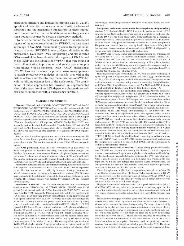
gyre. To find if there are key structural features consistent withour DNA footprinting data, we have reexamined the structureof yeast SWI/SNF utilizing cryoelectron microscopy (cryoEM)to avoid potential artifacts inherent in previous studies due tonegative staining and drying of the sample. The single-particlereconstruction of cryoEM data (resolution of 23 Å at an FSCof 0.5) (see Materials and Methods) revealed a shape closelyrelated to the negative-stain reconstruction (42), with most ofthe differences being attributable to flattening in stain andindistinct boundaries between stain penetration and exclusion.The foremost feature of the reconstruction in ice is the deep-ened trough whose base closely matches the contour of anucleosome. The trough has a base and three walls referred toas back, high, and low, with heights of 56 Å, 120 Å, and 85 Å,respectively, and with defined features at their upper edgesidentified as clamp and lip (Fig. 2A, C, and E).
The contours of the trough (Fig. 2C and E) suggest that anucleosome could be bound on edge with limited rotationalfreedom about the dyad axis but with a large degree of free-dom perpendicular to it. The DNA footprinting data suggestthat the nucleosome is located inside this trough, abutting the
high wall of SWI/SNF, thereby protecting one gyre of thenucleosome. The other gyre, facing the low wall, would bereadily accessible (Fig. 2B, D, and F). Based on this model, theGal4 binding site would be located in the outer surface of theback wall, and the dyad axis of the nucleosome would beexposed, facing out of the trough, rotated toward the back wall.
The Swi2/Snf2 subunit of SWI/SNF is closely associatedwith nucleosomal DNA two helical turns from the dyad axis.The subunits of SWI/SNF bound near nucleosomal and ex-tranucleosomal DNA were identified by site-directed DNAcross-linking. In these experiments, a photoreactive deoxyribo-nucleotide was enzymatically incorporated adjacent to a radio-active nucleotide(s) in DNA at 20 different sites throughoutthe nucleosome (Fig. 3A). These DNA modifications wereshown previously to not interfere with SWI/SNF binding andremodeling of nucleosomes (38) (results not shown). De-oxyuridine and deoxycytidine nucleotide analogs contained ei-ther a photoreactive aryl azide or diazirine moiety. The arylazide preferentially cross-links to nucleophilic side chains,while the diazirine has no amino acid side chain preference(46). The subunits of SWI/SNF bound proximal to these siteswere covalently linked to DNA by UV irradiation and theDNA degraded by DNase I and S1 nuclease. The short radio-labeled DNA left cross-linked to the protein was previouslyshown to add only about 3 to 5 kDa to the apparent molecularmass of the protein (38). Gel shift assays were used to monitorthe binding of SWI/SNF to nucleosomes, and SWI/SNF bind-ing was found to be dependent on Gal4-VP16 under theseconditions (Fig. 1B). Nucleosome assembly for all 20 positionswas as high as 97%, indicating that the binding and cross-linking of SWI/SNF to free DNA were negligible.
The region of nucleosomal DNA from positions �62 to �18/�17 bound by SWI/SNF was cross-linked almost exclusively tothe Swi2/Snf2 subunit (Fig. 3B to D). The site of strongest cross-linking is at a region two helical turns from the dyad axis at bp�17 and �18 and corresponds to where the translocation ofSWI/SNF along DNA was shown to be required for nucleosomemobilization (54). Swi2/Snf2 was reproducibly cross-linked atleast 10-fold more efficiently at bp �18/�17 than at any otherposition examined (Fig. 3E). These data indicate that althoughthe Swi2/Snf2 subunit makes extensive contact with nucleosomalDNA, its primary contact with the major groove of DNA is twohelical turns from the dyad axis, consistent with its catalytic role inchromatin remodeling. Cross-linking of SWI/SNF was highly spe-cific, as shown by its strong dependency on Gal4-VP16 (compareodd versus even lanes in Fig. 3B to D). Based on the model ofnucleosome binding to the trough region of SWI/SNF, the strongcross-linking of Swi2/Snf2 at bp �18 and �17 would be located atthe base of the trough next to the back wall (see Fig. 8C and D).
The Swp73 and Swp82 subunits are less efficiently cross-linked to nucleosomal DNA than Swi2/Snf2 from bp �40 to�52 and bp �21 to �34, respectively. Snf5 weakly cross-linkedDNA at three positions (bp �48 to �52 and �73 to �85 andnear the dyad axis from bp 0 to �14) proximal to each other onthe nucleosome surface. Although these contacts are weak,they appear to be specific and are not likely to be due to minoramounts of SWI/SNF bound to free DNA, as for the weakcross-linking of Snf6 that is observed throughout the nucleo-some-bound region (see below).
FIG. 2. cryoEM reconstruction of SWI/SNF and model of the SWI/SNF-nucleosome complex. Panels A, C, and E show three different viewsof the SWI/SNF structure obtained from cryoEM. Panels B, D, and F arethe models of the SWI/SNF-nucleosome complex obtained by fitting thecrystal structure of the nucleosome low pass filtered to 25 Å into theputative nucleosome binding surface of SWI/SNF. Features of the nu-cleosome binding face of SWI/SNF are a trough whose base (TB) ismet by a high wall (HW), a low wall (LW), and a back wall (BW). Thebase slopes very gently up into the high wall and down to the nearlyrimless front; junctions with the low and back walls are more pro-nounced. Along the rim of the trough are two prominent features,labeled lip (L) and clamp (CL). The dyad axis of the nucleosome isindicated by an arrow (B, D, and F).
VOL. 28, 2008 ARCHITECTURE OF SWI/SNF 6013
Dow
nloa
ded
from
http
s://j
ourn
als.
asm
.org
/jour
nal/m
cb o
n 05
Dec
embe
r 20
21 b
y 11
6.86
.249
.11.
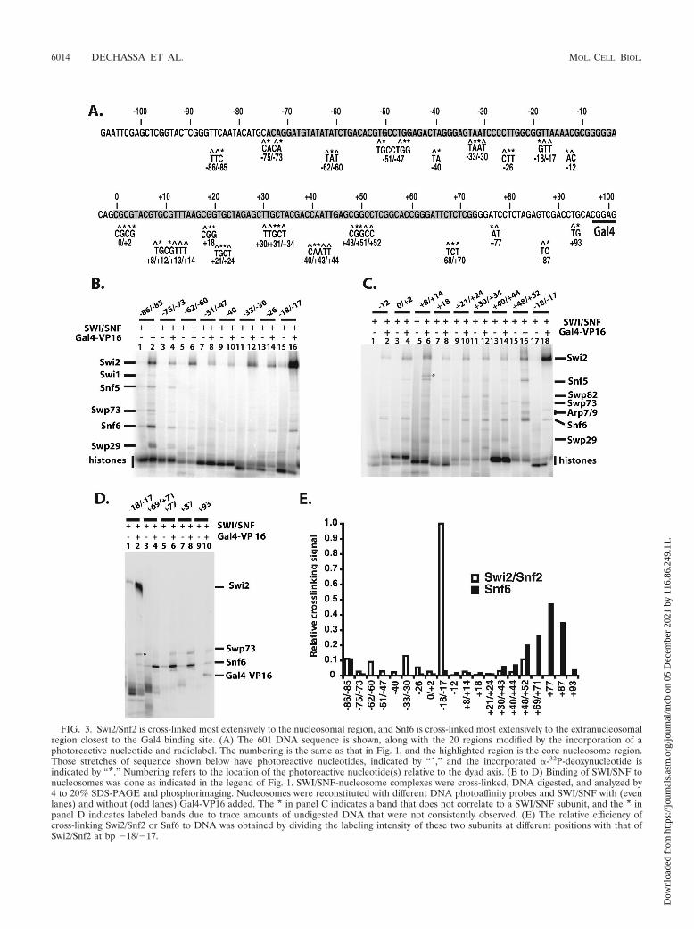
FIG. 3. Swi2/Snf2 is cross-linked most extensively to the nucleosomal region, and Snf6 is cross-linked most extensively to the extranucleosomalregion closest to the Gal4 binding site. (A) The 601 DNA sequence is shown, along with the 20 regions modified by the incorporation of aphotoreactive nucleotide and radiolabel. The numbering is the same as that in Fig. 1, and the highlighted region is the core nucleosome region.Those stretches of sequence shown below have photoreactive nucleotides, indicated by “ˆ,” and the incorporated �-32P-deoxynucleotide isindicated by “*.” Numbering refers to the location of the photoreactive nucleotide(s) relative to the dyad axis. (B to D) Binding of SWI/SNF tonucleosomes was done as indicated in the legend of Fig. 1. SWI/SNF-nucleosome complexes were cross-linked, DNA digested, and analyzed by4 to 20% SDS-PAGE and phosphorimaging. Nucleosomes were reconstituted with different DNA photoaffinity probes and SWI/SNF with (evenlanes) and without (odd lanes) Gal4-VP16 added. The * in panel C indicates a band that does not correlate to a SWI/SNF subunit, and the * inpanel D indicates labeled bands due to trace amounts of undigested DNA that were not consistently observed. (E) The relative efficiency ofcross-linking Swi2/Snf2 or Snf6 to DNA was obtained by dividing the labeling intensity of these two subunits at different positions with that ofSwi2/Snf2 at bp �18/�17.
6014 DECHASSA ET AL. MOL. CELL. BIOL.
Dow
nloa
ded
from
http
s://j
ourn
als.
asm
.org
/jour
nal/m
cb o
n 05
Dec
embe
r 20
21 b
y 11
6.86
.249
.11.

The Snf6 subunit binds near the Gal4-VP16 activator pro-tein, and the Swp29 subunit is associated close to the dyadaxis. The cross-linking of extranucleosomal DNA to SWI/SNFrevealed the association of several other SWI/SNF subunitswith the nucleosome. Extranucleosomal DNA closest to theGal4-VP16 binding site efficiently cross-linked primarily theSnf6 subunit in a region spanning from the edge of the nucleo-some (position �69/�71) to the side of the Gal4 binding site(position �87) as shown in Fig. 3D (compare lanes 4 to 8 tolane 2). Snf6 was most efficiently cross-linked at bp �77 andwas only half as efficient as optimal Swi2/Snf2 cross-linking atbp �18/�17 (Fig. 3E). In contrast, extranucleosomal DNA atbp �85/�86 cross-linked the Swi2/Snf2, Snf6, and Swp29 sub-units �10-fold less efficiently than did Swi2/Snf2 at bp �18/�17 (Fig. 3B, compare lane 2 to lane 16).
The Snf6 subunit was found to be most readily cross-linkedto DNA independent of the base pair position in SWI/SNF-DNA complexes (Fig. 4A). In other experiments where thebinding of SWI/SNF to DNA is restricted by Gal4-VP16 re-cruitment, DNA cross-linking revealed that Snf6 was prefer-entially cross-linked to sites nearest the Gal4-VP16 binding site(Fig. 4B). These data indicate that the recruitment of SWI/SNF to either free DNA or nucleosomes by Gal4-VP16 causesthe placement of the Snf6 subunit proximal to Gal4-VP16 andthat Snf6 likely interacts with Gal4-VP16. In vivo studies haveshown that Snf6 along with Snf5 and Swi3 are required for theoptimal recruitment of SWI/SNF by Gcn4 (51). Other cross-linking studies using photoreactive Gcn4 and Hap4 have foundSnf5, Swi1, and Swi2/Snf2 instead of Snf6 to be associated withthese two acidic transcription activators (30).
The DNA cross-linking data reveal new details about SWI/SNF interactions with nucleosomal and extranucleosomalDNA that were not uncovered in our previous cross-linkingstudies using nucleosomes reconstituted with 5S rDNA (39).Previously, the Swi2/Snf2 and other subunits were not seen tobe as well localized to regions of the nucleosome as observedin this study. The two reasons that the previous results likelyfailed to show the specificity of these contacts are that (i) 5SrDNA nucleosomes do not have a single nucleosomal transla-tional position and (ii) the recruitment of SWI/SNF to nucleo-somes eliminates the possibility of SWI/SNF docking nucleo-
somes in two equivalent and symmetrical orientations onnucleosomes.
Additional DNA photoaffinity experiments utilized DNAthat contained the photoreactive diazirine group instead of thearyl azido group. An advantage of diazirine is that upon pho-tolysis, it forms the more reactive and less selective carbenecompared to the aryl nitrene formed upon the photolysis ofaryl azide (46). The same positions on DNA were scannedusing diazirine-modified DNA (Fig. 5). A key difference in thecross-linking pattern with diazirine-modified DNA was thestrong and almost exclusive cross-linking of the Swp29 subunitat bp �8 to �14 (Fig. 5B, lane 3). Cross-linking of the Swp29subunit places this subunit close to the dyad axis in a region notstrongly protected by SWI/SNF binding (Fig. 6B). The associ-ation of the Swp29 subunit with this area of the nucleosome isconsistent with the cross-linking of Swp29 observed in theextranucleosomal region at bp �85 and �86 (Fig. 3B, lane 2).The likely reason for Swp29 not being cross-linked well by arylazide containing DNA at bp �8 to �14 is that the proteinsurface in close proximity to this DNA site is deficient innucleophilic side chains (46). The cross-linking of Swi2/Snf2was also different with DNA probes containing diazirine versusthose with aryl azide. The Swi2/Snf2 subunit was most effi-ciently cross-linked 17 to 18, 30 to 33, and 60 to 62 bp from thedyad axis (Fig. 5A, lanes 6, 12, and 16), consistent with Swi2/Snf2 being the principal subunit of SWI/SNF that contactsnucleosomal DNA in the region protected from hydroxyl rad-ical cleavage.
Swi2/Snf2 and Snf5 bind discrete regions of the histoneoctamer. DNA cross-linking provided a limited view of thenucleosome interaction surface of SWI/SNF by focusing on theedge of the nucleosome involved in binding DNA. Site-di-rected modification of histones in the nucleosome was used toexamine the interaction of SWI/SNF with the other two sidesof the nucleosome comprising the histone octamer faces. Pho-toreactive aryl azides were conjugated to different sites in thehistone octamer using recombinant histones engineered withunique cysteines (8). Residues in the histone proteins that werechanged to cysteine were selected based on (i) surface acces-sibility, (ii) not being in secondary structures, and (iii) notbeing essential or well conserved. Histones were expressed and
FIG. 4. Snf6 is the subunit of SWI/SNF most proximal to the Gal4-VP16 binding site. The same DNA probes as those in Fig. 3 were used inbinding reactions with SWI/SNF without (A) and with (B) Gal4-VP16 added but without prior assembly into nucleosomes. Samples were analyzedas described in the legend to Fig. 3, and the relative electrophoretic mobilities of the SWI/SNF subunits are shown.
VOL. 28, 2008 ARCHITECTURE OF SWI/SNF 6015
Dow
nloa
ded
from
http
s://j
ourn
als.
asm
.org
/jour
nal/m
cb o
n 05
Dec
embe
r 20
21 b
y 11
6.86
.249
.11.

purified (26), and histone octamers were prepared by replacingone of the wild-type histones (cysteine minus) with a mutanthistone protein (single cysteine). The introduction of cysteineat these sites and its subsequent modification did not affectnucleosome reconstitution or the ability of nucleosomes to beremodeled, as shown by gel shift and remodeling assays (datanot shown). The cross-linking reagent used was PEAS, whichcan be radioiodinated and cleaved by disulfide reduction (12).PEAS contains a 2-thiopyridyl moiety for conjugation to cys-teine, which creates a cleavable disulfide bond and has thephotoreactive 4-azidosalicylamide moiety that can be radio-iodinated. The specificity of PEAS conjugation to the mutantoctamers was tested using 125I-labeled PEAS and analyzed bySDS-PAGE and phosphorimaging. Conjugation site specificitywas confirmed using nucleosomes without cysteine as well asvarying the histone containing the unique cysteine as controls,and cross-linking and label transfer were UV irradiation de-pendent (data not shown).
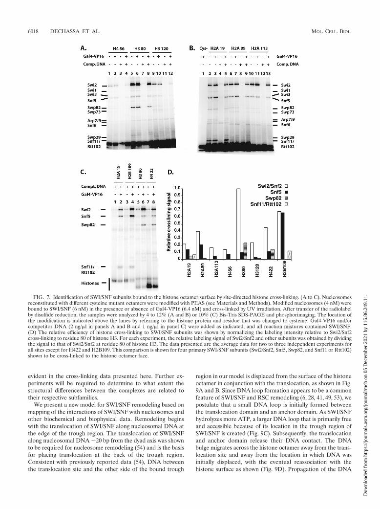
Histone cross-linking showed the association of Swi2/Snf2with a part of the histone octamer. Swi2/Snf2 was most effi-ciently cross-linked to residue 80 of histone H3, while residues22, 89, and 109 of histone H4, H2A, and H2B, respectively,were one-third, or more, as efficiently cross-linked (Fig. 7 anddata not shown). These four residues cluster to a region of thehistone octamer that coincides well with the part of the nu-cleosome shown by DNA footprinting to be bound to SWI/SNF (Fig. 6C). Also consistent with this idea is that residue 80of histone H3 is proximal to the position in DNA that iscross-linked most efficiently to Swi2/Snf2. The Swi2/Snf2 obvi-ously not only extensively interacts with nucleosomal DNA butalso interacts with a large portion of one or both faces of thehistone octamer not bound to DNA. For each mutant histoneoctamer, there are two symmetrical modification sites due tothe dual copies of each histone in the octamer such that SWI/SNF could be cross-linked from either side of the histoneoctamer. The DNA footprinting data, however, indicate thatone face of the nucleosome is tightly bound to SWI/SNF whilethe other face is fairly accessible. Given these data and theobservation of the asymmetry of the putative nucleosome bind-
ing trough of SWI/SNF, most of the histone cross-linking willlikely arise from just one face of the nucleosome.
Although Snf5 does not appear to be bound stably near nu-cleosomal DNA, at residue 109 of H2B, it was found to be effi-ciently cross-linked and was comparable to Swi2 cross-linking atthe same position (Fig. 6C and 7C, compare lane 4 to lanes 2, 6,and 8). The relative efficiency of Snf5 cross-linking progressivelydiminished at more distal positions. At the next most proximalpositions at residues 89 of H2A and 19 of H2A, the relativeefficiency is reduced �3.5-fold and even more extensively at moredistal positions such as residues 113 of H2A and 56 of H4 (Fig.7D). The Swp82 subunit was found to be located near residue 80of histone H3 and residue 22 of histone H4 (Fig. 6C and 7C, lanes6 and 8, and D). Likewise, one of the smallest subunits of SWI/SNF (Snf11 or Rtt102) was found to be discretely positioned at anopposite side of the nucleosome, near residue 19 of histone H2Aand residue 109 of histone H2B (Fig. 6C and 7B, lanes 2 to 5, andC, lanes 2 and 4).
The specificity of histone cross-linking to SWI/SNF was dem-onstrated by (i) cross-linking requiring the presence of mutantcysteine histone protein, (ii) competition by free DNA eliminat-ing SWI/SNF cross-linking, (iii) radiolabeling of SWI/SNF requir-ing UV irradiation, (iv) the cross-linking pattern varying in aposition-dependent manner, and (v) SWI/SNF cross-linking re-quiring Gal4-VP16 under competitive conditions. The interactionof SWI/SNF with the histone octamer surface was not substan-tially different whether it bound nucleosomes directly or was re-cruited by Gal4-VP16. These data suggest that the interactions ofSWI/SNF with the histone portion of the nucleosome, althoughfacilitated by recruitment, do not appear to be altered by recruit-ment. Conditions for the Gal4-VP16-dependent binding of SWI/SNF required the addition of sufficient competitor DNA to avoidthe direct binding of SWI/SNF to nucleosomes, as seen by gelshift assay (data not shown).
DISCUSSION
The structure of SWI/SNF and its interaction with nucleo-somes has been studied using a combination of DNA footprint-
FIG. 5. DNA cross-linking of SWI/SNF with photoreactive diazirine probes. DNA cross-linking was done as described in the legend to Fig. 3with photoreactive nucleosomes using the more photoreactive diazirine-containing DNA probes. (A) Samples contained SWI/SNF with (evenlanes) and without (odd lanes) Gal4-VP16. (B) All samples contained SWI/SNF and Gal4-VP16.
6016 DECHASSA ET AL. MOL. CELL. BIOL.
Dow
nloa
ded
from
http
s://j
ourn
als.
asm
.org
/jour
nal/m
cb o
n 05
Dec
embe
r 20
21 b
y 11
6.86
.249
.11.
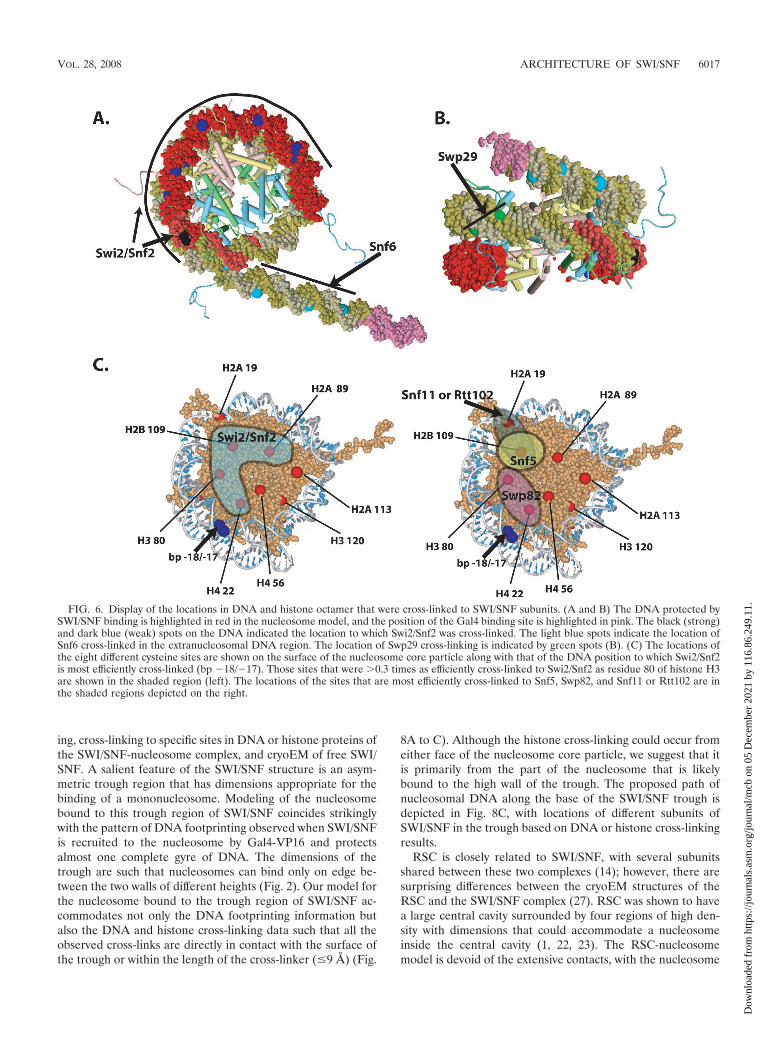
ing, cross-linking to specific sites in DNA or histone proteins ofthe SWI/SNF-nucleosome complex, and cryoEM of free SWI/SNF. A salient feature of the SWI/SNF structure is an asym-metric trough region that has dimensions appropriate for thebinding of a mononucleosome. Modeling of the nucleosomebound to this trough region of SWI/SNF coincides strikinglywith the pattern of DNA footprinting observed when SWI/SNFis recruited to the nucleosome by Gal4-VP16 and protectsalmost one complete gyre of DNA. The dimensions of thetrough are such that nucleosomes can bind only on edge be-tween the two walls of different heights (Fig. 2). Our model forthe nucleosome bound to the trough region of SWI/SNF ac-commodates not only the DNA footprinting information butalso the DNA and histone cross-linking data such that all theobserved cross-links are directly in contact with the surface ofthe trough or within the length of the cross-linker (�9 Å) (Fig.
8A to C). Although the histone cross-linking could occur fromeither face of the nucleosome core particle, we suggest that itis primarily from the part of the nucleosome that is likelybound to the high wall of the trough. The proposed path ofnucleosomal DNA along the base of the SWI/SNF trough isdepicted in Fig. 8C, with locations of different subunits ofSWI/SNF in the trough based on DNA or histone cross-linkingresults.
RSC is closely related to SWI/SNF, with several subunitsshared between these two complexes (14); however, there aresurprising differences between the cryoEM structures of theRSC and the SWI/SNF complex (27). RSC was shown to havea large central cavity surrounded by four regions of high den-sity with dimensions that could accommodate a nucleosomeinside the central cavity (1, 22, 23). The RSC-nucleosomemodel is devoid of the extensive contacts, with the nucleosome
FIG. 6. Display of the locations in DNA and histone octamer that were cross-linked to SWI/SNF subunits. (A and B) The DNA protected bySWI/SNF binding is highlighted in red in the nucleosome model, and the position of the Gal4 binding site is highlighted in pink. The black (strong)and dark blue (weak) spots on the DNA indicated the location to which Swi2/Snf2 was cross-linked. The light blue spots indicate the location ofSnf6 cross-linked in the extranucleosomal DNA region. The location of Swp29 cross-linking is indicated by green spots (B). (C) The locations ofthe eight different cysteine sites are shown on the surface of the nucleosome core particle along with that of the DNA position to which Swi2/Snf2is most efficiently cross-linked (bp �18/�17). Those sites that were 0.3 times as efficiently cross-linked to Swi2/Snf2 as residue 80 of histone H3are shown in the shaded region (left). The locations of the sites that are most efficiently cross-linked to Snf5, Swp82, and Snf11 or Rtt102 are inthe shaded regions depicted on the right.
VOL. 28, 2008 ARCHITECTURE OF SWI/SNF 6017
Dow
nloa
ded
from
http
s://j
ourn
als.
asm
.org
/jour
nal/m
cb o
n 05
Dec
embe
r 20
21 b
y 11
6.86
.249
.11.
evident in the cross-linking data presented here. Further ex-periments will be required to determine to what extent thestructural differences between the complexes are related totheir respective subfamilies.
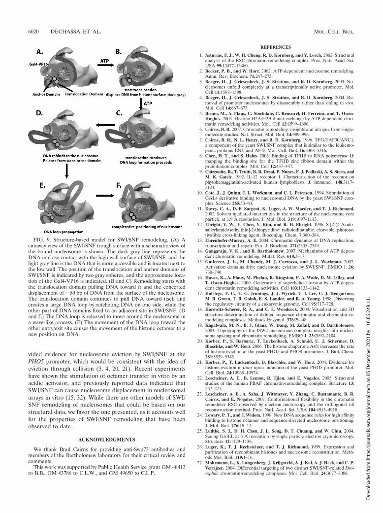
We present a new model for SWI/SNF remodeling based onmapping of the interactions of SWI/SNF with nucleosomes andother biochemical and biophysical data. Remodeling beginswith the translocation of SWI/SNF along nucleosomal DNA atthe edge of the trough region. The translocation of SWI/SNFalong nucleosomal DNA �20 bp from the dyad axis was shownto be required for nucleosome remodeling (54) and is the basisfor placing translocation at the back of the trough region.Consistent with previously reported data (54), DNA betweenthe translocation site and the other side of the bound trough
region in our model is displaced from the surface of the histoneoctamer in conjunction with the translocation, as shown in Fig.9A and B. Since DNA loop formation appears to be a commonfeature of SWI/SNF and RSC remodeling (6, 28, 41, 49, 53), wepostulate that a small DNA loop is initially formed betweenthe translocation domain and an anchor domain. As SWI/SNFhydrolyzes more ATP, a larger DNA loop that is primarily freeand accessible because of its location in the trough region ofSWI/SNF is created (Fig. 9C). Subsequently, the translocationand anchor domain release their DNA contact. The DNAbulge migrates across the histone octamer away from the trans-location site and away from the location in which DNA wasinitially displaced, with the eventual reassociation with thehistone surface as shown (Fig. 9D). Propagation of the DNA
FIG. 7. Identification of SWI/SNF subunits bound to the histone octamer surface by site-directed histone cross-linking. (A to C). Nucleosomesreconstituted with different cysteine mutant octamers were modified with PEAS (see Materials and Methods). Modified nucleosomes (4 nM) werebound to SWI/SNF (6 nM) in the presence or absence of Gal4-VP16 (6.4 nM) and cross-linked by UV irradiation. After transfer of the radiolabelby disulfide reduction, the samples were analyzed by 4 to 12% (A and B) or 10% (C) Bis-Tris SDS-PAGE and phosphorimaging. The location ofthe modification is indicated above the lanes by referring to the histone protein and residue that was changed to cysteine. Gal4-VP16 and/orcompetitor DNA (2 ng/�l in panels A and B and 1 ng/�l in panel C) were added as indicated, and all reaction mixtures contained SWI/SNF.(D) The relative efficiency of histone cross-linking to SWI/SNF subunits was shown by normalizing the labeling intensity relative to Swi2/Snf2cross-linking to residue 80 of histone H3. For each experiment, the relative labeling signal of Swi2/Snf2 and other subunits was obtained by dividingthe signal to that of Swi2/Snf2 at residue 80 of histone H3. The data presented are the average data for two to three independent experiments forall sites except for H422 and H2B109. This comparison is shown for four primary SWI/SNF subunits (Swi2/Snf2, Snf5, Swp82, and Snf11 or Rtt102)shown to be cross-linked to the histone octamer face.
6018 DECHASSA ET AL. MOL. CELL. BIOL.
Dow
nloa
ded
from
http
s://j
ourn
als.
asm
.org
/jour
nal/m
cb o
n 05
Dec
embe
r 20
21 b
y 11
6.86
.249
.11.

loop in this direction is favored because it first proceeds in theregion of the nucleosome facing away from the cleft, and as itproceeds around to the other side of the nucleosome, it is inthe area of the cleft with the low wall, where there is sufficientspace for the passage of a DNA loop. The loop will continue topropagate until it reaches the other entry/exit site and therebycomplete the movement of DNA around the nucleosome (Fig.9E and F). A key difference in this model compared to othersis the extent of the remodeler’s interaction with nucleosomes.RSC has been suggested to contact only a small region ofnucleosomal DNA, whereas SWI/SNF makes extensive contactand could reasonably cause a larger disruption of the histone-DNA interface due to compensating interactions of SWI/SNFwith nucleosomal DNA and thus move the nucleosome inlarger steps (6, 37, 54).
A natural outcome of this process will be the progressivereduction in the length of linker DNA at the side where theDNA is displaced and then pulled into the translocation do-main of SWI/SNF. As DNA from this entry/exit site is pulledinto SWI/SNF, proteins bound to neighboring DNA, such asadjacent nucleosomes or DNA-bound transcription factors,will be brought into closer proximity to the SWI/SNF-nucleo-some complex. This process will ultimately cause a clash be-tween any protein bound to the linker DNA and SWI/SNF withits bound nucleosome. Either the SWI/SNF-bound nucleo-some may be displaced causing remodeling to terminate or thefactor or nucleosome bound to the linker DNA may give way.Given this type of mechanism, it can be envisaged how SWI/SNF remodeling in a nucleosomal array might cause the dis-placement through nucleosome collision. In vivo studies pro-
FIG. 8. Subunit topology in the nucleosome binding pocket of SWI/SNF. The views in panels A, C, and E are in the same orientation as thatshown in Fig. 2A, C, and E. Nucleosomal DNA is displayed with the same color scheme as that in Fig. 6A and B and refers to sites that cross-linkedSwi2, Snf6, and Swp29. The histone octamer is not shown for better visualization of the proximity of DNA cross-linking sites to the surface ofSWI/SNF. The DNA protected by SWI/SNF binding is shown in red, and that not protected is shown in gray (A to B and E and F). In this model,the linker trajectory is arbitrarily assigned. (C) The locations of particular DNA and histone sites are shown with the nucleosome removed andare based on the SWI/SNF-nucleosome model described above. The legend for the symbols used to mark these locations is shown (D), along witha summary of the SWI/SNF subunits cross-linked at these sites and their relative efficiencies, i.e., strong (��) and weak (�). The proposed pathof nucleosomal DNA along the surface of the SWI/SNF trough is shown as a dashed line. Arrows in panels A and E indicate the dyad axis of thenucleosome.
VOL. 28, 2008 ARCHITECTURE OF SWI/SNF 6019
Dow
nloa
ded
from
http
s://j
ourn
als.
asm
.org
/jour
nal/m
cb o
n 05
Dec
embe
r 20
21 b
y 11
6.86
.249
.11.
vided evidence for nucleosome eviction by SWI/SNF at thePHO5 promoter, which would be consistent with the idea ofeviction through collision (3, 4, 20, 21). Recent experimentshave shown the stimulation of octamer transfer in vitro by anacidic activator, and previously reported data indicated thatSWI/SNF can cause nucleosome displacement in nucleosomalarrays in vitro (15, 32). While there are other models of SWI/SNF remodeling of nucleosomes that could be based on ourstructural data, we favor the one presented, as it accounts wellfor the properties of SWI/SNF remodeling that have beenobserved to date.
ACKNOWLEDGMENTS
We thank Brad Cairns for providing anti-Swp73 antibodies andmembers of the Bartholomew laboratory for their critical review andcomments.
This work was supported by Public Health Service grant GM 48413to B.B., GM 43786 to C.L.W., and GM 49650 to C.L.P.
REFERENCES
1. Asturias, F. J., W. H. Chung, R. D. Kornberg, and Y. Lorch. 2002. Structuralanalysis of the RSC chromatin-remodeling complex. Proc. Natl. Acad. Sci.USA 99:13477–13480.
2. Becker, P. B., and W. Horz. 2002. ATP-dependent nucleosome remodeling.Annu. Rev. Biochem. 71:247–273.
3. Boeger, H., J. Griesenbeck, J. S. Strattan, and R. D. Kornberg. 2003. Nu-cleosomes unfold completely at a transcriptionally active promoter. Mol.Cell 11:1587–1598.
4. Boeger, H., J. Griesenbeck, J. S. Strattan, and R. D. Kornberg. 2004. Re-moval of promoter nucleosomes by disassembly rather than sliding in vivo.Mol. Cell 14:667–673.
5. Bruno, M., A. Flaus, C. Stockdale, C. Rencurel, H. Ferreira, and T. Owen-Hughes. 2003. Histone H2A/H2B dimer exchange by ATP-dependent chro-matin remodeling activities. Mol. Cell 12:1599–1606.
6. Cairns, B. R. 2007. Chromatin remodeling: insights and intrigue from single-molecule studies. Nat. Struct. Mol. Biol. 14:989–996.
7. Cairns, B. R., N. L. Henry, and R. D. Kornberg. 1996. TFG/TAF30/ANC1,a component of the yeast SWI/SNF complex that is similar to the leukemo-genic proteins ENL and AF-9. Mol. Cell. Biol. 16:3308–3316.
8. Chen, H. T., and S. Hahn. 2003. Binding of TFIIB to RNA polymerase II:mapping the binding site for the TFIIB zinc ribbon domain within thepreinitiation complex. Mol. Cell 12:437–447.
9. Chizzonite, R., T. Truitt, B. B. Desai, P. Nunes, F. J. Podlaski, A. S. Stern, andM. K. Gately. 1992. IL-12 receptor. I. Characterization of the receptor onphytohemagglutinin-activated human lymphoblasts. J. Immunol. 148:3117–3124.
10. Cote, J., J. Quinn, J. L. Workman, and C. L. Peterson. 1994. Stimulation ofGAL4 derivative binding to nucleosomal DNA by the yeast SWI/SNF com-plex. Science 265:53–60.
11. Davey, C. A., D. F. Sargent, K. Luger, A. W. Maeder, and T. J. Richmond.2002. Solvent mediated interactions in the structure of the nucleosome coreparticle at 1.9 Å resolution. J. Mol. Biol. 319:1097–1113.
12. Ebright, Y. W., Y. Chen, Y. Kim, and R. H. Ebright. 1996. S-[2-(4-Azido-salicylamido)ethylthio]-2-thiopyridine: radioiodinatable, cleavable, photoac-tivatible cross-linking agent. Bioconjug. Chem. 7:380–384.
13. Ehrenhofer-Murray, A. E. 2004. Chromatin dynamics at DNA replication,transcription and repair. Eur. J. Biochem. 271:2335–2349.
14. Gangaraju, V. K., and B. Bartholomew. 2007. Mechanisms of ATP depen-dent chromatin remodeling. Mutat. Res. 618:3–17.
15. Gutierrez, J. L., M. Chandy, M. J. Carrozza, and J. L. Workman. 2007.Activation domains drive nucleosome eviction by SWI/SNF. EMBO J. 26:730–740.
16. Havas, K., A. Flaus, M. Phelan, R. Kingston, P. A. Wade, D. M. Lilley, andT. Owen-Hughes. 2000. Generation of superhelical torsion by ATP-depen-dent chromatin remodeling activities. Cell 103:1133–1142.
17. Holstege, F. C., E. G. Jennings, J. J. Wyrick, T. I. Lee, C. J. Hengartner,M. R. Green, T. R. Golub, E. S. Lander, and R. A. Young. 1998. Dissectingthe regulatory circuitry of a eukaryotic genome. Cell 95:717–728.
18. Horowitz-Scherer, R. A., and C. L. Woodcock. 2004. Visualization and 3Dstructure determination of defined sequence chromatin and chromatin re-modeling complexes. Methods Enzymol. 376:29–48.
19. Kagalwala, M. N., B. J. Glaus, W. Dang, M. Zofall, and B. Bartholomew.2004. Topography of the ISW2-nucleosome complex: insights into nucleo-some spacing and chromatin remodeling. EMBO J. 23:2092–2104.
20. Korber, P., S. Barbaric, T. Luckenbach, A. Schmid, U. J. Schermer, D.Blaschke, and W. Horz. 2006. The histone chaperone Asf1 increases the rateof histone eviction at the yeast PHO5 and PHO8 promoters. J. Biol. Chem.281:5539–5545.
21. Korber, P., T. Luckenbach, D. Blaschke, and W. Horz. 2004. Evidence forhistone eviction in trans upon induction of the yeast PHO5 promoter. Mol.Cell. Biol. 24:10965–10974.
22. Leschziner, A. E., B. Lemon, R. Tjian, and E. Nogales. 2005. Structuralstudies of the human PBAF chromatin-remodeling complex. Structure 13:267–275.
23. Leschziner, A. E., A. Saha, J. Wittmeyer, Y. Zhang, C. Bustamante, B. R.Cairns, and E. Nogales. 2007. Conformational flexibility in the chromatinremodeler RSC observed by electron microscopy and the orthogonal tiltreconstruction method. Proc. Natl. Acad. Sci. USA 104:4913–4918.
24. Lowary, P. T., and J. Widom. 1998. New DNA sequence rules for high affinitybinding to histone octamer and sequence-directed nucleosome positioning.J. Mol. Biol. 276:19–42.
25. Ludtke, S. J., D. H. Chen, J. L. Song, D. T. Chuang, and W. Chiu. 2004.Seeing GroEL at 6 A resolution by single particle electron cryomicroscopy.Structure 12:1129–1136.
26. Luger, K., T. J. Rechsteiner, and T. J. Richmond. 1999. Expression andpurification of recombinant histones and nucleosome reconstitution. Meth-ods Mol. Biol. 119:1–16.
27. Mohrmann, L., K. Langenberg, J. Krijgsveld, A. J. Kal, A. J. Heck, and C. P.Verrijzer. 2004. Differential targeting of two distinct SWI/SNF-related Dro-sophila chromatin-remodeling complexes. Mol. Cell. Biol. 24:3077–3088.
FIG. 9. Structure-based model for SWI/SNF remodeling. (A) Acutaway view of the SWI/SNF trough surface with a schematic view ofthe bound nucleosome is shown. The dark gray line represents theDNA in close contact with the high wall surface of SWI/SNF, and thelight gray line is the DNA that is more accessible and is located next tothe low wall. The position of the translocation and anchor domains ofSWI/SNF is indicated by two gray spheres, and the approximate loca-tion of the Gal4-VP16 is indicated. (B and C) Remodeling starts withthe translocation domain pulling DNA toward it and the concerteddisplacement of �50 bp of DNA from the surface of the nucleosome.The translocation domain continues to pull DNA toward itself andcreates a large DNA loop by ratcheting DNA on one side, while theother part of DNA remains fixed to an adjacent site in SWI/SNF. (Dand E) The DNA loop is released to move around the nucleosome ina wave-like process. (F) The movement of the DNA loop toward theother entry/exit site causes the movement of the histone octamer to anew position on DNA.
6020 DECHASSA ET AL. MOL. CELL. BIOL.
Dow
nloa
ded
from
http
s://j
ourn
als.
asm
.org
/jour
nal/m
cb o
n 05
Dec
embe
r 20
21 b
y 11
6.86
.249
.11.
28. Narlikar, G. J., M. L. Phelan, and R. E. Kingston. 2001. Generation andinterconversion of multiple distinct nucleosomal states as a mechanism forcatalyzing chromatin fluidity. Mol. Cell 8:1219–1230.
29. Natarajan, K., B. M. Jackson, H. Zhou, F. Winston, and A. G. Hinnebusch.1999. Transcriptional activation by Gcn4p involves independent interactionswith the SWI/SNF complex and the SRB/mediator. Mol. Cell 4:657–664.
30. Neely, K. E., A. H. Hassan, C. E. Brown, L. Howe, and J. L. Workman. 2002.Transcription activator interactions with multiple SWI/SNF subunits. Mol.Cell. Biol. 22:1615–1625.
31. Neely, K. E., A. H. Hassan, A. E. Wallberg, D. J. Steger, B. R. Cairns, A. P.Wright, and J. L. Workman. 1999. Activation domain-mediated targeting ofthe SWI/SNF complex to promoters stimulates transcription from nucleo-some arrays. Mol. Cell 4:649–655.
32. Owen-Hughes, T., R. T. Utley, J. Cote, C. L. Peterson, and J. L. Workman.1996. Persistent site-specific remodeling of a nucleosome array by transientaction of the SWI/SNF complex. Science 273:513–516.
33. Persinger, J., and B. Bartholomew. 1996. Mapping the contacts of yeastTFIIIB and RNA polymerase III at various distances from the major grooveof DNA by DNA photoaffinity labeling. J. Biol. Chem. 271:33039–33046.
34. Peterson, C. L., and J. Cote. 2004. Cellular machineries for chromosomalDNA repair. Genes Dev. 18:602–616.
35. Phelan, M. L., G. R. Schnitzler, and R. E. Kingston. 2000. Octamer transferand creation of stably remodeled nucleosomes by human SWI-SNF and itsisolated ATPases. Mol. Cell. Biol. 20:6380–6389.
36. Phelan, M. L., S. Sif, G. J. Narlikar, and R. E. Kingston. 1999. Reconstitu-tion of a core chromatin remodeling complex from SWI/SNF subunits. Mol.Cell 3:247–253.
37. Saha, A., J. Wittmeyer, and B. R. Cairns. 2005. Chromatin remodelingthrough directional DNA translocation from an internal nucleosomal site.Nat. Struct. Mol. Biol. 12:747–755.
38. Sengupta, S. M., J. Persinger, B. Bartholomew, and C. L. Peterson. 1999.Use of DNA photoaffinity labeling to study nucleosome remodeling by SWI/SNF. Methods 19:434–446.
39. Sengupta, S. M., M. VanKanegan, J. Persinger, C. Logie, B. R. Cairns, C. L.Peterson, and B. Bartholomew. 2001. The interactions of yeast SWI/SNF andRSC with the nucleosome before and after chromatin remodeling. J. Biol.Chem. 276:12636–12644.
40. Shen, F., S. J. Triezenberg, P. Hensley, D. Porter, and J. R. Knutson. 1996.Critical amino acids in the transcriptional activation domain of the herpes-virus protein VP16 are solvent-exposed in highly mobile protein segments.An intrinsic fluorescence study. J. Biol. Chem. 271:4819–4826.
41. Shundrovsky, A., C. L. Smith, J. T. Lis, C. L. Peterson, and M. D. Wang.
2006. Probing SWI/SNF remodeling of the nucleosome by unzipping singleDNA molecules. Nat. Struct. Mol. Biol. 13:549–554.
42. Smith, C. L., R. Horowitz-Scherer, J. F. Flanagan, C. L. Woodcock, and C. L.Peterson. 2003. Structural analysis of the yeast SWI/SNF chromatin remod-eling complex. Nat. Struct. Biol. 10:141–145.
43. Smith, C. L., and C. L. Peterson. 2003. Coupling tandem affinity purificationand quantitative tyrosine iodination to determine subunit stoichiometry ofprotein complexes. Methods 31:104–109.
44. Sudarsanam, P., V. R. Iyer, P. O. Brown, and F. Winston. 2000. Whole-genome expression analysis of snf/swi mutants of Saccharomyces cerevisiae.Proc. Natl. Acad. Sci. USA 97:3364–3369.
45. Swanson, M. J., H. Qiu, L. Sumibcay, A. Krueger, S. J. Kim, K. Natarajan,S. Yoon, and A. G. Hinnebusch. 2003. A multiplicity of coactivators isrequired by Gcn4p at individual promoters in vivo. Mol. Cell. Biol. 23:2800–2820.
46. Tate, J. J., J. Persinger, and B. Bartholomew. 1998. Survey of four differentphotoreactive moieties for DNA photoaffinity labeling of yeast RNA poly-merase III transcription complexes. Nucleic Acids Res. 26:1421–1426.
47. Treich, I., B. R. Cairns, T. de los Santos, E. Brewster, and M. Carlson. 1995.SNF11, a new component of the yeast SNF-SWI complex that interacts witha conserved region of SNF2. Mol. Cell. Biol. 15:4240–4248.
48. Tsukiyama, T., and C. Wu. 1997. Chromatin remodeling and transcription.Curr. Opin. Genet. Dev. 7:182–191.
49. Wang, H., R. Bash, S. M. Lindsay, and D. Lohr. 2005. Solution AFM studiesof human Swi-Snf and its interactions with MMTV DNA and chromatin.Biophys. J. 89:3386–3398.
50. Yang, X., R. Zaurin, M. Beato, and C. L. Peterson. 2007. Swi3p controlsSWI/SNF assembly and ATP-dependent H2A-H2B displacement. Nat.Struct. Mol. Biol. 14:540–547.
51. Yoon, S., H. Qiu, M. J. Swanson, and A. G. Hinnebusch. 2003. Recruitmentof SWI/SNF by Gcn4p does not require Snf2p or Gcn5p but dependsstrongly on SWI/SNF integrity, SRB mediator, and SAGA. Mol. Cell. Biol.23:8829–8845.
52. Yudkovsky, N., C. Logie, S. Hahn, and C. L. Peterson. 1999. Recruitment ofthe SWI/SNF chromatin remodeling complex by transcriptional activators.Genes Dev. 13:2369–2374.
53. Zhang, Y., C. L. Smith, A. Saha, S. W. Grill, S. Mihardja, S. B. Smith, B. R.Cairns, C. L. Peterson, and C. Bustamante. 2006. DNA translocation andloop formation mechanism of chromatin remodeling by SWI/SNF and RSC.Mol. Cell 24:559–568.
54. Zofall, M., J. Persinger, S. R. Kassabov, and B. Bartholomew. 2006. Chro-matin remodeling by ISW2 and SWI/SNF requires DNA translocation insidethe nucleosome. Nat. Struct. Mol. Biol. 13:339–346.
VOL. 28, 2008 ARCHITECTURE OF SWI/SNF 6021
Dow
nloa
ded
from
http
s://j
ourn
als.
asm
.org
/jour
nal/m
cb o
n 05
Dec
embe
r 20
21 b
y 11
6.86
.249
.11.