article – discoveries pbluescript ii with aci i chromosomal fragment containing (hawkins, orc2 and...
TRANSCRIPT

Evolution of Genome Architecture in Archaea: SpontaneousGeneration of a New Chromosome in Haloferax volcanii
Darya Ausiannikava,†,1 Laura Mitchell,1 Hannah Marriott,1 Victoria Smith,1 Michelle Hawkins,‡,1
Kira S. Makarova,2 Eugene V. Koonin,2 Conrad A. Nieduszynski,3 and Thorsten Allers*,1
1School of Life Sciences, University of Nottingham, Queen’s Medical Centre, Nottingham, United Kingdom2National Center for Biotechnology Information, National Library of Medicine, NIH, Bethesda, MD3Sir William Dunn School of Pathology, University of Oxford, Oxford, United Kingdom†Present address: School of Biological Sciences, Institute of Cell Biology, University of Edinburgh, Kings Buildings, Edinburgh, UnitedKingdom‡Present address: Department of Biology, University of York, Wentworth Way, York, United Kingdom
*Corresponding author: E-mail: [email protected].
Associate editor: Mary O’ConnellSequencing data sets generated and analyzed during this study are available in the NCBI Gene Expression Omnibus under accessionnumber GSE108201.
Abstract
The common ancestry of archaea and eukaryotes is evident in their genome architecture. All eukaryotic and severalarchaeal genomes consist of multiple chromosomes, each replicated from multiple origins. Three scenarios have beenproposed for the evolution of this genome architecture: 1) mutational diversification of a multi-copy chromosome; 2)capture of a new chromosome by horizontal transfer; 3) acquisition of new origins and splitting into two replication-competent chromosomes. We report an example of the third scenario: the multi-origin chromosome of the archaeonHaloferax volcanii has split into two elements via homologous recombination. The newly generated elements are bonafide chromosomes, because each bears “chromosomal” replication origins, rRNA loci, and essential genes. The newchromosomes were stable during routine growth but additional genetic manipulation, which involves selective bottle-necks, provoked further rearrangements. To the best of our knowledge, rearrangement of a naturally evolved prokaryoticgenome to generate two new chromosomes has not been described previously.
Key words: chromosome, genome architecture, multipartite genome, homologous recombination, genome stability,archaea, Haloferax volcanii.
IntroductionBacterial genomes usually consist of a single circular chromo-some with a unique origin of DNA replication oriC, which isrecognized by the initiator protein DnaA. Some bacteria,mainly from the phylum Proteobacteria (e.g. Agrobacterium,Brucella, Rhizobium, Vibrio), have large secondary repliconstermed chromids (Harrison et al. 2010; diCenzo and Finan2017). Unlike plasmids, chromids are often comparable to themain chromosome in size and carry core genes that are usu-ally found on the main chromosome. However, in contrast tothe main chromosome, chromids have been shown to relyexclusively on plasmid-type DNA replication initiation mech-anisms (often in the form of a RepABC system), and not onthe DnaA/oriC system (Egan et al. 2005; Pinto et al. 2012).
Archaea are similar to bacteria in terms of the size andoverall organization of their genomes (Koonin and Wolf2008). However, the core DNA replication proteins foundin archaea are more closely related to those of eukaryotesthan to their bacterial counterparts. Archaea commonly havemore than one origin on the main chromosome and rely onOrc1/Cdc6 replication initiator proteins, which are
homologous to the eukaryotic origin recognition complexsubunit Orc1 (Makarova and Koonin 2013; Ausiannikavaand Allers 2017). Archaeal genomes often have large second-ary replicons, which are referred to as mega-plasmids or mini-chromosomes. Unlike bacterial chromids, archaeal mini-chromosomes depend predominantly on Orc1 initiator pro-teins for their replication, similar to the main chromosome(Ng et al. 1998, 2000; Baliga et al. 2004; Wang et al. 2015).
Eukaryotic genomes consist of multiple chromosomes thatare almost always linear and are each replicated from multipleorigins. New extrachromosomal elements arise relatively fre-quently in eukaryotes (Gaubatz 1990; Moller et al. 2015;Turner et al. 2017), but these elements are often transientand low in abundance. Extrachromosomal circular DNAs arecommon in yeast and may cover up to 23% of the genome(Moller et al. 2015), and cancer cells often generate highlyamplified circular mini-chromosomes called double minutechromosomes (Storlazzi et al. 2010).
How did multiple chromosomes with multiple originsevolve? The ancestral state is unlikely to have been a singlechromosome with a single origin, but it is the simplest one to
Article
� The Author(s) 2018. Published by Oxford University Press on behalf of the Society for Molecular Biology and Evolution.This is an Open Access article distributed under the terms of the Creative Commons Attribution License (http://creativecommons.org/licenses/by/4.0/), which permits unrestricted reuse, distribution, and reproduction in any medium, provided the original work isproperly cited. Open AccessMol. Biol. Evol. doi:10.1093/molbev/msy075 Advance Access publication April 16, 2018 1Downloaded from https://academic.oup.com/mbe/advance-article-abstract/doi/10.1093/molbev/msy075/4972485
by gueston 28 June 2018

consider. (i) If present in multiple copies, a single chromo-some could diversify by the accumulation of mutations. (ii)More likely, a new element could be acquired by horizontaltransfer—over time, the secondary chromosome would gaincore genes from the main chromosome (diCenzo and Finan2017). (iii) Alternatively, the new element could integrate intothe main one, producing a multi-origin chromosome that hasthe potential to split into two replication-competent chro-mosomes, thereby giving rise to the state encountered inmodern genomes (Egan et al. 2005; diCenzo and Finan2017). In bacteria, the presence of plasmid-like replicationorigins on secondary replicons and the uneven distributionof core genes argues against scenario (i) and in favor of sce-nario (ii) (Harrison et al. 2010). Phylogenetic analysis of themultiple replication origins found on archaeal chromosomesindicates that they were independently acquired throughhorizontal gene transfer (HGT) and not by duplication ofpre-existing origins (Robinson and Bell 2007; Wu et al.2012), again apparently ruling out scenario (i) and insteadsupporting scenario (ii). Because features that are commonto all eukaryotic replication origins are elusive, little can bededuced about the evolution of eukaryotic genome organi-zation but scenario (iii) might be the most parsimonious.
Whatever the evolutionary scenario, genome architectureis not random in prokaryotes (Rocha 2004, 2008; Press et al.2016). One of the strongest constraints is the location ofreplication origins and termination regions; a striking X-shaped pattern of inversions, with endpoints symmetricallylocated around the origin and terminus of replication, hascommonly been observed in bacteria and archaea (Eisen et al.2000; Novichkov et al. 2009; Repar and Warnecke 2017). It hasbeen shown experimentally that altering the size ratio of thetwo replication arms (replichores) by >10% is deleterious forEscherichia coli (Esnault et al. 2007). A strong bias for codir-ectionality of transcription and replication, which is thoughtto reduce the collision of RNA and DNA polymerases, alsoexists in prokaryotic genomes (Wang et al. 2007; Srivatsanet al. 2010; Ivanova et al. 2015). The distribution of repetitiveand mobile elements shapes the genome as well, with bothhomologous and site-specific recombination acting as a po-tent driving force of chromosome architecture evolution inbacteria and archaea (Brugger et al. 2004; Papke et al. 2004;Whitaker et al. 2005; White et al. 2008; Bryant et al. 2012;Cossu et al. 2017; Mao and Grogan 2017).
Haloferax volcanii, a halophilic archaeon, is a tractablemodel to study prokaryotic genome plasticity and the evolu-tion of new chromosomes (Mullakhanbhai and Larsen 1975;Charlebois et al. 1991; Hartman et al. 2010). Its main chromo-some has three origins, oriC1, oriC2, and oriC3 (Norais et al.2007; Hawkins, Malla, et al. 2013). Three additional originsexist on the three mini-chromosomes, pHV4, pHV3, andpHV1 (Hartman et al. 2010). Haloferax volcanii is highly poly-ploid, with the entire genome present in�20 copies (Breuertet al. 2006). Consistent with the highly dynamic nature ofarchaeal genomes (Redder and Garrett 2006; Bridger et al.2012), two cases of genome rearrangements have beendetected in vivo for H. volcanii, namely fusion of the pHV4mini-chromosome with the main chromosome, and
inversion of part of this fused chromosome by recombinationbetween two insertion sequence (IS) elements (Hawkins,Malla, et al. 2013). The former rearrangement has increasedthe number of replication origins on the main chromosometo four. The involvement of HGT in archaeal genome evolu-tion is evident from the presence of many additional copies ofreplication genes. In the H. volcanii genome, there are 16 orcgenes encoding the Orc1 initiator protein but only six origins(Hartman et al. 2010; Raymann et al. 2014).
Here we report an unusual genome rearrangement in H.volcanii. In our investigation of DNA replication, we generatedstrains with serial deletions of orc genes. It came to our at-tention that one of these strains had undergone a genomerearrangement. Unexpectedly, the main chromosome splitinto two parts via homologous recombination betweentwo near-identical sod (superoxide dismutase) genes; there-fore, it was not due to excision of the integrated pHV4. Thetwo resulting DNA molecules exhibit all the features of bonafide chromosomes: they bear replication origins, rRNA loci,and essential core genes.
To the best of our knowledge, the evolution of a newchromosome without interspecies HGT has so far not beenobserved in prokaryotes. Thus, we have witnessed in vivo arealization of the scenario (iii) posited above: a multi-originchromosome splits into two replication-competent chromo-somes. This finding contrasts with our previous report show-ing fusion of the pHV4 mini-chromosome with the mainchromosome (Hawkins, Malla, et al. 2013) and demonstratesthat genome rearrangements do not inexorably lead to largerchromosomes. Instead, they can give rise to the multi-origin/multi-chromosome state encountered in modern genomes.
Results
Large-Scale Genome Rearrangement in the StrainDeleted for Orc1/Cdc6 Initiator Gene orc5In our study of Orc1-type initiator proteins and their role inDNA replication in H. volcanii, we focused on the four orcgenes, orc1, orc5, orc2, and orc3, which are genetically linkedto the four chromosomal origins, oriC1, oriC2, oriC3, and ori-pHV4, respectively (fig. 1A). The four origins create eightreplichores on the chromosome, with oriC1 being the mostactive origin and ori-pHV4 the least (Hawkins, Malla, et al.2013). We obtained replication profiles by marker frequencyanalysis using whole genome sequencing (Muller et al. 2014).We noted that upon deletion of orc5 gene, which is locatednext to oriC2, the mutant strain H1689 had acquired large-scale genome rearrangements. This was manifested as twoclear discontinuities in the replication profile (indicated byarrows in fig. 1B; Skovgaard et al. 2011), when compared withthe wild type (WT).
To verify the genome rearrangement by an independentmethod, we performed restriction digests with SfaAI and an-alyzed the fragment sizes by pulsed field gel electrophoresis(PFGE). We have previously used this method to detect ge-nome rearrangements in H. volcanii (Hawkins, Malla, et al.2013). We observed the disappearance of a band correspond-ing to a 390 kb fragment, and the appearance of a novel
Ausiannikava et al. . doi:10.1093/molbev/msy075 MBE
2Downloaded from https://academic.oup.com/mbe/advance-article-abstract/doi/10.1093/molbev/msy075/4972485by gueston 28 June 2018

579 kb fragment in the SfaAI digest of Dorc5 DNA, confirminga large-scale genome rearrangement (fig. 1C).
New Genome Architecture of Dorc5 StrainThe two interruptions in the replication profile of Dorc5 mu-tant (fig. 1B) correspond to the locations of the sod1(HVO_A0475; 689201–689803 bp) and sod2 genes(HVO_2913; 3385084–3385683 bp). The sod1 and sod2 super-oxide dismutase genes are 603 bp and 600 bp, respectively,and have 100% nucleotide sequence identity (apart from theinitial 8 bp); however, their flanking sequences are unique.This provides an opportunity for intrachromosomal homol-ogous recombination of the sod1 and sod2 genes, and twooutcomes are possible: splitting of the main chromosome
into two circular replicons (termed new chr 1 and new chr2, fig. 2A), or chromosomal inversion of the region betweenthe two sod genes. Given that the two sod genes are in thesame orientation (direct repeats), only the former outcome ispossible, as the latter would require the sod genes to be ar-ranged as inverted repeats.
To investigate the genome architecture of the Dorc5 strain,intact genomic DNA was analyzed by PFGE and a Southernblot was probed with sod1 and sod2 sequences (fig. 2B). In thewild isolate DS2 (Mullakhanbhai and Larsen 1975), the sod1and sod2 genes are located on pHV4 and the main chromo-some, respectively. In the WT laboratory strain H26, pHV4 isfused with the main chromosome and therefore both sodgenes are on the same molecule (Hawkins, Malla, et al.2013). In DNA prepared from the Dorc5 strain H1689, thesod1 and sod2 probes hybridized with two molecules thatcorrespond in size to new chr 1 (2,696 kb) and new chr 2(787 kb). Using PCR with primers to the unique sequencesflanking sod1 and sod2, we determined that these two genesunderwent recombination in the Dorc5 strain (fig. 2C). DNAsequencing of the PCR products confirmed that the uniqueflanking sequences of sod1 and sod2 had been exchanged inthe Dorc5 strain.
We constructed maps of the rearranged chromosomes(new chr 1 and new chr 2) and analyzed the predictedsod1/sod2 break points in the Dorc5 mutant by restrictiondigests and Southern blotting. As expected, a StyI digestgenerated one band of 7.8 kb in the WT and a larger 13 kbfragment (plus a faint WT-sized band) in the Dorc5 strain,which hybridize with a probe adjacent to sod1 (fig. 3A).Similarly, an EcoRV digest of DNA from the WT straingenerated a fragment of 8.9 kb, which hybridizes with aprobe adjacent to sod2 gene, whereas a smaller 5.5 kbfragment (plus a faint WT-sized band) was seen in theDorc5 strain (fig. 3A). The presence of the faint fragmentof WT size in both digests of the Dorc5 mutant suggeststhat the genome architecture of this strain is not mono-morphic, and that the two states (with and without ge-nome rearrangement), coexist in the population.
To confirm the splitting of the chromosome into two cir-cular replicons, genomic DNA was digested with SfaAI, ana-lyzed by PFGE and a Southern blot was probed with the oriC1downstream region (fig. 3B). In the WT, this probe will hy-bridize with a fragment of 390 kb that includes sod2. If themain chromosome is split into two, the 390 kb fragment willbe fused with a 215 kb fragment that includes sod1, to gen-erate a product of 579 kb. Such a rearrangement would ac-count for the disappearance of the 390 kb band, and theappearance of a novel 579 kb band, as seen in the SfaAI digestin figure 1C. The SfaAI-digested Dorc5 DNA in figure 3Bshowed the presence of such a 579 kb band that hybridizeswith the oriC1 probe. A faint 390 kb fragment correspondingto the WT was also present in the Dorc5 sample, indicatingthat the genome architecture of this strain is not monomor-phic, confirming the observation made in figure 3A.
To further confirm fragmentation of the chromosome intotwo replicons, genomic DNA was digested with AvrII andSwaI, and the fragments were analyzed by PFGE (fig. 3C).
FIG. 1. Genome rearrangement of Dorc5 strain. (A) Location of rep-lication origins and adjacent orc genes on Haloferax volcanii mainchromosome (þpHV4). Positions of the two rRNA loci are indicatedwith black arrows. The integrated pHV4 mini-chromosome is indi-cated by a thick line. The eight replichores representing the directionof replication forks are shown by colored arrows, corresponding totheir respective origins. SfaAI sites are indicated by tick marks. (B)Replication profiles of the Dorc5 mutant H1689 and a reference wild-type (WT) laboratory strain H26. The number of reads is plottedagainst the chromosomal location. The linearized H. volcanii chromo-some showing positions of oriC and orc genes is shown below (coloredas in A). Two discontinuities in the Dorc5 replication profile are in-dicated by vertical arrows. (C) Restriction fragment length polymor-phisms in WT and Dorc5 strain as shown by digestion with SfaAI andPFGE. The 390 kb SfaAI fragment (shown on the map in panel A) isabsent from the digest of Dorc5 DNA, and a novel 579 kb SfaAI frag-ment is present; these bands are indicated by arrows.
Evolution of Genome Architecture in Archaea . doi:10.1093/molbev/msy075 MBE
3Downloaded from https://academic.oup.com/mbe/advance-article-abstract/doi/10.1093/molbev/msy075/4972485by gueston 28 June 2018

The two largest AvrII fragments of WT are 1,028 kb and438 kb, and include the sod2 and sod1 genes, respectively.When the main chromosome is split into two elements,the largest fragments are 754 kb and 711 kb, and are foundon new chr 1 and new chr 2, respectively. The AvrII digest ofDorc5 DNA generated two such fragments of 711 kb and754 kb, alongside the disappearance of fragments of1,028 kb and 438 kb. The largest SwaI fragments of WT are1,718 kb, 1,428 kb, and 417 kb (the latter is found on pHV3,which is not affected by the genome rearrangement).Splitting the main chromosome into two would eliminatethe 1,428 kb SwaI fragment and generate a new fragmentof 640 kb on new chr 1; these fragments were observed inthe SwaI digest of Dorc5 DNA.
Taken together, the PCR and restriction digests indicatethat ectopic recombination between the two sod genes hasled to fragmentation of the main chromosome into two cir-cular replicons. However, the genome architecture of theDorc5 strain is polymorphic; that is, a WT chromosome isstill present alongside the two new elements.
orc5 Deletion Does Not Increase Rate of Large-ScaleGenome Rearrangements
The genome rearrangement in the Dorc5 strain might havebeen provoked by asymmetric and unbalanced replichores. Inthe archaeon Sulfolobus islandicus, deletion of orc1-1 or orc1-3genes abolishes replication initiation from the adjacent oriC1or oriC2 origins, respectively (Samson et al. 2013). A functionallinkage of orc genes and origins is also found in H. volcanii: thereplication profile in figure 1B shows that deletion of orc5abolishes replication initiation from oriC2, which is adjacentto orc5. The replichores that derive from the remaining ori-gins oriC1, oriC3 and ori-pHV4 are predicted to be highlyasymmetrical and unbalanced (fig. 1A vs. fig. 4A).Furthermore, in an Dorc5 strain, transcription of the rRNAlocus that is located adjacent to oriC2 might no longer pro-ceed in the same direction as DNA replication, provokinghead-on collisions of the transcription and replication ma-chinery. Thus, the absence of orc5 might make the genomeunstable and prone to rearrangements. However, the Dorc5
FIG. 2. Novel genome architecture of Dorc5 strain. (A) Scheme for outcome of recombination between sod1 and sod2 genes to split the mainchromosome (þpHV4) and generate two new chromosomes (new chr 1 and new chr 2). (B) PFGE and Southern blot confirming two newchromosomes in Dorc5 strain. Intact genomic DNA of wild isolate DS2, WT H26 and Dorc5 H1689 strains was probed with sod1 and sod2sequences. (C) Recombination of sod1 and sod2 genes in Dorc5 strain H1689 was confirmed by end-point PCR using primers to unique sequencesflanking sod1 and sod. The identity of the PCR products was validated by DNA sequencing.
Ausiannikava et al. . doi:10.1093/molbev/msy075 MBE
4Downloaded from https://academic.oup.com/mbe/advance-article-abstract/doi/10.1093/molbev/msy075/4972485by gueston 28 June 2018

strain H1689 shows no major growth defects. The growth ratewas determined by competition assay to be 5.5% slower thanthe WT strain (data not shown). This decrease in growth rateis comparable to the 4% growth defect previously reportedfor a DoriC2 strain, which does not have a genome rearrange-ment (Hawkins, Malla, et al. 2013).
To test the effect of asymmetric (unbalanced) replichores,we investigated the scale of genome rearrangements in strainswith different combinations of orc and origin deletions. Atotal of 16 additional strains were analyzed by SfaAI digestionand PFGE. In all 16 strains, the five largest bands generated by
SfaAI were identical in the size to those seen in the WT strain(fig. 4B). Therefore, only the Dorc5 strain underwent a large-scale genome rearrangement. This rearrangement could haveoccurred by chance or due to the deletion of orc5, whichpotentially might increase the rearrangement rate.
This hypothesis was tested statistically. As an initial control,we estimated the rate of spontaneous genome rearrange-ment during H. volcanii genome manipulation, by testing100 independent mutants where the orc4 gene had beendeleted. This gene was chosen because it is not expected to
13 kb
7.8 kb8.9 kb
5.5 kb
kb
10
8
6
5
4
3
EcoRV cutsod2D probe
WTH26
orc5H1689
StyI cutsod1U probe
A
sod1U probeStyI
StyI
StyI
StyI
7.8 kb
13 kb
EcoRV EcoRV
EcoRV EcoRV
sod2D probe
sod1
sod1/2
sod2
5.5 kb
8.9 kb
WTH26
orc5H1689 B
oriC1
sod1/2
New chr 2787 kb
579 kbSfaAI
SfaAI
oriC1sod2
sod1Chr+pHV43,483 kb
SfaAISfaAI
390 kb
(215 kb)
390 kb
579 kb
437388340291
243
194
146
485
97
kb
SfaAI cutoriC1 probe
533
WTH26
orc5H1689
750
1028
438
754
711
kb
2200 1600
1125
1020 945
825 785
680
610 565
450
365
285
kb
AvrII cut
C
AvrII
1028 kb
AvrII
AvrII
438 kb
1718 kb SwaI
SwaI1428 kb
Chr+pHV43,483 kb
New chr 2787 kb
AvrII 711 kb
1718
640
1428
SwaI cut
kb
417New chr 12,696 kb
640 kbSwaI
SwaI
1718 kb
AvrII
AvrII
754 kb
X
WTH26
orc5H1689
WTH26
orc5H1689
FIG. 3. Genome architecture of the Dorc5 strain is polymorphic. (A)Southern blot conforming location of breakpoints of genome rear-rangement in Dorc5 strain. Genomic DNA of WT H26 and Dorc5H1689 was digested with StyI or EcoRV and probed with sequencesadjacent to sod1 or sod2, respectively. A WT-sized band is present inthe Dorc5 lanes. (B) Southern blot of PFGE confirming relocation oforiC1 to new chr 2 in Dorc5 strain. SfaAI-digested DNA of WT H26 andDorc5 H1689 strains was probed with sequences adjacent to oriC1.Relevant SfaAI sites are indicated on the maps, the new chr 1 does nothybridize with oriC1 (map not shown). A faint 390 kb WT-sized bandis present in the Dorc5 lane. (C) PFGE confirming new genome archi-tecture of Dorc5 strain. Genomic DNA of WT H26 and Dorc5 H1689was digested with AvrII or SwaI. Relevant AvrII and SwaI sites areindicated on the outside and inside of chromosome maps, respec-tively. The 417 bp SwaI fragment is found on pHV3 (not shown),which is not affected by the genome rearrangement.
FIG. 4. Deletion of orc5 does not increase the rate of genome rear-rangement. (A) Scheme showing new replichores in the absence oforc5 (replichores and rRNA loci indicated as in fig. 1A). (B) SfaAIrestriction fragment length polymorphisms were not seen in unre-lated strains with different combinations of orc and oriC deletion.Strain genotypes are indicated below. (C) SfaAI-digested genomicDNA of 25 independently derived Dorc4 mutants and 25 indepen-dently derived Dorc5 mutants. Representative images, the Dorc4clone and Dorc5 clone with a genome rearrangement are indicatedby an asterisk.
Evolution of Genome Architecture in Archaea . doi:10.1093/molbev/msy075 MBE
5Downloaded from https://academic.oup.com/mbe/advance-article-abstract/doi/10.1093/molbev/msy075/4972485by gueston 28 June 2018
play a role in DNA replication: it is not located next to areplication origin or actively transcribed genes, and as judgedby synonymous codon usage (SCU), was acquired by HGT(Hartman et al. 2010). Only 1 of the 100 Dorc4 clones testedexhibited large-scale genome rearrangements as determinedby SfaAI digestion (fig. 4C). The same analysis was conductedwith 115 independently generated Dorc5 mutants, and onlyone of the 115 clones tested exhibited a genome rearrange-ment (fig. 4C). When combined with the Dorc5 strain H1689,the estimated rate of large-scale genome rearrangements inthe absence of orc5 is 1.7% (2/116), which is not statisticallydifferent from the 1% background rate obtained with Dorc4deletion (P-value 0.65, chi-squared test). Thus, deletion of orc5and any associated change in the size of the replichores doesnot appear to lead to an increase in large scale genomerearrangements.
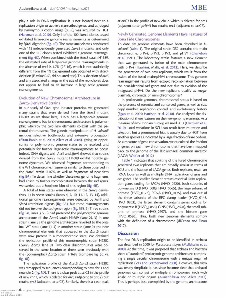
Evolution of New Chromosomal Architecture inDorc5-Derivative StrainsIn our study of Orc1-type initiator proteins, we generatedmany strains that were derived from the Dorc5 mutantH1689. As we show here, H1689 has a large-scale genomerearrangement but its chromosomal architecture is polymor-phic, whereby the two new elements co-exist with the pa-rental chromosome. The genetic manipulation of H. volcaniiincludes selective bottlenecks and extensive propagation(Bitan-Banin et al. 2003; Allers et al. 2004), giving an oppor-tunity for polymorphic genome states to be resolved, andpotentially for further large-scale rearrangements to occur.Indeed, DNA digests with AvrII and SfaAI showed that strainsderived from the Dorc5 mutant H1689 exhibit notable ge-nome dynamics. We observed fragments corresponding tothe WT chromosome, fragments similar to those observed inthe Dorc5 strain H1689, as well as fragments of new sizes(fig. 5A). To determine whether these new genome fragmentshad arisen by further recombination between the sod genes,we carried out a Southern blot of this region (fig. 5B).
A total of four states were observed in the Dorc5 deriva-tives. 1) In seven strains (lanes 4, 7, 10, 11, 12, 13, 14), addi-tional genome rearrangements were detected by AvrII andSfaAI restriction digests (fig. 5A), but these rearrangementsdid not involve the sod gene region (fig. 5B). 2) Three strains(fig. 5B, lanes 3, 5, 6) had preserved the polymorphic genomearchitecture of the Dorc5 strain H1689 (lane 2). 3) In onestrain (lane 8), the genome architecture reverted to the orig-inal WT state (lane 1). 4) In another strain (lane 9), the newchromosomal elements that appeared in the Dorc5 strainwere now present in a monomorphic state. We obtainedthe replication profile of this monomorphic strain H2202(Dorc5 Dorc3, lane 9). Two clear discontinuities were ob-served in the same location as those seen previously withthe (polymorphic) Dorc5 strain H1689 (compare fig. 5C vs.fig. 1B).
The replication profile of the Dorc5 Dorc3 strain H2202was remapped to sequences corresponding to new chr 1 andnew chr 2 (fig. 5D). There is a clear peak at oriC3 in the profileof new chr 1, which is deleted for orc5 (adjacent to oriC2) butretains orc2 (adjacent to oriC3). Similarly, there is a clear peak
at oriC1 in the profile of new chr 2, which is deleted for orc3(adjacent to ori-pHV4) but retains orc1 (adjacent to oriC1).
Newly Generated Genome Elements Have Features ofBona Fide ChromosomesTo date, six genome elements have been described in H.volcanii (table 1). The original strain DS2 contains the mainchromosome, pHV4, pHV3, pHV2, and pHV1 (Charleboiset al. 1991). The laboratory strain features a new elementthat was generated by fusion of the main chromosomewith pHV4 (Hawkins, Malla, et al. 2013). Here, we describethe generation of two new replicons, which result from thefission of the fused main/pHV4 chromosome. This genomerearrangement results from ectopic recombination betweenthe near-identical sod genes and not due to excision of theintegrated pHV4. Do the new replicons qualify as mega-plasmids, chromids, or mini-chromosomes?
In prokaryotic genomes, chromosomal status is based onthe presence of essential and conserved genes, as well as size,copy number, replication control, and evolutionary history(Egan et al. 2005; Harrison et al. 2010). We analyzed the dis-tribution of these features on the new genome elements. As ameasure of evolutionary history, we used SCU (Hartman et al.2010). Local variations in SCU can result from mutation andselection, but a pronounced bias is usually due to HGT fromanother species as indicated by a large fraction of rare codons.As a measure of gene conservation, we calculated the fractionof genes on each new chromosome that have been mappedback to the genome of the last archaeal common ancestor(LACA; Wolf et al. 2012).
Table 1 indicates that splitting of the fused chromosomegenerated two replicons that are broadly similar in terms ofSCU and the fraction of LACA genes. Both replicons retain anrRNA locus as well as multiple DNA replication origins andorc genes. The smaller element retains essential DNA replica-tion genes coding for MCM (HVO_0220), both subunits ofpolymerase D (HVO_0003, HVO_0065), the large subunit ofprimase (HVO_0173), PCNA (HVO_0175), and two out ofthe three subunits of the RFC clamp loader (HVO_0145,HVO_0203); the larger element contains genes coding forpolymerase B (HVO_0858), GINS (HVO_2698), the small sub-unit of primase (HVO_2697), and the histone gene(HVO_0520). Thus, both new genome elements complywith the definition of a chromosome (diCenzo and Finan2017).
DiscussionThe first DNA replication origin to be identified in archaeawas described in 2000 for Pyrococcus abyssi (Myllykallio et al.2000). At the time, it was proposed that archaea and bacteriashare a “standard” prokaryotic genome architecture, compris-ing a single circular chromosome with a unique origin ofreplication (Vas and Leatherwood 2000). However, this viewwas overly simplistic. It has since become clear that archaealgenomes can consist of multiple chromosomes, each withsingle or multiple origins (Ausiannikava and Allers 2017).This is perhaps best exemplified by the genome architecture
Ausiannikava et al. . doi:10.1093/molbev/msy075 MBE
6Downloaded from https://academic.oup.com/mbe/advance-article-abstract/doi/10.1093/molbev/msy075/4972485by gueston 28 June 2018
of H. volcanii, which has one large chromosome with threeorigins and three mini-chromosomes with one origin each(table 1). About 10% of bacteria have more than one replicon(diCenzo and Finan 2017), the best studied example being
Vibrio cholerae which has a large chromosome and a smallerchromid, each with one origin (Jha et al. 2012). In both H.volcanii and V. cholerae, genome rearrangements have beendocumented where two replicons have fused to become one.
FIG. 5. New genome architectures of Dorc5 derivatives. (A) AvrII and SfaAI digests of genomic DNA from derivatives of Dorc5 strain H1689identifying four different genome states. Strain genotypes and genome architecture state is indicated below, polymorphic and monomorphic referto strains with H1689-type genome rearrangements. The monomorphic Dorc5 Dorc3 strain H2202 is indicated. (B) Southern blots showing thatadditional genome rearrangements in derivatives of Dorc5 strain H1689 did not involve recombination of the sod gene region. Genomic DNA wasdigested with StyI or EcoRV and probed with sequences adjacent to sod1 or sod2, respectively (for key to restriction fragments, see fig. 3A). (C)Replication profile of Dorc5 Dorc3 strain H2202 (lane 9 in panels A and B) where the genome is in a monomorphic state. Labeled as in figure 1B, thetwo discontinuities in the replication profile are indicated by vertical arrows. (D) Replication profile of Dorc5 Dorc3 strain H2202 remapped tosequences corresponding to new chr 1 and new chr 2.
Evolution of Genome Architecture in Archaea . doi:10.1093/molbev/msy075 MBE
7Downloaded from https://academic.oup.com/mbe/advance-article-abstract/doi/10.1093/molbev/msy075/4972485by gueston 28 June 2018

We have previously reported that during generation of the H.volcanii laboratory strain, the pHV4 mini-chromosome fusedwith the main chromosome by recombination (Hawkins,Malla, et al. 2013). In V. cholerae, fusion of the chromosomewith the chromid can be induced deliberately or can occurspontaneously. Such spontaneous fusions arise as suppressorsof mutations that affect DNA replication (Val et al. 2014), butnaturally occurring V. cholerae strains with a single chromo-some have also been reported (Xie et al. 2017).
Here we describe a genome rearrangement in H. volcaniithat led to the generation of a new chromosome. The mainchromosome, which in the laboratory strain includes the in-tegrated pHV4 mini-chromosome, has split into two parts.The two resulting DNA molecules exhibit all the features ofbona fide chromosomes: they bear DNA replication origins,rRNA loci, and essential core genes. The genome rearrange-ment that gave rise to the new chromosome was not a simplereversal of the integration of pHV4, which had occurred byrecombination between two identical ISH18 ISs (Hawkins,Malla, et al. 2013). Instead, the genome rearrangementreported here occurred via homologous recombination be-tween the near-identical sod1 and sod2 genes. In the wildisolate DS2, these two genes are located on pHV4 and themain chromosome, respectively, but in the laboratory strainthey are located on the same DNA molecule.
Phylogenetic analysis of bacterial genomes indicates thatadditional chromosomal elements arise relatively rarely butonce a viable state is achieved, they remain stable over longevolutionary intervals (Harrison et al. 2010; diCenzo and Finan2017). It is unclear how the stability of the genome is main-tained in the multipartite state. Genetic engineering experi-ments in bacteria have shown that when parts of amultipartite genome are fused, growth rates remain largelyunaffected (Guo et al. 2003; Val et al. 2012). This finding isconsistent with our observation on the absence of a majorgrowth defect in any of the strains described above. However,multipartite genomes have the potential to be highly dy-namic because homologous genes are often found on differ-ent (or the same) chromosomal elements, providing ampleopportunity for recombination.
The constraints on genome architecture, such as the needto coordinate DNA replication with transcription, might be areason for the observed stability of multipartite genomes. Thefission or fusion of genome elements can potentially cause
unbalanced replichores (which will be exacerbated by therelocation of replication termination zones), conflicts be-tween replication and transcription, and/or changes in genedosage. In archaea such as H. volcanii, the equidistant locationof replication origins on the chromosome could reflect theevolutionary advantage in maintaining such a spatial arrange-ment. Surprisingly, we observed no immediate effect on ge-nome stability in H. volcanii when the replichores areunbalanced. The genome stability was assessed in strainswith different combinations of orc deletions, and there wasno measurable change in the rate of genome rearrangementfollowing deletion of orc5. This finding contrasts with bacte-rial systems, where replichore imbalance has been shown tolead to genome instability and reduced fitness (Esnault et al.2007; Dimude et al. 2016). For example, an E. coli strain wherethe origin was moved to an ectopic site has been found toharbor a large chromosomal inversion (Ivanova et al. 2015).
Several reasons might account for the lack of deleteriouseffects of replichore imbalance in H. volcanii. 1) In contrast tobacteria, which have discrete Ter replication termination sites,archaea and eukaryotes have broad termination zones whereconverging replication forks meet (Duggin et al. 2011). This ismost likely a consequence of having multiple origins perchromosome, and allows for greater flexibility in replicationinitiation. 2) Apart from the highly transcribed rRNA genes,transcription in H. volcanii is not consistently co-orientatedwith replication (Hartman et al. 2010). Such an arrangementis both more important and easier to maintain in bacteria,which have a single origin per chromosome. 3) The polyploidnature of H. volcanii genome (where each chromosome ispresent in 15–20 copies) could also account for the lack ofgenome instability, because deleterious genome rearrange-ments can be restored by gene conversion with a WT copyof the affected chromosome. 4) Little is known about theregulation of replication initiation in archaea. Haloferax volca-nii might use some origins as a “backup” to compensate forreplichore imbalance, thereby avoiding any potential con-flicts. Alternatively, differential origin usage within one cell,where some chromosomes use one origin and others use adifferent one, would ameliorate unbalanced replichores. Bothscenarios—compensatory and stochastic origin firing—havebeen observed in eukaryotic replication (Hawkins, Retkute,et al. 2013). 5) Recombination-dependent replication, whichis used in the absence of origins, leads to dispersed initiation
Table 1. Distribution of Features on Genome Elements in H. volcanii Wild Isolate DS2, Laboratory Strain H26, and Dorc5 Strain H1689.
Strain(s) GenomeElement
Size, bp Number ofGenes
SCU, RareCodons
GC Content LACAGenes
rRNA Loci ReplicationOrigins
DS2 Chromosome 2,847,757 2,960 7.3% 66.6% 37.3% 2 oriC1, oriC2, oriC3DS2 pHV4 635,786 636 15.5% 61.7% 28.3% 0 ori-pHV4H26 Chromosome 1 pHV4 3,482,975 3,596 8.7% 65.7% 35.5% 2 oriC1, oriC2, oriC3, ori-pHV4H1689 New chr1 2,695,880 2,781 8.3% 66.1% 37.4% 1 oriC2, oriC3H1689 New chr2 787,095 815 10.3% 64.6% 33% 1 oriC1, ori-pHV4DS2, H26, H1689 pHV3 437,906 380 7.7% 65.5% 35.9% 0 ori-pHV3DS2, H26, H1689 pHV1 85,092 88 26.3% 55.5% 18% 0 ori-pHV1
NOTE.—New genomic elements generated by fission of the fused chromosomeþ pHV4 are designated as New chr1 and New chr2. The fraction of rare codons was calculatedfrom SCU tables for each genome element (Hartman et al. 2010). The fraction of LACA genes was calculated with cut-off probability of 0.75 (Wolf et al. 2012).
Ausiannikava et al. . doi:10.1093/molbev/msy075 MBE
8Downloaded from https://academic.oup.com/mbe/advance-article-abstract/doi/10.1093/molbev/msy075/4972485by gueston 28 June 2018
throughout the genome and may relieve the spatial con-straints on replication origins. Thus, replichore imbalancewould have only minor effects on the viability of H. volcanii.
Nonetheless, it is notable that the Dorc5-derivative strainsexhibited considerable genome plasticity and the ability toevolve to different chromosome architectures (fig. 5). The twonew chromosomes were stable during routine growth butnew rounds of genetic manipulation appeared to provokefurther rearrangements. Following transformation, a select-able marker will initially be present on only one of the 20chromosome copies. This selectable marker will then spreadthroughout the genome by gene conversion, and may carrywith it genetically linked rearrangements. Therefore, the
selective bottleneck of genetic manipulation might allow anew chromosome architecture to become monomorphic.
Eukaryotic cells contain multiple linear chromosomes thatare replicated from multiple origins. For this type of genomearchitecture to arise, three steps are required (but not neces-sarily in this order): multiplication of origins, multiplication ofchromosomes, and linearization of chromosomes. Given theshared evolutionary history of eukaryotes and archaea, it isnot surprising that two of these three features are found inarchaeal genomes as well. Up to four replication origins canbe present on some archaeal chromosomes, and multiplechromosomes that use an Orc-type replication initiationmechanism co-exist in haloarchaeal species; however, no
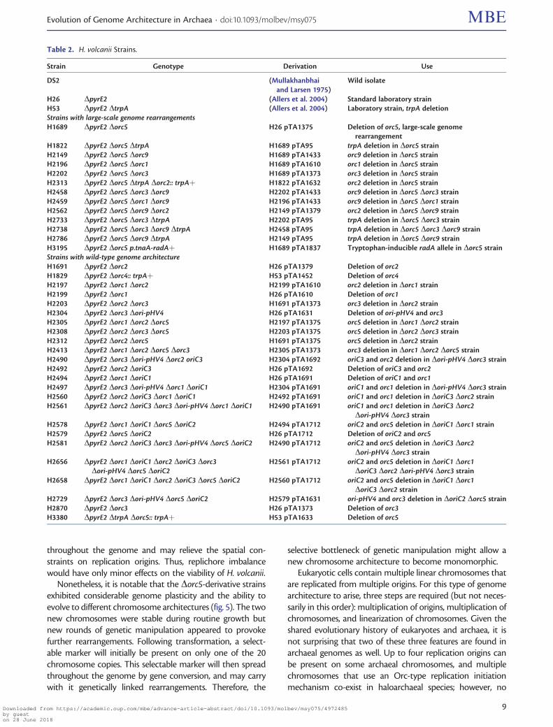
Table 2. H. volcanii Strains.
Strain Genotype Derivation Use
DS2 (Mullakhanbhaiand Larsen 1975)
Wild isolate
H26 DpyrE2 (Allers et al. 2004) Standard laboratory strainH53 DpyrE2 DtrpA (Allers et al. 2004) Laboratory strain, trpA deletionStrains with large-scale genome rearrangementsH1689 DpyrE2 Dorc5 H26 pTA1375 Deletion of orc5, large-scale genome
rearrangementH1822 DpyrE2 Dorc5 DtrpA H1689 pTA95 trpA deletion in Dorc5 strainH2149 DpyrE2 Dorc5 Dorc9 H1689 pTA1433 orc9 deletion in Dorc5 strainH2196 DpyrE2 Dorc5 Dorc1 H1689 pTA1610 orc1 deletion in Dorc5 strainH2202 DpyrE2 Dorc5 Dorc3 H1689 pTA1373 orc3 deletion in Dorc5 strainH2313 DpyrE2 Dorc5 DtrpA Dorc2:: trpAþ H1822 pTA1632 orc2 deletion in Dorc5 strainH2458 DpyrE2 Dorc5 Dorc3 Dorc9 H2202 pTA1433 orc9 deletion in Dorc5 Dorc3 strainH2459 DpyrE2 Dorc5 Dorc1 Dorc9 H2196 pTA1433 orc9 deletion in Dorc5 Dorc1 strainH2562 DpyrE2 Dorc5 Dorc9 Dorc2 H2149 pTA1379 orc2 deletion in Dorc5 Dorc9 strainH2733 DpyrE2 Dorc5 Dorc3 DtrpA H2202 pTA95 trpA deletion in Dorc5 Dorc3 strainH2738 DpyrE2 Dorc5 Dorc3 Dorc9 DtrpA H2458 pTA95 trpA deletion in Dorc5 Dorc3 Dorc9 strainH2786 DpyrE2 Dorc5 Dorc9 DtrpA H2149 pTA95 trpA deletion in Dorc5 Dorc9 strainH3195 DpyrE2 Dorc5 p.tnaA-radAþ H1689 pTA1837 Tryptophan-inducible radA allele in Dorc5 strainStrains with wild-type genome architectureH1691 DpyrE2 Dorc2 H26 pTA1379 Deletion of orc2H1829 DpyrE2 Dorc4:: trpAþ H53 pTA1452 Deletion of orc4H2197 DpyrE2 Dorc1 Dorc2 H2199 pTA1610 orc2 deletion in Dorc1 strainH2199 DpyrE2 Dorc1 H26 pTA1610 Deletion of orc1H2203 DpyrE2 Dorc2 Dorc3 H1691 pTA1373 orc3 deletion in Dorc2 strainH2304 DpyrE2 Dorc3 Dori-pHV4 H26 pTA1631 Deletion of ori-pHV4 and orc3H2305 DpyrE2 Dorc1 Dorc2 Dorc5 H2197 pTA1375 orc5 deletion in Dorc1 Dorc2 strainH2308 DpyrE2 Dorc2 Dorc3 Dorc5 H2203 pTA1375 orc5 deletion in Dorc2 Dorc3 strainH2312 DpyrE2 Dorc2 Dorc5 H1691 pTA1375 orc5 deletion in Dorc2 strainH2413 DpyrE2 Dorc1 Dorc2 Dorc5 Dorc3 H2305 pTA1373 orc3 deletion in Dorc1 Dorc2 Dorc5 strainH2490 DpyrE2 Dorc3 Dori-pHV4 Dorc2 oriC3 H2304 pTA1692 oriC3 and orc2 deletion in Dori-pHV4 Dorc3 strainH2492 DpyrE2 Dorc2 DoriC3 H26 pTA1692 Deletion of oriC3 and orc2H2494 DpyrE2 Dorc1 DoriC1 H26 pTA1691 Deletion of oriC1 and orc1H2497 DpyrE2 Dorc3 Dori-pHV4 Dorc1 DoriC1 H2304 pTA1691 oriC1 and orc1 deletion in Dori-pHV4 Dorc3 strainH2560 DpyrE2 Dorc2 DoriC3 Dorc1 DoriC1 H2492 pTA1691 oriC1 and orc1 deletion in DoriC3 Dorc2 strainH2561 DpyrE2 Dorc2 DoriC3 Dorc3 Dori-pHV4 Dorc1 DoriC1 H2490 pTA1691 oriC1 and orc1 deletion in DoriC3 Dorc2
Dori-pHV4 Dorc3 strainH2578 DpyrE2 Dorc1 DoriC1 Dorc5 DoriC2 H2494 pTA1712 oriC2 and orc5 deletion in DoriC1 Dorc1 strainH2579 DpyrE2 Dorc5 DoriC2 H26 pTA1712 Deletion of oriC2 and orc5H2581 DpyrE2 Dorc2 DoriC3 Dorc3 Dori-pHV4 Dorc5 DoriC2 H2490 pTA1712 oriC2 and orc5 deletion in DoriC3 Dorc2
Dori-pHV4 Dorc3 strainH2656 DpyrE2 Dorc1 DoriC1 Dorc2 DoriC3 Dorc3
Dori-pHV4 Dorc5 DoriC2H2561 pTA1712 oriC2 and orc5 deletion in DoriC1 Dorc1
DoriC3 Dorc2 Dori-pHV4 Dorc3 strainH2658 DpyrE2 Dorc1 DoriC1 Dorc2 DoriC3 Dorc5 DoriC2 H2560 pTA1712 oriC2 and orc5 deletion in DoriC1 Dorc1
DoriC3 Dorc2 strainH2729 DpyrE2 Dorc3 Dori-pHV4 Dorc5 DoriC2 H2579 pTA1631 ori-pHV4 and orc3 deletion in DoriC2 Dorc5 strainH2870 DpyrE2 Dorc3 H26 pTA1373 Deletion of orc3H3380 DpyrE2 DtrpA Dorc5:: trpAþ H53 pTA1633 Deletion of orc5
Evolution of Genome Architecture in Archaea . doi:10.1093/molbev/msy075 MBE
9Downloaded from https://academic.oup.com/mbe/advance-article-abstract/doi/10.1093/molbev/msy075/4972485by gueston 28 June 2018

archaeon with linear chromosomes has been found to date.Here, we show that an increase in the number of circularchromosomes is easily achievable through natural evolution.To the best of our knowledge, rearrangement of a naturallyevolved prokaryotic genome that generates two new chro-mosomes, each with pre-existing multiple origins that dependon the same type of replication initiation, has not been de-scribed previously. Interestingly, the H. volcanii genome mightalready contain an imprint of a similar event, where the
ancestral chromosome fragmented leading to the generationof a new chromosome. Indeed, the pHV3 mini-chromosomehas one Orc-dependent replication origin, a native SCU andGC content similar to the main chromosome, and a highproportion of LACA genes (table 1); thus, the generation ofpHV3 is compatible with the recombinational route de-scribed here.
Newly generated chromosomal elements must find effec-tive solutions for segregation and replication, and the ability
Table 3. Plasmids.
Plasmid Relevant Properties Derivation
pTA95 Integrative plasmid for trpA gene deletion (Allers et al. 2004)pTA131 Integrative plasmid based on pBluescript II, with pyrE2þ marker (Allers et al. 2004)pTA298 pUC19 with trpAþ marker flanked by BamHI sites (Lestini et al. 2010)pTA333 pUC19 with SacI-NspI chromosomal fragment containing orc4 gene This studypTA415 pBluescript II SK1 with MluI chromosomal fragment containing hel308 helicase gene This studypTA416 pBluescript II with SacI chromosomal fragment containing orc5 and oriC2 (Norais et al. 2007)pTA419 pTA131 with NheI-EcoRI fragment of pTA416 containing orc5 and oriC2 This studypTA1100 pBluescript II with AciI chromosomal fragment containing orc2 and oriC3 (Hawkins, Malla, et al. 2013)pTA1329 pTA131 with Dori-pHV4 construct (Hawkins, Malla, et al. 2013)pTA1343 pTA131 with p.tnaA-radAþ:: hdrBþ construct flanked by upstream and downstream radA
regions(Hawkins, Malla, et al. 2013)
pTA1370 pBluescript II SK1 with HindIII-KpnI chromosomal fragment containing orc1 gene andoriC1 origin
This study
pTA1371 pBluescript II SK1 with BstBI chromosomal fragment containing orc3 gene This studypTA1373 pTA131 with Dorc3 construct, comprising ClaI-BamHI fragment of upstream flanking
region of orc3 and BamHI-XbaI fragment of downstream flanking region of orc3, PCRamplified from pTA1371
This study
pTA1375 pTA131 with Dorc5 construct, comprising KpnI-BamHI fragment of downstream flankingregion of orc5 and BamHI-XbaI fragment of upstream flanking region of orc5, PCRamplified from pTA416
This study
pTA1379 pTA131 with Dorc2 construct, comprising KpnI-BamHI upstream flanking region of orc2and BamHI-XbaI fragment of downstream flanking region of orc2, PCR amplified frompTA1100
This study
pTA1431 pTA131 with inactivation of unique BamHI site in MCS by filling-in with Klenow This studypTA1432 pBluescript II SK1 with NotI chromosomal fragment containing orc9 gene This studypTA1433 pTA1431 with Dorc9 construct, comprising XbaI-BstXI upstream flanking region of orc9
and XbaI-BstXI fragment of downstream flanking region of orc9, PCR amplified frompTA1432
This study
pTA1610 pTA131 with Dorc1 construct, comprising KpnI-BamHI upstream flanking region of orc1and BamHI-XhoI fragment of downstream flanking region of orc1, PCR amplified frompTA1370
This study
pTA1631 Dorc3 Dori-pHV4 construct, where orc3 upstream region of pTA1373 was replaced byKpnI-BamHI fragment of ori-pHV4 upstream region from pTA1329
This study
pTA1632 pTA1379 with insertion of BamHI trpAþ fragment from pTA298 This studypTA1633 pTA1375 with insertion of BamHI trpAþ fragment from pTA298 This studypTA1691 pTA131 with Dorc1 DoriC1 construct, comprising StuI-BamHI upstream flanking region of
oriC1 and BamHI-XbaI fragment of downstream flanking region of orc1, PCR amplifiedfrom pTA1370
This study
pTA1692 pTA131 with Dorc2 DoriC3 construct, comprising AatII-BamHI upstream flanking regionof oriC3 and BamHI-KpnI fragment of downstream flanking region of orc2, PCR am-plified from pTA1100
This study
pTA1712 pTA131 with Dorc5 DoriC2 construct, comprising XbaI-BamHI upstream flanking regionof oriC2 and BamHI-XbaI fragment of downstream flanking region of orc5, PCR am-plified from pTA416
This study
pTA1837 pTA131 with p.tnaA-radAþ construct. XbaI-BamHI fragment of hdrBþ marker was re-moved from pTA1343, and 890 bp EcoRV-PvuII fragment of radA upstream flankingregion (PCR amplified from H26 genomic DNA) was used to replace 315 bp EcoRV-PvuIIfragment of radA upstream flanking region in pTA1343
This study
pID19T-HVO_2042 pTA131 with Dorc4:: trpAþ construct, comprising XhoI-HindIII fragment of upstreamflanking region of orc4 and BamHI-XbaI fragment of downstream flanking region oforc4, PCR amplified from H26 genomic DNA, joined using HindIII-BamHI trpAþ
fragment
Jerry Eichler
Ausiannikava et al. . doi:10.1093/molbev/msy075 MBE
10Downloaded from https://academic.oup.com/mbe/advance-article-abstract/doi/10.1093/molbev/msy075/4972485by gueston 28 June 2018

to spread throughout a population would be beneficial.Haloarchaea have developed potential solutions to thesechallenges. The proclivity of H. volcanii to userecombination-dependent replication in the absence of ori-gins weakens the requirement for newly generated chromo-somal elements to maintain balanced replichores, or evenorigins (Hawkins, Malla, et al. 2013). Haloferax volcanii doesnot strictly depend on orderly segregation of its chromo-somes, because its genome is highly polyploid and new chro-mosomal elements can rely on random partitioning intodaughter cells; furthermore, archaea lack the centromeresfound on eukaryotic chromosomes. Haloarchaea have a re-markable capacity for rapid genome evolution by HGT. Theexchange of up to 530 kb of DNA between different Haloferaxspecies has been detected after cell fusion (Naor et al. 2012),
thus providing the opportunity for a newly generated chro-mosome (and eventually, a new species) to arise. And becausearchaeal origins are nearly always linked to an orc gene encod-ing their cognate initiator protein, a “foreign” chromosomewill be efficiently replicated in its new host cell. The remark-able plasticity of haloarchaeal genomes thus presents a testbed for probing the evolution of genome organization andreplication initiation.
Materials and Methods
Strains and PlasmidsHaloferax volcanii strains (table 2) were grown at 45 �C oncomplete (Hv-YPC) or casamino acids (Hv-Ca) agar, or in Hv-YPC broth, as described previously (Allers et al. 2004).Isolation of genomic and plasmid DNA, and transformation
Table 4. Oligonucleotides.
Primer Sequence (50–30) Relevant Properties Use
MHorc3F1 CGTTCAtCGATTTGACGAGGTCATCCACG orc3 deletion, upstream pTA1373MHorc3R1 GTCCCGGaTCCCGATAGATCTCGGTGTCC orc3 deletion, upstream pTA1373MHorc3F2 ACGACTggATCcAGCAGTAGGTAGGTCG orc3 deletion, downstream pTA1373MHorc3R2 CCTCCGtCtAGAACACGACGTGCGCGACC orc3 deletion, downstream pTA1373MHorc2F1 CAGCGgTAcCGACCCGTCGCAGAGGTACG orc2 deletion, upstream pTA1379MHorc2R1 CGCAGGatCCGAGGCCGCCTGACCCCACG orc2 deletion, upstream pTA1379MHorc2F2 GCTCGgAtCCGGCGCATTAGCGTCGGTCC orc2 deletion, downstream pTA1379, pTA1692MHorc2R2 CCGAGGTctAGACATTTCGAGGGGCGG orc2 deletion, downstream pTA1379, pTA1692MHorc5F1 GTGCTAGGTacCTGAACACCCATAAGTG orc5/oriC2orc5 deletions, downstream pTA1375, pTA1712MHorc5R1 GCTCGAGGATCCGGACGTGGTGAGGGACG orc5/oriC2orc5 deletions, downstream pTA1375, pTA1712MHorc5F2 GTGAAGAGGaTCcTCGCTGGCGTTAGGC orc5 deletion, upstream pTA1375MHorc5R2 GGGGAAtcTAGAGAACCGGAAAACCCGG orc5 deletion, upstream pTA1375delorc9USR TCTTCGGGaTCCTCCCTCATCGAG orc9 deletion, upstream pTA1433delorc9DSF CGGTCGgAtCCGCGCCATCTCGCTCG orc9 deletion, downstream pTA1433pBSR3 ACCCCAGGCTTTACACTTTATGC orc9 deletion, downstream pTA1433pBSF2 TTAAGTTGGGTAACGCCAGGG orc9 deletion, upstream, and oriC1orc1
deletion, downstreampTA1433, pTA1691
MHorc1F1 ACGAGCgGTaCCGGACGATGCGCGCCGGC orc1 deletion, downstream pTA1610dorc1DF AGAACGggaTCCCGAAGTCCGACGC orc1/oriC1orc1 deletion, downstream pTA1610, pTA1691MHorc1F2 GTTCCCGGaTCCCCTCGTGCGCCGCCTCG orc1 deletion, upstream pTA1610MHorc1R2 CCACAGTCTaGaCCTCGCCGCAGTAGCCG orc1 deletion, upstream pTA1610oriC1-BamHL GTACTCCGGATCCATGCTCGGTATCCG oriC1orc1 deletion, upstream pTA1691pBSR2 CGCGCAATTAACCCTCACTAAAG oriC1orc1 and oriC3orc2 deletions,
upstreampTA1691, pTA1692
oriC3-BamHL GGTGTCGGAtCcCGGCTTTCGCGTTCCG oriC3orc2 deletion, upstream pTA1692OriC2-BamL CCGGTCTCGGATCCAACTTAGCTCTCACTCG oriC2orc5 deletion, upstream pTA1712OriC2-XbaR CGACCCTCTAGAGCGAGGCGAGGTCGCCCC oriC2orc5 deletion, upstream pTA171250HVO_2042_XhoI_F cccctcgagTCTTTGCAGTCTATTTCCTTC orc4 deletion, upstream pID19T-HVO_204250HVO_2042_HindIII_R gggaagcttACGTGTTGCAGACCTGTATAC orc4 deletion, upstream pID19T-HVO_204230HVO_2042_BamHI_F cccggatccCCCACAGAACAGATGAAGTG orc4 deletion, downstream pID19T-HVO_204230HVO_2042_XbaI_R gggtctagaCGTGCTTCCGAGTCAGAAAC orc4 deletion, downstream pID19T-HVO_2042radAUSNdeR TTCTGCCATAtgCAGTCGTTCCGCCTATACCC p.tnaA: radAþ construct, upstream pTA1837radAextraUS AGACCAGCTGAGTTCCGATGGGGCTGTTC p.tnaA: radAþ construct, upstream pTA1837sod1F AGTACAGGCCGAACTCGACGACGCC sod1 Southern blot probe, diagnostic
PCR and sequencing of sod1Figure 2B, C
sod1R TCTCACGGTAACCTGTGGTCGCGCG sod1 Southern blot probe, diagnosticPCR and sequencing of sod1
Figure 2B, C
sod2F GAAATCGCCGACGCCGTCTCGACG sod2 Southern blot probe, diagnosticPCR and sequencing of sod2
Figure 2B, C
sod2R GAGCAGTTTCGGACCTTCGTCGGCG sod2 Southern blot probe, diagnosticPCR and sequencing of sod2
Figure 2B, C
sod1 US-left ACAGGCTCCGAACGTATCAT sod1U Southern blot probe Figures 3A, 5Bsod1 US-right CAGTCGGTGAGTCCCTGTAA sod1U Southern blot probe Figures 3A, 5Bsod2 DS-left GATGACCTCCGCGACCTC sod2D Southern blot probe Figures 3A, 5Bsod2 DS-right GGGTCGCTGAACAGGTCC sod2D Southern blot probe Figures 3A, 5B
Evolution of Genome Architecture in Archaea . doi:10.1093/molbev/msy075 MBE
11Downloaded from https://academic.oup.com/mbe/advance-article-abstract/doi/10.1093/molbev/msy075/4972485by gueston 28 June 2018

of H. volcanii, were carried out as described previously (Allerset al. 2004). Standard molecular techniques were used(Sambrook and Russell 2001). Deletion mutants were con-structed and confirmed by colony hybridization and/orSouthern blotting as described previously (Allers et al.2004). Plasmids for gene deletion are shown in table 3 andwere generated by PCR using oligonucleotides shown in ta-ble 4. Probes for Southern blots are shown in table 5. Growthcompetition assays were carried out as described previously(Hawkins, Malla, et al. 2013).
Screening for Genome Rearrangements in Dorc5 andDorc4-Deleted BackgroundsTwelve independent “pop-in” strains were generated usingDorc5 and Dorc4 plasmids pTA1375 and pID19T-HVO_2042,respectively, and ten deletion (“pop-out”) strains were de-rived from each “pop-in.” Gene deletions were confirmedby colony hybridization with the relevant orc5 or orc4 probes.The deletion strains were assessed for SfaAI restriction frag-ment length polymorphisms by PFGE.
Marker Frequency Analysis by Deep SequencingFor exponential-phase samples, strains were grown overnightin Hv-YPC broth, diluted 500-fold in fresh media and incu-bated at 45 �C with vigorous aeration until an A650 of 0.4,then diluted 500-fold in fresh media and grown until an A650of 0.2. For a stationary-phase sample, a WT culture was grownat 45 �C for 3 days until saturation (no further increase inA650). Genomic DNA was isolated from 50 ml cultures fol-lowed by phenol: chloroform extraction as described previ-ously (Hawkins, Malla, et al. 2013). Marker frequency analysiswas performed by Deep Seq (University of Nottingham) usingIllumina HiSeq 2000 sequencing to measure sequence copynumber. Enrichment of uniquely mapping sequence tags wascalculated (in 1-kb windows) for exponentially growing sam-ples relative to a stationary phase WT sample, to correct fordifferences in read depth across the genome (Skovgaard et al.2011; Muller et al. 2014). Sequence reads were mapped to theH. volcanii genome and replication profiles were calculated asdescribed previously (Hawkins, Malla, et al. 2013).
Pulsed Field Gel ElectrophoresisFor PFGE, genomic DNA was prepared in agarose plugs anddigested as described previously (Hawkins, Malla, et al. 2013).For analysis of intact genomic DNA, agarose plugs were
subjected to 100 Gy of c radiation using a 137Cs source(Gammacell 1000), to linearize circular chromosomes(Beverley 1989). PFGE was performed using a CHEF Mapperapparatus (Bio-Rad). Intact and SfaAI-digested DNA frag-ments were separated on a 1.2% agarose gel in 0.5� TBE at14 �C, with a gradient voltage of 6 V/cm, linear ramping, anincluded angle of 120�, initial and final switch times of 0.64 sand 1 min 13.22 s, respectively, and a run time of 40 h (intactDNA) or 20 h 46 min (SfaAI-digested DNA). AvrII-digestedand SwaI-digested genomic DNA were separated on 1% aga-rose gel in 0.5� TBE at 14 �C, with a gradient voltage of 6 V/cm, linear ramping, an included angle of 120�, initial and finalswitch times of 1 min and 2 min, respectively, and a run timeof 24 h. The gel was stained with ethidium bromide.
AcknowledgmentsWe thank Uri Gophna and Nathan Jones for helpful com-ments on the manuscript, Sunir Malla (Deep Seq,Nottingham) for DNA sequencing, Christopher Turley forassistance with PFGE in figure 4, and Jerry Eichler (BenGurion University, Israel) for the Dorc4:: trpAþ plasmidpID19T-HVO_2042. This work was supported by theBiotechnology and Biological Sciences Research Council(BBSRC) [grant number BB/M001393/1]. The funders hadno role in study design, data collection and interpretation,or the decision to submit the work for publication.
Author ContributionsD.A. and T.A. conceived the study and wrote the manuscriptwith input from all authors. D.A., L.M., H.M., and T.A. per-formed the genetic experiments. M.H. generated the Dorc5strain H1689. V.S. performed the PFGE in figure 1C. C.N. an-alyzed the DNA replication profiles. K.M. and E.K. analyzedthe LACA gene distribution. All authors read and approvedthe manuscript.
ReferencesAllers T, Ngo HP, Mevarech M, Lloyd RG. 2004. Development of addi-
tional selectable markers for the halophilic archaeon Haloferax vol-canii based on the leuB and trpA genes. Appl Environ Microbiol.70(2):943–953.
Ausiannikava D, Allers T. 2017. Diversity of DNA replication in the ar-chaea. Genes (Basel) 8(2):56.
Baliga NS, Bonneau R, Facciotti MT, Pan M, Glusman G, Deutsch EW,Shannon P, Chiu Y, Weng RS, Gan RR, et al. 2004. Genome sequence
Table 5. Probes.
Probe Usage Location Source
sod1 Figure 2B sod1 gene 813 bp PCR using sod1F and sod1Rsod2 Figure 2B sod2 gene 1074 bp PCR using sod2F and sod2Rsod1U Figures 3A, 5B Upstream of sod1 gene 359 bp PCR using sod1 US-left and sod1 US-rightsod2D Figures 3A, 5B Downstream of sod2 gene 347 bp PCR using sod2 DS-left and sod2 DS-rightoriC1 Figure 3B Downstream of oriC1 origin 763 bp StyI fragment of pTA415orc4 Confirmation of orc4 deletion
by colony hybridizationorc4 gene 959 bp BglII-PstI fragment of pTA333
orc5 Confirmation of orc5 deletionby colony hybridization
orc5 gene 784 bp AatII fragment of pTA419
Ausiannikava et al. . doi:10.1093/molbev/msy075 MBE
12Downloaded from https://academic.oup.com/mbe/advance-article-abstract/doi/10.1093/molbev/msy075/4972485by gueston 28 June 2018
of Haloarcula marismortui: a halophilic archaeon from the Dead Sea.Genome Res. 14(11):2221–2234.
Beverley SM. 1989. Estimation of circular DNA size using gamma-irradiation and pulsed-field gel electrophoresis. Anal Biochem.177(1):110–114.
Bitan-Banin G, Ortenberg R, Mevarech M. 2003. Development of a geneknockout system for the halophilic archaeon Haloferax volcanii byuse of the pyrE gene. J Bacteriol. 185(3):772–778.
Breuert S, Allers T, Spohn G, Soppa J. 2006. Regulated polyploidy inhalophilic archaea. PLoS ONE. 1:e92.
Bridger SL, Lancaster WA, Poole FL 2nd, Schut GJ, Adams MW. 2012.Genome sequencing of a genetically tractable Pyrococcus furiosus strainreveals a highly dynamic genome. J Bacteriol. 194(15):4097–4106.
Brugger K, Torarinsson E, Redder P, Chen L, Garrett RA. 2004. Shuffling ofSulfolobus genomes by autonomous and non-autonomous mobileelements. Biochem Soc Trans. 32(Pt 2):179–183.
Bryant J, Chewapreecha C, Bentley SD. 2012. Developing insights into themechanisms of evolution of bacterial pathogens from whole-genome sequences. Future Microbiol. 7(11):1283–1296.
Charlebois RL, Schalkwyk LC, Hofman JD, Doolittle WF. 1991. Detailedphysical map and set of overlapping clones covering the genome ofthe archaebacterium Haloferax volcanii DS2. J Mol Biol.222(3):509–524.
Cossu M, Badel C, Catchpole R, Gadelle D, Marguet E, Barbe V, Forterre P,Oberto J. 2017. Flipping chromosomes in deep-sea archaea. PLoSGenet. 13(6):e1006847.
diCenzo GC, Finan TM. 2017. The divided bacterial genome: structure,function, and evolution. Microbiol Mol Biol Rev. 81:e00019–17.
Dimude JU, Midgley-Smith SL, Stein M, Rudolph CJ. 2016. Replicationtermination: containing fork fusion-mediated pathologies inEscherichia coli. Genes (Basel) 7(8):40.
Duggin IG, Dubarry N, Bell SD. 2011. Replication termination and chro-mosome dimer resolution in the archaeon Sulfolobus solfataricus.EMBO J. 30(1):145–153.
Egan ES, Fogel MA, Waldor MK. 2005. Divided genomes: negotiating thecell cycle in prokaryotes with multiple chromosomes. Mol Microbiol.56(5):1129–1138.
Eisen JA, Heidelberg JF, White O, Salzberg SL. 2000. Evidence for sym-metric chromosomal inversions around the replication origin inbacteria. Genome Biol. 1(6):RESEARCH0011.
Esnault E, Valens M, Espeli O, Boccard F. 2007. Chromosome structuringlimits genome plasticity in Escherichia coli. PLoS Genet. 3(12): e226.
Gaubatz JW. 1990. Extrachromosomal circular DNAs and genomic se-quence plasticity in eukaryotic cells. Mutat Res. 237(5–6):271–292.
Guo X, Flores M, Mavingui P, Fuentes SI, Hernandez G, Davila G, PalaciosR. 2003. Natural genomic design in Sinorhizobium meliloti: novelgenomic architectures. Genome Res. 13(8):1810–1817.
Harrison PW, Lower RP, Kim NK, Young JP. 2010. Introducing the bac-terial ‘chromid’: not a chromosome, not a plasmid. Trends Microbiol.18(4):141–148.
Hartman AL, Norais C, Badger JH, Delmas S, Haldenby S, Madupu R,Robinson J, Khouri H, Ren Q, Lowe TM, et al. 2010. The completegenome sequence of Haloferax volcanii DS2, a model archaeon. PLoSONE. 5(3):e9605.
Hawkins M, Malla S, Blythe MJ, Nieduszynski CA, Allers T. 2013.Accelerated growth in the absence of DNA replication origins.Nature 503(7477):544–547.
Hawkins M, Retkute R, Muller CA, Saner N, Tanaka TU, de Moura AP,Nieduszynski CA. 2013. High-resolution replication profiles definethe stochastic nature of genome replication initiation and termina-tion. Cell Rep. 5(4):1132–1141.
Ivanova D, Taylor T, Smith SL, Dimude JU, Upton AL, Mehrjouy MM,Skovgaard O, Sherratt DJ, Retkute R, Rudolph CJ. 2015. Shaping thelandscape of the Escherichia coli chromosome: replication–transcription encounters in cells with an ectopic replication origin.Nucleic Acids Res. 43(16):7865–7877.
Jha JK, Baek JH, Venkova-Canova T, Chattoraj DK. 2012. Chromosomedynamics in multichromosome bacteria. Biochim Biophys Acta.1819(7):826–829.
Koonin EV, Wolf YI. 2008. Genomics of bacteria and archaea: the emerg-ing dynamic view of the prokaryotic world. Nucleic Acids Res.36(21):6688–6719.
Lestini R, Duan Z, Allers T. 2010. The archaeal Xpf/Mus81/FANCM ho-molog Hef and the Holliday junction resolvase Hjc define alternativepathways that are essential for cell viability in Haloferax volcanii.DNA Repair (Amst). 9(9):994–1002.
Makarova KS, Koonin EV. 2013. Archaeology of eukaryotic DNA repli-cation. Cold Spring Harb Perspect Biol. 5(11):a012963.
Mao D, Grogan DW. 2017. How a genetically stable extremophileevolves: modes of genome diversification in the archaeonSulfolobus acidocaldarius. J Bacteriol. 199(17): e00177–17.
Moller HD, Parsons L, Jorgensen TS, Botstein D, Regenberg B. 2015.Extrachromosomal circular DNA is common in yeast. Proc NatlAcad Sci U S A. 112(24):E3114–E3122.
Mullakhanbhai MF, Larsen H. (76060741 co-authors). 1975.Halobacterium volcanii spec. nov., a Dead Sea halobacterium witha moderate salt requirement. Arch Microbiol. 104(3):207–214.
Muller CA, Hawkins M, Retkute R, Malla S, Wilson R, Blythe MJ, Nakato R,Komata M, Shirahige K, de Moura AP, et al. 2014. The dynamics ofgenome replication using deep sequencing. Nucleic Acids Res. 42(1):e3.
Myllykallio H, Lopez P, Lopez-Garcia P, Heilig R, Saurin W, Zivanovic Y,Philippe H, Forterre P. 2000. Bacterial mode of replication witheukaryotic-like machinery in a hyperthermophilic archaeon.Science 288(5474):2212–2215.
Naor A, Lapierre P, Mevarech M, Papke RT, Gophna U. 2012. Low speciesbarriers in halophilic archaea and the formation of recombinanthybrids. Curr Biol. 22(15):1444–1448.
Ng WV, Ciufo SA, Smith TM, Bumgarner RE, Baskin D, Faust J, Hall B,Loretz C, Seto J, Slagel J, et al. 1998. Snapshot of a large dynamicreplicon in a halophilic archaeon: megaplasmid or minichromo-some? Genome Res. 8(11):1131–1141.
Ng WV, Kennedy SP, Mahairas GG, Berquist B, Pan M, Shukla HD, LaskySR, Baliga NS, Thorsson V, Sbrogna J, et al. 2000. Genome sequence ofHalobacterium species NRC-1. Proc Natl Acad Sci U S A.97(22):12176–12181.
Norais C, Hawkins M, Hartman AL, Eisen JA, Myllykallio H, Allers T. 2007.Genetic and physical mapping of DNA replication origins inHaloferax volcanii. PLoS Genet. 3(5):e77.
Novichkov PS, Wolf YI, Dubchak I, Koonin EV. 2009. Trends in prokary-otic evolution revealed by comparison of closely related bacterialand archaeal genomes. J Bacteriol. 191(1):65–73.
Papke RT, Koenig JE, Rodriguez-Valera F, Doolittle WF. 2004. Frequentrecombination in a saltern population of Halorubrum. Science306:1928–1929.
Pinto UM, Pappas KM, Winans SC. 2012. The ABCs of plasmid replica-tion and segregation. Nat Rev Microbiol. 10(11):755–765.
Press MO, Queitsch C, Borenstein E. 2016. Evolutionary assembly pat-terns of prokaryotic genomes. Genome Res. 26(6):826–833.
Raymann K, Forterre P, Brochier-Armanet C, Gribaldo S. 2014. Globalphylogenomic analysis disentangles the complex evolutionary his-tory of DNA replication in archaea. Genome Biol Evol. 6(1):192–212.
Redder P, Garrett RA. 2006. Mutations and rearrangements in the ge-nome of Sulfolobus solfataricus P2. J Bacteriol. 188(12):4198–4206.
Repar J, Warnecke T. 2017. Non-random inversion landscapes in pro-karyotic genomes are shaped by heterogeneous selection pressures.Mol Biol Evol. 34(8):1902–1911.
Robinson NP, Bell SD. 2007. Extrachromosomal element capture and theevolution of multiple replication origins in archaeal chromosomes.Proc Natl Acad Sci U S A. 104(14):5806–5811.
Rocha EP. 2008. Evolutionary patterns in prokaryotic genomes. CurrOpin Microbiol. 11(5):454–460.
Rocha EP. 2004. Order and disorder in bacterial genomes. Curr OpinMicrobiol. 7(5):519–527.
Sambrook J, Russell DW. 2001. Molecular cloning: a laboratory manual.Cold Spring Harbor (NY): Cold Spring Harbor Laboratory Press.
Samson RY, Xu Y, Gadelha C, Stone TA, Faqiri JN, Li D, Qin N, Pu F, LiangYX, She Q, et al. 2013. Specificity and function of archaeal DNAreplication initiator proteins. Cell Rep. 3(2):485–496.
Evolution of Genome Architecture in Archaea . doi:10.1093/molbev/msy075 MBE
13Downloaded from https://academic.oup.com/mbe/advance-article-abstract/doi/10.1093/molbev/msy075/4972485by gueston 28 June 2018

Skovgaard O, Bak M, Lobner-Olesen A, Tommerup N. 2011. Genome-wide detection of chromosomal rearrangements, indels, and muta-tions in circular chromosomes by short read sequencing. GenomeRes. 21(8):1388–1393.
Srivatsan A, Tehranchi A, MacAlpine DM, Wang JD. 2010. Co-orientationof replication and transcription preserves genome integrity. PLoSGenet. 6(1):e1000810.
Storlazzi CT, Lonoce A, Guastadisegni MC, Trombetta D, D’Addabbo P,Daniele G, L’Abbate A, Macchia G, Surace C, Kok K, et al. 2010. Geneamplification as double minutes or homogeneously staining regionsin solid tumors: origin and structure. Genome Res. 20(9):1198–1206.
Turner KM, Deshpande V, Beyter D, Koga T, Rusert J, Lee C, Li B, Arden K,Ren B, Nathanson DA, et al. 2017. Extrachromosomal oncogeneamplification drives tumour evolution and genetic heterogeneity.Nature 543(7643):122–125.
Val ME, Skovgaard O, Ducos-Galand M, Bland MJ, Mazel D. 2012.Genome engineering in Vibrio cholerae: a feasible approach to ad-dress biological issues. PLoS Genet. 8(1):e1002472.
Val ME, Soler-Bistue A, Bland MJ, Mazel D. 2014. Management of mul-tipartite genomes: the Vibrio cholerae model. Curr Opin Microbiol.22:120–126.
Vas A, Leatherwood J. 2000. Where does DNA replication start in ar-chaea? Genome Biol. 1(3):REVIEWS1020.
Wang H, Peng N, Shah SA, Huang L, She Q. 2015. Archaeal extrachro-mosomal genetic elements. Microbiol Mol Biol Rev. 79(1):117–152.
Wang JD, Berkmen MB, Grossman AD. 2007. Genome-wide coorienta-tion of replication and transcription reduces adverse effects on rep-lication in Bacillus subtilis. Proc Natl Acad Sci U S A.104(13):5608–5613.
Whitaker RJ, Grogan DW, Taylor JW. 2005. Recombination shapes thenatural population structure of the hyperthermophilic archaeonSulfolobus islandicus. Mol Biol Evol. 22(12):2354–2361.
White JR, Escobar-Paramo P, Mongodin EF, Nelson KE, DiRuggiero J.2008. Extensive genome rearrangements and multiple horizontalgene transfers in a population of pyrococcus isolates fromVulcano Island, Italy. Appl Environ Microbiol. 74(20):6447–6451.
Wolf YI, Makarova KS, Yutin N, Koonin EV. 2012. Updated clusters oforthologous genes for Archaea: a complex ancestor of the Archaeaand the byways of horizontal gene transfer. Biol Direct. 7:46.
Wu Z, Liu H, Liu J, Liu X, Xiang H. 2012. Diversity and evolution ofmultiple orc/cdc6-adjacent replication origins in haloarchaea. BMCGenomics. 13:478.
Xie G, Johnson SL, Davenport KW, Rajavel M, Waldminghaus T, DetterJC, Chain PS, Sozhamannan S. 2017. Exception to the rule: genomiccharacterization of naturally occurring unusual Vibrio choleraestrains with a single chromosome. Int J Genomics. 2017:1.
Ausiannikava et al. . doi:10.1093/molbev/msy075 MBE
14Downloaded from https://academic.oup.com/mbe/advance-article-abstract/doi/10.1093/molbev/msy075/4972485by gueston 28 June 2018