aspergillus fumigatus conidia from - biomed central | the open
TRANSCRIPT
Elimination of Aspergillus fumigatus conidia fromthe airways of mice with allergic airwayinflammationShevchenko et al.
Shevchenko et al. Respiratory Research 2013, 14:78http://respiratory-research.com/content/14/1/78
Shevchenko et al. Respiratory Research 2013, 14:78http://respiratory-research.com/content/14/1/78
RESEARCH Open Access
Elimination of Aspergillus fumigatus conidia fromthe airways of mice with allergic airwayinflammationMarina A Shevchenko1*, Elena L Bolkhovitina1, Ekaterina A Servuli1,2 and Alexander M Sapozhnikov1
Abstract
Background: Aspergillus fumigatus conidia can exacerbate asthma symptoms. Phagocytosis of conidia is a principalcomponent of the host antifungal defense. We investigated whether allergic airway inflammation (AAI) affects theability of phagocytic cells in the airways to internalize the resting fungal spores.
Methods: Using BALB/c mice with experimentally induced AAI, we tested the ability of neutrophils, macrophages,and dendritic cells to internalize A. fumigatus conidia at various anatomical locations. We used light microscopy anddifferential cell and conidium counts to determine the ingestion potential of neutrophils and macrophages presentin bronchoalveolar lavage (BAL). To identify phagocyte-conidia interactions in conducting airways, conidia labeledwith tetramethylrhodamine-(5-(and-6))-isothiocyanate were administered to the oropharyngeal cavity of mice. Confocalmicroscopy was used to quantify the ingestion potential of Ly-6G+ neutrophils and MHC II+ antigen-presenting cellslocated in the intraepithelial and subepithelial areas of conducting airways.
Results: Allergen challenge induced transient neutrophil recruitment to the airways. Application of A. fumigatusconidia at the acute phase of AAI provoked recurrent neutrophil infiltration, and consequently increased the numberand the ingestion potential of the airway neutrophils. In the absence of recurrent allergen or conidia provocation, boththe ingestion potential and the number of BAL neutrophils decreased. As a result, conidia were primarily internalizedby alveolar macrophages in both AAI and control mice at 24 hours post-inhalation. Transient influx of neutrophils toconducting airways shortly after conidial application was observed in mice with AAI. In addition, the ingestion potentialof conducting airway neutrophils in mice with induced asthma exceeded that of control mice. Although the numberof neutrophils subsequently decreased, the ingestion capacity remained elevated in AAI mice, even at 24 hourspost-conidia application.
Conclusions: Aspiration of allergen to sensitized mice enhanced the ingestion potential of conducting airwayneutrophils. Such activation primes neutrophils so that they are sufficient to control dissemination of non-germinatingA. fumigatus conidia. At the same time, it can be a reason for the development of sensitivity to fungi and subsequentasthma exacerbation.
Keywords: Allergic airway inflammation, Aspergillus fumigatus, Neutrophils, Mucosal dendritic cells, Conducting airway,Confocal laser scanning microscopy
* Correspondence: [email protected] of Immunology, Laboratory of Cell Interactions,Shemyakin-Ovchinnikov Institute of Bioorganic Chemistry, Russian Academyof Sciences, Miklukho-Maklaya St. 16/10, 117997 Moscow, RussiaFull list of author information is available at the end of the article
© 2013 Shevchenko et al.; licensee BioMed Central Ltd. This is an Open Access article distributed under the terms of theCreative Commons Attribution License (http://creativecommons.org/licenses/by/2.0), which permits unrestricted use,distribution, and reproduction in any medium, provided the original work is properly cited.
Shevchenko et al. Respiratory Research 2013, 14:78 Page 2 of 12http://respiratory-research.com/content/14/1/78
BackgroundAspergillus are among the most common airborne fungi[1]. Fungal spores are present in both outdoor and in-door air, primarily in a resting or dormant state [2]. Theinhalation of Aspergillus conidia can induce allergicsensitization and exacerbate the allergic airway inflam-mation (AAI) [3,4]. Moreover, cardinal features of aller-gic asthma, such as IL-13-mediated mucus productionand goblet cell hyperplasia, are important for fungalclearance and antifungal host defense [5,6]. Despite theallergic potential of fungi, only 13% of asthmatic patientsshow sensitization to Aspergillus species [7]. The lowsensitivity can be partially explained by the resting ordormant state of the inhaled conidia [2,8]. A hydropho-bic surface layer covers resting conidia and masks thefungal molecular patterns, thereby protecting conidiafrom immune system recognition [8,9]. Such inertnessallows a tolerance to airborne fungi by immunocompe-tent hosts [10].Although resting A. fumigatus conidia do not activate
the immune system [8,11], they can enhance pre-existingAAI. Thus, inactivated A. fumigatus spores trigger Th2cytokine production by CD4+ T-cells that have been previ-ously stimulated with antigen-pulsed antigen-presentingcells (APCs) [12]. Fukushima et al. [4] injected miceexhibiting mite-induced AAI with heat inactivatedconidia, which mimic the resting fungal spores, andobserved increased bronchoalveolar lavage (BAL) eosino-phil number, as well as IL-5 and IL-13 expression by thelung cells. Hence, daily exposure to ubiquitous dormantA. fumigatus conidia induces tolerance in healthy people,but can provoke exacerbation in asthmatics.Neutrophils are the predominant inflammatory cells
that infiltrate airways during acute exacerbation of asthma[13]. Neutrophils also play an essential role in antifungaldefense [14,15]. They rapidly migrate to the infected lungsand suppress conidial germination through phagocytosisor extracellular trap formation [16,17]. Allergen challengealso induces transient neutrophil recruitment to theairways [18,19]. However, neutrophils from the sputumof asthmatics produce lower levels of IL-8, IL-1β, andTNF-α, and show reduced levels of TLR4 mRNA expres-sion [20]. Therefore, the alteration of neutrophil functionsin asthma can be a trigger for tolerance-to-sensitivity shift.To determine whether allergic asthma alters the inges-
tion activity of phagocytic airway cells, we examined theability of neutrophils, macrophages, and dendritic cells(DCs) to control resting A. fumigatus conidial dissemin-ation in the airways of mice with AAI. To accomplish this,mice with ovalbumin (OVA)-induced AAI were inoculatedvia inhalation with fixed A. fumigatus conidia at the acutestage of inflammation. Analysis of the phagocytic cells andconidial interactions at different time points, and in vari-ous anatomical compartments of the respiratory tract,
enabled us to identify the elevated ingestion potential ofconducting airway neutrophils as a sustained attribute ofAAI, independent from the recruited neutrophil number.
MethodsAnimalsEight-week-old BALB/c mice (Charles River Laboratories,Sulzfeld, Germany, and the Central Laboratory of theAnimal Breeding Facility, Russian Academy of MedicalSciences, Moscow, Russia) were used in the study. Animalswere fed with OVA-free laboratory food and tap water adlibitum, and held in regular 12-hour dark:light cycles at22°C. Animal experiments were performed in concordancewith the German animal protection law, under a protocolapproved by the appropriate governmental authority(Niedersächsisches Landesamt für Verbraucherschutz undLebensmittelsicherheit), or in accordance with the Guidefor the Care and Use of Laboratory Animals, under aprotocol approved by the Institutional Animal Care andUse Committee of the Shemyakin-Ovchinnikov Institute ofBioorganic Chemistry RAS.
A. fumigatus strain, media, and growth conditionsA. fumigatus clinical isolate D141 [21] was used in thepresent study. The fungus was grown at 37°C on Aspergil-lus minimal medium (AMM) containing 1% D-glucose asthe carbon source and 70 mM NaNO3 as the nitrogensource. A fungal suspension was transferred to AMM agarplates and incubated for 3 days at 37°C. Harvested conidiawere fixed overnight with 3% paraformaldehyde (Sigma-Aldrich, St. Louis, MO, USA), washed twice with PBS,and then labeled with tetramethylrhodamine-(5-(and-6))-isothiocyanate (TRITC) (Molecular Probes, Eugene, Oregon,USA) according to the manufacturer's instructions. TRITC-labeled spores were filtered through Steriflip Filter Units(Millipore, Cork, Ireland), aliquoted, and stored at 4°C untiluse. Viability tests were performed by inoculating an aliquotof stained conidia onto an AMM agar plate and incubatingfor 3 days at 37°C.
Induction of AAIBALB/c mice were sensitized with 10 μg OVA Grade VI(Sigma-Aldrich) adsorbed to 1.5 mg of Imject alum(Thermo Scientific, Rockford, IL, USA) diluted in PBS.OVA inoculation was carried out on days 0, 14, and 21 viaintraperitoneal (i.p.) injection. The animals were thenchallenged with 1% OVA aerosol in PBS for 20 minuteson day 27 (OVA/OVA). The negative control groups(OVA/PBS) were exposed to PBS instead of OVA aerosol.
Application of A. fumigatus conidiaAt the acute stage of AAI, 12 hours after single OVAaerosol challenge, mice received 5 × 10⁶ TRITC-labeledA. fumigatus conidia/mouse (n = 3–5 per group per time
Shevchenko et al. Respiratory Research 2013, 14:78 Page 3 of 12http://respiratory-research.com/content/14/1/78
point). Mice were anesthetized with 2-bromo-2-chloro-1,1,1-trifluoroethane (Sigma-Aldrich). Conidia weredissolved in PBS to a concentration of 1 × 10⁸ conidia/mL and administrated to the oropharyngeal cavity ofmice [22] in a total volume of 50 μL. Then mice weresubjected to either BAL collection or whole-mount air-way dissection.
BAL collectionAnimals were lethally anesthetized with an overdose ofi.p.-administered pentobarbital (Narcoren; Rhone Merieux,Laupheim, Germany) immediately before A. fumigatus co-nidia application (0 hours), and at 2, 4, 8, and 24 hourspost-application of conidia. BAL was performed twice with0.8 mL of ice-cold sterile PBS. Cells were quantified usinga Goryaev-chamber (Minimed, Suponevo, Bryansk, Russia),and diluted to a concentration of 1 × 10⁶ cells/mL. Cellswere then transferred onto glass slides using a Cytospin 2centrifuge (Shandon, London, UK). Cells were stainedusing a Diff-Quik stain set (Diachem, Saint-Petersburg,Russia), and the differential cell counts were assessed. Co-nidia that were internalized by BAL phagocytic cells andnon internalized conidia were counted.
Tissue processing and whole-mount immunostainingThe tissue was processed as described earlier [23]. Briefly,animals were lethally anesthetized as described above; thelungs were inflation-fixed with 2% paraformaldehyde(Sigma-Aldrich), and the main axial pathways of each lobewere microdissected. The airways were then washed withPBS for 1 hour and permeabilized with 0.3% Triton X-100in PBS for 2 hours. Washings with PBS (3 times for 10minutes) accompanied each step of the staining. Bronchiwere then blocked with 1% BSA in PBS and immuno-stained as whole-mounts. The left lung and the right infer-ior lobe were used for specific antibody staining analysis,and the right middle and post-caval lobes were used forisotype controls (Additional file 1: Figure S1). All anti-bodies were diluted in PBS supplemented with 0.5% BSAand 4% mouse, rat, or donkey serum according to theantibody type. Directly FITC-conjugated rat monoclonalanti-mouse Ly-6G antibody (clone RB6-8C5; eBioscience,San Diego, CA, USA) and primary rat anti-mouse I-A/I-Eantibody (clone M5/114.15.2; Biolegend, San Diego, CA,USA) were used at a dilution of 1/50. The Cy5-conjugateddonkey anti-rat IgG secondary antibody (Jackson ImmunoResearch, West Grove, PA, USA) was used at a dilution of1/400. Finally, all samples were mounted in Prolong Goldmounting medium (Molecular Probes, Eugene, OR, USA).
Confocal microscopyImages were acquired using an LSM 510 META (CarlZeiss, Jena, Germany) confocal microscope using 20× and40× (water immersion) objectives. Laser wavelengths of
488 nm, 543 nm, and 633 nm were used for the excitationof the fluorochromes FITC, TRITC, and Cy-5, respect-ively. Specimens were scanned in Z-stack "channel/multi-tracking" mode with the appropriate emission filters. Forquantitative analysis, the image stacks containing the air-way epithelium and smooth muscle layer were scannedwith an XYZ-resolution of 1024 × 1024 × 70, with dimen-sions of 318.2 μm × 318.2 μm × 39.81 μm, respectively.Airways were divided into four segments: two proximal(dorsal and ventral), and two distal (dorsal and ventral).At least two image stacks were taken at each segment, oneon the ventral and one on the dorsal side of the main axialpathway. Preference was given to locations containing aminimum of one A. fumigatus conidium. Confocal imageswere shown as two-dimensional maximum intensityprojections (MIPs) or three-dimensional surface objects.The final image processing was carried out using AdobePhotoshop CS version 5 software (Adobe Systems,Mountain View, CA, USA).
Quantitative image analysisImage stacks were analyzed using Imaris 6.2.1 softwaredeveloped by Bitplane (Zurich, Switzerland). Surface ren-dering was performed using optimal threshold settingsin the Ly-6G (neutrophils), A. fumigatus conidia, andMHC II (APC) channels by "region growing" [23]. Thresh-old and filter settings were optimized by visually comparingthe result with the MIP. Cell numbers were automaticallycalculated from the respective surface objects. To quantifythe number of conidia taken up by neutrophils and APCs,in the A. fumigatus conidia channel the mean intensity filterwas adjusted for the Ly-6G+ and MHC II+ channels, re-spectively. To identify Ly-6G+ and MHC II+ cells that hadinternalized conidia, cells with a mean fluorescence intensityabove a certain threshold in the A. fumigatus conidia chan-nels were selected. Visual control of selection regions wascarried out.
Statistical analysisData are expressed as mean ± SEM. To compare groups ofmice, and airway segments 2-way ANOVA test andBonferroni post-test were applied. Cell and conidia num-bers or ratios between the indicated time point and theinitial time point were compared using one-way ANOVAand Dunnett's multiple comparison tests. Tests wereperformed using GraphPad Prism version 4.03 for Windows(GraphPad Software, San Diego, CA, USA). Differences withp < 0.05 were considered statistically significant.
ResultsContribution of different BAL phagocytic cells toA. fumigatus conidial internalization in mice with AAIIn the present study, we used a well-established OVA-induced mouse model of AAI. Mice were inoculated with

Shevchenko et al. Respiratory Research 2013, 14:78 Page 4 of 12http://respiratory-research.com/content/14/1/78
A. fumigatus conidia via inhalation at the acute phase ofAAI, 12 hours post-single allergen challenge (Figure 1A).To mimic the resting state of conidia, and to exclude anactivation of the immune system as a result of conidialgermination, freshly harvested conidia were fixed withparaformaldehyde. The control inoculation revealed theabsence of conidial growth within 72 hours of incubationat 37°C.The total leukocyte recruitments and differential BAL
cell compositions were compared for OVA/PBS, OVA/OVA, and intact mice 12 hours following the allergen in-halation immediately before conidia application. Both totalcell number and the number of macrophages, neutrophils,eosinophils, and lymphocytes elevated significantly in theBAL of mice with acute phase AAI (Figure 1B).The numbers of macrophages, neutrophils, eosinophils,
lymphocytes, and A. fumigatus conidia in the BALs of micewere quantified at 2, 4, and 24 hours post-application ofconidia. We used the ratios of neutrophils to macrophagesto identify the prevalence of the respective phagocytes at
A
C
B
0 14 21 27 29
OVA/Alum i.p.10µg/mouse
OVA 1%challenge
A. fumigatusconidia 5x10⁶
i.ph. application
samplecollection
Days28
total
0.00
0.25
0.501.00
1.25*
cell,
mln
/ml
0 2 4 240
1
2
3
*
***
***
ns
§
§
§§
time after A. fumigatus conidia application, h
neu
trop
hil /m
acro
phag
e
Figure 1 Internalization of A. fumigatus conidia by BAL cells during A10 μg/mouse/injection), and were challenged on day 27 with OVA (1% in Pwith A. fumigatus conidia via inhalation at 24 hours post-allergen challengeadministration. (B) Total and differential cell numbers immediately before cneutrophil to macrophage ratio in BALs of OVA/PBS (open bars) or OVA/OVapplication. (D) The ratio of internalized by neutrophils to internalized by mconidial application to OVA/PBS (open bars) and OVA/OVA (black bars) micwith three and five mice per group. OVA/OVA versus OVA/PBS group or OVANeutrophil to macrophage ratio at the indicated time point versus the time p
different time points. Instilled conidia induced neutrophilrecruitment in both OVA/PBS and OVA/OVA mice(Figure 1C, Additional file 2: Figure S2A,B), but only inOVA/OVA group neutrophils outnumbered macrophages(Figure 1C). The percentage of conidia internalized by BALphagocytes referred to hereafter as the ingestion potentialof the phagocytes, and the ratios of neutrophil ingestionpotentials to these of macrophages indicated the domin-ance of neutrophils or macrophages in conidia interna-lization at the different time points. We observed thecorrelation between the number and ingestion potential ofBAL neutrophils in mice with AAI. Thus, at 4 hours afterconidial application, the ingestion potential of neutrophilssignificantly exceeded that of alveolar macrophages in OVA/OVA animals (Figure 1D, Additional file 2: Figure S2D)but not in OVA/PBS mice (Figure 1D, Additional file 2:Figure S2C). At 24 hours post-inhalation of conidia, boththe percentage (Figure 1C) and the ingestion potential(Figure 1D) of BAL neutrophils decreased, and macro-phages became the cells predominantly responsible for
D
cell
neutrophil
macrophage
lymphocyte
eosinophil
OVA/PBSOVA/OVANM
**
******
ns
***
ns
nsns
ns
ns
2 4 240
1
2
3
4
5
6
7
OVA/PBSOVA/OVA
**
ns
ns
time after A. fumigatus conidia application, h
con
idia
inn
eutr
op
hil/
mac
rop
hag
e
AI. (A) Mice received three i.p. injections of OVA (on days 0, 7, and 21;BS) (OVA/OVA group) or PBS (OVA/PBS group). Mice were inoculated. BAL samples were analyzed at 0, 2, 4, and 24 hours post-conidialonidia application were evaluated. NM – non-treated mice. (C) TheA (black bars) mice at the indicated time points following conidialacrophages conidia numbers at the different time points followinge. Data are shown as means ± SEM for two representative experiments,/PBS versus NM: * (p < 0.05), *** (p < 0.001), and ns: not significant.oint immediately before conidial application: § (p < 0.01).
Shevchenko et al. Respiratory Research 2013, 14:78 Page 5 of 12http://respiratory-research.com/content/14/1/78
conidial internalization in both OVA/PBS and OVA/OVAmice (Figure 1C,D, Additional file 2: Figure S2C,D). Theseresults demonstrated that within 24 hours, BAL phago-cytic cells internalized more than 80% of aspiratedA. fumigatus conidia. Consequently, the majority of co-nidia was captured, and therefore could not penetrate theepithelial barrier and disseminate into the airway tissues.
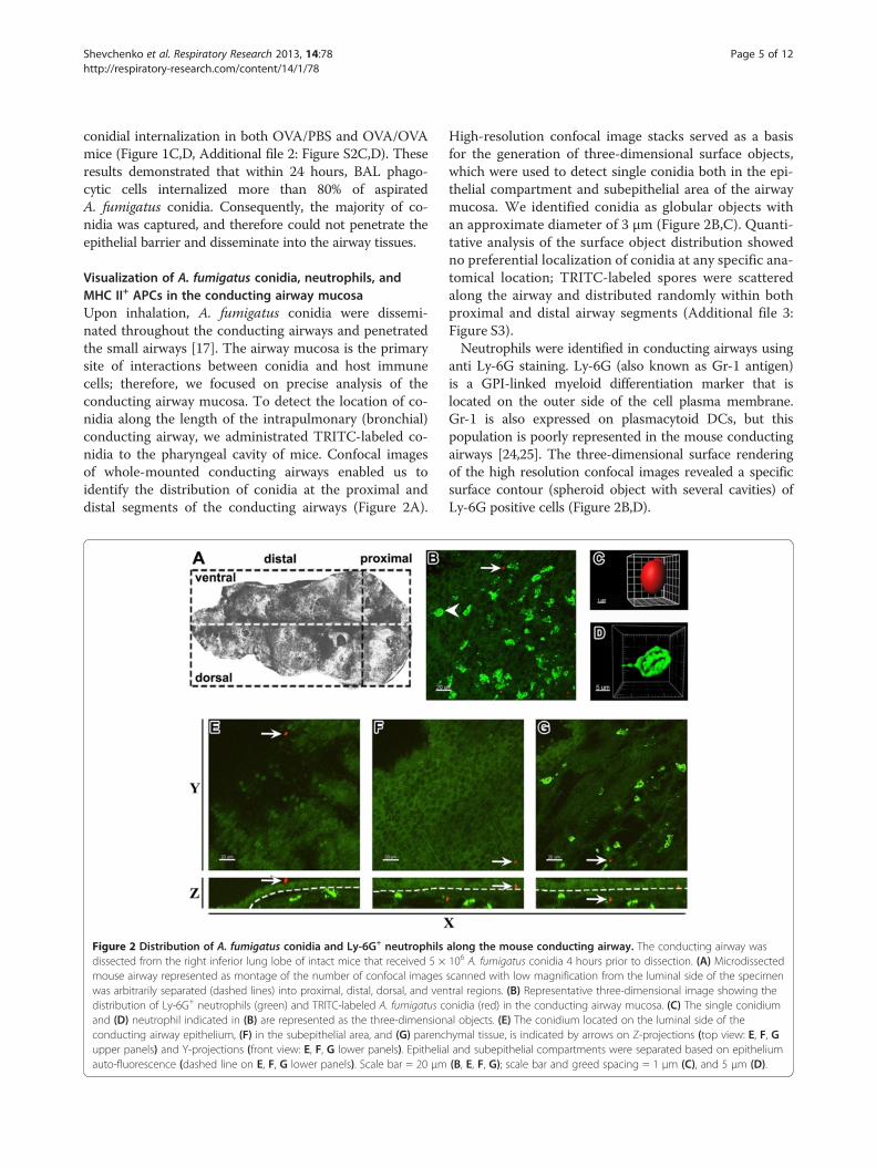
Visualization of A. fumigatus conidia, neutrophils, andMHC II+ APCs in the conducting airway mucosaUpon inhalation, A. fumigatus conidia were dissemi-nated throughout the conducting airways and penetratedthe small airways [17]. The airway mucosa is the primarysite of interactions between conidia and host immunecells; therefore, we focused on precise analysis of theconducting airway mucosa. To detect the location of co-nidia along the length of the intrapulmonary (bronchial)conducting airway, we administrated TRITC-labeled co-nidia to the pharyngeal cavity of mice. Confocal imagesof whole-mounted conducting airways enabled us toidentify the distribution of conidia at the proximal anddistal segments of the conducting airways (Figure 2A).
Figure 2 Distribution of A. fumigatus conidia and Ly-6G+ neutrophilsdissected from the right inferior lung lobe of intact mice that received 5 ×mouse airway represented as montage of the number of confocal imageswas arbitrarily separated (dashed lines) into proximal, distal, dorsal, and vendistribution of Ly-6G+ neutrophils (green) and TRITC-labeled A. fumigatus cand (D) neutrophil indicated in (B) are represented as the three-dimensionconducting airway epithelium, (F) in the subepithelial area, and (G) parencupper panels) and Y-projections (front view: E, F, G lower panels). Epitheliaauto-fluorescence (dashed line on E, F, G lower panels). Scale bar = 20 μm
High-resolution confocal image stacks served as a basisfor the generation of three-dimensional surface objects,which were used to detect single conidia both in the epi-thelial compartment and subepithelial area of the airwaymucosa. We identified conidia as globular objects withan approximate diameter of 3 μm (Figure 2B,C). Quanti-tative analysis of the surface object distribution showedno preferential localization of conidia at any specific ana-tomical location; TRITC-labeled spores were scatteredalong the airway and distributed randomly within bothproximal and distal airway segments (Additional file 3:Figure S3).Neutrophils were identified in conducting airways using
anti Ly-6G staining. Ly-6G (also known as Gr-1 antigen)is a GPI-linked myeloid differentiation marker that islocated on the outer side of the cell plasma membrane.Gr-1 is also expressed on plasmacytoid DCs, but thispopulation is poorly represented in the mouse conductingairways [24,25]. The three-dimensional surface renderingof the high resolution confocal images revealed a specificsurface contour (spheroid object with several cavities) ofLy-6G positive cells (Figure 2B,D).
along the mouse conducting airway. The conducting airway was106 A. fumigatus conidia 4 hours prior to dissection. (A) Microdissectedscanned with low magnification from the luminal side of the specimentral regions. (B) Representative three-dimensional image showing theonidia (red) in the conducting airway mucosa. (C) The single conidiumal objects. (E) The conidium located on the luminal side of thehymal tissue, is indicated by arrows on Z-projections (top view: E, F, Gl and subepithelial compartments were separated based on epithelium(B, E, F, G); scale bar and greed spacing = 1 μm (C), and 5 μm (D).
Shevchenko et al. Respiratory Research 2013, 14:78 Page 6 of 12http://respiratory-research.com/content/14/1/78
Conidia administrated to intact mice were detected notonly on the luminal side of the epithelium (Figure 2E,red), but also beneath the epithelium (Figure 2F, red), andeven in the lung tissue (Figure 2G, red). Neutrophils fromcontrol mice generally did not penetrate the epithelial bar-rier, and occupied the subepithelial compartment and par-enchymal tissue (Figure 2E-G, green).To identify conducting airway APCs, which can also take
part in internalization of conidia, we used anti-MHC IIstaining. Using the findings of Veres et al. [26], we identifiedtwo populations of conducting airway mucosal MHC II+
APCs, which were distinguished by the morphology andanatomical location: an epithelial (or intraepithelial) DC po-pulation, and a population containing subepithelial DCs(Additional file 4: Figure S4A,D,G and C,F,I: epithelialDCs, arrows; subepithelial DCs, arrowheads). Althoughseveral studies using flow cytometry of peripheral bloodand lymph node leukocyte samples have reported thatneutrophils are capable of expressing MHC class II [27],we did not identify expression of MHC II molecules onthe surface of Ly-6G+-conducting airway neutrophils byconfocal microscopy of the main bronchus specimen(Additional file 4: Figure S4B,E,H,C,F,I: MHC II, yellow;Ly6-G, green).
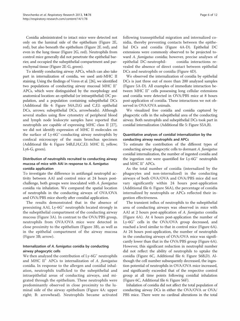
Distribution of neutrophils recruited to conducting airwaymucosa of mice with AAI in response to A. fumigatusconidia applicationTo investigate the difference in antifungal neutrophil ac-tivity between AAI and control mice at 24 hours post-challenge, both groups were inoculated with A. fumigatusconidia via inhalation. We compared the spatial locationof neutrophils in the conducting airways of OVA/OVAand OVA/PBS mice shortly after conidial application.The results demonstrated that in the absence of
preexisting AAI, Ly-6G+ neutrophils located strongly inthe subepithelial compartment of the conducting airwaymucosa (Figure 3A). In contrast to the OVA/PBS group,neutrophils from OVA/OVA mice were detected inclose proximity to the epithelium (Figure 3B), as well asin the epithelial compartment of the airway mucosa(Figure 3B; arrow).
Internalization of A. fumigatus conidia by conductingairway phagocytic cellsWe then analyzed the contribution of Ly-6G+ neutrophilsand MHC II+ APCs to internalization of A. fumigatusconidia. In response to the allergen and conidial inhal-ation, neutrophils trafficked to the subepithelial andintraepithelial areas of conducting airways, and mi-grated through the epithelium. These neutrophils werepredominantly observed in close proximity to the lu-minal side of the airway epithelium (Figure 4A: upperright; B: arrowhead). Neutrophils became activated
following transepithelial migration and internalized co-nidia, thereby preventing contacts between the epithe-lial DCs and conidia (Figure 4A-D). Epithelial DCextensions were commonly observed to be projected to-ward A. fumigatus conidia; however, precise analyses ofepithelial DC-neutrophil- conidia interactions re-vealed the absence of direct contact between epithelialDCs and neutrophils or conidia (Figure 4D).We observed the internalization of conidia by epithelial
DCs is just three out of more than 200 analyzed samples(Figure 5A-D). All examples of immediate interaction be-tween MHC II+ cells possessing long cellular extensionsand conidia were detected in OVA/PBS mice at 8 hourspost-application of conidia. These interactions we not ob-served in OVA/OVA animals.We visualized free conidia and conidia captured by
phagocytic cells in the subepithelial area of the conductingairway. Both neutrophils and subepithelial DCs took part inconidial internalization (Additional file 5: Figure S5A,B).
Quantitative analyses of conidial internalization by theconducting airway neutrophils and APCsTo estimate the contribution of the different types ofconducting airway phagocytic cells to dormant A. fumigatusconidial internalization, the number of ingested conidia andthe ingestion rate were quantified for Ly-6G+ neutrophilsand MHC II+ APCs.As the total number of conidia (internalized by the
phagocytes and non-internalized) in the conductingairways of both OVA/OVA and OVA/PBS mice did notvary significantly within 24 hours post-application(Additional file 6: Figure S6A), the percentage of conidiainternalized by neutrophils or APCs reflected their in-gestion effectiveness.The transient influx of neutrophils to the subepithelial
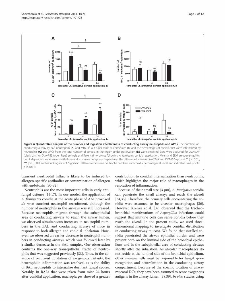
area of conducting airways was observed in mice withAAI at 2 hours post-application of A. fumigatus conidia(Figure 6A). At 4 hours post-application the number ofLy-6G+ cells in the OVA/OVA group decreased, andreached a level similar to that in control mice (Figure 6A).At 24 hours post-application, the number of neutrophilsin the conducting airways of OVA/OVA mice was signifi-cantly lower than that in the OVA/PBS group (Figure 6A).However, this significant reduction in neutrophil numberdid not reflect the ability of neutrophils to uptake theconidia (Figure 6C, Additional file 6: Figure S6B,D). Al-though the cell number subsequently decreased, the inges-tion potential of neutrophils in OVA/OVA mice increased,and significantly exceeded that of the respective controlgroup at all time points following conidial inhalation(Figure 6C, Additional file 6: Figure S6F).Inhalation of conidia did not affect the total population of
conducting airway DCs in either the OVA/OVA or OVA/PBS mice. There were no cardinal alterations in the total
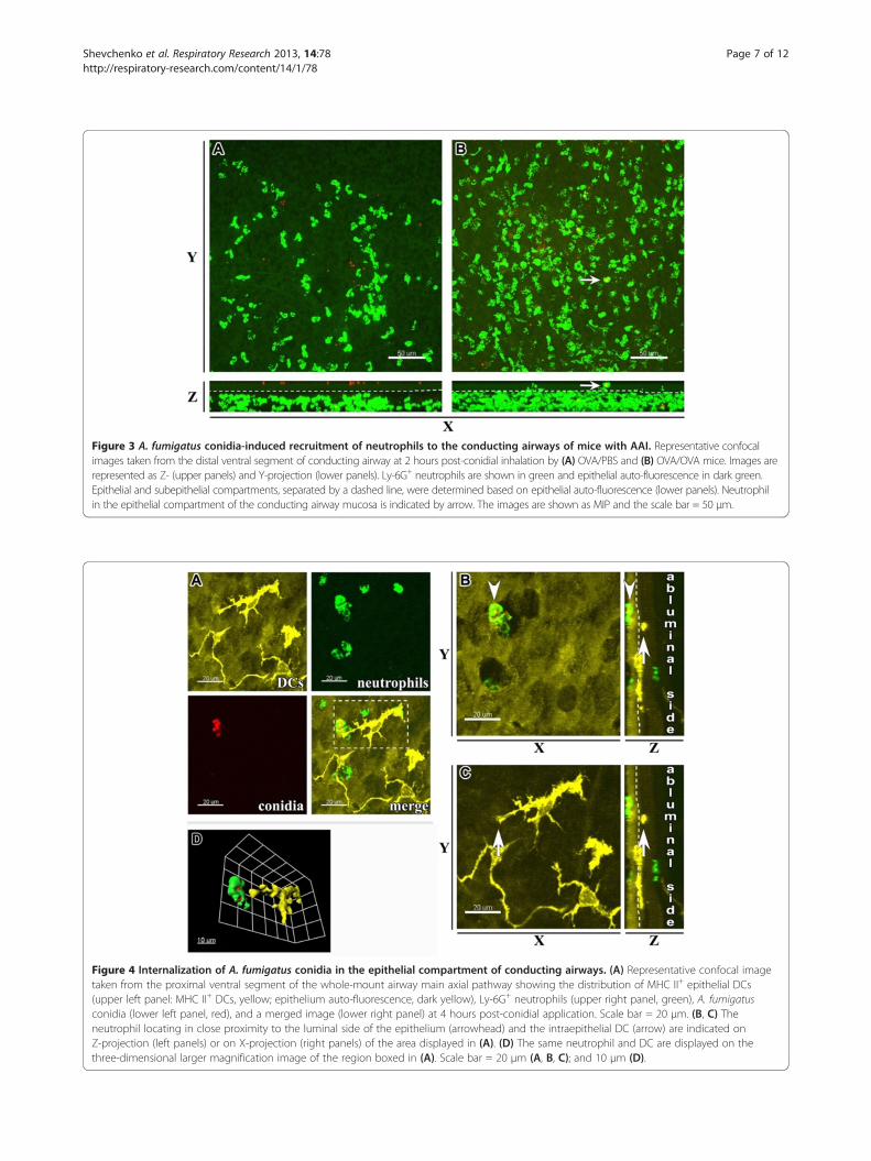
Figure 3 A. fumigatus conidia-induced recruitment of neutrophils to the conducting airways of mice with AAI. Representative confocalimages taken from the distal ventral segment of conducting airway at 2 hours post-conidial inhalation by (A) OVA/PBS and (B) OVA/OVA mice. Images arerepresented as Z- (upper panels) and Y-projection (lower panels). Ly-6G+ neutrophils are shown in green and epithelial auto-fluorescence in dark green.Epithelial and subepithelial compartments, separated by a dashed line, were determined based on epithelial auto-fluorescence (lower panels). Neutrophilin the epithelial compartment of the conducting airway mucosa is indicated by arrow. The images are shown as MIP and the scale bar = 50 μm.
Figure 4 Internalization of A. fumigatus conidia in the epithelial compartment of conducting airways. (A) Representative confocal imagetaken from the proximal ventral segment of the whole-mount airway main axial pathway showing the distribution of MHC II+ epithelial DCs(upper left panel: MHC II+ DCs, yellow; epithelium auto-fluorescence, dark yellow), Ly-6G+ neutrophils (upper right panel, green), A. fumigatusconidia (lower left panel, red), and a merged image (lower right panel) at 4 hours post-conidial application. Scale bar = 20 μm. (B, C) Theneutrophil locating in close proximity to the luminal side of the epithelium (arrowhead) and the intraepithelial DC (arrow) are indicated onZ-projection (left panels) or on X-projection (right panels) of the area displayed in (A). (D) The same neutrophil and DC are displayed on thethree-dimensional larger magnification image of the region boxed in (A). Scale bar = 20 μm (A, B, C); and 10 μm (D).
Shevchenko et al. Respiratory Research 2013, 14:78 Page 7 of 12http://respiratory-research.com/content/14/1/78
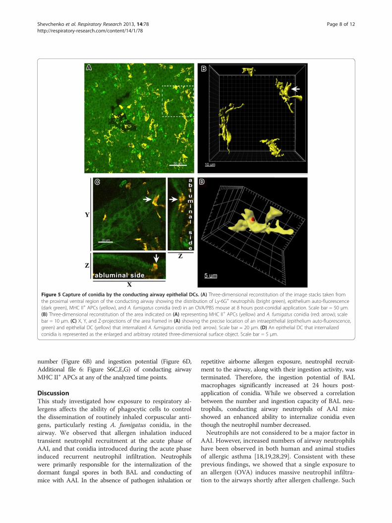
Figure 5 Capture of conidia by the conducting airway epithelial DCs. (A) Three-dimensional reconstitution of the image stacks taken fromthe proximal ventral region of the conducting airway showing the distribution of Ly-6G+ neutrophils (bright green), epithelium auto-fluorescence(dark green), MHC II+ APCs (yellow), and A. fumigatus conidia (red) in an OVA/PBS mouse at 8 hours post-conidial application. Scale bar = 50 μm.(B) Three-dimensional reconstitution of the area indicated on (A) representing MHC II+ APCs (yellow) and A. fumigatus conidia (red: arrow); scalebar = 10 μm. (C) X, Y, and Z-projections of the area framed in (A) showing the precise location of an intraepithelial (epithelium auto-fluorescence,green) and epithelial DC (yellow) that internalized A. fumigatus conidia (red: arrow). Scale bar = 20 μm. (D) An epithelial DC that internalizedconidia is represented as the enlarged and arbitrary rotated three-dimensional surface object. Scale bar = 5 μm.
Shevchenko et al. Respiratory Research 2013, 14:78 Page 8 of 12http://respiratory-research.com/content/14/1/78
number (Figure 6B) and ingestion potential (Figure 6D,Additional file 6: Figure S6C,E,G) of conducting airwayMHC II+ APCs at any of the analyzed time points.
DiscussionThis study investigated how exposure to respiratory al-lergens affects the ability of phagocytic cells to controlthe dissemination of routinely inhaled corpuscular anti-gens, particularly resting A. fumigatus conidia, in theairway. We observed that allergen inhalation inducedtransient neutrophil recruitment at the acute phase ofAAI, and that conidia introduced during the acute phaseinduced recurrent neutrophil infiltration. Neutrophilswere primarily responsible for the internalization of thedormant fungal spores in both BAL and conducting ofmice with AAI. In the absence of pathogen inhalation or
repetitive airborne allergen exposure, neutrophil recruit-ment to the airway, along with their ingestion activity, wasterminated. Therefore, the ingestion potential of BALmacrophages significantly increased at 24 hours post-application of conidia. While we observed a correlationbetween the number and ingestion capacity of BAL neu-trophils, conducting airway neutrophils of AAI miceshowed an enhanced ability to internalize conidia eventhough the neutrophil number decreased.Neutrophils are not considered to be a major factor in
AAI. However, increased numbers of airway neutrophilshave been observed in both human and animal studiesof allergic asthma [18,19,28,29]. Consistent with theseprevious findings, we showed that a single exposure toan allergen (OVA) induces massive neutrophil infiltra-tion to the airways shortly after allergen challenge. Such
2 4 240
500
1000
1500
2000 ***
ns
**
§
§
time after A. fumigatus conidia application, h
Num
ber
of
Ly-6
G+ c
ells
/mm
2 ep
ithe
lium
2 4 240
500
1000
1500
2000
ns
ns
ns
time after A. fumigatus conidia application, h
Num
ber
of
MH
C II
+ cel
ls/m
m2 e
pith
eliu
m
2 4 240
20
40
60
80
ns
**
**
§
time after A. fumigatus conidia application, h
% o
fco
nidi
ain
tern
aliz
ed b
yLy
-6G
+ cel
ls
2 4 240
20
40
60
80OVA/PBSOVA/OVA
ns
nsns
time after A. fumigatus conidia application, h
% o
f co
nid
iain
tern
aliz
ed b
y M
HC
II+ c
ells
A
DC
B
Figure 6 Quantitative analysis of the number and ingestion effectiveness of conducting airway neutrophils and APCs. The numbers ofconducting airway Ly-6G+ neutrophils (A) and MHC II+ APCs per mm2 of epithelium (B) and the percentages of conidia that were internalized byneutrophils (C) and APCs from the total number of conidia in the region under observation (D) were detected. Data were acquired for OVA/OVA(black bars) or OVA/PBS (open bars) animals at different time points following A. fumigatus conidial application. Mean and SEM are presented fortwo independent experiments with three and four mice per group, respectively. The difference between OVA/OVA and OVA/PBS groups: ** (p< 0.01),*** (p< 0.001), and ns: not significant. Significant difference between neutrophil numbers and conidia percentages at initial and indicated time points:§ (p<0.01).
Shevchenko et al. Respiratory Research 2013, 14:78 Page 9 of 12http://respiratory-research.com/content/14/1/78
transient neutrophil influx is likely to be induced byallergen-specific antibodies or contamination of allergenwith endotoxin [30-32].Neutrophils are the most important cells in early anti-
fungal defense [14,17]. In our model, the application ofA. fumigatus conidia at the acute phase of AAI provokedde novo transient neutrophil recruitment, although thenumber of neutrophils in the airways was still increased.Because neutrophils migrate through the subepithelialarea of conducting airways to reach the airway lumen,we observed simultaneous increases in neutrophil num-bers in the BAL and conducting airways of mice inresponse to both allergen and conidial inhalation. How-ever, we observed an earlier decrease in neutrophil num-bers in conducting airways, which was followed later bya similar decrease in the BAL samples. Our observationconfirms the one-way transepithelial traffic of neutro-phils that was suggested previously [33]. Thus, in the ab-sence of recurrent inhalation of exogenous irritants, theneutrophilic inflammation was resolved, as is the abilityof BAL neutrophils to internalize dormant fungal spores.Notably, in BALs that were taken from mice 24 hoursafter conidial application, macrophages showed a greater
contribution to conidial internalization than neutrophils,which highlights the major role of macrophages in theresolution of inflammation.Because of their small size (3 μm), A. fumigatus conidia
can penetrate the small airways and reach the alveoli[34,35]. Therefore, the primary cells encountering the co-nidia were assumed to be alveolar macrophages [36].However, Krenke et al. [37] observed that the tracheo-bronchial manifestations of Aspergillus infections couldsuggest that immune cells can sense conidia before theyreach the alveoli. In the present study, we used three-dimensional mapping to investigate conidial distributionin conducting airway mucosa. We found that instilled co-nidia penetrated the airway epithelial border, and werepresent both on the luminal side of the bronchial epithe-lium and in the subepithelial area of conducting airwaysshortly after the inhalation. As alveolar macrophages donot reside at the luminal side of the bronchial epithelium,other immune cells must be responsible for fungal sporerecognition and neutralization in the conducting airwaycompartment. Because of the specific location of airwaymucosal DCs, they have been assumed to sense exogenousantigens in the airway lumen [38,39]. In vivo studies using
Shevchenko et al. Respiratory Research 2013, 14:78 Page 10 of 12http://respiratory-research.com/content/14/1/78
triple cell co-cultures or silicone matrixes have shown thatmonocyte-derived DCs can internalize corpuscular anti-gens of 1 μm or larger [40,41]. However, ex vivo results ofboth the present study and the investigations of Vereset al. [26] provide evidence that conducting airway epithe-lial DCs sense corpuscular antigens (E. coli particles, poly-styrene beads, or A. fumigatus conidia) without immediatecontact. Our data revealed that conducting airway epithe-lial DCs can occasionally take up conidia, but not in micethat exhibited AAI. This finding may result from anenhanced neutrophil ingestion activity that was observedin conducting airways of challenged OVA mice.We demonstrated that allergen challenge activated the
transepithelial migration, followed by the massive neu-trophil recruitment to the airways. Subsequently, onthe luminal side of the conducting airway epithelium,neutrophils captured conidia and thereby preventedepithelial DC-conidia contacts. Although epithelial DC-conidial interactions in conducting airways rarely occur,the phenomenon should be investigated further.In contrast to the epithelial DCs, spherical MHC II+
APCs captured conidia in the subepithelial space. Never-theless, these APCs had a lower contribution than neutro-phils in conidial internalization in both AAI and controlmice. Thus, our data indicated that along with alveolarmacrophages, conducting airway neutrophils are the pri-mary cells to encounter aspirated A. fumigatus conidia.In addition, while more than 80% of A. fumigatus co-
nidia were internalized by BAL phagocytes only about50% of spores were internalized by the joint efforts ofconducting airway neutrophils and APCs 24 hours afterthe application of conidia to both OVA/OVA and OVA/PBS mice. The fungal spores that remained unbound bythe main phagocytes can hide in the airway tissue untilfavorable for germination condition and therefore theycan be considered as a life-threating factor.To summarize, we demonstrated that application of
conidia at the acute phase of AAI induces a massiveneutrophil influx to conducting airways, bringing themigrated cells into close proximity with the conductingairway epithelium, followed by transmigration to the lu-minal side. The more than two-fold increase in neutro-phil number indicated an intensification of the processof neutrophil-conidial interaction and spore internaliza-tion. Surprisingly, when the number of neutrophils inthe conducting airways of mice with AAI reduced, theeffectiveness of ingestion by these neutrophils remainedsignificantly higher than in control mice. In addition, theincreased ability to internalize corpuscular antigens wascaused by the activated state rather than by the numberof conducting airway neutrophils. In vitro studies haveshown that internalization of fungal spores is crucial forconidia killing [17]. On the other hand, internalizationof resting conidia and subsequent degradation of the
hydrophobic surface layer in phagolysosomes enablestheir recognition by the intracellular pattern recognitionreceptors and initiates inflammatory response [9,42].Hence, the allergen-induced enhancement of the capacityof conduction airway neutrophils (that we observed inmice with AAI but not in control animals) may indicate abreak in tolerance to resting fungal spores that is normallyexhibited by immunocompetent hosts [8]. Epidemiologicstudies have associated sensitivity to Aspergillus fungiwith the severe persistent asthma in adults [2]. Thus, theallergen-induced skewing from tolerance to sensitizationcan partially explain the potential of inhaled fungal sporesto exacerbate asthma symptoms.
ConclusionsIn conclusion, our data show that at the acute phase ofAAI, neutrophils are sufficient to control resting fungalspore dissemination. We demonstrated that the ingestionpotential of neutrophils strongly depends on both the timeand the location of the cell-pathogen interaction. Our datasuggest that allergen-induced enhancement of the ingestionpotential of conduction airway neutrophils may be a reasonfor the susceptibility of asthmatic patients to Aspergillusinfection.
Additional files
Additional file 1: Figure S1. Whole-mount immunostaining isotypecontrol. Whole-mount conducting airways were immunostained withFITC-conjugated (A) monoclonal rat anti-mouse Ly-6G antibody toidentify neutrophils or (B) rat IgG2b Isotype Control. To visualize MHC II+
APCs the other specimens were immunostained with primary (C) ratanti-mouse I-A/I-E antibody or (D) purified rat IgG2b followed byCy5-conjugated donkey anti-rat IgG. Representative three-dimensionalimages showing epithelium auto-fluorescence (A, B, dark green; C, D,dark yellow), neutrophils (A, green), and APCs (C, yellow) were obtainedas Z-stacks (30 optical slices × 50 μm) scanned from the luminal side ofthe airway. Scale bar = 20 μm.
Additional file 2: Figure S2. Contribution of BAL macrophages andneutrophils to Internalization of A. fumigatus conidia. (A) Percentages ofmacrophages (open bars), and neutrophils (black bars) in BALs at theindicated time points following conidial application to OVA/PBS and (B)OVA/OVA mice. (C) Percentages of conidia that were internalized bymacrophages (open bars), and neutrophils (black bars) at 2, 4, and 24hours following conidial application to OVA/PBS and (D) OVA/OVA mice.Data are shown as means ± SEM for two representative experiments,with three and five mice per group. Significant difference betweenneutrophil and conidia percentages at the time point 0 and indicatedtime point: § (p<0.01).
Additional file 3: Figure S3. Quantitative assay of A. fumigatus conidialdistribution in the proximal and distal regions of conducting airways. Thenumbers of conidia were quantified for the proximal (open bars) anddistal (black bars) regions of conducting airways of OVA/PBS mice at 2, 4,and 24 hours post-conidial-application. Mean and SEM are shown for tworepresentative experiments with three and four mice per group.Statistical analyses revealed non-significant differences in conidia numberbetween the airway segments at all analyzed time points.
Additional file 4: Figure S4. Visualization of conducting airway MHC II+
APCs. Representative three-dimensional images of the proximal dorsalsegment of the whole-mount conducting airway of an OVA/OVA mouse atacute stage of AAI. (A, D, G) MHC II+ APCs (yellow), (B, E, H) Ly-6G+
Shevchenko et al. Respiratory Research 2013, 14:78 Page 11 of 12http://respiratory-research.com/content/14/1/78
neutrophils (green) were visualized. (C), (F), and (I) Merged representationsof the images shown in (A, B), (D, E), and (G, H), respectively. ConfocalZ-stacks are represented as optical sections showing DCs and neutrophils inthe (A, B, C) epithelial layer, (D, E, F) in close proximity to the epithelium,and (C, H, I) in the subepithelial area. (A, D, G, C, F, I) Structural cellauto-fluorescence is shown in dark yellow and (B, E, H) dark green. Epithelialand subepithelial DCs are indicated by arrows and arrowheads, respectively.Scale bar = 50 μm.
Additional file 5: Figure S5. Internalization of A. fumigatus conidia inthe subepithelial area of conducting airways by subepithelial DCs.Representative images of the proximal ventral segment of a whole-mountconducting airway excised from an OVA/PBS mouse at 4 hourspost-conidial-application. The pictures are represented as X- (left panels) andZ-projections (right panels) showing interaction of (A) DCs (yellow) andconidia (red), or (B) neutrophil (green) and conidia (red). Conidium incontact to subepithelial DC is indicated by arrowhead; conidia insideneutrophil are indicated by arrow. Epithelial and subepithelialcompartments were separated based on epithelium auto-fluorescence(dashed line). Scale bar = 20 μm.
Additional file 6: Figure S6. Quantitative analysis of the number ofA. fumigatus conidia, ingestion rate and ingestion capacity of neutrophilsand APCs in conducting airways. The numbers of A. fumigatus conidiaper 1 mm2 of conducting airway epithelium (A); the number ofconducting airway Ly-6G+ neutrophils (B) and MHC II+ APCs (C) thatinternalized A. fumigatus conidia as well as the number of conidia thatwere internalized by Ly-6G+ neutrophils (F) and MHC II+ APCs (G) werequantified. The percentage of Ly-6G+ neutrophils (D) and MHC II+ APCs(E) that internalized conidia from the total number of the neutrophils andAPCs respectively were calculated. Data were acquired for OVA/OVA(black bars) or OVA/PBS (open bars) animals at different time pointsfollowing A. fumigatus conidial application. Mean and SEM are presentedfor two independent experiments with three and four mice per group,respectively. Significant difference between OVA/OVA and OVA/PBSgroups: * (p<0.05) and ** (p< 0.01), *** (p< 0.001), and ns: not significant.
AbbreviationsAAI: Allergic airway inflammation; AMM: Aspergillus minimal medium;APC: Antigen presenting cell; BAL: Bronchoalveolar lavage; Cy5: Cyanine 5;DC: Dendritic cell; FITC: Fluorescein isothiocyanate;GPI: Glycosylphosphatidylinisotol; i.p: Intraperitoneal; MHC: Majorhistocompatibility complex; MIP: Maximum intensity projection; OVA: Chickenovalbumin; OVA/OVA mice: A group of mice that received intraperitonealinjection of ovalbumin and subsequent aerosol challenges with ovalbumin;OVA/PBS mice: A group of mice that received intraperitoneal injection ofovalbumin and subsequent aerosol challenge with PBS; PBS: Phosphatebuffered saline; TRITC: Tetramethylrhodamine-(5-(and-6))-isothiocyanate.
Competing interestsThe authors declare that they have no competing interests.
Authors’ contributionsMAS designed the study, performed the experiments, analyzed the data, anddrafted the manuscript. ELB and EAS performed the experiments andanalyzed the data. AMS drafted the manuscript. All authors read andapproved the final manuscript.
AcknowledgementsThis work was partially supported by Russian Foundation for Basic Researchproject No. 11-04-01954a, and by Molecular and Cellular Biology Program ofRussian Academy of Sciences in. We also acknowledge the support of theEuropean Respiratory Society, Fellowship number 620. The authors thankPhilipp S. Schmalhorst for providing the Aspergillus fumigatus conidia clinicalisolate D141. This work was partially carried out at the Department of AirwayImmunology, Fraunhofer Institute for Toxicology and Experimental Medicine,Hannover, Germany, and we thank Drs Armin Braun and Tibor Z. Veres fortheir insightful comments, and Emma Spies for technical assistance.
Author details1Department of Immunology, Laboratory of Cell Interactions,Shemyakin-Ovchinnikov Institute of Bioorganic Chemistry, Russian Academy
of Sciences, Miklukho-Maklaya St. 16/10, 117997 Moscow, Russia. 2Faculty ofPostgraduate Education, Pirogov Russian National Research MedicalUniversity, Ministry of Health, Ostrovitianov St. 1, 117997 Moscow, Russia.
Received: 14 March 2013 Accepted: 23 July 2013Published: 27 July 2013
References1. Latge JP: The pathobiology of Aspergillus fumigatus. Trends Microbiol
2001, 9(8):382–389.2. Knutsen AP, Bush RK, Demain JG, Denning DW, Dixit A, Fairs A,
Greenberger PA, Kariuki B, Kita H, Kurup VP, et al: Fungi and allergiclower respiratory tract diseases. J Allergy Clin Immunol 2012,129(2):280–291. quiz 292–283.
3. Porter PC, Roberts L, Fields A, Knight M, Qian Y, Delclos GL, Han S,Kheradmand F, Corry DB: Necessary and sufficient role for T helper cellsto prevent fungal dissemination in allergic lung disease. Infect Immun2011, 79(11):4459–4471.
4. Fukushima C, Matsuse H, Fukahori S, Tsuchida T, Kawano T, Senjyu H, KohnoS: Aspergillus fumigatus synergistically enhances mite-induced allergicairway inflammation. Med Sci Monit 2010, 16(7):BR197–202.
5. Porter P, Susarla SC, Polikepahad S, Qian Y, Hampton J, Kiss A, Vaidya S, SurS, Ongeri V, Yang T, et al: Link between allergic asthma and airwaymucosal infection suggested by proteinase-secreting household fungi.Mucosal Immunol 2009, 2(6):504–517.
6. Palm NW, Rosenstein RK, Medzhitov R: Allergic host defences. Nature 2012,484(7395):465–472.
7. Agarwal R, Aggarwal AN, Gupta D, Jindal SK: Aspergillus hypersensitivityand allergic bronchopulmonary aspergillosis in patients with bronchialasthma: systematic review and meta-analysis. Int J Tuberc Lung Dis 2009,13(8):936–944.
8. Aimanianda V, Bayry J, Bozza S, Kniemeyer O, Perruccio K, Elluru SR,Clavaud C, Paris S, Brakhage AA, Kaveri SV, et al: Surface hydrophobinprevents immune recognition of airborne fungal spores. Nature 2009,460(7259):1117–1121.
9. Luther K, Torosantucci A, Brakhage AA, Heesemann J, Ebel F: Phagocytosisof Aspergillus fumigatus conidia by murine macrophages involvesrecognition by the dectin-1 beta-glucan receptor and Toll-like receptor2. Cell Microbiol 2007, 9(2):368–381.
10. Bayry J, Aimanianda V, Guijarro JI, Sunde M, Latge JP: Hydrophobins-unique fungal proteins. PLoS Pathog 2012, 8(5):e1002700.
11. Fukahori S, Matsuse H, Tsuchida T, Kawano T, Tomari S, Fukushima C,Kohno S: Aspergillus fumigatus regulates mite allergen-pulseddendritic cells in the development of asthma. Clin Exp Allergy 2010,40(10):1507–1515.
12. Rivera A, Van Epps HL, Hohl TM, Rizzuto G, Pamer EG: Distinct CD4+−T-cellresponses to live and heat-inactivated Aspergillus fumigatus conidia.Infect Immun 2005, 73(11):7170–7179.
13. Dougherty RH, Fahy JV: Acute exacerbations of asthma: epidemiology,biology and the exacerbation-prone phenotype. Clin Exp Allergy 2009,39(2):193–202.
14. Mircescu MM, Lipuma L, van Rooijen N, Pamer EG, Hohl TM: Essential role forneutrophils but not alveolar macrophages at early time points followingAspergillus fumigatus infection. J Infect Dis 2009, 200(4):647–656.
15. Hasenberg M, Behnsen J, Krappmann S, Brakhage A, Gunzer M: Phagocyteresponses towards Aspergillus fumigatus. Int J Med Microbiol 2011,301(5):436–444.
16. Bonnett CR, Cornish EJ, Harmsen AG, Burritt JB: Early neutrophilrecruitment and aggregation in the murine lung inhibitgermination of Aspergillus fumigatus Conidia. Infect Immun 2006,74(12):6528–6539.
17. Bruns S, Kniemeyer O, Hasenberg M, Aimanianda V, Nietzsche S, ThywissenA, Jeron A, Latge JP, Brakhage AA, Gunzer M: Production of extracellulartraps against Aspergillus fumigatus in vitro and in infected lung tissue isdependent on invading neutrophils and influenced by hydrophobinRodA. PLoS Pathog 2010, 6(4):e1000873.
18. Lommatzsch M, Julius P, Kuepper M, Garn H, Bratke K, Irmscher S, LuttmannW, Renz H, Braun A, Virchow JC: The course of allergen-induced leukocyteinfiltration in human and experimental asthma. J Allergy Clin Immunol2006, 118(1):91–97.
Shevchenko et al. Respiratory Research 2013, 14:78 Page 12 of 12http://respiratory-research.com/content/14/1/78
19. Page K, Lierl KM, Hughes VS, Zhou P, Ledford JR, Wills-Karp M: TLR2-mediated activation of neutrophils in response to German cockroachfrass. J Immunol 2008, 180(9):6317–6324.
20. Baines KJ, Simpson JL, Scott RJ, Gibson PG: Immune responses of airwayneutrophils are impaired in asthma. Exp Lung Res 2009, 35(7):554–569.
21. Reichard U, Buttner S, Eiffert H, Staib F, Ruchel R: Purification andcharacterisation of an extracellular serine proteinase from Aspergillusfumigatus and its detection in tissue. J Med Microbiol 1990,33(4):243–251.
22. Rao GV, Tinkle S, Weissman DN, Antonini JM, Kashon ML, Salmen R, BattelliLA, Willard PA, Hoover MD, Hubbs AF: Efficacy of a technique for exposingthe mouse lung to particles aspirated from the pharynx. J Toxicol EnvironHealth A 2003, 66(15):1441–1452.
23. Veres TZ, Shevchenko M, Krasteva G, Spies E, Prenzler F, Rochlitzer S,Tschernig T, Krug N, Kummer W, Braun A: Dendritic cell-nerve clusters aresites of T cell proliferation in allergic airway inflammation. Am J Pathol2009, 174(3):808–817.
24. von Garnier C, Wikstrom ME, Zosky G, Turner DJ, Sly PD, Smith M, ThomasJA, Judd SR, Strickland DH, Holt PG, et al: Allergic airways disease developsafter an increase in allergen capture and processing in the airwaymucosa. J Immunol 2007, 179(9):5748–5759.
25. Veres TZ, Voedisch S, Spies E, Valtonen J, Prenzler F, Braun A: AeroallergenChallenge Promotes Dendritic Cell Proliferation in the Airways. J Immunol2013, 190(3):897–903.
26. Veres TZ, Voedisch S, Spies E, Tschernig T, Braun A: Spatiotemporal andfunctional behavior of airway dendritic cells visualized by two-photonmicroscopy. Am J Pathol 2011, 179(2):603–609.
27. Gresnigt MS, Joosten LA, Verschueren I, van der Meer JW, Netea MG,Dinarello CA, van de Veerdonk FL: Neutrophil-mediated inhibition ofproinflammatory cytokine responses. J Immunol 2012,189(10):4806–4815.
28. Bhakta NR, Woodruff PG: Human asthma phenotypes: from the clinic, tocytokines, and back again. Immunol Rev 2011, 242(1):220–232.
29. Herz U, Braun A, Ruckert R, Renz H: Various immunological phenotypesare associated with increased airway responsiveness. Clin Exp Allergy1998, 28(5):625–634.
30. Taube C, Dakhama A, Rha YH, Takeda K, Joetham A, Park JW, Balhorn A,Takai T, Poch KR, Nick JA, et al: Transient neutrophil infiltration afterallergen challenge is dependent on specific antibodies and Fc gamma IIIreceptors. J Immunol 2003, 170(8):4301–4309.
31. Delayre-Orthez C, de Blay F, Frossard N, Pons F: Dose-dependent effects ofendotoxins on allergen sensitization and challenge in the mouse.Clin Exp Allergy 2004, 34(11):1789–1795.
32. Peters M, Dudziak K, Stiehm M, Bufe A: T-cell polarization depends onconcentration of the danger signal used to activate dendritic cells.Immunol Cell Biol 2010, 88(5):537–544.
33. Zemans RL, Colgan SP, Downey GP: Transepithelial migration ofneutrophils: mechanisms and implications for acute lung injury.Am J Respir Cell Mol Biol 2009, 40(5):519–535.
34. Latge JP: Aspergillus fumigatus and aspergillosis. Clin Microbiol Rev 1999,12(2):310–350.
35. Bozza S, Gaziano R, Spreca A, Bacci A, Montagnoli C, di Francesco P, RomaniL: Dendritic cells transport conidia and hyphae of Aspergillus fumigatusfrom the airways to the draining lymph nodes and initiate disparate Thresponses to the fungus. J Immunol 2002, 168(3):1362–1371.
36. Ibrahim-Granet O, Philippe B, Boleti H, Boisvieux-Ulrich E, Grenet D,Stern M, Latge JP: Phagocytosis and intracellular fate of Aspergillusfumigatus conidia in alveolar macrophages. Infect Immun 2003,71(2):891–903.
37. Krenke R, Grabczak EM: Tracheobronchial manifestations of Aspergillusinfections. ScientificWorldJournal 2011, 11:2310–2329.
38. Lambrecht BN, Hammad H: Lung dendritic cells in respiratory viralinfection and asthma: from protection to immunopathology. Annu RevImmunol 2012, 30:243–270.
39. Stumbles PA, Upham JW, Holt PG: Airway dendritic cells: co-ordinators ofimmunological homeostasis and immunity in the respiratory tract.APMIS 2003, 111(7–8):741–755.
40. Blank F, Rothen-Rutishauser B, Gehr P: Dendritic cells and macrophagesform a transepithelial network against foreign particulate antigens.Am J Respir Cell Mol Biol 2007, 36(6):669–677.
41. Behnsen J, Narang P, Hasenberg M, Gunzer F, Bilitewski U, Klippel N, RohdeM, Brock M, Brakhage AA, Gunzer M: Environmental dimensionalitycontrols the interaction of phagocytes with the pathogenic fungiAspergillus fumigatus and Candida albicans. PLoS Pathog 2007, 3(2):e13.
42. Faro-Trindade I, Willment JA, Kerrigan AM, Redelinghuys P, Hadebe S, ReidDM, Srinivasan N, Wainwright H, Lang DM, Steele C, et al: Characterisationof innate fungal recognition in the lung. PLoS One 2012, 7(4):e35675.
doi:10.1186/1465-9921-14-78Cite this article as: Shevchenko et al.: Elimination of Aspergillusfumigatus conidia from the airways of mice with allergic airwayinflammation. Respiratory Research 2013 14:78.
Submit your next manuscript to BioMed Centraland take full advantage of:
• Convenient online submission
• Thorough peer review
• No space constraints or color figure charges
• Immediate publication on acceptance
• Inclusion in PubMed, CAS, Scopus and Google Scholar
• Research which is freely available for redistribution
Submit your manuscript at www.biomedcentral.com/submit