assembly, organization, and function of the copii coat · a multi-subunit complex, the copii coat....
TRANSCRIPT

Histochem Cell Biol (2008) 129:129–151
DOI 10.1007/s00418-007-0363-xREVIEW
Assembly, organization, and function of the COPII coat
Helen Hughes · David J. Stephens
Accepted: 20 November 2007 / Published online: 4 December 2007© Springer-Verlag 2007
Abstract A full mechanistic understanding of how secre-tory cargo proteins are exported from the endoplasmicreticulum for passage through the early secretory pathwayis essential for us to comprehend how cells are organized,maintain compartment identity, as well as how they selec-tively secrete proteins and other macromolecules to theextracellular space. This process depends on the function ofa multi-subunit complex, the COPII coat. Here we describeprogress towards a full mechanistic understanding ofCOPII coat function, including the latest Wndings in thisarea. Much of our understanding of how COPII functionsand is regulated comes from studies of yeast genetics, bio-chemical reconstitution and single cell microscopy. Newdevelopments arising from clinical cases and model organ-ism biology and genetics enable us to gain far greaterinsight in to the role of membrane traYc in the context of awhole organism as well as during embryogenesis anddevelopment. A signiWcant outcome of such a full under-standing is to reveal how the machinery and processes ofmembrane traYcking through the early secretory pathwayfail in disease states.
Introduction
The early secretory pathway is made up of the endoplasmicreticulum (ER), Golgi apparatus and intermediate compart-ments between these two major stations (including transportvesicles and tubules) and the ER-Golgi intermediate
compartment itself (Palade 1975). These intracellular com-partments are in constant communication, with moleculesbeing passed from one donor compartment to a target com-partment via discrete vesicles or convoluted networks oftubules and vesicles (Bannykh and Balch 1997; Martinez-Menarguez et al. 1999). Evidence also exists for direct con-nections between compartments (e.g. Stinchcombe et al.1995) that would signiWcantly complicate sorting and segre-gation processes. Membrane traYcking must be a highlyorganized process with each outward movement counterbal-anced by a retrieval step whereby membrane and selectedproteins are returned to their original compartment of origin(Bonifacino and Glick 2004; Salama and Schekman 1995).Each transport vesicle must be highly selective for the pro-teins that make up vesicle cargo, and for the fusion proteinsspecifying the target membrane, so that traYcking withinthe cell is kept well-organized and eYcient (Bonifacino andGlick 2004; Salama and Schekman 1995). Here, we try toprovide a perspective on the formation of transport vesiclesfrom the ER. For further mechanistic detail, including subtlediVerences between the animal systems primarily describedhere and those in plants (Hanton et al. 2005), the reader isreferred to other excellent recent reviews in this area(Gurkan et al. 2006; Kirk and Ward 2007; Lee and Miller2007; Mancias and Goldberg 2005).
ER exit sites
The export of proteins from the ER has been well deWned inyeast (both Saccharomyces cerevisiae (Schekman and Nov-ick 2004) and Pichia pastoris (Rossanese et al. 1999)) andmammalian cells (Bannykh and Balch 1997). In S. cerevi-siae, budding appears to occur stochastically across theentire ER membrane. In P. pastoris and metazoans, the
H. Hughes · D. J. Stephens (&)Cell Biology Laboratories, Department of Biochemistry, University of Bristol, School of Medical Sciences, University Walk, Bristol BS8 1TD, UKe-mail: [email protected]
123
130 Histochem Cell Biol (2008) 129:129–151
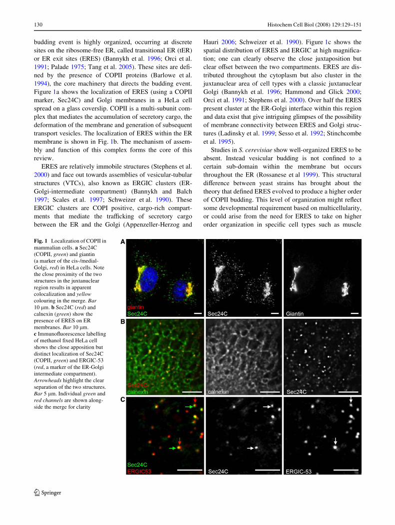
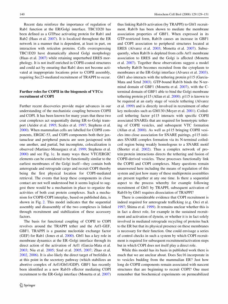
budding event is highly organized, occurring at discretesites on the ribosome-free ER, called transitional ER (tER)or ER exit sites (ERES) (Bannykh et al. 1996; Orci et al.1991; Palade 1975; Tang et al. 2005). These sites are deW-ned by the presence of COPII proteins (Barlowe et al.1994), the core machinery that directs the budding event.Figure 1a shows the localization of ERES (using a COPIImarker, Sec24C) and Golgi membranes in a HeLa cellspread on a glass coverslip. COPII is a multi-subunit com-plex that mediates the accumulation of secretory cargo, thedeformation of the membrane and generation of subsequenttransport vesicles. The localization of ERES within the ERmembrane is shown in Fig. 1b. The mechanism of assem-bly and function of this complex forms the core of thisreview.
ERES are relatively immobile structures (Stephens et al.2000) and face out towards assemblies of vesicular-tubularstructures (VTCs), also known as ERGIC clusters (ER-Golgi-intermediate compartment) (Bannykh and Balch1997; Scales et al. 1997; Schweizer et al. 1990). TheseERGIC clusters are COPI positive, cargo-rich compart-ments that mediate the traYcking of secretory cargobetween the ER and the Golgi (Appenzeller-Herzog and
Hauri 2006; Schweizer et al. 1990). Figure 1c shows thespatial distribution of ERES and ERGIC at high magniWca-tion; one can clearly observe the close juxtaposition butclear oVset between the two compartments. ERES are dis-tributed throughout the cytoplasm but also cluster in thejuxtanuclear area of cell types with a classic juxtanuclearGolgi (Bannykh et al. 1996; Hammond and Glick 2000;Orci et al. 1991; Stephens et al. 2000). Over half the ERESpresent cluster at the ER-Golgi interface within this regionand data exist that give intriguing glimpses of the possibilityof membrane connectivity between ERES and Golgi struc-tures (Ladinsky et al. 1999; Sesso et al. 1992; Stinchcombeet al. 1995).
Studies in S. cerevisiae show well-organized ERES to beabsent. Instead vesicular budding is not conWned to acertain sub-domain within the membrane but occursthroughout the ER (Rossanese et al 1999). This structuraldiVerence between yeast strains has brought about thetheory that deWned ERES evolved to produce a higher orderof COPII budding. This level of organization might reXectsome developmental requirement based on multicellularity,or could arise from the need for ERES to take on higherorder organization in speciWc cell types such as muscle
Fig. 1 Localization of COPII in mammalian cells. a Sec24C (COPII, green) and giantin (a marker of the cis-/medial-Golgi, red) in HeLa cells. Note the close proximity of the two structures in the juxtanuclear region results in apparent colocalization and yellow colouring in the merge. Bar 10 �m. b Sec24C (red) and calnexin (green) show the presence of ERES on ER membranes. Bar 10 �m. c ImmunoXuorescence labelling of methanol Wxed HeLa cell shows the close apposition but distinct localization of Sec24C (COPII, green) and ERGIC-53 (red, a marker of the ER-Golgi intermediate compartment). Arrowheads highlight the clear separation of the two structures. Bar 5 �m. Individual green and red channels are shown along-side the merge for clarity
123

Histochem Cell Biol (2008) 129:129–151 131
cells (Ralston et al. 2001) and neurons (Horton and Ehlers2004). COPII coated ERES are able to undergo Wssion andfusion events that appear to control ERES size (Stephens2003), as well as being able to form de novo (Bevis et al.2002; Stephens 2003). The core COPII components areperipheral membrane proteins; their constant cycling onand oV membranes (Stephens et al. 2000) makes such con-trol possible. Further examination of the mechanism under-lying ERES organization requires a discussion of thecomponents of the COPII coat. Here, we attempt to providesome historical background to our understanding of COPIIassembly and function, but from the start we attempt toincorporate the latest Wndings in this area.
An overview of the COPII coat
COPII complex components undergo several rounds ofexport from the ER. COPII recruitment is initiated by theactivation of the small GTPase Sar1 (Nakano et al. 1989)by its ER-localized GEF Sec12 (Nakano et al. 1988). Uponexchange of GDP for GTP, Sar1 exposes an N-terminalamphipathic tail, which inserts into the lipid bilayer (Bielliet al. 2005; Lee et al. 2005). Once tightly associated withthe membrane, Sar1 recruits a heterodimeric complex com-prising of Sec23/24 (Barlowe et al. 1994; Hicke and Schek-man 1989; Hicke et al. 1992; Kaiser and Schekman 1990).Sec23 acts as a GAP for Sar1 (Yoshihisa et al. 1993)increasing the very low rate of hydrolysis by Sar1. Sec24 isrequired for cargo binding, and together with cargo, Sec23,and Sar1 forms the prebudding complex (Aridor et al.1998). COPII budding can be reproduced in vitro usingsynthetic liposomes with which the Wve core COPII compo-nents alone (Sar1, Sec23/24 and Sec13/31) are suYcient todeform the membrane and generated coated vesicles (Mats-uoka et al. 1998). Inner coat subunits Sec23 and Sec24 arestructurally similar and form a “bow-tie” shape (Lederkr-emer et al. 2001). The presence of basic residues on theconcave inner side of the heterodimer potentially enhancesmembrane association (Bi et al. 2002). Subsequent recruit-ment of the heterotetramer Sec13/31, consisting of twoSec13 and two Sec31 subunits, permits minimal cage for-mation acting as a structural scaVold for the outer layer ofthe COPII coat (Fath et al. 2007; Stagg et al. 2006).
In mammals, multiple isoforms exist of nearly all COPIIsubunits, each encoded by a diVerent gene. These are gen-erally denoted with an alphabetical suYx and we use thisnomenclature here. Database searching reveals two iso-forms of Sar1 (Sar1A and Sar1B), two Sec16 isoforms(which we denote A and B, see below), two of Sec23,(Sec23A and Sec23B) four Sec24 isoforms (A-D), onebona Wde Sec13 isoform, and two of Sec31 (A and B). Thishas important implications for coat assembly and cargo
selection, and, as we shall discuss below, has great rele-vance to COPII function in certain disease states. Furthercomplexity arises from splicing and posttranslational modi-Wcation of some components (e.g. see Dudognon et al.2004; Salama et al. 1997; Stankewich et al. 2006).
Organization of ERES: a platform for COPII assembly
Given the diVerences in organization of COPII buddingbetween species, an obvious question arises—how thisorganization is achieved? Sec16 was identiWed in the origi-nal screen for secretory pathway mutants in yeast (Novicket al. 1980; for an overview see Schekman and Novick2004). Upon mutation of the Sec16 gene it was observedthat secretory protein precursors rapidly accumulated intheir core-glycosylated forms suggesting COPII transportto the Golgi had been blocked (Novick et al. 1981). Elec-tron microscopy of Sec16 mutants has also shown anabsence of the distinct 40–80 nm COPII transport vesicles(Kaiser and Schekman 1990). Sec16 encodes a large(»240 kDa) protein proposed as having three functionaldomains separated by clusters of proline residues acting asstructural spacers (Espenshade et al. 1995). Overexpressionof Sec16 in yeast is lethal, a function that maps to the N-ter-minus of the protein deWning one functional domain(Espenshade et al. 1995); two other domains can be deWnedby a »250 amino acid central conserved domain and asmaller C-terminal domain that interacts directly withCOPII coat protein subunits Sec23, Sec24 and Sec13/31(Espenshade et al. 1995; Gimeno et al. 1996). Yeast twohybrid and direct binding experiments have identiWed thatboth inner and outer COPII subunits, Sec24 and Sec13/31,bind to the central conserved domain of Sec16. Sec23 inter-acts with the C-terminal domain, to which Sec24 also bindsweakly (Gimeno et al. 1996; Shaywitz et al. 1997). There-fore, Sec16 probably serves as a scaVold onto which COPIIsubunits assemble (Supek et al. 2002).
Further information relating to the function of Sec16came with the identiWcation and characterization of Sec16from other species. Initial key work in this area arose fromthe use of P. pastoris as a model for the study of membranetraYcking. This system has proven so useful as it shows alevel of organization above that of S. cerevisiae but is stillgenetically tractable (Gould et al. 1992; Payne et al. 2000).P. pastoris Sec16 localizes to ERES (transitional ER)(Connerly et al. 2005) and is speciWcally involved in thedetermination of these sites and potential stabilization ofthe COPII coat. A screen for temperature sensitive mutants,generated from chemical mutagenesis, identiWed one partic-ular missense mutation shown to be signiWcant to ERexport (Connerly et al. 2005). This mutant, termed dot1,lies within the central conserved domain region of Sec16
123

132 Histochem Cell Biol (2008) 129:129–151
consequently causing disruption of the typical ERESphenotype (Connerly et al. 2005). Conversely, upon over-expression of a GDP-restricted Sar1 mutant Sar1T34N,known to block COPII vesicle formation, ERES remainintact (Connerly et al. 2005; Kuge et al. 1994). Further-more, the membrane-bound GEF Sec12 is not responsiblefor deWning ERES formation as when it is delocalized tothe general ER, ERES remain intact (Connerly et al. 2005).However, one should note that mammalian Sec12 is local-ized through the entire ER anyway (Weissman et al. 2001and DJS unpublished observations). This fragmentation ofERES resulting from the dot1 mutation, suggests a speciWcrole for Sec16 in coordinating COPII subunit assembly. Itis speculated that Sec16 prevents premature COPII subunitdisassembly upon Sar1-GTP hydrolysis with by linkingsubunits via Sec16 self-association (Connerly et al. 2005).This self-association mechanism results in the accumula-tion of COPII subunits at speciWc domains of Wxed loca-tions within the cell, in proximity to the cis-Golgi.
More recently, metazoan orthologues of Sec16 havebeen identiWed, the best characterized of which is now thatfrom human. Sec16 sequences from yeast species S. cerevi-siae and P. pastoris show highest homology within the cen-tral conserved domain; homology searching with thiscentral domain of yeast Sec16 led to the identiWcation ofthe human orthologue (Bhattacharyya and Glick 2007;Iinuma et al. 2007; Watson et al. 2006), which was origi-nally annotated as KIAA0310 (by the Kazusa consortiumwho identiWed this gene in a collection of large humancDNA clones, Kikuno et al. 2004). The large (»250 kDa)protein, KIAA0310 shows only 19.8% sequence similarityto S. cerevisiae Sec16 protein with the majority lyingwithin the central conserved domain. Both endogenous(Watson et al. 2006) and GFP-tagged Sec16 (Bhatta-charyya and Glick 2007; Iinuma et al. 2007; Watson et al.2006) localize to ERES. Sec16 clusters in the juxtanucleararea, aligned with yet distinct from Golgi membrane (Wat-son et al. 2006). Peripheral punctate spots colocalize withERES markers Sec24C and Sec31A and are both long-livedand stable (Watson et al. 2006). The localization of Sec16to ERES is dependent upon Sar1-GTP as Sec16 mutationsare shown to be partially suppressed by the overexpressionof Sar1 (Saito-Nakano and Nakano 2000). In addition,when a GDP-restricted form of Sar1 was expressed inmammalian cells Sec16, Sec24C and Sec31A were almostcompletely lost from punctate structures (Iinuma et al.2007; Watson et al. 2006).
The distribution of Sec16 throughout the cell has beenanalysed by subcellular fractionation experiments, andalthough immunoXuorescence shows a large presence ofSec16 at ERES throughout the cell, Sec16 is predominantlycytosolic and is thought to cycle on and oV the ERmembrane in a Sar1 dependent fashion (Iinuma et al. 2007;
Watson et al. 2006). The S. cerevisiae protein becomesincorporated in to budding vesicles (Espenshade et al.1995). It remains to be determined whether this is also trueof the metazoan orthologues.
The depletion (using RNA interference) or overexpres-sion of Sec16 gives rise to phenotypes that support a rolefor Sec16 in the organization of COPII at ERES. Followingdepletion of Sec16 expression, the number of ERES in acell decreases and protein transport from the ER is delayed,but signiWcantly, it is not abolished (Bhattacharyya andGlick 2007; Iinuma et al. 2007; Watson et al. 2006). In con-trast, overexpression of Sec16 causes displacement of otherCOPII subunits (Sec24 and Sec31) from ERES (Watsonet al. 2006), presumably due to sequestration of otherCOPII subunits by recombinant Sec16. As seen in P. pasto-ris, Sec16 is present at ERES at a concentration that is anorder of magnitude lower than that of COPII subunits, as aresult of this Sec16 is suggested to be a modulator of COPIIfunction rather than a stoichiometric subunit of the COPIIcoat (Connerly et al. 2005). FRAP (Xuorescence recoveryafter photobleaching) shows YFP-Sec16A, expressed atlow levels, to be rapidly recycling on and oV the membrane(Watson et al. 2006). There are two immediately obviousexplanations for this; either Sec16 becomes incorporatedinto the COPII vesicles, or it is being exchanged betweenthe membrane bound pool on the ER and the cytoplasmicpool (Iinuma et al. 2007; Watson et al. 2006).
Database searching using the central conserved domainof Sec16 also identiWed a second, shorter, mammalianhomologue of Sec16 (Bhattacharyya and Glick 2007). Thelonger and shorter isoforms of Sec16 have been referred toas Sec16L and Sec16S respectively; here, we use thenomenclature of all other COPII subunit isoforms and referto the longer isoform as Sec16A and second, shorter one asSec16B. It is hoped that this nomenclature is also thenapplicable to species in which there are two isoforms ofintermediate length such as Arabidopsis. Sec16B isencoded by a diVerent gene to Sec16A and results in asmaller 117 kDa protein; Sec16B is required for ER exportand a GFP-tagged form has been shown to localize toERES (Bhattacharyya and Glick 2007). There are also twostriking diVerences in their sequence homologies; Sec16Bdoes not contain the small conserved C-terminal domainthat is present in both yeast Sec16 and mammalian Sec16A,and is also truncated at the N-terminus by >1,000 aminoacids (Bhattacharyya and Glick 2007). Therefore, it hasbeen proposed that Sec16A is analogous to yeast Sec16,and that higher eukaryotes have evolved Sec16B as anadditional component.
The two isoforms are not functionally redundant(Bhattacharyya and Glick 2007) suggesting potentiallycomplex functions in COPII-dependent export from the ER.Evidence for Sec16A and Sec16B working in tandem with
123
Histochem Cell Biol (2008) 129:129–151 133
one another is accumulating. When GFP-tagged and trans-fected into cells both constructs colocalize completely(Bhattacharyya and Glick 2007). Both Sec16A and Sec16Bgenes are known to be expressed in multiple tissue types,and when knocked down singly, or together, they bothinhibit ER export demonstrating the same phenotypic dis-ruption of ERES (Bhattacharyya and Glick 2007). The twoproteins probably also function together in a complex, sinceco-expression of FLAG-tagged Sec16A or FLAG-taggedSec16B with GFP-Sec16A or GFP-Sec16B enables co-immunoprecipitation of the two isoforms (Bhattacharyyaand Glick 2007). However, depletion of endogenousSec16B by siRNA appears to have no eVect on the pheno-typic distribution of Sec16A (Bhattacharyya and Glick2007; Iinuma et al. 2007). Although Sec16 is thought to berequired for ER export, the eVect of Sec16 depletion on thetransport of the classical transmembrane secretory cargotsO45-G from the ER is fairly minimal; tsO45-G can stillexit the ER but at a slower rate. It is possible that Sec16 isrequired for eYcient export of certain cargo, but overallremains nonessential (Watson et al. 2006). It is important tonote that the remaining endogenous levels of Sec16 can stillassociate to apparently fully functional ERES (Watsonet al. 2006), thus it is possible that in fact the steady-statenumber of ERES in a HeLa cell is in vast excess to what isrequired and a much smaller number (around 15%) is infact suYcient for eYcient ER-to-Golgi transport. Thiscould have implications for systems in which a rapid up-regulation of ER export is required such as plasma celldiVerentiation.
The mechanism by which either isoform of Sec16 localizedto ERES in metazoans remains unclear. The minimal EREStargeting regions that have been deWned show only weakhomology (Bhattacharyya and Glick 2007). It is alsounclear whether they truly localize to ERES independentlyof one another. It remains possible that these regions havesomething to do with hetero-oligomer formation (Bhatta-charyya and Glick 2007) and that in fact the ERES target-ing information lies elsewhere.
Other potential COPII assembly factors have also beenidentiWed including the nucleoside diphosphate kinaseNm23H2 (Kapetanovich et al. 2005), which appears tofacilitate COPII vesicle formation independent of its kinaseactivity. In addition, the Rab-interacting protein Yip1p isalso implicated in COPII vesicle formation (Heidtman et al.2003). Yip1p interacts with the Ypt1p/Rab1 GTPase(Calero and Collins 2002; Yang et al. 1998) and geneticallywith several COPII subunits (Heidtman et al. 2003). Itforms a heterodimeric complex with Yif1p (Matern et al.2000) and this in turn interacts with Yos1p (Heidtman et al.2005). Genetic defects in each of these components showreduced COPII vesicle formation. The mammalian ortho-logue of Yip1p, Yip1A localizes to ERES (Tang et al.
2001). More recent data implicate another S. cerevisiaeprotein, Smy2p, in the formation of COPII vesicles(Higashio et al. 2007). The precise mode of action of theseproteins and roles either directly in vesicle formation, orparticularly in the case of Yip1p-Yif1p, later in vesicle teth-ering and fusion (Barrowman et al. 2003), remains to befully deWned.
Quality control and ER export
A vital aspect to eYcient export of proteins from the ER isto ensure that only those proteins that are correctly foldedand assembled are competent for export. This is a relativelyunder-researched area at present that is sparking renewedinterest, particularly since the majority of “traYcking-related diseases” (Aridor and Hannan 2000, 2002) are infact attributable to folding and assembly defects. In order toexit the ER proteins must be properly folded and assembledinto their multimeric protein complexes (Ellgaard and Hel-enius 2003). Misfolded or aggregated proteins are recog-nized by chaperones as part of a quality control mechanismfor proteins leaving the ER (Ellgaard and Helenius 2003).Chaperones cover up ER export signals, or anchor proteinsin the ER, as export of incorrectly formed proteins mayhave detrimental eVects if they are transported onwards.One proposed mechanism for overcoming ER retentionmotifs comes from initial work on T cell and immunoglobulinE receptors (Letourneur et al. 1995; Mallabiabarrena et al.1992). Masking of the ER retention motif is thought tooccur during the association of protein subunits as they foldinto their quaternary multimeric complexes; unassembledand incorrectly assembled proteins are prevented from exit-ing the ER (Letourneur et al. 1995; Mallabiabarrena et al.1992). Studies in neuronal cells support this theory, show-ing that the retention of plasma membrane destined multi-meric channels and receptors within the ER, occurs via adibasic motif. Upon protein folding this motif is maskedand the resulting complexes can undergo ER export (Maet al. 2002).
More recently, a second model for quality control ofexported proteins has been suggested involving mediationby the chaperone complex 14-3-3 (O’Kelly et al. 2002;Yuan et al. 2003). This mechanism for overcoming basicsignal mediated protein retention was discovered as a resultof direct binding assays showing a speciWc interactionbetween both 14-3-3 and the COPI subunits �-COP(O’Kelly et al. 2002; Yuan et al. 2003). These foundationssuggest newly synthesized proteins are continually probedas they are exported from the ER, along microtubules,towards ERGIC or the cis-Golgi. Any misfolding thatoccurs along route can be detected by 14-3-3 proteins,which adjudicate the subsequent binding of COPI to the
123

134 Histochem Cell Biol (2008) 129:129–151
exposed dibasic motif so that unassembled cargo can berecycled back to the ER (Nufer and Hauri 2003).
A requirement for multiple cargo export signals helps achieve eYcient ER export
Oligomerization of cargo not only helps in overcoming ERretention signals, but also plays a role in mediating eYcientER export (Hurtley and Helenius 1989). Studying the rolethat oligomerization of cargo proteins play on ER exporthas proven diYcult as preventing proteins from oligomerizingoften results in protein misfolding and ER retention bychaperones. The type I membrane protein ERGIC-53 pro-vides and excellent model cargo protein for the study of ERexport and has so far formed the basis for much of thisresearch (Appenzeller-Herzog and Hauri 2006; Appenzel-ler et al. 1999; Ben-Tekaya et al. 2005; Kappeler et al.1997; Schindler et al. 1993). ERGIC-53 is a mannose-bind-ing lectin, which serves as a receptor packaging selectedsecretory cargo into COPII vesicles (Appenzeller et al.1999; Moussalli et al. 1999). ERGIC-53 is an importantcomponent of the cargo packaging machinery since humanswith an inherited mutation in this gene were unable to pack-age blood clotting factors, Factor V and Factor VIII, result-ing in excessive bleeding (Nichols et al. 1998).
Cargo capture via the COPII coat can be mediated bydihydrophobic (e.g. –FF-) or diacidic (e.g. –DxE-) motifs inthe cytoplasmic domain (reviewed in Barlowe 2003). Forexample, export of ERGIC-53 is dependent upon an FFmotif in the C-terminus of the cytoplasmic domain (Itinet al. 1995). Indeed other work has shown that C-terminalvaline residue, appropriately spaced from the membrane, isalone suYcient to direct eYcient export (Nufer et al. 2002).Current thinking favours a model whereby cooperationbetween features within both the cytoplasmic and trans-membrane domains, as well as the correct optimal oligo-meric presentation of cargo, are required for eYcient exportof Type I membrane proteins from the ER (Nufer et al.2003; Otte and Barlowe 2004; Sato and Nakano 2003).When the FF motif of ERGIC-53 is appended to reporterproteins, it alone is not suYcient for ER export (Kappeleret al. 1997). Subsequently, when the FF motif from thecytoplasmic domain of ERGIC-53 is removed, ERGIC-53can still export the ER but does so at a slower rate than wildtype ERGIC-53 (Itin et al. 1995; Kappeler et al. 1997). Thisstarted the search for other signals required for a coopera-tive export mechanism. Homology searches identiWed resi-dues that could potentially prove important for export(Nufer et al. 2003). Three possible export determinantswere probed further; phenylalanine F509 at position-2 fromthe C-terminus, glutamine Q501 (working as part of anRSSQE motif) in the cytoplasmic domain, and a non-con-
tiguous motif in the transmembrane domain that is requiredfor the correct oligomerization of ERGIC-53 (Nufer et al.2002, 2003). In the cytoplasmic domain F484 mediatesCOPII binding, whereas Q488 accelerates transport (Nuferet al. 2003). The mechanisms for this are still uncertain,current views suggest the less highly conserved Q488 assistsF484 by mediating its optimal presentation to the COPIIsubunit Sec24 (Nufer et al. 2003). This mechanism is likelyto be a result of hydrophobicity and structure of the cyto-plasmic domain.
Immediately following its synthesis, ERGIC-53 formshomodimers and homohexamers via disulphide bond for-mation between two highly conserved cysteine residues,C466 and C475, close to the membrane (Nufer et al. 2003). Ifeither, or both, cysteines are absent ERGIC-53 is unable tohexamerize, although ERGIC-53 homodimers remain sta-ble and have similar stability to the wild-type protein. Sta-bilization of oligomers by disulphide-bond formation istherefore not a requirement for export but instead appearsto be a non-obligatory but preferable event resulting ineYcient ER export. The results of chemical crosslinkingexperiments between constructs unable to form disulphide-bonds helped conWrm this theory (Nufer et al. 2003). Theability to oligomerize eYciently is contributed to three resi-dues in the TMD, F484, Q488, Y498, whose absence results inthe formation of aggregates. Q488 is strictly conserved,whereas both F484 and Y498 aromatics are less highly con-served (Nufer et al. 2003). Helical wheels show all threeresidues to be present on the same side of the alpha helix;this allows the polar properties of Q488 to drive oligomeri-zation via association of these hydrophobic stretches of theTMD amphipathic helixes in the membrane (Nufer et al.2003). In summary, dimerization is mediated by disulphidebond formation and further hexamerization occurs viaTMD associations between amphipathic helices. Bothmechanisms alone are suYcient for ER export, however,when dimerization occurs prior to hexamerization the disul-phide bonds formed help stabilize helices and mediate helixpacking events via the correct presentation of Q488 (Eilerset al. 2000; Nufer et al. 2003). This cooperation betweenmechanisms results in the most eYcient ER export.
The length of the transmembrane domain aids the sortingof various transmembrane proteins within the exo-endo-cytic system; this includes protein retention in the Golgi,segregation of membrane proteins into intracellular organ-elles in Toxoplasma gondii, and tail-anchored protein sort-ing from the ER to the plasma membrane to name a few(Bulbarelli et al. 2002; Karsten et al. 2004; Munro 1995).For ERGIC-53, the length of the transmembrane domaindid not seem to aVect membrane association, but insteadshowed an eVect on transport eYciency (Nufer et al. 2003).The optimal transmembrane length for eYcient ER exportis 21 amino acids, when past this optimal number export
123

Histochem Cell Biol (2008) 129:129–151 135
from the ER directly slows (Nufer et al. 2003). This sug-gests that a transmembrane domain of 21 amino acidsallows the clustering of ERGIC-53 into ERES domains.The precise means by which TMD length dictates incorpo-ration into COPII clusters remains to be determined. Uponswitching the transmembrane domain for a 18 leucine resi-dues, ERGIC-53 became retained in the ER (Nufer et al.2003). Cooperation between recognition signals and pre-sentation of the optimal oligomeric form achieves an ERexport mechanism that is more controlled and selective.This applies in multiple systems (e.g. see Otte and Barlowe2004; Sato and Nakano 2003) and includes proper recogni-tion of fusion competent SNARE pairings (Allan et al.2000; Sato and Nakano 2005).
Kinetic assembly of the COPII coat
Stabilization of the pre-budding complex via a combination of GTPase, GEF and GAP activities
Several pieces of evidence point to a direct role for thecontrolled rate of GTP hydrolysis by Sar1 as an importantfactor in the incorporation of cargo into nascent buds.First, inhibition of GTP hydrolysis by Sar1 using a muta-tion that restricts Sar1 to the GTP-bound state (Sar1H79G)causes transmembrane and soluble cargo to become accu-mulated in pre-budding complexes that often accumulatein the juxtanuclear region of mammalian cells; in contrast,GPI-anchored cargo remains restricted to the bulk of theER (Stephens and Pepperkok 2004). Elegant reconstitu-tion experiments have suggested that GTP hydrolysis bySar1, and its regulation by cargo proteins, acts to ensurethat improper cargoes are excluded from nascent buds(Sato and Nakano 2004). Data remain controversial as towhether all COPII vesicles are the same or diVer in theircomposition or mechanism of production. Biochemicaldata have shown that transmembrane and GPI-anchoredcargo can be found in distinct populations of COPII-derived vesicles in yeast (Muniz et al. 2001). Similarly,imaging of cells expressing GFP-tagged procollagenshows that this particular cargo appears in vesicular-tubu-lar transport carriers that appear to exclude other cargoproteins (Stephens and Pepperkok 2002). It remains possi-ble that kinetic diVerences in the rates of vesicle forma-tion account for these distinctions. Furthermore, it islikely that procollagen presents a particular biophysicalproblem in terms of incorporating a 300 nm rod-likestructure in to a transport container. However, mechanis-tic data implicate tethering and fusion machineries in thegeneration and subsequent consumption of these vesiclepopulations (Morsomme and Riezman 2002; Morsommeet al. 2003).
Initial work on COPII reconstitution showed that it waspossible to generate COPII vesicles from minimal compo-nents on lipid membranes of appropriate (generally nega-tively charged) membranes (Antonny et al. 2001; Matsuokaet al. 1998). Recently an assay was developed that enabledanalysis of one full cycle of COPII turnover on a membraneincorporating transport-competent cargo. Exploiting Xuo-rescence resonance energy transfer (FRET) between cargoand coat, Sato and Nakano (2005) reconstituted cargo mol-ecules in the form of SNARE proteins in to proteolipo-somes along with puriWed, Xuorescently labelled Sec23/24complexes (Sato and Nakano 2005) and Sar1. This systemallows a direct comparison between the time-course ofcargo disassembly, as monitored by FRET, alongside thetime-course for Sar1-GTP turnover, as monitored by trypto-phan Xuorescence (Sato and Nakano 2005). The kinetics ofSar1 binding to COPII inner coat subunits were measuredusing the two Xuorophores; CFP-labelled SNARE proteinsas donors, and YFP-labelled subunits Sec23/24 as accep-tors. Upon addition of GMPPNP, a poorly hydrolysableanalogue of GTP resulting in stabilization of COPII sub-units on the synthetic liposomes, energy transfer wasobserved between subunits Sec23/24 and various SNAREproteins (Sato and Nakano 2005). In other systems that lackinclusion of cargo proteins, after GTP hydrolysis, COPIIsubunits disassociate from the membrane due to lack of sta-bility (Antonny et al. 2003; Matsuoka et al. 1998). In Satoand Nakano’s system Sec23/24 remained membrane boundupon interacting with v-SNARE Bet1p. For stabilization ofCOPII inner proteins by t-SNARE Sec22p (Sato and Nak-ano 2005), it was necessary for Sec22 to be present alongwith its partner proteins as part of the t-SNARE complexpresumably to eYciently engage with Sec23/24 (Milleret al. 2003; Mossessova et al. 2003).
These important experiments allowed for analysis of therequirement for Sar1-GTP in the maintenance of cargo-coatinteractions. This showed that Sec23/24 remained associ-ated with the membrane (through interaction with cargo)for much longer time frames than the rate of GTP hydroly-sis by Sar1. Further to this, the continual reactivation ofSar1 was facilitated by the presence of Sec12p�lum, thecytosolic domain of the Sar1 GEF, Sec12. In this case, theinteraction between Sec23/24 and SNARE proteins is pro-longed further still suggesting that multiple rounds of Sar1-GTP cycling facilitate sustained interaction of Sec23/24with cargo (Sato and Nakano 2005). To test this ideadirectly, the authors added unlabelled Sec23/24p subunitsand found that in the presence of incomplete t-SNAREcomplexes, exchange of these subunits with their YFP ver-sions on the membrane occurred indicative of continuedSec23/24 turnover. When acting as part of the fully formedt-SNARE complex the exchange of Xuorescent subunits fornon-Xuorescent subunits is not observed, consistent with a
123
136 Histochem Cell Biol (2008) 129:129–151
sustained interaction of Sec23/24 with the t-SNAREcomplex. Interactions between membrane recruited Sec23/24 and anionic phospholipids alone cannot bring aboutstabilization (Matsuoka et al. 1998); this Sar1-independent,SNARE-based, COPII subunit stabilization allows time forthe maximum possible accuracy of cargo recognition andsorting. SNAREs have not only been shown to stabilizeSec23/24 on the membrane, but have recently been pro-posed to play a role in modulating the GAP activity ofSec23 (Sato and Nakano 2004). It is suggested that throughbinding to SNAREs, Sec23 becomes optimally orientatedin order for Sar1 to catalyze GTP hydrolysis (Sato andNakano 2004).
COPII subunit dynamics in living cells
Initial work using live cell imaging to deWne COPII dynamicsin cells showed that COPII-coated ERES are relativelyimmobile, undergoing only short range movements within aconWned area (Hammond and Glick 2000; Stephens et al.2000). Occasional long-range movements are observed butthese are not directed towards any particular cellular targetand could result from repositioning of the underlying ER.More recent work suggests that opposing motor proteinsmight act in positioning ERES within cells (V. Gupta andD.J.S., unpublished observations) but it remains unclearhow the position of ERES relates to their function.
Early measurements using Xuorescence recovery afterphotobleaching (FRAP) of GFP-tagged ERES markersshowed relatively slow turnover times of the order ofaround 40 s (Stephens et al. 2000). The ability to bleachindividual ERES, and measure their subsequent recoveryrates, has improved sampling frequencies and led to a re-evaluation of this original data. If one bleaches a large areaof cytoplasm, then one observes a slow turnover time forCOPII subunits (Stephens et al. 2000) owing to bleachingof both the ERES and surrounding cytosol. In contrast,photobleaching of a much smaller area incorporating a sin-gle ERES reveals a half time for recovery of Xuorescenceof around 4 s (Forster et al. 2006). This suggests that thereis a local concentration of COPII subunits within the cyto-sol in the immediate vicinity of ERES. Other work hashinted at such a possibility before (Orci et al. 1991) but itremains unclear as to how this local concentration isachieved.
FRAP analysis of the turnover activities of COPII com-ponents Sar1, Sec23, and Sec13 has allowed further analy-sis of how the balance between GTPase and GAP activitycontrols the membrane association of the COPII coat(Forster et al. 2006). Advances in imaging technology haveenabled us to determine turnover rates of each individualcomponent at a single ERES. Each turnover rate is now of
the order of <5 s. It should be noted of course that thesemeasurements are global population measurements forCOPII subunits within a single ERES. Experiments inwhich the amount of cargo within the ER was modulatedwere also entirely consistent with in vitro data describedabove in that they showed that membrane association ofCOPII subunits is modulated (i.e. sustained) by the pres-ence of transport competent cargo in the ER (Forster et al.2006). This mechanism of cargo regulating coat subunitkinetics is also employed by the COPI coat assembling ofVTC and Golgi membranes (Presley et al. 2002). A signiW-cant Wnding in terms of the mechanism of COPII assemblywas that the coat components could continue to cycle onand oV the membrane in the presence of a GTP-restrictedform of Sar1 (Sar1H79G) (Ward et al. 2001), albeit withreduced recovery. These data showed dynamic associationof COPII components with membranes despite perturbationof the GTPase cycle of Sar1. Cargo (YFP-p58, the rodentERGIC-53 orthologue) could also dynamically associatewith these structures (Ward et al. 2001), consistent with theidea that these are in many ways reminiscent of pre-bud-ding complexes.
Role of Sec12 in maintaining COPII coat assembly
Sec12 is a type II transmembrane protein that acts primarilyas a GEF for Sar1 (Barlowe and Schekman 1993; Weiss-man et al. 2001). It is required to generate the active pool ofSar1 in cells to trigger COPII recruitment and in vitro isnecessary for the formation of COPII on synthetic lipo-somes in the presence of GTP. In the absence of Sec12,COPII does not assemble unless a non-hydrolysable formof GTP is included in the assay (Futai et al. 2004). Theexperiments of Sato and Nakano (2005) and Futai et al.(2004) clearly demonstrate a role for Sec12 in the continualsupply of Sar1-GTP during cargo capture by COPII. This ispresumably necessary to provide a suYcient membrane-associated pool of Sar1 and Sec23/24 for cargo concentra-tion to occur. Further complexity occurs upon recruitmentof Sec13/31 to the membrane. This outer layer of COPIIfurther stimulates the GAP activity of Sec23/24 by an orderof magnitude (Antonny et al. 2001; Bi et al. 2007). Theratio of inner coat protein subunits Sec23/24 to theexchange factor Sec12 is 14:1 (Futai et al. 2004). This sug-gests the tenfold increase in GAP activity, occurring onSec13/31 binding, is required to overcome the approximatetenfold ratio of Sec12 GEF activity to Sec23/24 GAP activityto drive COPII uncoating once Wssion is complete (Futaiet al. 2004). Sec12 binds preferentially to nucleotide-freeSar1 and is predicted to form a �-propeller consisting of 7WD repeats (Chardin and Callebaut 2002). Upon mutationof a highly conserved asparagine, N40, present in the
123

Histochem Cell Biol (2008) 129:129–151 137
asparagine-rich loop of the Wrst blade, the catalytic activityof Sec12�Cp is reduced to only »2% of wild-type proteinactivity (Futai et al. 2004). This mutation, along with threeothers, shows critical defects in Sar1 binding andSec12�Cp activity, which results in diminished coat stability(Futai et al. 2004). This provides further evidence fornucleotide exchange by Sec12p being responsible for sta-bilising COPII coat assembly. In S. cerevisiae, a putativeSec12 homologue, Sed4p, is present. Sed4p is an integralmembrane protein localized on the ER, which shares 45%identity with the cytoplasmic domain of Sec12p (Gimenoet al. 1995). SED4 exhibits genetic interactions with otherCOPII components SEC12, SEC16, and SAR1 (Gimenoet al. 1995; Saito et al. 1999). The role of Sed4 in COPIIvesicle formation is unclear and no metazoan orthologuehas been identiWed.
In summary, after Sar1-GDP activation by membrane-bound Sec12, Sar1-GTP inserts into the membrane andrecruits Sec23/24. The presence of cargo dramaticallyincreases the time Sec23/24 spends associated with themembrane after GTP hydrolysis and subsequent membranerelease. Therefore, cargo is required to retain Sec23/24 onthe membrane via a “holding” mechanism providing timefor sustained, eYcient cargo sorting (Forster et al. 2006;Futai et al. 2004; Sato and Nakano 2004).
An N-terminal amphipathic helix in Sar1 initiates membrane curvature
Upon activation of Sar1 by the membrane-bound GEFSec12, Sar1 undergoes a conformational change resultingin the exposure of an N-terminal amphipathic helix, whichinserts into the lipid bilayer anchoring Sar1-GTP to the ER(Huang et al. 2001). This membrane recruitment mecha-nism is reversed by Sar1 GTP hydrolysis inducing a confor-mational change in Sar1, which results in retraction of thehelix from its hydrophobic pocket in the membrane (Huanget al. 2001). Subsequently, Sar1-GDP is released into thecytosol. The eVect of this membrane insertion can be wit-nessed on synthetic liposomes observed by thin-section EM(Bielli et al. 2005; Lee et al. 2005). Incubating Sar1 and lip-osomes alone showed no distinguishable eVects, however,upon addition of GMPPNP these large spherical liposomesdeformed into long, narrow tubules (Lee et al. 2005). Thesetubules have an average diameter of »26 nm, much smallerthan the average COPII vesicle. This dramatic eVect on themembrane was conWrmed to be a direct result of Sar1N-terminal helix insertion as Sar1 mutants were producedthat cannot undergo membrane deformation (Lee et al.2005). These mutants have bulky hydrophobic residues,which line the membrane contacting side of the amphi-pathic helix, mutation to alanine removes the hydrophobic
force which provides the drive required for membraneinsertion of amphipathic helices.
Coupling coat protein assembly and cargo selection to membrane curvature
The bilayer couple hypothesis suggests that membranedeformation results from asymmetric insertion of amphi-pathic compounds into the lipid membrane; the asymmetrybetween inner and outer lipid bilayers induces spontaneousmembrane curvature (Chernomordik and Kozlov 2003;Farsad and De Camilli 2003). However, a second mecha-nism proposes that after membrane insertion of the Sar1-GTP N-terminal helix it is the subsequent binding of Sec23/24, via electrostatic interactions with the lipid bilayer, that“captures” these deformations (Bi et al. 2002; Lee et al.2005). In this model, recruitment and binding of outerCOPII subunits Sec13/31 acts to propagate membrane cur-vature from the initial membrane deformation until a fullycoated vesicle is formed (Lee et al. 2005).
Like other members of the Ras superfamily of G proteinsSar1 requires GTP hydrolysis to disrupt switch regions inthe protein which cause disruption to the hydrophobicpockets in which the amphipathic helix usually resides(Huang et al. 2001); this disruption results in Sar1-GDPmembrane release. Upon deletion of the amphipathic helix,Sar1 can still bind and turnover GTP in the cytosol, as wellas retaining its ability to recruit and bind Sec23/24 (Leeet al. 2005). As expected, in this case synthetic liposomesremained large and spherical, as membrane deformationcould not be induced. Upon restoring the ability of Sar1 toattach to the membrane, the kinetic turnover rates of themembrane-bound truncated form of Sar1 could be deter-mined using tryptophan Xuorescence. The GTPase activityof Sar1 was found to be independent of its membrane inser-tion properties in conditions with and without Sec12,Sec23/24, and Sec13/31. It was possible for the cytosolicdomain of Sec12 to stimulate nucleotide exchange on thetruncated form of Sar1. Altogether results show that mem-brane insertion by the N-terminal helix of Sar1 is purely alocalization mechanism and does not appear to induce anyconformational changes in the protein but does serve todeform the underlying lipid bilayer (Lee et al. 2005).
The outer layer: Sec13/31 structure and function
The outer layer of the COPII coat comprises two subunits,Sec13 and Sec31, which assemble into heterotetramers con-taining two copies of each subunit (Gurkan et al. 2006).These heterotetramers oligomerize forming a cage, whichencompasses the prebudding vesicle. Empty COPII cages
123
138 Histochem Cell Biol (2008) 129:129–151
assemble in vitro from puriWed recombinant proteins mak-ing it possible to solve their structure from cryo-EM andsingle particle analysis (Stagg et al. 2006). The Sec13/31cage can assemble into cuboctahedrons, polyhedrons witheight triangular faces and six square faces. This representsthe minimal assembly of the outer layer. A cuboctahedronhas 12 identical vertices, with two triangles and twosquares meeting at each; and 24 identical edges, each sepa-rating a triangle from a square. Each vertices consists offour asymmetric edges, and it is the assembly of these verti-ces that forms the geometric characteristic that directs theassembly of the COPII cage (Stagg et al. 2006). There aretwo basic structural features behind COPII cage formation,WD40 motifs and �-solenoid motifs (Fath et al. 2007;Stagg et al. 2006). Together they provide the COPII cagewith structural stability as well as functional Xexibility. The35 kDa Sec13 subunit is almost entirely composed of aWD40 domain, providing six of the seven �-propellers ofthe seven-bladed �-propeller motif (Fath et al. 2007). Theseventh �-propeller is provided by the N-terminus ofSec31, with the remaining protein comprising two regionsof �-solenoid separated by a region of low complexity.These motifs are required to contribute towards protein–protein interactions at the cage vertices (Stagg et al. 2006).
Formation of the cage begins with the association ofSec13 with Sec31 to form one complete WD40 domain(Stagg et al. 2006). The second stage involves the assemblyof two Sec13/31 dimers to form a (Sec13-Sec31)–(Sec31-Sec13) heterotetramer with the C-terminal �-solenoidmotifs of the Sec31 subunits interacting to form the centreof one asymmetric edge (Stagg et al. 2006). Lastly, theassociation of multiple heterotetramers, via contactsbetween four asymmetric edges forming one vertex,Wnalizes the basic conWguration of the COPII cage (Stagget al. 2006). In total the COPII cage consist of a minimumof 24 Sec13/31 heterotetramers. Ultimately, the number ofheterotetramers present determines the vesicle size. ThisXexibility in size is made possible by making small changesin local contacts that form the vertices; and stems from theability of both WD40 and �-solenoid motifs to overlap tovarying degrees (Stagg et al. 2007). It is also probably thatby being comprised of asymmetric associations betweenedges this allows the generation of greater combinations ofasymmetrical vesicle cages (Stagg et al. 2007). The assem-bly mechanism and a comparison to other coat assemblyprocesses are very nicely described in more detail in(Gurkan et al. 2006).
The initial Sec13/31 crystal structure of the Sec13/31complex (Fath et al. 2007) is incomplete, missing a largepart of the central domain, likely to be the part that contactsthe other COPII components. The minimal fragment ofSec31 that can stimulate Sar1-GTPase activity has recentlybeen identiWed and turns out to lie within this proline rich
region. This proline rich domain in fact directly contactsSar1 and Sec23 (Bi et al. 2007) and the crystal structure ofthe Sar1-Sec23-Sec31 ternary complex has been solved (Biet al. 2007). Sec31 directly contributes to the active site onSar1 positioning it for high eYciency GTPase activity. Thisimportant crystallographic work provides a framework forthe entire assembly of the COPII coat and provides keyinsight in to the likely positioning of the inner (Sec23/24)layer relative to the outer (Sec13/31) layer. In addition tothe obvious interest for COPII biochemistry, this has keyimplications in clinical cases where COPII function isdefective (see Boyadjiev et al. 2006; Fromme et al. 2007;Lang et al. 2006 and described in detail below). The overallassembly process for the COPII coat is summarized inFig. 3. Key aspects of this model are discussed in morebelow. Figure 4 shows the completed minimal cage struc-ture. The positioning of the inner Sec23/24 layer relative tothe outer Sec13/31 layer shows the potential accessibility ofboth the inner layer of the coat and the underlyingmembrane could be in the Wnal structure. This has impor-tant implications for downstream vesicle function such astethering and signalling.
The role of the COPII coat during and after budding
Until recently, completion of COPII vesicle formation wasbelieved to be rapidly followed by uncoating. In the sim-plest models of ER-to-Golgi transport in mammalian cells,COPII vesicles uncoat and fuse to form larger carriers thatare subsequently transported along microtubules to theGolgi (Mizuno and Singer 1994; Palmer et al. 2005b;Presley et al. 1997; Thyberg and Moskalewski 1985;Watson et al. 2005). The role of homotypic fusion and therole of microtubules in transport are likely to be less signiW-cant in yeast owing to the considerably shorter distancesalong which transport carriers must travel to reach theirdestination. However, there is now very good evidence forhomotypic fusion of COPII vesicles shortly after theirformation in mammalian systems.
In contrast to these simple models, recent data suggestthat in fact, the COPII coat, and speciWcally the Sec23/24layer might in fact act in the recruitment of machinerymolecules necessary for subsequent processes. Two keypieces of evidence point to this. First, Sec23 has beenshown to recruit the dynactin complex (Watson et al. 2005),which works with dynein to drive motility towards theGolgi (Presley et al. 1997). Second, tethering complexessuch as TRAPPI have been shown to associate with theSec23/24 layer (Cai et al. 2007) suggesting that in fact teth-ering of vesicles together occurs before full uncoating. Thisleads one to speculate on the nature of uncoating and per-haps indicates that the Sec13/31 layer is lost at an early
123

Histochem Cell Biol (2008) 129:129–151 139
point with the inner, Sec23/24 layer being more stablyassociated. This is emphasized by the recent discovery thatSec23/24 is likely to be positioned beneath the Sec13/31layer of the COPII coat. In a cuboctahedral structure(Fig. 4) these inner components (including Sec23/24) couldtherefore be accessible from the cytosolic side.
These Wndings have prompted the theory that tethers arenot simply structural links between donor and acceptormembranes, the role they play in preceding membranefusion events is anticipated to be more complex. Broadlyspeaking there are two forms of tethers; long coiled-coilproteins, and more globular multi-subunit complexes. Fromsingle particle EM analysis the TRAPP complexes,TRAPPI and TRAPPII, are the best structurally elucidatedof all eight tethering complexes (Sacher et al. 2001). TheTRAPPI complex acts to speciWcally promote ER-to-GolgitraYcking and is a large 300 kDa complex with the proWleof a Xattened dumbbell (Kim et al. 2006; Sacher et al.2001). Each lobe consists of one of two heterotrimers;either trs20-trs31-bet3, or bet3-trs33-bet5, with a seventhsubunit, trs23, linking the two lobes (Kim et al. 2006).TRAPPII is required for intra-Golgi or endosomal traYck-ing to the late Golgi and includes all of same subunits asTRAPPI, but with an additional three proteins; trs130,trs120, and trs65 (Cai et al. 2005; Sacher et al. 2001).
When COPII vesicles exit the ER in yeast cells theyindividually target and fuse to the cis-Golgi apparatus. Inmammalian cells after COPII budding occurs, the majorityof COPII vesicles undergo homotypic fusion events result-ing in the formation of larger structures known as ERGIC/VTCs (Xu and Hay 2004). This homotypic fusion step isrequired for soluble cargo concentration into fewer largertransport carriers, making traYcking between the ER andGolgi a less congested, and more energy eYcient process.Tethering events involved in these homotypic fusion eventsappear at be mediated by the TRAPPI complex (Yu et al.2006). The TRAPPI subunit mBet3 is the most highly con-served of all the TRAPP complex subunits and has beendetected by immuno-EM to be present on both COPII vesi-cles and budding proWles (Sacher et al. 1998; Yu et al.2006). Protein binding assays preformed in both yeast andmammalian cells has shown a direct interaction betweenmBet3 and the COPII inner subunits Sec23 (Cai et al.2007). Based on the structure of the TRAPPI complex(Kim et al. 2006), a mechanism for TRAPPI-mediated vesi-cle fusion can be hypothesized. The TRAPPI complex mayact as a structural bridge between COPII vesicles with eachTRAPPI lobe heterotrimer becoming associated with adiVerent COPII vesicle through an interaction betweenBet3 (located at each end of the TRAPPI molecule) andSec23 (Kim et al. 2006; Sacher et al. 2001). AlthoughTRAPPII also contains one Bet3 subunit per lobe the addi-tional three component subunits could act to obscure the
Sec23 binding site (Cai et al. 2007). Thus, one can con-clude that for these tethering events to occur COPII vesiclesmust retain their inner coat subunits until recognized bytheir speciWc tethering complexes. Further evidence in sup-port of this idea comes from work showing that the Sec23/24 layer can also interact directly with the GRASP homo-logue in S. cerevisiae (Behnia et al. 2007). Together thesedata strongly support the idea that recruitment of tethersprecedes full uncoating. The discovery of Rab exchangefactors binding coat protein subunits implies a potentialregulatory role for Rabs in vesicle uncoating (Cai et al.2007). It is not yet known at what stage of uncoating vesi-cle tethering occurs. COPII vesicles may require the loss ofSec13/31 for the Bet3 binding site on Sec23 to becomeexposed for tethering; likewise Sec13/31 may be requiredon the budded vesicle for attracting accessory factors asso-ciated with this or other processes.
Regulatory role for Rabs in COPII vesicle formation and fusion
The majority of membrane traYcking events within the cellare regulated by Rab GTPases (Gillingham and Munro2007). Rabs cycle between their cytoplasmic and mem-brane-bound forms through the actions of GDI (guaninenucleotide inhibitor) and GDF (GDI displacement factor)proteins acting on the membrane-inserting prenyl group. Sofar over 60 Rab proteins have been identiWed in mammaliancells (Gillingham and Munro 2007). DiVerent subclasses ofRab GTPases can be deWned by their compartment speciWcproperties, and recruitment of downstream signalling eVec-tors. Through interacting with these eVectors Rabs regulatemultiple traYcking events such as; formation, tethering,docking and fusion of various transport vesicles. Antero-grade traYcking from ER to Golgi requires at least two Rabisoforms, Rab1a and Rab1b (Tisdale et al. 1992). TheseGTPases have been seen to localize at the ER–Golgi inter-face and within the Golgi apparatus. They are associatedwith two main roles; they interact with tethering proteins,such as p115, GM130 and golgin-84, as well as playing arole in COPI recruitment (Alvarez et al. 2003; Gillinghamand Munro 2007). Each of these features directly impacts onCOPII function. Many tethering proteins are known to beeither Rab eVectors or Rab exchange factors; examplesinclude EEA1 (Christoforidis et al. 1999) and TRAPPI (Caiet al. 2005; Sacher et al. 2001) in endosomal membranefusion and, more relevant here, TRAPPI (Sacher et al.2001). Rab1 (known in yeast as Ypt1p), is required to pre-cede SNARE complex formation for yeast homotypic vacu-ole fusion (Cao et al. 1998). Rab1 is also known to berequired for COPII tethering in vitro, with the TRAPPI com-plex acting as a Rab1 exchange factor (Sacher et al. 2001).
123
140 Histochem Cell Biol (2008) 129:129–151
Recent data reinforce the importance of regulation ofRab1 function at the ER/Golgi interface. TBC1D20 hasbeen deWned as a GTPase activating protein for Rab1 andRab2 (Haas et al. 2007). It is localized throughout the ERnetwork in a manner that is dependent, at least in part, oninteraction with reticulon proteins. Cells overexpressingTBC1D20 have dramatically altered Golgi morphology(Haas et al. 2007) while retaining unperturbed ERES mor-phology. It is not itself enriched in COPII-coated structuresand could act by ensuring that Rab1 does not become acti-vated at inappropriate locations prior to COPII assembly,requiring Sec23-mediated recruitment of TRAPPI to occur.
Further roles for COPII in the biogenesis of VTCs: recruitment of COPI
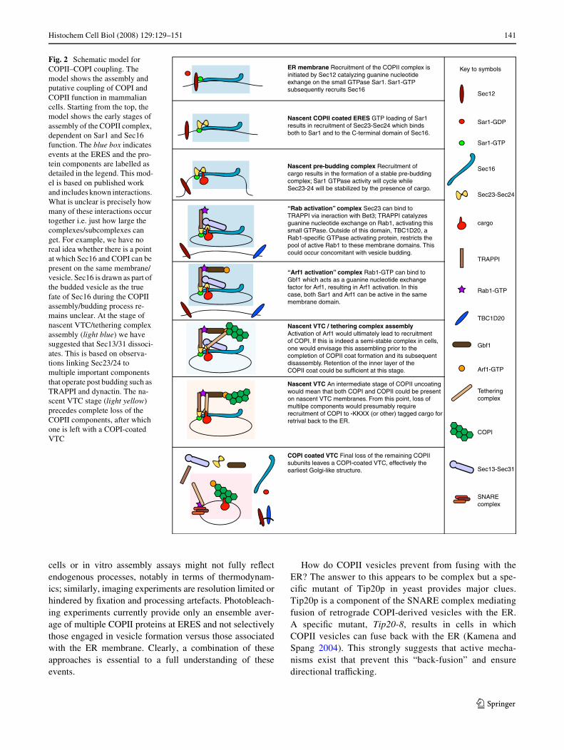
Further recent discoveries provide major advances in ourunderstanding of the mechanistic coupling between COPIIand COPI. It has been known for many years that these twocoat complexes act sequentially during ER-to-Golgi trans-port (Aridor et al. 1995; Scales et al. 1997; Stephens et al.2000). When mammalian cells are labelled for COPII com-ponents, ERGIC-53, and COPI components both their jux-tanuclear and peripheral puncta are seen juxtaposed withone another, and partial, but incomplete, colocalization isobserved (Martinez-Menarguez et al. 1999; Stephens et al.2000) and see Fig. 1c. In many ways these VTC/ERGICelements can be considered to be functionally similar to theearliest membranes of the Golgi itself—they contain bothanterograde and retrograde cargo and recruit COPI therebybeing the Wrst physical location for COPI-mediatedretrieval. The events that keep these components in closecontact are not well understood but it seems logical to sug-gest there would be a mechanism in place to organize theactivities of both coat protein complexes. Such a mecha-nism for COPII-COPI interplay, based on published data, isshown in Fig. 2. This model indicates that the sequentialassembly and disassembly of the two complexes is linkedthrough recruitment and stabilization of these accessoryfactors.
The basis for functional coupling of COPII to COPIrevolves around the TRAPPI tether and the Arf1-GEF,GBF1. TRAPPI is a guanine nucleotide exchange factor(GEF) for Rab1 (Jones et al. 2000). Gbf1 has a key role inmembrane dynamics at the ER–Golgi interface through itsdirect action of the activation of Arf1 (Garcia-Mata et al.2003; Niu et al. 2005; Szul et al. 2005, 2007; Zhao et al.2002, 2006). It is also likely the direct target of brefeldin Aat this point in the secretory pathway (which stabilizes anabortive complex of Arf1/Gbf1/GDP). GBF1 has recentlybeen identiWed as a new Rab1b eVector mediating COPIrecruitment to the ER-Golgi interface (Monetta et al. 2007)
thus linking Rab1b activation (by TRAPPI) to Gbf1 recruit-ment. Rab1b has been shown to mediate the membraneassociation properties of GBF1. When expressed in itsGTP-restricted form, Rab1b causes an increase in GBF1and COPI association to peripheral structures located atERES (Alvarez et al. 2003; Monetta et al. 2007). Subse-quently, when Rab1b is depleted from cells Arf1 membraneassociation to ERES and the Golgi is aVected (Monettaet al. 2007). Together these observations suggest a modelwhereby Rab1b becomes recruited from the cytoplasm tomembranes at the ER-Golgi interface (Alvarez et al. 2003).Gbf1 also interacts with the tethering protein p115 (Garcia-Mata and Sztul 2003). GTP-bound Rab1b binds the N-ter-minal domain of GBF1 (Monetta et al. 2007), with the C-terminal domain of GBF1 able to bind the Golgi membranetethering protein p115 (Allan et al. 2000). p115 is known tobe required at an early stage of vesicle tethering (Alvarezet al. 1999) and is directly involved in recruitment of otherkey molecules such as GM130 (Moyer et al. 2001). Coiled-coil tethering factor p115 interacts with speciWc COPIIassociated SNAREs that are required for homotypic tether-ing of COPII vesicles, and subsequent VTC formation(Allan et al. 2000). As well as p115 bringing COPII vesi-cles into close association for SNARE pairings, p115 initi-ates SNARE complex formation via its N-terminal coiled-coil region being weakly homologous to a SNARE motif(Shorter et al. 2002). Thus a complex network of pro-tein:protein interactions directs the tethering and fusion ofCOPII-derived vesicles. These processes functionally linkthe COPII and COPI complexes. Many questions remainunanswered here including the means of regulation of thissystem and just how many of these multiprotein assembliesare present together at any one time. Is there a sequentialaspect to the process whereby for example followingrecruitment of Gbf1 by TRAPPI, subsequent activation ofRab1b by Gbf1 requires dissociation of TRAPPI?
There is considerable evidence that COPI recruitment isindeed required for anterograde traYcking (e.g. Orci et al.1997; Shima et al. 1999). It remains unclear whether this isin fact a direct role, for example in the sustained recruit-ment and activation of dynein, or whether it is in fact solelyinvolved in mediated retrograde recycling of proteins backto the ER but that its physical presence on these membranesis necessary for their function. One could envisage a seriesof control checks in such a system by which COPI recruit-ment is required for subsequent recruitment/activation stepsbut in which COPI does not itself play a direct role.
While this model has its basis in published work there ismuch that we are unclear about. Does Sec16 incorporate into vesicles budding from the mammalian ER? Just howlong do COPII components remain associated with nascentstructures that are beginning to recruit COPI? One mustremember that biochemical experiments on permeabilized
123
Histochem Cell Biol (2008) 129:129–151 141
cells or in vitro assembly assays might not fully reXectendogenous processes, notably in terms of thermodynam-ics; similarly, imaging experiments are resolution limited orhindered by Wxation and processing artefacts. Photobleach-ing experiments currently provide only an ensemble aver-age of multiple COPII proteins at ERES and not selectivelythose engaged in vesicle formation versus those associatedwith the ER membrane. Clearly, a combination of theseapproaches is essential to a full understanding of theseevents.
How do COPII vesicles prevent from fusing with theER? The answer to this appears to be complex but a spe-ciWc mutant of Tip20p in yeast provides major clues.Tip20p is a component of the SNARE complex mediatingfusion of retrograde COPI-derived vesicles with the ER.A speciWc mutant, Tip20-8, results in cells in whichCOPII vesicles can fuse back with the ER (Kamena andSpang 2004). This strongly suggests that active mecha-nisms exist that prevent this “back-fusion” and ensuredirectional traYcking.
Fig. 2 Schematic model for COPII–COPI coupling. The model shows the assembly and putative coupling of COPI and COPII function in mammalian cells. Starting from the top, the model shows the early stages of assembly of the COPII complex, dependent on Sar1 and Sec16 function. The blue box indicates events at the ERES and the pro-tein components are labelled as detailed in the legend. This mod-el is based on published work and includes known interactions. What is unclear is precisely how many of these interactions occur together i.e. just how large the complexes/subcomplexes can get. For example, we have no real idea whether there is a point at which Sec16 and COPI can be present on the same membrane/vesicle. Sec16 is drawn as part of the budded vesicle as the true fate of Sec16 during the COPII assembly/budding process re-mains unclear. At the stage of nascent VTC/tethering complex assembly (light blue) we have suggested that Sec13/31 dissoci-ates. This is based on observa-tions linking Sec23/24 to multiple important components that operate post budding such as TRAPPI and dynactin. The na-scent VTC stage (light yellow) precedes complete loss of the COPII components, after which one is left with a COPI-coated VTC
ER membrane Recruitment of the COPII complex is initiated by Sec12 catalyzing guanine nucleotideexhange on the small GTPase Sar1. Sar1-GTP subsequently recruits Sec16
Nascent COPII coated ERES GTP loading of Sar1results in recruitment of Sec23-Sec24 which binds both to Sar1 and to the C-terminal domain of Sec16.
Nascent pre-budding complex Recruitment ofcargo results in the formation of a stable pre-buddingcomplex; Sar1 GTPase activity will cycle whileSec23-24 will be stabilized by the presence of cargo.
“Rab activation” complex Sec23 can bind to TRAPPI via ineraction with Bet3; TRAPPI catalyzesguanine nucleotide exchange on Rab1, activating thissmall GTPase. Outside of this domain, TBC1D20, a Rab1-specific GTPase activating protein, restricts the pool of active Rab1 to these membrane domains. Thiscould occur concomitant with vesicle budding.
“Arf1 activation” complex Rab1-GTP can bind to Gbf1 which acts as a guanine nucleotide exchangefactor for Arf1, resulting in Arf1 activation. In this case, both Sar1 and Arf1 can be active in the same membrane domain.
Nascent VTC / tethering complex assembly Activation of Arf1 would ultimately lead to recruitment of COPI. If this is indeed a semi-stable complex in cells, one would envisage this assembling prior to the completion of COPII coat formation and its subsequent disassembly. Retention of the inner layer of the COPII coat could be sufficient at this stage.
Nascent VTC An intermediate stage of COPII uncoatingwould mean that both COPI and COPII could be presenton nascent VTC membranes. From this point, loss ofmultilpe components would presumably require recruitment of COPI to -KKXX (or other) tagged cargo forretrival back to the ER.
COPI coated VTC Final loss of the remaining COPII subunits leaves a COPI-coated VTC, effectively theearliest Golgi-like structure.
Sec12
Sar1-GDP
Sar1-GTP
Sec16
Sec23-Sec24
cargo
TRAPPI
Rab1-GTP
TBC1D20
Gbf1
Arf1-GTP
Tethering complex
COPI
Sec13-Sec31
SNAREcomplex
Key to symbols
123
142 Histochem Cell Biol (2008) 129:129–151
Regulation of COPII coat formation via signalling mechanisms
A key function of the ER in most eukaryotic cells is thesequestration and storage of calcium from the cytosol. Inresponse to extracellular signals, calcium from the ER canundergo rapid release and reuptake in order to trigger intra-cellular signalling cascades (Berridge et al. 2000). To date,the downstream eVects of highly Xuctuating calcium con-centrations on the ER membrane surface have been specu-lative as little research has been done into the eVects ofcalcium Xuctuation on early secretory pathway traYcking.A recent observation regarding the intracellular localizationof penta-EF-hand Ca2+-binding protein ALG-2, apoptosislinked gene 2, has prompted research into the potential roleof calcium signalling in COPII vesicle formation. WhenALG-2 was expressed in HeLa cells, it localizes to ERES(Shibata et al. 2007; Yamasaki et al. 2006). To conWrm this,ALG-2 and ERES marker p125 (Tani et al. 1999) were co-expressed and their partial colocalization was observed(Yamasaki et al. 2006). When intracellular calcium levelswere increased, ALG-2 was distributed at the juxtanucleararea colocalizing with COPII outer subunit Sec31A (laCour et al. 2007). Upon lowering intracellular calcium con-centrations ALG-2 is seen to disperse from the juxtanucleararea, which also shows a loss of Sec31A staining asSec31A dissociated from the membrane and remains cyto-solic (la Cour et al. 2007). Experiments undertaken toknockdown and mislocalize ALG-2 also exhibit a decreasein the number of Sec31A puncta within the cells (la Couret al. 2007; Shibata et al. 2007). Although ALG-2 clearlyaVects intracellular distribution of Sec31A, there appears tobe no eVect on function as the amount of Sec13 binding toSec31 in ALG-2 knockdown cells remains unchanged (Shi-bata et al. 2007).
Interestingly, this eVect of ALG-2 upon Sec31 is isoformspeciWc and only appears to aVect Sec31A; ALG-2 showsno phenotypic eVect on Sec31B (la Cour et al. 2007). Thisprovides the Wrst key functional distinction between theseisoforms (Stankewich et al. 2006). Reasons for this areunknown. Current results implicate a model in which cal-cium binds to ALG-2 when intracellular calcium levelsreach a certain threshold value (la Cour et al. 2007). Thisinteraction induces a conformational change in ALG-2,which allows ALG-2 binding to a proline-rich region ofSec31 (Yamasaki et al. 2006). The conformational changein ALG-2 may confer its ability to discriminate betweenSec31 subunit isoforms. A further conformational changein Sec31A could result in a tighter association between theouter coat of COPII and the inner coat, stabilizing Sec31Aat ERES.
Protein phosphorylation regulates many events in traYcbetween the ER and Golgi. Roles have been proposed for
protein phosphorylation in the regulation of anterogradetransport (Aridor and Balch 2000; Muniz et al. 1996),export of speciWc cargo (Scott et al. 2003), and in retro-grade transport back to the ER (Cabrera et al. 2003). Manyof these eVects were initially ascribed to protein kinaseA (PKA) (Muniz et al. 1996) based on use of the inhibitorH-89; more recent evidence suggests that H-89 may be act-ing through inhibition of a diVerent kinase (Aridor andBalch 2000; Jamora et al. 1999; Lee and Linstedt 2000).PCTAIRE protein kinases interact directly with COPII andregulate secretory cargo transport (Palmer et al. 2005a).PCTAIREs are cyclin-dependent kinase (cdk) family mem-bers with a single substitution (Ser!Cys) in the classicalcyclin binding motif. PCTAIREs do not appear to be regu-lated during the cell cycle, or to associate with any cyclin(Graeser et al. 2002). All cells and tissues examinedexpress at least one PCTAIRE isoform. These data (Palmeret al. 2005a) show that mutations in the ATP binding sitethat abolish kinase activity (K194R) (Graeser et al. 2002)result in fragmentation of the Golgi and an inhibition ofanterograde cargo transport (transport of tsO45-G-GFP);expression of activated mutants causes an equivalentincrease in the rate of cargo transport.
COPII-dependent budding from the ER has long beenknown to be ATP- as well as GTP-dependent (Balch et al.1986) and can be inhibited by the kinase inhibitor H-89(Aridor and Balch 2000); it is conceivable that PCTAIREactivity represents this ATP requirement. PCTAIREkinases bind 14-3-3 proteins (Sladeczek et al. 1997), whichhave themselves been implicated in the control of ERexport. 14-3-3 proteins are believed to act in export bymasking speciWc retention signals in proteins, facilitatingtheir forward transport (O’Kelly et al. 2002). The associa-tion of 14-3-3 with some cargoes (e.g. the KCNK3 K+
channel) in this scheme is regulated by phosphorylation(O’Kelly et al. 2002); it is possible that PCTAIRE mediatescargo phosphorylation and acts to transfer 14-3-3 to facili-tate cargo export. A recent large-scale aYnity column pro-teomic analysis has identiWed the Sec23/24 complex asinteracting with 14-3-3 proteins (Rubio et al. 2004). Thus14-3-3 proteins could integrate PCTAIRE and COPII func-tion. PCTAIRE kinases are highly expressed in terminallydiVerentiated and transformed cells and appear to regulateneurite outgrowth (Graeser et al. 2002).
So far the mechanisms of ERES formation, COPII ves-icle formation, and eYcient ER export have been dis-cussed in terms of cooperative protein–proteininteractions. Additional protein–lipid, and lipid–lipidinteractions provide regulated modiWcation of ER mem-brane phospholipid composition which are required tosupport the cooperative interactions required for selectivetransport. These include phospholipase D (PLD, Pathreet al. 2003). Phospholipase D catalyses the hydrolysis of
123

Histochem Cell Biol (2008) 129:129–151 143
the phosphodiester bond of glycerophospholipids to gen-erate phosphatidic acid (PA) and a free head group. Trans-port from the ER to the Golgi is PLD dependent (Bi et al.1997); the role of PLD in COPII vesicle formation is com-plex. It does not directly enhance COPII recruitment tomembranes but appears to act in facilitating the recruit-ment of Sec23/24 following Sar1 recruitment (Pathreet al. 2003). The authors invoke a model of localizedSar1-stimulated phosphatidic acid production in COPIIvesicle formation. This would certainly provide a spatialcue for COPII assembly but is diYcult to reconcile withthe localization of Sec12 to the entire ER membranethereby presumably leading to Sar1 activation across theentire ER. Once more, the mechanism of restriction ofCOPII dependent budding to ERES remains the elusivecomponent. The activation of Sar1p only supports coatrecruitment when a high proportion of acidic phospholipidsare present (Matsuoka et al. 1998). PA could be requiredto stabilize activated Sar1 and Sec23/24 as both innerCOPII subunits have basic residues exposed on their sur-face (Bi et al. 2002) where possible PA interactions occur.It is likely that these protein lipid interactions are tran-sient, indicating a role in the initial recruitment; sustainedstability could then be provided through subunit protein–protein interactions. PA could also play an allosteric role,modulating Sec23/24 for cargo recognition. The Sec23-interacting protein p125 (Tani et al. 1999) has been identi-Wed that appears to play a role in the organization ofERES (Shimoi et al. 2005). The protein shows homologyto phosphatidic acid preferring phospholipase A1.Recruitment to ERES appears to be mediated by a combi-nation of the PA-binding domain and interaction withSec23. The cholesterol content of the membrane also hasa direct eVect on the export of secretory cargo from theER (Runz et al. 2006). SpeciWcally, reduction in the sterolcontent of cells caused reduced mobility of a model cargoprotein within the ER membrane that correlated with areduced incorporation in to ERES. The rate of turnover ofSec23 on the membrane was also reduced in sterol-depleted cells. The interplay between COPII and sterolbiochemistry is illustrated beautifully but our understand-ing of the pathways that sense the sterol requirements ofcells. In mammalian cells, sterol synthesis is regulated bysterols themselves. Sterols regulatory element bindingproteins (SREBPs) require transport to the Golgi and sub-sequent proteolytic cleavage in order to translocate to thenucleus and act on the relevant genes for cholesterol syn-thesis. High levels of sterols block the ER-to-Golgi trans-port of SREBP cleavage-activating protein (SCAP), asterol-sensing protein that escorts SREBPs (Espenshadeet al. 2002). The mechanism underlying this appears to bethe selective inhibition of SCAP incorporation in toCOPII vesicles. Thus, SREBP/SCAP complexes remain
in the ER and SREBP remains transcriptionally inactive.Incorporation of SCAP into COPII vesicles is mediatedthrough binding of a sorting motif-MELADL in SCAP toSec23/24 (Sun et al. 2005). A further component of thesystem called Insig renders this motif inaccessible whenbound (Sun et al. 2007). The reduction in sterols in the ERleads to dissociation of Insig and transport of SCAP/SREBP to the Golgi.
Further data supporting a role for lipid composition inregulating protein assembly at ERES comes from studies ofphosphatidylinositiol-4-kinases (PI4k) (Blumental-Perryet al. 2006). The authors show that the FAPP1-PH domain,through binding to PI4P inhibits ER export. Further to this,they showed apparent recruitment of a GFP-FAPP1-PH-domain to ERES in permeabilized cells. Interestingly, addi-tion of PI4P appears to overcome the requirement for ATPin COPII recruitment in vitro (Blumental-Perry et al. 2006).Much of the data within this paper relies on the use of theFAPPI-PH domain as a PI4P biosensor. It has been welldocumented that this in fact requires the dual binding ofboth PI4P and Arf1 for its recruitment to Golgi membranes(Godi et al. 2004; Shin and Nakayama 2004); given theproximity of Arf1 activation to COPII assembly, and thefact that COPII and COPI show such close interplay interms of function (for examples see Scales et al. 1997; Ste-phens et al. 2000), one could conclude that the observedeVects could reXect PI4P-dependent processes adjacent torather than at ERES. Furthermore, use of the PH domain ofFAPP1 likely sequesters PI4P and potentially activatesArf1-GTP, which could cause signiWcant perturbation ofERES/VTC function.
Defects in secretory pathway functions can lead to disease
Our increased knowledge of the fundamental mechanismunderlying ER-to-Golgi transport has enabled us to gaininsight in to the molecular basis of a number of diseasestates. As mentioned above, many diseases that are attrib-uted to “traYcking defects” are in fact primary disorders ofprotein folding and assembly. The traYcking element herereally relates to the important question of quality control interms of export of properly assembled cargo. However, anincreasing number of disease states are now directly attrib-utable to defects in traYcking machinery. A good exampleof this is the resulting defects in secretion of blood clottingfactors and delivery of lysosomal enzymes in patients withmutations in ERGIC-53, a key cargo receptor for ERexport. This is described in previous sections and coveredin depth elsewhere (e.g. Zhang et al. 2003). Here, we willdeal with mutations in the core traYcking machinery i.e.COPII.
123
144 Histochem Cell Biol (2008) 129:129–151
Cranio-lenticulosutural dysplasia and integration of COPII coat assembly
Cranio-lenticulo sutural dysplasia (CLSD) is an autosomalrecessive syndrome that results from a missense mutationin Sec23A (F382L). This disorder aVects cranio-facialdevelopment and skeletogenesis and appears to be due to adefect in the deposition of extracellular matrix (Boyadjievet al. 2006). A chemical mutagenesis screen for phenotypesaVecting craniofacial development in zebraWsh generated aseries of mutants with similar phenotypes (Neuhauss et al.1996). One such mutant, crusher, is caused by a nonsensemutation L402X in Sec23A and results in a truncated formof Sec23A that is not functionally active (Lang et al. 2006).The truncated domain is known to interact with Sar1; how-ever, as this truncation aVects full Sec23A activity it is diY-cult to specify the outcome of the mutation (Lang et al.2006). Cartilage of the craniofacial region primarily con-sists of type II collagen and is derived from cranial neuralcrest cells that diVerentiate into chondrocytes. These colla-gen-secreting cells with Sec23A mutations show a largebuild up of procollagen which is retained inside distendedER structures (Boyadjiev et al. 2006). The Sec23A muta-tion is not lethal as presumably the majority of secretorycargoes can still become exported from the ER. One inter-pretation is that this results from the ongoing, normal, func-tion of the second Sec23 isoform Sec23B. This could becompounded by a preferential requirement for Sec23A inthe biogenesis of collagen-containing transport carriers.Such isoform selectivity has been shown for other cargoes(Wendeler et al. 2007). Recent data support the Wrst ofthese conclusions (Bi et al. 2007; Fromme et al. 2007).Cells from CLSD patients show a redistribution of Sec13/31 to the cytoplasm (Boyadjiev et al. 2006); this suggeststhat a failure to recruit and stabilize Sec13/31 at ERES pre-vents procollagen secretion. Now Orci, Schekman and col-leagues (Fromme et al. 2007) show that in vitro assays forCOPII-dependent budding, Sec23A containing the F382Lmutation is less eYcient in coupling to the Sec13/31 layerand consequently inhibits COPII-dependent budding. Theyfurther show that Sec23B is only expressed at very low lev-els in calvarial osteoblasts (responsible for ossiWcation ofthe cranial suture which is signiWcantly defective in CLSDand Wbroblasts) which could explain several other aspectsof the clinical presentation of CLSD (Boyadjiev et al.2003). Thus, the combination of mutation in Sec23A anddeWciency of Sec23B is proposed to result in ineYcientcoupling of Sec13/31 and the ensuing phenotypes. Furthercomplexity was revealed in these experiments in that Sar1isoform selectivity in budding assays was revealed for theWrst time (Fromme et al. 2007). Intriguingly, Sar1A, but notSar1B, was found to partially rescue the defect in buddingobserved when paired with Sec23A-F382L. Sar1A is in fact
expressed at high level in calvarial osteoblasts but evidentlynot a suYcient level to overcome the defect arising fromF382L mutation in Sec23A (Fromme et al. 2007). This sug-gests that Sar1A is not a limiting factor in COPII-dependentbudding and the authors suggest that in fact this is causedby the low expression level of Sec23B. An alternativeexplanation would be that impaired recruitment of Sec13/31 has a greater eVect on the ER export of certain cargoessuch as collagen and other extracellular matrix components.This could be caused by a speciWc requirement to generatea transport carrier of such unusual dimensions [to accom-modate the large, rod-like structure of assembled collagenprotoWbrils (Canty and Kadler 2005)]. Some evidenceexists for this possibility; coupling of Sar1A with Sec23A-F382L leads to robust packaging of ERGIC-53 (Frommeet al. 2007). siRNA silencing of Sec13 expression in mam-malian cells does not signiWcantly reduce ER-to-Golgitransport of tsO45-G (A.K. Townley and D.J.S., unpub-lished observations).
A key outcome of the analysis of CLSD patient cells isthe clear accumulation of budding proWles on distended ER(Fromme et al. 2007). These images show clearly thatCOPII vesicle Wssion is inhibited in these cells. The natureof COPII vesicle scission has remained elusive, notably inthe absence of any constricting force generator. These newdata suggest that the defect in coupling of Sec23/24 toSec13/31 causing ineYcient recruitment of the outer layerleads to a failure of vesicle scission. This is elegantly rein-forced by crystallographic data showing that Sec31 directlycontacts Sec23 in very close proximity to the F382 residue(Bi et al. 2007). Furthermore, Sec31 also contacts Sar1directly (Bi et al. 2007) and signiWcantly, this interface lieson the same surface as the F382 residue in Sec23A, i.e. indirect apposition with Sec31. Mechanistically, Sec31 con-tributes to the GTP binding site of Sar1 and facilitates rapidGTP hydrolysis by optimising the geometry of the activesite. In a complete vesicle, this would also position Sec23/24 directly underneath Sec13/31 in the vesicle coat struc-ture (Bi et al. 2007; Fath et al. 2007; Stagg et al. 2006).Thus, Sec13/31 itself provides the driving force for COPIIvesicle scission. This role for Sec13/31 is shown in Fig. 3in which recruitment of Sec13/31 occurs after membranecurvature and cargo accumulation but precedes scission ofthe vesicle (Fig. 4).
Chylomicron retention diseases
Mutation in Sar1B, one of the two human isoforms ofSar1, causes clinically important defects in lipoproteinmetabolism (Jones et al. 2003). A number of diseases thatcan be classiWed together as chylomicron retention dis-eases (CMRDs) have now been conWrmed to be caused bya speciWc mutation in the SARA2 gene encoding Sar1B.
123
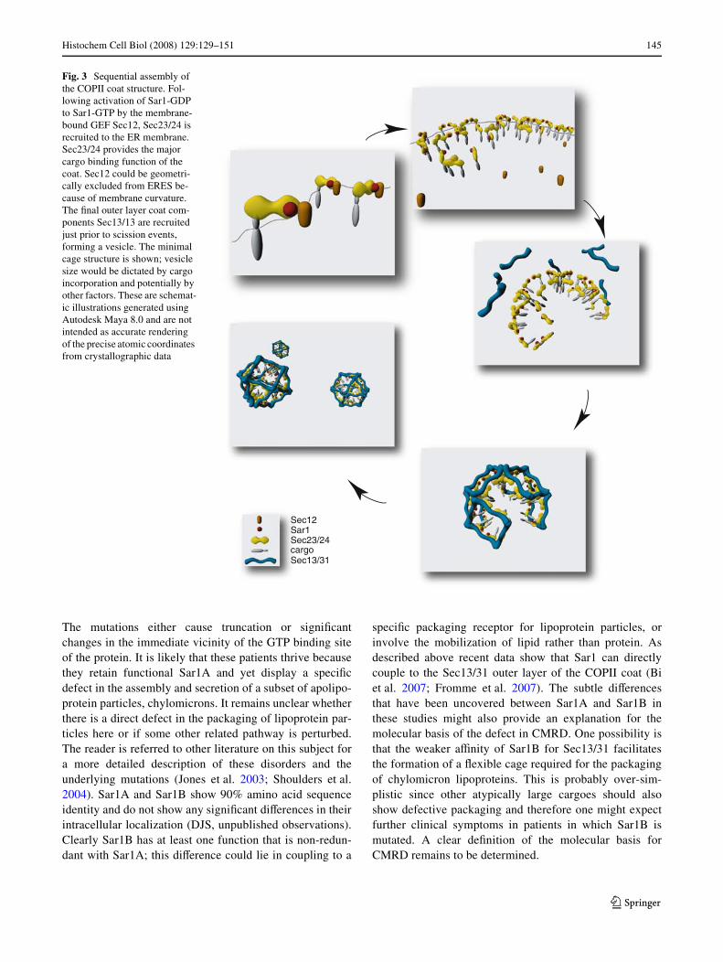
Histochem Cell Biol (2008) 129:129–151 145
The mutations either cause truncation or signiWcantchanges in the immediate vicinity of the GTP binding siteof the protein. It is likely that these patients thrive becausethey retain functional Sar1A and yet display a speciWcdefect in the assembly and secretion of a subset of apolipo-protein particles, chylomicrons. It remains unclear whetherthere is a direct defect in the packaging of lipoprotein par-ticles here or if some other related pathway is perturbed.The reader is referred to other literature on this subject fora more detailed description of these disorders and theunderlying mutations (Jones et al. 2003; Shoulders et al.2004). Sar1A and Sar1B show 90% amino acid sequenceidentity and do not show any signiWcant diVerences in theirintracellular localization (DJS, unpublished observations).Clearly Sar1B has at least one function that is non-redun-dant with Sar1A; this diVerence could lie in coupling to a
speciWc packaging receptor for lipoprotein particles, orinvolve the mobilization of lipid rather than protein. Asdescribed above recent data show that Sar1 can directlycouple to the Sec13/31 outer layer of the COPII coat (Biet al. 2007; Fromme et al. 2007). The subtle diVerencesthat have been uncovered between Sar1A and Sar1B inthese studies might also provide an explanation for themolecular basis of the defect in CMRD. One possibility isthat the weaker aYnity of Sar1B for Sec13/31 facilitatesthe formation of a Xexible cage required for the packagingof chylomicron lipoproteins. This is probably over-sim-plistic since other atypically large cargoes should alsoshow defective packaging and therefore one might expectfurther clinical symptoms in patients in which Sar1B ismutated. A clear deWnition of the molecular basis forCMRD remains to be determined.
Fig. 3 Sequential assembly of the COPII coat structure. Fol-lowing activation of Sar1-GDP to Sar1-GTP by the membrane-bound GEF Sec12, Sec23/24 is recruited to the ER membrane. Sec23/24 provides the major cargo binding function of the coat. Sec12 could be geometri-cally excluded from ERES be-cause of membrane curvature. The Wnal outer layer coat com-ponents Sec13/13 are recruited just prior to scission events, forming a vesicle. The minimal cage structure is shown; vesicle size would be dictated by cargo incorporation and potentially by other factors. These are schemat-ic illustrations generated using Autodesk Maya 8.0 and are not intended as accurate rendering of the precise atomic coordinates from crystallographic data
Sec12Sar1Sec23/24cargoSec13/31
123

146 Histochem Cell Biol (2008) 129:129–151
Hijacking of COPII pathways by viruses and microbial pathogens
Many pathogenic bacteria and viruses are known to modu-late host cell traYcking machineries. We know most aboutmodulation of endocytic systems during viral or bacterialentry, notably through hijacking of clathrin-dependent
endocytic pathways (see Veiga et al. 2007 for an elegantrecent illustration of this). Increasingly, examples are com-ing to light of viruses that exploit the COPII pathway dur-ing their life cycle, for example the biogenesis of thepoliovirus particles (Rust et al. 2001) and the formation ofvacuoles used by Legionella for its replication (Robinsonand Roy 2006). Bacterial systems also co-opt host path-ways to their own advantage. A good recent example of thiscomes from the NleA protein from Escherichia coli. Thisprotein is transferred from the bacteria to host cells througha type III secretion system where it localizes primarily to ajuxtanuclear compartment (Gruenheid et al. 2004). Recentuse of biochemical reconstitution of COPII-dependent bud-ding has shown that NleA in fact binds to Sec24 modulatesCOPII vesicle formation from the ER (Kim et al. 2007).NleA appears to stabilize the binding of Sec23/24 to lipo-somes in the presence of GTP, inducing the formation ofirregular aggregates of COPII proteins. These data suggestthat inhibition of COPII function is a contributing factor tobacterial virulence. Many of these factors also aVect otherclosely linked pathways such as the COPI system [forrecent examples see (Belov et al. 2007; Wessels et al.2006)] and highlight the importance of a full molecularunderstanding of host membrane traYcking pathways.
Conclusions
Clearly, our knowledge of membrane dynamics at the ER/Golgi interface is far from complete. The combination oftechnology available to us today, including yeast genetics,biochemical reconstitution, live cell imaging and wholeorganism physiology, should ensure that we rapidly reach agreater understanding of the mechanistic basis for selectivecargo export and intracellular organization in health anddisease. Increasingly, our understanding will tend moretowards physiologically relevant systems and the diVer-ences that underlie intracellular patterning and function indiVerent species and cell types during and after diVerentia-tion.
Acknowledgments We thank Pete Watson for Fig. 1 and construc-tive comments throughout. Work in the Stephens lab is supported byNon-Clinical Senior Fellowship from the MRC and research grantsfrom BBSRC and the Royal Society. HH is funded through a BBSRCDoctoral Training Account. We would particularly like to thank AngusLaurie-Pile for his work in generating the 3D rendered images used inFigs. 3 and 4.
References
Allan BB, Moyer BD, Balch WE (2000) Rab1 recruitment of p115 intoa cis-SNARE complex: programming budding COPII vesicles forfusion. Science 289:444–448
Fig. 4 Sec16 spatially restricts assembly of the COPII coat. a Outercoat components Sec13/31 form cuboctahedrons in their minimal state.The proline-rich region of Sec13 contacts Sec23/24 and Sar1 GTP sta-bilizing contacts between the inner and outer coat complexes. This alsohas the capacity to position Sec23/24 underneath the edges of the cagemaking the membrane surface (and associated proteins) more accessi-ble. These are schematic illustrations generated using Autodesk Maya8.0 and are not intended as accurate rendering of the precise atomiccoordinates from crystallographic data
123
Histochem Cell Biol (2008) 129:129–151 147
Alvarez C, Fujita H, Hubbard A, Sztul E (1999) ER to Golgi transport:requirement for p115 at a pre-Golgi VTC stage. J Cell Biol147:1205–1222
Alvarez C, Garcia-Mata R, Brandon E, Sztul E (2003) COPI Recruit-ment is modulated by a Rab1b-dependent mechanism. Mol BiolCell 14:2116–2127
Antonny B, Madden D, Hamamoto S, Orci L, Schekman R (2001)Dynamics of the COPII coat with GTP and stable analogues. NatCell Biol 3:531–537
Antonny B, Gounon P, Schekman R, Orci L (2003) Self-assembly ofminimal COPII cages. EMBO Rep 4:419–424
Appenzeller-Herzog C, Hauri HP (2006) The ER-Golgi intermediatecompartment (ERGIC): in search of its identity and function.J Cell Sci 119:2173–2183
Appenzeller C, Andersson H, Kappeler F, Hauri HP (1999) The lectinERGIC-53 is a cargo transport receptor for glycoproteins. NatCell Biol 1:330–334
Aridor M, Bannykh SI, Rowe T, Balch WE (1995) Sequential couplingbetween COPII and COPI vesicle coats in endoplasmic reticulumto Golgi transport. J Cell Biol 131:875–893
Aridor M, Weissman J, Bannykh S, NuoVer C, Balch WE (1998) Car-go selection by the COPII budding machinery during export fromthe ER. J Cell Biol 141:61–70
Aridor M, Balch WE (2000) Kinase signaling initiates coat complex II(COPII) recruitment and export from the mammalian endoplas-mic reticulum. J Biol Chem 275:35673–35676
Aridor M, Hannan LA (2000) TraYc jam: a compendium of humandiseases that aVect intracellular transport processes. TraYc1:836–851
Aridor M, Hannan LA (2002) TraYc jams II: an update of diseases ofintracellular transport. TraYc 3:781–790
Balch WE, Elliott MM, Keller DS (1986) ATP-coupled transport ofvesicular stomatitis virus G protein between the endoplasmicreticulum and the Golgi. J Biol Chem 261:14681–14689
Bannykh SI, Rowe T, Balch WE (1996) The organization of endoplas-mic reticulum export complexes. J Cell Biol 135:19–35
Bannykh SI, Balch WE (1997) Membrane dynamics at the endoplas-mic reticulum–Golgi interface. J Cell Biol 138:1–4
Barlowe C, Schekman R (1993) SEC12 encodes a guanine-nucleotide-exchange factor essential for transport vesicle budding from theER. Nature 365:347–349
Barlowe C, Orci L, Yeung T, Hosobuchi M, Hamamoto S, Salama N,Rexach MF, Ravazzola M, Amherdt M, Schekman R (1994)COPII: a membrane coat formed by Sec proteins that drive vesiclebudding from the endoplasmic reticulum. Cell 77:895–907
Barlowe C (2003) Signals for COPII-dependent export from the ER:what’s the ticket out? Trends Cell Biol 13:295–300
Barrowman J, Wang W, Zhang Y, Ferro-Novick S (2003) TheYip1p.Yif1p complex is required for the fusion competence ofendoplasmic reticulum-derived vesicles. J Biol Chem278:19878–19884
Behnia R, Barr FA, Flanagan JJ, Barlowe C, Munro S (2007) The yeastorthologue of GRASP65 forms a complex with a coiled-coil pro-tein that contributes to ER to Golgi traYc. J Cell Biol 176:255–261
Belov GA, Altan-Bonnet N, Kovtunovych G, Jackson CL, Lippincott-Schwartz J, Ehrenfeld E (2007) Hijacking components of the cel-lular secretory pathway for replication of poliovirus RNA. J Virol81:558–567
Ben-Tekaya H, Miura K, Pepperkok R, Hauri HP (2005) Live imagingof bidirectional traYc from the ERGIC. J Cell Sci 118:357–367
Berridge MJ, Lipp P, Bootman MD (2000) The versatility and univer-sality of calcium signalling. Nat Rev Mol Cell Biol 1:11–21
Bevis BJ, Hammond AT, Reinke CA, Glick BS (2002) De novo forma-tion of transitional ER sites and Golgi structures in Pichia pasto-ris. Nat Cell Biol 4:750–756
Bhattacharyya D, Glick BS (2007) Two mammalian Sec16 homo-logues have nonredundant functions in endoplasmic reticulum(ER) export and transitional ER organization. Mol Biol Cell18:839–849
Bi K, Roth MG, Ktistakis NT (1997) Phosphatidic acid formation byphospholipase D is required for transport from the endoplasmicreticulum to the Golgi complex. Curr Biol 7:301–307
Bi X, Corpina RA, Goldberg J (2002) Structure of the Sec23/24-Sar1pre-budding complex of the COPII vesicle coat. Nature 419:271–277
Bi X, Mancias JD, Goldberg J (2007) Insights into COPII coat nucle-ation from the structure of Sec23*Sar1 complexed with the activefragment of Sec31. Dev Cell 13:635–645
Bielli A, Haney CJ, Gabreski G, Watkins SC, Bannykh SI, Aridor M(2005) Regulation of Sar1 NH2 terminus by GTP binding andhydrolysis promotes membrane deformation to control COPIIvesicle Wssion. J Cell Biol 171:919–924
Blumental-Perry A, Haney CJ, Weixel KM, Watkins SC, Weisz OA,Aridor M (2006) Phosphatidylinositol 4-phosphate formation atER exit sites regulates ER export. Dev Cell 11:671–682
Bonifacino JS, Glick BS (2004) The mechanisms of vesicle buddingand fusion. Cell 116:153–166
Boyadjiev SA, Justice CM, Eyaid W, McKusick VA, Lachman RS,Chowdry AB, Jabak M, Zwaan J, Wilson AF, Jabs EW (2003) Anovel dysmorphic syndrome with open calvarial sutures and su-tural cataracts maps to chromosome 14q13-q21. Hum Genet113:1–9
Boyadjiev SA, Fromme JC, Ben J, Chong SS, Nauta C, Hur DJ, ZhangG, Hamamoto S, Schekman R, Ravazzola M et al (2006) Cranio-lenticulo-sutural dysplasia is caused by a SEC23A mutation lead-ing to abnormal endoplasmic-reticulum-to-Golgi traYcking. NatGenet 38:1192–1197
Bulbarelli A, Sprocati T, Barberi M, Pedrazzini E, Borgese N (2002)TraYcking of tail-anchored proteins: transport from the endoplas-mic reticulum to the plasma membrane and sorting between sur-face domains in polarised epithelial cells. J Cell Sci 115:1689–1702
Cabrera M, Muniz M, Hidalgo J, Vega L, Martin ME, Velasco A(2003) The retrieval function of the KDEL receptor requires PKAphosphorylation of its C-terminus. Mol Biol Cell 14:4114–4125
Cai H, Zhang Y, Pypaert M, Walker L, Ferro-Novick S (2005) Mutantsin trs120 disrupt traYc from the early endosome to the late Golgi.J Cell Biol 171:823–833
Cai H, Yu S, Menon S, Cai Y, Lazarova D, Fu C, Reinisch K, Hay JC,Ferro-Novick S (2007) TRAPPI tethers COPII vesicles by bind-ing the coat subunit Sec23. Nature 445:941–944
Calero M, Collins RN (2002) Saccharomyces cerevisiae Pra1p/Yip3pinteracts with Yip1p and Rab proteins. Biochem Biophys ResCommun 290:676–681
Canty EG, Kadler KE (2005) Procollagen traYcking, processing andWbrillogenesis. J Cell Sci 118:1341–1353
Cao X, Ballew N, Barlowe C (1998) Initial docking of ER-derived ves-icles requires Uso1p and Ypt1p but is independent of SNAREproteins. Embo J 17:2156–2165
Chardin P, Callebaut I (2002) The yeast Sar exchange factor Sec12,and its higher organism orthologs, fold as beta-propellers. FEBSLett 525:171–173
Chernomordik LV, Kozlov MM (2003) Protein–lipid interplay in fu-sion and Wssion of biological membranes. Annu Rev Biochem72:175–207
Christoforidis S, McBride HM, Burgoyne RD, Zerial M (1999) TheRab5 eVector EEA1 is a core component of endosome docking.Nature 397:621–625
Connerly PL, Esaki M, Montegna EA, Strongin DE, Levi S,Soderholm J, Glick BS (2005) Sec16 is a determinant of transi-tional ER organization. Curr Biol 15:1439–1447
123
148 Histochem Cell Biol (2008) 129:129–151
Dudognon P, Maeder-Garavaglia C, Carpentier JL, Paccaud JP (2004)Regulation of a COPII component by cytosolic O-glycosylationduring mitosis. FEBS Lett 561:44–50
Eilers M, Shekar SC, Shieh T, Smith SO, Fleming PJ (2000) Internalpacking of helical membrane proteins. Proc Natl Acad Sci USA97:5796–5801
Ellgaard L, Helenius A (2003) Quality control in the endoplasmicreticulum. Nat Rev Mol Cell Biol 4:181–191
Espenshade P, Gimeno RE, Holzmacher E, Teung P, Kaiser CA (1995)Yeast SEC16 gene encodes a multidomain vesicle coat proteinthat interacts with Sec23p. J Cell Biol 131:311–324
Espenshade PJ, Li WP, Yabe D (2002) Sterols block binding of COPIIproteins to SCAP, thereby controlling SCAP sorting in ER. ProcNatl Acad Sci USA 99:11694–11699
Farsad K, De Camilli P (2003) Mechanisms of membrane deformation.Curr Opin Cell Biol 15:372–381
Fath S, Mancias JD, Bi X, Goldberg J (2007) Structure and organiza-tion of coat proteins in the COPII cage. Cell 129:1325–1336
Forster R, Weiss M, Zimmermann T, Reynaud EG, Verissimo F, Ste-phens DJ, Pepperkok R (2006) Secretory cargo regulates the turn-over of COPII subunits at single ER exit sites. Curr Biol 16:173–179
Fromme JC, Ravazzola M, Hamamoto S, Al-Balwi M, Eyaid W, Boy-adjiev SA, Cosson P, Schekman R, Orci L (2007) The genetic ba-sis of a craniofacial disease provides insight into COPII coatassembly. Dev Cell 13:623–634
Futai E, Hamamoto S, Orci L, Schekman R (2004) GTP/GDP ex-change by Sec12p enables COPII vesicle bud formation on syn-thetic liposomes. EMBO J 23:4146–4155
Garcia-Mata R, Sztul E (2003) The membrane-tethering protein p115interacts with GBF1, an ARF guanine-nucleotide-exchange fac-tor. EMBO Rep 4:320–325
Garcia-Mata R, Szul T, Alvarez C, Sztul E (2003) ADP-ribosylationfactor/COPI-dependent events at the endoplasmic reticulum–Gol-gi interface are regulated by the guanine nucleotide exchange fac-tor GBF1. Mol Biol Cell 14:2250–2261
Gillingham AK, Munro S (2007) The small g proteins of the arf familyand their regulators. Annu Rev Cell Dev Biol 23:579–611
Gimeno RE, Espenshade P, Kaiser CA (1995) SED4 encodes a yeastendoplasmic reticulum protein that binds Sec16p and participatesin vesicle formation. J Cell Biol 131:325–338
Gimeno RE, Espenshade P, Kaiser CA (1996) COPII coat subunitinteractions: Sec24p and Sec23p bind to adjacent regions ofSec16p. Mol Biol Cell 7:1815–1823
Godi A, Di Campli A, Konstantakopoulos A, Di Tullio G, Alessi DR,Kular GS, Daniele T, Marra P, Lucocq JM, De Matteis MA(2004) FAPPs control Golgi-to-cell-surface membrane traYc bybinding to ARF and PtdIns(4)P. Nat Cell Biol 6:393–404
Gould SJ, McCollum D, Spong AP, Heyman JA, Subramani S (1992)Development of the yeast Pichia pastoris as a model organism fora genetic and molecular analysis of peroxisome assembly. Yeast8:613–628
Graeser R, Gannon J, Poon RY, Dubois T, Aitken A, Hunt T (2002)Regulation of the CDK-related protein kinase PCTAIRE-1 and itspossible role in neurite outgrowth in Neuro-2A cells. J Cell Sci115:3479–3490
Gruenheid S, Sekirov I, Thomas NA, Deng W, O’Donnell P, Goode D,Li Y, Frey EA, Brown NF, Metalnikov P et al (2004) IdentiWca-tion and characterization of NleA, a non-LEE-encoded type IIItranslocated virulence factor of enterohaemorrhagic Escherichiacoli O157:H7. Mol Microbiol 51:1233–1249
Gurkan C, Stagg SM, Lapointe P, Balch WE (2006) The COPII cage:unifying principles of vesicle coat assembly. Nat Rev Mol CellBiol 7:727–738
Haas AK, Yoshimura S, Stephens DJ, Preisinger C, Fuchs E, Barr FA(2007) Analysis of GTPase-activating proteins reveals that Rab1
and Rab43 are key Rabs required for the maintenance of a func-tional Golgi in human cells. J Cell Sci 120:2997–3010
Hammond AT, Glick BS (2000) Dynamics of transitional endoplasmicreticulum sites in vertebrate cells. Mol Biol Cell 11:3013–3030
Hanton SL, Bortolotti LE, Renna L, Stefano G, Brandizzi F (2005)Crossing the divide-transport between the endoplasmic reticulumand Golgi apparatus in plants. TraYc 6:267–277
Heidtman M, Chen CZ, Collins RN, Barlowe C (2003) A role forYip1p in COPII vesicle biogenesis. J Cell Biol 163:57–69
Heidtman M, Chen CZ, Collins RN, Barlowe C (2005) Yos1p is a nov-el subunit of the Yip1p–Yif1p complex and is required for trans-port between the endoplasmic reticulum and the Golgi complex.Mol Biol Cell 16:1673–1683
Hicke L, Schekman R (1989) Yeast Sec23p acts in the cytoplasm topromote protein transport from the endoplasmic reticulum to theGolgi complex in vivo and in vitro. Embo J 8:1677–1684
Hicke L, Yoshihisa T, Schekman R (1992) Sec23p and a novel 105-kDa protein function as a multimeric complex to promote vesiclebudding and protein transport from the endoplasmic reticulum.Mol Biol Cell 3:667–676
Higashio H, Sato K, Nakano A (2007) Smy2p participates in COPIIvesicle formation through the interaction with Sec23p/Sec24psubcomplex. TraYc doi: 10.1111/j.1600–0854.2007.00668.x
Horton AC, Ehlers MD (2004) Secretory traYcking in neuronal den-drites. Nat Cell Biol 6:585–591
Huang M, Weissman JT, Beraud-Dufour S, Luan P, Wang C, Chen W,Aridor M, Wilson IA, Balch WE (2001) Crystal structure of Sar1-GDP at 1.7 A resolution and the role of the NH2 terminus in ERexport. J Cell Biol 155:937–948
Hurtley SM, Helenius A (1989) Protein oligomerization in the endo-plasmic reticulum. Annu Rev Cell Biol 5:277–307
Iinuma T, Shiga A, Nakamoto K, O’Brien MB, Aridor M, Arimitsu N,Tagaya M, Tani K (2007) Mammalian Sec16/p250 plays a role inmembrane traYc from the endoplasmic reticulum. J Biol Chem282:17632–17639
Itin C, Schindler R, Hauri HP (1995) Targeting of protein ERGIC-53to the ER/ERGIC/cis-Golgi recycling pathway. J Cell Biol131:57–67
Jamora C, Yamanouye N, Van Lint J, Laudenslager J, VandenheedeJR, Faulkner DJ, Malhotra V (1999) Gbetagamma-mediated reg-ulation of Golgi organization is through the direct activation ofprotein kinase D. Cell 98:59–68
Jones B, Jones EL, Bonney SA, Patel HN, Mensenkamp AR, Eichen-baum-Voline S, Rudling M, Myrdal U, Annesi G, Naik S et al(2003) Mutations in a Sar1 GTPase of COPII vesicles are associ-ated with lipid absorption disorders. Nat Genet 34:29–31
Jones S, Newman C, Liu F, Segev N (2000) The TRAPP complex is anucleotide exchanger for Ypt1 and Ypt31/32. Mol Biol Cell11:4403–4411
Kaiser CA, Schekman R (1990) Distinct sets of SEC genes governtransport vesicle formation and fusion early in the secretory path-way. Cell 61:723–733
Kamena F, Spang A (2004) Tip20p prohibits back-fusion of COPIIvesicles with the endoplasmic reticulum. Science 304:286–289
Kapetanovich L, Baughman C, Lee TH (2005) Nm23H2 facilitatescoat protein complex II assembly and endoplasmic reticulum ex-port in mammalian cells. Mol Biol Cell 16:835–848
Kappeler F, Klopfenstein DR, Foguet M, Paccaud JP, Hauri HP (1997)The recycling of ERGIC-53 in the early secretory pathway. ER-GIC-53 carries a cytosolic endoplasmic reticulum-exit determi-nant interacting with COPII. J Biol Chem 272:31801–31808
Karsten V, Hegde RS, Sinai AP, Yang M, Joiner KA (2004) Trans-membrane domain modulates sorting of membrane proteins inToxoplasma gondii. J Biol Chem 279:26052–26057
Kikuno R, Nagase T, Nakayama M, Koga H, Okazaki N, Nakajima D,Ohara O (2004) HUGE: a database for human KIAA proteins, a
123
Histochem Cell Biol (2008) 129:129–151 149
2004 update integrating HUGEppi and ROUGE. Nucleic AcidsRes 32:D502–D504
Kim J, Thanabalasuriar A, Chaworth-Musters T, Fromme JC, Frey EA,Lario PI, Metalnikov P, Rizg K, Thomas NA, Lee SF et al (2007)The bacterial virulence factor NleA inhibits cellular protein secre-tion by disrupting mammalian COPII function. Cell Host Microb2:160–171
Kim YG, Raunser S, Munger C, Wagner J, Song YL, Cygler M, WalzT, Oh BH, Sacher M (2006) The architecture of the multisubunitTRAPP I complex suggests a model for vesicle tethering. Cell127:817–830
Kirk SJ, Ward TH (2007) COPII under the microscope. Semin CellDev Biol
Kuge O, Dascher C, Orci L, Rowe T, Amherdt M, Plutner H, Ravaz-zola M, Tanigawa G, Rothman JE, Balch WE (1994) Sar1 pro-motes vesicle budding from the endoplasmic reticulum but notGolgi compartments. J Cell Biol 125:51–65
la Cour JM, Mollerup J, Berchtold MW (2007) ALG-2 oscillates insubcellular localization, unitemporally with calcium oscillations.Biochem Biophys Res Commun 353:1063–1067
Ladinsky MS, Mastronarde DN, McIntosh JR, Howell KE, StaehelinLA (1999) Golgi structure in three dimensions: functional in-sights from the normal rat kidney cell. J Cell Biol 144:1135–1149
Lang MR, Lapierre LA, Frotscher M, Goldenring JR, Knapik EW(2006) Secretory COPII coat component Sec23a is essential forcraniofacial chondrocyte maturation. Nat Genet 38:1198–1203
Lederkremer GZ, Cheng Y, Petre BM, Vogan E, Springer S, SchekmanR, Walz T, Kirchhausen T (2001) Structure of the Sec23p/24p andSec13p/31p complexes of COPII. Proc Natl Acad Sci USA98:10704–10709
Lee MC, Orci L, Hamamoto S, Futai E, Ravazzola M, Schekman R(2005) Sar1p N-terminal helix initiates membrane curvature andcompletes the Wssion of a COPII vesicle. Cell 122:605–617
Lee MC, Miller EA (2007) Molecular mechanisms of COPII vesicleformation. Semin Cell Dev Biol
Lee TH, Linstedt AD (2000) Potential role for protein kinases in regu-lation of bidirectional endoplasmic reticulum-to-Golgi transportrevealed by protein kinase inhibitor H89. Mol Biol Cell 11:2577–2590
Letourneur F, Hennecke S, Demolliere C, Cosson P (1995) Stericmasking of a dilysine endoplasmic reticulum retention motif dur-ing assembly of the human high aYnity receptor for immunoglob-ulin E. J Cell Biol 129:971–978
Ma D, Zerangue N, Raab-Graham K, Fried SR, Jan YN, Jan LY (2002)Diverse traYcking patterns due to multiple traYc motifs in G pro-tein-activated inwardly rectifying potassium channels from brainand heart. Neuron 33:715–729
Mallabiabarrena A, Fresno M, Alarcon B (1992) An endoplasmic retic-ulum retention signal in the CD3 epsilon chain of the T-cell recep-tor. Nature 357:593–596
Mancias JD, Goldberg J (2005) Exiting the endoplasmic reticulum.TraYc 6:278–285
Martinez-Menarguez JA, Geuze HJ, Slot JW, Klumperman J (1999)Vesicular tubular clusters between the ER and Golgi mediate con-centration of soluble secretory proteins by exclusion from COPI-coated vesicles. Cell 98:81–90
Matern H, Yang X, Andrulis E, Sternglanz R, Trepte HH, Gallwitz D(2000) A novel Golgi membrane protein is part of a GTPase-bind-ing protein complex involved in vesicle targeting. Embo J19:4485–4492
Matsuoka K, Orci L, Amherdt M, Bednarek SY, Hamamoto S, Schek-man R, Yeung T (1998) COPII-coated vesicle formation reconsti-tuted with puriWed coat proteins and chemically deWnedliposomes. Cell 93:263–275
Miller EA, Beilharz TH, Malkus PN, Lee MC, Hamamoto S, Orci L,Schekman R (2003) Multiple cargo binding sites on the COPII
subunit Sec24p ensure capture of diverse membrane proteins intotransport vesicles. Cell 114:497–509
Mizuno M, Singer SJ (1994) A possible role for stable microtubules inintracellular transport from the endoplasmic reticulum to the Gol-gi apparatus. J Cell Sci 107:1321–1331
Monetta P, Slavin I, Romero N, Alvarez C (2007) Rab1b interacts withGBF1 and modulates both ARF1 dynamics and COPI association.Mol Biol Cell 18:2400–2410
Morsomme P, Riezman H (2002) The Rab GTPase Ypt1p and tether-ing factors couple protein sorting at the ER to vesicle targeting tothe Golgi apparatus. Dev Cell 2:307–317
Morsomme P, Prescianotto-Baschong C, Riezman H (2003) The ERv-SNAREs are required for GPI-anchored protein sorting fromother secretory proteins upon exit from the ER. J Cell Biol162:403–412
Mossessova E, Bickford LC, Goldberg J (2003) SNARE Selectivity ofthe COPII Coat. Cell 114:483–495
Moussalli M, Pipe SW, Hauri HP, Nichols WC, Ginsburg D, KaufmanRJ (1999) Mannose-dependent endoplasmic reticulum (ER)-Gol-gi intermediate compartment-53-mediated ER to Golgi traYckingof coagulation factors V and VIII. J Biol Chem 274:32539–32542
Moyer BD, Allan BB, Balch WE (2001) Rab1 interaction with aGM130 eVector complex regulates COPII vesicle cis-Golgi teth-ering. TraYc 2:268–276
Muniz M, Alonso M, Hidalgo J, Velasco A (1996) A regulatory rolefor cAMP-dependent protein kinase in protein traYc along the ex-ocytic route. J Biol Chem 271:30935–30941
Muniz M, Morsomme P, Riezman H (2001) Protein sorting upon exitfrom the endoplasmic reticulum. Cell 104:313–320
Munro S (1995) An investigation of the role of transmembrane do-mains in Golgi protein retention. Embo J 14:4695–4704
Nakano A, Brada D, Schekman R (1988) A membrane glycoprotein,Sec12p, required for protein transport from the endoplasmic retic-ulum to the Golgi apparatus in yeast. J Cell Biol 107:851–863
Nakano A, Muramatsu M, Murakami A, Kimura K (1989) A novelGTP-binding protein, Sar1p, is involved in transport from theendoplasmic reticulum to the Golgi apparatus. J Cell Biol109:2677–2691
Neuhauss SC, Solnica-Krezel L, Schier AF, Zwartkruis F, StempleDL, Malicki J, Abdelilah S, Stainier DY, Driever W (1996) Muta-tions aVecting craniofacial development in zebraWsh. Develop-ment 123:357–367
Nichols WC, Seligsohn U, Zivelin A, Terry VH, Hertel CE, WheatleyMA, Moussalli MJ, Hauri HP, Ciavarella N, Kaufman RJ, Gins-burg D (1998) Mutations in the ER-Golgi intermediate compart-ment protein ERGIC-53 cause combined deWciency ofcoagulation factors V and VIII. Cell 93:61–70
Niu TK, Pfeifer AC, Lippincott-Schwartz J, Jackson CL (2005)Dynamics of GBF1, a Brefeldin A-sensitive Arf1 exchange factorat the Golgi. Mol Biol Cell 16:1213–1222
Novick P, Field C, Schekman R (1980) IdentiWcation of 23 comple-mentation groups required for post-translational events in theyeast secretory pathway. Cell 21:205–215
Novick P, Ferro S, Schekman R (1981) Order of events in the yeastsecretory pathway. Cell 25:461–469
Nufer O, Guldbrandsen S, Degen M, Kappeler F, Paccaud JP, Tani K,Hauri HP (2002) Role of cytoplasmic C-terminal amino acids ofmembrane proteins in ER export. J Cell Sci 115:619–628
Nufer O, Hauri HP (2003) ER export: call 14-3-3. Curr Biol 13:R391–R393
Nufer O, Kappeler F, Guldbrandsen S, Hauri HP (2003) ER export ofERGIC-53 is controlled by cooperation of targeting determinantsin all three of its domains. J Cell Sci 116:4429–4440
O’Kelly I, Butler MH, Zilberberg N, Goldstein SA (2002) Forwardtransport. 14-3-3 binding overcomes retention in endoplasmicreticulum by dibasic signals. Cell 111:577–588
123
150 Histochem Cell Biol (2008) 129:129–151
Orci L, Ravazzola M, Meda P, Holcomb C, Moore HP, Hicke L,Schekman R (1991) Mammalian Sec23p homologue is restrictedto the endoplasmic reticulum transitional cytoplasm. Proc NatlAcad Sci USA 88:8611–8615
Orci L, Stamnes M, Ravazzola M, Amherdt M, Perrelet A, Söllner TH,Rothman JE (1997) Bidirectional transport by distinct popula-tions of COPI-coated vesicles. Cell 90:335–349
Otte S, Barlowe C (2004) Sorting signals can direct receptor-mediatedexport of soluble proteins into COPII vesicles. Nature Cell Biol-ogy 6:1189–1194
Palade G (1975) Intracellular aspects of the process of protein synthe-sis. Science 189:347–358
Palmer KJ, Konkel JE, Stephens DJ (2005a) PCTAIRE protein kinasesinteract directly with the COPII complex and modulate secretorycargo transport. J Cell Sci
Palmer KJ, Watson P, Stephens DJ (2005b) The role of microtubulesin transport between the endoplasmic reticulum and Golgi appa-ratus in mammalian cells. Biochem Soc Symp 1–13
Pathre P, Shome K, Blumental-Perry A, Bielli A, Haney CJ, Alber S,Watkins SC, Romero G, Aridor M (2003) Activation of phospho-lipase D by the small GTPase Sar1p is required to support COPIIassembly and ER export. Embo J 22:4059–4069
Payne WE, Kaiser CA, Bevis BJ, Soderholm J, Fu D, Sears IB, GlickBS (2000) Isolation of Pichia pastoris genes involved in ER-to-Golgi transport. Yeast 16:979–993
Presley JF, Cole NB, Schroer TA, Hirschberg K, Zaal KJ, Lippincott-Schwartz J (1997) ER-to-Golgi transport visualized in livingcells. Nature 389:81–85
Presley JF, Ward TH, Pfeifer AC, Siggia ED, Phair RD, Lippincott-Schwartz J (2002) Dissection of COPI and Arf1 dynamics in vivoand role in Golgi membrane transport. Nature 417:187–193
Ralston E, Ploug T, Kalhovde J, Lomo T (2001) Golgi complex,endoplasmic reticulum exit sites, and microtubules in skeletalmuscle Wbers are organized by patterned activity. J Neurosci21:875–883
Robinson CG, Roy CR (2006) Attachment and fusion of endoplasmicreticulum with vacuoles containing Legionella pneumophila. CellMicrobiol 8:793–805
Rossanese OW, Soderholm J, Bevis BJ, Sears IB, O’Connor J, Wil-liamson EK, Glick BS (1999) Golgi structure correlates with tran-sitional endoplasmic reticulum organization in Pichia pastoris andSaccharomyces cerevisiae. J Cell Biol 145:69–81
Rubio MP, Geraghty KM, Wong BH, Wood NT, Campbell DG, Mor-rice N, Mackintosh C (2004) 14-3-3-aYnity puriWcation of over200 human phosphoproteins reveals new links to regulation ofcellular metabolism, proliferation and traYcking. Biochem J379:395–408
Runz H, Miura K, Weiss M, Pepperkok R (2006) Sterols regulate ER-export dynamics of secretory cargo protein ts-O45-G. Embo J25:2953–2965
Rust RC, Landmann L, Gosert R, Tang BL, Hong W, Hauri HP, EggerD, Bienz K (2001) Cellular COPII proteins are involved in pro-duction of the vesicles that form the poliovirus replication com-plex. J Virol 75:9808–9818
Sacher M, Jiang Y, Barrowman J, Scarpa A, Burston J, Zhang L,Schieltz D, Yates JR 3rd, Abeliovich H, Ferro-Novick S (1998)TRAPP, a highly conserved novel complex on the cis-Golgi thatmediates vesicle docking and fusion. Embo J 17:2494–2503
Sacher M, Barrowman J, Wang W, Horecka J, Zhang Y, Pypaert M,Ferro-Novick S (2001) TRAPP I implicated in the speciWcity oftethering in ER-to-Golgi transport. Mol Cell 7:433–442
Saito-Nakano Y, Nakano A (2000) Sed4p functions as a positive regu-lator of Sar1p probably through inhibition of the GTPase activa-tion by Sec23p. Genes Cells 5:1039–1048
Saito Y, Yamanushi T, Oka T, Nakano A (1999) IdentiWcation ofSEC12, SED4, truncated SEC16, and EKS1/HRD3 as multicopy
suppressors of ts mutants of Sar1 GTPase. J Biochem (Tokyo)125:130–137
Salama NR, Schekman RW (1995) The role of coat proteins in the bio-synthesis of secretory proteins. Curr Opin Cell Biol 7:536–543
Salama NR, Chuang JS, Schekman RW (1997) Sec31 encodes anessential component of the COPII coat required for transport ves-icle budding from the endoplasmic reticulum. Mol Biol Cell8:205–217
Sato K, Nakano A (2003) Oligomerization of a cargo receptor directsprotein sorting into COPII-coated transport vesicles. Mol BiolCell 14:3055–3063
Sato K, Nakano A (2004) Reconstitution of coat protein complex II(COPII) vesicle formation from cargo-reconstituted proteolipo-somes reveals the potential role of GTP hydrolysis by Sar1p inprotein sorting. J Biol Chem 279:1330–1335
Sato K, Nakano A (2005) Dissection of COPII subunit-cargo assemblyand disassembly kinetics during Sar1p-GTP hydrolysis. NatStruct Mol Biol 12:167–174
Scales SJ, Pepperkok R, Kreis TE (1997) Visualization of ER-to-Golgitransport in living cells reveals a sequential mode of action forCOPII and COPI. Cell 90:1137–1148
Schekman R, Novick P (2004) 23 genes, 23-years later. Cell 116:S13–S15, 11 p following S19
Schindler R, Itin C, Zerial M, Lottspeich F, Hauri HP (1993) ERGIC-53, a membrane protein of the ER–Golgi intermediate compart-ment, carries an ER retention motif. Eur J Cell Biol 61:1–9
Schweizer A, Fransen JA, Matter K, Kreis TE, Ginsel L, Hauri HP(1990) IdentiWcation of an intermediate compartment involved inprotein transport from endoplasmic reticulum to Golgi apparatus.Eur J Cell Biol 53:185–196
Scott DB, Blanpied TA, Ehlers MD (2003) Coordinated PKA and PKCphosphorylation suppresses RXR-mediated ER retention and reg-ulates the surface delivery of NMDA receptors. Neuropharmacol-ogy 45:755–767
Sesso A, de Faria FP, Iwamura ESM, Correa H (1992) A three-dimen-sional reconstruction study of the rough ER–Golgi interface in se-rial thin sections of the pancreatic acinar cell of the rat. J Cell Sci107:517–528
Shaywitz DA, Espenshade PJ, Gimeno RE, Kaiser CA (1997) COPIIsubunit interactions in the assembly of the vesicle coat. J BiolChem 272:25413–25416
Shibata H, Suzuki H, Yoshida H, Maki M (2007) ALG-2 directly bindsSec31A and localizes at endoplasmic reticulum exit sites in aCa2+-dependent manner. Biochem Biophys Res Commun353:756–763
Shima DT, Scales SJ, Kreis TE, Pepperkok R (1999) Segregation ofCOPI-rich and anterograde-cargo-rich domains in endoplasmic-reticulum-to-Golgi transport complexes. Curr Biol 9:821–824
Shimoi W, Ezawa I, Nakamoto K, Uesaki S, Gabreski G, Aridor M,Yamamoto A, Nagahama M, Tagaya M, Tani K (2005) p125 islocalized in endoplasmic reticulum exit sites and involved in theirorganization. J Biol Chem 280:10141–10148
Shin HW, Nakayama K (2004) Dual control of membrane targeting byPtdIns(4)P and ARF. Trends Biochem Sci 29:513–515
Shorter J, Beard MB, Seemann J, Dirac-Svejstrup AB, Warren G (2002)Sequential tethering of Golgins and catalysis of SNAREpin assem-bly by the vesicle-tethering protein p115. J Cell Biol 157:45–62
Shoulders CC, Stephens DJ, Jones B (2004) The intracellular transportof chylomicrons requires the small GTPase, Sar1b. Curr OpinLipidol 15:191–197
Sladeczek F, Camonis JH, Burnol AF, Le BouVant F (1997) The Cdk-like protein PCTAIRE-1 from mouse brain associates with p11and 14-3-3 proteins. Mol Gen Genet 254:571–577
Stagg SM, Gurkan C, Fowler DM, LaPointe P, Foss TR, Potter CS,Carragher B, Balch WE (2006) Structure of the Sec13/31 COPIIcoat cage. Nature 439:234–238
123

Histochem Cell Biol (2008) 129:129–151 151
Stagg SM, LaPointe P, Balch WE (2007) Structural design of cage andcoat scaVolds that direct membrane traYc. Curr Opin Struct Biol17:221–228
Stankewich MC, Stabach PR, Morrow JS (2006) Human Sec31B: afamily of new mammalian orthologues of yeast Sec31p that asso-ciate with the COPII coat. J Cell Sci 119:958–969
Stephens DJ, Lin-Marq N, Pagano A, Pepperkok R, Paccaud JP (2000)COPI-coated ER-to-Golgi transport complexes segregate fromCOPII in close proximity to ER exit sites. J Cell Sci 113:2177–2185
Stephens DJ, Pepperkok R (2002) Imaging of procollagen transport re-veals COPI-dependent cargo sorting during ER-to-Golgi trans-port in mammalian cells. J Cell Sci 115:1149–1160
Stephens DJ (2003) De novo formation, fusion and Wssion of mamma-lian COPII-coated endoplasmic reticulum exit sites. EMBO Rep4:210–217
Stephens DJ, Pepperkok R (2004) DiVerential eVects of a GTP-restrict-ed mutant of Sar1p on segregation of cargo during export from theendoplasmic reticulum. J Cell Sci 117:3635–3644
Stinchcombe JC, Nomoto H, Cutler DF, Hopkins CR (1995) Antero-grade and retrograde traYc between the rough endoplasmic retic-ulum and the Golgi complex. J Cell Biol 131:1387–1401
Sun LP, Li L, Goldstein JL, Brown MS (2005) Insig required for sterol-mediated inhibition of Scap/SREBP binding to COPII proteins invitro. J Biol Chem 280:26483–26490
Sun LP, Seemann J, Goldstein JL, Brown MS (2007) Sterol-regulatedtransport of SREBPs from endoplasmic reticulum to Golgi: Insigrenders sorting signal in Scap inaccessible to COPII proteins.Proc Natl Acad Sci USA 104:6519–6526
Supek F, Madden DT, Hamamoto S, Orci L, Schekman R (2002)Sec16p potentiates the action of COPII proteins to bud transportvesicles. J Cell Biol 158:1029–1038
Szul T, Garcia-Mata R, Brandon E, Shestopal S, Alvarez C, Sztul E(2005) Dissection of membrane dynamics of the ARF-guaninenucleotide exchange factor GBF1. TraYc 6:374–385
Szul T, Grabski R, Lyons S, Morohashi Y, Shestopal S, Lowe M, SztulE (2007) Dissecting the role of the ARF guanine nucleotide ex-change factor GBF1 in Golgi biogenesis and protein traYcking. JCell Sci
Tang BL, Ong YS, Huang B, Wei S, Wong ET, Qi R, Horstmann H,Hong W (2001) A membrane protein enriched in endoplasmicreticulum exit sites interacts with COPII. J Biol Chem276:40008–40017
Tang BL, Wang Y, Ong YS, Hong W (2005) COPII and exit from theendoplasmic reticulum. Biochim Biophys Acta 1744:293–303
Tani K, Mizoguchi T, Iwamatsu A, Hatsuzawa K, Tagaya M (1999)p125 is a novel mammalian Sec23p-interacting protein with struc-tural similarity to phospholipid-modifying proteins. J Biol Chem274:20505–20512
Thyberg J, Moskalewski S (1985) Microtubules and the organizationof the Golgi complex. Exp Cell Res 159:1–16
Tisdale EJ, Bourne JR, Khosravi-Far R, Der CJ, Balch WE (1992)GTP-binding mutants of rab1 and rab2 are potent inhibitors ofvesicular transport from the endoplasmic reticulum to the Golgicomplex. J Cell Biol 119:749–761
Veiga E, Guttman JA, Bonazzi M, Boucrot E, Toledo-Arana A, LinAE, Enninga J, Pizarro-Cerda J, Finlay BB, Kirchhausen T, Coss-art P (2007) Invasive and adherent bacterial pathogens Co-Opthost clathrin for infection. Cell Host Microb 2:340–351
Ward TH, Polishchuk RS, Caplan S, Hirschberg K, Lippincott-Sch-wartz J (2001) Maintenance of Golgi structure and function de-pends on the integrity of ER export. J Cell Biol 155:557–570
Watson P, Forster R, Palmer KJ, Pepperkok R, Stephens DJ (2005)Coupling of ER exit to microtubules through direct interaction ofCOPII with dynactin. Nat Cell Biol 7:48–55
Watson P, Townley AK, Koka P, Palmer KJ, Stephens DJ (2006)Sec16 deWnes endoplasmic reticulum exit sites and is required forsecretory cargo export in mammalian cells. TraYc 7:1678–1687
Weissman JT, Plutner H, Balch WE (2001) The mammalian guaninenucleotide exchange factor mSec12 is essential for activation ofthe Sar1 GTPase directing endoplasmic reticulum export. TraYc2:465–475
Wendeler MW, Paccaud JP, Hauri HP (2007) Role of Sec24 isoformsin selective export of membrane proteins from the endoplasmicreticulum. EMBO Rep 8:258–264
Wessels E, Duijsings D, Niu TK, Neumann S, Oorschot VM, de LangeF, Lanke KH, Klumperman J, Henke A, Jackson CL et al (2006)A viral protein that blocks Arf1-mediated COP-I assembly byinhibiting the guanine nucleotide exchange factor GBF1. DevCell 11:191–201
Xu D, Hay JC (2004) Reconstitution of COPII vesicle fusion to gener-ate a pre-Golgi intermediate compartment. J Cell Biol 167:997–1003
Yamasaki A, Tani K, Yamamoto A, Kitamura N, Komada M (2006)The Ca2+-binding protein ALG-2 is recruited to endoplasmicreticulum exit sites by Sec31A and stabilizes the localization ofSec31A. Mol Biol Cell 17:4876–4887
Yang X, Matern HT, Gallwitz D (1998) SpeciWc binding to a novel andessential Golgi membrane protein (Yip1p) functionally links thetransport GTPases Ypt1p and Ypt31p. Embo J 17:4954–4963
Yoshihisa T, Barlowe C, Schekman R (1993) Requirement for a GT-Pase-activating protein in vesicle budding from the endoplasmicreticulum. Science 259:1466–1468
Yu S, Satoh A, Pypaert M, Mullen K, Hay JC, Ferro-Novick S (2006)mBet3p is required for homotypic COPII vesicle tethering inmammalian cells. J Cell Biol 174:359–368
Yuan H, Michelsen K, Schwappach B (2003) 14-3-3 dimers probe theassembly status of multimeric membrane proteins. Curr Biol13:638–646
Zhang B, Cunningham MA, Nichols WC, Bernat JA, Seligsohn U,Pipe SW, McVey JH, Schulte-Overberg U, de Bosch NB, Ruiz-Saez A et al (2003) Bleeding due to disruption of a cargo-speciWcER-to-Golgi transport complex. Nat Genet 34:220–225
Zhao X, Lasell TK, Melancon P (2002) Localization of large ADP-ribosylation factor-guanine nucleotide exchange factors to diVer-ent Golgi compartments: evidence for distinct functions in proteintraYc. Mol Biol Cell 13:119–133
Zhao X, Claude A, Chun J, Shields DJ, Presley JF, Melancon P (2006)GBF1, a cis-Golgi and VTCs-localized ARF-GEF, is implicatedin ER-to-Golgi protein traYc. J Cell Sci 119:3743–3753
123