association of newborn vitamin d status with
TRANSCRIPT

ASSOCIATION OF NEWBORN VITAMIN D STATUS WITH PREGNANCY
OUTCOME AND INFANT HEALTH
A Thesis Submitted to the College of
Graduate Studies and Research
In Partial Fulfillment of the Requirements
For the Degree of Master’s of Science
In the College of Medicine
University of Saskatchewan
Saskatoon
By Miriam Katzman
Copyright Miriam Leah Katzman, 2012. All rights reserved.

i
PERMISSION TO USE
In presenting this thesis in partial fulfilment of the requirements for a Postgraduate degree
from the University of Saskatchewan, I agree that the Libraries of this University may
make it freely available for inspection. I further agree that permission for copying of this
thesis in any manner, whole or in part, for scholarly purposes may be granted by the
professors who supervised my thesis work or, in their absence, by the Dean of the College
in which my thesis work was done. It is understood that any copying or publication or use
of this theses or parts thereof for financial gain shall not be allowed without my written
permission. It is also understood that due recognition shall be given to me and the
University of Saskatchewan in any scholarly use which may be made of any material in my
thesis.
Requests for permission to copy or to make other use of material in this thesis in whole or
part should be addressed to:
Dean of the College of Medicine
University of Saskatchewan
107 Wiggins Road
Saskatoon, Saskatchewan
S7N 5E5

ii
ABSTRACT
There is little information available about the relationship of newborn vitamin D status
with pregnancy outcome and infant health. The purpose of this cross-sectional study was to
estimate the prevalence of vitamin D deficiency and insufficiency in newborns in the Saskatoon
Health Region, identify risk factors for low neonatal levels of vitamin D, and determine whether
any association exists between low levels of vitamin D and adverse pregnancy and neonatal
outcomes.
The Newborn Vitamin D Study was conducted between December, 2011 and February,
2012. Sixty-five maternal-fetal dyads delivering in the Saskatoon Health Region were included
in the study. Mean cord blood vitamin D level was 64.1 nmol/L (standard deviation = 19.8
nmol/L), which is in the insufficient range. Cord blood vitamin D level was deficient (<50
nmol/L) in 22% and insufficient (50-75 nmol/L) in 48% of the 65 newborns studied. Simple
linear regression indicated that low weight gain during pregnancy is significantly associated with
low vitamin D levels (p = 0.04). However, younger maternal age (p < 0.01) and urban area of
residence (p = 0.09) were the strongest predictors of low cord blood vitamin D levels in a
multiple linear regression model (R2 of 0.519, p = 0.003). Cord blood vitamin D levels were not
significantly associated with any pregnancy or neonatal outcomes.
Despite 85% of mothers reporting having taken a daily prenatal supplement, 70% of
newborns in our study population had either an insufficient or deficient cord blood vitamin D
status. This suggests that prenatal supplements, which typically contain 400 IU of vitamin D,
contain an inadequate dose of vitamin D to produce sufficient cord blood vitamin D levels in
most newborns. Further research is necessary to inform maternal vitamin D supplementation
guidelines and to investigate the role of vitamin D in pregnancy outcomes and infant health.

iii
ACKNOWLEDGEMENTS
I would like to thank my supervisors, Dr Alan Rosenberg and Dr Susan Whiting, for initiating
the project and for their unwavering support and guidance throughout the year. I am very
grateful to Dr Josh Lawson, without whose help I would not have been able to complete my
statistical analysis. Thank you to Lynn Maenz for her help in processing the vitamin D samples
and to Shauna Richards for her help with project design. I also appreciate the financial support
of the College of Medicine and the flexibility of the College in allowing me to pursue my
Master’s in a short time frame. Finally, thank you also to the participants of the Newborn
Vitamin D Study, without whom this research would not have been possible.

iv
TABLE OF CONTENTS
PERMISSION TO USE i
ABSTRACT ii
ACKNOWLEDGMENTS
iii
TABLE OF CONTENTS
iv
LIST OF TABLES LIST OF FIGURES
vii viii
LIST OF ABBREVIATIONS
ix
1. INTRODUCTION
1.1 Introduction 1 1.2 Objectives 3 1.3 Hypotheses
3
2. LITERATURE REVIEW
2.1 Vitamin D Physiology 5 2.1.1 Biochemistry of Vitamin D 5 2.1.2 Vitamin D in Bone Health 6 2.1.2.1 Normal Vitamin D Physiology 6 2.1.2.2 Effects of Low Vitamin D on Bone Health 7 2.1.3 Nonskeletal Roles of Vitamin D 2.1.4 The Vitamin D Receptor 2.1.5 Vitamin D Genetics
7 9 10
2.1.6 Definition of Vitamin D Deficiency 11 2.1.7 Risk Factors for Low Levels of Vitamin D 13 2.1.7.1 Risk Factors for Low Levels of Vitamin D in Newborns 15 2.2 Vitamin D in Pregnancy 16 2.2.1 Guidelines for Vitamin D Supplementation in Pregnancy 16 2.2.2 Prevalence of Vitamin D Insufficiency and Deficiency in Pregnancy 17 2.2.3 Outcomes of Vitamin D Sufficiency, Insufficiency and Deficiency in
Pregnancy 19
2.2.3.1 Maternal Effects of Gestational Vitamin D Deficiency 19 2.2.3.2 Effects of Gestational Vitamin D Deficiency on Neonatal Health 19 2.2.3.3 Effects of Gestational Vitamin D Deficiency on Infant and Child
Health 23
2.2.4 Conclusion 24

v
2.3 Vitamin D in Newborns 25 2.3.1 Prevalence of Cord Blood Vitamin D Insufficiency and Deficiency 25
2.3.2 Outcomes of Cord Blood Vitamin D Sufficiency, Insufficiency and Deficiency
26
2.3.3 Conclusion
29
2.4 Vitamin D in Children 29 2.4.1 Prevalence of Vitamin D Insufficiency and Deficiency in Children 29 2.4.2 Outcomes of Childhood Vitamin D Insufficiency and Deficiency 31 2.4.3 Conclusion
38
2.5 Health and Economic Burdens of Vitamin D Deficiency
38
2.6 Conclusions From the Literature Review and Rationale for the Current Study
39
3. NEWBORN VITAMIN D STUDY
3.1 Materials and Methods 42 3.1.1 Experimental Design 42 3.1.2 Subjects 43 3.1.3 Data Collection 44 3.1.3.1 Cord Blood Collection, Storage and Analysis 44 3.1.3.2 Socio-Demographic, Obstetric and Personal Health Information 44 3.1.3.3 Anthropometric, Pregnancy and Delivery-Related Information 47 3.1.3.4 Skin Tone 48 3.1.4 Statistical Analysis 3.1.4.1 Description of Study Population and Vitamin D Status 3.1.4.2 Risk Factors for Low Cord Blood Vitamin D Levels
3.1.4.3 Outcomes Associated with Low Cord Blood Vitamin D Levels
48 48 48 50
3.1.5 Confidentiality and Ethics
50
3.2 Results 51 3.2.1 Description of Study Population and Vitamin D Status 51 3.2.2 Risk Factors for Low Cord Blood Vitamin D Levels 54 3.2.3 Outcomes Associated with Cord Blood Vitamin D Levels 3.2.4 Analysis of Statistical Power
58 60
3.3 Discussion 61 3.3.1 Interpretation of Results 3.3.1.1 Prevalence of Cord Blood Vitamin D Insufficiency and Deficiency 3.3.1.2 Risk Factors for Cord Blood Vitamin D Insufficiency and Deficiency 3.3.1.3 Vitamin D Level as a Predictor for Obstetric and Neonatal Outcomes
61 61 62 65
3.3.2 Assessment of External Validity 3.3.2.1 Comparison of Study Population with General Population
3.3.2.2 Sampling Bias
65 66 67

vi
3.3.3 Assessment of Internal Validity 3.3.3.1 Information Bias 3.3.3.1.1 Accuracy of Collected Data 3.3.3.2 Confounding 3.3.3.3 Sample Size and Statistical Power
68 69 70 73 74
3.3.4 Other Strengths and Limitations 3.3.5 Conclusions and Future Research Directions
76 78
4. REFERENCES
82
5. APPENDICES
A. Consent Form 96 Participant Questionnaire 104 Recruitment Materials
107
B. Cord Blood 25(OH)D Levels 111

vii
LIST OF TABLES
Table 1. Vitamin D Status Based on Serum 25(OH)D Level
13
Table 2. Categorical Demographic and Obstetric Characteristics of Participants
51
Table 3. Continuous Demographic and Obstetric Characteristics of Participants
53
Table 4. Mean (SD) Levels of Cord Blood Vitamin D by Categorical Independent Variables
55
Table 5. Correlation Between Continuous Variables and Cord Blood Vitamin D Level
56
Table 6. Crude and Adjusted Results from Linear Regression Analyses Predicting Vitamin D Levels
57
Table 7. Frequency (%) of Categorical Obstetric and Neonatal Outcomes (n = 65)
58
Table 8. Descriptives of Continuous Obstetric and Neonatal Outcomes (n = 65)
58
Table 9. Mean (SD) Levels of Cord Blood 25(OH)D Stratified by Categorical Variables
59
Table 10. Correlation Between Continuous Variables and Cord Blood 25(OH)D 59

viii
LIST OF FIGURES
Figure 1. Histogram of Cord Blood Vitamin D Levels (n = 65) 54

ix
LIST OF ABBREVIATIONS
ALRI: acute lower respiratory infection
BMI: body mass index
COPD: chronic obstructive pulmonary disease
GDM: gestational diabetes mellitus
HIV: human immunodeficiency virus
IU: international units
LGA: large for gestational age
PTH: parathyroid hormone
RCT: randomized controlled trial
RSV: respiratory syncytial virus
RXR: retinoid X receptor
SGA: small for gestational age
SNP: single nucleotide polymorphism
VDBP: vitamin D binding protein
VDR: vitamin D receptor
1,25(OH)2D: calcitriol, the active form of vitamin D
25(OH)D: calcidiol, the inactive form of vitamin D

1
CHAPTER 1: INTRODUCTION
1.1 Introduction
In addition to its well-established role in bone health and calcium homeostasis, vitamin D
is also a regulator of immune function and is implicated in predisposing to infection,
autoimmune and cardiovascular diseases, mental illness, and cancer (McGrath, Saari et al. 2004;
Nagpal, Na et al. 2005; Wang, Pencina et al. 2008). In fact, it has been estimated that by
optimizing vitamin D status in Canadians mortality could be reduced by 37,000 annually (a
16.1% reduction) and reduce the annual economic burden by $14.4 billion (6.9%) (Grant,
Schwalfenberg et al. 2010). Vitamin D’s role in preventing adverse health outcomes may begin
as early as the first trimester of intra-uterine development; low maternal vitamin D intake during
pregnancy has been linked with increased risk of asthma and diabetes later in the offspring’s life
(Camargo, Rifas-Shiman et al. 2007; Devereux, Litonjua et al. 2007; Erkkola, Kaila et al. 2009;
Miyake, Sasaki et al. 2010; Krishnaveni, Veena et al. 2011). However, few studies have
assessed the relationship of newborn vitamin D levels with pregnancy outcome and neonatal
health.
The prevalence of maternal vitamin D insufficiency and deficiency in Canada is between
46-80% (Weiler, Fitzpatrick-Wong et al. 2005; Newhook, Sloka et al. 2009). In Saskatchewan,
the prevalence and health and economic burdens of maternal vitamin D deficiency are likely to
be particularly high due to the population’s risk factors for low vitamin D status, including
northern latitude ( 49 - 60°N), lack of sun exposure during the winter months, high prevalence of
obesity, and high proportion of Aboriginal ethnicity (Camargo, Ingham et al. 2010; Dror, King et
al. 2011). However, no studies have investigated the vitamin D status of Saskatchewan mothers,
and few studies anywhere have investigated maternal risk factors for having newborns with low

2
levels of vitamin D.
The lack of consensus on guidelines for vitamin D supplementation during pregnancy
may be contributing to the lack of adequate maternal vitamin D supplementation in Canada and
abroad (Hypponen and Boucher 2010). Different recommendations by the Canadian Pediatric
Society, the Endocrine Society and Health Canada may cause confusion among mothers and
health care practitioners alike (Canadian Pediatric Society 2007; Health Canada 2010; Holick,
Binkley et al. 2011). Research that examines the pregnancy and neonatal outcomes associated
with newborn vitamin D status may aid in determining how much supplementation is necessary
for healthy maternal and fetal development and thus help guide development of consensus
guidelines for maternal vitamin D supplementation.
The prevalence of newborn vitamin D deficiency ranges from 11 - 93%, depending on
the definition of deficiency used and the population studied (Maghbooli 2007; Bowyer, Catling-
Paull et al. 2009). Because vitamin D crosses the placenta, the vitamin D level of the newborn is
entirely dependent on the maternal vitamin D level (Hillman and Haddad 1974). Therefore, a
high prevalence of vitamin D deficiency or insufficiency in pregnant women correlates with a
correspondingly high prevalence of vitamin D deficiency or insufficiency in newborns.
However, the clinical significance of low newborn vitamin D levels has not been established.
Although some studies indicate low neonatal levels of vitamin D may be associated with adverse
neonatal outcomes (Belderbos 2011; Camargo, Ingham et al. 2011), it is unknown if low levels
of vitamin D may harm infants or whether they are simply reflective of a lower vitamin D
requirement.
This research aimed to estimate the prevalence of low neonatal levels of vitamin D in the
Saskatoon Health Region, identify maternal risk factors for low neonatal vitamin D levels, and

3
examine the relationship between low neonatal levels of vitamin D and adverse pregnancy and
neonatal outcomes. Information generated by this research will help identify socio-demographic,
obstetric and personal health factors that combine to influence vitamin D levels in the newborn,
and will help empower pregnant women to optimize their vitamin D status and thereby
contribute to improving the long term health of their offspring. These findings will help to
inform future studies in order to prevent adverse effects of low levels of vitamin D in utero and
in childhood.
1.2 Objectives
1) To estimate the prevalence of vitamin D sufficiency, insufficiency and deficiency in newborns
delivered in the Saskatoon Health Region.
2) To identify associations between neonatal levels of vitamin D based on cord blood samples
and socio-demographic, obstetric, and personal health factors in this population.
3) To identify associations between neonatal levels of vitamin D based on cord blood samples
and obstetric and neonatal outcomes such as method of delivery, delivery complications, infant
weight, infant length, infant head circumference, Apgar scores, and illness at birth.
1.3 Hypotheses
Hypothesis 1: The majority of newborns in the Saskatoon Health Region will have deficient or
insufficient cord blood levels of vitamin D.
Hypothesis 2: Risk factors for low levels of D in newborns will include dark skin pigmentation,
high pre-pregnancy BMI, and a lack of vitamin D supplementation.
Hypothesis 3: Low levels of cord blood vitamin D will be associated with Cesarean delivery,

4
increased risk of delivery complications, low birth weight, low birth length, smaller head
circumference, low Apgar scores, and increased risk of illness at birth.
5
2. LITERATURE REVIEW
2.1 Vitamin D Physiology
2.1.1 Biochemistry of Vitamin D
The term “vitamin D” is in fact a misnomer. Whereas true vitamins are ingested only
through exogenous supplementation, vitamin D may be produced endogenously and moves to
other areas in the body to exert its action. Vitamin D is therefore a hormone, not a vitamin, but it
is referred to as a vitamin due to current convention.
There are two forms of vitamin D. Vitamin D2 (ergocalciferol) is derived from the
ultraviolet irradiation of plant ergosterol, and vitamin D3 (cholecalciferol) is found in fish oils
and is made in the skin. Vitamin D3 is produced from 7-dehydrocholesterol in skin exposed to
ultraviolet B (UVB) radiation (Holick 2006), and both vitamins D2 and D3 may be ingested
through the diet or through supplementation (Holick 2007). In this thesis, vitamin D will refer to
both vitamin D2 and D3.
Vitamin D is hydroxylated in the liver and becomes 25(OH)D, or calcidiol, the primary
circulating form of vitamin D. 25(OH)D may be converted to 1,25(OH)2D (calcitriol), the active
form of vitamin D, by 1-alpha hydroxylase (CYP27B1) in the kidneys and other organs. The
production of 1,25(OH)2D in the kidney is regulated by plasma parathyroid hormone levels as
well as serum calcium and phosphorus levels (Holick 2007). 1,25(OH)2D is broken down into its
inactive metabolite by 24-hydroxylase (CYP24). Negative feedback aids in regulating
1,25(OH)2D levels, as 1,25(OH)2D inhibits renal 1-alpha hydroxylase and stimulates 24-
hydroxylase, and this maintains circulating levels within a limited range (Aranow 2011). As
well, excess vitamin D in the skin is broken down by sunlight, inhibiting the production of
excessive vitamin D after prolonged sun exposure.

6
More than 80% of the vitamin D requirement is derived from cutaneous synthesis (Holick
2007). Vitamin D can also be ingested through supplementation or through vitamin D-rich
foods, including oily fish such as salmon, mackerel and sardines, some fish oils such as cod liver
oil, egg yolk, and vitamin D-enriched foods (milk, infant formula, cereal, orange juice, yogurt
and margarine) (Holick 2006). However, dietary intake alone, including fortified foods, is
unlikely to provide the daily recommended quantity of vitamin D to prevent insufficiency.
Supplements are usually required to reach the minimum recommended daily intake of vitamin D,
especially during the winter months (Holick 2006).
2.1.2 Vitamin D in Bone Health
2.1.2.1 Normal Vitamin D Physiology
Calcitriol increases levels of ionized calcium in blood by binding to the vitamin D
receptor (VDR). In the intestine, the calcitriol-VDR complex acts as a transcription factor for
the expression of transport proteins involved in calcium absorption (Bouillon 2003), and an
increase in intestinal absorption of calcium results. In the kidney, calcitriol increases renal
tubular reabsorption of calcium, and in bone, calcitriol indirectly stimulates osteoclast action to
increase calcium release into blood. Calcitriol also acts directly on the parathyroid gland to
decrease parathyroid hormone (PTH) production and indirectly decreases PTH by increasing
serum calcium concentration (Holick 2006).
Vitamin D is primarily responsible for regulating absorption of calcium and phosphorus
from the intestine. Low levels of vitamin D result in only 10% to 15% of dietary calcium and a
low level of dietary phosphorus being absorbed. However, when vitamin D levels are sufficient,
30% to 40% of dietary calcium and a higher level of phosphorus are absorbed (Heaney, Dowell

7
et al. 2003; Holick 2007). Low levels of vitamin D result in insufficient calcium absorption to
maintain calcium homeostasis, and low serum calcium stimulates PTH release. PTH increases
serum calcium by acting on bone to increase calcium release, acting on the kidney to increase
renal tubular calcium reabsorption, and increasing formation of calcitriol (Holick 2006).
2.1.2.2 Effects of Low Vitamin D on Bone Health
Chronic, severe vitamin D deficiency results in impaired calcium absorption. Low
calcium stimulates the parathyroid gland resulting in secondary hyperparathyroidism, which
allows the body to avoid hypocalcemia by mobilizing bone stores of calcium. In infants and
adolescents, these bone stores of calcium occasionally cannot be released quickly enough to meet
the demand during periods of rapid growth, and this may result in hypocalcemic seizures or
tetany (Narchi, El Jamil et al. 2001; Ladhani, Srinivasan et al. 2004). In school-aged children,
chronic mobilization of calcium from bone is usually sufficient to maintain normal serum
calcium, but demineralization and subsequent deformity of the bone can result in rickets
(Ladhani, Srinivasan et al. 2004). Bone breakdown is also the reason that low vitamin D in
pregnancy and in childhood may impair the attainment of peak bone mass in children (Cooper,
Javaid et al. 2005). The same process in adults may result in bone pain, proximal muscle
weakness, osteomalacia, osteoporosis, and increased risk of fractures (Exton-Smith, Hodkinson
et al. 1966; Passeri, Pini et al. 2003).
2.1.3 Nonskeletal Roles of Vitamin D
The presence of the vitamin D receptor in a wide variety of nonskeletal organs and
tissues suggests that vitamin D likely plays a role in a many physiological processes. A role for

8
vitamin D has been implicated in cardiovascular health (Wang, Pencina et al. 2008), autoimmune
disease (Munger, Zhang et al. 2004), cancer (Chang, Smedby et al. 2005), neurological
development (Eyles, Burne et al. 2011) and immune function (Aranow 2011). As well, vitamin
D levels have been associated with preeclampsia (Halhali, Tovar et al. 2000), dermatologic
diseases (Kragballe, Barnes et al. 1998), obesity (Gilbert-Diamond 2010), and mental health,
specifically schizophrenia (McGrath, Saari et al. 2004) and seasonal affective disorder (Gloth,
Alam et al. 1999). Vitamin D receptor ligands have been implicated in; “inflammation
(rheumatoid arthritis, psoriatic arthritis), dermatological indications (psoriasis, actinic keratosis,
seborrheic dermatitis, photoaging), osteoporosis (postmenopausal and steroid-induced
osteoporosis), cancers (prostate, colon, breast, myelodysplasia, leukemia, head and neck
squamous cell carcinoma, and basal cell carcinoma), secondary hyperparathyroidism, and
autoimmune diseases (systemic lupus erythematosus, type I diabetes, multiple sclerosis, and
organ transplantation).” (Nagpal, Na et al. 2005)
The mechanisms by which vitamin D exerts its nonskeletal effects are manifold.
Calcitriol directly or indirectly controls more than 200 genes. It is involved in the regulation of
cellular differentiation, apoptosis, proliferation and angiogenesis (Nagpal, Na et al. 2005). It
increases myocardial contractility (Zittermann 2006), inhibits renin synthesis (Li 2003) and is
involved in insulin production (Chiu, Chu et al. 2004). It also acts as an immunomodulator
(Penna, Roncari et al. 2005), as it dampens systemic inflammatory responses through functional
vitamin D receptors present on all major immune cells. In short, researchers are only beginning
to elucidate the role of vitamin D in physiology, and additional research is necessary before the
importance of vitamin D supplementation is fully understood.

9
2.1.4 The Vitamin D Receptor
The vitamin D receptor (VDR) is a steroid hormone receptor that binds calcitriol and acts
as a transcription factor during gene expression (Holick 2006). After the VDR is bound to
calcitriol, the complex dimerizes with the retinoid X receptor (RXR). The calcitriol-VDR-RXR
heterodimer translocates into the nucleus and binds to vitamin D responsive elements in the
promoter regions of vitamin D responsive genes, inducing their expression (Aranow 2011).
The VDR is expressed in many organs including those not typically involved in calcium
homeostasis or bone metabolism, such as the placenta, brain, gonads, heart, lung, prostate, skin,
breast, pancreas, small intestine, colon, immune system and vascular wall (Holick 2007). The
placenta, brain, lymph nodes and skin also express alpha-1 hydroxylase, and they are therefore
capable of producing calcitriol as an autocrine hormone (Hewison 2000; Eyles, Smith et al.
2005). The presence of the VDR in this wide variety of tissue types suggests that vitamin D’s
role in physiology is more multifaceted than previously recognized. Further research is
necessary to confirm the nature of vitamin D’s interaction with these tissue types.
Mutations in the VDR have been associated with many clinical outcomes. VDR genetic
mutations can confer susceptibility to bone demineralization, particularly in the context of other
risk factors such as age, suboptimal calcium intake, and physical inactivity (Viitanen AM 1996;
Gong, Stern et al. 1999; Ralston 2003). Genetic mutations in the VDR have also been associated
with increased risk of developing cancers (Slattery, Neuhausen et al. 2004; Uitterlinden, Fang et
al. 2004; John, Schwartz et al. 2005) and type 1 diabetes mellitus (McDermott, Ramachandran et
al. 1997; Slattery, Neuhausen et al. 2004). Vitamin D and its receptor may both play a role in
disease prevention and the maintenance of skeletal and extraskeletal health.
10
2.1.5 Vitamin D Genetics
Mutations in the proteins involved in vitamin D absorption, metabolism, and excretion
may affect serum vitamin D levels and therefore susceptibility to vitamin D-related disease
outcomes. The best-studied mutations effecting serum 25(OH)D levels are single nucleotide
polymorphisms in the vitamin D receptor (VDR), the group-specific component of the vitamin D
binding protein (VDBP), and the cytochrome P450s involved in vitamin D metabolism. The
single nucleotide polymorphisms (SNPs) in the VDBP most strongly associated with serum
vitamin D levels include rs4588 and rs7041 (McGrath, Saha et al. 2010). The former SNP is
associated with increased 25(OH)D levels while the latter is associated with decreased vitamin
D levels. The SNP rs2228570 (formerly rs10735810) in the VDR is associated with increased
vitamin D concentrations. While the SNP rs10877012 in CYP27B1 (also known as 1-alpha
hydroxylase) was found to be significantly associated with vitamin D levels in two studies, the
direction of the association differed between the studies (McGrath, Saha et al. 2010).
The mechanism of action for SNP influence on vitamin D levels is poorly understood
(McGrath, Saha et al. 2010). However, it is possible that individuals with different vitamin D-
related genotypes require varying levels of vitamin D to optimize their vitamin D status. It is
also likely that genotype plays a role in susceptibility to vitamin D-related disease outcomes.
More studies are required to investigate the relationship between SNPs in the VDR and VDBP,
vitamin D levels, and disease pathogenesis.
Evidence that SNPs in vitamin D-related proteins are related to clinical outcomes is
mounting. SNPs in the VDR have been linked with multiple primary melanoma (Mandelcorn-
Monson, Marrett et al. 2011), prostate cancer (John, Schwartz et al. 2005), colorectal cancer
(Slattery, Neuhausen et al. 2004), Alzheimer’s disease (Lehmann, Refsum et al. 2011), type 1
11
diabetes mellitus (McDermott, Ramachandran et al. 1997; Slattery, Neuhausen et al. 2004),
multiple sclerosis (Agliardi 2011), and RSV infection in children (Kresfelder, Janssen et al.
2011). VDR genotype has been associated with decreased bone mineral density and increased
fracture risk in several studies (Viitanen AM 1996; Feskanich, Hunter et al. 1998; Gong, Stern et
al. 1999; Ralston 2003), and maternal VDR genotype has been associated with increased risk of
infants being born small for gestational age (Bodnar, Catov et al. 2010). SNPs in the VDBP
have been associated with breast cancer (Anderson, Cotterchio et al. 2011), inflammatory bowel
disease (Eloranta, Wenger et al. 2011), asthma (Li, Jiang et al. 2011) and COPD (Wood,
Bassford et al. 2011). A SNP in CYP2R1 (also called 25-hydroxylase) was associated with
increased risk of asthma (Pillai, Iqbal et al. 2011). The existing body of evidence therefore
favours the role of vitamin D in the pathophysiology of many diseases. However, further studies
must be conducted to clarify the role of genotype in disease susceptibility.
2.1.6 Definition of Vitamin D Deficiency
Although 1,25(OH)2D is the active form of vitamin D, due to a tight regulation of its
production as well as a relatively short half-life (4–6 hours), it is not a good indicator of vitamin
D status (Papandreou, Malindretos et al. 2010). As well, 1,25(OH)2D may be normal or even
elevated in those with vitamin D deficiency due to secondary hyperparathyroidism and does not
accurately reflect vitamin D stores (Holick, Binkley et al. 2011). Serum or plasma 25(OH)D has
a half-life of two to three weeks and is the major circulating form of vitamin D (Holick, Binkley
et al. 2011). 25(OH)D is therefore the most appropriate biochemical marker of vitamin D status
(Papandreou, Malindretos et al. 2010).
The optimal level of vitamin D for bone health was determined to be the one at which a

12
minimal level of PTH was released and a maximal level of calcium was absorbed. PTH levels
plateau at their minimum level at a 25(OH)D concentration of 75 nmol/L (Chapuy, Preziosi et al.
1997; McKenna and Freaney 1998; Heaney 2004; Holick, Siris et al. 2005) and serum 25(OH)D
levels greater than 75 nmol/L may be required to maximize intestinal calcium absorption
(Heaney, Dowell et al. 2003). A 25(OH)D level greater than 75 nmol/L may also be necessary
to prevent secondary hyperparathyroidism-induced skeletal conditions (van der Wielen, Lowik et
al. 1995; Feskanich, Willett et al. 2003). Based on this and similar evidence, vitamin D
sufficiency, insufficiency and deficiency have been defined by the Endocrine Society as
25(OH)D levels greater than 75 nmol/L, 50-75 nmol/L, and less than 50 nmol/L, respectively
(Holick, Binkley et al. 2011). Vitamin D intoxication occurs when 25(OH)D levels exceed 374
nmol/L (Holick 2007) (Table 1). Despite these proposed recommendations for categorizing
vitamin D status, there is still debate about the correct definition of vitamin D deficiency in both
adults and children.
There are no age-specific reference ranges for serum vitamin D status. The Endocrine
Society definitions of vitamin D sufficiency, insufficiency and deficiency are the same for
children as they are for adults (Holick, Binkley et al. 2011). Some research supports these
definitions, as an inverse relationship has been demonstrated between 25(OH)D and PTH levels
in children and adolescents (Cheng, Tylavsky et al. 2003; Abrams, Griffin et al. 2005).
Furthermore, PTH levels plateau at 25(OH)D levels >80 nmol/L in adolescents (Guillemant,
Taupin et al. 1999). However, little is known about the normal 25(OH)D range in neonates,
infants, and children. In his review article on the role of vitamin D in child and adolescent
health, Daniel Roth argues that because umbilical cord 25(OH)D samples usually contain 50-
60% of the maternal 25(OH)D concentration (Waiters, Godel et al. 1999), “it might be inferred
13
that normal neonatal concentration is >40 nmol/L”(Roth 2007). He notes that additional research
on the clinical outcomes of vitamin D deficiency in neonates is necessary to establish age-
specific reference ranges (Roth 2007). Until new information is available, the acceptable
vitamin D levels for children will remain the same as they are for adults.
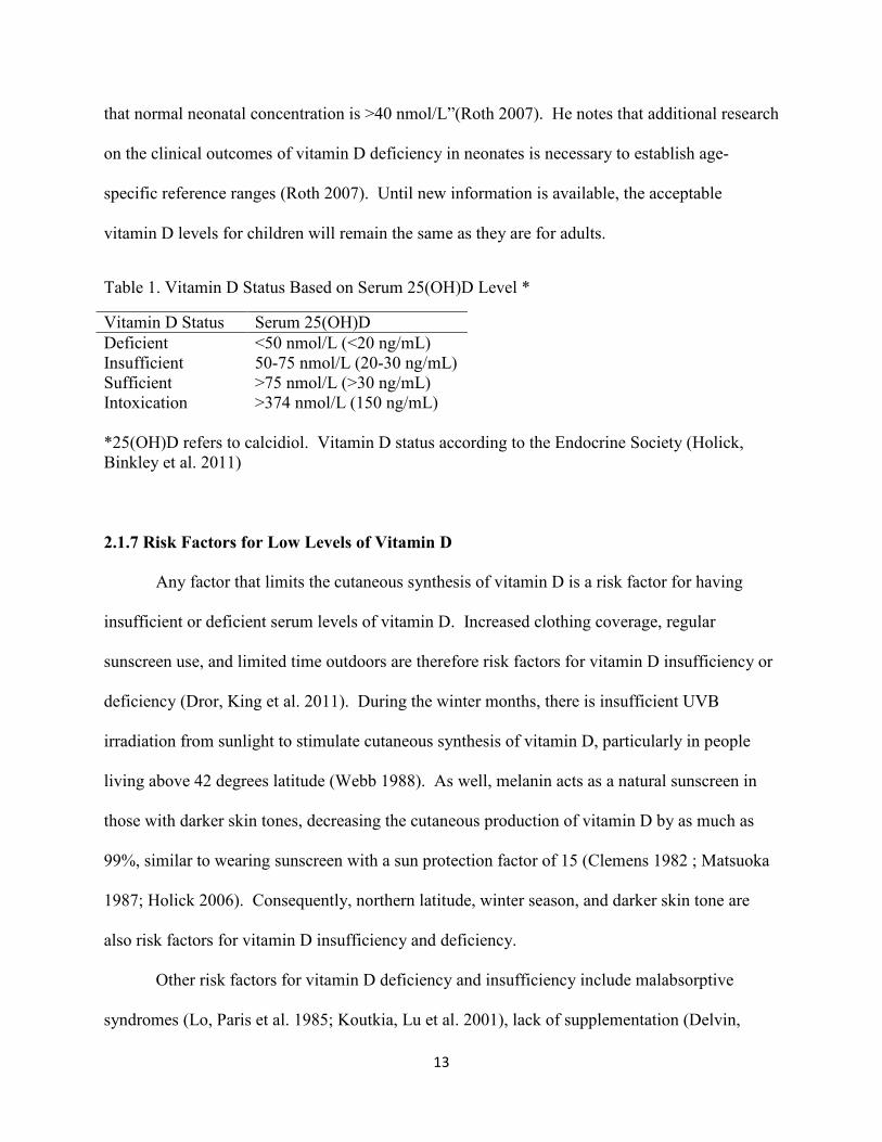
Table 1. Vitamin D Status Based on Serum 25(OH)D Level *
Vitamin D Status Serum 25(OH)D
Deficient <50 nmol/L (<20 ng/mL) Insufficient 50-75 nmol/L (20-30 ng/mL) Sufficient >75 nmol/L (>30 ng/mL) Intoxication >374 nmol/L (150 ng/mL)
*25(OH)D refers to calcidiol. Vitamin D status according to the Endocrine Society (Holick, Binkley et al. 2011)
2.1.7 Risk Factors for Low Levels of Vitamin D
Any factor that limits the cutaneous synthesis of vitamin D is a risk factor for having
insufficient or deficient serum levels of vitamin D. Increased clothing coverage, regular
sunscreen use, and limited time outdoors are therefore risk factors for vitamin D insufficiency or
deficiency (Dror, King et al. 2011). During the winter months, there is insufficient UVB
irradiation from sunlight to stimulate cutaneous synthesis of vitamin D, particularly in people
living above 42 degrees latitude (Webb 1988). As well, melanin acts as a natural sunscreen in
those with darker skin tones, decreasing the cutaneous production of vitamin D by as much as
99%, similar to wearing sunscreen with a sun protection factor of 15 (Clemens 1982 ; Matsuoka
1987; Holick 2006). Consequently, northern latitude, winter season, and darker skin tone are
also risk factors for vitamin D insufficiency and deficiency.
Other risk factors for vitamin D deficiency and insufficiency include malabsorptive
syndromes (Lo, Paris et al. 1985; Koutkia, Lu et al. 2001), lack of supplementation (Delvin,

14
Salle et al. 1986), high body mass index (BMI) (Bell, Epstein et al. 1985; Wortsman, Matsuoka
et al. 2000; Arunabh, Pollack et al. 2003; Snijder, van Dam et al. 2005), certain medications
(Pascussi, Robert et al. 2005), age (MacLaughlin and Holick 1985; Holick, Matsuoka et al.
1989), socioeconomic status (Laitinen, Rasanen et al. 1995; Pehlivan, Hatun et al. 2003;
Rasanen, Kronberg-Kippila et al. 2006), and genetic mutations in the vitamin D receptor or the
vitamin D binding protein (McGrath, Saha et al. 2010).
When cutaneous synthesis does not produce adequate levels of vitamin D, the body relies
on absorption of vitamin D from the intestine. Malabsorptive syndromes, such as inflammatory
bowel disease, celiac disease or cystic fibrosis, decrease the ability of the intestine to absorb any
ingested vitamin D (Lo, Paris et al. 1985; Koutkia, Lu et al. 2001). A lack of dietary vitamin D
makes less vitamin D available for absorption by the intestine, and medication use, particularly
anticonvulsants, corticosteroids and rifampin, can interfere with its subsequent metabolism (Bell,
Epstein et al. 1985; Pascussi, Robert et al. 2005). Metabolized vitamin D is less physiologically
available to those with a high BMI, as the fat-soluble vitamin becomes sequestered in the fat and
results in lower serum vitamin D levels (Bell, Epstein et al. 1985; Wortsman, Matsuoka et al.
2000; Arunabh, Pollack et al. 2003; Snijder, van Dam et al. 2005). Increased age results in lower
vitamin D levels in both children (Roth, Martz et al. 2005) and the elderly (MacLaughlin and
Holick 1985; Holick, Matsuoka et al. 1989), and socioeconomic status, reflected by education
level, occupation and household income, is a risk factor for low levels of vitamin D because low
socioeconomic status families are less likely to consume sufficient vitamin D in their diets
(Laitinen, Rasanen et al. 1995; Rasanen, Kronberg-Kippila et al. 2006). Finally, genetic
mutations in the vitamin D receptor and vitamin D binding protein play a role in modulating
vitamin D levels (McNally, Leis et al. 2009), though the mechanism of this is poorly understood

15
(McGrath, Saha et al. 2010).
Ethnicity is also a risk factor for low serum levels of vitamin D (Camargo, Ingham et al.
2010). Although skin pigmentation likely accounts for much of the observed ethnic differences,
genetic and environmental differences among ethnic groups may also contribute to lower serum
25(OH)D levels and these warrant further study.
2.1.7.1 Risk Factors for Low Levels of Vitamin D in Newborns
Because vitamin D is transported across the placenta, newborns are entirely dependent on
their mothers for vitamin D. Therefore, through contribution to low maternal vitamin D status,
the risk factors listed above are also risk factors for low vitamin D status in the newborn. Other
established risk factors for vitamin D deficiency in infancy include maternal vitamin D
deficiency and breastfeeding without supplementation, as human milk contains only 20 IU/L of
vitamin D (Canadian Pediatric Society 1988). Longer gestational age and younger maternal age
were associated with low cord blood vitamin D levels in a New Zealand study involving 929
newborns (Camargo, Ingham et al. 2010), and an Australian study involving 901 newborns also
found an association between low newborn vitamin D and younger maternal age (Bowyer,
Catling-Paull et al. 2009). Longer gestational age provides a longer period for a fetus to draw
upon its mother’s declining vitamin D levels, and lower maternal age may lead to less sun
exposure or less compliance in taking prenatal vitamins (Camargo, Ingham et al. 2010).
However, these findings require replication and further investigation before they are accepted as
risk factors for low levels of vitamin D in newborns.
16
2.2 Vitamin D in Pregnancy
2.2.1 Guidelines for Vitamin D Supplementation in Pregnancy
There are no consensus guidelines for vitamin D supplementation during pregnancy. The
Canadian Pediatric Society (CPS) recommends vitamin D supplementation of 2000 IU/day
during pregnancy and lactation (Canadian Pediatric Society 2007), while Health Canada
recommends 600 IU/day for pregnant and breast-feeding women (Health Canada 2010). The
Health Canada guidelines are based on the 2010 U.S. Institute of Medicine report, which is
primarily intended for food manufacturers, while the CPS guidelines are based a thorough
literature review and are intended for clinicians.
Meanwhile, the National Institute for Health and Clinical Excellence in the United
Kingdom recommends 400 IU/day of vitamin D during pregnancy (National Institute for Health
and Clinical Excellence 2008), the World Health Organization recommends 200 IU/day (World
Health Organization 2004), and the Endocrine Society recommends 600 IU/day (Holick, Binkley
et al. 2011). However, the Endocrine Society also recognizes that doses of up to 1500 – 2000
IU/day may be required to maintain sufficient serum vitamin D levels (>75 nmol/L) during
pregnancy and lactation (Holick, Binkley et al. 2011).
Recent studies have also suggested the need for much higher intakes during pregnancy,
with one suggesting that supplementation of 4000 IU/day was safe and most effective in
achieving sufficiency in pregnant mothers and their offspring (Hollis, Johnson et al. 2011), and
another showing that doses of up to 10,000 IU/day for five months in pregnancy did not elevate
levels into the toxic range (Hollis and Wagner 2004). Studies have also indicated that doses of
400 IU/day are inadequate to achieve sufficient serum vitamin D levels in mothers (Dror, King et
al. 2011) and newborns (Bodnar, Simhan et al. 2007). The author of a British review article

17
concluded that, “The lack of unified advice on vitamin D supplementation of pregnant mothers
in the UK hinders the implementation of primary prevention strategies and is likely to leave
some deficient mothers without supplementation (Hypponen and Boucher 2010).” The same
could be said of Canada.
The Endocrine Society Clinical Practice Guidelines suggest that pregnancy and lactation
are an indication for 25(OH)D screening (Holick, Binkley et al. 2011), but the British Journal of
Nutrition argues that given the prevalence of deficiency and the cost of the assay, it would be
much cheaper and more effective to have universal guidelines for Vitamin D supplementation
during pregnancy than screening every pregnant woman for deficiency (Hypponen and Boucher
2010).
2.2.2 Prevalence of Vitamin D Insufficiency and Deficiency in Pregnancy
This review will use the definition of vitamin D insufficiency and deficiency espoused by
the Endocrine Society, which defines vitamin D sufficiency as a serum vitamin D level of >75
nmol/L, insufficiency as 50-75 nmol/L, and deficiency as <50 nmol/L (Holick, Binkley et al.
2011). Though the definitions of vitamin D deficiency and insufficiency differ among studies,
most conclude that low levels of vitamin D are prevalent among pregnant mothers (Kazemi A
2009; Camargo, Ingham et al. 2010; Dror, King et al. 2011). In Britain’s Avon Longitudinal
Study of Parents and Children, 90% of the 10,000 white pregnant mothers had 25(OH)D
concentrations in the deficiency range (<50 nmol/l) during winter and spring while 28% were
seriously deficient (<25 nmol/l) (Golding, Pembrey et al. 2001). In Oakland, California, 54% of
275 mothers and 90% of their neonates had insufficient serum 25(OH)D levels (<75 nmol/L)
over a one year period (Dror, King et al. 2011). Similar studies in France (Madelenat, Bastian et

18
al. 2001), India (Sachan 2005), Iran (Kazemi A 2009), China (Specker, Ho et al. 1992) and New
Zealand (Camargo, Ingham et al. 2010) have shown that maternal serum levels of vitamin D are
frequently deficient, and a review article on serum vitamin D levels in pregnancy found that 35
of 76 studies reviewed had mean or median vitamin D levels below 35 nmol/L (Schroth, Lavelle
et al. 2005).
In Canada, several studies have assessed the vitamin D status of pregnant mothers. In
Vancouver, 24% and 65% of 336 multiethnic pregnant women had deficient (<50 nmol/L) and
insufficient (<75 nmol/L) serum levels of vitamin D, respectively (Li 2011). In Newfoundland
and Labrador, a study of 50 pregnant women found that 38% of pregnant women had insufficient
(50-75 nmol/L) and 42% had deficient (<50 nmol/L) levels of vitamin D (Newhook, Sloka et al.
2009). In the Northwest Territories, a study of 121 pregnant mothers revealed that the mean
vitamin D level for Caucasian and Native Indian mothers was insufficient (< 75nmol/L), while
the mean vitamin D level for Inuit mothers was deficient (<50 nmol/L) (Waiters, Godel et al.
1999). Three small studies in Northern Manitoba (n= 32, 35, 37) found that the median serum
vitamin D concentrations in pregnant mothers was seriously deficient (<25 nmol/L) (Smith
2000), and in Montreal, a small study (n = 27) of mothers with twin pregnancies found a mean
serum vitamin D concentration in the deficiency range (<50 nmol/L) (Reddy, Norman et al.
1983). Finally, a Winnipeg study (n = 50) found that 46% of mothers who had delivered within
the past 48 hours were deficient (<37.5 nmol/L) (Weiler, Fitzpatrick-Wong et al. 2005).
Therefore, existing research indicates that vitamin D deficiency in pregnant mothers is prevalent
in Canada.

19
2.2.3 Outcomes of Vitamin D Sufficiency, Insufficiency and Deficiency in Pregnancy
While one 2011 review article concluded that there is insufficient evidence to suggest
that low vitamin D levels in the first trimester are associated with adverse pregnancy and
neonatal outcomes (Nassar, Halligan et al. 2011), a second 2011 review article concluded that,
“Recent evidence supports a role of maternal vitamin D status, particularly early in pregnancy, in
modulating the risk of pregnancy complications and in sustaining fetal growth, bone
development, and immune maturation” (Dror 2011). Though existing evidence on the role of
maternal vitamin D in pregnancy, neonatal, and child health outcomes is sparse and sometimes
conflicting, it suggests that maternal vitamin D supplementation is required to prevent adverse
health outcomes in mothers, infants and children.
2.2.3.1 Maternal Effects of Gestational Vitamin D Deficiency
Low levels of vitamin D throughout pregnancy have been associated adverse health
outcomes for mothers, including preeclampsia (Bodnar, Catov et al. 2007), gestational diabetes
(GDM) (Soheilykhah, Mojibian et al. 2010), intrahepatic cholestasis of pregnancy (Wikstrom
Shemer and Marschall 2010), periodontal disease (Dietrich, Joshipura et al. 2004; Boggess,
Espinola et al. 2011), Caesarean section (Merewood, Mehta et al. 2009) and HIV progression
and mortality (Mehta, Giovannucci et al. 2010). This thesis will not cover maternal outcomes of
vitamin D deficiency in detail as its focus is on neonatal health.
2.2.3.2 Effects of Gestational Vitamin D Deficiency on Neonatal Health
Size
The existing body of evidence indicates that low vitamin D levels during pregnancy are a
20
significant predictor for newborn size. An American study found that serum 25(OH)D <37.5
nmol/L was a significant risk factor for infants being born SGA (small for gestational age)
among white, but not black, women (Bodnar, Catov et al. 2010). A Dutch study of 3730 mothers
found that infants born to mothers with serum 25(OH)D <29.9 nmol/L were more likely to be
born SGA and had a significantly lower birth weight than those born to mothers with serum
25(OH)D >50 nmol/L (Leffelaar, Vrijkotte et al. 2010). An Iranian study of 449 pregnant
women found that mothers who received the daily recommended doses of calcium and vitamin D
had infants with higher birth length and were less likely to have infants with low birth weight
(Sabour 2006). Similarly, a US study including 2251 pregnant women found that vitamin D
intake was a significant predictor for infant birth weight (Scholl and Chen 2009). However, an
Australian study found that first trimester vitamin D levels were not associated with birth weight,
head circumference, knee-heel length, or crown-heel length (Morley, Carlin et al. 2006).
Randomized controlled trials (RCTs) of vitamin D supplementation have conflicting
evidence on the role of vitamin D in infant size. An RCT in 80 British mothers of Asian descent
found that the incidence of SGA was higher in the placebo group than in mothers supplemented
with 1000 IU of vitamin D per day (Brooke and Wood 1980; Brooke, Brown et al. 1981).
Infants of the placebo group mothers subsequently gained less weight and had lower rates of
linear growth than infants of the supplemented group (Brooke, Butters et al. 1981). By contrast,
a French RCT involving pregnant women in their third trimester gave one dose of 200,000 IU to
one group (n = 27), 1000 IU daily to one group (n = 21), and a placebo to a third group (n = 29)
and found no differences in birth weight between groups (Mallet, Gugi et al. 1986).
Studies based on dietary recall support the role of vitamin D in infant size. A study
performed in Calgary, Alberta showed that mothers who consumed more than one cup (>250

21
mL, n = 207) of milk per day during pregnancy had infants who weighed more than mothers who
consumed less milk (n = 72). Each additional cup of milk per day was associated with a 41g
increase in birth weight. Vitamin D was also a significant predictor of birth weight and each
additional microgram of vitamin D per day was associated with an 11g increase in birth weight.
However, there was no significant difference in infant lengths and head circumferences between
groups (Mannion, Gray-Donald et al. 2006).
The Danish National Birth Cohort included data from 50,117 mother-infant pairs and
found similar results. Findings were adjusted for potential confounding variables and milk
consumption was inversely associated with the risk of SGA birth and directly associated with
large for gestational age (LGA) birth and birth weight. Milk consumption was also associated
with very small increases in head circumference and birth length (Olsen, Halldorsson et al.
2007). Though this evidence is compelling, it is difficult to control for all confounding factors
and larger, randomized-controlled trials are needed to assess the role of vitamin D in birth size.
Apgar scores
Only one study investigated the relationship of maternal vitamin D with Apgar scores.
This study assessed the vitamin D intake of 449 pregnant women in Iran and found that adequate
maternal calcium and vitamin D intake was associated with higher 1-minute Apgar scores
(Sabour 2006). However, it did not measure serum levels of 25(OH)D, the effects of calcium
and vitamin D were not assessed separately, and there was no adjustment for potential
confounding factors. Further research is required to support this finding.

22
Skeletal development
Studies have associated maternal vitamin D status during pregnancy with bone
development in utero and bone size during infancy and childhood. One study involved 424
pregnant women and used a high-resolution 3D ultrasound to measure fetal femur length and
distal metaphyseal cross-sectional area. This study found that lower maternal 25(OH)D
concentrations were associated with greater femoral metaphyseal cross-sectional area and a
higher femoral splaying index at 19 and 34 weeks’ gestation, which suggests that maternal
vitamin D status can influence fetal bone development early in pregnancy (Mahon, Harvey et al.
2010). In another study, newborn tibial bone mineral content and cross-sectional area, but not
bone mineral density, were higher in a group of newborns whose mothers had 25(OH)D
concentrations >42.5 nmol/L compared with newborns whose mothers’ 25(OH)D concentrations
were below this level, further suggesting the maternal vitamin D concentration influences bone
size (Viljakainen, Saarnio et al. 2010). When a subset of the participants in this study were
followed at age 14 months, tibial cross-section area remained higher in children whose mothers
had better vitamin D status during pregnancy (Viljakainen, Korhonen et al. 2011). Finally, a
study in the UK of 198 mother-child dyads found that maternal serum 25(OH)D deficiency or
insufficiency was associated with reduced whole-body and lumbar spine bone mineral
concentration in their 9-year-old children (Javaid, Crozier et al. 2006). The universal conclusion
of these studies is that vitamin D supplementation of pregnant women may lead to increased
skeletal development and decreased fracture risk in their offspring.
Severe maternal vitamin D deficiency may result in congenital rickets and hypocalcemic
seizures (Russell and Hill 1974; Orbak, Karacan et al. 2007), although this is rare.

23
2.2.3.3 Effects of Gestational Vitamin Deficiency on Infant and Child Health
Neurological development
Low vitamin D levels during pregnancy may have long-lasting effects on the developing
fetus. The presence of vitamin D receptors throughout the brain and its role in the development
of the nervous system implies that maternal vitamin D levels may have an impact on fetal brain
maturation (Garcion, Wion-Barbot et al. 2002). Only one study has assessed the role of vitamin
D in the neurological development of the child. A study performed in the UK (n = 178) reported
that sufficient maternal serum 25(OH)D status (>75 nmol/L) did not appear to impact the 9-year-
old child’s psychological health or intelligence (Gale, Robinson et al. 2008). However, no
studies have investigated the relationship of maternal or newborn vitamin D status with
neurological development in infancy.
Asthma and atopy
Low dietary and supplemental intake of vitamin D during pregnancy has been associated
with increased risk of wheezing illness, asthma and allergic rhinitis. In Scotland, a study
assessing the vitamin D intake of 1,212 mothers found that 5-year-old children were more likely
to ever wheeze, wheeze in the previous year, or have a persistent wheeze if their mothers had a
low vitamin D intake during pregnancy (Devereux, Litonjua et al. 2007). A US study similarly
found that 3-year-old children were more likely to have a recurrent wheeze if their mothers had a
low vitamin D intake during pregnancy (Camargo, Rifas-Shiman et al. 2007). In Finland, a
study of 1669 5-year-old children found that the risk of asthma and allergic rhinitis was
increased in children whose mothers had low vitamin D intake during pregnancy (Erkkola, Kaila
et al. 2009), and in Japan, a study of 763 mother-child pairs found that low vitamin D intake

24
during pregnancy was a risk factor for eczema and wheeze in infants aged 16-24 months
(Miyake, Sasaki et al. 2010).
Diabetes
Maternal vitamin D deficiency has also been related to the risk of Type 1 and Type 2
diabetes. An Indian study of 568 mothers found that 5 and 9.5-year-old children of vitamin D-
deficient (<50 nmol/L) mothers had higher insulin resistance than children whose mothers had
sufficient vitamin D levels during pregnancy (Krishnaveni, Veena et al. 2011). A Norwegian
survey of 85 diabetic patients and 1,071 control patients found that when mothers took cod liver
oil during their pregnancy, their offspring had a lower risk of Type 1 diabetes (Stene, Ulriksen et
al. 2000).
2.2.4 Conclusion
Evidence suggests that low levels of vitamin D are extremely prevalent during pregnancy
and may adversely affect neonatal, infant and child health. In Saskatchewan, the prevalence of
low levels of vitamin D is likely to be high due to the northern latitude of the province, the high
Aboriginal population, the high prevalence of obesity, and the lack of sun exposure during the
winter months. Further studies will need to be conducted in order to estimate the prevalence of
low levels of vitamin D in Saskatchewan’s pregnant population and appreciate the role of
maternal vitamin D supplementation in infant and child health.
25
2.3 Vitamin D in Newborns
2.3.1 Prevalence of Cord Blood Vitamin D Insufficiency and Deficiency
25(OH)D easily crosses the placenta (Hillman and Haddad 1974), and maternal levels of
25(OH)D are slightly higher than cord blood 25(OH)D levels (Sachan 2005; Bodnar, Simhan et
al. 2007). Therefore, given the high prevalence of maternal vitamin D deficiency throughout the
world, a similarly high prevalence of newborn vitamin D deficiency is expected. Deficient cord
blood vitamin D has been documented in India (n = 207, mean cord blood 21.0 +/- 14.2 nmol/L)
(Sachan 2005), Iran (n = 552, 93% <35 nmol/L) (Maghbooli 2007), Australia (n = 901, 11% <25
nmol/L) (Bowyer, Catling-Paull et al. 2009), Greece (n = 123, 8.1% <25 nmol/L) and the United
States (n = 40, 65% <30nmol/L) (Lee, Smith et al. 2007), among other countries. In an
American study of 200 black and 200 white neonates, vitamin D deficiency (<37.5 nmol/L) and
insufficiency (37.5-80 nmol/L) occurred in 45.6% and 46.8% of black neonates, respectively,
compared with 9.7% and 56.4% of white neonates (Bodnar, Simhan et al. 2007). Another study
of 210 newborns performed in California found that 90% were either insufficient or deficient
(<75 nmol/L) in vitamin D (Dror, King et al. 2011). Even in lower latitudes and warm climates,
vitamin D deficiency in newborns is extremely prevalent. However, few studies have been
performed in Canada.
Three Canadian studies have assessed cord blood vitamin D concentrations and had
similar findings. A Winnipeg study (n = 50) found that 36% of infants had deficient cord blood
25(OH)D levels (<27.5 nmol/L). Average cord blood 25(OH)D in the deficient group was 9.0 ±
6.0 nmol/L, while average cord blood in the sufficient group was 39.7 ± 10.6 nmol/L (Weiler,
Fitzpatrick-Wong et al. 2005). In the Northwest Territories, a study of 121 newborns found that
both native and non-native newborns were on average deficient (34.2+/-13.1 nmol/L and 41.4+/-

26
23.5 nmol/L, respectively) (Waiters, Godel et al. 1999). In Newfoundland, a study of 51
newborns showed that average cord blood vitamin D was 48.6 nmol/L ± 17.5 nmol/L in the
winter and 63.3 nmol/L ± 13.5 nmol/L in the summer. Approximately 49% of infants were
insufficient and 35% were deficient (Newhook, Sloka et al. 2009). No studies have assessed
cord blood vitamin D status in Saskatchewan.
2.3.2 Outcomes of Cord Blood Vitamin D Sufficiency, Insufficiency and Deficiency
Although many studies have commented on the prevalence of low cord blood vitamin D
levels, few have studied its relationship with infant health. The following studies represent the
majority of the literature available on the association of cord blood vitamin D status with infant
and child health outcomes.
Neonatal heart failure and symptomatic neonatal hypocalcemia
Similar to the case reports of maternal vitamin D deficiency in relation to neonatal rickets
and hypocalcemia (Russell and Hill 1974; Orbak, Karacan et al. 2007), case reports have
associated low serum 25(OH)D in newborns with neonatal heart failure (Maiya, Sullivan et al.
2008) and symptomatic neonatal hypocalcemia (Shenoy, Swift et al. 2005; Teaema and Al
Ansari 2010). In Qatar, 19 cases of neonatal hypocalcemia due to neonatal serum vitamin D
deficiency were ultimately attributed to maternal vitamin D deficiency (Teaema and Al Ansari
2010). Similarly, in England, a review of 16 infants with rickets-associated dilated
cardiomyopathy found that no infant or mother was receiving the recommended vitamin D
supplementation (Maiya, Sullivan et al. 2008), suggesting that these life-threatening
presentations of heart failure could have been prevented.

27
Infection
Two studies have associated low cord blood vitamin D levels with an increased risk of
infections. Low cord blood vitamin D has been associated with an increased risk of RSV
(respiratory syncytial virus) in the first year of life (Belderbos 2011) and an increased risk of
respiratory infections (Camargo, Ingham et al. 2011). The increased infection rate could be
explained by the role of vitamin D in immunity, as one study demonstrated that low cord blood
vitamin D decreased in vitro monocyte responses (Walker, Zhang et al. 2011), and another found
that high cord blood vitamin D levels were associated with lower numbers of T regulatory cells
(Chi, Wildfire et al. 2011). (For more information on the role of vitamin D in the immune
system, please see Section 2.4.2). These studies suggest that vitamin D status in utero may
influence immune function in infancy. Additional studies are required to determine the role of
vitamin D in neonatal health and particularly in the risk of infection.
Asthma and atopy
Evidence surrounding the role of neonatal vitamin D levels in asthma and allergy is
conflicting. One study found that both high and low levels of cord blood 25(OH)D were
associated with increased isoallergen sensitization, but that vitamin D levels were not associated
with asthma or allergic rhinitis (Rothers, Wright et al. 2011). Similarly, another study found that
low cord blood 25(OH)D was associated with increased risk of respiratory infections and
wheezing, but not with an increased risk of asthma (Camargo, Ingham et al. 2011). In one study
(n = 649), cord blood vitamin D deficiency (< 27 nmol/L) was not associated with food
sensitivity (Liu, Wang et al. 2011). However, vitamin D deficiency did increase the risk of food
sensitivity among children carrying single nucleotide polymorphisms (SNPs) in the genes

28
involved in regulating IgE and 25(OH)D concentrations (Liu, Wang et al. 2011). This gene-
vitamin D interaction is likely not unique to food sensitivity and probably exists with many other
conditions. Future studies should search for associations between health outcomes and genetic
mutations, as relationships that are not initially apparent may surface once genetic variation is
accounted for.
The mechanism underlying these observations may pertain to vitamin D’s role as an
immunomodulator. One study found that vitamin D supplementation in mothers resulted in the
induction of tolerogenic dendritic cells in cord blood, which suggests that vitamin D status in
utero may influence the development of atopic diseases later in life (Rochat, Ege et al. 2010).
Another found that cord blood vitamin D levels were correlated with interleukin-10 (IL-10)
levels, and that IL-10 levels were highest in the summer (Zittermann 2004). Because IL-10 is
also involved in tolerizing exogenous antigens, it may also predispose infants to increased atopic
risk. Research is necessary to further characterize the role of vitamin D in atopy.
Other health outcomes
One small Canadian study (n = 50) reported that low cord blood 25(OH)D concentration
was related to lower bone mineral content in newborns (Weiler, Fitzpatrick-Wong et al. 2005). It
also found that low vitamin D concentrations were associated with greater infant weight and
length. This particular finding contradicts the general consensus of existing research, which has
associated lower gestational vitamin D levels with smaller infant size (see Section 2.2.3.2).

29
2.3.3 Conclusion
Several studies have indicated a high prevalence of low vitamin D levels in newborns
globally. In Canada, low levels of cord blood vitamin D were demonstrated in Newfoundland,
the Northwest Territories, and Winnipeg. As previously mentioned, Saskatchewan’s many risk
factors for vitamin D deficiency put its newborns at high risk for having low levels of vitamin D.
An estimate of the prevalence of vitamin D deficiency in Saskatchewan’s newborns would
establish the relevance of this problem to our province, while the identification of risk factors
may indicate populations that require screening and primary prevention strategies. Finally,
research that identifies the neonatal outcomes of vitamin D deficiency may aid in clarifying the
role of vitamin D in physiology and consequently reinforce the importance of vitamin D
supplementation.
2.4 Vitamin D in Children
2.4.1 Prevalence of Vitamin D Insufficiency and Deficiency in Children
It is conceivable, though not proven, that those born with a low level of vitamin D may
continue to have a low level of vitamin D later in life. As well, it may not be uncommon for a
child to develop vitamin D deficiency or insufficiency during childhood. A solid understanding
of the risk factors for vitamin D deficiency may improve health behaviours of families during
pregnancy, infancy and childhood and prevent vitamin D deficiency at all stages of life. As well,
an understanding of the physiologic mechanisms of vitamin D in utero and infancy are the first
steps to understanding the physiology of vitamin D in later life. Intrauterine vitamin D
deficiency may predispose to chronic diseases which effect individuals at all ages. It is
imperative to make connections between studies performed during pregnancy, infancy,

30
childhood, adolescence, adulthood and old age to fully understand how vitamin D levels may
impact an individual’s future health.
A study analyzing the results of the 2007/2008 Canadian Health Measures survey
indicated that for Canadians aged 6–11 years, only 52% have vitamin D levels in the sufficient
range (>75 nmol/L), while only 35% of children aged 12-19 years have vitamin D levels in the
sufficient range (Langlois, Greene-Finestone et al. 2010). The prevalence of vitamin D
deficiency in young children is high in many countries, including England (Callaghan, Moy et al.
2006), Greece (Nicolaidou, Hatzistamatiou et al. 2006), Iran (Neyestani, Hajifaraji et al. 2012),
India (Babu and Calvo 2010), Qatar (Bener, Al-Ali et al. 2009) and the United States (Gordon,
DePeter et al. 2004; Gordon, Feldman et al. 2008; Cole, Grant et al. 2010; Merewood, Mehta et
al. 2010). Several Canadian studies also indicate a high prevalence of low vitamin D levels in
children. In Manitoba, a cross-sectional study of 80 mother-infant pairs showed that 43% of
infants 3-24 months had a 25(OH)D level below normal (Lebrun, Moffatt et al. 1993). In
Quebec, a study of 1753 school-aged children showed that more than 93% had 25(OH)D levels
below 75 nmol/L, and mean levels ranged from 40 to 50 nmol/L (Mark, Gray-Donald et al.
2008). Another Quebec study of 159 children aged 8-11 whose parents were obese or had
metabolic syndrome showed that over 90% of them had serum 25(OH)D levels in the deficiency
range (Mark 2010). In Edmonton, the mean serum 25(OH)D level of 68 pediatric patients
presenting to the emergency room was 47 nmol/L, and all but one of the participants had a
vitamin D concentration <80 nmol/L (Roth, Martz et al. 2005). Finally, data collected from our
research group in Saskatoon indicated a mean vitamin D level of 49 +/- 24 nmol/L among a
group of children admitted to the pediatric intensive care unit with acute lower respiratory
infection (McNally, Leis et al. 2009).

31
The high prevalence of low vitamin D levels among Canadian mothers and children can
be explained by the reduced skin synthesis of vitamin D during the winter as well as an
inadequate intake of vitamin D in the diet. A study which analyzed the results of the 2004
Canadian Community Health Survey Cycle 2.2 showed that the majority of Canadians consume
less than the recommended intake of vitamin D from food, suggesting that more vitamin D
should be added to fortified foods, and that a wider range of foods should be fortified
(Vatanparast, Calvo et al. 2010). It also showed that white Canadians had a higher vitamin D
intake than non-white Canadians in most age sex groups (Vatanparast, Calvo et al. 2010), which,
along with darker skin tone and genetic diversity, aids in explaining the discrepancy in vitamin D
levels observed between white and non-white ethnicities. Given that the same risk factors may
create low levels of vitamin D in utero as in infancy and childhood, early detection and
prevention of low levels of vitamin D in pregnancy may substantially improve long-term child
health.
2.4.2 Outcomes of Childhood Vitamin D Insufficiency and Deficiency
Rickets
Severe vitamin D deficiency, which causes rickets, has been well-known and documented
since the 1800s (Welch, Bergstrom et al. 2000). However, despite the relatively recent
fortification of foods with vitamin D, nutritional rickets appears to be re-emerging in developed
countries (Welch, Bergstrom et al. 2000; Allgrove 2004). In Canada, a survey of 2325
pediatricians over two years estimated the incidence of rickets in Canada to be 2.9 per 100,000
(Ward, Gaboury et al. 2007). Of the 104 cases identified by the survey, the mean age at
diagnosis was 1.4 years, 89% had darker skin tones, and 94% had been breast fed. None of the

32
breast-fed infants had been supplemented according to current guidelines (400 IU/day) (Ward,
Gaboury et al. 2007), highlighting the importance of supplementing darker-skinned infants
during the first year of life. In a Winnipeg hospital between 1972-1984, 48 cases of rickets were
identified (Haworth and Dilling 1986) and in a Toronto hospital between 1988 and 1993, 17
cases of rickets were identified (Binet and Kooh 1996). Given the high prevalence of vitamin D
insufficiency and deficiency among Canadian children, clinically apparent cases of deficiency
represent only a small fraction of vitamin D-related child health outcomes.
Infections and Immunity
Vitamin D is able to act in an autocrine or paracrine manner in regulating immune
function (Aranow 2011). The vitamin D receptor is expressed on B cells, T cells, and antigen-
presenting cells, and these cells are also capable of producing 1-alpha hydroxylase, the enzyme
responsible for converting 25(OH)D to the active form of vitamin D, 1,25(OH)2D. The
metabolism of vitamin D is also regulated locally by the immune environment, resulting in local
levels different from systemic levels. The macrophage 1-alpha hydroxylase is not PTH-
dependant (Wu, Ren et al. 2007) and instead responds to circulating levels of 25(OH)D,
interferon (IFN) gamma, interleukin (IL) 1, or tumor necrosis factor (TNF) alpha (van Etten,
Stoffels et al. 2008). The macrophage 24-hydroxylase is non-functional, so there is no negative
feedback of 1,25(OH)2D production (Aranow 2011). The significance of vitamin D as an
autocrine hormone continues to be investigated.
Vitamin D is an important factor influencing inflammatory responses (Abu-Amer 1993;
Wang, Nestel et al. 2004). Vitamin D dampens systemic inflammatory response by inhibiting
antigen-induced T-cell proliferation, antagonizing the pro-inflammatory Th1 response,

33
suppressing macrophage release of pro-inflammatory cytokines, altering gene expression, and
decreasing adherence and chemotaxis of neutrophils (Bhalla, Amento et al. 1984; Rigby,
Denome et al. 1987). These roles are pertinent to inflammatory responses common to many
diseases.
Vitamin D status has been associated with infection risk. In 1981, Hope-Simpson
published a study that recognized the increased incidence of influenza A during the winter in
both northern and southern latitudes, while equatorial countries had a steady incidence of
influenza A (Hope-Simpson 1981). Since then, many studies have demonstrated a direct
association between hypovitaminosis D and increased risk of infection (McNally, Leis et al.
2009; Hewison 2010; Walker, Zhang et al. 2011). One study examined data from 18,883
individuals over age 12 in the US and found an association between low vitamin D levels (<30
ng/mL) and upper respiratory tract infection (Ginde, Mansbach et al. 2009). Low vitamin D
levels have also been associated with increased risk of bacterial vaginosis (Bodnar, Krohn et al.
2009), influenza (Cannell, Vieth et al. 2006), and HIV (Villamor 2006). Most convincingly, a
randomized, double-blind, placebo-controlled trial involving 167 schoolchildren showed that
vitamin D supplementation reduced influenza A incidence by 42% (Urashima, Segawa et al.
2010).
In children, many studies have associated low vitamin D levels with an increased risk of
respiratory infections (Muhe 1997; McNally, Leis et al. 2009). In an Ethiopian study of 500
children with pneumonia and 500 controls, children with pneumonia were 13 times more likely
to have rickets, suggesting that low levels of vitamin D may predispose children to respiratory
infection (Muhe 1997). An Indian study of 80 cases of acute lower respiratory infection (ALRI)
and 70 controls aged 2-60 months found that children with vitamin D levels >22.5 nmol/L had a

34
decreased risk of ALRI (Wayse, Yousafzai et al. 2004). A study conducted by our research
group in Saskatoon found no difference in vitamin D levels between children diagnosed with
pneumonia or bronchiolitis (n = 105) and children with no respiratory symptoms (n = 92).
However, children admitted to the pediatric intensive care unit with ALRI were more likely to be
vitamin D deficient than their healthy counterparts (McNally, Leis et al. 2009). However, no
studies have investigated the relationship of cord blood vitamin D status with the risk of
infection or illness at birth.
Autoimmune disease and diabetes
Low 25(OH)D levels have been associated with increased risk of autoimmune diseases,
such as multiple sclerosis (McMichael and Hall 1997; Munger, Zhang et al. 2004; Willer,
Dyment et al. 2005), type 1 diabetes mellitus (Staples, Ponsonby et al. 2003; Willer, Dyment et
al. 2005; Svoren, Volkening et al. 2009), rheumatoid arthritis (Merlino, Curtis et al. 2004), lupus
(Kamen and Aranow 2008), and inflammatory bowel disease (Cantorna, Munsick et al. 2000).
Northern latitude is associated with an increased risk of these diseases, further implicating
vitamin D in their pathogenesis (Holick 2007). In fact, the risk of multiple sclerosis is decreased
by approximately 50% in those living below 35 degrees latitude for the first 10 years of life
(Ponsonby, McMichael et al. 2002; Van Amerongen, Dijkstra et al. 2004). However, a Finnish
study had an opposite finding; vitamin D supplementation during the first year of life was
associated with an increased risk of asthma, atopy and allergic rhinitis at age 31 years
(Hypponen, Sovio et al. 2004). Despite the conflicting findings of present research, existing
studies suggest that vitamin D supplementation during infancy may have long-lasting impacts on
future health.

35
Several studies have associated childhood vitamin D levels with the risk of developing
Type 1 diabetes. A systemic review and meta-analysis of five studies found that the risk of type
1 diabetes was reduced in infants who were supplemented with vitamin D in all studies (Zipitis
2008). The odds ratio for the risk of diabetes in those supplemented compared with those not
supplemented was 0.71 (95% CI 0.60 to 0.84), and there was evidence for a dose-response effect
with supplementation (Zipitis 2008).
Studies also support the role of vitamin D in the pathogenesis of diabetes. Vitamin D
receptors have been found in both beta islet cells and immune cells, and a protective effect of
vitamin D in cytokine-induced beta cell dysfunction has been demonstrated (Mathieu, Gysemans
et al. 2005). Another study found that vitamin D deficiency was associated with increased
insulin resistance and metabolic syndrome (Chiu, Chu et al. 2004). Finally, low vitamin D status
has also been associated with increased fasting blood glucose and other indicators of lipid
metabolism in Canadian children, which suggests a role for vitamin D in cardiovascular health as
well as diabetes (Delvin, Lambert et al. 2010).
Cancer
In adults, serum 25(OH)D levels have been associated with increased risk of many types
of cancer, including colorectal (Lappe, Travers-Gustafson et al. 2007), breast (Garland, Garland
et al. 1990; Grant 2002), prostate (Bodiwala, Luscombe et al. 2003; Tuohimaa, Tenkanen et al.
2004), and colon cancer (Garland, Comstock et al. 1989; Pritchard, Baron et al. 1996). People
living at high latitudes are also at increased risk for these and other cancers (Holick 2007). A
randomized controlled trial involving 1179 postmenopausal women found that vitamin D
supplementation reduced the risk of cancer by 60 to 77% (Lappe, Travers-Gustafson et al. 2007),

36
and there is a growing consensus that vitamin D supplementation may reduce the risk of cancer
in all age groups.
Fewer studies have been conducted in the pediatric population. In children and young
adults, sunlight exposure reduces the risk of non-Hodgkin’s lymphoma by 40% (Chang, Smedby
et al. 2005), and decreases the risk of death from malignant melanoma once it develops
(Berwick, Armstrong et al. 2005). Lab studies show that vitamin D concentrations >75 nmol/L
maintain normal cell growth and prevent cells from undergoing malignant transformation
(Mawer, Hayes et al. 1994; Cross, Bareis et al. 2001; Tangpricha, Flanagan et al. 2001). Its role
in cellular differentiation, apoptosis, proliferation and angiogenesis (Nagpal, Na et al. 2005)
makes vitamin D a promising target for cancer research and increases the importance of vitamin
D supplementation in protecting future health.
Cardiovascular Disease
Vitamin D’s role in cardiovascular health is also being investigated (Wang, Pencina et al.
2008). As previously mentioned, low vitamin D levels have been associated with increased
fasting blood glucose and other indicators of lipid metabolism in Canadian children, an
observation that could have implications for future cardiovascular disease (Delvin, Lambert et al.
2010). Another study reported that deficient levels of vitamin D may predispose children to
obesity (Gilbert-Diamond 2010). Other studies show that higher latitude increases the risk of
hypertension and cardiovascular disease (Rostand 1997; Zittermann 2006), and that low vitamin
D levels are associated with congestive heart failure in adults (Zittermann 2006). Interestingly,
hypertensive patients who were exposed to ultraviolet light were able to normalize their blood
pressure (Krause, Buhring et al. 1998), which suggests a role for vitamin D influencing

37
cardiovascular health. Further studies are warranted to investigate the role of vitamin D in
cardiovascular health.
Neurological Development and Mental Health
Many studies have documented the presence of vitamin D, its receptor and its metabolites
in the brain and recognized that vitamin D may be an important modulator of brain development
(Eyles, Smith et al. 2005; Eyles, Feron et al. 2009; Eyles, Burne et al. 2011; Harms, Burne et al.
2011). Vitamin D may affect the developing brain through its roles in cellular differentiation,
neurotransmitter synthesis, neurotrophic factor expression, and the expression of proteins
involved in neuronal cell structure and metabolism (Eyles, Burne et al. 2011). VDR-deficient
mice exhibit neurobehavioural abnormalities, including increased anxiety, reduced social
behavior, abnormal grooming, pup cannibalism, impaired nest building, and neophobia (Kalueff,
Keisala et al. 2006). They also exhibit increased susceptibility to epilepsy and impaired
vestibular function (Kalueff, Minasyan et al. 2006; Keisala, Minasyan et al. 2009). These
findings suggest that vitamin D is necessary for normal neurological development and that low
levels of vitamin D may result in neurological impairment. However, as previously mentioned,
only one study has investigated the role of maternal vitamin D deficiency on child psychological
health, and it found no association between maternal vitamin D level and child psychological
health or intelligence at 9 years of age (Gale, Robinson et al. 2008).
Insufficient or deficient Vitamin D status during infancy and childhood may predispose
individuals to an increased risk of mental health diseases. Vitamin D levels have been associated
with seasonal affective disorder (Gloth, Alam et al. 1999), and sufficient levels of vitamin D
during the first year of life have been associated with a decreased risk of schizophrenia in males
38
(McGrath, Saari et al. 2004). Low vitamin D levels are occasionally responsible for neonatal
hypocalcemic seizures (Camadoo, Tibbott et al. 2007), and researchers are investigating a
possible role for vitamin D in epilepsy (Janjoppi, Katayama et al. 2008). Low levels of vitamin
D are frequently found in patients with minor and major depression (Hoogendijk, Lips et al.
2008; Arvold, Odean et al. 2009), and supplemental vitamin D and light therapy reduce
depressive symptoms (Jorde, Sneve et al. 2008; Shirani and St Louis 2009). It is likely that
vitamin D status plays a role in the pathogenesis of many types of mental illness and that
supplementation during pregnancy and infancy may improve outcomes in depression and other
disorders.
2.4.3 Conclusion
Vitamin D deficiency and insufficiency is prevalent in Canadian children, and the myriad
outcomes associated with vitamin D deficiency can adversely affect long term health. While
many of these studies are observational and only associations have been made, randomized-
controlled trials are unlikely to occur due to the health detriment to any group assigned to a
placebo arm. Vitamin D deficiency is associated with many adverse outcomes during infancy,
childhood, adolescence and adulthood. Further research is necessary to ascertain vitamin D’s
role in the pathogenesis of these diseases. It is possible that fetal and infant vitamin D levels
may have more long-lasting effects on child and adult health than previously imagined.
2.5 Health and Economic Burdens of Vitamin D Deficiency
Given the substantial evidence for an association between vitamin D levels and health
status, including potential life-threatening associations, one study estimated that the Canadian
39
death rate could fall by 37,000 deaths (22,300-52,300 deaths) if the mean serum 25(OH)D level
of Canadians was increased to 105 nmol/L. This represents 16.1% of annual deaths and could
reduce the annual economic burden by approximately $14.4 billion (6.9%) (Grant,
Schwalfenberg et al. 2010). In Saskatchewan, the health and economic burdens of low vitamin
D levels are likely to be higher than the national average because of the northern latitude in
which much of the province’s population resides, the inadequate winter sun exposure and
reduced UVB radiation from winter sunlight strength, the high prevalence of obesity, the high
Aboriginal population and the divergence of the Aboriginal population from their traditional
fish-based diet. Saskatchewan’s pregnant mothers, and therefore their newborns, are likely to
exhibit a high prevalence of vitamin D insufficiency and deficiency. If low vitamin D levels
occur during a critical period in fetal development, it is possible that the fetus may experience
long term adverse health consequences that would have been preventable through adequate
maternal supplementation.
2.6 Conclusions From the Literature Review and Rationale for the Current Study
The role of vitamin D in the pathogenesis of many non-skeletal infant and child health
outcomes is only beginning to be elucidated. Studies are difficult to interpret because of
differences in the definitions of vitamin D insufficiency and deficiency. Furthermore, the
acceptable level of vitamin D for neonates, infants and children is still debated. Vitamin D
studies in newborns are few and far between despite the inherent vulnerability of the fetus in
utero.
Canada’s, and particularly Saskatchewan’s, populations are at high risk of being vitamin
D deficient due to their high Aboriginal populations, northern latitude, and limited sun intensity

40
and exposure during the winter. Results of Canadian and non-Canadian studies indicating the
prevalence of maternal vitamin D deficiency during pregnancy are alarming. Given that vitamin
D travels through the placenta, a similarly prevalent level of newborn vitamin D deficiency
would be expected. However, no studies have examined vitamin D levels of Saskatchewan’s
newborn population.
Both low maternal and cord blood vitamin D levels have been linked with adverse
outcomes in infancy and childhood. Maternal vitamin D levels have been linked with infant size
and Apgar scores, and low levels of vitamin D during pregnancy may increase a child’s risk of
wheezing, asthma, and type 1 diabetes. Cord blood vitamin D levels have been associated with
neonatal heart failure and hypocalcemia, increased risk of childhood infections, and increased
risk of atopic disease. Maternal supplementation of vitamin D may mitigate the risk of several
infant and childhood diseases. Therefore, research indicates that the intrauterine environment
may have a long-lasting impact on the fetus and eventually the individual. However, clinical
associations have not been adequately explored in newborns, and the pathophysiology of the
associations identified in children remains unknown.
Although many studies have demonstrated that cord blood vitamin D deficiency and
insufficiency is prevalent, it remains uncertain what constitutes “low” levels of vitamin D for
newborns because correlations between low vitamin D levels and clinical outcomes are lacking.
It is unclear whether socio-demographic, obstetric or personal health factors may influence
newborn vitamin D levels, and it is unknown whether low cord blood vitamin D levels are
associated with adverse clinical outcomes at birth. Finally, there are few Canadian studies and
no Saskatchewan studies that describe cord blood vitamin D levels.
This research aims to estimate the prevalence of low levels of vitamin D among

41
Saskatoon’s newborns, which may aid in establishing the extent of vitamin D deficiency and
insufficiency in this population. This research also aims to identify socio-demographic, obstetric
and personal health factors that may influence cord blood levels of vitamin D, and identify
whether adverse clinical outcomes may be associated with low vitamin D levels.
The association of newborn vitamin D status with neonatal outcomes will aid the
understanding of the role of vitamin D’s role in utero and its impact on a newborn’s health.
Clinical associations may also aid in the development of age-specific reference ranges for
vitamin D levels, as “low” vitamin D levels for adults may be normal in newborns and may not
result in any adverse clinical outcomes.
Finally, despite compelling evidence of the importance of vitamin D sufficiency during
pregnancy, there is no consensus on guidelines for supplementation. The prevalence of vitamin
D deficiency and insufficiency in newborns and any adverse outcomes to the fetus or child may
be decreased or prevented with adequate maternal vitamin D supplementation. The final goal of
this research is to educate ourselves and our communities about the importance of vitamin D
supplementation. This knowledge will empower pregnant mothers, families and communities to
make informed decisions about the benefits of vitamin D supplementation. We hope to promote
adequate vitamin D supplementation throughout all stages of life.

42
CHAPTER 3: NEWBORN VITAMIN D STUDY
3.1 MATERIALS AND METHODS
3.1.1 Study design
This study had a cross-sectional design. Participants were recruited between December,
2011 and February, 2012. The study was advertised through posters in Royal University
Hospital (RUH), presentations at prenatal classes, and brochures available through midwives and
the West Winds Primary Health Centre.
Some participants were recruited at prenatal classes and informed consent was obtained
after the class. Most participants were approached on the Assessment, Antepartum and Labour
and Delivery wards at RUH. Participants were approached by the researcher when nurses
deemed they were appropriate candidates for the study and depending on the researcher’s
availability. Nurses of potential participants were always approached prior to entering any room
to ensure that potential participants were eligible for the study and comfortable enough to give
informed consent. The study was explained to potential participants and their families and they
were given approximately half an hour to consider participating.
After consent was obtained, participants were asked to fill out a questionnaire containing
questions about the mother’s health and demographic information. At delivery, nurses collected
a cord blood sample for vitamin D measurement and this was stored in a refrigerator until
transport to the research laboratory. After delivery, a spectrophotometer was used to measure the
skin reflectance of the infant. The medical records pertaining to labour, birth and immediate
post-partum history for the mother and the infant were used to collect information pertaining to
the pregnancy and delivery as well as anthropometric measurements and health status of the baby
at birth. This information was entered into an electronic database. (See Appendix 1 for Consent

43
Form, Participant Questionnaire and Recruitment Materials.)
3.1.2 Subjects
The study population included 65 maternal-fetal dyads delivering in the Saskatoon Health
Region (latitude 52°N). Consenting mothers of singleton pregnancies delivering in the
Saskatoon Health Region were included. Any pregnant women with pre-existing parathyroid or
calcium conditions, those who took medications that interfere with calcium metabolism (such as
diuretics or calcium channel blockers), those with thyroid disease and those without informed
consent were excluded.
Of the 89 participants recruited for the study, proper informed consent was not assured
for five participants and their information was not used in the analysis. Of the 84 participants
enrolled in the study, 19 failed to undergo cord blood collection at delivery and although their
demographic information was collected the variable of interest (cord blood vitamin D level) was
not available and therefore the information from these participants was excluded from statistical
analysis. As well, these participants were not followed post-partum, and therefore no
information about the obstetric or neonatal outcomes of these participants is available. For two
of these cases, samples were not collected due to precipitous deliveries. For the remaining 17
cases, the reason for failed blood collection was unknown but was likely due to a failure to
communicate participatory status with nurses. The remaining 65 samples were included in the
statistical analysis.

44
3.1.3 Data Collection
3.1.3.1 Cord Blood Collection, Storage and Analysis
At the time of delivery, cord blood (3cc) was collected by needle/syringe withdrawal
from an umbilical cord vein into a 3cc capacity heparinized vacutainer. Samples were stored in a
refrigerator (4oC) until they were transported to the testing laboratory. There, they were
centrifuged at 2500 revolutions per minute for 10 minutes, the supernatant was removed, and the
supernatant was stored at -80°C until analysis.
Quantitative plasma 25(OH)D was determined by enzyme immunoassay
(Immunodiagnostic Systems Ltd.). The assays were performed by the study investigators in the
Pediatric Rheumatic Disease Research Laboratory, University of Saskatchewan as we have
previously described (McNally, Matheson et al. 2008; McNally, Matheson et al. 2008) and were
run in accordance with the manufacturer’s specifications. Vitamin D External Quality
Assessment Scheme (DEQAS; www.deqas.org) samples were run in parallel with subject
samples.
3.1.3.2 Socio-Demographic, Obstetric and Personal Health Information
At enrolment, a study number was assigned to each participant and the following
enrolment information documented (See Appendix 1 for Study Questionnaire):
• Date of enrolment
• Maternal age
• Marital status: married, single, divorced, separated, common-law, widowed (Statistics
Canada 2006). Due to the small number of participants in other categories, marital status
was dichotomized into married and common-law for the purposes of analysis

45
• Maternal area of residence: determined by forward sortation area (the first three
characters of postal code; as an approximate indicator of rural residence a ‘zero’ as the
second character was considered rural and any other number as the second character as
urban) (Statistics Canada 2008)
• Maternal education level: elementary, high school, some university, technical training
beyond high school, one or more university degrees, other (Statistics Canada 2006). For
analysis purposes the education level was dichotomized as elementary or high school
education and some post-secondary education
• Household income: <$10,000, $10,000 – $25,000, $25,001 – $50,000, $50,001 –
$100,000, >$100,000. Due to the small number of participants in other categories, these
five categories were truncated into three categories (<$50,000, $50,001 - $100,000, and
>$100,000) for the purposes of analysis
• Parity: number of previous births trichotomized into no previous births, 1 previous birth
and greater than 1 previous birth
• Self-reported pre-pregnancy height (cm) and weight (kg)
• Maternal vitamin D supplementation: amount, frequency, duration. Dichotomized as
yes/no, where ‘yes’ indicated some vitamin D supplementation other than a prenatal
supplement
• Infant ethnicity: referred to all ethnic origins of the fetus. Non-white ethnicities included
East and Southeast Asian, Aboriginal, Chinese, South Asian, American Indian, East
Indian, Caribbean or Arab origins (Statistics Canada 2006). For analysis purposes
ethnicity was dichotomized into Caucasian origins only and some non-Caucasian
ethnicity

46
• Maternal occupation: Management Occupations, Business, Finance and Administrative
Occupations, Natural and Applied Sciences and Related Occupations, Health
Occupations, Occupations in Social Science, Education, Government Service and
Religion, Occupations in Art, Culture, Recreation and Sport, Sales and Service
Occupations, Trades, Transport and Equipment Operators and Related Occupations,
Occupations Unique to Primary Industry, Occupations Unique to Processing,
Manufacturing and Utilities, Student, Other (list) (Statistics Canada 2006). Mother’s
occupation was not used during statistical analysis as many participants answered
“Other.” As well, it was not clear if the participant had previously worked or was
currently working outside the home
• Pre-pregnancy medical conditions: listed. Dichotomized as yes/no (has medical
diagnosis for which medication is taken and no medical conditions)
• Infections or illnesses during the pregnancy. Dichotomized as yes/no
• Pre-pregnancy weight was confirmed using pre-pregnancy or early pregnancy weight in
the medical chart. Pre-pregnancy height and weight were used to calculate pre-
pregnancy BMI (kg/m2)
All information was entered into an electronic database under the study number of the
participant.

47
3.1.3.3 Anthropometric, Pregnancy and Delivery-Related Information
After delivery, health records pertaining to the pregnancy and delivery were consulted to
obtain the following information:
• Maternal weight gain during pregnancy: kg (determined by subtracting weight at first
prenatal visit from current weight or weight at last prenatal visit)
• Gestational age: weeks
• Method of delivery (vaginal or Cesarean birth)
• Presence of complications with delivery. Dichotomized as no complications or any
complications during delivery; any complications were specified as a string variable and
included, as examples, fetal decelerations, shoulder dystocia, vacuum or forceps-assisted
delivery, and Cesarean section for non-reassuring fetal heart rate, failure to progress, or
malpresentation,
• Infant height (cm), weight (g), head circumference (cm)
• Apgar scores: a measurement of neonatal vitality, measured at 1, 5 and 10 minutes.
Apgar scores are very infrequently measured at 10 minutes and 91% of this data was
missing. Therefore only Apgar scores at 1 and 5 minutes were included in the analysis
• Presence of any illness in the newborn. Dichotomized as yes/no, where ‘no’ indicated no
illness and ‘yes’ indicated any neonate who was admitted to the neonatal intensive care
unit or required any assisted ventilation or medical intervention immediately after birth
This information was entered into an electronic database under the study number of the
participant.

48
3.1.3.4 Skin Tone
Skin pigmentation was estimated by measuring skin reflectance using a portable
spectrophotometer (Model cm-600d, Konica Minolta). Skin reflectance readings (L*) were
taken at the inner aspect of the middle right forearm as well as on the sternum. L* is an index of
reflectance ranging from 0 (perfect black) to 100 (perfect white).
3.1.4 Statistical Analysis
All statistical tests were performed using SPSS Statistics 19 software. The definitions of
vitamin D deficiency, insufficiency and sufficiency are as follows: vitamin D sufficiency (> 75
nmol/L), vitamin D insufficiency (50 to 74.99 nmol/L) and vitamin D deficiency (< 50 nmol/L)
(Holick, Binkley et al. 2011).
3.1.4.1 Description of Study Population and Vitamin D Status
Descriptive demographic data were used to provide information about the population
under investigation. A comparison between the 65 included and 19 excluded participants was
performed using chi-square tests for categorical variables with only two categories and t tests
were used for continuous variables.
A histogram and tests for skewness and kurtosis were used to determine whether the cord
blood vitamin D level had a normal distribution (see Figure 1 in Section 3.2.1). Once the
assumption of normality was satisfied, further statistical analysis using cord blood vitamin D
level as a continuous dependent variable could be pursued.

49
3.1.4.2 Risk Factors for Low Cord Blood Vitamin D Levels
Mean levels of vitamin D were reported for each category within each categorical
variable. Independent t-tests and one-way ANOVAs were used to determine whether any
statistically significant difference existed between the mean vitamin D levels between categories
for each categorical variable. The Pearson correlation coefficient was used to evaluate the
correlation between continuous variables and vitamin D levels.
Linear regression was used to determine whether any association existed between cord
blood vitamin D levels and the independent variables. Multiple linear regression analysis was
performed to find the most parsimonious and best fitting model to predict cord blood vitamin D
levels. It was performed in three steps:
1) The crude analysis included all available data within a variable (most variables had
some missing data) and involved a bivariate analysis between each independent variable and
cord blood vitamin D level (the dependent variable).
2) A Full Model was created which included all variables from the Crude Model with a p
< 0.25 (Hosmer and Lemeshow 2000).
3) A Reduced Model was created using all variables from the Full Model with a p < 0.10.
Partial F tests were used to examine whether any variable from the Full Model that was excluded
from the Reduced Model (i.e because p > 0.10) should be included in the Reduced Model.
Following this, all variables that were included in the Full Model but excluded from the Reduced
Model were added individually to the Reduced Model to test for their role as a confounder. If
any added variable caused greater than a twenty percent change in the coefficients of the original
variables, confounding was determined to be present and that variable was retained in the
reduced model.

50
3.1.4.3 Outcomes Associated with Low Cord Blood Vitamin D Levels
The obstetrical and neonatal outcomes of the study population were described. Mean
cord blood vitamin D level for each category within each categorical variable was described.
Independent t-tests were used to compare the mean cord blood vitamin D levels between
categories for each categorical variable. Simple linear regression was used to determine whether
any association existed between continuous variables and cord blood vitamin D levels. Finally,
cord blood vitamin D level was used as an independent variable in the prediction of each
obstetric and neonatal outcome.
3.1.5 Confidentiality and Ethics
The protocol Bio 11-185 for the Newborn Vitamin D Study was approved by the
Research Ethics Board on November 3, 2011 at the University of Saskatchewan as well as by the
Saskatoon Health Region. Informed assent/consent was obtained from participants included in
the study. For 5 participants, reliable informed consent was not obtained and therefore these
subjects were excluded from analysis. Data and samples were coded by study number only and
stored securely.

51
3.2 RESULTS
3.2.1 Description of Study Population and Vitamin D Status
Of the 89 participants recruited for the study, proper informed consent was not assured
for five participants and their information was not used in the analysis. Of the 84 participants
enrolled in the study, 19 failed to undergo cord blood collection at delivery and their information
was therefore excluded from the statistical analysis. For two of these cases, samples were not
collected due to precipitous deliveries. For the remaining 17 cases, the reason for failed blood
collection was unknown but was likely due to a failure to communicate participatory status with
nurses. The remaining 65 samples were included in the analysis.
Univariate statistics for the sociodemographic variables of all enrolled participants are
shown in Tables 2 and 3. Comparisons were made between those who were included in the
analysis and those who were not because of failed blood collections. There were no statistically
significant differences between the included and excluded populations. Overall, 85% of included
mothers reported taking prenatal supplements and 26% reported taking supplemental vitamin D
in addition to prenatal supplements.
Table 2. Categorical Demographic and Obstetric Characteristics of Participants
Included (n = 65)
Excluded (n = 19)
Variable n (%) n (%) p-value*
Residence Rural Urban
21 (32%) 44 (68%)
6 (32%)
13 (68%)
0.95
Mother’s Education Level Elementary or High School Some Post-Secondary Missing
8 (12%)
55 (85%) 2 (3%)
5 (26%)
14 (74%) 0
0.15

52
Marital Status Married Single Separated Common-law Missing
43 (66%)
1 (2%) 0
18 (28%) 3 (5%)
13 (68%)
1 (5%) 1 (5%)
4 (21%) 0
0.22
Household Income ≤ $50,000 $50,001 - $100,000 >$100,000 Missing
13 (20%) 23 (35%) 21 (32%) 8 (12%)
5 (26%) 4 (21%) 8 (42%) 2 (11%)
0.45
Ethnicity of Infant Any Non-White Ethnicity No Non-White Ethnicity Missing
17 (26%) 43 (66%)
5 (8%)
6 (32%)
13 (68%) 0
0.79
Parity
0 1 >1 Missing
37 (57%) 19 (29%) 7 (11%) 2 (3%)
8 (42%) 6 (32%) 3 (16%) 2 (11%)
0.64
Prenatal Supplement Yes No Missing
55 (85%) 7 (11%) 3 (5%)
11 (58%) 3 (16%) 5 (26%)
0.31
Vitamin D Supplement Yes No Missing
17 (26%) 44 (68%)
4 (6%)
1 (5%)
12 (63%) 6 (32%)
0.12
Maternal Medical Condition Yes No Missing
16 (25%) 48 (74%)
1 (2%)
5 (26%)
14 (74%) 0
0.91
Illness During Pregnancy Yes No Missing
23 (35%) 41 (63%)
1 (2%)
7 (37%)
12 (63%) 0
0.94
* p-value comparing the included to excluded groups

53
Table 3. Continuous Demographic and Obstetric Characteristics of Participants
Included (n=65)
Excluded (n=19)
Variable Mean (SD)
n (%) Mean (SD)
n (%) p-value *
Age (years) 29.6 (4.4) 63 (97%) 30.2 (4.3) 9 (47%) 0.67 Pre-Pregnancy BMI (kg/m2)
25.2 (5.3) 59 (91%) 26.5 (5.7) 18 (95%) 0.37
Weight Gain During Pregnancy (kg)
13.5 (5.1) 59 (91%)
Gestational Age (weeks)
39.8 (1.5) 0
Skin tone forearm (L*) 62.5 (3.5) 52 (80%) Skin tone sternum (L*) 63.0 (2.6) 39 (60%)
* p-value comparing the included to excluded groups
Cord blood vitamin D levels had a relatively normal distribution (Figure 1). Average
cord blood vitamin D was 64.1 nmol/L ± 19.8 nmol/L. Samples ranged from 17.5 nmol/L to
106.3 nmol/L. The cord blood vitamin D values were 50.2 nmol/L, 64.2 nmol/L and 78.5
nmol/L for the 25th, 50th and 75th percentiles respectively. When compared with previously
established categories of deficient (<50 nmol/L), insufficient (50-75 nmol/L), and sufficient (>75
nmol/L) (Holick, Binkley et al. 2011), cord blood vitamin D level was deficient in 22% and
insufficient in 48% of the 65 newborns studied.

54
Figure 1. Histogram of Cord Blood Vitamin D Levels (n = 65)
3.2.2 Risk Factors for Low Cord Blood Vitamin D Levels
Mean cord blood vitamin D level and standard deviation for each level of the categorical
variables is presented in Table 4. There was no statistically significant difference in vitamin D
level between categories for any variable. However, there was a trend towards higher vitamin D
levels in those with rural residence (p = 0.09).

55
Table 4. Mean (SD) Levels of Cord Blood Vitamin D by Categorical Independent Variables
Variable
n (%) Mean Cord Blood Vitamin D Level (nmol/L) (SD)
p-value*
Residence Rural Urban
21 (32) 44 (68)
70.2 (17.8) 61.2 (20.3)
0.09
Education Elementary or High School Some Post-Secondary
8 (12)
55 (85)
61.3 (29.7) 64.8 (18.3)
0.65
Marital Status Married Common-law
43 (66) 18 (28)
65.4 (19.3) 63.4 (21.2)
0.72
Income ≤ $50,000 $50,001 - $100,000 >100,000
13 (20) 23 (35) 21 (32)
65.5 (18.6) 64.5 (22.6) 67.1 (18.6)
0.91
Ethnicity of Infant Any Non-White Ethnicity No Non-White Ethnicity
17 (26) 43 (66)
65.0 (21.7) 63.9 (19.3)
0.84
Parity
0 1 >1
37 (57) 19 (29) 7 (11)
61.3 (19.2) 70.6 (18.1) 63.4 (26.0)
0.20
Prenatal Supplement Yes No
55 (85) 7 (11)
65.0 (19.6) 55.4 (22.4)
0.23
Vitamin D Supplement Yes No
17 (26) 44 (68)
65.5 (25.0) 62.4 (17.0)
0.59
Maternal Medical Condition Yes No
16 (25) 48 (74)
67.8 (19.0) 63.4 (20.0)
0.45
Illness During Pregnancy Yes No
23 (35) 41 (63)
63.4 (18.5) 65.1 (20.5)
0.74
*p value comparing the vitamin D levels between categories within each variable
The r and p-values for the correlation between each continuous independent variable and
cord blood vitamin D level are presented in Table 5. Low weight gain during pregnancy was
associated with low cord blood vitamin D levels (r = 0.27, p = 0.04). Vitamin D level was not
significantly correlated with any other maternal demographic or obstetric risk factors. However,

56
lower maternal age and higher pre-pregnancy BMI showed trends of an association with lower
cord blood vitamin D levels (p = 0.07 and p = 0.09, respectively).
Table 5. Correlation Between Continuous Variables and Cord Blood Vitamin D Level
Variable n (%) r value p-value
Age (years) 63 (97) 0.227 0.07 Pre-pregnancy BMI (kg/m2) 59 (91) 0.226 0.09 Less Weight Gain During Pregnancy (kg)
59 (91) 0.274 0.04
Gestational Age (weeks) 65 (100) 0.085 0.50 Skin tone forearm (L*) 52 (80) 0.194 0.17 Skin tone sternum (L*) 39 (60) 0.107 0.52
The results from the linear regression analyses are presented in Table 6. This included
the examination of the association between each risk factor and cord blood vitamin D level at a
crude level and two levels of adjustment. The Full Model contains all variables from the Crude
Model with a p < 0.25, while the Reduced Model contains all variables from the Full Model with
a p < 0.10 (Age, Rural). The Full Model has an R2 of 0.344 (p = 0.02) in predicting vitamin D
levels (n = 41). The Reduced Model has an R2 of 0.270 (p < 0.01) in predicting vitamin D levels
(n = 41). None of the variables included in the full model that were excluded from the reduced
model affected the β estimate of those variables included in the reduced model. As such, these
variables were not included as confounders.

57
Table 6. Crude and Adjusted Results from Linear Regression Analyses Predicting Vitamin D Levels
57
Crude Full (n = 41) Reduced (n = 41) Variable n (%) of 65 β (SE) p-value β (SE) p-value β (SE) p-value
Age 63 (97%) 1.016 (0.553) 0.07 1.809 (0.568) <0.01 1.840 (0.557)
<0.01
Rural Address (ref: No) 65 (100%) 9.049 (5.171) 0.09 10.651 (5.727)
0.07 9.296 (5.395)
0.09
Pre-pregnancy BMI 59 (91%) -0.844 (0.482) 0.09 -0.759 (0.628)
0.24
Prenatal Supplement (ref: No)
62 (95%) 9.588 (7.983) 0.23 -1.486 (8.037)
0.85
Skin tone forearm (L*) 39 (60%) -1.045 (0.746) 0.17 -1.084 (0.729)
0.15
Weight Gain During Pregnancy (kg)
59 (91%) 1.047 (0.488) 0.04 0.573 (0.583) 0.33
Post-secondary Education (ref: No)
63 (97%) 3.487 (7.537) 0.65
Marital Status 62 (95%) -0.506 (1.393) 0.72 Household Income 53 (82%) 1.004 (3.523) 0.78 Non-white Ethnicity 60 (92%) 1.137 (5.735) 0.84 Parity 63 (97%) 3.534 (3.633) 0.33 Vitamin D Supplement (ref: No)
61 (94%) 3.051 (5.562) 0.59
Maternal Medical Condition (ref: No)
64 (98%) 4.330 (5.700) 0.45
Illness During Pregnancy (ref: No)
64 (98%) -1.737 (5.163) 0.74
Gestational Age (weeks)
65 (100%) -1.151 (1.700) 0.50
Skin tone sternum (L*) 39 (60%) -0.810 (1.240) 0.52

58
3.2.3 Outcomes Associated with Cord Blood Vitamin D Levels
The obstetric and neonatal outcomes that occurred among participants (n = 65) are
presented in Tables 7 and 8. Most participants had vaginal births (82%) and most of their infants
(85%) did not have any illness or require any medical intervention at birth. Their infants had a
mean length of 52.8 ± 2.9cm, a mean weight of 3616 ± 523g, a mean head circumference of 34.9
± 1.4cm, and mean Apgar scores of 7.8 ± 1.6 at 1 minute and 8.8 ± 0.6 at 5 minutes. Apgar score
at 10 minutes was not considered due to the large number of missing values (Table 8).
Table 7. Frequency (%) of Categorical Obstetric and Neonatal Outcomes (n = 65)
Variable n (%) of 65
Method of Delivery Vaginal Cesarean
53 (82%) 12 (19%)
Delivery Complication Yes No Missing
28 (43%) 34 (52%)
3 (5%) Infant Illness at Birth Yes No Missing
7 (11%)
55 (85%) 3 (5%)
Table 8. Descriptives of Continuous Obstetric and Neonatal Outcomes (n = 65)
Variable Mean (SD) Missing n (%) of 65
Infant Length (cm) 52.8 (2.9) 0 Infant Weight (g) 3616 (523) 0 Infant Head Circumference (cm) 34.9 (1.4) 0 Apgar 1 minute 7.8 (1.6) 0 Apgar 5 minute 8.8 (0.6) 0 Apgar 10 minute 8.2 (1.0) 59 (91%)
The mean cord blood vitamin D level and standard deviation for each categorical variable
are presented in Table 9. There was no significant difference between categories within each
variable.

59
Table 9. Mean (SD) Levels of Cord Blood 25(OH)D Stratified by Categorical Variables
Variable Mean Cord Blood Vitamin D Level (nmol/L) (SD)
p-value*
Method of Delivery Vaginal Cesarean
63.2 (21.2) 67.8 (12.0)
0.47
Delivery Complication Yes No
63.8 (17.1) 63.3 (21.4)
0.92
Infant Illness at Birth Yes No
74.1 (18.3) 62.1 (19.3)
0.12
*p value comparing the vitamin D levels between categories within each variable
The r and p-values for the correlation between each continuous independent variable and
cord blood vitamin D level are presented in Table 10. Vitamin D level was not significantly
associated with any neonatal outcomes.
Table 10. Correlation Between Continuous Variables and Cord Blood 25(OH)D
Variable R value p-value
Infant Length (cm) 0.054 0.67 Infant Weight (g) 0.113 0.37 Infant Head Circumference (cm) 0.029 0.82 Apgar 1 minute 0.159 0.21 Apgar 5 minute 0.128 0.31
Vitamin D level was investigated as a risk factor for various obstetric and neonatal
outcomes. Vitamin D level was not significantly associated with any obstetric or neonatal
outcomes including Cesarean delivery (OR = 1.002, 95% CI = 0.998 – 1.006, p-value = 0.47),
delivery complications (OR = 0.000, 95% CI = 1.006 – 0.994, p-value = 0.92), infant illness at
birth (OR = 1.003, 95% CI = 1.007 – 0.999, p-value = 0.12), infant length (β = -0.008, SE =
0.018, p-value = 0.67), infant weight (β = -2.973, SE = 3.305, p = 0.37), infant head

60
circumference (β = -0.002, SE = 0.009, p = 0.82), Apgar 1 minute (β = -0.013, SE = 0.010, p =
0.21), and Apgar 5 minute (β = -0.004, SE = 0.004, p = 0.31).
3.2.4 Analysis of Statistical Power
This post-hoc analysis estimated the power achieved for all categorical variables with a
between-group significance of p < 0.25. The categorical variable with the highest power was
“Residence,” which had an effect size of 0.47, a power of 42% and would have required a
sample size of 160 participants to achieve a power of 80%. “Infant Illness at Birth” had an
effect size of 0.64, a power of 35% and would have required a sample size of greater than 200
participants to achieve a power of 80%. “Parity” had an effect size of 0.21, a power of less than
30%, and would have required a sample size of 228 to achieve a power of 80%, while “Prenatal
Supplement” had an effect size of 0.45, a power of 20% and would have required a sample size
of more than 400 to achieve a power of 80%. Because all other categorical variables had lower p
values than the four described above, these variables would have required much higher sample
sizes to achieve a power of 80%. Therefore, if a statistically significant difference was present in
the population, our study was inappropriately powered to detect it most of the time. In the best
scenario, for the variable “Residence,” a power of 42% means that our probability of committing
Type II error was 58% (our study would not detect a significant difference if one was present
58% of the time).

61
3.3 DISCUSSION
“Readers of medical literature need to consider two types of validity, internal and external.
Internal validity means that the study measured what it set out to; external validity is the ability
to generalise from the study to the reader’s patients.”
-David A Grimes and Kenneth F Schulz, The Lancet, 2002
3.3.1 Interpretation of Results
3.3.1.1 Prevalence of Cord Blood Vitamin D Insufficiency and Deficiency
The estimated prevalence of cord blood vitamin D deficiency and insufficiency in the
Saskatoon Health Region was lower than the prevalence reported by other Canadian studies. In
Winnipeg, 36% of 50 infants had deficient cord blood vitamin D levels (deficiency defined as
< 27.5 nmol/L) (Weiler, Fitzpatrick-Wong et al. 2005), in the Northwest Territories, the average
newborn cord blood vitamin D level was deficient (n = 121, deficiency defined as < 50 nmol/L)
(Waiters, Godel et al. 1999), and in Newfoundland, 35% and 49% of 51 infants had deficient
(defined as < 50 nmol/L) and insufficient (defined as 50 – 75 nmol/L) cord blood vitamin D
levels, respectively (Newhook, Sloka et al. 2009). Given the Saskatoon population’s many risk
factors for having low levels of vitamin D, it was expected that the prevalence of vitamin D
deficiency and insufficiency in our study population would be similar to or higher than the
prevalence found in other Canadian studies. Results indicated that 22% and 48% of our 65
newborns had deficient (< 50 nmol/L) and insufficient (50 – 75 nmol/L) cord blood vitamin D
levels, respectively. While the prevalence of cord blood vitamin D deficiency and insufficiency
in the Saskatoon Health Region was estimated to be lower than the prevalence reported in the
other Canadian studies, it is possible that our study underestimated the prevalence of vitamin D

62
insufficiency and deficiency (see Sections 3.3.1 and 3.3.2).
Similar to many previous studies, our study found that the 400 IU per day contained in
prenatal vitamins was not sufficient to produce sufficient maternal and cord blood levels of
vitamin D. For example, in Vancouver, Li et al. found that although 80% of 336 pregnant
women took a vitamin D-containing supplement containing ≥ 400 IU of vitamin D, 24% and
65% of women had deficient (< 50 nmol/L) and insufficient (< 75 nmol/L) serum levels of
vitamin D, respectively (Li et al., 2011). Bodnar et al. found that although 90% of women used
prenatal vitamins in the last trimester of pregnancy, vitamin D deficiency (cord blood vitamin D
levels < 37.5 nmol/L) and insufficiency (cord blood vitamin D levels 37.5 – 80 nmol/L) occurred
in 45.6% and 46.8% of black neonates and 9.7% and 56.4% of white neonates (Bodnar, Simhan
et al. 2007). Additionally, Dror et al. found that of the mothers who took a daily prenatal
supplement containing 400 IU of vitamin D, 50% had a serum vitamin D concentration that was
either insufficient or deficient (<75 nmol/L) (Dror, King et al. 2011). Similarly, this study
showed that despite daily prenatal multivitamin use by 85% of mothers (typically containing 400
IU of vitamin D) and additional vitamin D supplements taken by 26% of mothers, 22% of
newborns were deficient and 48% were insufficient in their cord blood vitamin D levels.
Maternal vitamin D supplementation of 400 IU per day was not enough to prevent vitamin D
insufficiency and deficiency in most newborns. This finding highlights the need for an urgent
revision of maternal vitamin D supplementation guidelines in order to produce sufficient cord
blood vitamin D levels in newborns.
3.3.1.2 Risk Factors for Cord Blood Vitamin D Insufficiency and Deficiency
Our study aimed to determine whether any socio-demographic, obstetric, or personal

63
health risk factors existed for low cord blood vitamin D levels in our study population. Each
categorical variable was tested to determine whether a significant difference existed between
mean cord blood vitamin D levels for each category within each variable (see Table 4).
Although there was no significant difference in mean cord blood vitamin D levels between
categories for each categorical variable, there was a trend towards higher levels of vitamin D in
those with a rural area of residence compared with an urban area of residence (p = 0.09).
The finding of a trend towards higher levels of vitamin D in those with a rural area of
residence may be due to higher socioeconomic status, better diet and genetic differences between
Caucasian and darker-skinned ethnicities. Bedroom communities that are a short distance from
Saskatoon likely have populations with a higher socioeconomic status who commute into
Saskatoon for employment. Those with higher socioeconomic status are more likely to consume
vitamin D-rich foods (Laitinen, Rasanen et al. 1995; Rasanen, Kronberg-Kippila et al. 2006). As
well, according to the 2011 Statistics Canada census, rural towns, such as Dundurn and
Rosetown, have predominantly Caucasian populations based on their reported mother tongues
and languages spoken at home (Statistics Canada, 2011). While the increased absorption of
vitamin D from sunlight in Caucasians would normally help to explain differences in vitamin D
levels between Caucasian and non-Caucasian populations, our study was conducted during the
winter months, and therefore this effect should have been negligible. Instead, genetic differences
between Caucasians and darker-skinned ethnicities may aid in explaining differences in vitamin
D levels, but little research exists to confirm this hypothesis.
Bivariate analysis revealed that low weight gain during pregnancy was associated with
low cord blood vitamin D levels (R = 0.274, p = 0.04). It also indicated that lower maternal age,
higher pre-pregnancy BMI, and urban area of residence were modestly associated with lower
64
cord blood vitamin D levels (p = 0.07, p = 0.09, and p = 0.09, respectively). The association of
low vitamin D levels with urban area of residence is discussed above. High BMI is a well-
established risk factor for having low levels of vitamin D (Bell, Epstein et al. 1985; Wortsman,
Matsuoka et al. 2000; Arunabh, Pollack et al. 2003), as the fat-soluble vitamin becomes
sequestered in the fat and is subsequently physiologically unavailable. As well, high pre-
pregnancy BMI has been associated with low pregnancy and neonatal vitamin D levels in a study
involving 400 mother-infant dyads (Bodnar, Catov et al. 2007). It is possible that participants
with higher BMIs gained less weight during pregnancy and that this could account for the low
vitamin D levels among those with low weight gain during pregnancy. This hypothesis is likely,
as a bivariate analysis using pre-pregnancy BMI as a predictor for weight gain during pregnancy
reveals a strong trend between the two variables given the small sample size (n = 56, p = 0.07).
It is also possible that those with a low weight gain during pregnancy had an inadequate diet to
produce both normal weight gain and sufficient vitamin D levels.
Low maternal age has been associated with cord blood vitamin D status in previous
literature. Younger maternal age was associated with lower neonatal vitamin D status in a New
Zealand study involving 929 newborns (Camargo, Ingham et al. 2010) as well as in an Australian
study involving 901 newborns (Bowyer, Catling-Paull et al. 2009). One author speculated that
younger mothers may be less compliant with prenatal vitamins or may have less sun exposure
(Camargo, Ingham et al. 2010). It is also possible that younger mothers may have a smaller
income than older mothers and may consume less expensive, less nutritious foods.
The linear regression model which best predicted cord blood vitamin D levels included
only maternal age and rural area of residence. This likely differs from the bivariate analysis due
to confounders which affect the relationship between the independent variables and the

65
dependent variable (cord blood vitamin D level).
3.3.1.3 Vitamin D Level as a Predictor for Obstetric and Neonatal Outcomes
Cord blood vitamin D level did not significantly predict any obstetric and neonatal
outcomes. There is little literature that examines the role of cord blood vitamin D levels in the
prediction of infant health outcomes, but a substantial body of literature supports the role of
maternal vitamin D intake in predicting birth weight and length (Sabour 2006; Scholl and Chen
2009; Leffelaar, Vrijkotte et al. 2010). It is possible that with a larger sample size, an association
between cord blood vitamin D levels and obstetric or neonatal outcomes may become apparent.
It is also possible that “normal” vitamin D levels for children differ substantially from “normal”
vitamin D levels for adults. Although some research conducted in children and adolescents
indicates that normal levels of vitamin D for children may be the same as they are for adults
(Guillemant, Taupin et al. 1999; Cheng, Tylavsky et al. 2003; Abrams, Griffin et al. 2005), little
or no research has been performed in neonates, infants or small children. In fact, one author
argues that because cord blood vitamin D samples typically contain 50-60% of maternal vitamin
D concentration, “normal” cord blood vitamin D concentration may be >40 nmol/L (Roth 2007).
Further research on the clinical implications of cord blood vitamin D deficiency or insufficiency
must be conducted before age-specific reference ranges are established.
3.3.2 Assessment of External Validity
The assessment of external validity will compare the study population with the general
population and comment on the presence and effect of selection bias.

66
3.3.2.1 Comparison of Study Population with General Population
The median income of the population under investigation is similar to the median income
of Saskatoon’s general population. In 2010, the median family income in Saskatoon was
$80,570 (Statistics Canada 2009). In this study, 32% of participants reported a household
income of greater than $100,000, 35% reported a household income of $50,001 - $100,000, and
20% reported a household income of less than $50,000. Therefore, median income in the study
population was in the range of $50,001 - $100,000, which is similar to the median family income
of $80,750 reported for the general population.
The study population is also similar to the Saskatoon Health Region population in terms
of ethnicity, area of residence, and maternal age at delivery. The most recent census data for
Saskatoon indicated that 17.2% of Saskatoon’s population had some non-white ethnicity
(Statistics Canada 2006), 73% of the Saskatoon Health Region population lived in the city of
Saskatoon (Neudorf 2009), and the highest fertility rates for women in the Saskatoon Health
Region were in the age groups of 25-29 and 30-34, respectively (Neudorf 2009). In comparison,
26% of our study participants indicated that their newborn had some non-white ethnicity, 68%
indicated an urban area of residence, and our study population had a mean maternal age of 29.6 ±
4.4 years. Therefore, for a small sample size, the incomes, ethnicities, area of residence and
mean maternal age at delivery of the study population closely matched those of the general
population in the Saskatoon Health Region.
Differences between the general population and the study population were most
prominent in maternal educational level and marital status. In 2006, 46% of adult women living
in Saskatchewan had some form of post-secondary education and 55% had elementary or high
school education (Saskatchewan Ministry of Advanced Employment and Labour 2009). In this
67
study, 85% of women reported some post-secondary education, while only 12% reported
elementary or high school education. An analysis of data collected from 5,643 mothers between
1992 and 1994 indicated that 27.3% of pregnant women had never been married, while 70.5%
were currently married (Jackson 2003). In our study, 94% of participants indicated their marital
status as either “married” or “common-law.” These differences in educational and marital status
between study and general populations may indicate either a response bias or a genuine
difference between populations. In any case, both education level and marital status may be
regarded as socioeconomic indicators, and low socioeconomic status is a risk factor for having a
low level of vitamin D (Laitinen, Rasanen et al. 1995; Rasanen, Kronberg-Kippila et al. 2006).
Our study population indicated that they were more educated and more likely to be married than
the general population. If our study population has a higher socioeconomic status than the
general population, we may have underestimated the prevalence of vitamin D deficiency and
insufficiency in the newborn population of the Saskatoon Health Region.
3.3.2.2 Sampling Bias
Sampling bias is a type of selection bias which causes some members of the population to
be less likely to be included than others due to a non-random selection of participants. This type
of bias refers to a skewing of the results due to the inclusion of study population that is not
reflective of the general population.
The study design may have favoured a healthier population. Potential participants were
recruited at prenatal tours as well as on the Labour and Delivery ward at Royal University
Hospital. While the population present at prenatal tours (and the population who receives any
prenatal care) tends to be a healthier, higher income, and more educated sub-section of the

68
population, it was hoped that recruitment on Labour and Delivery would allow for the inclusion
of more marginalized sections of the population. Although the women who chose to participate
in the study were more educated and more likely to be married than the general population, they
were similar to the general population in terms of income, ethnicity, area of residence, and
maternal age at delivery (see Section 3.3.2 Assessment of External Validity). Women who are
more educated may be more likely to consume nutrient-rich foods, and this may skew results in
favour of an underestimation of low levels of vitamin D in the general population. However, due
to the voluntary nature of participation in this study, this bias could not be controlled for.
The study design specifically excluded individuals with medical conditions or drugs that
might affect calcium or vitamin D levels. The exclusion criteria of the study ensured that all
mothers with thyroid or parathyroid problems, problems with calcium regulation, or those who
took medications that would interact with calcium regulation were excluded. Therefore, in the
absence of any undiagnosed conditions that may interfere with calcium or vitamin D regulation,
the study population did not have any medical conditions that would have skewed the results.
However, the general population has a small percentage of people with calcium regulation
problems who were not represented in our study. This bias is likely irrelevant in a population
with a small sample size.
3.3.3 Assessment of Internal Validity
“Bias undermines the internal validity of research” (Grimes 2002). Research bias refers
to deviation of the research results from the truth. Many types of bias in study design,
implementation and interpretation exist. The following type of bias are often present in
observational studies: selection bias, information bias, and confounding (Grimes 2002). In this

69
study, a single group was enrolled, the prevalence of an outcome (low cord blood vitamin D
levels) was estimated, and associations between that outcome and several independent variables
were examined. The following section will therefore explore selection bias, information bias and
confounding in the context of this study.
3.3.3.1 Information Bias
This type of bias results from the incorrect determination of exposure or outcome, or both
(Grimes 2002).
Missing maternal demographic data ranged between 0 – 12% for each variable measured
by the Participant Questionnaire (see Table 2), and this may have skewed the outcomes
measured in the study. A number of participants chose not to answer questions concerning their
age, marital status, education level, household income and ethnicity, among other questions. As
well, some information, such as weight gain during pregnancy, was not always present in the
medical records. The Participant Questionnaire stated clearly that participants did not have to
answer any questions that they feel uncomfortable answering, and this may have been one reason
that participants chose not to answer certain questions. It is also possible that participants felt a
lack of anonymity with their responses, as the questionnaires were collected from them
personally by the researcher (despite no names being present on the questionnaire itself). This
type of bias could potentially be avoided in the future by providing participants with a postage-
paid, addressed envelope so that questionnaires could be mailed in anonymously (identified only
by participant number). Although the missing data may have skewed the results towards more
socially acceptable responses, this is unlikely given the small amount of missing data for each
variable.

70
The demographic information of the included participants (n = 65) did not differ
significantly from the demographic information of the excluded participants (n = 19) (see Tables
2 and 3). Therefore, the exclusion of those participants from the analysis likely did not bias the
data. As well, because samples were not collected from 19 participants, these participants were
not followed post-partum and no information is available about their obstetric or neonatal
outcomes. It is therefore impossible to compare the included to the excluded groups in terms of
obstetric and neonatal outcomes. However, given their demographic similarity, the missing data
likely did not significantly skew the available data. Future studies could avoid this problem
through a study design that improved communication with labour and delivery staff so that
samples were not missed.
Information about the mother’s occupation was collected but not analyzed. The Statistics
Canada categories for occupation did not include “Homemaker” and it could not be discerned
whether participants had previously been or were currently working outside the home. As well,
many participants chose several categories or indicated “Other,” making it very difficult to
analyze these results. This could be avoided by asking, “Do you work outside the home?”
(Yes/No) prior to asking the participants to choose the single Statistics Canada category that best
describes their occupation. Because occupation is used as an indicator of socioeconomic status
(and low socioeconomic status is a risk factor for low levels of vitamin D), it is possible that an
association would have been present between employment status and cord blood vitamin D
levels. However, income and education level, other indicators of socioeconomic status, were
analyzed and were not significantly associated with cord blood vitamin D levels.
3.3.3.1.1 Accuracy of Collected Data: The Participant Questionnaire was answered
directly by participants and asked participants to answer only questions they felt comfortable

71
answering. This may have led to some non-response bias. As well, some questions were
difficult to answer. For example, “Maximum Education Level” was listed in 6 categories:
“elementary,” “high school,” “some university,” “technical training beyond high school,” “one
or more university degrees,” or “other (list).” These categories were loosely based on the
Statistics Canada 2006 Census. However, those who chose “other” were left without an
analyzable category. Also, the term “technical training beyond high school” makes any non-
university training, such as a Basic Life Support course, equivalent to a college degree. In this
study, due to the small sample size, Education Level was reduced to “Elementary or high
school,” and “Some post-secondary education.” Future studies would do better to use the
categories “elementary,” “high school,” “training other than college or university,” “college
degree,” and “university degree.”
The Questionnaire asked whether or not participants took an “additional vitamin D
supplement” but did not ask about prenatal supplements. Due to this lack of clarity, the
researcher asked each participant directly about her prenatal supplement use and vitamin D
intake in order to quantify the amount of vitamin D taken. This could be avoided through the use
of two separate questions stating, “Did you take a daily prenatal supplement?” and “Did you take
an additional vitamin D supplement?”
The data collected for ethnicity was difficult to interpret. Participants were instructed to
select all ethnicities that applied to their newborns (ethnic groups listed according to the
Statistics Canada 2006 Census). Several ethnicities were often selected, and this makes
ethnicities present several generations ago equivalent to ethnicities present within one
generation. For the purposes of analysis, this data was reduced to “No non-white ethnicity,” and
“Any non-white ethnicity” based on the ethnicities listed. Non-white ethnicities included East

72
and Southeast Asian, Aboriginal, Chinese, South Asian, American Indian, East Indian,
Caribbean or Arab origins (Statistics Canada 2006). The question could be clarified by asking
participants if they or their partners would identify themselves as visible minorities, as the
purpose of the question was to search for an association between darker skin tones and lower
vitamin D levels.
As previously mentioned, the Statistics Canada categories for maternal occupation were
difficult to use. It would have been useful to include the question, “Do you work outside the
home?” as there was no option for “Homemaker.” The question that asked participants to list
any infections or illnesses during pregnancy could have been clarified by asking participants to
list only infections or illnesses which required antibiotics or other medical treatment. Finally, the
term “household income” was unclear because it did not specify gross or net income.
Although the Participant Questionnaire could have been clearer, the collection of data
from participants was standardized. All participants were asked to fill out the Participant
Questionnaire and asked directly about their prenatal supplement use and vitamin D intake by the
researcher. After delivery, participant and newborn medical charts were consulted to obtain all
obstetric and newborn outcome information. Although different health care providers may have
completed different charts, this information was likely unbiased as the physicians caring for the
participants had no knowledge or consideration of the study.
Finally, the dependent variable, vitamin D, was objectively measured by a lab technician
who had no knowledge of participants as samples were identified by participant number only.
Levels were determined by enzyme immunoassay (Immunodiagnostic Systems Ltd.) which were
run in accordance with the manufacturer’s specifications. Vitamin D External Quality
Assessment Scheme (DEQAS; www.deqas.org) samples were run in parallel with subject

73
samples.
In conclusion, although the socio-demographic information could have been collected
through clearer questions on the Participant Questionnaire, the questions were identical for all
participants. There may have been some non-response bias which could not be controlled for.
Vitamin D intake was the only question assessed directly by the researcher. The obstetric and
neonatal outcome information derived from medical charts was likely unbiased and the cord
blood vitamin D levels were assessed objectively.
3.3.3.2 Confounding
Confounding occurs when a third factor interferes with a relationship between an
independent variable and the outcome variable. A confounding factor is associated with the
independent variable and effects the outcome variable (Grimes 2002). These relationships
should always be assessed when critically appraising research. By completing an adjusted
analysis, we attempted to control for potential confounders.
All variables included in the full model but not initially in the reduced model were added
individually to the Reduced Model (see Table 6) to test for confounding and because no
variables significantly changed the β values of the variables in the Reduced Model, it was
concluded that no confounders were present in the data set. However, the small sample size may
have masked confounding that would have been significant with a larger data set. As well,
variables outside the data set may have been confounders that were not controlled for.
For example, diet and sun exposure were not measured in the study population and may
have acted as confounders. Future studies may rectify this by taking detailed diet histories or
asking participants to recollect how much time was spent outdoors. Given that the study was

74
conducted over the winter months, sun exposure was likely minimal for all participants. Diet
histories, however, may have been a worthwhile endeavour.
In this study, each categorical variable creates several groups of participants being
studied, and these groups cannot be compared to each other without accounting for confounding
variables. For example, those with income < $50,000 may be have differences from those with
incomes $50,000 - $100,000 and > $100,000 other than income. They may have lower education
levels, take fewer prenatal supplements or vitamin D supplements, have an increased risk of
illness during pregnancy or have different ethnicities than those with higher incomes. Unless
those variables are accounted for, any association between vitamin D level and income is
assumed to be directly related to income. Therefore, bivariate analysis may be misleading, as it
attributes causation to only a single variable. Multivariate model building controls for all
potential confounders included in the model and decreases the risk of selection bias.
In this study, although bivariate analysis was used in the initial testing of variables, a
multivariate model was later used which controlled for many confounding variables. However,
other differences between groups within each variable, such as better response rates, cannot be
measured, as it is impossible to know whether participants with missing data belonged to one
group or another. This response bias could not be controlled for.
3.3.3.3 Sample Size and Statistical Power
Statistical power is the probability of rejecting the null hypothesis when it is false (the
probability of not making a Type II error). Power is influenced by the sample size, the effect
size and the levels of error we are willing to accept (significance criterion). Effect size refers to
the detectable difference in the outcome variable between groups (a higher-powered study will

75
detect smaller differences in the outcome variable). Given that the significance criterion
commonly used by the scientific community is 0.05, there is only a 5% chance that any study
using this criterion will find a false positive result (Type I, or α, error). Power may be increased
(and Type II error decreased) by increasing the significance criterion (for example, to p = 0.10),
but this increases the risk of Type I error. The accepted balance between Type I and Type II
errors is to use a significance criterion of 0.05.
Most researchers consider a power of 80% adequate, as this achieves a 4-to-1 trade-off
between Type II (β) and Type I (α) error (β = 0.20, α = 0.05) (Hulley, Cummings et al. 2007).
However, the choice of power level and alpha depends on the type of study, as studies reporting
adverse events require the highest possible power to avoid Type II error, while studies reporting
the success of a treatment should minimize alpha and Type I error. While a significance
criterion of α = 0.05 was used for this study, the post-hoc analysis of statistical power showed
that a power of 80% was not achieved for any categorical variable due to small sample size.
A conservative post-hoc analysis uses categorical rather than continuous variables to
estimate power, as categorical variables generally have lower power than continuous variables
(Hulley, Cummings et al. 2007). Our power analysis (Section 3.2.4) estimated the power
achieved for all categorical variables with a p < 0.25 and indicated that our probability of
committing Type II error was very high (58% for the highest-powered categorical variable) due
to our small sample size. Therefore, a larger sample size may be necessary to rule out the role of
chance in this study. Despite this, the effect sizes observed with the aforementioned variables,
other than “parity”, in this study were all greater than 0.45 indicating a moderate effect size.
Lower power may also indicate that any difference detected by our study may be very
likely to be present in the general population (our study only detects large differences).

76
However, given the number of comparisons made during bivariate analysis, it is equally likely
that those findings of significance are due to chance. For example, if 20 independent variables
are tested at an α = 0.05, there is a very high probability of making a Type 1 error (64%, or 1 –
0.9520) (Hulley, Cummings et al. 2007). While an adjustment for multiple comparisons would
control for this error, the Bonferroni approach, which divides the significance level by the
number of hypotheses tested, is usually too stringent. The Bayesian approach asserts that if each
individual variable has a reasonable probability of being correct based on previous research, no
adjustment for multiple comparisons is necessary (Hulley, Cummings et al. 2007). In this study,
each variable was included based on the results of previous research and physiological
plausibility. This study was also exploratory in nature. Therefore no adjustment for multiple
comparisons was made.
3.3.4 Other Strengths and Limitations
Due to the lack of data about the risk factors and neonatal outcomes associated with cord
blood vitamin D levels, this study was exploratory in its intent. This study assessed sixteen
potential socio-demographic and personal health risk factors and eight potential obstetric and
neonatal outcomes for association with cord blood vitamin D level. In the assessment for risk
factors, there was no correction for multiple comparisons because the bivariate analysis was only
used as a basis for multivariate model building. All variables with a p < 0.25 in bivariate
analysis were included in a Full Model, and all variables from the Full Model with a p < 0.10
were included in the Reduced Model. Due to the small sample size (n = 41 due to missing data)
and the small number of variables in the Full and Reduced Models (six and two variables,
respectively), no adjustment for multiple comparisons was made. Furthermore, none of the

77
variables included in the full model that were excluded from the reduced model affected the β
estimate of those variables included in the reduced model. Therefore, no confounding was
found to exist.
No neonatal outcomes had statistically significant associations with vitamin D levels
during the bivariate analysis and therefore no model building was pursued.
The small sample size of this study indicates that the study has little power, or little
probability of not making Type II error (falsely accepting the null hypothesis). A study with
little power has limited ability to detect associations that are present. However, this also means
that any statistical significance detected denotes a stronger association between the independent
and dependant variables. Therefore, the association of younger maternal age and urban area of
residence with low cord blood vitamin D levels is likely to be strong in this population. A larger
sample size may be necessary to confirm or refute this finding as well as ascertain whether
further risk factors are associated with low cord blood levels of vitamin D.
The largest strength of this study was that the data is likely to be both reliable (precise
and reproducible) and valid (reflective of the general population). Obstetric and neonatal
outcomes as well as cord blood vitamin D levels were measured objectively. Although the
measurement of socio-demographic factors was subject to non-response bias, the measurement
was standardized for all participants. The prevalence of cord blood vitamin D deficiency and
insufficiency in the study population is likely to be either reflective or an underestimation of that
in the general population. Although a larger sample size would be necessary to help rule out the
role of chance to determine whether any risk factors or neonatal outcomes are associated with
cord blood vitamin D levels, this small study’s negative results are likely to be reproducible.

78
3.3.5 Conclusions and Future Research Directions
Adequate vitamin D supplementation during pregnancy is likely a factor important to
ensuring optimal health outcomes for offspring. Despite a compelling body of research showing
that vitamin D deficiency is prevalent among both mothers and infants in Canada and worldwide
(see Sections 2.2.2 and 2.3.1), the problem of widespread vitamin D deficiency remains
insufficiently addressed. Researchers are only now beginning to appreciate the potential clinical
and economic implications of in utero vitamin D deficiency; studies have linked maternal
vitamin D levels with chronic diseases such as asthma (Devereux, Litonjua et al. 2007; Erkkola,
Kaila et al. 2009) and type 1 diabetes (Krishnaveni, Veena et al. 2011) in the offspring. Vitamin
D’s associations with infection, autoimmune disease, cancer, and bone health are better
understood, and low vitamin D status throughout life is associated with adverse health outcomes.
In fact, one study estimated that optimizing vitamin D levels in Canadians could reduce mortality
by 37,000 annually (a 16.1% reduction) and reduce the annual economic burden by $14.4 billion
(6.9%) (Grant, Schwalfenberg et al. 2010). Given the health and economic benefits afforded to
the very inexpensive supplement, the prevention of low levels of vitamin D should be a priority
for the medical and public health fields as well as the general public. Sufficient vitamin D status
should begin in utero and be maintained throughout life.
Our research estimates that the prevalence of vitamin D deficiency and insufficiency
among newborns in the Saskatoon Health Region is 22% and 48%, respectively. The
population’s many risk factors for low vitamin D levels, including its northern latitude, lack of
sun exposure during the winter months, high Aboriginal population and high prevalence of
obesity may aid in explaining these results. However, similar results have been found in warmer
climates that lack some or all of these risk factors (Bowyer, Catling-Paull et al. 2009; Dror, King
et al. 2011), which presents a stronger case for the role of maternal diet and supplementation in

79
preventing low newborn levels of vitamin D. The provision of adequate maternal vitamin D
supplementation may represent a tremendous opportunity for health and economic gain in
Saskatoon and around the world.
A lack of research that demonstrates safe and effective vitamin D dosages during
pregnancy has impeded consensus on maternal vitamin D supplementation guidelines. Health
Canada, the Endocrine Society, and the Canadian Pediatric Society have different
recommendations for vitamin D supplementation during pregnancy (see Section 2.2.1). Further
randomized-controlled trials of vitamin D supplementation in pregnancy may be necessary prior
to any consensus being reached.
A review of recent literature as well as the results of this study suggest that 400 IU of
vitamin D per day is insufficient to prevent vitamin D deficiency and insufficiency in mothers
and newborns (Bodnar, Simhan et al. 2007; Ward, Gaboury et al. 2007; Dror 2011; Li 2011). A
recent randomized controlled trial demonstrated the safety and efficacy of 4,000 IU of vitamin D
per day during pregnancy (Hollis, Johnson et al. 2011), and another study showed that doses of
up to 10,000 IU per day did not elevate vitamin D levels into the toxic range (Hollis and Wagner
2004). Although the Endocrine Society recommends a dose of 600 IU of vitamin D per day for
pregnant and lactating women, it also recognizes that doses of up to 1500 – 2000 IU per day may
be necessary to maintain a serum vitamin D level of greater than 75 nmol/L (Holick, Binkley et
al. 2011). The Canadian Pediatric Society’s recommendation of 2000 IU per day for pregnant or
lactating women (Canadian Pediatric Society 2007) is based on a recent literature review and
should perhaps be adopted by other agencies. However, further research is needed to confirm
this recommendation or to aid the creation of new guidelines for maternal vitamin D
supplementation.

80
Maternal vitamin D supplementation guidelines may need to be stratified according to
risk factors. Skin colour, BMI, sun exposure and a lack of dietary vitamin D consumption are all
well-recognized risk factors for having low levels of vitamin D. As well, genotype may play a
role in modulating vitamin D levels and requirements. Mothers with darker skin, mutations in
vitamin D-specific proteins, and other risk factors for having a low level of vitamin D may
require higher levels of supplementation than mothers without risk factors. Our research
indicates that young, urban mothers in the Saskatoon Health Region are at the highest risk of
having infants with a low cord blood vitamin D level, but further research is necessary to
confirm or refute this observation.
The role of vitamin D in fetal and neonatal life has yet to be elucidated. Our research
found no association between low neonatal vitamin D levels and obstetric or neonatal outcomes.
It is possible that this lack of association is because age-specific reference ranges for vitamin D
levels have yet to be determined. “Normal” vitamin D levels for neonates may be a fraction of
“normal” levels for adults. It is also possible that low fetal levels of vitamin D do not manifest
clinically until later in life, or that a larger study may discover associations between vitamin D
status and neonatal outcomes. Research that establishes the clinical relevance of low cord blood
vitamin D levels will shed light on both age-specific reference ranges for vitamin D levels and
our general understanding of vitamin D in physiology.
In conclusion, this study highlights a growing awareness that vitamin D deficiency and
insufficiency is a prevalent problem with a relatively simple solution. Low newborn vitamin D
levels are only the proverbial “tip of the iceberg” as vitamin D deficiency and insufficiency are
pervasive throughout life. Healthy diets, sun exposure and vitamin D supplementation may be
the “wonder-drugs” of the future. Multidisciplinary teams including nutritionists, dieticians,

81
nurses, physicians, and teachers will be needed to effect change in both supplementation
guidelines and public attitudes. Setting up a newborn for a healthy future begins at conception,
but we must collaborate to maintain health throughout infancy, childhood and beyond.

82
6. REFERENCES
Abrams, S., I. Griffin, et al. (2005). "Relationships among vitamin D levels, parathyroid
hormone, and calcium absorption in young adolescents." Journal of Clinical
Endocrinology & Metabolism 90(10): 5576-5581.
Abu-Amer, Y., Bar-Shavit, Z. (1993). "Impaired bone marrow-derived macrophage
differentiation in vitamin D deficiency." Cellular Immunology 151(2): 356-368.
Agliardi, C., Guerini, F, Saresella, M, Caputo, D, Leone, M, Zanzottera, M, Bolognesi, E,
Marventano, I, Barizzone, N, Fasano, M, Al-Daghri, N, Clerici, M (2011). "Vitamin
D receptor (VDR) gene SNPs influence VDR expression and modulate protection
from multiple sclerosis in HLA-DRB1*15-positive individuals." Brain, Behavior, &
Immunity 25(7): 1460-1467.
Allgrove, J. (2004). "Is nutritional rickets returning?" Archives of Diseases in Childhood
89(8): 699-701.
Anderson, L. N., M. Cotterchio, et al. (2011). "Vitamin D-related genetic variants,
interactions with vitamin D exposure, and breast cancer risk among Caucasian
women in Ontario." Cancer Epidemiology, Biomarkers & Prevention 20(8): 1708-
1717.
Aranow, C. (2011). "Vitamin D and the immune system." Journal of Investigative
Medicine 59(6): 881-886.
Arunabh, S., S. Pollack, et al. (2003). "Body fat content and 25-hydroxyvitamin D levels in
healthy women." Journal of Clinical Endocrinology & Metabolism 88(1): 157-161.
Arvold, D. S., M. J. Odean, et al. (2009). "Correlation of symptoms with vitamin D
deficiency and symptom response to cholecalciferol treatment: a randomized
controlled trial." Endocrine Practice 15(3): 203-212.
Babu, U. S. and M. S. Calvo (2010). "Modern India and the vitamin D dilemma: evidence
for the need of a national food fortification program." Molecular Nutrition & Food
Research 54(8): 1134-1147.
Belderbos, M. E., Houben, M.L., Wilbrink, B., Lentjes, E., Bloemen, E.M., Kimpen, J.L.,
Rovers, M., Bont, L. (2011). "Vitamin D deficiency in cord blood linked to RSV
infections." Pediatrics: 2010-3054.
Bell, N. H., S. Epstein, et al. (1985). "Evidence for alteration of the vitamin D-endocrine
system in obese subjects." Journal of Clinical Investigation 76(1): 370-373.
Bener, A., M. Al-Ali, et al. (2009). "Vitamin D deficiency in healthy children in a sunny
country: associated factors." International Journal of Food Sciences & Nutrition 60
Suppl 5: 60-70.
Berwick, M., B. K. Armstrong, et al. (2005). "Sun exposure and mortality from
melanoma." Journal of the National Cancer Institute 97(3): 195-199.
Bhalla, A. K., E. P. Amento, et al. (1984). "1,25-Dihydroxyvitamin D3 inhibits antigen-
induced T cell activation." Journal of Immunology 133(4): 1748-1754.
Binet, A. and S. W. Kooh (1996). "Persistence of Vitamin D-deficiency rickets in Toronto in
the 1990s." Canadian Journal of Public Health. Revue Canadienne de Sante
Publique 87(4): 227-230.
Bodiwala, D., C. J. Luscombe, et al. (2003). "Susceptibility to prostate cancer: studies on
interactions between UVR exposure and skin type." Carcinogenesis 24(4): 711-717.

83
Bodnar, L. M., J. M. Catov, et al. (2007). "Prepregnancy obesity predicts poor vitamin D
status in mothers and their neonates." Journal of Nutrition 137(11): 2437-2442.
Bodnar, L. M., J. M. Catov, et al. (2007). "Maternal vitamin D deficiency increases the risk
of preeclampsia." Journal of Clinical Endocrinology & Metabolism 92(9): 3517-
3522.
Bodnar, L. M., J. M. Catov, et al. (2010). "Maternal serum 25-hydroxyvitamin D
concentrations are associated with small-for-gestational age births in white women."
Journal of Nutrition 140(5): 999-1006.
Bodnar, L. M., M. A. Krohn, et al. (2009). "Maternal vitamin D deficiency is associated
with bacterial vaginosis in the first trimester of pregnancy." Journal of Nutrition
139(6): 1157-1161.
Bodnar, L. M., H. N. Simhan, et al. (2007). "High prevalence of vitamin D insufficiency in
black and white pregnant women residing in the northern United States and their
neonates." Journal of Nutrition 137(2): 447-452.
Boggess, K. A., J. A. Espinola, et al. (2011). "Vitamin D status and periodontal disease
among pregnant women." Journal of Periodontology 82(2): 195-200.
Bouillon, R., Van Cromphaut, S., Carmeliet, G. (2003). "Intestinal calcium absorption:
Molecular vitamin D mediated mechanisms." Journal of Cellular Biochemistry
88(2): 332-339.
Bowyer, L., C. Catling-Paull, et al. (2009). "Vitamin D, PTH and calcium levels in pregnant
women and their neonates." Clinical Endocrinology 70(3): 372-377.
Brooke, O. G., I. R. Brown, et al. (1981). "Observations on the vitamin D state of pregnant
Asian women in London." British Journal of Obstetrics & Gynaecology 88(1): 18-
26.
Brooke, O. G., F. Butters, et al. (1981). "Intrauterine vitamin D nutrition and postnatal
growth in Asian infants." British Medical Journal Clinical Research Ed. 283(6298):
1024.
Brooke, O. G. and C. Wood (1980). "Growth in British Asians: longitudinal data in the
first year." Journal of Human Nutrition 34(5): 355-359.
Callaghan, A. L., R. J. Moy, et al. (2006). "Incidence of symptomatic vitamin D deficiency."
Archives of Diseases in Childhood 91(7): 606-607.
Camadoo, L., R. Tibbott, et al. (2007). "Maternal vitamin D deficiency associated with
neonatal hypocalcaemic convulsions." Nutrition Journal 6: 23.
Camargo, C. A., Jr., T. Ingham, et al. (2011). "Cord-blood 25-hydroxyvitamin D levels and
risk of respiratory infection, wheezing, and asthma." Pediatrics 127(1): e180-187.
Camargo, C. A., Jr., T. Ingham, et al. (2010). "Vitamin D status of newborns in New
Zealand." British Journal of Nutrition 104(7): 1051-1057.
Camargo, C. A., Jr., S. L. Rifas-Shiman, et al. (2007). "Maternal intake of vitamin D
during pregnancy and risk of recurrent wheeze in children at 3 y of age." American
Journal of Clinical Nutrition 85(3): 788-795.
Canadian Pediatric Society (2007). Vitamin D supplementation: Recommendations for
Canadian mothers and infants. Paediatrics and Child Health. 12: 583-589.
Canadian Pediatric Society, I. a. I. H. C. (1988). Vitamin D supplementation for northern
native communities. . Canadian Medical Association Journal. 138: 229-230.
Cannell, J. J., R. Vieth, et al. (2006). "Epidemic influenza and vitamin D." Epidemiology &
Infection 134(6): 1129-1140.

84
Cantorna, M. T., C. Munsick, et al. (2000). "1,25-Dihydroxycholecalciferol prevents and
ameliorates symptoms of experimental murine inflammatory bowel disease."
Journal of Nutrition 130(11): 2648-2652.
Chang, E. T., K. E. Smedby, et al. (2005). "Family history of hematopoietic malignancy and
risk of lymphoma." Journal of the National Cancer Institute 97(19): 1466-1474.
Chapuy, M. C., P. Preziosi, et al. (1997). "Prevalence of vitamin D insufficiency in an adult
normal population." Osteoporosis International 7(5): 439-443.
Cheng, S., F. Tylavsky, et al. (2003). "Association of low 25-hydroxyvitamin D
concentrations with elevated parathyroid hormone concentrations and low cortical
bone density in early pubertal and prepubertal Finnish girls.[Erratum appears in
Am J Clin Nutr. 2006 Jan;83(1):174]." American Journal of Clinical Nutrition
78(3): 485-492.
Chi, A., J. Wildfire, et al. (2011). "Umbilical cord plasma 25-hydroxyvitamin D
concentration and immune function at birth: the Urban Environment and
Childhood Asthma study." Clinical & Experimental Allergy 41(6): 842-850.
Chiu, K. C., A. Chu, et al. (2004). "Hypovitaminosis D is associated with insulin resistance
and beta cell dysfunction." American Journal of Clinical Nutrition 79(5): 820-825.
Clemens, T. L., Adams, J.S., Henderson, S.L., and Holick, M.F. (1982 ). "Increased skin
pigment reduces the capacity of skin to synthesise vitamin D3." The Lancet 1(8263):
74-76.
Cole, C. R., F. K. Grant, et al. (2010). "25-hydroxyvitamin D status of healthy, low-income,
minority children in Atlanta, Georgia." Pediatrics 125(4): 633-639.
Cooper, C., K. Javaid, et al. (2005). "Developmental origins of osteoporotic fracture: the
role of maternal vitamin D insufficiency." Journal of Nutrition 135(11): 2728S-
2734S.
Cross, H. S., P. Bareis, et al. (2001). "25-Hydroxyvitamin D(3)-1alpha-hydroxylase and
vitamin D receptor gene expression in human colonic mucosa is elevated during
early cancerogenesis." Steroids 66(3-5): 287-292.
Delvin, E. E., M. Lambert, et al. (2010). "Vitamin D status is modestly associated with
glycemia and indicators of lipid metabolism in French-Canadian children and
adolescents." Journal of Nutrition 140(5): 987-991.
Delvin, E. E., B. L. Salle, et al. (1986). "Vitamin D supplementation during pregnancy:
effect on neonatal calcium homeostasis." Journal of Pediatrics 109(2): 328-334.
Devereux, G., A. A. Litonjua, et al. (2007). "Maternal vitamin D intake during pregnancy
and early childhood wheezing." American Journal of Clinical Nutrition 85(3): 853-
859.
Dietrich, T., K. J. Joshipura, et al. (2004). "Association between serum concentrations of
25-hydroxyvitamin D3 and periodontal disease in the US population." American
Journal of Clinical Nutrition 80(1): 108-113.
Dror, D. K. (2011). "Vitamin D status during pregnancy: maternal, fetal, and postnatal
outcomes." Current Opinion in Obstetrics & Gynecology 23(6): 422-426.
Dror, D. K., J. C. King, et al. (2011). "Association of modifiable and nonmodifiable factors
with vitamin D status in pregnant women and neonates in Oakland, CA." Journal of
the American Diet Association 111(1): 111-116.
85
Eloranta, J. J., C. Wenger, et al. (2011). "Association of a common vitamin D-binding
protein polymorphism with inflammatory bowel disease." Pharmacogenetics and
Genomics 21(9): 559-564.
Erkkola, M., M. Kaila, et al. (2009). "Maternal vitamin D intake during pregnancy is
inversely associated with asthma and allergic rhinitis in 5-year-old children."
Clinical & Experimental Allergy 39(6): 875-882.
Exton-Smith, A. N., H. M. Hodkinson, et al. (1966). "Nutrition and metabolic bone disease
in old age." The Lancet 2(7471): 999-1001.
Eyles, D., T. Burne, et al. (2011). "Vitamin D in fetal brain development." Seminars in Cell
& Developmental Biology 22(6): 629-636.
Eyles, D. W., F. Feron, et al. (2009). "Developmental vitamin D deficiency causes abnormal
brain development." Psychoneuroendocrinology 34 Suppl 1: S247-257.
Eyles, D. W., S. Smith, et al. (2005). "Distribution of the vitamin D receptor and 1 alpha-
hydroxylase in human brain." Journal of Chemical Neuroanatomy 29(1): 21-30.
Feskanich, D., D. J. Hunter, et al. (1998). "Vitamin D receptor genotype and the risk of
bone fractures in women." Epidemiology 9(5): 535-539.
Feskanich, D., W. C. Willett, et al. (2003). "Calcium, vitamin D, milk consumption, and hip
fractures: a prospective study among postmenopausal women." American Journal
of Clinical Nutrition 77(2): 504-511.
Gale, C. R., S. M. Robinson, et al. (2008). "Maternal vitamin D status during pregnancy
and child outcomes." European Journal of Clinical Nutrition 62(1): 68-77.
Garcion, E., N. Wion-Barbot, et al. (2002). "New clues about vitamin D functions in the
nervous system." Trends in Endocrinology & Metabolism 13(3): 100-105.
Garland, C. F., G. W. Comstock, et al. (1989). "Serum 25-hydroxyvitamin D and colon
cancer: eight-year prospective study." The Lancet 2(8673): 1176-1178.
Garland, F. C., C. F. Garland, et al. (1990). "Geographic variation in breast cancer
mortality in the United States: a hypothesis involving exposure to solar radiation."
Preventive Medicine 19(6): 614-622.
Gilbert-Diamond, e. a. (2010). "Vitamin D deficiency and anthropometric indicators of
adiposity in school-age children: a prospective study." American Journal of Clinical
Nutrition 92(6): 1446-1451.
Ginde, A. A., J. M. Mansbach, et al. (2009). "Association between serum 25-
hydroxyvitamin D level and upper respiratory tract infection in the Third National
Health and Nutrition Examination Survey." Archives of Internal Medicine 169(4):
384-390.
Gloth, F. M., 3rd, W. Alam, et al. (1999). "Vitamin D vs broad spectrum phototherapy in
the treatment of seasonal affective disorder." Journal of Nutrition, Health & Aging
3(1): 5-7.
Golding, J., M. Pembrey, et al. (2001). "ALSPAC--the Avon Longitudinal Study of Parents
and Children. I. Study methodology." Paediatric and Perinatal Epidemiology 15(1):
74-87.
Gong, G., H. S. Stern, et al. (1999). "The association of bone mineral density with vitamin
D receptor gene polymorphisms." Osteoporosis International 9(1): 55-64.
Gordon, C. M., K. C. DePeter, et al. (2004). "Prevalence of vitamin D deficiency among
healthy adolescents." Archives of Pediatrics and Adolescent Medicine 158(6): 531-
537.

86
Gordon, C. M., H. A. Feldman, et al. (2008). "Prevalence of vitamin D deficiency among
healthy infants and toddlers." Archives of Pediatrics & Adolescent Medicine 162(6):
505-512.
Grant, W. B. (2002). "An ecologic study of dietary and solar ultraviolet-B links to breast
carcinoma mortality rates." Cancer 94(1): 272-281.
Grant, W. B., G. K. Schwalfenberg, et al. (2010). "An estimate of the economic burden and
premature deaths due to vitamin D deficiency in Canada." Molecular Nutrition and
Food Research.
Grimes, D. a. S., Kenneth. (2002). "Bias and causal associations in observational research."
The Lancet 359: 248-252.
Guillemant, J., P. Taupin, et al. (1999). "Vitamin D status during puberty in French
healthy male adolescents." Osteoporosis International 10(3): 222-225.
Halhali, A., A. R. Tovar, et al. (2000). "Preeclampsia is associated with low circulating
levels of insulin-like growth factor I and 1,25-dihydroxyvitamin D in maternal and
umbilical cord compartments." Journal of Clinical Endocrinology & Metabolism
85(5): 1828-1833.
Harms, L. R., T. H. J. Burne, et al. (2011). "Vitamin D and the brain." Best Practice &
Research Clinical Endocrinology & Metabolism 25(4): 657-669.
Haworth, J. C. and L. A. Dilling (1986). "Vitamin-D-deficient rickets in Manitoba, 1972-
84." Canadian Medical Association Journal 134(3): 237-241.
Health Canada. (2010). "Vitamin D and Calcium; Updated Dietary Reference Intakes."
from http://www.hc-sc.gc.ca/fn-an/nutrition/vitamin/vita-d-eng.php.
Heaney, R. P. (2004). "Functional indices of vitamin D status and ramifications of vitamin
D deficiency." American Journal of Clinical Nutrition 80(6 Suppl): 1706S-1709S.
Heaney, R. P., M. S. Dowell, et al. (2003). "Calcium absorption varies within the reference
range for serum 25-hydroxyvitamin D." Journal of the American College of
Nutrition 22(2): 142-146.
Hewison, M. (2010). "Vitamin D and the immune system: new perspectives on an old
theme." Endocrinology & Metabolism Clinics of North America 39(2): 365-379,
table of contents.
Hewison, M., Zehnder, D., Bland, R, and Stewart, P.M. (2000). "1alpha-Hydroxylase and
the action of vitamin D." Journal of Molecular Endocrinology 25: 141-148.
Hillman, L. S. and J. G. Haddad (1974). "Human perinatal vitamin D metabolism. I. 25-
Hydroxyvitamin D in maternal and cord blood." Journal of Pediatrics 84(5): 742-
749.
Holick, M. F. (2006). "High Prevalence of Vitamin D Inadequacy and Implications for
Health." Mayo Clinic Proceedings 81: 353-373.
Holick, M. F. (2006). "Resurrection of vitamin D deficiency and rickets." Journal of
Clinical Investigation 116(8): 2062-2072.
Holick, M. F. (2007). "Vitamin D deficiency." New England Journal of Medicine 357(3):
266-281.
Holick, M. F., N. C. Binkley, et al. (2011). "Evaluation, treatment, and prevention of
vitamin D deficiency: an Endocrine Society clinical practice guideline." Journal of
Clinical Endocrinology & Metabolism 96(7): 1911-1930.
Holick, M. F., L. Y. Matsuoka, et al. (1989). "Age, vitamin D, and solar ultraviolet." The
Lancet 2(8671): 1104-1105.

87
Holick, M. F., E. S. Siris, et al. (2005). "Prevalence of Vitamin D inadequacy among
postmenopausal North American women receiving osteoporosis therapy." Journal
of Clinical Endocrinology & Metabolism 90(6): 3215-3224.
Hollis, B. W., D. Johnson, et al. (2011). "Vitamin D supplementation during pregnancy:
Double blind, randomized clinical trial of safety and effectiveness." Journal of Bone
and Mineral Research.
Hollis, B. W. and C. L. Wagner (2004). "Assessment of dietary vitamin D requirements
during pregnancy and lactation." American Journal of Clinical Nutrition 79(5):
717-726.
Hoogendijk, W. J. G., P. Lips, et al. (2008). "Depression is associated with decreased 25-
hydroxyvitamin D and increased parathyroid hormone levels in older adults."
Archives of General Psychiatry 65(5): 508-512.
Hope-Simpson, R. E. (1981). "The role of season in the epidemiology of influenza." Journal
of Hygiene 86(1): 35-47.
Hosmer, D. W. and S. Lemeshow, Eds. (2000). Appled logistic regression. Toronto, John
Wiley & Sons Inc.
Hulley, S. B., S. R. Cummings, et al., Eds. (2007). Designing Clinical Research Philadelphia,
PA, USA., Lippincott Williams & Wilkins.
Hypponen, E. and B. J. Boucher (2010). "Avoidance of vitamin D deficiency in pregnancy
in the United Kingdom: the case for a unified approach in National policy." British
Journal of Nutrition 104(3): 309-314.
Hypponen, E., U. Sovio, et al. (2004). "Infant vitamin d supplementation and allergic
conditions in adulthood: northern Finland birth cohort 1966." Annals of the New
York Academy of Sciences 1037: 84-95.
Jackson, M. (2003). Low Birth Weight and Neighbourhood of Residence: a Multi-Level
Analysis. Master's of Science, Saskatchewan.
Janjoppi, L., M. H. Katayama, et al. (2008). "Expression of vitamin D receptor mRNA in
the hippocampal formation of rats submitted to a model of temporal lobe epilepsy
induced by pilocarpine." Brain Research Bulletin 76(5): 480-484.
Javaid, M. K., S. R. Crozier, et al. (2006). "Maternal vitamin D status during pregnancy
and childhood bone mass at age 9 years: a longitudinal study.[Erratum appears in
Lancet. 2006 May 6;367(9521):1486]." The Lancet 367(9504): 36-43.
John, E. M., G. G. Schwartz, et al. (2005). "Sun exposure, vitamin D receptor gene
polymorphisms, and risk of advanced prostate cancer." Cancer Research 65(12):
5470-5479.
Jorde, R., M. Sneve, et al. (2008). "Effects of vitamin D supplementation on symptoms of
depression in overweight and obese subjects: randomized double blind trial."
Journal of Internal Medicine 264(6): 599-609.
Kalueff, A. V., T. Keisala, et al. (2006). "Behavioural anomalies in mice evoked by "Tokyo"
disruption of the Vitamin D receptor gene." Neuroscience Research 54(4): 254-260.
Kalueff, A. V., A. Minasyan, et al. (2006). "Increased severity of chemically induced
seizures in mice with partially deleted Vitamin D receptor gene." Neuroscience
Letters 394(1): 69-73.
Kamen, D. and C. Aranow (2008). "Vitamin D in systemic lupus erythematosus." Current
Opinion in Rheumatology 20(5): 532-537.

88
Kazemi A, S. F., Jafari N, Mousavinasab N. (2009). "High prevalence of vitamin D
deficiency among pregnant women and their newborns in an Iranian population. ."
Journal of Women's Health 18(6): 835-839.
Keisala, T., A. Minasyan, et al. (2009). "Premature aging in vitamin D receptor mutant
mice." Journal of Steroid Biochemistry & Molecular Biology 115(3-5): 91-97.
Koutkia, P., Z. Lu, et al. (2001). "Treatment of vitamin D deficiency due to Crohn's disease
with tanning bed ultraviolet B radiation." Gastroenterology 121(6): 1485-1488.
Kragballe, K., L. Barnes, et al. (1998). "Calcipotriol cream with or without concurrent
topical corticosteroid in psoriasis: tolerability and efficacy." British Journal of
Dermatology 139(4): 649-654.
Krause, R., M. Buhring, et al. (1998). "Ultraviolet B and blood pressure." The Lancet
352(9129): 709-710.
Kresfelder, T. L., R. Janssen, et al. (2011). "Confirmation of an association between single
nucleotide polymorphisms in the VDR gene with respiratory syncytial virus related
disease in South African children." Journal of Medical Virology 83(10): 1834-1840.
Krishnaveni, G. V., S. R. Veena, et al. (2011). "Maternal vitamin D status during
pregnancy and body composition and cardiovascular risk markers in Indian
children: the Mysore Parthenon Study." American Journal of Clinical Nutrition
93(3): 628-635.
Ladhani, S., L. Srinivasan, et al. (2004). "Presentation of vitamin D deficiency." Archives
of Diseases in Childhood 89(8): 781-784.
Laitinen, S., L. Rasanen, et al. (1995). "Diet of Finnish children in relation to the family's
socio-economic status." Scandinavian Journal of Social Medicine 23(2): 88-94.
Langlois, K., L. Greene-Finestone, et al. (2010). "Vitamin D status of Canadians as
measured in the 2007 to 2009 Canadian Health Measures Survey." Health Reports
21(1): 47-55.
Lappe, J. M., D. Travers-Gustafson, et al. (2007). "Vitamin D and calcium supplementation
reduces cancer risk: results of a randomized trial." American Journal of Clinical
Nutrition 85(6): 1586-1591.
Lappe, J. M., D. Travers-Gustafson, et al. (2007). "Vitamin D and calcium supplementation
reduces cancer risk: results of a randomized trial.[Erratum appears in Am J Clin
Nutr. 2008 Mar;87(3):794]." American Journal of Clinical Nutrition 85(6): 1586-
1591.
Lebrun, J. B., M. E. Moffatt, et al. (1993). "Vitamin D deficiency in a Manitoba
community." Canadian Journal of Public Health 84(6): 394-396.
Lee, J. M., J. R. Smith, et al. (2007). "Vitamin D deficiency in a healthy group of mothers
and newborn infants." Clinical Pediatrics 46(1): 42-44.
Leffelaar, E. R., T. G. Vrijkotte, et al. (2010). "Maternal early pregnancy vitamin D status
in relation to fetal and neonatal growth: results of the multi-ethnic Amsterdam Born
Children and their Development cohort." British Journal of Nutrition 104(1): 108-
117.
Lehmann, D. J., H. Refsum, et al. (2011). "The vitamin D receptor gene is associated with
Alzheimer's disease." Neuroscience Letters 504(2): 79-82.
Li, F., L. Jiang, et al. (2011). "Vitamin D binding protein variants associate with asthma
susceptibility in the Chinese Han population." BMC Medical Genetics 12: 103.

89
Li, W., Green, T.J., Innis, S.M., Barr, S.I., Whiting, S.J., Shand, A., von Dadelszen, P.
(2011). "Suboptimal vitamin D levels in pregnant women despite supplement use."
Canadian Journal of Public Health 102(4): 308-312.
Li, Y. C. (2003). "Vitamin D regulation of the renin-angiotensin system." Journal of
Cellular Biochemistry 88(2): 327-331.
Liu, X., G. Wang, et al. (2011). "Gene-vitamin D interactions on food sensitization: a
prospective birth cohort study." Allergy 66(11): 1442-1448.
Lo, C. W., P. W. Paris, et al. (1985). "Vitamin D absorption in healthy subjects and in
patients with intestinal malabsorption syndromes." American Journal of Clinical
Nutrition 42(4): 644-649.
MacLaughlin, J. and M. F. Holick (1985). "Aging decreases the capacity of human skin to
produce vitamin D3." Journal of Clinical Investigation 76(4): 1536-1538.
Madelenat, P., H. Bastian, et al. (2001). "[Winter supplementation in the 3rd trimester of
pregnancy by a dose of 80,000 IU of vitamin D]." Journal de gynécologie,
obstétrique et biologie de la reproduction (Paris) 30(8): 761-767.
Maghbooli, Z., Hossein-Nezhad, A, Shafaei, A, Karimi, F, Madani FS and Larijani B
(2007). "Vitamin D status in mothers and their newborns in Iran." BMC Pregnancy
and Childbirth 7(1).
Mahon, P., N. Harvey, et al. (2010). "Low maternal vitamin D status and fetal bone
development: cohort study." Journal of Bone & Mineral Research 25(1): 14-19.
Maiya, S., I. Sullivan, et al. (2008). "Hypocalcaemia and vitamin D deficiency: an
important, but preventable, cause of life-threatening infant heart failure." Heart
94(5): 581-584.
Mallet, E., B. Gugi, et al. (1986). "Vitamin D supplementation in pregnancy: a controlled
trial of two methods." Obstetrics & Gynecology 68(3): 300-304.
Mandelcorn-Monson, R., L. Marrett, et al. (2011). "Sun exposure, vitamin D receptor
polymorphisms FokI and BsmI and risk of multiple primary melanoma." Cancer
Epidemiology 35(6): e105-110.
Manicourt and Devogelaer (2008). "Urban Tropospheric Ozone Increases the Prevalence
of Vitamin D Deficiency among Belgian Postmenopausal Women with Outdoor
Activities during Summer " Journal of Endocrinology and Metabolism 93(10): 3893.
Mannion, C. A., K. Gray-Donald, et al. (2006). "Association of low intake of milk and
vitamin D during pregnancy with decreased birth weight." Canadian Medical
Association Journal 174(9): 1273-1277.
Mark, S. (2010). "Vitamin D status and recommendations to improve vitamin D status in
Canadian youth." Applied Physiology, Nutrition, & Metabolism = Physiologie
Appliquee, Nutrition et Metabolisme 35(5): 718.
Mark, S., K. Gray-Donald, et al. (2008). "Low vitamin D status in a representative sample
of youth from Quebec, Canada." Clinical Chemistry 54(8): 1283-1289.
Mathieu, C., C. Gysemans, et al. (2005). "Vitamin D and diabetes." Diabetologia 48(7):
1247-1257.
Matsuoka, L. Y., Ide, L., Wortsman, J., MacLaughlin, J.A., and Holick, M.F. (1987).
"Sunscreens suppress cutaneous vitamin D3 synthesis." Journal of Clinical
Endocrinology & Metabolism 64(6): 1165-1168.

90
Mawer, E. B., M. E. Hayes, et al. (1994). "Constitutive synthesis of 1,25-dihydroxyvitamin
D3 by a human small cell lung cancer cell line." Journal of Clinical Endocrinology
& Metabolism 79(2): 554-560.
McDermott, M. F., A. Ramachandran, et al. (1997). "Allelic variation in the vitamin D
receptor influences susceptibility to IDDM in Indian Asians." Diabetologia 40(8):
971-975.
McGrath, J., K. Saari, et al. (2004). "Vitamin D supplementation during the first year of
life and risk of schizophrenia: a Finnish birth cohort study." Schizophrenia
Research 67(2-3): 237-245.
McGrath, J. J., S. Saha, et al. (2010). "A systematic review of the association between
common single nucleotide polymorphisms and 25-hydroxyvitamin D
concentrations." Journal of Steroid Biochemisty & Molecular Biology.
McKenna, M. J. and R. Freaney (1998). "Secondary hyperparathyroidism in the elderly:
means to defining hypovitaminosis D." Osteoporosis International 8 Suppl 2: S3-6.
McMichael, A. J. and A. J. Hall (1997). "Does immunosuppressive ultraviolet radiation
explain the latitude gradient for multiple sclerosis?" Epidemiology 8(6): 642-645.
McNally, J., L. A. Matheson, et al. (2008). "Capillary blood sampling as an alternative to
venipuncture in the assessment of serum 25 hydroxyvitamin D levels." Journal of
Steroid Biochemistry & Molecular Biology 112(1-3): 164-168.
McNally, J. D., K. Leis, et al. (2009). "Vitamin D deficiency in young children with severe
acute lower respiratory infection." Pediatric Pulmonology 44(10): 981-988.
McNally, J. D., L. A. Matheson, et al. (2008). "Epidemiologic Considerations in
Unexplained Pediatric Arthralgia: The Role of Season, School, and Stress." Journal
of Rheumatology.
Mehta, S., E. Giovannucci, et al. (2010). "Vitamin D status of HIV-infected women and its
association with HIV disease progression, anemia, and mortality." PLoS ONE
[Electronic Resource] 5(1): e8770.
Merewood, A., S. D. Mehta, et al. (2009). "Association between vitamin D deficiency and
primary cesarean section." Journal of Clinical Endocrinology & Metabolism 94(3):
940-945.
Merewood, A., S. D. Mehta, et al. (2010). "Widespread vitamin D deficiency in urban
Massachusetts newborns and their mothers." Pediatrics 125(4): 640-647.
Merlino, L. A., J. Curtis, et al. (2004). "Vitamin D intake is inversely associated with
rheumatoid arthritis: results from the Iowa Women's Health Study." Arthritis &
Rheumatism 50(1): 72-77.
Miyake, Y., S. Sasaki, et al. (2010). "Dairy food, calcium and vitamin D intake in
pregnancy, and wheeze and eczema in infants." European Respiratory Journal
35(6): 1228-1234.
Morley, R., J. B. Carlin, et al. (2006). "Maternal 25-hydroxyvitamin D and parathyroid
hormone concentrations and offspring birth size." Journal of Clinical
Endocrinology & Metabolism 91(3): 906-912.
Muhe, L., Lulseged, S., Mason, K.E., Simoes, E.A. (1997). "Case-control study of the role of
nutritional rickets in the risk of developing pneumonia in Ethiopian children." The
Lancet 349(9068): 1801-1804.
Munger, K. L., S. M. Zhang, et al. (2004). "Vitamin D intake and incidence of multiple
sclerosis." Neurology 62(1): 60-65.
91
Nagpal, S., S. Na, et al. (2005). "Noncalcemic actions of vitamin D receptor ligands."
Endocrine Reviews 26(5): 662-687.
Narchi, H., M. El Jamil, et al. (2001). "Symptomatic rickets in adolescence." Archives of
Diseases in Childhood 84(6): 501-503.
Nassar, N., G. H. Halligan, et al. (2011). "Systematic review of first-trimester vitamin D
normative levels and outcomes of pregnancy." American Journal of Obstetrics &
Gynecology.
National Institute for Health and Clinical Excellence. (2008). "Improving the nutrition of
pregnant and breastfeeding mothers and children in low-income households. ."
from http://www.nice.org.uk/nicemedia/pdf/PH011guidance.pdf.
Neudorf, C., Marko, J., Wright, J., Ugolini, C., Kershaw, T., Whitehead, S., Opondo, J.,
Findlater, R. (2009). "Health Status Report 2008: A Report of the Chief Medical
Health Officer." Saskatoon: Saskatoon Health Region.
Newhook, L. A., S. Sloka, et al. (2009). "Vitamin D insufficiency common in newborns,
children and pregnant women living in Newfoundland and Labrador, Canada."
Maternal & Child Nutrition 5(2): 186-191.
Neyestani, T. R., M. Hajifaraji, et al. (2012). "High prevalence of vitamin D deficiency in
school-age children in Tehran, 2008: a red alert." Public Health Nutrition 15(2):
324-330.
Nicolaidou, P., Z. Hatzistamatiou, et al. (2006). "Low vitamin D status in mother-newborn
pairs in Greece." Calcified Tissue International 78(6): 337-342.
Olsen, S. F., T. I. Halldorsson, et al. (2007). "Milk consumption during pregnancy is
associated with increased infant size at birth: prospective cohort study." American
Journal of Clinical Nutrition 86(4): 1104-1110.
Orbak, Z., M. Karacan, et al. (2007). "Congenital rickets presenting with hypocalcaemic
seizures." West Indian Medical Journal 56(4): 364-367.
Papandreou, D., P. Malindretos, et al. (2010). "Possible Health Implications and Low
Vitamin D Status during Childhood and Adolescence: An Updated Mini Review."
International Journal of Endocrinology 2010: 472173.
Pascussi, J. M., A. Robert, et al. (2005). "Possible involvement of pregnane X receptor-
enhanced CYP24 expression in drug-induced osteomalacia." Journal of Clinical
Investigation 115(1): 177-186.
Passeri, G., G. Pini, et al. (2003). "Low vitamin D status, high bone turnover, and bone
fractures in centenarians." Journal of Clinical Endocrinology & Metabolism 88(11):
5109-5115.
Pehlivan, I., S. Hatun, et al. (2003). "Maternal vitamin D deficiency and vitamin D
supplementation in healthy infants." Turkish Journal of Pediatrics 45(4): 315-320.
Penna, G., A. Roncari, et al. (2005). "Expression of the inhibitory receptor ILT3 on
dendritic cells is dispensable for induction of CD4+Foxp3+ regulatory T cells by
1,25-dihydroxyvitamin D3." Blood 106(10): 3490-3497.
Pillai, D. K., S. F. Iqbal, et al. (2011). "Associations between genetic variants in vitamin D
metabolism and asthma characteristics in young African Americans: a pilot study."
Journal of Investigative Medicine 59(6): 938-946.
Ponsonby, A.-L., A. McMichael, et al. (2002). "Ultraviolet radiation and autoimmune
disease: insights from epidemiological research." Toxicology 181-182: 71-78.

92
Pritchard, R. S., J. A. Baron, et al. (1996). "Dietary calcium, vitamin D, and the risk of
colorectal cancer in Stockholm, Sweden." Cancer Epidemiology, Biomarkers and
Prevention 5(11): 897-900.
Ralston, S. H. (2003). "Genetic determinants of susceptibility to osteoporosis." Current
Opinion in Pharmacology 3(3): 286-290.
Rasanen, M., C. Kronberg-Kippila, et al. (2006). "Intake of vitamin D by Finnish children
aged 3 months to 3 years in relation to sociodemographic factors." European
Journal of Clinical Nutrition 60(11): 1317-1322.
Reddy, G. S., A. W. Norman, et al. (1983). "Regulation of vitamin D metabolism in normal
human pregnancy." Journal of Clinical Endocrinology & Metabolism 56(2): 363-
370.
Rigby, W. F., S. Denome, et al. (1987). "Regulation of lymphokine production and human
T lymphocyte activation by 1,25-dihydroxyvitamin D3. Specific inhibition at the
level of messenger RNA." Journal of Clinical Investigation 79(6): 1659-1664.
Rochat, M. K., M. J. Ege, et al. (2010). "Maternal vitamin D intake during pregnancy
increases gene expression of ILT3 and ILT4 in cord blood." Clinical &
Experimental Allergy 40(5): 786-794.
Rostand, S. G. (1997). "Ultraviolet light may contribute to geographic and racial blood
pressure differences." Hypertension 30(2 Pt 1): 150-156.
Roth, D. E. (2007). "Bones and beyond: an update on the role of vitamin D in child and
adolescent health in Canada." Applied Physiology, Nutrition, & Metabolism =
Physiologie Appliquee, Nutrition et Metabolisme 32(4): 770-777.
Roth, D. E., P. Martz, et al. (2005). "Are national vitamin D guidelines sufficient to
maintain adequate blood levels in children?" Canadian Journal of Public Health
96(6): 443-449.
Rothers, J., A. L. Wright, et al. (2011). "Cord blood 25-hydroxyvitamin D levels are
associated with aeroallergen sensitization in children from Tucson, Arizona."
Journal of Allergy & Clinical Immunology 128(5): 1093-1099.e1091-1095.
Russell, J. G. and L. F. Hill (1974). "True fetal rickets." British Journal of Radiology
47(562): 732-734.
Sabour, H., Hossein-Nezhad, A., Maghbooli, Z., Madani, F., Mir, E., Larijani, B. (2006).
"Relationship between pregnancy outcomes and maternal vitamin D and calcium
intake: A cross-sectional study." Gynecological Endocrinology 22(10): 585-589.
Sachan, A., Gupta, R., Das, V., Agarwal, A., Awasthi, P.K., Bhatia, V. (2005). "High
prevalence of vitamin D deficiency among pregnant women and their newborns in
northern India. ." American Journal of Clinical Nutrition 81(5): 1060-1064.
Saskatchewan Ministry of Advanced Employment and Labour. (2009). "Socio-
Demographic Profiles of Saskatchewan Women. Education." from
http://www.socialservices.gov.sk.ca/education.pdf.
Scholl, T. O. and X. Chen (2009). "Vitamin D intake during pregnancy: association with
maternal characteristics and infant birth weight." Early Human Development
85(4): 231-234.
Schroth, R. J., C. L. B. Lavelle, et al. (2005). "Review of vitamin D deficiency during
pregnancy: who is affected?" International Journal of Circumpolar Health 64(2):
112-120.

93
Shenoy, S. D., P. Swift, et al. (2005). "Maternal vitamin D deficiency, refractory neonatal
hypocalcaemia, and nutritional rickets." Archives of Diseases in Childhood 90(4):
437-438.
Shirani, A. and E. K. St Louis (2009). "Illuminating rationale and uses for light therapy."
Journal of Clinical Sleep Medicine 5(2): 155-163.
Slattery, M. L., S. L. Neuhausen, et al. (2004). "Dietary calcium, vitamin D, VDR genotypes
and colorectal cancer.[Erratum appears in Int J Cancer. 2004 Oct 10;111(6):983]."
International Journal of Cancer 111(5): 750-756.
Smith, P. (2000). "Vitamin D deficiency in three northern Manitoba communities." PhD
thesis. University of Manitoba.
Snijder, M. B., R. M. van Dam, et al. (2005). "Adiposity in relation to vitamin D status and
parathyroid hormone levels: a population-based study in older men and women."
Journal of Clinical Endocrinology & Metabolism 90(7): 4119-4123.
Soheilykhah, S., M. Mojibian, et al. (2010). "Maternal vitamin D status in gestational
diabetes mellitus." Nutrition in Clinical Practice 25(5): 524-527.
Specker, B. L., M. L. Ho, et al. (1992). "Prospective study of vitamin D supplementation
and rickets in China." Journal of Pediatrics 120(5): 733-739.
Staples, J. A., A. L. Ponsonby, et al. (2003). "Ecologic analysis of some immune-related
disorders, including type 1 diabetes, in Australia: latitude, regional ultraviolet
radiation, and disease prevalence." Environmental Health Perspectives 111(4): 518-
523.
Statistics Canada. (2006). "2006 Community Profiles. ." from
http://www12.statcan.gc.ca/census-recensement/2006/dp-pd/prof/92-
591/details/page.cfm?Lang=E&Geo1=CMA&Code1=725&Geo2=CSD&Code2=471
1066&Data=Count&SearchText=Saskatoon&SearchType=Begins&SearchPR=01&
B1=All&Custom=.
Statistics Canada (2006). "Population by selected ethnic origins, by province and territory
(2006 Census). ."
Statistics Canada. (2008). "Forward Sortation Area." from
http://www12.statcan.ca/census-recensement/2006/ref/notes/FSA-RTR-eng.cfm.
Statistics Canada. (2009). "Median total income, by family type, by census metropolitan
area. ." from http://www.statcan.gc.ca/tables-tableaux/sum-
som/l01/cst01/famil107a-eng.htm.
Statistics Canada (2011). "Census Profile: Dundurn." from
http://www12.statcan.gc.ca/census-recensement/2011/dp-
pd/prof/details/page.cfm?Lang=E&Geo1=CSD&Code1=4711063&Geo2=CD&Code
2=4711&Data=Count&SearchText=dundurn&SearchType=Begins&SearchPR=01
&B1=All&Custom=&TABID=1
Statistics Canada (2011). "Census Profile: Rosetown." from
http://www12.statcan.gc.ca/census-recensement/2011/dp-
pd/prof/details/page.cfm?Lang=E&Geo1=CSD&Code1=4712006&Geo2=CD&Code
2=4712&Data=Count&SearchText=Rosetown&SearchType=Begins&SearchPR=01
&B1=All&Custom=&TABID=1
Stene, L. C., J. Ulriksen, et al. (2000). "Use of cod liver oil during pregnancy associated
with lower risk of Type I diabetes in the offspring.[Erratum appears in Diabetologia
2000 Nov;43(11):1451]." Diabetologia 43(9): 1093-1098.
94
Svoren, B. M., L. K. Volkening, et al. (2009). "Significant vitamin D deficiency in youth
with type 1 diabetes mellitus." Journal of Pediatrics 154(1): 132-134.
Tangpricha, V., J. N. Flanagan, et al. (2001). "25-hydroxyvitamin D-1alpha-hydroxylase in
normal and malignant colon tissue." The Lancet 357(9269): 1673-1674.
Teaema, F. H. and K. Al Ansari (2010). "Nineteen cases of symptomatic neonatal
hypocalcemia secondary to vitamin D deficiency: a 2-year study." Journal of
Tropical Pediatrics 56(2): 108-110.
Tuohimaa, P., L. Tenkanen, et al. (2004). "Both high and low levels of blood vitamin D are
associated with a higher prostate cancer risk: a longitudinal, nested case-control
study in the Nordic countries." International Journal of Cancer 108(1): 104-108.
Uitterlinden, A. G., Y. Fang, et al. (2004). "Vitamin D receptor gene polymorphisms in
relation to Vitamin D related disease states." Journal of Steroid Biochemistry &
Molecular Biology 89-90(1-5): 187-193.
Urashima, M., T. Segawa, et al. (2010). "Randomized trial of vitamin D supplementation to
prevent seasonal influenza A in schoolchildren." American Journal of Clinical
Nutrition 91(5): 1255-1260.
Van Amerongen, B. M., C. D. Dijkstra, et al. (2004). "Multiple sclerosis and vitamin D: an
update." European Journal of Clinical Nutrition 58(8): 1095-1109.
van der Wielen, R. P., M. R. Lowik, et al. (1995). "Serum vitamin D concentrations among
elderly people in Europe." The Lancet 346(8969): 207-210.
van Etten, E., K. Stoffels, et al. (2008). "Regulation of vitamin D homeostasis: implications
for the immune system." Nutrition Reviews 66(10 Suppl 2): S125-134.
Vatanparast, H., M. S. Calvo, et al. (2010). "Despite mandatory fortification of staple foods,
vitamin D intakes of Canadian children and adults are inadequate." Journal of
Steroid Biochemistry & Molecular Biology 121(1-2): 301-303.
Viitanen AM, K. M., Laitinen K et al (1996). "Common Polymorphism of the Vitamin D
Receptor Gene is Associated with Variation of Peak Bone Mass in Young Finns. ."
Calcified Tissue International 59(4): 231-234.
Viljakainen, H. T., T. Korhonen, et al. (2011). "Maternal vitamin D status affects bone
growth in early childhood--a prospective cohort study." Osteoporosis International
22(3): 883-891.
Viljakainen, H. T., E. Saarnio, et al. (2010). "Maternal vitamin D status determines bone
variables in the newborn." Journal of Clinical Endocrinology & Metabolism 95(4):
1749-1757.
Villamor, E. (2006). "A potential role for vitamin D on HIV infection?" Nutrition Reviews
64(5 Pt 1): 226-233.
Waiters, B., J. C. Godel, et al. (1999). "Perinatal vitamin D and calcium status of northern
Canadian mothers and their newborn infants." Journal of the American College of
Nutrition 18(2): 122-126.
Walker, V. P., X. Zhang, et al. (2011). "Cord blood vitamin D status impacts innate
immune responses." Journal of Clinical Endocrinology & Metabolism 96(6): 1835-
1843.
Wang, T. J., M. J. Pencina, et al. (2008). "Vitamin D deficiency and risk of cardiovascular
disease." Circulation 117(4): 503-511.

95
Wang, T. T., F. P. Nestel, et al. (2004). "Cutting edge: 1,25-dihydroxyvitamin D3 is a direct
inducer of antimicrobial peptide gene expression." Journal of Immunology 173(5):
2909-2912.
Ward, L. M., I. Gaboury, et al. (2007). "Vitamin D-deficiency rickets among children in
Canada." Canadian Medical Association Journal 177(2): 161-166.
Wayse, V., A. Yousafzai, et al. (2004). "Association of subclinical vitamin D deficiency with
severe acute lower respiratory infection in Indian children under 5 y." European
Journal of Clinical Nutrition 58(4): 563-567.
Webb, A. R., Kline, L., Holick, M.F. (1988). "Influence of season and latitude on the
cutaneous synthesis of vitamin D3: exposure to winter sunlight in Boston and
Edmonton will not promote vitamin D3 synthesis in human skin. ." Journal of
Clinical Endocrinology & Metabolism 67(373-8).
Weiler, H., S. Fitzpatrick-Wong, et al. (2005). "Vitamin D deficiency and whole-body and
femur bone mass relative to weight in healthy newborns." Canadian Medical
Association Journal 172(6): 757-761.
Welch, T. R., W. H. Bergstrom, et al. (2000). "Vitamin D-deficient rickets: the reemergence
of a once-conquered disease." Journal of Pediatrics 137(2): 143-145.
Wikstrom Shemer, E. and H.-U. Marschall (2010). "Decreased 1,25-dihydroxy vitamin D
levels in women with intrahepatic cholestasis of pregnancy." Acta Obstetricia et
Gynecologica Scandinavica 89(11): 1420-1423.
Willer, C. J., D. A. Dyment, et al. (2005). "Timing of birth and risk of multiple sclerosis:
population based study." British Medical Journal 330(7483): 120.
Wood, A. M., C. Bassford, et al. (2011). "Vitamin D-binding protein contributes to COPD
by activation of alveolar macrophages." Thorax 66(3): 205-210.
World Health Organization, F. a. A. O. o. t. U. N. (2004). "Vitamin and Mineral
Requirements in Human Nutrition. Geneva: World Health Organization and Food
and Agriculture Organization of the United Nations. Vitamin D ", from
http://whqlibdoc.who.int/publications/2004/9241546123_chap3.pdf.
Wortsman, J., L. Y. Matsuoka, et al. (2000). "Decreased bioavailability of vitamin D in
obesity.[Erratum appears in Am J Clin Nutr. 2003 May;77(5):1342]." American
Journal of Clinical Nutrition 72(3): 690-693.
Wu, S., S. Ren, et al. (2007). "Splice variants of the CYP27b1 gene and the regulation of
1,25-dihydroxyvitamin D3 production." Endocrinology 148(7): 3410-3418.
Zipitis, C., and Akobeng, AK (2008). "Vitamin D supplementation in early childhood and
risk of type 1 diabetes: a systematic review and meta-analysis." Archives of Diseases
in Childhood 93(512-517).
Zittermann, A. (2006). "Vitamin D and disease prevention with special reference to
cardiovascular disease." Progress in Biophysics & Molecular Biology 92(1): 39-48.
Zittermann, A., Dembinski, J., Stehle, P. (2004). "Low vitamin D status is associated with
low cord blood levels of the immunosuppressive cytokine interleukin-10. ." Pediatric
Allergy & Immunology 15(3): 242-246.

96
APPENDIX A:
PARTICIPANT INFORMATION AND
CONSENT FORM
STUDY TITLE
Association of Newborn Vitamin D Status with Pregnancy Outcome and Infant Health
PRINCIPAL INVESTIGATOR
Dr Alan Rosenberg, Professor of Pediatrics, University of Saskatchewan. Department of Pediatrics,
Royal University Hospital, 103 Hospital Drive, Saskatoon, SK S7N 0W8.
Phone: 306-966-8112.
SUB-INVESTIGATOR
Dr Susan Whiting, Professor of Nutrition and Dietetics, University of Saskatchewan. College of
Pharmacy and Nutrition, University of Saskatchewan, Saskatoon, SK S7N 5C9.
Phone: 306-966-5837.
STUDENT RESEARCHERS
Dr Miriam Katzman, MSc Candidate in Health Sciences, Department of Pediatrics, Royal
University Hospital, 103 Hospital Drive, Saskatoon, SK S7N 0W8. Phone: 306-966-8112
SPONSOR
University of Saskatchewan College of Medicine
CONTACT EMAIL
CONTACT PHONE
306-966-7608
________________________________________________________________________

97
INTRODUCTION
You are invited to take part in this research study because you will be delivering your baby prior to May
2012.
Your participation is voluntary. It is up to you to decide whether or not you wish to take part. If you wish
to participate, you will be asked to sign this form. If you do decide to take part in this study, you are still
free to withdraw at any time and without giving any reasons for your decision.
If you do not wish to participate, you will not lose the benefit of any medical care to which you are
entitled or are presently receiving.
Please take time to read the following information carefully. You can ask the researcher to explain any
words or information that you do not clearly understand. You may ask as many questions as you need.
Please feel free to discuss this with your family, friends or family physician before you decide.
WHO IS CONDUCTING THE STUDY?
This study is being conducted by researchers at the University of Saskatchewan, Dr. Alan Rosenberg, a pediatrician, Dr. Susan Whiting, a nutritionist, and Dr. Miriam Katzman, a graduate student.
WHY IS THIS STUDY BEING DONE?
We are studying vitamin D levels in newborns to help us understand how common low vitamin D levels
are, why some newborns are at higher risk of having low vitamin D levels, and what effect a low vitamin
D level has on a newborn’s health.
Saskatchewan newborns might be at risk of having low vitamin D levels. During pregnancy, you could
have low Vitamin D levels because the ultraviolet B rays from the sunlight during Saskatchewan’s winter
months are not intense enough to activate Vitamin D in your skin. Diet and genetics can also affect
Vitamin D levels in you and your newborn baby. We don’t know how many Saskatchewan newborns
have low vitamin D levels or what a low level would mean for your baby, but normal vitamin D levels
may be important for ensuring the health of your baby. The results of this research may help prevent low
vitamin D levels and any health risks associated with them.
In this study we aim to answer the following questions:
1. How common are low vitamin D levels in newborns delivered in the Saskatoon Health Region?
2. Why are some newborns at a higher risk for having a low vitamin D level than others?
3. What effect does a low vitamin D level have on a newborn’s health?

98
WHO CAN PARTICIPATE IN THE STUDY?
You are eligible to participate in this study if:
• You are 18 years of age or older • You will be delivering your baby at the Royal University Hospital prior to May, 2012 • You have a single pregnancy (not twins or triplets)
You may not participate if:
• You have thyroid or parathyroid problems • You have problems with calcium regulation • You take any medications that interfere with calcium regulation (such as diuretics or calcium
channel blockers); • If you are unsure of whether your medication is a diuretic or calcium channel blocker, please ask!
WHAT DOES THE STUDY INVOLVE?
Your participation in this study will take approximately 20 minutes of your time.
If you decide to participate, you will be asked to:
• Fill out a short questionnaire. You do not have to respond to any questions that you are uncomfortable answering.
• Allow the researchers to access your and your newborn’s health records to get information about this pregnancy and delivery
• Allow the researchers to measure your baby’s skin tone using a device that is placed for several seconds on the baby’s arm.
• Allow the researchers to take a sample of your newborn’s cord blood*
*Cord blood is blood taken from the leftover umbilical cord after it is cut. Cord blood samples are
normally taken by hospital staff. We would like to use a sample of cord blood to determine your
newborn’s vitamin D level.
Your medical information will be protected: You will assigned a Project ID number and all of the
medical information collected from the questionnaire, health records and cord blood sample will be
entered into an electronic database under this number. Only members of the research team will have
access to information linking your identity with your project ID number. Your identity will be protected
in all published material.
The information collected would include:
Information collected prior to delivery:
• Age

99
• Marital status • The first three characters of your postal code • Ethnic group • Education level • Occupation • Household income • Parity (number of pregnancies you have had) • Quantity and frequency of vitamin D supplementation you have taken during your pregnancy • Height and weight before you became pregnant • Medical conditions you had prior to becoming pregnant
Information collected after delivery:
• Medical problems during pregnancy • Weight gain during pregnancy • Gestation (if your baby was born at the expected time) • Method of delivery • Complications of delivery • Infant length, weight and head circumference • Apgar score (a measure of newborn health) • Infant medical problems • Infant skin reflectance (skin tone) • Infant vitamin D level
WHAT ARE THE BENEFITS OF PARTICIPATING IN THIS STUDY?
There may be no direct benefits to you. We expect that the information gained from this study will help
provide advice to women and health care providers about vitamin D supplementation during pregnancy.
We will also gain a better understanding of whether vitamin D has a role in ensuring the health of
newborn babies.
ARE THERE POSSIBLE RISKS AND DISCOMFORTS?
There are no risks to participating in this study.
WHAT HAPPENS IF I DECIDE TO WITHDRAW?
Your participation in this research is voluntary. You may withdraw from this study at any time. You do
not have to provide a reason. Your medical care will not be affected. Please contact the researchers via
phone to withdraw.
If you choose to enter the study and then decide to withdraw later, all data collected about you during
your enrolment will be retained for analysis.

100
WILL I BE INFORMED OF THE RESULTS OF THE STUDY?
If you would like to be informed of the results of the vitamin D level that is obtained on your baby’s cord
blood as part of this research study and if you would like us to inform your child’s primary care physician
(such as your child’s family doctor or pediatrician) of the results, you may indicate this in the “Consent to
Participate” section at the end of this document.
The results of the study will be available by January, 2013 on www.newbornvitamind.com. As well, the
results will be published in a Master’s of Health Science thesis paper.
WILL I BE REIMBURSED FOR PARTICIPATING?
You will not be charged for any research-related procedures. You will not be paid for participating in this study. You will not receive any compensation, or financial benefits for being in this study, or as a result of data obtained from research conducted under this study.
WILL MY TAKING PART IN THIS STUDY BE KEPT CONFIDENTIAL?
In Saskatchewan, the Health Information Protection Act (HIPA) protects the privacy of your personal
health information.
Your confidentiality will be respected. No information that discloses your identity will be released or
published without your specific consent to the disclosure. However, research records and medical records
identifying you may be inspected in the presence of the Investigator and the University of Saskatchewan
Research Ethics Board for the purpose of monitoring the research. However, no records, which identify
you by name or initials, will be allowed to leave the Investigators' offices. The results of this study may
be presented in a scientific meeting or published, but your identity will not be disclosed.
WHO DO I CONTACT IF I HAVE QUESTIONS ABOUT THE STUDY?
If you have any questions or desire further information about this study before or during participation,
you can refer to www.newbornvitamind.com or contact study personnel at [email protected]
or the Vitamin D Study Office at 306-966-7608.
If you have any concerns about your rights as a research participant and/or your experiences while
participating in this study, contact the Chair of the University of Saskatchewan Research Ethics Board, at

101
306-966-2975. The Research Ethics Board is a group of individuals (scientists, physicians, ethicists,
lawyers and members of the community) that provide an independent review of human research studies.
This study has been reviewed and approved on ethical grounds by the University of Saskatchewan
Research Ethics Board.

102
CONSENT TO PARTICIPATE
Study Title: Association of Newborn Vitamin D Status with Pregnancy Outcome and
Infant Health
• I have read (or someone has read to me) the information in this consent form.
• I understand the purpose and procedures and the possible risks and benefits of the study.
• I was given sufficient time to think about it.
• I had the opportunity to ask questions and have received satisfactory answers.
• I understand that I am free to withdraw from this study at any time for any reason and the decision to stop taking part will not affect my future relationships.
• I give permission to the use and disclosure of my de-identified information collected for the research purposes described in this form.
• I understand that by signing this document I do not waive any of my legal rights.
• I understand I will be given a signed copy of this consent.
I would like the researchers to contact me with the results of my child’s cord blood vitamin D level:
□ Yes
□ No
If you answered “Yes”:
Please provide your name and address:
Name:____________________________________________
Address: __________________________________________

103
I would like the researchers to contact my child’s primary care physician with the results of my child’s cord
blood vitamin D level:
□ Yes
□ No
If you answered “Yes”:
Please provide your child’s physician’s name and address:
Physician’s Name:____________________________________________
Physician’s Address: __________________________________________
I agree to be contacted for possible future studies related to vitamin D in infants and children:
□ Yes
□ No
I agree to participate in this study:
____________________________________________________________________________
Printed name of participant: Signature Date
____________________________________________________________________________
Printed name of person obtaining consent: Signature Date

104
Study Questionnaire for Participants Date of
Enrolment:
Project ID number:
You do not have to answer any questions that you feel uncomfortable answering .
Age:
Marital Status:
□ Married
□ Single
□ Divorced
□ Separated
□ Common-law
□ Widowed
Forward Sortation Area (first 3 characters of postal code):
Maximum Education level:
□ Elementary
□ High School
□ Some university
□ Technical training beyond high school
□ One or more university degrees
□ Other (specify) _____________
Household income:
□ Less than $10,000
□ Between $10,000 and $25,000
□ Between $25,001 and $50,000
□ Between $50,001 and $100,000
□ More than $100,000
Number of previous births (not including current):
Height (ft/inch):

105
Pre-pregnancy weight (lb):
Do you take additional vitamin D supplements? If yes:
How much vitamin D do you take?:
How many times per day?:
How long have you been taking vitamin D supplements?:
Ethnic group:
Low levels of vitamin D might be more common in newborns of certain ethnic groups. Please
place an ‘X’ in the box or boxes that will best describe your newborn’s origin/descent.
□ British
Isles origins
□ Other
North
American
origins
□ Canadian □ European
origins
□ English □ French
origins
□ French □ Scottish □ Western
European
origins
□ Irish □ German □ Eastern
European
origins
□ Southern
European
origins
□ East and
Southeast
Asian
origins
□ Aboriginal
origins
□ Italian □ Chinese □ South
Asian
origins
□ American
Indian
□ Ukrainian □ Northern
European
origins
□ Dutch
(Netherlands)
□Scandinavian
origins
□ Polish
□ East
Indian
□ Caribbean
origins
□ Russian □ Arab
origins
□ Welsh

106
Mother’s Occupation:
□ Management Occupations
□ Business, Finance and Administrative Occupations
□ Natural and Applied Sciences and Related Occupations
□ Health Occupations
□ Occupations in Social Science, Education, Government Service and Religion
□ Occupations in Art, Culture, Recreation and Sport
□ Sales and Service Occupations
□ Trades, Transport and Equipment Operators and Related Occupations
□ Occupations Unique to Primary Industry
□ Occupations Unique to Processing, Manufacturing and Utilities
□ Student
□ Other ________________
Please list any pre-pregnancy medical conditions:
Have you had any infections or illnesses during your pregnancy?
□ Yes
□ No
If yes, please specify and indicate how many months pregnant at the time:
____________________________________________________________

107
Recruitment Materials
Thank you very much for your interest in the Newborn Vitamin D Study. I am enclosing copies
of the information sheet, questionnaire and consent form for your review. These will provide
you with detailed information about the study purpose and procedures. Letters for your doctor
and delivery nurse have also been included.
You will have time to consider the information and may contact our research staff with any
questions or to participate in the study.
Congratulations and best wishes with the new member of your family!
Sincerely,
Miriam Katzman, MD
Newborn Vitamin D Study Research Team
Vitamin D Study Office: (306) 966-7608
Email: [email protected]
www.newbornvitamind.com

108
The Newborn Vitamin D Study:
Information for pregnant women interested in participating
What is the purpose of this research?
Our study aims to answer three questions:
1. How common are low vitamin D levels in newborns delivered in the Saskatoon Health
Region?
2. Why are some newborns at a higher risk for having a low vitamin D level than others?
3. What effect does a low vitamin D level have on a newborn’s health?
Why is this study important?
This study will help to determine how frequently low vitamin D levels are found in newborns
and if there is an association between vitamin D levels at birth and the health of the baby in the
newborn period.
Who is performing this study?
This study is being conducted by researchers at the University of Saskatchewan, Dr. Alan
Rosenberg and Dr. Susan Whiting and a graduate student, Dr. Miriam Katzman.
Who can participate in this study?
You are eligible to participate in this study if you are over 18 years old, have a single pregnancy
(not twins or triplets) and will be delivering at the Royal University Hospital prior to May, 2012.
What will the people participating in the study be asked to do?
Participating takes about 20 minutes of your time. If you decide to participate in the study, you will be asked to fill out a short questionnaire, allow the researchers to access your and your newborn’s health records that relate to your pregnancy, delivery and health of your baby at birth, allow the researchers to measure your baby’s skin tone, and allow the researchers to take a sample of your newborn’s cord blood.
Who can I contact for more information about this study?
If you have any questions or are interested in participating, please contact:
Dr. Miriam Katzman, Graduate Student, or Shauna Richards, Study Nurse
Email: [email protected] Website: www.newbornvitamind.com Phone: 306-966-
7608

109
Dear Nurse,
Your patient has consented to participate in the Newborn Vitamin D Study. We would very much
appreciate it if you would collect a 3cc sample of venous cord blood in the 3mL vacutainer tubes intended
for this study and store the sample in the study fridge behind the G wing desk in Labour and Delivery.
Please contact myself, the study administrator, Miriam Katzman, at 716-1937, once this participant has
delivered her newborn. As a thank-you for your participation, you may enter your name into a monthly
draw to win a Starbucks card. The draw box is located next to the study fridge.
Thank you very much for your help,
Miriam Katzman, MD
Graduate Student, Newborn Vitamin D Research Team
Email: [email protected]

110
Date
Dear Dr.
Re: Patient Name
DOB: PHN:
Address:
Parent Name:
Phone Number:
Newborn Vitamin D Study Participant (Study ID# )
Your patient, identified above, is a participant in a research study titled: Association of Newborn Vitamin
D Status with Pregnancy Outcome and Infant Health.
The purpose of this study is to determine vitamin D status in a population of newborns, to identify factors
that influence vitamin D levels, and to determine whether a low vitamin D status is associated with any
adverse neonatal outcomes. In Saskatchewan, and other northern latitude locations, vitamin D deficiency
is an increasing concern.
This letter is to inform you that we have received permission from your patient to participate in the study.
If she wishes, the results of the vitamin D test will be mailed to you.
Further information about this study can be found on the study’s website (www.newbornvitamind.com).
Please do not hesitate to contact me if you have any questions or suggestions relating to this research.
Yours truly,
Alan M. Rosenberg M.D.
Professor of Pediatrics
Royal University Hospital
Dr. Alan M. Rosenberg
Department of Pediatrics
Royal University Hospital
103 Hospital Drive

111
APPENDIX B: Newborn Vitamin D Study: Cord Blood 25(OH)D Levels
Table B.1. Cord blood 25(OH)D levels (nmol/L)
17.46 65.28
21.53 66.36
27.63 67.11
30.65 67.29
33.14 67.37
33.73 67.47
37.34 70.36
37.54 71.62
44.63 72.54
44.80 73.38
45.54 74.13
45.92 74.85
47.99 75.05
48.62 76.13
50.16 77.01
50.23 77.42
50.23 79.67
51.68 80.29
51.89 80.42
53.94 80.62
54.23 80.73
55.15 81.03
56.10 81.65
58.55 85.70
58.66 88.97
60.55 89.49
60.56 92.50
61.12 95.12
61.49 95.85
61.76 98.59
62.33 104.01
62.51 106.29
64.21