astrocytes play a key role in drosophila mushroom body axon pruning
DESCRIPTION
Axon pruning is an evolutionarily conserved strategy used to remodel neuronal connections during development. The Drosophila mushroom body (MB) undergoes neuronal remodeling in a highly stereotypical and tightly regulated manner, however many open questions remain. Although it has been previously shown that glia instruct pruning by secreting a TGFb ligand, myoglianin, which primes MB neurons for fragmentation and also later engulf the axonal debris once fragmentation has been completed, which glia subtypes participate in these processes as well as the molecular details are unknown. Here we show that, unexpectedly, astrocytes are the major glial subtype that is responsible for the clearance of MB axon debris following fragmentation, even though they represent only a minority of glia in the MB area during remodeling. Furthermore, we show that astrocytes both promote fragmentation of MB axons as well as clear axonal debris and that this process is mediated by ecdysone signaling in the astrocytes themselves. In addition, we found that blocking the expression of the cell engulfment receptor Draper in astrocytes only affects axonal debris clearance. Thereby we uncoupled the function of astrocytes in promoting axon fragmentation to that of clearing axonal debris after fragmentation has been completed. our study finds a novel role for astrocytes in the mb and suggests two separate pathways in which they affect developmental axon pruning.TRANSCRIPT

Astrocytes Play a Key Role in
Drosophila Mushroom Body
Axon Pruning
MINOR PROJECT
Submitted to : Dr. B.V.Shyamala Professor in Genetics Department of Zoology
Submitted by : Nethravathi.R GN113011 II sem, MSc.Genetics
Literature review

CONTENTS
• Abstract• Introduction• Experimental Procedures• Results• Discussion• Acknowledgments

INTRODUCTIONOF
RESEARCHGROUP

Yaniv Hakim, Postdoctoral Fellow
Principal Investigator Dept. Of Molecular Cell BiologyWolfson Bldg. for Biological Research Weizmann Institute of ScienceRehovot 76100, Israel
Dr. Oren Schuldiner
Dr. Shiri YanivSenior Intern

ABSTRACT

ABSTRACT
Axon pruning is an evolutionarily conserved strategy used to remodel neuronal connections during development. The Drosophila mushroom body (MB) undergoes neuronal remodeling in a highly stereotypical and tightly regulated manner, however many open questions remain. Although it has been previously shown that glia instruct pruning by secreting a TGFb ligand, myoglianin, which primes MB neurons for fragmentation and also later engulf the axonal debris once fragmentation has been completed, which glia subtypes participate in these processes as well as the molecular details are unknown. Here we show that, unexpectedly, astrocytes are the major glial subtype that is responsible for the clearance of MB axon debris following fragmentation, even though they represent only a minority of glia in the MB area during remodeling. Furthermore, we show that astrocytes both promote fragmentation of MB axons as well as clear axonal debris and that this process is mediated by ecdysone signaling in the astrocytes themselves. In addition, we found that blocking the expression of the cell engulfment receptor Draper in astrocytes only affects axonal debris clearance. Thereby we uncoupled the function of astrocytes in promoting axon fragmentation to that of clearing axonal debris after fragmentation has been completed. our study finds a novel role for astrocytes in the mb and suggests two separate pathways in which they affect developmental axon pruning.

INTRODUCTION TO THE
TERMINOLOGY

Drosophila MUSHROOM BODY
DROSOPHILA MUSHROOM
BODY
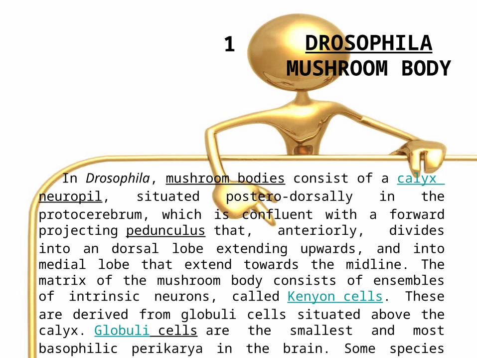
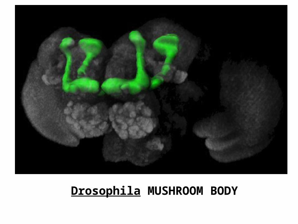
In Drosophila, mushroom bodies consist of a calyx neuropil, situated postero-dorsally in the protocerebrum, which is confluent with a forward projecting pedunculus that, anteriorly, divides into an dorsal lobe extending upwards, and into medial lobe that extend towards the midline. The matrix of the mushroom body consists of ensembles of intrinsic neurons, called Kenyon cells. These are derived from globuli cells situated above the calyx. Globuli cells are the smallest and most basophilic perikarya in the brain. Some species possess hundreds of thousands of Kenyon cells but a fruitfly mushroom body
comprises about 3,000 Kenyon cells
1

Drosophila MUSHROOM BODY
Drosophila MUSHROOM BODY


NEURONAL REMODELING
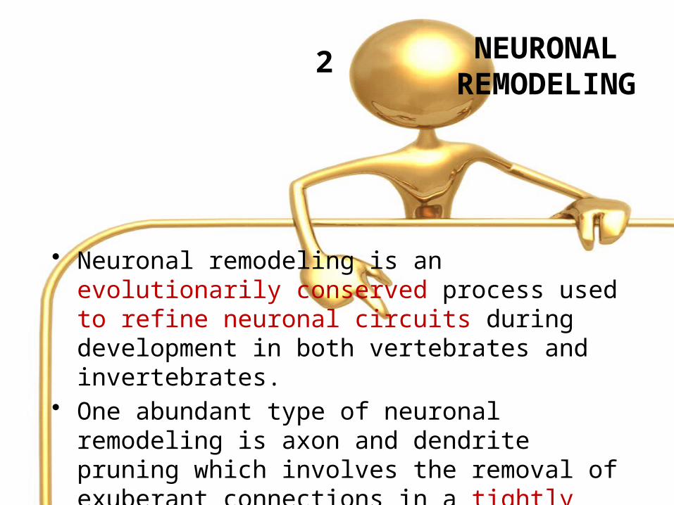
• Neuronal remodeling is an evolutionarily conserved process used to refine neuronal circuits during development in both vertebrates and invertebrates.
• One abundant type of neuronal remodeling is axon and dendrite pruning which involves the removal of exuberant connections in a tightly regulated process
2

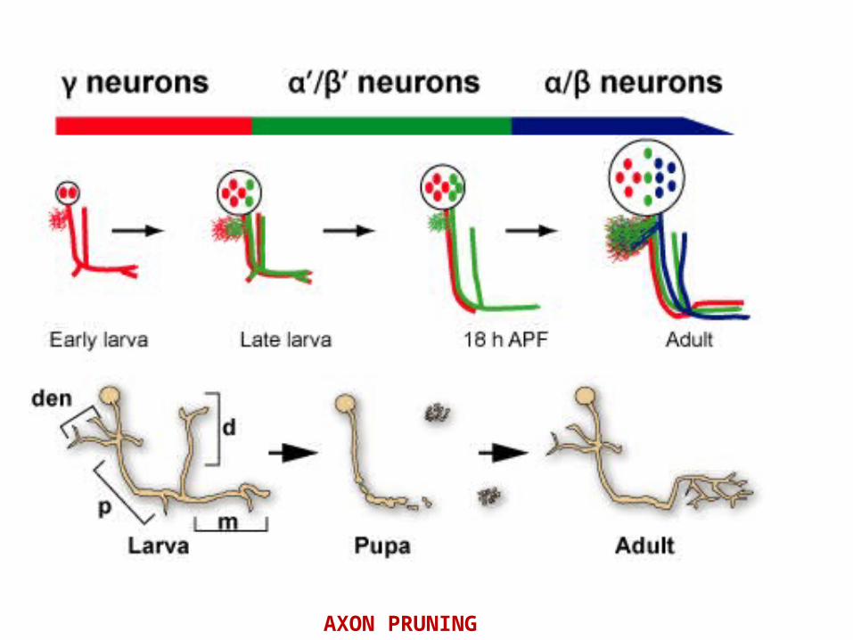
AXON PRUNING
• Axon pruning is an evolutionarily conserved strategy used to remodel neuronal connections during development.
• The Drosophila mushroom body (MB) undergoes neuronal remodeling in a highly stereotypical and tightly regulated manner.
3
AXON PRUNING

MB neurons undergo a highly stereotyped and tightly controlled process of dendrite and axon pruning

• Recently, glia were found to instruct axon fragmentation by secreting myoglianin (myo), a TGF-b ligand, which binds to the TGFb receptors baboon (babo) and punt/wishful thinking (put; wit) assisted by the TGF- accessory receptor plum on the membrane of c neurons.
4
• This in turn triggers the TGF-b pathway cell- autonomously within the MB c neurons resulting in an increase in the expression of ecdysone receptor B1 (EcR-B1) in c neurons
4…


QUESTIONS TO BE ANSWERED ????

QUESTIONS TO BE ANSWERED ????
1. Which glial subtypes participate in this process and their exact interaction with the neurons during pruning.
2.What is the relative contribution of each glial subtype in both the initiation of fragmentation as well as debris clearance?
?
QUESTIONS TO BE ANSWERED ????
3. Which glial type is involved in the refinement of the neuromuscular junction in Drosophila.
4. Are the same glial cells involved in secreting myo as those that are responsible for axon engulfment?
5. How separate are these processes?
?
EXPERIMENTAL PROCEDURES

Drosophila STRAINS
• Drosophila strains used: UAS-EcR.B-DC655.W650A; P{M2ETQF} ET40; P{QUAS-mtdTomato-3xHA}14; P{UAS-RedStinger}; Drpr-RNAi; P{UAS-FLP.Exel}2 and P{UAS-Cbb\DT-A.I}18 were obtained from Bloomington Drosophila stock center (Indiana University, USA).
1

Drosophila STRAINS
Tub.CD2.Gal4 was a kind gift from F. Pignoni, Upstate Medical University, Syracuse, NY; alrm-Gal4 and mZ0709-Gal4 were kindly provided by M. Freeman, UMass, Worcester, MA; UAS-myo was kindly provided by T. Lee, Janelia Farm, HHMI, Virginia; mZ1127-GAL4 was a kind gift from I. Salecker, National Institute for Medical Research, London; UAS-Shits1-pJFRC100 was provided by GM Rubin, Janelia Farm, HHMI, Virginia.
1…

ANTIBODY STAINING CONDITIONS
• Brains were dissected and stained & imaged on a Zeiss LSM710 confocal microscope.
Antibodies were used at the following dilutions:
Rat monoclonal anti-mouse CD8 a subunit, 1:200 (Caltag, Burlingame, CA, USA), Chicken anti-GFP, 1:500; Rabbit anti-HA (H6908), 1:100 (Sigma-Aldrich, Saint-Louis, MO, USA); The remaining antibodies were all obtained from the Developmental Studies Hybridoma Bank: mouse monoclonal anti-FasII (1D4), 1:50; mouse monoclon- al anti-EcR-B1 (AD4.4), 1:25 mouse monoclonal anti-Repo (8D1.2), 1:10; mouse monoclonal anti- Draper (8A1), 1:400. Alexa 488, Alexa546 or Alexa633 conjugated secondary antibodies were used at 1:300 (Invitrogen). DAPI (49,6- Diamidino-2-Phenylindole, Dihydrochloride) 1:1000. In order to stain for two anti-mouse anti- bodies concurrently we used Zenon (Invitrogen).
2

FLY GENETICS
Drosophila melanogaster were grown on standard media at 25’C or at indicated temperatures to manipulate Gal4 expression.
3

GLIA AND ASTROCYTES
NUCLEI QUANTIFICATION
• Glia and astrocytes cell number were quantified by counting the number of glial nuclei, as labeled with anti-Repo antibody and the nuclear RFP reporter (RedStinger) driven by alrm-Gal4.
• Confocal Z stacks were used as three-dimensional representations of the MB environment. To quantify the number of cells in the immediate vicinity of the MB lobe, we counted nuclei that were within 5 mM of the designated area
4

High magnification view of dorsal tip reveals that astrocytes (red) label only part of the repo+ nuclei in the vicinity of the MB lobe tip.

ECR-DN AND MYO FORCED EXPRESSION IN ASTROCYTES PRUNING AND DEBRIS CLEARANCE SEVERITY COMPARISON
• To analyze the severity of the pruning and debris clearance phenotype 18 and 20 confocal LSM files of EcR-DN and Myo forced expressed with EcR-DN, respectively, were blindly ranked from 0 (WT) to 5 (most severe pruning defect/debris clearance defect) by 4 independent researchers.
• Pruning severity was determined by comparing unpruned axonal branches that are significantly outside the a lobe, which is defined by strong FasII immunoreactivity.
• Analysis was done using Statistical software using repeated measures ANOVA.
5

QUANTIFICATION OF DRAPER EXPRESSION LEVELS
• Drpr antibody staining was examined in the immediate vicinity of the dorsal lobe.
• Quantification of intensity in confocal single slices was performed using ImageJ software.
• Levels of Drpr in WT and EcR-DN expressing animals were compared using student’s T-test.
6

RESULT

ASTROCYTES INFILTRATE AND ENGULF MB NEURONS DURING REMODELING
• Glia play a major role in the clearance of MB c axon fragments following developmental axon fragmentation.
• However the precise identity of these glia is unknown.• As a first step, they first wished to identify which glia are
located in the vicinity of the MB during metamorphosis.• They labeled the two major types of neuropil glia using
available glial subtype specific Gal4 drivers,
1. alrm-Gal4 for astrocytes and
2. mz0709-Gal4 for ensheathing glia.• Then they followed these cells at the time points relevant for
axon pruning.• The ensheathing glia specific driver, mz0709- Gal4, is
expressed in repo positive cells and in antenna lobe(AL) in the adult MB.
RESULT 1

• However, this driver was not expressed in repo-positive cells at the axon pruning relevant time point of 6 h APF in either the vicinity of the MB or the AL.
• Indicating that, at least in these regions, this driver does not label glia at this developmental time point.
• Therefore necessary to study their role during remodeling.
• In contrast, labeling astrocytes with alrm-Gal4 showed that astrocytes are adjacent to the c lobe at 0 h APF and 6 h APF and even remained in the vicinity after axon fragmentation has been completed.
• They also noticed a morphological change in the astrocytes throughout this developmental period.
RESULT 1… ASTROCYTES INFILTRATE AND ENGULF MB NEURONS DURING REMODELING

(B) At the onset of metamorphosis (0 h APF) astrocytic membranesare evenly dispersed in the region of the MB lobes (higher magnification in B2). (C) By 6 h APF, at the onset of pruning, the astrocytes have changedtheir morphology and have begun to infiltrate the degenerating lobes. (D) At 18 h astrocyte membranes surround axon fragments

• Between 0 h APF and 6 h APF astrocytic membranes became much more concentrated into discrete points that look like cysts, or beads on a string.
• Later, astrocytes gained ‘‘finger-like’’ extensions at 18 h APF while by 24 h APF very few membranes were labeled.
• Upon closer analysis, we noticed that not only are the astrocytes in the vicinity of the c lobes, they actually infiltrate them.
RESULT 1…
ASTROCYTES INFILTRATE AND ENGULF MB NEURONS DURING REMODELING

At 6 h APF FasII staining of c axons shows clear spherical regions that are devoid of staining in the both the dorsal & medial lobes.
RESULT 1…

The areas in the peduncle there is a slight decrease in FasII staining
RESULT…
This decrease is minor as compared to that which we observed in the medial and dorsal lobes
so they believe there is no significant amount of pruning.

It appears that these low FasII regions are actually filled with extensions of astrocytes
suggesting that astrocytes not only infiltrate the lobes but also may actually engulf axonal material.

• However, to test this hypothesis they wanted to express a stable transgene in neurons and at the same time visualize the astrocytes.
• We thus decided to use two binary systems within the same animal, labeling
1. neurons using the QF system (QF-ET40 driving QUAS-mtdt:HA
2. Astrocytes using the GAL4 system, alrm-Gal4 driving
UAS-mCD8-GFP)
RESULT 1…

Indeed they found that the astrocytes were clearly engulfing axonal debris at the dorsal tip of the c lobe at 6 h APF
RESULT 1…
In fact, they found that most, if not all, axonal fragments were engulfed by astrocytic membranes.
Interestingly, when we took a closer look at the peduncle area of the lobe they saw a different situation

RESULT 1…
part of the axon peduncle does not undergo extensive fragmentation and indeed while we do see exploratory infiltration of astrocytes into this region, there is less active engulfment.
lysosomal activity is delayed in the axon peduncle compared to the lobes at this time point
Results suggest that astrocytes, not ensheathing glia, are the main glial subtype that is responsible for the infiltration and engulfment of MB debris following axon fragmentation.

THE NUMBER OF ASTROCYTES AT THE MB STAYS CONSTANT THROUGHOUT REMODELING
• The decrease in alrm positive membrane staining during neuronal remodeling could be due to either a decrease in astrocyte cell number (due to cell death, differentiation or migration) or to a decrease in alrm-Gal4 expression leading to a reduction in CD8-GFP.
• . In order to discriminate between these options we expressed nuclear RFP (RedStinger) driven by alrm- Gal4.
• While it has been previously shown that there are 6–8 repo-positive cells in the vicinity of each MB lobe during pruning.
RESULT 2

THE NUMBER OF ASTROCYTES AT THE MB STAYS CONSTANT THROUGHOUT REMODELING
• we found only 1–2 astrocytes present at the tip of each c lobe throughout development
RESULT 2…
Labeling astrocyte cell bodies shows that the number of the astrocytes does not significantly change during remodeling.

THE NUMBER OF ASTROCYTES AT THE MB STAYS CONSTANT THROUGHOUT REMODELING
• Thus, we found that the number and distribution of astrocytes was largely unchanged throughout development suggesting that the observed lack of glial membranes after 24 h APF is a result of reduced CD8-GFP expression.
• Astrocytes are a small glia subgroup of 1–2 out of the 6–8 glia within the 5mM vicinity of the lobe.
RESULT 2…

THE NUMBER OF ASTROCYTES AT THE MB STAYS CONSTANT THROUGHOUT REMODELING
• Next we wanted to ask whether these 1–2 astrocytes that are within 5mM of the lobe are the only astrocytes engulfing the degenerating c lobe.
• Using sparse labeling(neuron labelling technique), they found that distant glia can also send processes to the lobe.
• despite the fact that we could see no labeled glia within the vicinity of the lobe, the degenerating lobe was still infiltrated by labeled glial membranes.
• suggesting that distant glia can also participate in the process of engulfment.
RESULT 2…

THE NUMBER OF ASTROCYTES AT THE MB STAYS CONSTANT THROUGHOUT REMODELING
They found four astrocyte cell bodies that were up to 50mM from the MB lobes but nonetheless sent highly branched processes into the degenerating lobe.
RESULT 2…

THE NUMBER OF ASTROCYTES AT THE MB STAYS CONSTANT THROUGHOUT REMODELING
• Therefore, although astrocytes are a small glia subpopulation, the effective number of astrocytes that contribute to pruning is more than we initially thought.
• To determine whether mz1127-Gal4 and alrm-Gal4 labeled the same or potentially a different population of astrocytes, we co-labeled glia with RedStinger driven by both alrm-Gal4 and by mz1127-Gal4 and counted the number of cell bodies in the vicinity of the MB tips at 6 h APF.
• We found that the number of cells remained 1–2 within 5mM of the lobe tip.
• suggesting that mz1127-Gal4 expression indeed overlaps with alrm-Gal4 at this developmental time point.
RESULT 2…

ASTROCYTES ARE NECESSARY FOR EFFICIENT AXON PRUNING
• to further delineate the specific role of astrocytes in axon pruning we decided to completely ablate them from the animal.
• They expressed the highly toxic diphtheria toxin (DTI) under the control of alrm-Gal4.
• Unfortunately, when the flies were reared at 29’C, where the expression of the Gal4 is high, this resulted in animal lethality.
• suggesting that astrocytes are essential for normal development.
RESULT 3

ASTROCYTES ARE NECESSARY FOR EFFICIENT AXON PRUNING
• when we reared the flies at 25’C to lower the expression level we obtained some escapers who reached adulthood.
• These flies showed only partial ablation of their astrocytes
RESULT 3…

ASTROCYTES ARE NECESSARY FOR EFFICIENT AXON PRUNING
• we detected a mild axon pruning defect in flies with partially ablated astrocytes that manifested in both intact larval c axons in 70% of the flies (9 out of 13 flies).
• uncleared axonal debris in 30% of flies (4 out of 13 flies).• these results suggest that astrocytes are necessary for
priming c axons to prune as well as to clear away the debris of fragmented axons.
RESULT 3…

ASTROCYTES ARE NECESSARY FOR EFFICIENT AXON PRUNING
RESULT 3…
(A–B) Confocal Z-projections of adult brains expressing CD8-GFP (A) additionally UAS-DTI (diphtheria toxin; B) driven by alrm-Gal4. Driving the expression of DTI in astrocytes resulted in their partial ablation(grey, A2, B2) and results in fragmentation defects (arrow in B1) and uncleared debris (arrowhead in B1) in escapers (13 out of 118 expectedflies).

ECDYSONE RECEPTOR IN ASTROCYTES REGULATES MULTIPLE ASPECTS OF AXON PRUNING
• One major determinant of Drosophila MB axon pruning is the insect hormone ECDYSONE that activates the ecdysone receptor, dominant negative version of the ecdysone receptor (EcR-DN) specifically in astrocytes.
• First, we noticed that expressing EcR-DN in astrocytes resulted in changes in their morphology and expression pattern.
RESULT 4

ECDYSONE RECEPTOR IN ASTROCYTES REGULATES MULTIPLE ASPECTS OF AXON PRUNING
• At 6 h APF astrocytes have acquired a cyst-like morphology, seem to surround the lobe fully and send small processes that infiltrate the lobe (20 out of 20 examined lobes)
• By 18 h APF, there are few GFP positive membranes in the area and they have become much longer and send ‘‘finger-like’’ extensions.
• At 24 h APF, there is almost no alrm-Gal4 positive membrane staining in the vicinity of the MB, even though the cell bodies remain.
• By adulthood, the astrocytes seem to obtain the classical astrocyte like morphology and occupy the entire brain.
RESULT 4..
In WT
ECDYSONE RECEPTOR IN ASTROCYTES REGULATES MULTIPLE ASPECTS OF AXON PRUNING
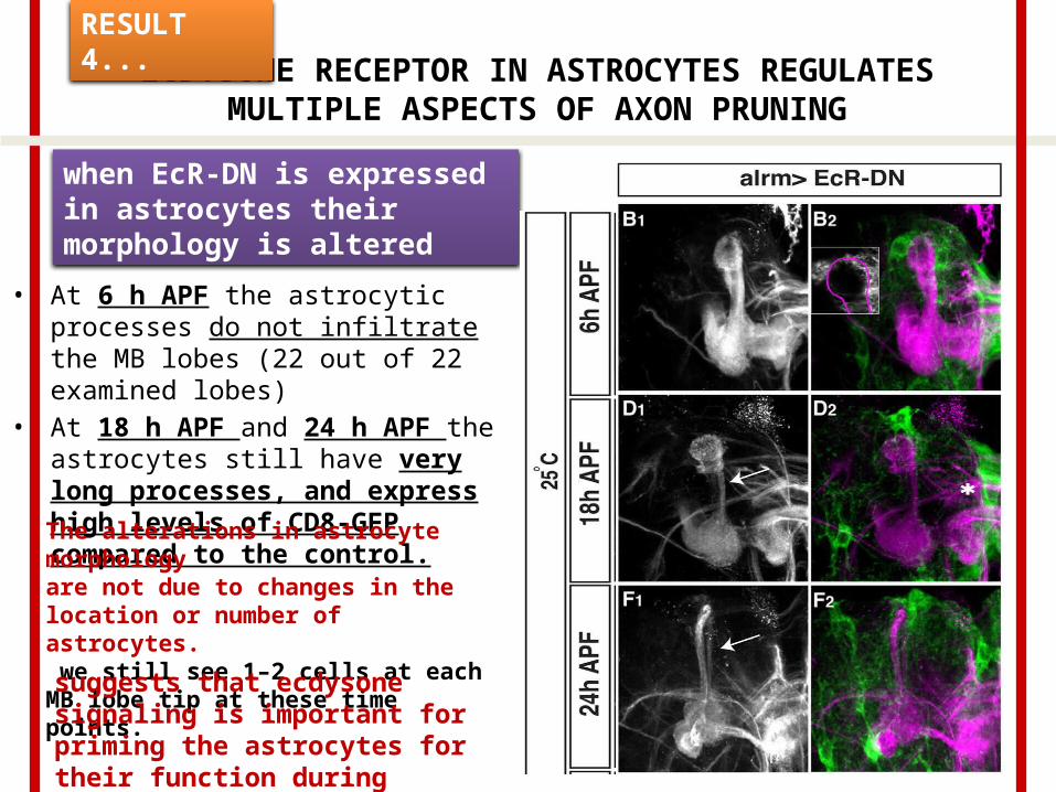
• At 6 h APF the astrocytic processes do not infiltrate the MB lobes (22 out of 22 examined lobes)
• At 18 h APF and 24 h APF the astrocytes still have very long processes, and express high levels of CD8-GFP compared to the control.
RESULT 4...
when EcR-DN is expressed in astrocytes their morphology is altered
The alterations in astrocyte morphologyare not due to changes in the location or number of astrocytes. we still see 1–2 cells at each MB lobe tip at these time points.
suggests that ecdysone signaling is important for priming the astrocytes for their function during pruning.

ECDYSONE RECEPTOR IN ASTROCYTES REGULATES MULTIPLE ASPECTS OF AXON PRUNING
• At 18 h APF & 24 h APF, expressing EcR-DN in astrocytes resulted in a significant delay in axon pruning.
• At 18APF WT MBs display the initiation of fragmentation (13 out of 18 partially fragmented c lobes) while some lobes have been completely fragmented at this time point (5 out of 18). In contrast, no MB c lobes in astrocytic EcR-DN flies show signs of fragmentation (18 out of 18).
RESULT 4..
EXAMINATION C NEURONS (FasII staining for new growing axon)

ECDYSONE RECEPTOR IN ASTROCYTES REGULATES MULTIPLE ASPECTS OF AXON PRUNING
Later during development, at 24 APF, most WT c lobes have been completely fragmented (13 out of 16) while none were completely fragmented in EcR-DN flies (12 out of 12).
RESULT 4…
EXAMINATION C NEURONS (FasII staining)
Indeed, elevated EcR-DN expression resulted in an exacerbation of both the fragmentation defect (9 out of 11 flies) and the debris clearance defect.

ECDYSONE RECEPTOR IN ASTROCYTES REGULATES MULTIPLE ASPECTS OF AXON PRUNING
• Myoglianin (myo) is a Drosophila TGF-b ligand secreted by glia that plays a major role in instructing the MB to undergo neuronal remodeling by increasing EcR-B1 levels in the c neurons, a necessary step in the progression of axon pruning.
• Myo has been previously shown to be secreted from both cortex glia and astrocytes although it seemed that cortex glia play a more crucial role in myo secretion.
• we decided to test whether expression of EcR-DN in astrocytes might affect pruning by affecting myo secretion.
• Thus, overexpressed myo in addition to EcR-DN in astrocytes
RESULT 4…
Myoglianin (myo)

ECDYSONE RECEPTOR IN ASTROCYTES REGULATES MULTIPLE ASPECTS OF AXON PRUNING
• Found that forced expression of myo partially suppressed both the fragmentation and the debris clearance defects apparent in EcR-DN animals.
• UAS-myo by itself had no effect on any MB morphology
RESULT 4..
DEBRIS CLEARANCE DEFECTS.

ASTROCYTES ENGULF MB C AXON DEBRIS VIA DRAPER
• The cell corpse engulfment receptor Draper (Drpr) is expressed in glial cells and is necessary for the engulfment of c axon fragments following pruning.
• We next wished to see if
a) the engulfment of c axons by astrocytes is also mediated by Drpr.
b) if engulfment by astrocytes is active or passive.
To do so we expressed Drpr RNAi in astrocytes.
RESULT 5
DRAPER RECEPTOR

ASTROCYTES ENGULF MB C AXON DEBRIS VIA DRAPER
saw a defect in debris clearance but not in them fragmentation process itself, as we could not see intact larval axons
RESULT 5…
The lack of effect onaxon fragmentation.
Is it due to low expression level of the RNAi?
Therefore, we decided to increase and prolong the expression of the RNAi by using a flip out tubulin-Gal4 system.

ASTROCYTES ENGULF MB C AXON DEBRIS VIA DRAPER
• While this increased expression of Drpr RNAi did exacerbate the amount of uncleared debris.
.
RESULT 5…
we still never observed a defect in axon fragmentation.
This indicates that while Drpr does play a role in engulfing fragmented axons.
astrocytes only caused a defect in debris clearance without affecting axon fragmentation

ASTROCYTES ENGULF MB C AXON DEBRIS VIA DRAPER
• To this end, we examined if there were alterations in the expression of Drpr in animals expressing EcR-DN in astrocytes.
• WT astrocytes express high amounts of Drpr at 6APF• EcRDN expressing astrocytes show substantially lower expression
RESULT 5…

ASTROCYTES ENGULF MB C AXON DEBRIS VIA DRAPER
• Taken together, it appears that astrocytes have two distinct roles:
1. they instruct c neuron fragmentation in an ecdysone dependent pathway and
2. they are the major glial type that clears axonal debris in a pathway that is also mediated by ecdysone signaling which increases Drpr expression and by endocytosis.
RESULT 5…

DISCUSSION

• The role of glia in shaping the nervous system has gained much traction in the past few years. In this study we further dissected the involvement of glia in the neuronal remodeling of the Drosophila MB.
• We show that astrocytes are one of the major glial subtypes that is responsible for MB c axon pruning, despite the fact that they make up a small subpopulation of glia surrounding the MB during this developmental period.
• Furthermore, we can block the ability of astrocytes to clear MB axonal debris, but not affect their morphological change or ability to promote axon fragmentation, by blocking endocytosis or knocking down the expression of the engulfment receptor Drpr.
DISCUSSION
• Thus decoupling these two processes.• We therefore propose that astrocytes play a key role in
both promoting axon fragmentation as well as clearing axonal debris once fragmentation has been completed.
DISCUSSION…

ASTROCYTES PLAY A KEY ROLE IN DEBRIS CLEARANCE DURING MB AXON PRUNING
• Here we show that astrocytes are the glial subtype that is responsible for most, if not all, of the infiltration and engulfment of neuronal debris following fragmentation. This was surprising for two major reasons.
• One is that ensheathing glia have already been shown to act as phagocytes.
• Another reason these results were surprising is that astrocytes make up only a minority of the glia surrounding the MB lobes during pruning, comprising of only 1–2 astrocytes out of 6–8 total glia in the vicinity of each lobe.
DISCUSSION …

ASTROCYTES PLAY A KEY ROLE IN DEBRIS CLEARANCE DURING MB AXON PRUNING
Although there are only 1–2 astrocytic cell bodies located near each tip, we found that cells that are located further away, up to 50mm, can also infiltrate the degenerating lobe, thereby potentially participating in debris clearance
DISCUSSION

ECDYSONE RECEPTOR CONTROLS ASTROCYTE FUNCTION DURING PRUNING
• One of the major determinants of metamorphosis and of axon pruning is the insect hormone ecdysone.
• Ecdysone receptor B1 (EcR-B1) is cell autonomously required in MB c axons to initiate a transcriptional program that, in turn, mediates axon pruning.
• EcR-B1 is also necessary for the dendrite pruning of dendritic arborization (DA) neurons as well as for the remodeling of embryonic olfactory projection neurons.
• In addition, ecdysone plays a role in the refinement of the fly neuromuscular junction.
• As all these neuronal remodeling processes occur in the pupae, this raises the hypothesis that ecdysone may act as a master regulator of multiple remodeling processes.
DISCUSSION
ECDYSONE RECEPTOR CONTROLS ASTROCYTE FUNCTION DURING PRUNING
• In addition, ecdysone being a systemic hormone, can function as a potential coordinator between fragmentation in neurons with phagocytic cells.
• To further investigate the role of astrocytes in MB axon pruning.
• we expressed a dominant negative version of the ecdysone receptor (EcR-DN) specifically in astrocytes and discovered defects in glial morphology, c axon fragmentation and axonal debris clearance.
• The defect in debris clearance is, at least partially, due to decreased expression of the cell corpse engulfment receptor Drpr.
DISCUSSION

ECDYSONE RECEPTOR CONTROLS ASTROCYTE FUNCTION DURING PRUNING
• This is consistent with previous results showing that expression of EcR-DN in glia leads to a defect in Drpr transcript upregulation following metamorphosis.
• glia in the neuronal remodeling of the MB c neurons has been through the secretion of myoglianin (myo), leading to an increase in c neuron EcR-B1 levels.
• Myo secretion has been shown to be crucial in cortex glia and less so in astrocytes.
• We found that expression of EcR-DN in astrocytes, resulted in a partial pruning defect, manifested as intact larval axons as well as decreased axonal debris clearance, with no evident decrease in EcR-B1 expression in MB c neurons.
DISCUSSION

ECDYSONE RECEPTOR CONTROLS ASTROCYTE FUNCTION DURING PRUNING
• We found that expressing high levels of Drpr RNAi or disrupting endocytosis in astrocytes disrupted axon debris clearance but, in contrast to the EcR pathway, did not cause a defect in axon fragmentation.
• It appears that ecdysone acts as a master regulator in astrocytes to regulate multiple functions during remodeling.
• Drpr and the endocytotic pathway mediate only debris clearance.
DISCUSSION
ECDYSONE RECEPTOR CONTROLS ASTROCYTE FUNCTION DURING PRUNING
• Furthermore, we found that forced expression of myo in addition to EcR-DN in astrocytes only partially rescues the pruning defect of MB c neurons.
• This suggests that the pruning defect apparent in these adult flies is not mediated solely by a decrease in myo secretion but that there is an additional mechanism at play.
• This might involve astrocytes morphology and distribution, which was previously shown to be important for proper neuronal circuit development in other systems.
• Thus, delineating ecdysone signaling in both glia and MB neurons during pruning requires further investigation.
DISCUSSION

ACKNOWLEDGEMENT

ACKNOWLEDGMENT
• Yaniv Hakim, Shiri P. Yaniv, Oren Schuldiner*
Department of Molecular Cell Biology, Weizmann Institute of Sciences, Rehovot, Israel for good paper.
• Our Guide Prof.BVS for support.• Friends for needful help.

