atlas & text of lung cytology - home | pathology · this atlas & text of lung cytology was...
TRANSCRIPT

Atlas & Text of
Lung Cytology
Gia-Khanh Nguyen Brenda Smith 2012

2
Atlas & Text of
Lung Cytology Gia-Khanh Nguyen, MD, FRCPC Professor Emeritus Department of Laboratory Medicine and Pathology University of Alberta Edmonton, Alberta, Canada And Brenda Smith, BSc, RT, CT (ASCP) Clinical Instructor and Cytotechnologist British Columbia Cancer Agency Vancouver, British Columbia, Canada
First edition 2012. All rights reserved. Legally deposited at Library and Archives Canada. ISBN 978-0-9881205-1-8

3
Table of Contents Preface 4 Related material by the same author and Key to abbreviations 5 Chapter 1. Cytologic investigation of the lung 6 Chapter 2. Nonneoplastic lung lesions 13 Chapter 3. Usual lung cancers 35 Chapter 4. Carcinoid tumors 60 Chapter 5. Other primary tumors and tumor-like lesions of the lung and pleura 67 Chapter 6. Secondary lung tumors 91

4
Preface This Atlas & Text of Lung Cytology was written at the request of a number of students in cytology who wished to have a concise and well-illustrated manual for easy consultation during their clinical laboratory training. Some materials in this book were taken from one (GKN) of the authors’ ebook on Essentials of Lung Tumor Cytology that was published online by The David F. Hardwick Pathology Learning Centre of The University of British Columbia in 2008. The newly proposed classification of lung adenocarcinomas, classification of lung cancer in small biopsies and cytologic materials and the importance of tumor typing for molecular target therapy for lung cancer are also briefly discussed. This atlas and text should be used in conjunction with the latest editions of the authors’ two monographs on lung tumor cytology and fluid cytology for additional information. For improvement of the future editions of this atlas and text, comments and suggestions from the reader will be appreciated. Last but not least, we wish to thank Dr. Jason Ford, Director and Mrs. Helen Dyck, Manager of The David F. Hardwick Pathology Learning Centre for their interest and enthusiasm in publishing this book on online. A free access to the educational materials posted on the webpage of the above-mentioned centre is valuable for students with limited financial resources worldwide. Gia-Khanh Nguyen, MD, FRCPC Brenda Smith, BSc, RT, CT (ASCP) Vancouver, BC, Canada Winter 2012

Related material by the same author Essentials of Needle Aspiration Biopsy Cytology, Igaku-Shoin, New York, 1991 Essentials of Exfoliative Cytology, Igaku-Shoin, New York, 1992 Essentials of Cytology. An Atlas, Igaku-Shoin, New York, 1993 Critical Issues in Cytopathology, Igaku-Shoin, New York, 1996 Essential of Lung Tumor Cytology, UBC Pathology, Vancouver, 2008 Essentials of Abdominal Fine Needle Aspiration Cytology, UBC Pathology, 2008 Essentials of Head and Neck Cytology, UBC Pathology, Vancouver, 2009 Essentials of Fluid Cytology, UBC Pathology, Vancouver, 2009 Essentials of Gynecologic Cytology, UBC Pathology, Vancouver, 2011 Essentials of Pap smear and Breast Cytology, UBC Pathology, Vancouver, 2012
Key to abbreviations ABC: Avidin-biotin complex technique BAL: Bronchoalveolar lavage BB: Bronchial brushing BW: Bronchial washing CB: cellblock CP: conventional preparation* DQ: Diff-Quick stain FNA: Fine-needle aspiration or aspirate HE: hematoxylin and eosin stain IHC: Immunohistochemistry/Immunohistochemical LBP: Liquid-based preparation MGG: May-Grünwald-Giema stain Pap: Papanicolaou stain TBFNA: Transbronchial/mucosal FNA TTFNA: Transthoracic FNA * Conventional preparations include smearing of cellular materials from sputum, FNA, BB and sediments from liquid materials obtained by BW and BAL, as well as cytospin preparations from liquid materials obtained by BW, BAL and FNA, in contrast to cell films prepared by LBP technique such as ThinPrep technique.
Chapter 1
Cytologic investigation of the lung The respiratory tract is divided into upper and lower parts. The upper respiratory tract is composed of the nose and larynx, and the lower respiratory tract consists of the trachea and lung. The tracheobronchial tree contains cartilage and submucosal mucus-secreting glands and is lined by a pseudostratified, ciliated columnar epithelium that contains, in addition, goblet cells, Clara cells and Kulchitsky cells (neuroendocrine cells). The bronchi ultimately branch into bronchioles that do not have cartilage and submucosal glands. Bronchioles are purely conducting ducts that divide into respiratory bronchioles which merge into alveolar ducts and alveoli. Pulmonary alveoli are lined by type I and II epithelial cells. Type I cells account for 40% of the alveolar cells, covers 95% of the alveolar surface and facilitate gas exchange. Type II cells produce surfactant and can reconstitute the alveolar surface after injury. The lung and the inner aspect of the pleural cavities are covered by a layer of mesothelial cells. Lung cell samples The lower respiratory tract is the target of respiratory cytology that can be studied by one or a variable combination of the following 7 types of cell sample: sputum, bronchial suction, bronchial washing (BW), bronchial brushing (BB), bronchoalveolar lavage (BAL), Transbronchial FNA (TBFNA) and Transthoracic FNA (TTFNA). Sputum Sputum cell samples are obtained by early morning deep cough after mouth washing. For a sputum specimen collected in 70% ethanol, the classic “pick and smear” technique is used, and 2 to 4 smears are prepared, immediately fixed in 95% ethanol and stained by the Pap technique. The rest of the specimen is fixed in formalin and embedded in paraffin for CB sections. Sputum cytology is more sensitive in detecting cancers involving large proximal bronchi than peripheral and metastatic cancers and its sensitivity increases with the number of specimens. An adequate or representative sputum cell sample must contain alveolar macrophages. (Fig.1.2).

˚
7
Fig.1.1. Adequate sputum sample showing dust-laden alveolar macrophages. (CP, Pap). Bronchial washing Bronchial secretions may be aspirated from the trachea via a tracheal tube or a tracheotomy stoma. BW is performed during bronchoscopy by instilling vials of 5 to 10 mL of warm normal saline into a bronchus. The fluid is then aspirated and usually 4 cytospin smears are prepared and stained by the Pap method. A BW from a normal individual should show a few bronchial columnar cells admixed with polymorphonuclear leukocytes and macrophages. (Fig.1.2). It is often contaminated with squamous cells exfoliated from the upper respiratory tract.
Fig.1.2. Normal BW showing bronchial epithelial cells, alveolar macrophages and a few metaplastic squamous cells. (CP, Pap).

˚
8
Bronchial brushing BB is performed during bronchoscopy. A cytobrush is used to scrape the surface of a bronchial lesion. The entrapped cells are transferred to a frosted slide by circular movements. Usually 2 smears are prepared and stained by the Pap technique. It can be done 2 to 3 times to secure an adequate number of diagnostic cells. The brush may be deposited in a vial of normal saline or alcohol fixative for cytospin preparation or LBP, and the rest of the cell sample can be used for making a CB. Cytologic material obtained by BB contains abundant bronchial epithelial cells and a small number of neutrophils as well as a few squamous cells exfoliated from the upper airways. (Figs. 1.3 and 1.4).
A B
C Fig.1.3. Normal bronchial epithelium showing in BB: A. Numerous bronchial glandular cells present singly, in clusters and sheets. B. Two bronchial epithelial fragments consisting of ciliated columnar cells with terminal plates and a benign metaplastic squamous cell. C. A few columnar bronchial epithelial cells and goblet cells with vacuolated cytoplasm. (CP, Pap).

˚
9
A B Fig.1.4. A, B. A BB of normal bronchial epithelium showing single and clustered columnar bronchial epithelial cells with terminal bar and cilia. (LBP, Pap). Bronchoalveolar lavage To obtain a BAL cell sample, a bronchoscope is wedged into position as far as it can advance. The distal airways are flushed with several vials of warm normal saline totaling 300 mL. The flushed samples are then aspirated. The first sample contains mainly bronchial secretion and is discarded. Other samples are pooled together and usually 4 cytospin smears are prepared and stained by the Pap and/or Diff-Quik technique. The remaining BAL cell sample is used for CB preparation. BAL reflects the cellular changes within alveolar spaces. An adequate BAL cell sample should contain abundant alveolar macrophages and a few lymphocytes and polymorphonuclear leukocytes. (Fig.1.5). The number of epithelial cell (bronchial columnar and squamous cells) should be less than 5% of all cells present in the sample. Differential cell counts are obtained by evaluating 200 cells. In normal, nonsmoking individuals polymorphonuclear leukocytes account for about 1% of all cells present. Neutrophils, up to 4%, can be found in the BAL from a cigarette smoker without any lung disease, however. BAL is useful in detecting infections of the alveolar spaces and it is less sensitive in detecting lung cancers.

˚
10
Fig.1.5. A BAL sample from a city resident showing numerous alveolar macrophages, with many containing dust and carbon particles. (CP, Pap). Transbronchial/transmucosal fine needle aspiration TBFNA is performed during bronchoscopy. It samples a submucosal mass lesion or a paratracheal or parabronchial lesion or enlarged lymph node. The sample is invariably contaminated with bronchial secretions containing exfoliated bronchial epithelial cells, and submucosal glandular cells may rarely be seen. An adequate TBFNA cell sample from a lymph node should show abundant lymphocytes. Endoscopic ultrasound-guided FNA via the bronchial tree or esophagus is used to obtain cytologic materials from posterior mediastinal lymph nodes for diagnosis and staging of lung and pleural cancers. Transthoracic fine needle aspiration TTFNA under imaging guidance is used for investigation of patients with a lung or pleural mass lesion, usually peripherally located, showing no diagnostic cells in sputum, BW, BB, BAL and TBFNA. An adequate TTFNA cell sample from a normal lung tissue may show alveolar macrophages, bronchial epithelial cells and sheets of mesothelium. (Fig.1.6). TTFNA is highly sensitive in detecting lung cancers. Tumor cells in a CB prepared from a needle aspirate are routinely studied by IHC for tumor typing.

˚
11
Fig.1.6. TTFNA from a normal lung showing a large fragment of mesothelium with folding and a few alveolar macrophages. (CP, Pap). Ancillary techniques Cytochemical and IHC studies can be done with satisfactory results on previously stained smears without prior de-staining. However, they are best performed on formalin-fixed minute tumor tissue fragments in CBs prepared from materials procured by BW, BB or FNA. Grossly identified minute tissue fragments should be removed and fixed in formalin for histologic, cytochemical and IHC studies. They may also be fixed in 2% glutaraldehyde for ultrastructural evaluation. It should be born in mind that ethanol is not a suitable fixative for electron microscopy as it destroys cellular ultrastructures. Diagnostic accuracy The sensitivity, specificity and predictive value of different types of respiratory specimen in the diagnosis of lung cancer vary with the tumor location, the type and number of specimens. In general a combination of different types of cell sample offers higher sensitivity, specificity and predictive value for a positive result than a single sample.
• Sputum cytology is more efficient in detecting cancers involving large proximal bronchi. Its sensitivity is low with one specimen (27-41%) and when 3 samples are obtained it increases to 57-89%. If 5 samples are used a sensitivity as high as 96.1% may be reached.
• The sensitivity of a BW in the diagnosis of lung cancer varies from 61 to 76%, and that of a BB ranges from 70 to 77%.
• For TBFNA, the sensitivity of the procedure alone is about 52%. When a TBFNA

˚
12
is combined with BW, BB and bite biopsy its sensitivity increases to 72%. The specificity of the biopsy technique is 70 to 74% and its positive and negative predictive values are 100% and 53 to 70%, respectively.
• For TTFNA, the sensitivity and specificity of the procedure are 89% and 96%, respectively. Its positive and negative predictive values are 98% and 70%, respectively; and its false-positive and false-negative rates are 0.85% and 6%, respectively.
• For tumor typing, the cytohistologic correlation rates of sputum, BW and BB, as reported by Johnston and Bossen, were 85% for squamous cell carcinoma, 79% for adenocarcinoma, 30% for large cell carcinoma and 93% for small cell carcinoma of the bronchial tree. Those two investigators have also reported that the cytohistologic correlation rates of TTFNA were 80%, 96%, 42% and 95% for squamous cell carcinoma, adenocarcinoma, large cell carcinoma and small cell carcinoma of the lung, respectively. Bibliography French CA. Respiratory tract. In Cytology. Diagnostic principles and clinical correlates. 3rd edition, 2009, Cibas ES, Ducatman BS, eds. Philadelphia, Saunders, p.65. Johnston WW, Bossen EH. Ten years of respiratory cytopathology at Duke University Medical Center. I. The cytopathologic diagnosis of lung cancer during the years 1970-1974, noting the significance of specimen number and type. Acta Cytol.1981;25: 103. Johnson WW, Bossen EH. Ten years of respiratory cytopathology at Duke University Medical Center. II. A comparison between cytopathology and histopathology in typing of lung cancer during the years 1970-1974. Acta Cytol. 1981;25:499. Johnston WW. Cytodiagnosis of lung cancer. Principles and problems. Path Res Pract. 1986;181:1. Koss LG, Melamed MR. Koss’ Diagnostic cytology, 5th edition, 2006, Koss LG, Melamed MR, editors, Philadelphia, Lippincott Williams & Wilkin, p. 568 and 643. The Papanicolaou Society of Cytopathology Task Force on Standard of Practice. (Suen, et al.). Guidelines of the Papanicolaou Society of Cytopathology for the examination of cytologic specimens obtained from the respiratory tract. Diagn Cytopathol.1999;21:61.

˚
13
Chapter 2
Nonneoplastic lung lesions Abnormal cellular findings A. Hyperplastic and reactive bronchial epithelial cells Hyperplastic and reactive bronchial epithelial cells are present in groups or clusters and they may form tridimensional papillary clusters with smooth contours or Creola bodies. Highly reactive bronchial epithelial cells with prominent nucleoli may mimic malignant glandular cells. However, these cells often retain a columnar or cuboidal shape with presence of terminal plates and cilia. They may be seen in sputum or bronchial cytologic materials from patients with acute and chronic bronchitis or viral pneumonitis and they usually disappear in 2 weeks after the recovery of a lung infection. (Figs.2.1 to 2.3).
A B Fig.2.1. A. Reactive/hyperplastic bronchial epithelial cells in a BB of a patient with viral pneumonitis. B. Large atypical reactive/hyperplastic bronchial epithelial cells in BB from another patient with viral pneumonitis. (CP, Pap).

˚
14
A B Fig.2.2. A, B. Reactive bronchial epithelial cells in a BB of an acute bronchitis mimicking malignant glandular cells. These cells are present in monolayered sheets and show prominent nucleoli. (LBP, Pap).
AB Fig.2.3. A. A cluster of hyperplastic bronchial epithelial cells in BW (A) and a creola body in sputum (B) of a patient with chronic bronchitis. (CP, Pap). 2. Hyperplastic alveolar cells Patients receiving hyperbaric oxygen therapy for respiratory failure or having a pulmonary infarct may exfoliate highly atypical reactive and hyperplastic alveolar cells. These cells are large and polygonal in shape, usually occur in small clusters and show enlarged nuclei with prominent nucleoli, mimicking malignant glandular cells. Their presence is usually transitory. (Fig.2.4).

˚
15
Fig.2.4. A cluster of atypical and hyperplastic alveolar cells with prominent nucleoli in a BW of a patient recovering from diffuse alveolar cell damage. (CP, Pap) 3. Hyperplastic reserve cells Hyperplastic reserve cells are usually seen in bronchial cell samples from patients with chronic bronchitis. These cells display rather distinctive cytologic features permitting their correct identification in almost all cases. They usually occur in compact clusters or sheets consisting of uniform small cells that often have a straight edge and may be mistaken for cells of a small cell carcinoma by an inexperienced observer. (Fig.2.5).
A B Fig.2.5. A, B. A sheet of hyperplastic reserve bronchial epithelial cells in BAL (A) and in BB (B) showing small cuboidal cells with scant cytoplasm and straight edges. (CP, Pap). 4. Metaplastic squamous cells Bronchial epithelium in long-term cigarette smokers and in patients with chronic bronchitis and chronic obstructive lung disease commonly undergoes squamous metaplasia that may display cellular atypia. Exfoliated metaplastic squamous cells in

˚
16
lung cell samples usually occur singly, in small aggregates or in small monolayered sheets. These cells show a “hard” basophilic or orangeophilic and well-defined cytoplasm. Atypical metaplastic squamous cells may display enlarged nuclei with a slightly hyperchromatic, granular, open chromatin pattern and inconspicuous nucleoli. (Fig.2.6). A cavitating mycetoma may be lined by an atypical metaplastic squamous epithelium that yields in TTNA highly atypical squamous cells mimicking malignant squamous cells.
Fig.2.6. Atypical metaplastic squamous cells in BB cell sample. Note the open chromatin pattern and inconspicuous nucleoli. (CP, Pap). 5. Radiation- and Chemotherapy-induced cellular changes Radiation and chemotherapy may induce cellular changes, mimicking cancer cells. Those cells appear as enlarged bizarre cells with smudgy nuclei, multinucleation and vacuolated cytoplasm. However, they lack unequivocal cytologic features of malignant cells such as a high N/C ratio, irregular nuclear contours and hyperchromatic coarsely granular chromatin clumping. (Figs. 2.7).
A B

˚
17
Fig.2.7. A. Highly atypical or suspicious epithelial cells in sputum of a patient having radiation therapy for mediastinal germ cell tumor. B. Atypical epithelial cells in sputum of a patient receiving chemotherapy for acute myelogenous leukemia. (CP, Pap). Abnormal noncellular findings 1. Curschmann spirals are formed by inspissated mucus within bronchiolar lumens of patients with small airway diseases, especially in chronic smokers. It has a dark-staining core with wispy mucous ends. Curschmann spirals are seen mainly in sputum and BAL specimens. (Fig.2.8).
Fig. 2.8. A Curschmann spiral in a BAL cell sample. (CP, Pap). 2. Corpora amylacea are composed of glycoprotein and are large, nonlaminated or poorly formed lamellar structures that stain yellow or orange with the Pap method. They are more commonly seen in sputum and BAL from patients with chronic pulmonary edema. (Fig.2.9).
Fig.2.9. Corpora amylacea in a sputum cell sample. (CP, Pap).

˚
18
3. Calcified concretions or calcospherites are laminated (psammoma bodies) or nonlaminated bodies with dense central parts. They are composed of calcium, phosphate, iron, magnesium and other materials and may be seen in sputum and BAL from patients with chronic obstructive lung disease, pulmonary tuberculosis, cor pulmonale and rarely in patients with papillary bronchial adenocarcinoma. (Fig.2.10).
Fig.2.10. Two calcospherites in a sputum cell sample. (CP, Pap). 4. Ferruginous and asbestos bodies. Inhaled asbestos fibers are phagocytosed by alveolar macrophages. These fibers are covered with iron and protein and become ferruginous bodies with heads having different shapes. (Fig.2.11). They are seen mainly in BAL cell samples.
Fig.2.11. A ferruginous body and alveolar macrophages in a BAL. (CP, Pap).

˚
19
5. Talcosis occurs in individuals with prolonged and heavy exposure to talc powder or in patients with intravenous drug abuse when talc powder is used as carrier material. Foreign-body granulomas with birefringent platlike talc particles in fibrotic interstitium with nodularity are observed in lung tissue, and talc particles within macrophages may be found in BAL cell samples. (Fig.2.12).
A B Fig.2.12. A: Histology of the lung with talcosis from an intravenous drug user. B: BAL showing alveolar macrophages with two of them containing intracytoplasmic birefringent talc particles. (CP, Pap). 6. Charcot-Leyden crystals are rhomboid crystals that are breakdown products of eosinophils in asthmatic lungs. They are seen mainly in BAL and sputum cell samples. (Fig.2.13).
Fig.2.13. Charcot-Leyden crystals in a sputum cell sample. (CP, Pap).

˚
20
7. Hemosiderin-laden macrophages are seen in pulmonary hemorrhage that occurs in Goodpasture syndrome, idiopathic pulmonary hemorrhage and lung infarct secondary to pulmonary thromboembolism. These macrophages can be seen in sputum and BAL cell samples. (Fig.2.14)
A B Fig.2.14. A. BW showing numerous hemosiderin-laden macrophages with intracytoplasmic coarsely granular hemosiderin granules. (CP, Pap). B. Hemosiderin-laden macrophages in a sputum cellblock. (Prussian blue) Lung infections 1. Nonspecific infections Nonspecific lung infection is caused by a number of bacteria and may be classified as acute or chronic. Acute pneumonitis is characterized by inflammatory exudates and pus formation. Chronic pneumonitis shows an increase in lymphoid cells, plasma cells and macrophages. Lung abscess is usually caused by Staphyloccus aureus and is commonly secondary to aspiration of food and gastric contents. 2. Tuberculosis A lung tuberculoma may destroy a bronchus, discharge its caseating contents and become a cavity. Bronchial cytologic materials or BAL may reveal necrotic debris, single and clustered epithelioid cells with elongated or bean-shaped nuclei and multinucleated giant cells of Langhans. (Figs.2.15 to 2.17). TBFNA from enlarged mediastinal lymph nodes with tuberculous lymphadenitis may reveal epithelioid cells, giant cells of Langhans and minute tissue fragments containing tuberculous granulomas. Acid-fast bacilli can be visualized in CB sections with Ziehl- Neelsen or acid-fast bacilli stain.

˚
21
A B Fig.2.15. A, B. BB from a tuberculous bronchitis reveals single spindle and polygonal epithelioid cells with elongated or bean-shaped nuclei. (CP, Pap).
A B Fig.2.16. A, B. TTFNA from a lung tuberculoma showing clustered epithelioid cells and multinucleated giant cells of Langhans. (CP, Pap).
A B Fig.2.17. Minute tissue fragment in TBFNA from a mediastinal tuberculous lymphadenitis showing a granuloma with multinucleated giant cells of Langhans in A. Acid-fast bacilli with Ziehl-Neelsen stain are seen in B.

˚
22
3. Fungal infections Fungal infections of the lung may be caused by pathologic fungi such as Blastomyces, Cryptococcus neoformans, Coccidioidomyces immitis and Histoplasma capsulatum. In patients with immune deficiencies infections by opportunistic fungi such as Candida, Phycomyces and Aspergillus species are common. These fungal elements may be seen in Pap-stained materials but they are best demonstrated by periodic acid-Schiff and Gomori methenamine-silver (GMS) stains. (Fig.2.18 to 2.20)
o North American Blastomyces are broad-base budding yeasts, 8 to 20 µm in greatest dimension.
o Cryptococcus neoformans are narrow-base budding yeasts, 4 to 15 µm in greatest dimension, extracellular clear zone.
o Coccidioidomyces immitis cysts are 15 to 60 µm thick-walled spherules with 1 to 2 µm endospores.
o Candida species are characterized by 2 to 10 µm round or oval budding yeasts and non-branching hyphae.
o Aspergillus species show 5 to 10 µm wide septate hyphae with 45º angle branching.
o Phycomyces elements are irregular, broken, 10 to 30 µm wide nonseptate hyphae with haphazard or 90º angle branching).
For detecting fungal infections of the lung, according to some reported series the sputum had a sensitivity and positive predictive value of 16.66% and 50%, respectively; and the sensitivities of BAL and bronchial biopsy were 80% and 18 to 20%, respectively,
A B

˚
23
C Fig.2.18. Fungal elements: A. North American Blastomyces round yeast with large-base budding. (CP, Pap). B. Cryptococcus yeasts with thick walls. (CP, Pap). C. Cryptococcus yeast with round budding and narrow base. (CP, GMS).
A B Fig.2.19. Coccidioides cyst with thick wall and endospores in smear (A) and CB (B). (CP, A: Pap, B: GMS).
A B

˚
24
C Fig.2.20. A and B. Aspergillus showing nonseptate hyphae with 45º angle branching. (CP: A, Pap; B, GMS). B. Phycomyces showing broken, irregular hyphae with haphazard or 90º angle branching. (CP, Pap). 4. Viral infections Viral pneumonitis is common in immunocompromised hosts. Etiologic agents include Cytomegalovirus and Herpes simplex viruses. The inflammatory process affects the interstitial tissue and bronchial epithelium.
o Cytomegalovirus infection is characterized by isolated large cells with a single intranuclear eosinophilic inclusion and perinuclear halo. These cells may be seen in all types of respiratory cell samples, including sputum, bronchial materials and BAL. In problematic cases a positive IHC staining with Cytomegalovirus antibody confirms the viral infection. (Fig.2.21).
A B Fig.2.21. An alveolar cell with large intranuclear round inclusion displaying an immune- positive reaction to Cytomegalovirus antibody. (CP, A: Pap, B: ABC).

˚
25
o Herpetic bronchitis shows single cells and multinucleated giant cells with intranuclear inclusions. (Fig.2.22). A positive IHC staining with a herpes antibody will confirm the viral infection in equivocal cases.
A B Fig.2.22. Herpetic bronchitis showing in sputum clustered epithelial cells with ground glass nuclei with nuclear molding (A). Intranuclear inclusions without perinuclear halos in BB material (B). (CP, Pap). 5. Pneumocystis pneumonitis Pneumocystis jiroveci (formerly carinii) is ubiquitous and often affects immunocompromised persons and caused an interstitial lung infiltrate of plasma cells and lymphocytes, diffuse alveolar damage with foamy alveolar exudates or casts with organisms appearing as tiny bubbles or vacuoles. These casts may be found in sputum and BW but they are best seen in BAL sample stained by the Pap technique. (Fig.2.23). In appropriate clinical settings these foamy casts are diagnostic of Pneumocystis pneumonia. The cysts represented by the vacuoles within alveolar casts are not stained by the Pap method. These cysts are spherical, oval or cup-shaped structures with one flat surface and measure 5 to 7 µm in greatest dimension. Within the cysts are 1 or 2 dot-like trophozoites or sporozoites measuring 0.5 to 1 µm in diameter. Pneumocystis jirovecci organisms may be detected by commercially available monoclonal antibody or by PCR technique. However, GMS staining of BAL cell samples is the preferred diagnostic method in most cytology laboratories.

˚
26
A B Fig.2.23. Pneumocystis pneumonitis: A. A large intraalveolar foamy cast. (CP, Pap). B. Pneumocystis jirovecci organisms with central, round nuclei in a CB. (CP, GMS). Other inflammatory and noninflammatory lung diseases 1. Eosinophilic pneumonia Eosinophilic pneumonia can be idiopathic or secondary to drugs, fungal infection or parasitic infestation. It is characterized by the presence of numerous eosinophils in alveolar spaces, and abundant eosinophils can be seen in respiratory cytologic materials. (Fig.2.24).
A B Fig.2.24. Eosinophilic pneumonia. A. Histology of the lung lesion. B. Abundant eosinophils in a BAL cell sample. (CP, Pap).

˚
27
2. Sarcoidosis Sarcoidosis is a disease of unknown etiology and is probably caused by an exaggerated helper T-cell response. It is characterized by mediastinal lymphadenopathy and pulmonary infiltration. Histologically, numerous non-necrotizing granulomas are present in lymph nodes, interstitial lung tissue and bronchial mucosa. Multinucleated giant cells with intracytoplasmic star-shaped crystals (asteroid bodies) and small lamellar calcified bodies (Schaumann bodies) may be seen in lung tissue sections and material obtained by BB. The BAL shows abundant lymphoid cells of T-cell type. The lymphoid cell population usually ranges from 10 to 70% in most cases. (Figs.2.25 and 2.26). Multinucleated giant cells with the above-mentioned intracytoplasmic bodies may be seen in BB but are rarely identified in BAL cell samples.
A B
CD Fig.2.25. Lung sarcoidosis: A. Lung tissue section showing nonnecrotizing granulomas in interstitial tissue. B-D. Numerous lymphocytes present in a BAL cell sample. An elongated epithelioid cell is seen in C. D showing clustered epithelioid cells in TBFNA of an enlarged parabronchial lymph node with sarcoidosis. (CP, Pap). (Reproduced from: Nguyen GK, Batoroev YK. Value of simultaneous transbronchial fine needle aspiration of

˚
28
hilar lymph nodes and lung biopsy in the diagnosis of pulmonary sarcoidosis. Russian Journal of Clinical Cytology. 2006:10:23, with permission).
Fig.2.26. Cell block prepared from a TBFNA of a parabronchial enlarged lymph node in a patient with sarcoidosis showing a noncaseating granuloma. (HE). 3. Lipid pneumonia and Aspiration pneumonia Lipid pneumonia may occur in patients aspiring mineral oil or using oily nose drops. It commonly affects the lower lobe of left lung that may resemble a lung tumor radiologically. Numerous lipid-laden macrophages are seen in sputum and BAL material. Intracellular fat droplets can be demonstrated in air-dried smears stained with Oil-red-O or Sudan black. (Fig.2.27). Aspiration pneumonia develops as the result of aspiration of food particles with subsequent development of a lung abscess. Cytologic material from the cavity of an aspiration pneumonitis reveals pus and food particles. (Fig.2.28).
A B

˚
29
C Fig.2.27. Lipid pneumonia: A. Histologic section of a lung with lipid pneumonia. B. Clustered foamy histiocytes in a sputum cell sample. (CP, Pap). C. BAL cell sample showing numerous lipid-laden macrophages. (CP, Oil-red-O).
A B
C Fig.2.28. Aspiration pneumonia showing in sputum: A. Well-preserved vegetable cells in flat fragments with thick transparent cellulose walls and homogenously stained nuclei. The cellulose walls are not stained by the Pap stain in this case. (CP, Pap). B. A fragment of vegetable showing cells with thick cellulose walls that stain weakly with the Pap stain. (CP, Pap). C. An irregular fragment of food surrounded by numerous polymorphonuclear leukocytes. (CP, Pap).

˚
30
4. Pulmonary infarct A pulmonary infarct may mimic a lung tumor radiologically. Highly reactive alveolar cells and numerous hemosiderin-laden macrophages are seen in respiratory materials. (Fig.2.29). Hemosiderin can be well-demonstrated by Prussian blue stain.
Fig.2.29. Reactive alveolar cells in a BW. (CP, Pap). 5. Chronic interstitial lung fibrosis This is a heterogenous group of lung diseases. These disorders may have similar clinical and radiological findings and consist of idiopathic and connective tissue disease-associated interstitial lung fibrosis and interstitial fibrosis caused by inhalation of organic and non-organic dusts. Disorders with chronic interstitial lung fibrosis may be divided into 2 cytologic groups on the basis of neutrophilic or lymphocytic reaction:
• Neutrophilic group is composed of idiopathic and connective tissue disease- associated interstitial fibrosis, asbestosis and histiocytosis X. The BAL shows numerous macrophages and neutrophils that may account for 5 to 50% of the total cell count. An increase in leukocytic count is an indication of the aggravation of the disease and a decrease in leukocytic count is an indication of a favorable response to treatment, in sequential BAL samplings. (Fig.2.30).

˚
31
A B Fig.2.30. A. Histology of pulmonary interstitial fibrosis. B. Numerous polymorpho-nuclear leukocytes are present in BAL fluid. (CP, Pap).
• Lymphocytic group consists of sarcoidosis and hypersensitivity pneumonitis. In sarcoidosis increased numbers of macrophages and lymphoid cells are noted. The lymphoid cells account for 10 to 70% of the differential cell counts. The number of helper T-cells is also increased. Progression of the disease to a more severe interstitial fibrosis is heralded by an increased number of neutrophils, and a resolving disease is heralded by a decrease of lymphoid cells in BAL fluid samples. (Fig.2.31).
Fig.2.31. Sarcoidosis progressing to a more severe interstitial fibrosis showing several polymorphonuclear leukocytes in a BAL sample. An epithelioid cell with elongated nucleus is noted at 9 o’ clock. (CP, DQ).

˚
32
6. Pulmonary alveolar proteinosis Pulmonary alveolar proteinosis (PAP) is a disease of unknown pathogenesis and its true incidence is unknown. It is more common in men than in women of 30 to 40 years of age, with a 3:1 male-to-female ratio. A defect in alveolar clearance and/or alveolar macrophage activity associated with an overproduction of lipid by alveolar type II lining cells have been suggested to play a role in the pathogenesis of PAP. Two forms PAP are encountered: idiopathic and secondary. The secondary PAP occurs in several settings: lung infection, hematologic malignancies, immune deficiencies including HIV infection and inhalation of silica, aluminum dust, titanium and insecticides. PAP is an unusual diffuse lung disease and characterized by an accumulation of large amounts of phospholipoprotein-rich material in alveolar spaces. Ultrastructural study of lung tissue reveals intraalveolar accumulation of concentric lamellar structures or myelin figures, suggesting surfactant. (Fig.2.32). The BAL fluid is turbid, milky, thick and granular. It stains strongly positively with periodic acid-Schiff with prior diastase digestion (PASD). Electron microscopic study of BAL sediment may reveal numerous well- or poorly preserved myelin figures, as seen in tissue section. Repeating BALs is an effective therapeutic procedure for PAP. (Fig.2.33).
A B Fig.2.32. A. Histologic section of a lung with PAP showing thick, granular, PASD positive intraalveolar material. (PASD). B. Ultrastructure of lung tissue showing concentric lamellar bodies, suggesting surfactant. (Uranyl acetate and lead citrate stain, x 51,000).

˚
33
A B
C Fig.2.33. Pulmonary alveolar proteinosis: A. Thick, amorphous, coarsely granular material in BAL sediment. (CP, Pap). B. Intraalveolar material stains strongly positively with PASD. (CP, PASD). C. Ultrastructure of BAL sediment showing degenerated and fragmented lamellar bodies, suggesting surfactant. (Uranyl acetate and lead citrate, X 48,000) Bibliography Akin MR, Nguyen GK. Pulmonary alveolar proteinosis. Pathol Res Pract.2004;200:693. French CA. Respiratory tract. In Cytology. Diagnostic principles and clinical correlates, 3rd edition, Cibas ES, Ducatman BS, eds. Edinburgh, Saunders, 2009, p.65. Garg S, et al. Comparative analysis of various cytohistological techniques in diagnosis of lung diseases. Diagn Cytopathol. 2007;35:26.

˚
34
Khan A, Agarwal R. Pulmonary alveolar proteinosis. Respir Care.2011;15 Koss LG, Melamed MR. The lower respiratory tract in absence of cancer: conventional and aspiration cytology. In Koss’ Diagnostic Cytology, 5th ed, 2006, Koss LG and Melamed MR, editors. Philadelphia, Lippincott Williams & Wilkins, p. 568. Martin WJ, et al. Interstitial lung diseases. Assessment by bronchoalveolar lavage. Mayo Clin Proc.1983;58:751. Nassar A, et al. Utility of reflex Gomori methenamine silver staining for Pneumocystis jirovecci on bronchoalveolar lavage cytologic specimens: a review. Diagn Cytopathol. 2006:34:719. Travis WD, et al. Non-neoplastic Disorders of the Lower Respiratory Tract. In Atlas of non-tumor pathology, 1st series, Washington DC, Armed Forces Institutes of Pathology, 2002

˚
35
Chapter 3
Usual lung cancers Bronchogenic carcinomas account for about 95% of all primary lung cancers and have a male predominance, but the number of affected women is increasing. These lung cancers occur most commonly in the 6th and 7th decades of life, and 95% of them may be classified into 4 major histologic types: squamous cell carcinoma, adenocarcinoma, large-cell carcinoma and small-cell carcinoma. Bronchogenic carcinomas are traditionally classified into 2 large groups: small-cell lung cancer (SCLC) and non-small-cell lung cancer (NSCLC) for management purposes. NSCLCs include squamous cell carcinomas, adenocarcinomas and large cell carcinomas. The main reason for this categorization is that almost all SCLCs are at advanced stages when first detected, and they are best treated by chemotherapy with or without radiation. NSCLCs, on the other hand, respond poorly to chemotherapy and are best treated by surgical resection. In general, up to 30% of all bronchogenic carcinomas are resectable when diagnosed. The prognosis of lung cancer is dismal: 5-year survival rate for all stages of lung cancer combined is about 15%. Bronchogenic carcinomas consist of many histologic types that are classified as follows by the World Health Organization (2004). 2004 WHO Classification of primary lung carcinomas Squamous cell carcinoma Papillary, clear cell, small cell, and basaloid subtypes Adenocarcinoma Acinar, papillary, solid, bronchioloalveolar, fetal, signet ring, clear cell, mucinous, and mixed subtypes. Large-cell carcinoma Large cell neuroendocrine carcinoma Basaloid carcinoma Lymphoepithelioma-like carcinoma Clear cell carcinoma Large cell carcinoma with rhabdoid features Small-cell carcinoma Combined small-cell carcinoma Adenosquamous carcinoma Sarcomatoid carcinomas Spindle cell carcinoma Giant-cell carcinoma

˚
36
Pleomorphic carcinoma Carcinosarcoma Pulmonary blastoma Carcinoid tumor Typical carcinoid Atypical carcinoid Salivary gland tumors Mucoepidermoid carcinoma Adenoid cystic carcinoma Epithelial-myoepithelial carcinoma Squamous cell carcinoma This tumor accounts for about 30% of all primary lung cancers. It commonly arises from a major or segmental bronchus and invades the surrounding lung parenchyma. Bronchogenic squamous cell carcinoma may be well- or poorly differentiated. (Fig.3.1). A well-differentiated neoplasm shows keratin pearls and intercellular bridges. A poorly differentiated tumor may mimic a poorly differentiated adenocarcinoma or large cell carcinoma histologically.
A B Fig.3.1. Lung squamous cell carcinoma: A. Well-differentiated tumor. B. Poorly differentiated tumor. The cytologic manifestations of squamous cell carcinomas in the sputum and in materials obtained by BW, BB and FNA are somewhat similar and vary with the tumor differentiation. (Figs.3.2 to 3.7).

˚
37
Common cytologic features of bronchogenic squamous cell carcinoma in sputum, BW, BB and FNA include:
• Abnormal squamous cells with large or pyknotic hyperchromatic nuclei. • Bizarre cell shapes, abnormal keratinization. • Cell dissociation especially in differentiated tumors. • Tumor tissue fragments and cell aggregates are often present in FNA. • Tumor cells usually appear less differentiated in FNA than in BB or sputum
because of a higher component of deeper non-keratinizing tumor tissue. • Tumor cells forming epithelial pearls and intercellular bridges may be seen in
well-differentiated tumors. • A poorly differentiated tumor shows cohesive clusters on non-keratinizing
malignant epithelial cells with ill-defined, opaque cytoplasm and hyperchromatic nuclei with prominent nucleoli.
• Necrotic debris. Bronchogenic squamous cell carcinoma subtypes such as clear cell or small cell variants may yield cells mimicking those of a large cell carcinoma, adenocarcinoma of the lung with extensive clear cell change and metastatic clear cell carcinoma from the kidney and ovary or cells derived from a small cell lung cancer. In these situations IHC studies of the cancer cells may yield important information for a more accurate tumor typing. Cells from a bronchogenic squamous cell carcinoma are CK5/6 and p63 positive and CK7, CK20 and Napsin A negative. Pitfalls in the cytodiagnosis of malignant squamous cells include benign cells with radiation effect, atypical metaplastic squamous cells, benign cells with chemotherapy effect, atypical metaplastic squamous cells in a mycetoma and vegetable cells or pollen.
A B Fig.3.2. Well-differentiated squamous cell carcinoma showing: A. Necrotic and viable keratinized malignant squamous cells in sputum. (CP, Pap). B. Single and clustered keratinized malignant squamous cells in sputum CB. (HE).

˚
38
Fig.3.3. Poorly differentiated squamous cell carcinoma showing in sputum a fragment of non-keratinized malignant squamous epithelium. (CP, Pap).
A B Fig.3.4. Well-differentiated squamous cell carcinoma showing: A. Isolated keratinized malignant squamous cells in BB. B. Isolated keratinized tumor cells in TBFNA. (CP, Pap).
AB

˚
39
C Fig.3.5. Well-differentiated squamous cell carcinoma showing: A, B. Isolated keratinizing squamous cells in BB. (LBP, Pap). C. A fragment of keratinized malignant squamous cell epithelium in BB CB. (HE).
Fig.3.6. Poorly differentiated squamous cell carcinoma showing in TTFNA a fragment of non-keratinized malignant squamous epithelium. (CP, Pap).
Fig.3.7. Poorly differentiated squamous cell carcinoma showing in BB loosely clustered nonkeratinized malignant epithelial cells. (LBP, Pap).

˚
40
Adenocarcinoma Bronchogenic adenocarcinomas account for about 30% of all primary lung cancers. About 75% of the tumors arise in the lung periphery and present radiologically as a “coin lesion”. In the remaining 25% of the cases the neoplasms are located in a lobar or segmental bronchus. Lung adenocarcinomas display several histologic patterns and are classified according to the above 2004 WHO classification. In 2011 a multidisciplinary classification of lung adenocarcinomas was proposed by a joint study of the International Association for the Study of Lung Cancer (IALC), American Thoracic Society (AST) and European Respiratory Society (ERS). IASLC/ATS/ERS Classification of Lung Adenocarcinomas in Resection Specimens 1. Preinvasive lesions Atypical adenomatous hyperplasia Adenocarcinoma in situ (≤3 cm, formerly Bronchioloalveolar carcinoma) Nonmucinous Mucinous Mixed mucinous/nonmucinous 2. Minimally invasive adenocarcinoma (≤3 cm lepidic predominant, ≤ 5mm invasion) Nonmucinous Mucinous Mixed mucinous/nonmucinous 3. Invasive adenocarcinoma Lepidic predominant (formerly nonmucinous BAC pattern, with > 5mm invasion) Acinar predominant Papillary predominant Micropapillary predominant Solid predominant with mucinous production 4. Variants of invasive adenocarcinoma Invasive mucinous adenocarcinoma (formerly mucinous BAC) Colloid Fetal (low and high grade) Enteric Adenocarcinoma, NOS is histologically an invasive lung adenocarcinoma that consists of monomorphic malignant glandular cells with conspicuous nucleoli or pleomorphic malignant cells with prominent nucleoli. (Fig.3.8).

˚
41
A B Fig.3.9. Histology of invasive lung adenocarcinoma: Tumor with acinar pattern (A). Tumor with solid pattern (B). The cytologic manifestations of bronchogenic adenocarcinomas are somewhat similar in sputum and in materials obtained by BW, BB and FNA. (Figs.3.9 to 3.12).
• The malignant glandular cells are present predominantly in small groups. • Cells from a well-differentiated tumor show fairly uniform nuclei with smooth
nuclear contours and conspicuous nucleoli. • Cells from a poorly differentiated adenocarcinoma are more pleomorphic and
show single or multiple macronucleoli. • Intracellular mucus may be demonstrated with mucicarmine or PASD.
Bronchogenic adenocarcinoma cells are CEA, CK7, Napsin A and TTF-1 positive and p63 and CK20 negative. Pitfalls in the cytodiagnosis of bronchogenic adenocarcinoma include Creola bodies, numerous goblet cells misinterpreted as mucinous adenocarcinoma, atypical pneumocytes, cells with viral cytopathic changes, and reactive mesothelial cells seen in TTFNA.

˚
42
A B Fig.3.9. A. Well-differentiated adenocarcinoma showing in sputum clustered mono-morphic tumor cells with vacuolated cytoplasm and conspicuous nucleoli. (CP, Pap). B. Poorly differentiated adenocarcinoma showing in sputum clustered pleomorphic malignant glandular cells with prominent nucleoli. (CP, Pap).
Fig.3.10. Sputum cell block showing a cluster of malignant glandular cells with vacuolated cytoplasm. (HE).

˚
43
Fig.3.11. Lung adenocarcinoma showing in TTFNA a cohesive cluster of malignant glandular cells with prominent nucleoli. (CP, Pap).
A B
C D Fig.3.12. Invasive, low-grade lung adenocarcinoma showing in BB: A-C. Monomorphic cuboidal tumor cells with eccentrically located nuclei, nucleoli and well-defined, granular cytoplasm are present singly and in cohesive sheets. (LBP, Pap). D. A cluster of tumor cells in BB CB showing TTF-1 positive nuclei. (ABC).

˚
44
Lung adenocarcinoma with lepidic growth pattern (ADL, previously called nonmucinous bronchioloalveolar carcinoma) is rarely encountered. It can be unifocal or multifocal and is characterized by cuboidal or low columnar tumor cells with conspicuous nucleoli growing along preexisting alveolar walls. The mucinous bronchioloalveolar carcinoma was renamed as mucinous adenocarcinoma in the 2011 IALC/AST/ERS proposed classification of lung adenocarcinomas. (Fig.3.13). Cytologic features of lung ADL in sputum, BW, BB, FNA: (Figs.3.14 to 3.16).
• In sputum, small cuboidal tumor cells with oval nuclei are seen predominantly in tridimensional papillary clusters and cell balls.
• In materials obtained by BB or FNA the tumor cells are commonly seen in large monolayered sheets with nuclear crowding and overlapping.
• Cellular aspirate. • Regular and monotonous relatively small cells with ample cytoplasm. • Vesicular nuclei with prominent nucleoli, hyperchromatic nucleoli. • Intranuclear cytoplasmic inclusions may be present. • Clean mucoid background. • Tumor cells are TTF-1 negative and may express surfactant proteins (SP-A, pro-
SP-B, pro-SP-C).
A B Fig.3.13. Histology of lung adenocarcinoma: A. Adenocarcinoma with lepidic growth pattern (formerly nonmucinous bronchioloalveolar carcinoma). B. Mucinous adenocarcinoma (formerly mucinous bronchioloalveolar carcinoma). (HE)

˚
45
Fig.3.14. An ADL exfoliates in sputum a cohesive cluster of tumor cells with nuclear crowding and molding. (CP, Pap).
A B Fig.3.15. Mucinous adenocarcinoma showing in TTFNA a cohesive sheet of mucus-secreting tumor cells with nuclear crowding. (CP, Pap).
A B

˚
46
Fig.3.16. Lung ADL showing in TTFNA tumor cells predominantly in irregular, large, cohesive sheets (A). At higher magnification focal glandular spaces, crowded tumor cells with slightly pleomorphic nuclei and conspicuous nucleoli are observed, as well as intranuclear cytoplasmic inclusions (B). (CP, Pap). Fetal adenocarcinoma is a rare tumor, related to cigarette smoking and commonly occurs in 5th or 6th decade of life. It usually pursues a less aggressive clinical course. Histologically, the tumor is composed of low-grade malignant glandular cells arranged in acinar pattern. Focal tumor cell morulae containing intracytoplasmic neurosecretory granules, as demonstrated by electron microscopy and by IHC staining with neuron-specific enolase and chromogranin antibodies, are present. In one case the tumor TTFNA revealed large monolayered and folded sheets of low-grade malignant columnar epithelial cells with clear or granular cytoplasm and uniformly oval, small nuclei with inconspicuous nucleoli. Focal glandular arrangement may be visualized within a tumor cell sheet. (Fig.3.17)
A B
C Fig.3.17. Fetal adenocarcinoma, low-grade: A. Tumor forming intraglandular morules. B. Tumor cells in morule showing chromogranin positive cytoplasm. (ABC). C. Tumor showing in TTFNA a large sheet of tumor cells with honeycomb pattern. Round glandular space is noted elsewhere. (CP, Pap).

˚
47
Small cell carcinoma Small cell carcinoma or “oat cell carcinoma” accounts for about 20% of all primary lung cancers. The tumor is related to cigarette smoking and may be associated with a paraneoplastic syndrome such as diabetes insipidus or Cushing syndrome. It arises most commonly from major bronchi and has a rapid growth with early hilar lymph node and distant metastases. About 70% of patients with small cell carcinoma are at an advanced stage when the tumor is detected. Rarely, a small cell carcinoma presents as a “coin lesion”. Histologically, the tumor has a solid growth pattern with extensive necrosis. The tumor cells are small, two to three times the size of a mature lymphocyte and show scant cytoplasm, oval nuclei with finely granular chromatin pattern and inconspicuous nucleolus. Nuclear molding is a prominent feature and mitotic index is high. Tumor necrosis is a common finding. (Fig.3.18). In some cases the neoplasm is of intermediate cell type and it is composed of tumor cells that are larger than those of the classic small cell carcinoma, but the tumor cells essentially show the nuclear features of the latter. Small cell carcinoma may coexist with a nonsmall cell carcinoma.
Fig.3.18. Histology of a small cell carcinoma. Cytologically, the tumor cells are seen singly, in groups or along mucus threads with nuclear molding in sputum and materials obtained by BW. Most tumor cells are necrotic and show pyknotic and darkly stained nuclei. The smear background contains linear basophilic necrotic debris. In BB and FNA the tumor cells are well-preserved and display a salt and pepper chromatin pattern with inconspicuous nucleoli. (Figs.3.19 to 3.23). Cytologic features of small cell carcinoma in sputum, BW, BB and FNA: Sputum and BW:
• Small dissociated tumor cells. • Scant cytoplasm, nuclear molding.

˚
48
• Coarsely stipped chromatin. • Inconspicuous nucleoli. • Degenerative changes, common.
BB and FNA:
• Better preserved larger cells than in sputum • Some nuclear molding and cell clustering • Open chromatin pattern • Nucleoli visible • Artifactually crushed basophilic nuclear debris
Cells from a bronchogenic small cell carcinoma are CK7, chromogranin, synaptophysin, CD56 and TTF-1 positive and CK20 negative. Pitfalls in the diagnosis of small cell carcinoma include hyperplastic reserve cells, pools of lymphocytes, small cell adenocarcinoma cells, lymphoma cells, carcinoid tumor cells, cells derived from small blue cell tumors (Ewing sarcoma, Wilms' tumor, neuroblastoma, embryonal rhabdomyosarcoma, pleuropulmonary blastoma) and droplets of condensed mucus.
A B Fig.3.19. A. Small cell cancer showing in sputum clustered small tumor cells with scant cytoplasm, oval nuclei and no nucleoli. Nuclear molding is noted in some tumor cell clustered. B. The tumor showing in BW loosely aggregated tumor cells. (CP, Pap).

˚
49
A B Fig.3.20. A. Small cell carcinoma showing in BB tumor cells with salt and pepper chromatin pattern and linear, basophilic nuclear debris. B. Small cell carcinoma, intermediate cell type showing in BB larger tumor cells and crushing, linear, basophilic nuclear debris. (CP, Pap).
A B Fig.3.21. A, B. Small cell carcinoma showing single and clustered small cancer cells. Minimal nuclear molding is noted in some cell clusters, but basophilic linear nuclear debris is not present, as seen in CP smears. (LBP, Pap).

˚
50
A B Fig.3.22. A, B. Small cell carcinoma showing in TTFNA isolated and clustered malignant cells with hyperchromatic nuclei and nuclear molding. (CP, Pap).
AB
C Fig.3.23. A. Lung small cell cancer showing in BB cellblock a cluster of tumor cells with nuclear molding. (HE). B. TTF-1 positive tumor cells. C. Chromogranin positive tumor cells. (ABC).

˚
51
Large cell carcinoma Large cell carcinoma constitutes about 10% of all bronchogenic carcinomas. Most of these tumors arise from segmental or lobar bronchi. Histologically, the tumor is composed of large malignant cells with abundant, granular cytoplasm and macronucleoli and shows no squamous or glandular cell differentiation. In cytologic materials of all types (sputum, BW, BB and FNA) the tumor cells are seen singly and in loose or cohesive aggregates. These are large malignant cells with variably abundant cytoplasm, large nuclei with single or multiple eosinophilic macronucleoli. (Fig.3.24). Cells from a bronchogenic large cell carcinoma are usually CEA, CK7 and TTF-1 positive and CK20 negative.
Fig.3.24. Large cell carcinoma showing in BW clustered large tumor cells with macronucleoli. (CP, Pap). Lymphoepithelial carcinoma is a rare morphologic variant of large cell carcinoma of the lung. The tumor presents as a peripheral lung nodule and yields in FNA single and cohesive sheets of pleomorphic malignant epithelial cells with prominent nucleoli and numerous lymphocytes. Large cell neuroendocrine carcinoma (LCNEC) is rare and highly aggressive tumor occurring in adults with a median age of 64 years. The neoplasm may be centrally or peripherally located and averages 3 cm in greatest dimensions. Histologically, LCNEC consists of large pleomorphic malignant cells arranged in neuroendocrine pattern with focal rosette formation. Mitotic figures are abundant and large geographic necrosis is common. The tumor cells express neuroendocrine markers. In cell samples obtained by BB or FNA the tumor cells are seen singly and in loose aggregates. They are large, pleomorphic and display well-defined, granular cytoplasm and oval nuclei with granular chromatin pattern and prominent nucleoli, mimicking

˚
52
those of a large cell carcinoma. Naked tumor cell nuclei and necrotic debris may be observed. Tumor cells arranged in rosettes and linear pattern may be observed. (Fig.3.25). Staining with NSE, synaptophysin, chromogranin and CD56 antibodies will be helpful for confirming the neuroendocrine differentiation of the tumor cells. About 50% of LCNEC are TTF-1 positive.
A B Fig.3.25. Large cell NE carcinoma. A. Histology of the tumor. B. The tumor showing in TTFNA large, pleomorphic malignant epithelial cells with abundant cytoplasm, oval nuclei and prominent nucleoli. Some cells show a plasmacytoid configuration. (DQ) Sarcomatoid carcinoma Giant cell and spindle cell carcinomas are rare a variant of large cell carcinoma (1%) with very poor prognosis. Histologically, these 2 tumors are characterized by giant, bizarre malignant cells with single or multiple nuclei or spindle malignant cells. A giant cell carcinoma yields in sputum and in materials obtained by bronchial washing and brushing or FNA single and loosely clustered giant, bizarre malignant cells with variably abundant cytoplasm, single, multiple, lobulated nuclei with macronucleoli. (Figs.3.26 and 3.27).

˚
53
A B
C Fig.3.26. A. Histology of giant cell carcinoma. B, C. The tumor showing in BB large multinucleated malignant cells. (CP, Pap).
A B Fig.3.27. Large cell carcinoma, spindle cell variant. A. Histology of the tumor. B. The tumor showing in TTFNA dissociated spindle malignant cells. (CP, Pap) Important comparative cytologic and immunocytochemical features of usual bronchogenic carcinomas are tabulated in Table 3.1.

˚
54
Table 3.1. Comparative Cellular, Histochemical and Immunohistochemical Features of Usual Bronchogenic Carcinomas
CELLULAR FEATURES
SQUAMOUS CARCINOMA ADENOCARCINOMA LARGE CELL
CARCINOMA SMALL CELL CARCINOMA
Arrangement Singly Syncytia Sheets
Acini Aggregates, Sheets Papillary clusters Cell balls
Singly or Aggregates
Singly, Clusters, loose
Tumor cell configuration
Pleomorphic Columnar Cuboidal
Polygonal, Pleomorphic, Giant cells
Small, Round, Oval
Cytoplasm Well-defined, abundant, keratinized Ill-defined, variable, nonkeratinized
Vacuolated Granular
Variable Ill-defined, Scant
Nucleus Central, Bizarre Chromatin, clumped
Central/Eccentric Oval Chromatin, clumped or fine
Central/Eccentric Oval Single or Multiple
Central Chromatin, fine Nuclear molding
Nucleolus Variable Macronucleoli, Variable Macronucleoli Absent
Cellular mucin - + ± -
Immunocytochemistry
TTF-1 - + + + p63 + - ± - Chromogranin - - -* + Synaptophysin - - -* + CD56 - - -* + CK7/CK20 CK7-/CK20- CK7+/CK20- CK7+/CK20- CK7+/CK20- **Adapted from Nguyen GK, Kline TS: Essentials of Cytology. An Atlas, New York, Igaku-Shoin. 1993, p. 44, with modification. * Positive in Large cell carcinoma with neuroendocrine differentiation.

˚
55
Classification of lung cancers in small biopsies and cytologic materials Bronchogenic carcinomas are traditionally and clinically classified as small cell lung cancer (SCLC) and Non-SCLC (NSCLC) for management purposes. These 2 types of lung malignancy account for 15% and 85% of usual lung cancers, respectively. SCLCs are almost always at advanced stages at diagnosis and treated by chemotherapy. For NSCLCs, resection is reserved for localized tumors; and radiotherapy, chemotherapy and molecular targeted therapy are for unresectable and advanced tumors. Only less than 30% of NSCLCs are diagnosed at early stages, but the rate of resection is only 6% to 15%, due to co-morbid illnesses. Therefore, over 80% of NSCLCs are diagnosed only by small biopsy and/or cytology prior to treatment. The 2004 WHO classification of lung tumors is currently found unsuitable for small biopsies and cytologic specimens. In 2011 the International Association for the Study of Lung Cancer, American Thoracic Society and European Respiratory Society has proposed a classification of lung cancer for small biopsies and cytology. In this classification the term “large cell carcinoma” is not used and it is replaced by “non-small cell carcinoma” and IHC characteristic features of NSCLCs are emphasized and included. Lung cancer classification for small biopsies and cytology A. Adenocarcinoma - Adenocarcinoma with identifiable pattern (papillary, micropapillary, acinar, solid, mixed) - Adenocarcinoma with lepidic pattern - Mucinous adenocarcinoma - Adenocarcinoma with colloid pattern - Adenocarcinoma with fetal pattern - Adenocarcinoma with signet ring cell features - Adenocarcinoma with clear cell features B. Squamous cell carcinoma C. Small cell carcinoma D. Non-small cell carcinoma (NSCC/NSCLC) - NSCC with NE morphology (+ NE markers), possible LCNEC - NSCC with NE morphology (- NE markers) - NSCC, with squamous cell and adenocarcinoma patterns (Adenosquamous) - NSCC, favor SQCC (if morphologic SQCC absent, and supported by IHC) - NSCC, favor ADA (if morphologic ADA absent, and supported by IHC) - Poorly differentiated NSCC with spindle & giant cell carcinoma features - NSCC, NOS (specify IHC stain results with interpretation)

˚
56
Diagnosis of lung cancers in small biopsies and cytologic materials can be made based on the tumor histology and tumor cell morphology with or without histochemical and/or IHC characteristics. According to some recently published reports, the light microscopy can accurately type 75% of HE stained small biopsies and 80 to 90% of Pap stained cytologic preparations. Histochemical and/or IHC studies are necessary for subclassification in the remaining 25% biopsies and 10 to 20% cell samples. Four antibodies are commonly used to distinguish an adenocarcinoma from a squamous cell carcinoma: TTF-1, Napsin A, p63 and CK5/6. A positive reaction to TTF-1 and Napsin A and a negative reaction to p63 and CK5/6 of the tumor cells indicate a lung adenocarcinoma while cells of a squamous cell carcinoma usually express p63 and CK5/6. (Figs.3.28 and 3.29). It should be born in mind that benign type II pneumocytes express TTF-1, and that CK5/6 and p63 antibodies stain positively normal basal bronchial epithelial cells. Alveolar macrophages and type II pneumocytes also express Napsin A. In the above lung cancer classification the term “NSCC-NOS” is advised to be used as little as possible, and it should only be used if no clear differentiation by morphology, histochemistry or IHC is found, and when the morphology and IHC results are conflicting.
A B
C

˚
57
Fig.3.28. Non-small cell carcinoma, favor squamous cell carcinoma: Fragment of tumor tissue that is difficult to be differentiated from a poorly differentiated adenocarcinoma (A). Tumor cells displaying positive cytoplasmic reaction to CK5/6 antibody (B). Tumor cells showing positive nuclear staining with p63 antibody (C). (ABC).
A B
C Fig.3.29. Non-small cell carcinoma, favor adenocarcinoma: A minute tumor tissue fragment (A) in BW material from a case of bronchial non-small cell carcinoma showing Napsin A positive cells (B) and TTF-1 positive nuclei (C). (ABC) Molecular target therapy for lung cancer A number of molecular abnormalities are present in about 50% of NSCLCs and include EGFR mutation (about 10% in Caucasian versus 50% in Asian), KRAS mutation (up to 30% in Caucasian versus 15% in Asian), anaplastic lymphoma kinase (ALK) translocation (about 7% in Caucasian versus 5% in Asian), and other rare mutations of BRAF, MET, HER2 and PIK3CA genes (about 1 to 2% each). These abnormalities exist

˚
58
in a mutually exclusionary fashion each other except PIK3CA gene and show some racial variations. In recent years, a subset of patients with advanced NSCLC with activating of tyrosine kinase domain of EGFR show excellent response to EGFR-TKI agents (gefetinib and erlotinib), especially in never-smoking female patients and those with lung adenocarcinomas. EGFR-TKI agents have been an approved and standard treatment for advanced NSCLCs in many industrialized countries around the world. Advanced lung adenocarcinomas are usually treated with EGFR-TKI combined with vascular endothelial growth factor (VEGF) inhibitor (bevacizumab), pemetrexed (antifolate) and chemotherapy. These agents are not effective in the treatment of lung squamous cell carcinomas, and bevacizumab has caused fatal or life threatening hemoptysis in about 30% of patients with bronchogenic squamous cell cancer. KRAS mutations are more commonly in Caucasian male smokers and are associated with a resistance to a treatment with EGFR-TKIs. For EGFR testing, the test is ordered at morphologic diagnosis of NSCLC and biopsied samples are preferred over cytology specimens. Recent studies have demonstrated that routinely stained recent or archived cytologic smears are also suitable for DNA extraction for EGFR and KRAS mutations testing. Tested materials are best fixed 10% neutral buffered formalin and should contain a high number of tumor cells. However, if a higher sensitive technique is used a lower percentage of tumor cell content is acceptable. Two methods of molecular testing are currently used for EGFR mutation testing: DNA sequencing and amplified refractory mutation system (ARMS) method. The minimum number of tumor cells optimal for mutational analysis is not known with certainty. However, over 50% of tumor cells per sample are adequate for DNA sequencing. If the ARMS method (using PCR technique and more sensitive than DNA sequencing) is used, ≤ 10% of tumor cells may be adequate. Usually, a manual dissection of tumor cells is needed to obtain a sample with high tumor cell contents. The number of tumor cells in FNA and bite biopsy varies with the size of the biopsy needle and number of biopsy:
• For FNA, if a 21-gauge needle is used, the number of tumor cells is usually ≥ 100 per aspirate. If a 19-gauge needle is used, the number of tumor cells is ≥150 per aspirate.
• For transbronchial tissue biopsy: the number of tumor cells is usually ≥300 per biopsy. For CT-guided biopsy the number of tumor cells is ≥500 per biopsy.
The diagnostic process usually takes 5 to 7 working days to complete. A few commercially available kits are currently used in lung cancer EGFR molecular testing using either DNA sequencing technique or ARMS method with PCR. KRAS mutation analysis using FISH technique and ALK translocation testing using IHC are also commercially available.
˚
59
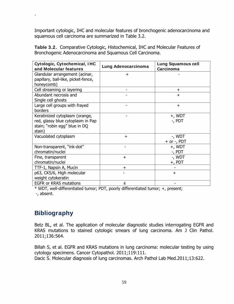
Important cytologic, IHC and molecular features of bronchogenic adenocarcinoma and squamous cell carcinoma are summarized in Table 3.2. Table 3.2. Comparative Cytologic, Histochemical, IHC and Molecular Features of Bronchogenic Adenocarcinoma and Squamous Cell Carcinoma. Cytologic, Cytochemical, IHC and Molecular features Lung Adenocarcinoma Lung Squamous cell
Carcinoma Glandular arrangement (acinar, papillary, ball-like, picket-fence, honeycomb)
+ -
Cell streaming or layering - + Abundant necrosis and Single cell ghosts
- +
Large cell groups with frayed borders
- +
Keratinized cytoplasm (orange, red, glassy blue cytoplasm in Pap stain; “robin egg” blue in DQ stain)
- +, WDT -, PDT
Vacuolated cytoplasm + -, WDT + or -, PDT
Non-transparent, “ink-dot” chromatin/nuclei
- +, WDT -, PDT
Fine, transparent chromatin/nuclei
+ -, WDT +, PDT
TTF-1, Napsin A, Mucin + - p63, CK5/6, High molecular weight cytokeratin
- +
EGFR or KRAS mutations ± - * WDT, well-differentiated tumor; PDT, poorly differentiated tumor; +, present; -, absent. Bibliography Betz BL, et al. The application of molecular diagnostic studies interrogating EGFR and KRAS mutations to stained cytologic smears of lung carcinoma. Am J Clin Pathol. 2011;136:564. Billah S, et al. EGFR and KRAS mutations in lung carcinoma: molecular testing by using cytology specimens. Cancer Cytopathol. 2011;119:111. Dacic S. Molecular diagnosis of lung carcinomas. Arch Pathol Lab Med.2011;13:622.

˚
60
Francis H, Solomon B. The current status of targeted therapy for non-small cell lung cancer. Internal Med J. 2010;40:611. Geisinger KR, et al. Localized lung diseases. In Modern Cytopathology. Philadelphia, Churchill Livingston, 2004, p 399. Hammar SP, Dacic S. Immunohistochemistry of lung and pleural neoplasms. In Diagnostic Immunohistochemistry, Theranostic and Genomic applications. 3rd edition, 2010. Dabbs DJ, editor. Philadelphia, Saunders Elsevier. p.369. Koss LG, Melamed MR. Tumors of the lung: conventional cytology and aspiration biopsy. In Koss’ Diagnostic Cytology and Its Histopathologic Bases, Koss LG and Melamed MR, editors. 5th ed, 2006. Philadelphia, Lippincott Williams & Wilkins, p. 643. Nguyen GK, Kline TS. Essentials of cytology. An atlas. New York, Igaku-Shoin, 1993, p 43. Ocque R, et al. Usefulness of immunohistochemical and histochemical studies in the classification of lung adenocarcinoma and squamous cell carcinoma in cytologic specimens. Am J Clin Pathol. 2011;136:81. Pirker R, et al. Consensus for EGFR mutation testing in non-small cell lung cancer. Results from a European workshop. J Thorac Oncol. 2010;5:1706. Rekhtman N, et al. Suitability of thoracic cytology for new therapeutic paradigms in non-small cell lung carcinoma: high accuracy of tumor subtyping and feasibility of EGFR and KRAS molecular testing. J Thorac Oncol. 2011;6:451. Sigel CS, et al. Subtyping of non-small cell lung carcinoma: a comparison of small biopsy and cytology specimens. J Thorac Oncol. 2011;6:1849. Travis WD, et al. Pathology and genetics of tumours of the lung, pleura, thymus and heart. In WHO Classification of Tumours, Lyon, IARC Press, 2004. Travis WD, et al. International Association for the Study of Lung Cancer/American Thoracic Society/European Respiratory Society international multidisciplinary classification of lung adenocarcinoma. J Thorac Oncol. 2011;6:244. Travis WD, Rekhtman N. Pathological diagnosis and classification of lung cancer in small biopsies and cytology: strategic management of tissue for molecular testing. Semin Respir Crit Care Med. 2011;32:22.

˚
61
Chapter 4
Carcinoid tumors Pulmonary neuroendocrine (NE) neoplasms are one of the most complicated and confusing topics in human pathology. The histogenesis of these neoplasms has been controversial, and their classification has undergone several revisions. According to Travis, the spectrum of lung NE tumors includes: A. Tumors with NE morphology:
• Typical carcinoid tumor • Atypical carcinoid tumor • Large cell NE carcinoma (LCNEC) and combined LCNENC • Small cell carcinoma and combined small cell carcinoma
B. Non-small cell carcinoma with NE differentiation C. Other tumors with NE properties:
• Pulmonary blastoma • Primary neuroectodermal tumor • Desmoplastic round cell tumor • Carcinomas with rhabdoid phenotype • Paraganglioma
In this chapter, only the cytologic manifestations of typical and atypical carcinoid tumors are presented. The cytology of other NE neoplasms may be found elsewhere in the monograph. Typical carcinoid tumor Typical carcinoid tumors (TCT) of the lung account for 1% to 2% of all primary lung cancers, occur in all age groups (20 to 70 years), and affect men and women equally. About 80% of TCT are centrally located and 10% to 20% are found in the periphery of the lung. At initial diagnosis, metastasis to hilar lymph nodes is present in about 20% of cases. TCT usually pursue an indolent course, and the 5-year-disease-free survival rate is about 100%. TCT is usually covered with an intact bronchial or squamous metaplastic epithelium and it is composed of uniform small round or cuboidal cells arranged in NE growth patterns. The tumor cell nuclei are oval and show a granular chromatin pattern, conspicuous nucleoli, and a scant or moderate amount of pale, clear or eosinophilic cytoplasm. Peripheral TCT are well-circumscribed, non-encapsulated and generally unrelated to the bronchial tree. These uncommon peripheral tumors account for about 5% of all pulmonary carcinoid tumors and are usually composed of uniformly spindle cells with

˚
62
oblong nuclei showing granular chromatin pattern and inconspicuous nucleoli. Areas showing a TCT may be present elsewhere within the tumor. Fewer than 2 mitoses per 2 mm² and no necrosis are present. (Fig.4.1).
A B Fig.4.1. A, B. Histology of two typical carcinoid tumors. (HE). TCT cells may be detected in sputum and BW if the overlying bronchial mucosa is destroyed by ulceration or tumor invasion. BB, TTFNA or TBFNA are effective means to diagnose carcinoid tumors. The cytologic manifestations of a TCT in cell samples obtained by BB and FNA have characteristic features that are diagnostic of the tumor. (Figs.4.2 and 4.3).
• The tumor cells are seen singly, in loose aggregates or syncytial clusters. • They are polygonal in shape and show either a well-defined, moderately
abundant, granular cytoplasm or an ill-defined, scant, pale cytoplasm. • The nuclei are oval in shape and show a granular chromatin pattern and
conspicuous nucleoli, and nuclear molding is rarely observed. • Tumor cells wrapping around capillary blood vessels may be present. • Tumor cell cytoplasm stains positively with neuron-specific enolase,
synaptophysin, chromogranin and CD56 antibodies. It is important to note that the tumor cell nuclei of central TCT show some similarities with those of benign bronchial glandular epithelial cells. Therefore, cautions should be exercised when interpreting naked nuclei in cell samples taken by BB or FNA. A TCT may show oncocytic change and yields cells with abundant, granular and eosinophilic cytoplasm mimicking those of a granular cell tumor. Occasionally, a TCT is composed of cells with large intracytoplasmic vacuoles and it yields in TBFNA cells mimicking those of a signet-ring cell adenocarcinoma. IHC staining of the tumor cells with neuron-specific enolase, synaptophysin, chromogranin and CD56 antibodies will be helpful for confirmation of the NE nature of the tumor.

˚
63
A B
C D Fig.4.2. Typical carcinoid tumor showing in: A. Sputum, monomorphic tumor cells with round nuclei and scant cytoplasm. B. BB, dyshesive monomorphic tumor cells with plasmacytoid configuration. C. TBFNA, single and clustered monomorphic tumor cells. (CP, Pap). D. Single and clustered tumor cells aspirated from a typical carcinoid tumor showing immunopositive cytoplasmic reaction to chromogranin antibody. (CP, ABC).
A B Fig.4.3. A, B. TBFNA of a typical carcinoid tumor showing tumor cells wrapping around a capillary blood vessel. (CP, Pap).

˚
64
Peripheral TCT with spindle cells yields in FNA randomly arranged, uniform, spindle tumor cells with oval or spindle nuclei displaying a granular chromatin pattern and inconspicuous nucleoli. (Fig.4.4).
A B Fig.4.4. Peripheral spindle cell typical carcinoid tumor: A. Histology of the tumor. B. Tumor showing in TTFNA dyshesive spindle tumor cells with elongated nuclei and scant cytoplasm in no specific pattern. (CP, Pap). Cells from a central TCT should be differentiated from hyperplastic reserve cells, lymphoid cells, cells from a small-cell adenocarcinoma or small-cell carcinoma. Cells from a spindle-cell tumor may be mistaken for those of a metastatic melanoma, spindle-cell squamous cell carcinoma, metastatic thyroid medullary carcinoma, spindle-cell thymoma and soft tissue tumors. IHC staining with NSE, CD56, chromogranin, calcitonin and CEA antibodies is helpful in difficult cases. Atypical carcinoid tumor Atypical carcinoid tumors (ACT) are rare neoplasms and account for less than 25% of all pulmonary carcinoid tumors. At initial diagnosis 70% of patients with ACT already have hilar lymph node metastasis, and distant metastasis is present in about 20% of the cases. The treatment of choice for an ACT is surgical resection. Post-operative adjuvant chemotherapy with or without radiotherapy has limited affects, and the 5-year survival rate is about 70%. ACTs are composed of more pleomorphic and larger tumor cells arranged in NE patterns. Two to 10 mitoses per 2 mm² and/or foci of necrosis, often punctuate, are present. On the other hand an ATC may contain cells similar to those of a TCT, but the number of mitosis is higher than in TCT and punctuate necrosis is present. These features can only be observed in surgically removed tumors but not in small biopsied
˚
65
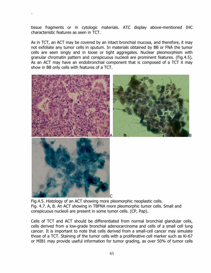
tissue fragments or in cytologic materials. ATC display above-mentioned IHC characteristic features as seen in TCT. As in TCT, an ACT may be covered by an intact bronchial mucosa, and therefore, it may not exfoliate any tumor cells in sputum. In materials obtained by BB or FNA the tumor cells are seen singly and in loose or tight aggregates. Nuclear pleomorphism with granular chromatin pattern and conspicuous nucleoli are prominent features. (Fig.4.5). As an ACT may have an endobronchial component that is composed of a TCT it may show in BB only cells with features of a TCT.
A B
C Fig.4.5. Histology of an ACT showing more pleomorphic neoplastic cells. Fig. 4.7. A, B. An ACT showing in TBFNA more pleomorphic tumor cells. Small and conspicuous nucleoli are present in some tumor cells. (CP, Pap). Cells of TCT and ACT should be differentiated from normal bronchial glandular cells, cells derived from a low-grade bronchial adenocarcinoma and cells of a small cell lung cancer. It is important to note that cells derived from a small-cell cancer may simulate those of a TCT. Staining of the tumor cells with a proliferative cell marker such as Ki-67 or MIB1 may provide useful information for tumor grading, as over 50% of tumor cells

˚
66
from a lung small-cell carcinoma show a positive nuclear reaction with Ki-67 antibody while fewer than 25% of tumor cells derived from a TCT or ACT react positively with this antibody. Bibliography Hammar SP, Dacic S. Immunohistology of lung and pleural neoplasms. In Diagnostic immunohistochemistry, Theranostic and Genomic applications. 3rd edition, 2010. Dabbs DJ, editor. Philadelphia, Saunders Elsevier. p. 369. Lin O, et al. Immunohistochemical staining of cytologic smears with MIB-1 helps distinguish low-grade from high-grade neuroendocrine neoplasms. Am J Clin Pathol 2003;120:209. Nguyen GK, et al. Transmucosal needle aspiration biopsy via the fiberoptic bronchoscope. Value and limitations in the cytodiagnosis of tumors and tumor-like lesions of the lung. Pathol Annu 1992;27 (1):105. Nguyen GK. Cytopathology of pulmonary carcinoid tumors in sputum and bronchial brushing. Acta Cytol.1995;39:1152. Renshaw AA, et al. Distinguishing carcinoid tumor from small cell carcinoma of the lung: correlating cytologic features and performance in the College of American Pathologists Non-Gynecologic Cytology Program. Arch Pathol Lab Med. 2005;129:614. Travis WD, et al. Pathology and genetics of tumours of the lung, pleura, thymus and heart. In WHO Classification of Tumours, Lyon, IARCPress, 2004. Travis WD. Neuroendocrine lung tumors. Pathology Case Reviews.2006;11:325.

˚
67
Chapter 5
Other primary tumors and tumor-like lesions of the lung and pleura Malignant lung tumors Salivary gland tumors Bronchial gland carcinomas are rare neoplasms occurring in adult patients. These neoplasms may manifest with hemoptysis or bronchial obstruction with distal lung infection. The tumors account for about 1% of all primary lung cancers and consist of two main lesions: Adenoid cystic carcinoma and mucoepidermoid carcinoma. Adenoid Cystic Carcinoma is the most common salivary gland-like tumor of the lower respiratory tract and it accounts for about 0.2% of all primary lung cancers. The neoplasm usually arises from the trachea, main stem bronchi or lobar bronchi. The patient’s age ranges from 18 to 79 years. It is a less aggressive neoplasm and has a distinct histologic pattern of growth consisting of cribiform and glandular arrays or tubules surrounding central spaces filled with epithelial mucin and solid foci. The tumor yields in BB and TBFNA single and clustered small, round cells with scant cytoplasm and round basophilic bodies. Tumor cells wrapping around basophilic bodies are a diagnostic feature of the lesion. (Figs.5.1 and 5.2).
A B

˚
68
C Fig.5.1. A. Histology of a bronchial adenoid cystic carcinoma. B, C. TBFNA of a bronchial adenoid cystic carcinoma showing ball-like clusters small cuboidal neoplastic cells wrapping around eosinophilic round bodies. A round body without wrapping cells is present in B. (Diff-Quik).
A B
C Fig.5.2. A, B. Bronchial adenoid cystic carcinoma showing in TBFNA irregular and round clusters of small cuboidal cells wrapping around eosinophilic bodies. (LBP, Pap). C. CB form the needle aspirate reveals minute tumor tissue fragments with histologic features of an adenoid cystic carcinoma described above. (HE)

˚
69
Mucoepidermoid Carcinoma is a rare tumor comprising 0.1 to 0.2% of all primary lung carcinomas. The patients range in age from 4 to 78 years but about 50% of patients are younger than 30 years. The tumor most commonly arises from the main or lobar bronchus and can measure up to 6 cm in greatest dimension. Histologically, it consists of a variable population of mucus-secreting cells, squamous cells and intermediate cells that display no particular differentiating characteristics. Bronchial mucoepidermoid carcinomas can be classified as low- and high-grade tumors, depending on the degree of cellular atypia. About 75 to 80% of mucoepidermoid carcinomas arising from the lung are of low histologic grade. A low-grade tumor yields in FNA single and clustered benign-appearing squamous cells admixed with benign-appearing mucus-secreting cells. A variable number of intermediate cells may be present. A high-grade tumor yields loosely clustered malignant squamoid cells with intracytoplasmic mucus. (Figs.5.3 and 5.4).
A B Fig.5.3. Low-grade mucoepidermoid carcinoma. A. Histology of the tumor. B. Tumor FNA loosely clustered squamoid tumor cells with some cells showing vacuolated “clear” cytoplasm. Small cuboidal tumor cells in the lower part of the figure are of intermediate type. (CP, Pap).
A B

˚
70
Fig.5.4. High-grade mucoepidermoid carcinoma: A. Histology of the tumor. B. Tumor TBFNA showing clustered malignant squamoid cells with intracytoplasmic mucus that stains positively with PASD. (CP, PASD). Soft tissue sarcomas Primary lung soft tissue sarcomas are exceedingly rare neoplasms and account for less than 1% of all primary lung cancers. Almost all histologic types of soft-tissue sarcomas have been reported in the lung. Of the primary lung sarcomas, leiomyosarcoma is the most common one. The tumor occurs mainly in adults and rarely in children. Lung leiomyosarcoma may arise from a bronchus or from intraparenchymal blood vessels. Its cytologic manifestations in BB and in FNA are similar and consist of scattered loosely aggregated, elongated slightly pleomorphic, hyperchromatic, naked tumor cell nuclei with blunt ends. Bundle of smooth muscle cells may be present. (Fig.5.5).
A B Fig.5.5. Well-differentiated leiomyosarcoma: A. TTFNA showing single and loosely clustered tumor cells with elongated nuclei with blunt ends. B. BB showing bundles of malignant smooth muscle cells with enlarged, elongated or oval nuclei. (CP, Pap). Embryonal or alveolar rhabdomyosarcoma of the lung yields in TTFNA malignant small round cells with scant cytoplasm. (Fig.5.6). A positive reaction of the tumor cell cytoplasm with MyoD1 or myogenin antibody will be helpful for a more accurate tumor typing.

˚
71
Fig.5.6. Primary lung embryonal rhabdomyosarcoma showing in TTFNA small round malignant cells with hyperchromatic nuclei and scant cytoplasm. (CP, Pap). Hematologic malignancy Primary Non-Hodgkin lymphoma (NHL) and Hodgkin disease (HD) of the lung are rare neoplasms. However, metastatic NHL and HD to the lung are noted in 20 to 50% of patients with those tumors at some point in their clinical courses. In patients with leukemia about 10% of diffuse lung infiltrations are caused by leukemic involvement, and rarely a mass lesion is observed. Multiple myeloma and mycosis fungoides rarely involve the lung. Lung NHL may be diagnosed cytologically by BAL or FNA with adjunct cell marker study. (Fig.5.7). The cytodiagnosis of HD is based on the identification of Reed-Sternberg cells. Cell marker studies by IHC and flow cytometry of BAL cell samples are useful for further confirmation of lymphoma and leukemia involving the lung.
A B Fig.5.7. A. Histology of primary non-Hodgkin lymphoma. B. Lung B-cell NHL showing in BB dissociated, monomorphic malignant lymphoid cells with conspicuous nucleoli and scant cytoplasm. (CP, Pap).

˚
72
Benign tumors and tumor-like lesions of the lung Hamartoma Lung hamartoma most commonly occurs in 6th decade of life. It is usually asymptomatic, often discovered incidentally by chest roentgenograms and it is usually located in the peripheral zone of the lung. If located in a bronchus it may cause bronchial obstruction with distal bronchial infection. The lesion is well-circumscribed, lobulated and usually measures 2 cm in greatest dimension. It is formed by elements that are normally present in the lung such as cartilage, fibromyxoid connective tissue, fat, smooth muscles and respiratory epithelium. It shows in TTFNA an admixture of the above-mentioned cytologic elements. (Fig. 5.8).
A B
C Fig.5.8. Lung hamartoma. A. Histology of the tumor. B. Tumor TTFNA showing in myxoid material, chondrocytes and clusters of benign bronchial glandular cells. (CP, Pap). C. Large fragment of benign cartilaginous tissue. (CP, Pap).

˚
73
Granular cell tumor This is a rare benign neoplasm arising from the Schwann cell. In over 90% of cases, the tumor has an endobronchial component, and in less than 10% of patients it presents as a parenchymal lesion and appears on chest roentgenograms as a “coin lesion”. It yields in BB and TBFNA sheets of benign tumor cells with eosinophilic, granular and PAS-positive cytoplasm and small, oval nuclei with conspicuous nucleoli. (Fig.5.9).
A B Fig.5.9. Bronchial granular cell tumor: A. Histology of the tumor showing cells with granular and PAS-positive cytoplasm. B. A thick fragment of tumor tissue in a TBFNA showing benign neoplastic cells with oval nuclei and ill-defined, granular cytoplasm. (A: PAS; B: Pap). Clear cell (Sugar) tumor This rare neoplasm is most likely arising from perivascular epithelioid cells and it occurs in all age groups. The tumor is usually asymptomatic, peripherally located and measures from 1 to 7 cm in greatest dimension. Histologically, it consists of benign-appearing polygonal or spindle tumor cells with oval or elongated nuclei and clear cytoplasm. (Fig.5.10). The tumor cell cytoplasm expresses S-100 protein, HMB-45 and Melan A, and it is negative for cytokeratins and CEA. It yields in FNA clustered polygonal and spindle cells with oval or elongated bland nuclei and clear cytoplasm. Intracytoplasmic glycogen can be demonstrated by staining of the tumor cells with PAS reagent. (Fig.5.10). The tumor cells should be differentiated from those of a clear cell carcinoma (primary and metastatic) and melanoma.

˚
74
A B
C Fig.5.10. Benign clear cell “sugar” tumor: A. Histology of the tumor. B. Tumor showing in TTFNA spindle tumor cells with round, bland elongated nuclei. (CP, HE). C. An aggregate of benign epithelial-like cells with ill-defined cytoplasm, round or oval nuclei and thin, semitransparent, “clear” cytoplasm. (CP, Pap). Squamous cell and glandular cell papillomas These are very rare benign endobronchial lesions that may cause hemoptysis or bronchial obstruction with distal bronchial and pulmonary infection. The squamous cell papilloma may be solitary, multiple, exophytic or endophytic. Solitary squamous cell papilloma is seen mainly in men in their fifth decade of life and is more commonly exophytic. It may be associated with human papilloma virus of both low and high risks subtypes. Cytologically, benign squamous cells and glandular cells are seen in materials obtained by BW and BB. The squamous cell tumor associated with HPV infection may show histologic features of a papillary condyloma and yields in bronchial cytologic materials dyskaryotic squamous cells with perinuclear halos. (Fig.5.11). The glandular cell papilloma exfoliates benign bronchial glandular cells and cannot be identified cytologically.

˚
75
A B
C Fig.5.11. Solitary bronchial squamous cell papilloma: A,B. Histology of the tumor showing its squamous epithelial lining displaying mild dysplasia and dyskaryotic koilocytes. C. Dyskaryotic squamous cells with one showing koilocytic change in BW. (CP, Pap). Pulmonary amyloidosis Pulmonary amyloidosis most commonly occurs in patients over 60 years of age. It usually diffusely involves the submucosa of the tracheobronchial tree but it may appear as a parenchymal mass lesion. The bronchial lesion may mimic a submucosal tumor and it yields in BB or TBFNA irregular masses of amorphous, granular, waxy material that stains slightly eosinophilic or basophilic with the Papanicolaou stain and orangeophilic with Congo red. (Fig.5.12).

˚
76
A B Fig.5.12. Bronchial amyloid deposit: A. Bronchial amyloid covered with a benign metaplastic squamous epithelium. B. BB of the lesion reveals irregular, ill-defined masses of amorphous, waxy, granular and orangeophilic amyloid material. (CP, Pap). Wegener granulomatosis This is a systemic necrotizing vasculitis of unknown etiology and it is characterized by granulomatous lesions in the nose, nasal sinuses, lung and kidney. In the lung the granulomata may measure up to 5 cm in greatest dimension and may mimic a neoplasm radiologically. A TTFNA or brushing of the lung lesion reveals granular debris of necrotic collagen admixed with chronic inflammatory cells. Multinucleated giant cells and epithelioid cells may be seen. Inflammatory pseudotumor It is also known as inflammatory fibroblastic tumor of the lung and it is a rare lesion that usually develops after a nonspecific pulmonary inflammation. It occurs in men or women, usually before the age of 40. Most of these lesions are contained within the lung and appear as a circumscribed, nodular mass consisting of an admixture of fibroblastic cells, myoepithelial cells and chronic inflammatory cells such as lymphocytes, plasma cells and macrophages. The majority of these lesions are benign, but about 5% of them are aggressive and invade adjacent structures such as esophagus, mediastinum, diaphragm and chest walls. The above-mentioned cellular elements may be seen in TTFNA. (Fig.5.13).

˚
77
A B Fig.5.13. TTFNA of an inflammatory pseudotumor of the lung reveals irregular bundles of fibroblastic cells admixed with scattered chronic inflammatory cells. (CP, Pap). Pleural Mesothelioma Pleural mesothelioma is a rare and aggressive cancer. Epidemiologic studies have linked occupational exposure to asbestos to the development of pleural mesotheliomas in 70 to 90% of the cases and the average latent period is 35 years. Other etiologic factors include exposure to erionite, therapeutic radiation and chronic infection. The tumor occurs mainly during the fifth and sixth decades of life and rarely in children. Males comprise 75% of all reported cases, and almost all patients with the disease die within 6 to 12 months after the diagnosis. It may present as a diffuse or localized growth, and the diffuse form accounts for about 75% of all cases. In over 90% of patients the disease manifests initially with recurrent, unilateral, bloody pleural effusions. In less than 10% of the cases, pleural tumors without pleural effusions are detected by chest radiography. Thus, pleural mesotheliomas can be evaluated either by cytologic examination of associated effusions or by TTFNA of a pleural mass lesion. Mesotheliomas are classified into 4 main histologic types: epithelioid, sarcomatous, biphasic or mixed, and poorly differentiated. About 50% of pleural mesotheliomas are of epithelioid in type, and they commonly show a tubulopapillary, microcystic and solid patterns. Usually, 2 or 3 histologic patterns coexist in almost all epithelial and mixed types. Sarcomatous and mixed mesotheliomas account for approximately 15 to 20% and 25 to 30% of all cases, respectively. A sarcomatous mesothelioma is characterized by spindle malignant cells arranged in a non-specific pattern. A mixed mesothelioma is composed of epithelioid and sarcomatous elements, and areas showing a transition between these 2 cellular elements may be seen. (Figs.5.14). Epithelioid and mixed mesotheliomas are commonly associated with pleural effusions containing exfoliated epithelioid tumor cells. In contrast, sarcomatous tumors are rarely associated with pleural effusions, and when they do, they seldom exfoliate their cells in the effusions.
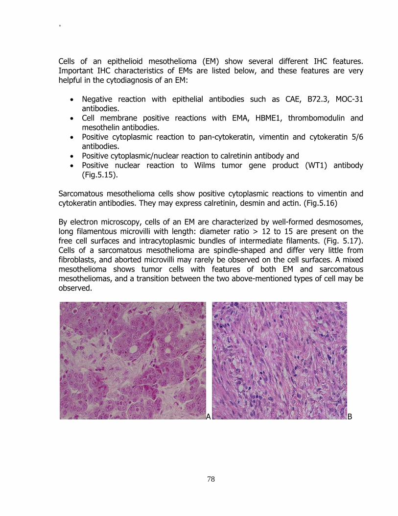
˚
78
Cells of an epithelioid mesothelioma (EM) show several different IHC features. Important IHC characteristics of EMs are listed below, and these features are very helpful in the cytodiagnosis of an EM:
• Negative reaction with epithelial antibodies such as CAE, B72.3, MOC-31 antibodies.
• Cell membrane positive reactions with EMA, HBME1, thrombomodulin and mesothelin antibodies.
• Positive cytoplasmic reaction to pan-cytokeratin, vimentin and cytokeratin 5/6 antibodies.
• Positive cytoplasmic/nuclear reaction to calretinin antibody and • Positive nuclear reaction to Wilms tumor gene product (WT1) antibody
(Fig.5.15). Sarcomatous mesothelioma cells show positive cytoplasmic reactions to vimentin and cytokeratin antibodies. They may express calretinin, desmin and actin. (Fig.5.16) By electron microscopy, cells of an EM are characterized by well-formed desmosomes, long filamentous microvilli with length: diameter ratio > 12 to 15 are present on the free cell surfaces and intracytoplasmic bundles of intermediate filaments. (Fig. 5.17). Cells of a sarcomatous mesothelioma are spindle-shaped and differ very little from fibroblasts, and aborted microvilli may rarely be observed on the cell surfaces. A mixed mesothelioma shows tumor cells with features of both EM and sarcomatous mesotheliomas, and a transition between the two above-mentioned types of cell may be observed.
A B

˚
79
C Figs.5.14. Histology of different types of pleural mesotheliomas: A. EM showing polygonal tumor cells in solid and glandular patterns. B. Sarcomatous mesothelioma showing spindle tumor cells in a nonspecific pattern. C. Mixed mesothelioma showing mixed epithelial and sarcomatous cells.
A B Fig. 5.15. Pleural EM shows tumor cells with positive cytoplasmic reaction to calretinin antibody in A and positive nuclear staining to WT1 antibody in B. (ABC).
Fig.5.16. Sarcomatous mesothelioma with spindle tumor cells shows a positive cytoplasmic reaction to pan-cytokeratin antibody. (ABC)

˚
80
Fig.5.17. Ultrastructure of an EM showing tumor cells with well-formed desmosomes and long filamentous microvilli without dense-core rootlets. (Uranyl acetate and lead citrate, x 36,000). Effusion cytology Serous effusions in patients with epithelioid and mixed mesotheliomas are usually cellular and show numerous EM cells that often display a wide range of nuclear changes, ranging from mild to marked atypia to frank malignancy. In about 10% of cases the effusions are acellular or contain only rare benign reactive mesothelial cells. Sarcomatous cells in a mixed mesothelioma and cells of a sarcomatous or desmoplastic mesothelioma do not usually and spontaneously exfoliate into associated effusions. The cytodiagnosis of EM in serous effusion requires first to cytologically diagnosis malignancy and then to identify mesothelial features of the cancer cells present. In about 50% of cases, cells from an EM or mixed mesothelioma occur singly, in small groups and in large tridimensional ball-like clusters consisting of up to several hundred cells. In about 25% of cases the tumor cells occur predominantly in tridimensional clusters with very few cells present singly and in small clusters. In the remaining 25% of cases the tumor cells occur predominantly singly. From the cytodiagnostic point of view an EM can be suspected in about 60% of cases by examination of routinely stained cytologic preparations. Classic cytologic manifestations of an EM consist of malignant cells showing the following features. (Figs. 5.18)
• Tumor cells occur singly, in small groups or clusters, as well as in large tridimensional clusters (>50 cells). Large cell clusters have smooth and lobulated contours.
• Tumor cells are usually large and resemble normal mesothelial cells except they have larger nuclei, prominent nucleoli and show a spectrum of

˚
81
nuclear changes ranging from benign to atypical to malignant. The presence of two distinct cell populations, one benign and the other malignant, as seen in metastatic cancers, is not obviously present.
• Small tumor cell clusters commonly show “cell-embracing-cell”, "push-in" cell junctions and a clear space or “window” between two adjacent cells.
• Thick papillary tumor tissue fragments with or without fibrovascular cores may be seen and are highly suggestive of an EM.
• Tumor cells have a thick endoplasm and a fuzzy ectoplasm that is due to the presence of long filamentous microvilli on free cell surfaces.
Cellblock from an effusion secondary to an EM may reveal papillary tumor tissue fragments with fibrovascular cores covered with a single layer of tumor cells. (Fig.5.19). This rare and interesting finding is highly suggestive of an EM.
A B
C Fig.5.18. Serous effusion in a pleural EM of showing: A and B. Tumor cells present singly, in small clusters and in large ball-like clusters. C. Tumor cells showing abundant, granular cytoplasm and cell-embracing-cell arrangement. (A-C, CP, Pap).
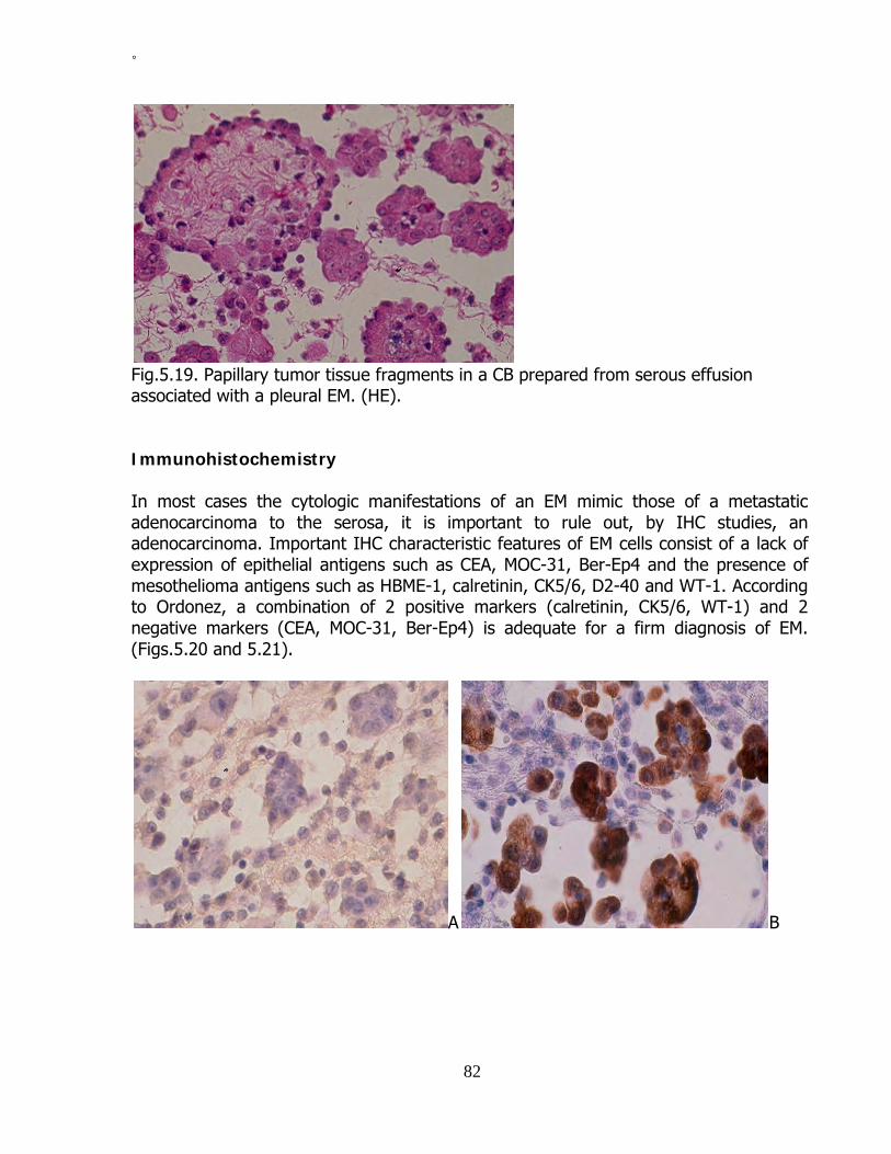
˚
82
Fig.5.19. Papillary tumor tissue fragments in a CB prepared from serous effusion associated with a pleural EM. (HE). Immunohistochemistry In most cases the cytologic manifestations of an EM mimic those of a metastatic adenocarcinoma to the serosa, it is important to rule out, by IHC studies, an adenocarcinoma. Important IHC characteristic features of EM cells consist of a lack of expression of epithelial antigens such as CEA, MOC-31, Ber-Ep4 and the presence of mesothelioma antigens such as HBME-1, calretinin, CK5/6, D2-40 and WT-1. According to Ordonez, a combination of 2 positive markers (calretinin, CK5/6, WT-1) and 2 negative markers (CEA, MOC-31, Ber-Ep4) is adequate for a firm diagnosis of EM. (Figs.5.20 and 5.21).
A B

˚
83
C D Fig.5.20. IHC of effusion CB in EM: A. Tumor cell cytoplasm reacts negatively with CEA antibody. B. Tumor cells show strong cytoplasmic and nuclear reactions to calretinin antibody. C. Strong, thick, membranous positive staining with spiking pattern with EMA antibody, reflecting the presence of long microvilli on tumor cell surfaces. D. EM cells showing positive nuclear staining with WT-1 antibody. (ABC).
A B
C Fig.5.21. IHC of lung adenocarcinoma: A. Tumor cells in CB showing a strong cytoplasmic reaction to CEA antibody. B. Tumor cell nuclei in CB stain positively with TTF-1 antibody. C. Tumor cells displaying a positive membranous reaction to MOC-31 antibody. (ABC).

˚
84
Some commonly encountered cytologic, IHC and ultrastructural features of epithelial mesothelioma, bronchogenic adenocarcinoma and reactive mesothelium are tabulated in Table 5.1. Table 5.1: Comparative Cytologic Manifestations of Reactive Mesothelium, Epithelial Mesothelioma and Bronchogenic Adenocarcinoma in Serous Effusions* CELLULAR FEATURES
REACTIVE MESOTHELIUM
EPITHELIOID MESOTHELIOMA
BRONCHOGENIC ADENOCARCINOMA
Architecture: -Singly, common -Monolayered sheets -Loose groups with “windows”
-Large cohesive clusters with lobulated borders -Small tight clusters with “windows” and “push-in” junctions
-Singly, rare -Tight multilayered, 3-dimensional clusters with smooth borders
Cells: -Configuration: -Cytoplasm: -Nucleus: -Anisonucleosis -Irregular contours -Molding -Chromatin -Nucleolus
-Polygonal, round -Well-defined -Foamy or homogenous - - - Fine Small
-Polygonal, round -Well-defined -Dense ectoplasm -Fuzzy periphery + + + Fine or coarse Small or large
-Polygonal, round -Ill-defined -Vacuolated + + + Coarse Large, single, multiple
Staining characteristics: - Mucin - - + - Calretinin, CK5/6, D2-40, Mesothelin, WT-1
+ + -
- CEA, MOC-31, Ber-Ep4, TTF-1 - - +
- Desmin +/- +/- - - EMA +, periphery ++, fuzzy, periphery +, periphery Ultrastructure: - Long microvilli, with L:D >12 - Intracytoplasmic mucous granules
+ -
+ -
-
+ *Adapted from Nguyen GK, Kline TS: Essentials of cytology. An Atlas. 1993, Igaku-Shoin, New York, p.88.

Diagnostic accuracy of mesothelioma by effusion cytology According to Whitaker a diagnosis of EM by effusion cytology may be suggested in the presence of many large clusters or aggregates of tumor cells together with abundant single neoplastic cells. Large cellular aggregates are of most value in facilitating a diagnosis of malignancy, and single cells or clusters of 2 to 6 cells are of most value in identifying the mesothelial characteristics of tumor cells. Nuclear atypias as commonly noted in metastatic adenocarcinomas are not usually seen in mesothelioma cases. IHC and/or electron microscopic studies of effusion CBs are necessary for distinguishing an EM from an adenocarcinoma. In the experience of Whitaker, et al. a diagnostic accuracy rate of 80% of mesotheliomas of has been reached by a combination of effusion cytology and IHC and/or electron microscopic studies of effusion CBs. The predictive value of a positive diagnosis of EM in serous effusion has been about 100% in those investigators’ hands. FNA cytology of mesothelioma Only a small number of pleural mesotheliomas with cytologic evaluation by TTFNA have been reported. For FNA diagnosis of pleural mesothelioma a sensitivity rate of 73-78% has been reported. An EM usually yields tumor cells singly, in thick clusters, in sheets and in ball-like or papillary clusters. The individual tumor cells display well-defined, optically dense cytoplasm, oval nuclei and prominent nucleoli. Occasional tumor cells show a vacuolated cytoplasm. (Figs. 5.22 and 5.23). TTFNA from a sarcomatous tumor may reveal spindle malignant cells with elongated nuclei and scant, granular or clear cytoplasm present singly and in loose clusters. (Fig. 5.24). A mixed mesothelioma is characterized by an admixture of single and clustered malignant spindle cells and malignant epithelioid cells displaying mesothelial cell features. A poorly differentiated mesothelioma yields large malignant cells with ill-defined or well-defined, granular cytoplasm and prominent nucleoli singly and in aggregates, similar to those of a large cell anaplastic carcinoma.

86
A B Fig. 5.22. TTFNA of a pleural EM showing in: A. Ball-like and papillary tumor cell clusters or fragment. B. Tumor cells with prominent nucleoli present in tridimensional clusters and singly. (Diff-Quik).
Fig. 5.23. EM shows in TTFNA tumor cells singly, in a loose sheet and in a tridimensional cluster. (HE).

87
Fig.5.24. Pleural EM showing in TTFNA an irregular and large cluster of fairly monomorphic tumor cells with oval nuclei and inconspicuous nucleoli. (CP, Pap)
Fig.5.25. TTFNA of a sarcomatous mesothelioma of the pleura showing single spindle malignant cells with elongated cytoplasm. (Pap). IHC staining of the tumor cells within the CB or aspirated minute tissue fragments with antibodies against CEA, Ber-Ep4, MOC-31, cytokeratins 5/6, calretinin and WT1 are helpful for further tumor typing. By electron microscopy the epithelial tumor cells show well-formed desmosomes and long slender microvilli. (Fig.5.26). Microvilli in direct contact with collagen fiber bundles in the tumor matrix may be seen in minute tumor tissue fragments, and this finding constitutes a strong evidence indicating an invasive EM, according to Ghadially.
Fig. 5.26. Ultrastructure of an aspirated minute tumor tissue fragment from a pleural EM showing tumor cells with well-formed cell junctions and long, filamentous microvilli on the tumor cell surface. (Uranyl acetate and lead citrate, x 24,000).

Cells aspirated from an EM should be differentiated from those of a lung adenocarcinoma, either primary or metastatic. Spindle cells aspirated from a sarcomatous mesothelioma should be differentiated from those of a benign solitary fibrous tumor of the pleura, and from cells of a fibrosarcoma, leiomyosarcoma, malignant schwannoma and malignant fibrous histiocytoma of the lung and pleura. IHC study of the needle aspirate with a number of selected antibodies will be useful for differential diagnosis and tumor typing Bibliography Awasthi A, et al. Pitfalls in the diagnosis of Wegener’s granulomatosis on fine needle aspiration cytology. Cytopathology. 2007; 18: 8. Bhargava R, Dabbs DJ. Immunohistology of metastatic carcinoma of unknown primary. In Diagnostic Immunohistochemistry. Theranostic and genomic applications, 3rd edition, 2010, Dabbs DJ, editor, Saunders Elsevier, p.206. DeMay R. Fluids. In Arts & Science of Cytopathology. Exfoliative cytology. Chicago, ASCP, 1996, p.257. Fletsch PA, Abati A. Immunocytochemistry in effusion cytology. A contemporary review. Cancer (Cancer Cytopathol). 2001;93:293. Giesinger KR, et al. Effusions. In Modern Cytopathology, Giesinger KR, et al, eds. Philadelphia, Churchill Livingstone, 2004, p.257. Geisinger KR, et al. Localized lung diseases. In Modern Cytopathology, Geisinger KR, et al, eds. Philadelphia, Churchill Livingstone. 2004, p 399. Ghadially FN. Diagnostic electron microscopic of tumours, 2nd ed, 1985. London, Butterworths. Husain M, Nguyen GK. Cytopathology of granular cell tumor of the lung. Diagn Cytopathol. 2000; 23: 294. Leong AS. Immunostaining of cytologic specimens. Am J Clin Pathol.1996;105:139. Machicao CN, et al. Transthoracic needle aspiration biopsy of inflammatory pseudotumors of the lung. Diagn Cytopathol. 1989; 5:400. Nguyen GK. Cytology of bronchial gland carcinoma. Acta Cytol. 1988; 32: 235.

89
Nguyen GK. Aspiration biopsy cytology of benign clear-cell “sugar” tumor of the lung. Acta Cytol. 1989; 33:511. Nguyen GK. Cytopathology of pleural mesotheliomas. Am J Clin Pathol. 2000; 114 (Suppl 1): S68. Nguyen GK, et al. Cytodiagnosis of malignant mesothelioma of the pleura by fine-needle aspiration biopsy. Diagn Cytopathol.1999;21:253. Obers VJ, et al. Primary pleural tumors (mesotheliomas) presenting as localized masses. Fine needle aspiration cytologic findings, clinical and radiological features and review of the literature. Acta Cytol.1988;32:567. Ordonez NG. Immunohistochemical diagnosis of epithelioid mesotheliomas: a critical review of old markers, new markers. Hum Pathol.2002;33:935. Odashiro AN, et al. Primary lung leiomyosarcoma detected by bronchoscopy cytology. Diagn Cytopathol. 2005; 33: 220. Policarpio-Nicolas ML, et al. Fine needle aspiration cytology of clear cell “sugar” tumor (PEComa) of the lung: report of a case. Diagn Cytopathol. 2008;36:89. Sterrett GF, et al. Fine needle aspiration cytology of malignant mesothelioma. Acta Cytol 1987; 31:185. Suster S, Moran CA. Applications and limitations of immunohistochemistry in the diagnosis of malignant mesothelioma. Adv Anat Pathol.2006;13:316 Travis WD, et al. Pathology and Genetics of Tumours of the Lung, Pleura, Thymus and Heart. In WHO Classification of Tumors. Lyon, IARCPress,2004. Whitaker D, et al. Mesothelioma. In Diagnostic Cytopathology, 2nd ed, Gray W, McKee GT, eds, 2003, London, Churchill Livingstone, p.205. Wick MR, et al. Immunohistochemical differential diagnosis of pleural effusions, with emphasis on malignant mesothelioma. Curr Opin Pul Med.2001;7:187. Wood B, et al. Diagnosis of pulmonary hamartoma by fine needle biopsy. Acta Cytol. 2008; 52:412.

90
Chapter 6
Secondary lung tumors The lung is one of the most common sites of metastasis from extrathoracic cancers. From 20 to 60% of individuals with extrathoracic solid cancers show, at autopsy, lung metastasis; and the lung is the only site of metastasis in 15 to 25% of these cases. Carcinomas arising from the breast, prostate, testicles and kidney, cutaneous melanoma, Ewing sarcoma, osteogenic sarcoma and rhabdomyosarcoma frequently metastasize to the lung. Metastatic cancers in the lung display some distinctive patterns of metastasis such as multiple tumor nodules, lymphangitic, endobronchial, endovascular, solitary and pleural. An awareness of these macroscopic patterns of metastasis is helpful for a more accurate cytodiagnosis of secondary lung cancers. Endobronchial metastatic cancers may exfoliate their cells in sputum and bronchoscopy cytologic specimens. Lung parenchymal deposits are best diagnosed with TTFNA and BAL may show malignant cells from a cancer with alveolar spread. The cytologic manifestations of metastatic cancers to the lung are somewhat similar in different types of pulmonary cell samples. Clinical history and a comparison of the metastatic cancer cells with the histologic sections or cytologic samples of the primary cancers, if available, are of diagnostic help in the majority of cases. Cytodiagnosis of solitary metastasis Diagnosis of solitary metastasis is important for patient care, as a second or a third primary cancer may develop in patients who had a surgically removed primary cancer many years prior. As in the liver, diagnosis of metastatic adenocarcinoma to the lung is challenging. An awareness of the incidences of metastasis of cancers arising from different organs or anatomic sites can be helpful. IHC studies of cell samples or aspirated minute tumor tissue fragments with selected antibodies may be required for a more accurate tumor typing in some cases. On rare occasions, electron microscopic study of aspirated tumor tissue fragments is needed for tumor typing. Morphologic evaluation of the FNA and routinely stained CB sections usually permit a proper classification of well-differentiated cancer cells into 4 broad categories: epithelial, lymphomatous, melanocytic and sarcomatous malignancies. However, in the case of a poorly differentiated or undifferentiated cancer, IHC studies are needed for a

91
more accurate tumor typing. By staining with antibodies to S-100, HMB-45, AE1/AE3, calretinin, MOC-31, CEA, LCA and vimentin, poorly differentiated cancer cells may be classified into 5 cell lines: lymphomatous, melanocytic, epithelial, mesothelial and sarcomatous. A coordinate staining of epithelial cells with CK7/CK20 antibodies, according to Bhargava and Dabbs, will further divide them into 4 different categories, each with only a few tumor types. Additional IHC expressions of some cell markers by metastatic cancer cells may further confirm the anatomic sites of their primary cancers. 1. CK7+/CK20+: Urothelial carcinoma and ovarian mucinous carcinoma. 2. CK7+/CK20-: Carcinomas of lung (small cell, non-small cell), breast, ovary (serous type), endometrium and thyroid; germ cell tumors and epithelial mesothelioma. 3. CK7-/CK20-: Squamous cell, prostatic, renal cell and hepatocellular carcinomas. 4. CK7-/CK20+: Colorectal and Merkel cell carcinomas. Cytologic manifestations of secondary lung cancers Metastatic breast cancer Mammary carcinomas frequently metastasize to the lung. The tumors usually yield malignant glandular cells with nonspecific features and tumor cells arranged in linear pattern may be observed. (Figs.6.1 and 6.2). These cells are usually CEA, estrogen and progesterone receptors and CK7 positive, and CK20 and TTF-1 negative.
Fig.6.1. Endobronchial metastatic mammary duct carcinoma showing in sputum loosely clustered malignant glandular cells with conspicuous nucleoli. (CP, Pap)

92
A B Fig.6.2. Metastatic mammary duct carcinoma showing in: A. TTFNA, clustered malignant glandular cells with focal nuclear crowding. Tumor cells in linear arrangement are noted elsewhere. (DQ). B. Tumor cells in a CB section showing positive nuclear staining with ER antibody. (ABC). Metastatic thyroid cancer About 15% of thyroid carcinomas metastasize to the lung. Metastatic cancers are more frequently derived from an anaplastic carcinoma then from a poorly differentiated or well-differentiated carcinoma. Metastatic papillary carcinoma shows papillary tissue fragments, sheets or groups of tumor cells with nuclear crowding, intranuclear cytoplasmic inclusions and nuclear grooves. (Fig.6.3). A follicular carcinoma yields cells in clusters with focal acinar arrangement. A Hürthle cell carcinoma shows tumor cells with granular cytoplasm singly and in monolayered sheets. A medullary carcinoma may show single and clustered polygonal and/or spindle tumor cells with elongated nuclei. Intranuclear cytoplasmic inclusions may be noted and cytoplasmic azurophil granules may be observed in tumor cells stained with MGG or DQ technique. (Fig.6.4). Cells derived from an anaplastic carcinoma are either large pleomorphic or spindle. Tumor cells from a papillary, follicular, Hürthle cell or insular carcinoma express thyroglobulin and TTF-1 and are negative for CEA while those of a medullary carcinoma stain positively with calcitonin and CEA antibodies. Cells derived from an anaplastic carcinoma do not usually express thyroglobulin or TTF-1.

93
A B Fig.6.3. Metastatic papillary carcinoma of the thyroid, follicular variant: A. Histology of the tumor. (HE). B. Tumor TTNA showing clustered tumor cells displaying nuclear crowding. Intranuclear cytoplasmic inclusions are observed in some tumor cells. (Diff-Quik).
Fig.6.4. Metastatic thyroid medullary carcinoma to the lung showing tumor cells with plasmacytoid configuration. (DQ). Metastatic GI tract, pancreatic and biliary tree cancers Metastatic tumors from a poorly differentiated adenocarcinomas arising from the stomach, small and large bowels, pancreas or biliary tree yields malignant cells with no specific features. Staining of the tumor cells with CDX2, CK7, CK20, MUC-2 and MUC-5 antibodies will be useful for determining the site of the primary tumor. Cells from a biliary or pancreatic tumor are usually monoclonal CEA positive, CDX2 negative, CK7 positive, CK20 negative and MUC-5 positive while those of colorectal origin are usually CDX2 positive, CK7 negative, CK20 positive and MUC-2 positive. Cells with signet-ring

94
configurations are most commonly derived from an anaplastic carcinoma of the stomach. Cells from a well- or moderately differentiated colonic adenocarcinoma are seen in sheets with elongated nuclei in picket fence pattern. A large amount of necrotic debris is commonly noted in FNAs from a metastatic colonic adenocarcinoma. (Fig. 6.5). In other cases oval or elongated tumor cells in syncytial sheets and clusters are observed. (Figs.6.6 and 6.7).
A B
C D
E

95
Fig.6.5. Metastatic colonic adenocarcinoma to the lung: A. Histology of the tumor. (HE). B, C. Irregular sheet of tumor cells in BB showing cells in palisade at periphery. (CP, Pap). D. Small tumor tissue fragment in CB showing glandular-type cells in nonspecific pattern (D, HE). E. Tumor cells with CDX2 positive nuclei. (ABC).
Fig. 6.6. Endobronchial metastatic colonic adenocarcinoma showing in BB a syncytial cluster of malignant cells with elongated nuclei. (CP, Pap).
A B Fig. 6.7. Metastatic poorly differentiated colonic adenocarcinoma to the lung showing in BAL: A, B. Dissociated polygonal cells with oval nuclei, conspicuous nucleoli and thin, foamy cytoplasm that stains positively with CEA antibody. (CP, Pap; B, ABC). Metastatic liver cancer Hepatocellular carcinomas commonly spread to the lung. Single and clustered polygonal cells with granular or vacuolated cytoplasm are seen, and intracytoplasmic globular

96
inclusions may be observed. Cells derived from a hepatocellular carcinoma do not express CK7 or CK20. A positive staining of the tumor cell cytoplasm with alpha-fetoprotein or HepPar 1 antibody will practically confirm the diagnosis of a metastatic hepatocellular carcinoma. (Fig.6.8).
A B Fig.6.8. Metastatic hepatocellular carcinoma to the lung: A. BAL showing a tumor cell with intra cytoplasmic globular inclusion. (Pap). B. Histology of the moderately differentiated hepatocellular carcinoma metastatic to the lung showing tumor cells with many of them showing intracytoplasmic globular inclusions. (HE). Metastatic salivary gland cancer Of the salivary gland carcinomas, adenoid cystic carcinoma most commonly metastasizes to the lung. It is characterized in FNA by the presence of abundant small cells with hyperchromatic nuclei and scant cytoplasm in acinar arrangement. Globular bodies of amorphous, basophilic material may be present in smear background and globular bodies wrapped with tumor cells are commonly seen. Metastatic renal, urinary tract and adrenal cancers Renal cell carcinomas (RCC) commonly metastasize to the lung. A conventional RCC yields cells with clear or granular cytoplasm singly, in clusters and in monolayered sheets. (Figs.6.9 and 6.10). A metastatic papillary RCC yields in FNA monolayered sheets of monomorphic tumor cells with clear or granular cytoplasm, and papillary tumor tissue fragments with fibrovascular core may be present. RCC cells stain positively with RCC and CD10 and negatively with inhibin, CK7 and CK20 antibodies.

A B Fig.6.9. Endobronchial metastatic RCC showing in: A. Clustered malignant glandular cells with scanty cytoplasm and prominent nucleoli in sputum. B. Clusters of malignant glandular cells with granular, well-defined cytoplasm, oval nuclei and prominent nucleoli in TBFNA. (CP, Pap)
A B Fig.6.10. Metastatic grade 3/3 renal cell carcinoma to the lung showing in: A. Sputum, 2 clusters of malignant glandular cells with prominent nucleoli. B. TTFNA, single and loosely aggregated pleomorphic malignant cells with granular cytoplasm, single or large multiple nuclei. (CP, HE). Cells from a conventional RCC and an adrenal cortical carcinoma are morphologically similar and express both cytokeratin and vimentin. Adrenal cortical carcinoma cells express, in addition, inhibin, Melan A or A103 but they do not express RCC, CD10. A metastatic chromophobe RCC yields cells similar to those of a conventional RCC. Perinuclear clear spaces and positive cytoplasmic staining with colloidal iron are other

98
characteristic cellular features of the tumor. Cells from a chromophobe RCC express CD117. Abundant intracytoplasmic microvesicles are seen by electron microscopic study of aspirated minute tumor tissue fragments. A metastatic high-grade transitional cell carcinoma (TCC) of the renal pelvis or urinary bladder is characterized by the presence of pleomorphic malignant epithelial cells singly and in clusters. Tumor cells with cytoplasmic extension or “tail” (cercariform cells) are commonly seen and constitute a fairly reliable feature for this type of neoplasm. (Figs.6.11 and 6.12). Urothelial cancer cells express uroplakin III (URO III), p63, CK5/6, thrombomodulin, CK7 and CK20.
A B
C Fig. 6.11. Metastatic transitional cell carcinoma, grade 2/3: A. Histology of the tumor. B, C. Slightly pleomorphic tumor cells with granular, ill-defined cytoplasm, small nucleoli in loosely cohesive sheets seen in tumor TTFNA. (CP, Pap).

99
A B Fig.6.12. Metastatic transitional cell carcinoma, grade 3/3 showing in TTFNA: A, B. Pleomorphic, single and clustered malignant cells. A few tumor cells with cytoplasmic tails or “cercariform cells” are noted in A. (CP, Pap). Metastatic prostatic cancer A metastatic prostatic adenocarcinoma shows clusters and sheets of small glandular cells with clear cytoplasm and round nuclei with prominent nucleoli. The tumor cell cytoplasm characteristically stains positively with prostatic specific antigen antibody. (Fig. 6.13).
A B Fig.6.13. A showing cohesive small malignant cells in bronchial brushing of an endobronchial metastatic prostatic adenocarcinoma. The tumor cells in B stain positively with prostatic specific antigen antibody. (A: CP, Pap; B: ABC).

100
Metastatic uterine cancer Cervical squamous cell carcinomas frequently spread to the lung while end cervical adenocarcinomas rarely do so. Endometrial adenocarcinoma also rarely metastasizes to the lung. Its tumor cells express ER and vimentin. A metastatic low-grade endometrial stromal sarcoma shows in TTFNA abundant single and clustered small round cells with scant cytoplasm. A metastatic myometrial leiomyosarcoma to the lung shows malignant spindle cells with elongated nuclei with blunt ends. Metastatic ovarian cancer Ovarian carcinoma metastatic to the lung frequently involves the pleura with associated malignant effusion. Parenchymal spread is uncommon and occurs late in the disease. Cells derived from an ovarian adenocarcinoma usually express CA-125, vimentin and ER and they are CEA negative. Metastatic melanoma Cutaneous melanoma frequently spreads to the lung and commonly yields single pleomorphic malignant cells. Intranuclear cytoplasmic inclusions are commonly seen and intracytoplasmic melanin pigment granules may be noted. The tumor cell cytoplasm characteristically expresses S100 protein, HMB-45, MART-1 and Melan A. (Fig. 6.14).
A B Fig.6.14. A, B. Metastatic melanoma to the lung showing in TTFNA pleomorphic dyshesive malignant cells that stain positively with HMB-45 antibody. (CP, A, Pap; B, CB, ABC).

101
Metastatic soft tissues and bone sarcomas Soft tissue and bone sarcomas commonly spread to the lung. In practice, the presence of malignant nonepithelial cells in a pulmonary cell sample from a patient with a known soft tissue or bone sarcoma is often indicative of a metastatic sarcoma. A metastatic Ewing sarcoma yields in TTFNA single and clustered round cancer cells with scant cytoplasm. (Fig.6.15). A metastatic well-differentiated leiomyosarcoma yields in TTFNA loosely clustered spindle cells with cigar-shaped nuclei and scant cytoplasm. (Fig. 6.16). A positive cytoplasmic reaction of the tumor cell cytoplasm with desmin antibody will confirm the diagnosis. A neurogenic sarcoma will yield similar tumor cells that stain positively with S-100 protein antibody.
Fig. 6.15. Single and clustered round tumor cells with scant cytoplasm in TFNA of a metastatic Ewing sarcoma to the lung. (CP, DQ)
Fig. 6.16. Metastatic well-differentiated uterine leiomyosarcoma to the lung showing in TTFNA a large cluster of malignant spindle cells with cigar-shaped and blunt end nuclei. (CP, Pap).

102
Metastatic testicular cancer and extragonadal germ cell tumors Testicular seminomas rarely spread to the lung, but other gonadal tumors often metastasize to the lung. Cells derived from a nonseminomatous tumor deposits are usually pleomorphic and occur singly or in syncytial clusters. The tumor cell cytoplasm expresses alpha-fetoprotein and placental alkaline phosphatase. Cells from a choriocarcinoma express beta human chorionic gonadotropin. Cells from an embryonal carcinoma are pleomorphic glandular-type cells with scant cytoplasm and prominent nucleoli. (Fig.6.17). Cells derived from a yolk sac tumor are usually present in clusters and show intracytoplasmic eosinophilic globular inclusions. (Fig. 6.18).
Fig.6.17. A. Testicular embryonal carcinoma showing clustered malignant cells with prominent nucleoli. (CP, Pap).
Fig.6.18. A metastatic yolk sac tumor showing pleomorphic malignant cells with ill-defined cytoplasm and intracytoplasmic, eosinophilic globular bodies. (CP, Pap). (Courtesy of Dr. K. C. Suen, Vancouver, BC, Canada).

103
Metastatic neuroendocrine cancer Neuroendocrine cancer arising from extrapulmonary locations (gastrointestinal tract, pancreas and ovary) may spread to the lung. These tumors yield single and clustered epithelial cells with eccentrically located nuclei (plasmacytoid configuration) and chromatin clumping. A positive cytoplasmic reaction with neuron-specific enolase, synaptophysin, chromogranin and CD56 antibodies will confirm the diagnosis. However, determination of the location of the primary tumor cannot be made with confidence on cytologic bases alone. Thymic tumor Thymoma may directly invade the lung or metastasizes to it. Thymomas are classified histologically into Type A and Type B tumors. Type A tumor consists of spindle neoplastic epithelial cells with bland nuclei, and type B tumor is characterized by polygonal epithelial tumor cells. Type B thymoma is further subdivided into subtypes B1, B2 and B3, based on the extent of lymphocytic infiltration and the degree of epithelial cell atypia, with B1 tumor containing abundant lymphoid cells and type B2 and B3 being rich in epithelial cells. Type B2 thymoma is characterized by highly atypical or malignant epithelial cells admixed with a small number of lymphocytes. Thymomas with features of type A and type B1 or B2 are designated type AB. Type A, B1 and AB thymomas have a low-malignant potential with rare local recurrence and late metastases, while type B2 and B3 thymomas and thymic carcinomas are more aggressive cancers. Cytologically, Type A thymoma yields in TFNA single, loosely clustered or tightly clustered spindle epithelial cells. (Fig.6.19). Type B1 thymoma is characterized by abundant lymphocytes admixed with polygonal epithelial cells. (Fig.6.20). The epithelial cells can be difficult to identify in routinely stained cellular materials and staining of the cell sample with a pancytokeratin antibody may easily identify them. Thymic squamous cell carcinoma is characterized by malignant squamous cells. Thymic neuroendocrine carcinomas have different histologic patterns such as typical and atypical carcinoid tumors and large and small cell carcinomas, and display in TTFNA cells similar to those of the lung with similar histologic patterns.

104
A B
C Fig.6.19. Type A thymoma: A. Histology of the tumor showing spindle cells with bland nuclei. B, C. Tumor FNA showing a thick bundle and a thin bundle of spindle tumor cells with bland nuclei and scant cytoplasm. (CP, HE).
A B Fig.6.20. Type B2 thymoma: A. Histology of the tumor showing abundant polygonal epithelial cells and lymphocytes. B. Tumor FNA showing cohesive clusters of epithelial tumor cells with pleomorphic nuclei and benign lymphocytes. (CP; B,C, Pap).

105
Metastatic Non-Hodgkin and Hodgkin lymphoma Non-Hodgkin lymphoma and Hodgkin disease involving the thymus, mediastinal lymph nodes and other anatomic sites may spread to the lung with formation of tumor masses. These lesions may be diagnosed by bronchial cytologic materials or TTFNA with IHC studies and flow cytometry. Bibliography Ali TZ, et al. Exfoliative sputum cytology of cancers metastatic to the lung. Diagn Cytopathol. 2005;33:147. Bhargava R, Dabbs DJ. Immunohistology of metastatic carcinoma of unknown primary. In Diagnostic immunohistochemistry. Theranostic and genomic applications. 3rd edition, 2010. D. Dabbs, editor, Philadelphia, Saunders Elsevier. p. 206. DeLellis RA, Hoda RS. Immunohistochemistry and molecular biology in cytological diagnosis. In Koss’ Diagnostic cytology and its histopathologic bases. 5th ed, Koss LG, Melamed MR, eds. Philadelphia, Lippincott Williams & Wilkins. 2006, p 1635. Koss LG, Melamed MR. Tumors of the lung: conventional cytology and aspiration biopsy. In Koss’ Diagnostic cytology and its histopathologic bases, 5th edition, 2006. Koss LG, Melamed MR, editors. Philadelphia, Lippincott Williams & Wilkins. p 643. Nguyen GK. Fine needle aspiration biopsy cytology of metastatic renal cell carcinoma. Acta Cytol. 1988; 32;409. Nguyen GK, et al. Transmucosal needle aspiration biopsy via fiberoptic bronchoscope. Value and limitations in the cytodiagnosis of tumors and tumor-like lesions of the lung. Pathol Annu. 1992; 27(1): 105. Park SY, et al. Panels of immunohistochemical markers help determine primary sites of metastatic adenocarcinoma. Arch Pathol Lab Med. 2007; 131: 1561. Raab SS, et al. Metastatic tumors in the lung: a practical approach to diagnosis. In Practical pulmonary pathology. Leslie KO and Wick WR, eds. Philadelphia, Churchill Livingstone, 2005, 603. Simsir A, et al. Utility of CD10 and RCCma in the diagnosis of metastatic conventional renal cell adenocarcinoma by fine-needle aspiration biopsy. Diagn Cytopathol. 2005; 33:3.