atm phosphorylates histone h2ax in response to …/67531/metadc742264/m2/1/high... · 1 atm...
TRANSCRIPT

1
ATM Phosphorylates Histone H2AX in Response to DNA Double-Strand Breaks
Sandeep Burma1, Benjamin P. Chen1, Michael Murphy1, Akihiro Kurimasa1, 2, and David J. Chen1, *
Lawrence Berkeley National Laboratory Life Sciences Division
1 Cyclotron Road, Berkeley, CA 94720
1Lawrence Berkeley National Laboratory, Berkeley, CA 2Current Address: Life Sciences Division, Tottori University, Tottori, JAPAN
* Corresponding author Tel: 510-495-2861 Fax: 510-486-6816 E.mail: [email protected]
Running title: Phosphorylation of H2AX by ATM

2
SUMMARY
A very early step in the response of mammalian cells to DNA double-strand breaks
(DSBs) is the phosphorylation of histone H2AX at serine 139 at the sites of DNA damage. While
the PI-3 kinases, DNA-PK (DNA-dependent protein kinase), ATM (ataxia telangiectasia
mutated), and ATR (ATM and Rad3-related), have all been implicated in H2AX
phosphorylation, the specific kinase involved has not yet been identified. In order to definitively
identify the specific kinase(s) that phosphorylates H2AX in vivo, we have utilized DNA-PKcs-/-
and Atm-/- cell lines and mouse embryonic fibroblasts. We find that H2AX phosphorylation and
nuclear focus formation are normal in DNA-PKcs-/- cells and severely compromised in Atm-/-
cells. We also find that ATM can phosphorylate H2AX in vitro and that ectopic expression of
ATM in Atm-/- fibroblasts restores H2AX phosphorylation in vivo. The minimal H2AX
phosphorylation in Atm-/- fibroblasts can be abolished by low concentrations of wortmannin
suggesting that DNA-PK, rather than ATR, is responsible for low levels of H2AX
phosphorylation in the absence of ATM. Our results clearly establish ATM as the major kinase
involved in the phosphorylation of H2AX and suggest that ATM is one of the earliest kinases to
be activated in the cellular response to DSBs.

3
INTRODUCTION DNA double-strand breaks (DSBs) are probably the most dangerous of the many
different types of DNA damage that occur within the cell. DSBs are generated by exogenous
agents such as ionizing radiation (IR) or by endogenously generated reactive oxygen species and
occur as intermediates during meiotic and V(D)J recombination (1). A very early step in the
cellular response to DSBs is the phosphorylation of a histone H2A variant, H2AX, at the sites of
DNA damage (2). H2AX is rapidly phosphorylated (within seconds) at serine 139 when DSBs
are introduced into mammalian cells (3) resulting in discrete γ-H2AX (phosphorylated-H2AX)
foci at the DNA damage sites (4). In experiments involving the use of ‘laser scissors’ to
introduce breaks into living cells, γ-H2AX foci localized specifically with the laser path through
the cell nuclei clearly demonstrating that H2AX phosphorylation is specific to the sites of DNA
damage (4,5). H2AX phosphorylation also appears to be a general cellular response to processes
involving DSB intermediates including V(D)J recombination in lymphoid cells (6) and meiotic
recombination in mice (7). Phosphorylation of yeast H2A at serine 129 (homologous to serine
139 of mammalian H2AX) causes chromatin decondensation and is required for efficient DNA
double-strand break repair (8). In mammals, phosphorylation of H2AX appears to play a critical
role in the recruitment of repair or damage-signaling factors to the sites of DNA damage (5,9).
As H2AX phosphorylation plays a very early and important role in the cellular
response to DNA double-strand breaks, it is important to specifically identify the kinase(s)
involved in this event. Members of the PI-3 kinase family, including DNA-PK (DNA-dependent
protein kinase), ATM (ataxia telangiectasia mutated), and ATR (ATM and Rad3-related), are
involved in the responses of mammalian cells to DSBs (10). γ-H2AX focus formation is
inhibited by the PI-3 kinase inhibitor wortmannin and H2AX phosphorylation is reduced in the

4
DNA-PK-deficient human cell line M059J (5). This led to the conclusion that DNA-PK, and at
least one other kinase, possibly ATM and/or ATR, can phosphorylate H2AX upon DNA damage
(2,5,10,11).
In order to unambiguously define the roles of ATM and DNA-PK in H2AX
phosphorylation, we utilized cells derived from knockout mice for ATM or DNA-PKcs (the
catalytic subunit of DNA-PK). We observed normal H2AX phosphorylation and γ-H2AX focus
formation in irradiated fibroblasts derived from wild type or DNA-PKcs-/- mice. In contrast,
H2AX phosphorylation and γ-H2AX focus formation were strikingly reduced to near
background levels in fibroblasts from Atm-/- mice. Ectopic expression of ATM in Atm-/- cells
restored H2AX phosphorylation. Moreover, we show that immunoprecipitated ATM can
phosphorylate recombinant H2AX in vitro. These results indicate that ATM, not DNA-PK, is the
major kinase responsible for modifying H2AX upon irradiation. The minimal H2AX
phosphorylation in Atm-/- cells could be abolished by low concentrations of wortmannin
suggesting that DNA-PK, rather than ATR, is responsible for low levels of γ-H2AX formation in
the absence of ATM.

5
EXPERIMENTAL PROCEDURES
Cell Culture and Induction of DNA Damage - Spontaneously immortalized mouse
fibroblasts, derived from wild type, DNA-PKcs-/- (12), or Atm-/- mice (13), were maintained in
a humidified atmosphere with 5% CO2 in α-MEM medium supplemented with 10% fetal calf
serum, 100 units / ml penicillin and 100 µg / ml streptomycin. Mouse embryonic fibroblasts
(MEFs) were isolated from 13.5-day old embryos and maintained in α-MEM medium
supplemented with 15% fetal calf serum. Cells were grown to about 70% confluence and
irradiated with X-rays (300 kV, 12 mA, 0.5 mm Cu) at the rate of 5.5 Gy / min to achieve a
cumulative dose of 10 Gy for all experiments unless otherwise mentioned. Cells were UV
irradiated at the rate of 0.15 J / m2 / sec to achieve a cumulative dose of 10 J / m2. Cells were
harvested after 30 min, except in case of time courses where they were harvested at time points
ranging from 5 min to 8 h. Drug treatment of cells was carried out by the addition of the
following DNA-damaging agents to the culture media for 1 h at the indicated concentrations:
neocarzinostatin (0.2 µg / ml), bleomycin (50 µg / ml), etoposide (30 µg / ml), methyl
methanesulfonate (50 µg / ml), hydroxyurea (1 mM).
Antibody Production and Western Blotting - Anti-γ-H2AX antibody was generated
against a synthetic peptide consisting of the last nine amino acids of H2AX with phospho-Ser–
139 as described before (3). SDS extracts for western blotting were prepared from mock-
irradiated or irradiated cells as described previously (14). The antibodies used for western
blotting are anti-γ-H2AX, anti-H2A (H-124; Santa Cruz Biotechnology Inc., Santa Cruz, CA),
and anti-ATM monoclonal antibody MAT3-4G10/8 (15).

6
Transient Transfection of Atm-/- Cells – Transient transfection of exponentially
growing Atm-/- spontaneously immortalized fibroblasts with the ATM cDNA expression vector
pMAT1 (16) was carried out using Superfect transfection reagent (Qiagen Inc., Valencia, CA) as
per manufacturer’s protocols. Immediately after transfection, cells were induced for ATM
expression with 5 µM CdCl2 for 16 h and then mock-irradiated or irradiated as described above.
ATM Kinase Assay – ATM immunoprecipitations were carried out as described in (17).
Approximately 1X10⁷ spontaneously immortalized mouse fibroblasts were grown to 70 %
confluence, mock-irradiated or irradiated, harvested after 30 min, and lysed in fresh cold lysis
buffer containing protease and phosphatase inhibitors. The lysate was cleared by centrifugation
and the supernatant incubated with 10 µg anti-ATM monoclonal antibody MAT3-4G10/8 (15)
for 2 h at 4oC followed by incubation with protein A/G sepharose beads for an additional 2 h.
The beads were washed repeatedly with lysis buffer, once with high salt buffer, and twice with
kinase buffer. The beads were then incubated in a kinase mix (20 µl kinase buffer, 500 ng
recombinant H2AX (purified from bacteria), 2 µl 100 µM ATP, and 10 µCi γ[32P]-ATP) at 30oC
for 10 min. After SDS-PAGE, the reaction products were visualized by autoradiography.
Immunofluorescence – Spontaneously immortalized fibroblasts were grown on chamber
slides to about 70% confluence, then mock-irradiated or irradiated and incubated for 30 min.
Cells were fixed in 4% paraformaldehyde for 10 min, permeabilized for 10 min in 0.2% Triton
X-100, and blocked in 10% normal goat serum for 1 h at room temperature. The slides were
incubated with anti-γ-H2AX antibody for 1 h, washed in PBS, and incubated with Alexa Fluor
488-conjugated goat anti-rabbit secondary antibody (Molecular Probes, Eugene, OR) for 1 h at

7
room temperature. Cells were washed in PBS and mounted using Vectashield mounting medium
with DAPI (Vector Laboratory, Burlingame, CA). Fluorescence images were captured using an
Olympus BH2 epifluorescent microscope equipped with a CCD camera and Cytovision software
(Applied Imaging, Santa Clara, CA). To allow direct comparisons, all the cells were irradiated
and processed simultaneously and all the images were obtained using the same parameters
(brightness, contrast, etc.).
8
RESULTS
Histone H2AX is Phosphorylated Specifically in Response to DNA Double-Strand
Breaks – To examine H2AX phosphorylation in mouse cells, a rabbit polyclonal antibody (anti-
γ-H2AX) was generated against a synthetic phosphorylated polypeptide consisting of the last
nine amino acids of H2AX with phospho-Ser-139 [CKATQAS(PO4)QEY]. The purified anti-γ-
H2AX antibody reacted specifically with the immunizing polypeptide (phosphorylated at serine
139) but not with the unphosphorylated peptide (CKATQASQEY) (Fig. 1a). Thus, the anti-γ-
H2AX antibody is immunoreactive only with H2AX specifically phosphorylated at serine 139.
Spontaneously immortalized wild type mouse fibroblasts were mock-irradiated or
irradiated with X-rays, harvested after 30 min, and H2AX phosphorylation was analyzed by
western blotting of SDS extracts with anti-γ-H2AX antibody. We observed significant
phosphorylation of histone H2AX in response to ionizing radiation (Fig. 1b). The observed
phosphorylation was specific to serine 139 as no signal was detected in irradiated samples when
the immunizing polypeptide (phosphorylated at serine 139) was used as competitor in western
blotting (data not shown). Significant phosphorylation of H2AX was also observed after
treatment of cells with the DSB-inducing agents neocarzinostatin (NCS), bleomycin (BLM), and
etoposide. In contrast, there was no increase in γ-H2AX formation when these cells were
irradiated with UV rays or treated with the DNA alkylating agent methyl methanesulfonate
(MMS) confirming that H2AX is phosphorylated at serine 139 specifically in response to DNA
double-strand breaks. Low levels of H2AX phosphorylation were also observed in cells treated
with the DNA replication inhibitor hydroxyurea (HU). This is probably because cells treated
with HU accumulate DSBs due to replication fork collapse (18,19).

9
Ionizing Radiation-Induced H2AX Phosphorylation Can be Inhibited by Low
Concentrations of Wortmannin - As the PI-3 kinases, DNA-PK, ATM, and ATR, have all been
implicated in H2AX phosphorylation (2,5,10,11) we wanted to determine which of these three
kinases played a major role in the process. The fungal PI-3 kinase inhibitor wortmannin inhibits
the kinase activities of ATM and DNA-PK in intact cells with half-maximal inhibition at
concentrations of about 5 µM (20). The kinase activity of ATR is significantly more resistant to
this drug with half-maximal inhibition at concentrations higher than 100 µM. Spontaneously
immortalized wild type mouse fibroblasts were treated with increasing concentrations of
wortmannin for 30 min, irradiated with X-rays, harvested after 30 min, and analyzed by western
blotting. We found that H2AX phosphorylation was inhibited by low concentrations of
wortmannin (1-10 µM) indicating that ATM and/or DNA-PK, but not ATR, is involved in this
process (Fig. 1c).
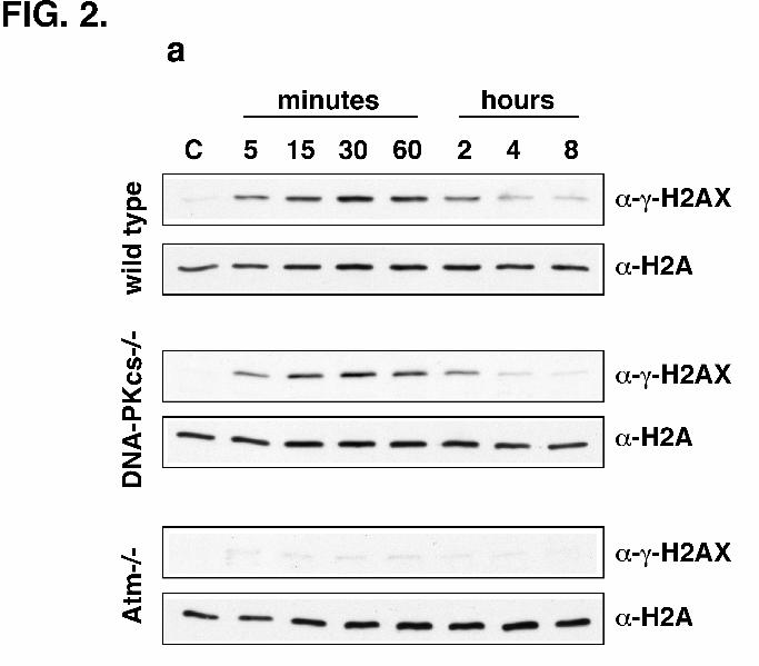
H2AX Phosphorylation is Abrogated in Atm-/-, but Not in DNA-PKcs-/-, Cells -
Spontaneously immortalized fibroblasts from wild type, DNA-PKcs-/-, or Atm-/- mice were
mock-irradiated or irradiated, harvested at time points ranging from 5 min to 8 h, and assayed for
H2AX phosphorylation by western blotting. H2AX phosphorylation in both wild type and DNA-
PKcs-/- cells occurred very rapidly (within 5 min) and lasted for about 2 h, with maximum levels
of phosphorylation observed at 30 min (Fig. 2a). In striking contrast, we observed minimal
H2AX phosphorylation in irradiated Atm-/- cells. While we observed robust Η2ΑX
phosphorylation in DNA-PKcs-/- cells at 30 min post-irradiation, γ-H2AX formation in Atm-/-
cells was reproducibly reduced to about 5% of that in wild type cells (Fig. 2b) indicating that
ATM is the major kinase responsible for H2AX phosphorylation upon DNA damage.

10
Atm-/- fibroblasts were treated with increasing concentrations of wortmannin for 30 min,
irradiated with X-rays, harvested after 30 min, and analyzed by western blotting. We found that
the minimal H2AX phosphorylation in Atm-/- cells was completely abolished by low
concentrations of wortmannin (1-10 µM) (Fig. 2c). As ATR is inhibited by high concentrations
of wortmannin (> 100 µM) (20), our results suggest that DNA-PK, rather than ATR, is
responsible for low levels of γ-H2AX formation in the absence of ATM.
Lack of H2AX Phosphorylation in Atm-/- Mouse Embryonic Fibroblasts (MEFs) –
It is possible that other mutations in the Atm-/- cell line used could also be responsible for the
lack of H2AX phosphorylation in these cells. We therefore examined H2AX phosphorylation in
a panel of isogenic, early passage (p2 or p3) ATM +/+ or -/- mouse embryonic fibroblasts
(MEFs). Normal Η2ΑX phosphorylation was observed in irradiated Atm+/+ MEFs (Fig. 3a;
upper panel). In contrast, very low levels of γ-H2AX formation was observed in two independent
Atm-/- MEFs confirming that ATM is required for H2AX phosphorylation in response to IR. No
significant difference in H2AX phosphorylation was observed between irradiated DNA-PKcs+/-
and -/- MEFs (Fig. 3a; lower panel).
Ectopic Expression of ATM Restores H2AX Phosphorylation in Atm-/- Cells – In
order to confirm that ATM is required in vivo for H2AX phosphorylation, the ATM cDNA
expression vector pMAT1 (16) was transiently transfected into Atm-/- spontaneously
immortalized fibroblasts. The ectopic expression of ATM in the transfected cells resulted in
restoration of H2AX phosphorylation upon irradiation (Fig. 3b; compare lanes 2 and 4). On the
other hand, cells transfected with the vector alone showed no increase in γ-H2AX formation

11
(Fig. 3b; compare lanes 2 and 6). The correlation between ATM expression and H2AX
phosphorylation establishes that ATM is required in vivo for γ-H2AX formation in response to
ionizing radiation.
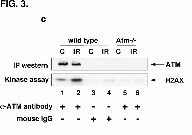
ATM Can Phosphorylate Recombinant H2AX In Vitro – In order to determine
whether ATM can directly phosphorylate H2AX in vitro, ATM was immunoprecipitated from
spontaneously immortalized wild type fibroblasts using an anti-ATM monoclonal antibody
raised against a peptide representing positions 1967-1988 of murine ATM (15). The
immunoprecipitated ATM efficiently phosphorylated recombinant H2AX in vitro (Fig. 3c; lane
1). Furthermore, irradiation of cells resulted in a significant increase in H2AX phosphorylation
(Fig. 3c; lane 2). Essentially no ATM protein or kinase activity was detected when
immunoprecipitation was performed with normal mouse IgG or from Atm-/- fibroblasts (Fig. 3c;
lanes 3-6). The in vitro phosphorylation of H2AX by ATM suggests that ATM could directly
phosphorylate H2AX within the cell in response to DNA damage.
Poor γγγγ-H2AX Focus Formation in Atm-/- Cells – H2AX phosphorylation in response
to DNA damage results in the formation of discrete γ-H2AX foci at the sites of DNA double-
strand breaks (4). To determine the status of γ-H2AX focus formation in wild type, DNA-PKcs-
/-, and Atm-/- spontaneously immortalized fibroblasts, these cells were irradiated and allowed to
recover for 30 min before fixation and immunostaining with anti-γ-H2AX antibody. We
observed robust γ-H2AX focus formation upon irradiation of both wild type and DNA-PKcs-/-
cells (Fig. 4). In striking contrast, focus formation was very poor in Atm-/- cells confirming that
ATM is required for γ-H2AX focus formation at the sites of DSBs.

12
DISCUSSION
Histone H2AX is rapidly phosphorylated at serine 139 in response to DNA double-strand
breaks (3). The PI-3 kinases - DNA-PK, ATM, and ATR - have all been implicated in this
process (2,5,10,11). While the substrate specificities of these kinases are overlapping in vitro,
they have clearly distinct functions in vivo (10). For example, while ATM phosphorylates p53,
Chk2, and Nbs1 leading to cell cycle arrest, DNA-PK is not required for any of these processes
(21,22). On the other hand, DNA-PK, unlike ATM, may be involved in the recruitment of
XRCC4 and DNA ligase IV to the sites of DSBs (23). It is therefore important to definitively
delineate the roles of these kinases in the phosphorylation of H2AX.
We demonstrate that ATM can phosphorylate H2AX in vitro and that H2AX
phosphorylation and γ-H2AX focus formation are severely compromised in Atm-/- cells. Ectopic
expression of ATM corrects this defect. In contrast, these functions are normal in DNA-PKcs-/-
cells. Interestingly, DNA-PK, but not ATR, may be responsible for the minimal levels of H2AX
phosphorylation in Atm-/- cells as this can be abolished by low concentrations of wortmannin.
We also find that immunoprecipitated ATM can directly interact with recombinant H2AX in
vitro (Sandeep Burma and David J. Chen; unpublished results) and experiments will be
performed to examine complex formation between H2AX and ATM in vivo.
Our results establish that ATM is the major kinase responsible for histone H2AX
phosphorylation in response to DNA double-strand breaks in murine fibroblasts. The reduced
H2AX phosphorylation reported in M059J cells (5) could, therefore, be due to the low levels of
ATM in these cells (24-26) rather than due to the absence of DNA-PK. ATM plays a crucial role
in the rapid induction of multiple signaling pathways in response to DSBs, leading to repair of
13
DNA damage, activation of cell cycle checkpoints, and cellular stress responses (27). As γ-
H2AX focus formation is a very early event occurring within seconds of DNA damage infliction
(3), our results indicate that ATM is one of the earliest kinases to be activated in the cellular
response to DNA double-strand breaks. Supporting ATM’s important role in damage-induced
chromatin modification is a report indicating that ATM is also required for the IR-induced
transient dephosphorylation of histone H1 which is thought to contribute to chromatin
decondensation (28).
Our results are consistent with a recent report indicating that a fraction of nuclear ATM
co-localizes with γ-H2AX at the sites of DSBs in response to DNA damage (15). A very striking
correlation was reported between the kinetics of appearance and dissolution of γ-H2AX foci and
DNA-bound ATM aggregates. This suggests that DNA localization by ATM and H2AX
phosphorylation occur concomitantly, very rapidly after DNA damage, and decrease at the same
rate thereafter. In the light of this report, our findings suggest that once ATM is activated at a
DSB, it could immediately phosphorylate histone H2AX at the site of the break thereby signaling
to the cell that a DSB has occurred. This very early event could then initiate the recruitment of
DNA repair or damage-signaling factors to the break mediated by chromatin modification and/or
direct interactions of these factors with phospho-H2AX.
14
ACKNOWLEDGEMENTS
We are grateful to Dr. William Bonner (National Institutes of Health) for the anti-γ-
H2AX antibody used in preliminary studies, Dr. Yosef Shiloh (Tel Aviv University) for anti-
ATM antibody, and Dr. Martin Lavin (Queensland Institute of Medical Research) for pMat1
plasmid. We thank Melinda Henrie for excellent technical assistance and Steve Yannone, David
Gilley, Janice Pluth, and Bipasha Mukherjee for critically reading the manuscript. This work was
funded by US Dept. of Energy and NIH grant CA50519 to D.J.C.
15
REFERENCES
1. Khanna, K. K., and Jackson, S. P. (2001) Nat. Genet. 27, 247-54
2. Modesti, M., and Kanaar, R. (2001) Current Biol. 11, R229-R232
3. Rogakou, E. P., Pilch, D. R., Orr, A. H., Ivanova, V. S., and Bonner, W. M. (1998) J.
Biol. Chem. 273, 5858-5868
4. Rogakou, E. P., Boon, C., Redon, C., and Bonner, W. M. (1999J. Cell Biol. 146, 905-915
5. Paull, T. T., Rogakou, E. P., Yamazaki, V., Kirchgessner, C. U., Gellert, M., and Bonner,
W. M. (2000) Current Biol. 10, 886-895
6. Chen, H. T., Bhandoola, A., Difilippantonio, M. J., Zhu, J., Brown, M. J., Tai, X.,
Rogakou, E. P., Brotz, T. M., Bonner, W. M., Ried, T., and Nussenzweig, A. (2000)
Science (Washington D C) 290, 1962-1964
7. Mahadevaiah, S. K., Turner, J. M. A., Baudat, F., Rogakou, E. P., de Boer, P., Blanco-
Rodriguez, J., Jasin, M., Keeney, S., Bonner, W. M., and Burgoyne, P. S. (2001) Nat.
Genet. 27, 271-276
8. Downs, J. A., Lowndes, N. F., and Jackson, S. P. (2000) Nature (London) 408, 1001-
1004
9. Rappold, I., Iwabuchi, K., Date, T., and Chen, J. (2001) J. Cell Biol. 153, 613-620
10. Durocher, D., and Jackson, S. P. (2001) Current Opinion in Cell Biol. 13, 225-231
11. van Gent, D. C., Hoeijmakers, J. H., and Kanaar, R. (2001) Nat. Rev. Genet. 2, 196-206
12. Kurimasa, A., Ouyang, H., Dong, L. J., Wang, S., Li, X., Cordon-Cardo, C., Chen, D. J.,
and Li, G. C. (1999) Proc. Natl. Acad. Sci. U.S.A. 96, 1403-8
16
13. Xu, Y., Ashley, T., Brainerd, E. E., Bronson, R. T., Meyn, M. S., and Baltimore, D.
(1996) Genes Dev. 10, 2411-2422
14. D'Anna, J. A., Valdez, J. G., Habbersett, R. C., and Crissman, H. A. (1997) Radiat. Res.
148, 260-71
15. Andegeko, Y., Moyal, L., Mitelman, L., Tsarfaty, I., Shiloh, Y., and Rotman, G. (2001) J.
Biol. Chem., July 13 [epub ahead of print]
16. Zhang, N., Chen, P., Khanna, K. K., Scott, S., Gatei, M., Kozlov, S., Watters, D., Spring,
K., Yen, T., and Lavin, M. F. (1997) Proc. Natl. Acad. Sci. U.S.A. 94, 8021-8026
17. Ziv, Y., Banin, S., Lim, D. S., Canman, C. E., Kastan, M. B., Shiloh, Y., Chan, D. W.,
Gately, D. P., Urban, S., Galloway, A. M., Lees-Miller, S. P., Yen, T., and Allalunis-
Turner, J. (2000) Methods Mol. Biol. 99, 99-108
18. Saintigny, Y., Delacote, F., Vares, G., Petitot, F., Lambert, S., Averbeck, D., and Lopez,
B. S. (2001) EMBO J. 20, 3861-3870
19. Skog, S., Heiden, T., Eriksson, S., Wallström, B., and Tribukait, B. (1992) Anti-Cancer
Drugs 3, 379-86
20. Sarkaria, J. N., Tibbetts, R. S., Busby, E. C., Kennedy, A. P., Hill, D. E., and Abraham,
R. T. (1998) Cancer Res. 58, 4375-4382
21. Burma, S., Kurimasa, A., Xie, G., Taya, Y., Araki, R., Abe, M., Crissman, H. A.,
Ouyang, H., Li, G. C., and Chen, D. J. (1999) J. Biol. Chem. 274, 17139-17143
22. Kastan, M. B., and Lim, D. S. (2000) Nat. Rev. Mol. Cell Biol. 1, 179-86
23. Smith, G. C., Jackson, S. P., Chan, D. W., Gately, D. P., Urban, S., Galloway, A. M.,
Lees-Miller, S. P., Yen, T., and Allalunis-Turner, J. (1999) Genes Dev. 13, 916-34
24. Hoppe, B. S., Jensen, R. B., and Kirchgessner, C. U. (2000) Radiat. Res. 153, 125-30
17
25. Gately, D. P., Hittle, J. C., Chan, G. K., and Yen, T. J. (1998) Mol. Biol. Cell 9, 2361-74
26. Chan, D. W., Gately, D. P., Urban, S., Galloway, A. M., Lees-Miller, S. P., Yen, T., and
Allalunis-Turner, J. (1998) Int. J. Radiat. Biol. 74, 217-24
27. Rotman, G., and Shiloh, Y. (1999) Oncogene 18, 6135-44
28. Guo, C. Y., Wang, Y., Brautigan, D. L., and Larner, J. M. (1999) J. Biol. Chem. 274,
18715-20.

18
LEGENDS TO FIGURES
FIG. 1. Inhibition of H2AX phosphorylation by wortmannin. (A) Anti-γ H2AX
antibody (directed against H2AX phosphorylated at serine 139) were used to immunoblot
synthetic peptides comprising of the last 9 amino acids of H2AX with (134-142ser139) or without
(134-142) phosphorylation at serine 139. (B) Spontaneously immortalized wild type mouse
fibroblasts were mock-irradiated or irradiated with X-rays or UV rays as indicated and harvested
after 30 min. Cells were mock-treated or treated for 1h with the DSB-inducing agents
neocarzinostatin (NCS), bleomycin (BLM), or etoposide or with the DNA alkylating agent
methyl methanesulfonate (MMS) as indicated and then harvested. SDS extracts were analyzed
for H2AX phosphorylation by western blotting with anti-γ-H2AX antibody. The blots were
stripped and re-probed with anti-H2A antibody as a normalizing control. (C) Wild type
fibroblasts were mock-irradiated (C) or irradiated with X-rays after a 30 min incubation with
increasing concentrations of wortmannin (0-100 µM). Cells were harvested 30 min after
irradiation and SDS extracts analyzed by western blotting with anti-γ-H2AX or anti-H2A
antibodies.
FIG. 2. Lack of H2AX phosphorylation in Atm-/- cells. (A) Wild type, DNA-PKcs-/-,
or Atm-/- spontaneously immortalized fibroblasts were mock-irradiated (C) or irradiated with X-
rays and harvested at time points ranging from 5 min to 8 h as indicated. SDS extracts were
analyzed for H2AX phosphorylation by western blotting with anti-γ-H2AX antibody or with
anti-H2A antibody as a normalizing control. (B) Wild type, DNA-PKcs-/-, or Atm-/- fibroblasts
were mock-irradiated (C) or irradiated (IR), harvested after 30 min, and analyzed by western
19
blotting. The autoradiograph in the figure was scanned, the bands quantified using NIH Image
software, and the relative levels of γ-H2AX plotted on the Y-axis. After IR the level of γ-H2AX
in Atm-/- cells is about 5% of that in wild type cells. A similar pattern of Η2ΑX phosphorylation
was observed in three independent experiments. (C) Atm-/- fibroblasts were incubated with
increasing concentrations of wortmannin for 30 min and then irradiated. Cells were harvested 30
min after irradiation and SDS extracts analyzed by western blotting.
FIG. 3. Phosphorylation of H2AX by ATM in vitro and in vivo. (A) Abrogated H2AX
phosphorylation in Atm-/- mouse embryonic fibroblasts (MEFs). Early passage isogenic Atm
+/+ or -/- MEFs were mock-irradiated (C) or irradiated with X-rays (IR), harvested after 30 min,
and analyzed for H2AX phosphorylation by western blotting with anti-γ-H2AX antibody or with
anti-H2A antibody as a normalizing control. DNA-PKcs+/- or -/- MEFs were similarly analyzed
for H2AX phosphorylation. (B) Restoration of H2AX phosphorylation in Atm-/- cells by ectopic
expression of ATM. Spontaneously immortalized Atm-/- fibroblasts were either untransfected
(lanes 1 and 2), transiently transfected with the ATM cDNA expression vector pMat1 (lanes 3
and 4), or transiently transfected with the vector alone (lanes 5 and 6). Cells were either mock-
irradiated (C) or irradiated (IR), harvested after 30 min, and SDS extracts analyzed for H2AX
phosphorylation by western blotting with anti-γ-H2AX antibody. The expression of ATM in
transfected cells was monitored by western blotting with anti-ATM antibody. (C) In vitro
phosphorylation of recombinant H2AX by ATM. ATM was immunoprecipitated from mock-
irradiated (C) or irradiated (IR) spontaneously immortalized fibroblasts with anti-ATM
monoclonal antibody or normal mouse IgG as control and kinase activity assayed using

20
recombinant H2AX as substrate. Top panel: ATM immunoprecipitates immunoblotted with anti-
ATM monoclonal antibody. Bottom panel: autoradiograph of phosphorylated H2AX.
FIG. 4. Poor γγγγ-H2AX focus formation in Atm-/- cells. Wild type, DNA-PKcs-/-, or
Atm-/- spontaneously immortalized fibroblasts were mock-irradiated or irradiated with X-rays
and incubated for 30 min before fixation, permeabilization, and staining with anti-γ-H2AX
antibody (green). Nuclei were stained with DAPI (blue). To allow direct comparisons, all the
slides were processed together and all the images were captured using the same parameters.