atomic resolution probe for allostery in the regulatory ... · the ctnc binding pocket have been...
TRANSCRIPT
Atomic resolution probe for allostery in the regulatorythin filamentMichael R. Williamsa,1, Sarah J. Lehmanb,1, Jil C. Tardiffc,d, and Steven D. Schwartza,2
aDepartment of Chemistry and Biochemistry, The University of Arizona, Tucson, AZ 85721; bPhysiological Sciences, The University of Arizona, Tucson, AZ85724; cDepartment of Medicine, The University of Arizona, Tucson, AZ 85724; and dDepartment of Cellular and Molecular Medicine, The University ofArizona, Tucson, AZ 85724
Edited by James A. Spudich, Stanford University School of Medicine, Stanford, CA, and approved February 5, 2016 (received for review October 1, 2015)
Calcium binding and dissociation within the cardiac thin filament (CTF)is a fundamental regulator of normal contraction and relaxation.Although the disruption of this complex, allosterically mediated pro-cess has long been implicated in human disease, the precise atomic-level mechanisms remain opaque, greatly hampering the developmentof novel targeted therapies. To address this question, we used a fullyatomistic CTF model to test both Ca2+ binding strength and the energyrequired to remove Ca2+ from the N-lobe binding site in WT and mu-tant troponin complexes that have been linked to genetic cardiomyop-athies. This computational approach is combined with measurementsof in vitro Ca2+ dissociation rates in fully reconstituted WT and cardiactroponin T R92L and R92W thin filaments. These human disease muta-tions represent known substitutions at the same residue, reside at asignificant distance from the calcium binding site in cardiac troponin C,and do not affect either the binding pocket affinity or EF-hand struc-ture of the binding domain. Both have been shown to have signifi-cantly different effects on cardiac function in vivo. We now show thatthese mutations independently alter the interaction between the Ca2+
ion and cardiac troponin I subunit. This interaction is a previously un-identified mechanism, in which mutations in one protein of a complexindirectly affect a third via structural and dynamic changes in a secondto yield a pathogenic change in thin filament function that results inmutation-specific disease states. We can now provide atom-level in-sight that is potentially highly actionable in drug design.
cardiac thin filament | hypertrophic cardiomyopathy | calciumhomeostasis | molecular modeling | steered molecular dynamics
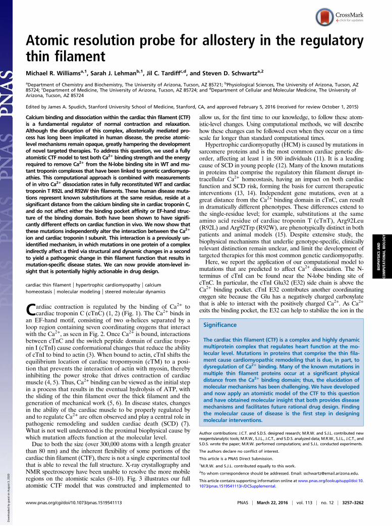
Cardiac contraction is regulated by the binding of Ca2+ tocardiac troponin C (cTnC) (1, 2) (Fig. 1). The Ca2+ binds in
an EF-hand motif, consisting of two α-helices separated by aloop region containing seven coordinating oxygens that interactwith the Ca2+, as seen in Fig. 2. Once Ca2+ is bound, interactionsbetween cTnC and the switch peptide domain of cardiac tropo-nin I (cTnI) cause conformational changes that reduce the abilityof cTnI to bind to actin (3). When bound to actin, cTnI shifts theequilibrium location of cardiac tropomyosin (cTM) to a posi-tion that prevents the interaction of actin with myosin, therebyinhibiting the power stroke that drives contraction of cardiacmuscle (4, 5). Thus, Ca2+ binding can be viewed as the initial stepin a process that results in the eventual hydrolysis of ATP, withthe sliding of the thin filament over the thick filament and thegeneration of mechanical work (5, 6). In disease states, changesin the ability of the cardiac muscle to be properly regulated byand to regulate Ca2+ are often observed and play a central role inpathogenic remodeling and sudden cardiac death (SCD) (7).What is not well understood is the proximal biophysical cause bywhich mutation affects function at the molecular level.Due to both the size (over 300,000 atoms with a length greater
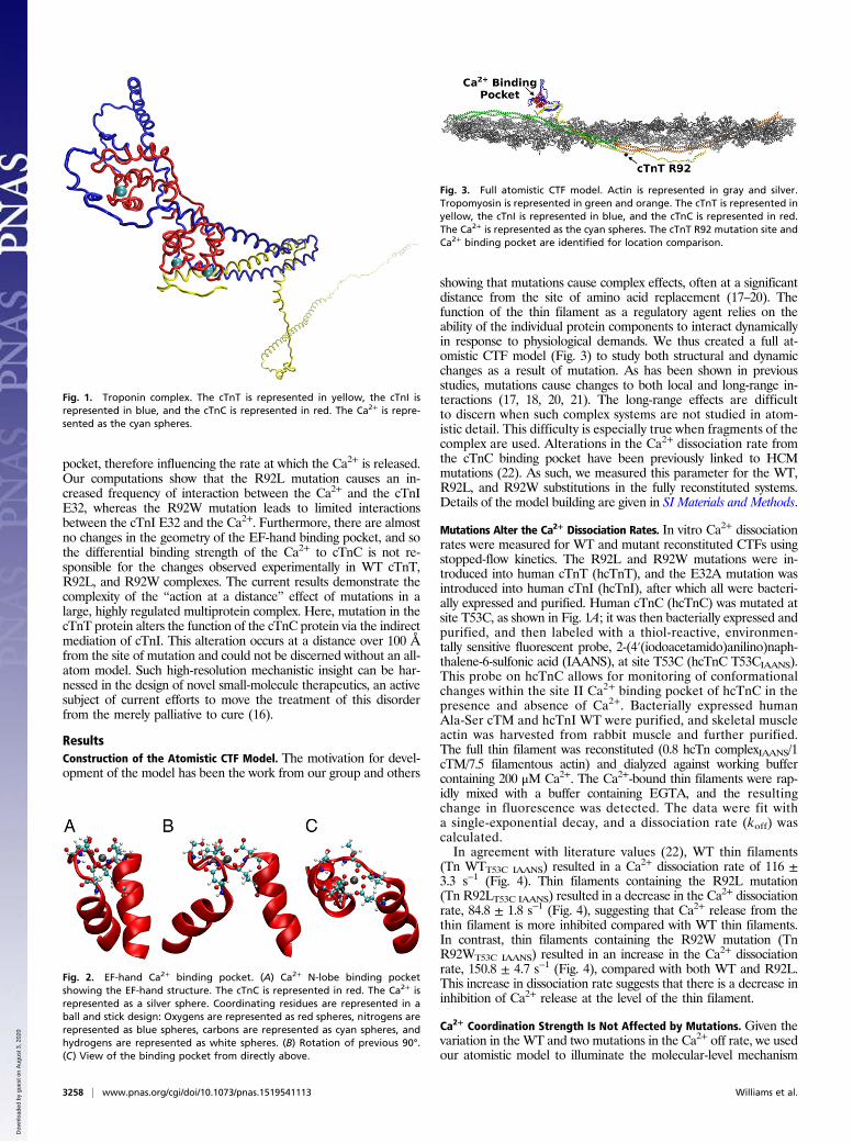
than 80 nm) and the inherent flexibility of some portions of thecardiac thin filament (CTF), there is not a single experimental toolthat is able to reveal the full structure. X-ray crystallography andNMR spectroscopy have been unable to resolve the more mobileregions on the atomistic scales (8–10). Fig. 3 illustrates our fullatomistic CTF model that was constructed and implemented to
allow us, for the first time to our knowledge, to follow these atom-istic-level changes. Using computational methods, we will describehow these changes can be followed even when they occur on a timescale far longer than standard computational times.Hypertrophic cardiomyopathy (HCM) is caused by mutations in
sarcomere proteins and is the most common cardiac genetic dis-order, affecting at least 1 in 500 individuals (11). It is a leadingcause of SCD in young people (12). Many of the known mutationsin proteins that comprise the regulatory thin filament disrupt in-tracellular Ca2+ homeostasis, having an impact on both cardiacfunction and SCD risk, forming the basis for current therapeuticinterventions (13, 14). Independent gene mutations, even at agreat distance from the Ca2+ binding domain in cTnC, can resultin dramatically different phenotypes. These differences extend tothe single-residue level; for example, substitutions at the sameamino acid residue of cardiac troponin T (cTnT), Arg92Leu(R92L) and Arg92Trp (R92W), are phenotypically distinct in bothpatients and animal models (15). Despite extensive study, thebiophysical mechanisms that underlie genotype-specific, clinicallyrelevant distinction remain unclear, and limit the development oftargeted therapies for this most common genetic cardiomyopathy.Here, we report the application of our computational model to
mutations that are predicted to affect Ca2+ dissociation. The N-terminus of cTnI can be found near the N-lobe binding site ofcTnC. In particular, the cTnI Glu32 (E32) side chain is above theCa2+ binding pocket. cTnI E32 contributes another coordinatingoxygen site because the Glu has a negatively charged carboxylatethat is able to interact with the positively charged Ca2+. As Ca2+
exits the binding pocket, the E32 can help to stabilize the ion in the
Significance
The cardiac thin filament (CTF) is a complex and highly dynamicmultiprotein complex that regulates heart function at the mo-lecular level. Mutations in proteins that comprise the thin fila-ment cause cardiomyopathic remodeling that is due, in part, todysregulation of Ca2+ binding. Many of the known mutations inmultiple thin filament proteins occur at a significant physicaldistance from the Ca2+ binding domain; thus, the elucidation ofmolecular mechanisms has been challenging. We have developedand now apply an atomistic model of the CTF to this questionand have obtained molecular insight that both provides diseasemechanisms and facilitates future rational drug design. Findingthe molecular cause of disease is the first step in designingmolecular interventions.
Author contributions: J.C.T. and S.D.S. designed research; M.R.W. and S.J.L. contributed newreagents/analytic tools; M.R.W., S.J.L., J.C.T., and S.D.S. analyzed data; M.R.W., S.J.L., J.C.T., andS.D.S. wrote the paper; M.R.W. performed computations; and S.J.L. conducted experiments.
The authors declare no conflict of interest.
This article is a PNAS Direct Submission.1M.R.W. and S.J.L. contributed equally to this work.2To whom correspondence should be addressed. Email: [email protected].
This article contains supporting information online at www.pnas.org/lookup/suppl/doi:10.1073/pnas.1519541113/-/DCSupplemental.
www.pnas.org/cgi/doi/10.1073/pnas.1519541113 PNAS | March 22, 2016 | vol. 113 | no. 12 | 3257–3262
BIOPH
YSICSAND
COMPU
TATIONALBIOLO
GY
Dow
nloa
ded
by g
uest
on
Aug
ust 3
, 202
0
pocket, therefore influencing the rate at which the Ca2+ is released.Our computations show that the R92L mutation causes an in-creased frequency of interaction between the Ca2+ and the cTnIE32, whereas the R92W mutation leads to limited interactionsbetween the cTnI E32 and the Ca2+. Furthermore, there are almostno changes in the geometry of the EF-hand binding pocket, and sothe differential binding strength of the Ca2+ to cTnC is not re-sponsible for the changes observed experimentally in WT cTnT,R92L, and R92W complexes. The current results demonstrate thecomplexity of the “action at a distance” effect of mutations in alarge, highly regulated multiprotein complex. Here, mutation in thecTnT protein alters the function of the cTnC protein via the indirectmediation of cTnI. This alteration occurs at a distance over 100 Åfrom the site of mutation and could not be discerned without an all-atom model. Such high-resolution mechanistic insight can be har-nessed in the design of novel small-molecule therapeutics, an activesubject of current efforts to move the treatment of this disorderfrom the merely palliative to cure (16).
ResultsConstruction of the Atomistic CTF Model. The motivation for devel-opment of the model has been the work from our group and others
showing that mutations cause complex effects, often at a significantdistance from the site of amino acid replacement (17–20). Thefunction of the thin filament as a regulatory agent relies on theability of the individual protein components to interact dynamicallyin response to physiological demands. We thus created a full at-omistic CTF model (Fig. 3) to study both structural and dynamicchanges as a result of mutation. As has been shown in previousstudies, mutations cause changes to both local and long-range in-teractions (17, 18, 20, 21). The long-range effects are difficultto discern when such complex systems are not studied in atom-istic detail. This difficulty is especially true when fragments of thecomplex are used. Alterations in the Ca2+ dissociation rate fromthe cTnC binding pocket have been previously linked to HCMmutations (22). As such, we measured this parameter for the WT,R92L, and R92W substitutions in the fully reconstituted systems.Details of the model building are given in SI Materials and Methods.
Mutations Alter the Ca2+ Dissociation Rates. In vitro Ca2+ dissociationrates were measured for WT and mutant reconstituted CTFs usingstopped-flow kinetics. The R92L and R92W mutations were in-troduced into human cTnT (hcTnT), and the E32A mutation wasintroduced into human cTnI (hcTnI), after which all were bacteri-ally expressed and purified. Human cTnC (hcTnC) was mutated atsite T53C, as shown in Fig. 1A; it was then bacterially expressed andpurified, and then labeled with a thiol-reactive, environmen-tally sensitive fluorescent probe, 2-(4′(iodoacetamido)anilino)naph-thalene-6-sulfonic acid (IAANS), at site T53C (hcTnC T53CIAANS).This probe on hcTnC allows for monitoring of conformationalchanges within the site II Ca2+ binding pocket of hcTnC in thepresence and absence of Ca2+. Bacterially expressed humanAla-Ser cTM and hcTnI WT were purified, and skeletal muscleactin was harvested from rabbit muscle and further purified.The full thin filament was reconstituted (0.8 hcTn complexIAANS/1cTM/7.5 filamentous actin) and dialyzed against working buffercontaining 200 μM Ca2+. The Ca2+-bound thin filaments were rap-idly mixed with a buffer containing EGTA, and the resultingchange in fluorescence was detected. The data were fit witha single-exponential decay, and a dissociation rate (koff) wascalculated.In agreement with literature values (22), WT thin filaments
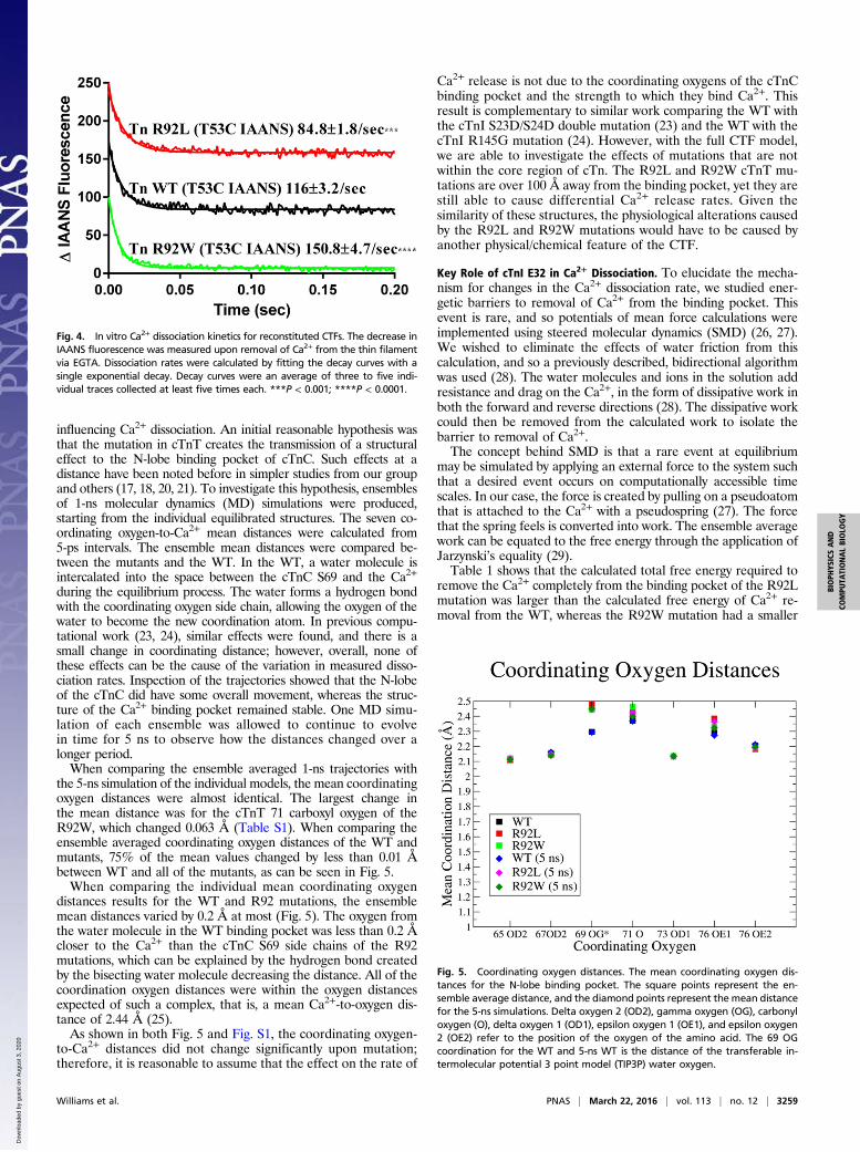
(Tn WTT53C IAANS) resulted in a Ca2+ dissociation rate of 116 ±3.3 s−1 (Fig. 4). Thin filaments containing the R92L mutation(Tn R92LT53C IAANS) resulted in a decrease in the Ca2+ dissociationrate, 84.8 ± 1.8 s−1 (Fig. 4), suggesting that Ca2+ release from thethin filament is more inhibited compared with WT thin filaments.In contrast, thin filaments containing the R92W mutation (TnR92WT53C IAANS) resulted in an increase in the Ca2+ dissociationrate, 150.8 ± 4.7 s−1 (Fig. 4), compared with both WT and R92L.This increase in dissociation rate suggests that there is a decrease ininhibition of Ca2+ release at the level of the thin filament.
Ca2+ Coordination Strength Is Not Affected by Mutations. Given thevariation in the WT and two mutations in the Ca2+ off rate, we usedour atomistic model to illuminate the molecular-level mechanism
Fig. 1. Troponin complex. The cTnT is represented in yellow, the cTnI isrepresented in blue, and the cTnC is represented in red. The Ca2+ is repre-sented as the cyan spheres.
Fig. 2. EF-hand Ca2+ binding pocket. (A) Ca2+ N-lobe binding pocketshowing the EF-hand structure. The cTnC is represented in red. The Ca2+ isrepresented as a silver sphere. Coordinating residues are represented in aball and stick design: Oxygens are represented as red spheres, nitrogens arerepresented as blue spheres, carbons are represented as cyan spheres, andhydrogens are represented as white spheres. (B) Rotation of previous 90°.(C) View of the binding pocket from directly above.
Fig. 3. Full atomistic CTF model. Actin is represented in gray and silver.Tropomyosin is represented in green and orange. The cTnT is represented inyellow, the cTnI is represented in blue, and the cTnC is represented in red.The Ca2+ is represented as the cyan spheres. The cTnT R92 mutation site andCa2+ binding pocket are identified for location comparison.
3258 | www.pnas.org/cgi/doi/10.1073/pnas.1519541113 Williams et al.
Dow
nloa
ded
by g
uest
on
Aug
ust 3
, 202
0
influencing Ca2+ dissociation. An initial reasonable hypothesis wasthat the mutation in cTnT creates the transmission of a structuraleffect to the N-lobe binding pocket of cTnC. Such effects at adistance have been noted before in simpler studies from our groupand others (17, 18, 20, 21). To investigate this hypothesis, ensemblesof 1-ns molecular dynamics (MD) simulations were produced,starting from the individual equilibrated structures. The seven co-ordinating oxygen-to-Ca2+ mean distances were calculated from5-ps intervals. The ensemble mean distances were compared be-tween the mutants and the WT. In the WT, a water molecule isintercalated into the space between the cTnC S69 and the Ca2+
during the equilibrium process. The water forms a hydrogen bondwith the coordinating oxygen side chain, allowing the oxygen of thewater to become the new coordination atom. In previous compu-tational work (23, 24), similar effects were found, and there is asmall change in coordinating distance; however, overall, none ofthese effects can be the cause of the variation in measured disso-ciation rates. Inspection of the trajectories showed that the N-lobeof the cTnC did have some overall movement, whereas the struc-ture of the Ca2+ binding pocket remained stable. One MD simu-lation of each ensemble was allowed to continue to evolvein time for 5 ns to observe how the distances changed over alonger period.When comparing the ensemble averaged 1-ns trajectories with
the 5-ns simulation of the individual models, the mean coordinatingoxygen distances were almost identical. The largest change inthe mean distance was for the cTnT 71 carboxyl oxygen of theR92W, which changed 0.063 Å (Table S1). When comparing theensemble averaged coordinating oxygen distances of the WT andmutants, 75% of the mean values changed by less than 0.01 Åbetween WT and all of the mutants, as can be seen in Fig. 5.When comparing the individual mean coordinating oxygen
distances results for the WT and R92 mutations, the ensemblemean distances varied by 0.2 Å at most (Fig. 5). The oxygen fromthe water molecule in the WT binding pocket was less than 0.2 Åcloser to the Ca2+ than the cTnC S69 side chains of the R92mutations, which can be explained by the hydrogen bond createdby the bisecting water molecule decreasing the distance. All of thecoordination oxygen distances were within the oxygen distancesexpected of such a complex, that is, a mean Ca2+-to-oxygen dis-tance of 2.44 Å (25).As shown in both Fig. 5 and Fig. S1, the coordinating oxygen-
to-Ca2+ distances did not change significantly upon mutation;therefore, it is reasonable to assume that the effect on the rate of
Ca2+ release is not due to the coordinating oxygens of the cTnCbinding pocket and the strength to which they bind Ca2+. Thisresult is complementary to similar work comparing the WT withthe cTnI S23D/S24D double mutation (23) and the WT with thecTnI R145G mutation (24). However, with the full CTF model,we are able to investigate the effects of mutations that are notwithin the core region of cTn. The R92L and R92W cTnT mu-tations are over 100 Å away from the binding pocket, yet they arestill able to cause differential Ca2+ release rates. Given thesimilarity of these structures, the physiological alterations causedby the R92L and R92W mutations would have to be caused byanother physical/chemical feature of the CTF.
Key Role of cTnI E32 in Ca2+ Dissociation. To elucidate the mecha-nism for changes in the Ca2+ dissociation rate, we studied ener-getic barriers to removal of Ca2+ from the binding pocket. Thisevent is rare, and so potentials of mean force calculations wereimplemented using steered molecular dynamics (SMD) (26, 27).We wished to eliminate the effects of water friction from thiscalculation, and so a previously described, bidirectional algorithmwas used (28). The water molecules and ions in the solution addresistance and drag on the Ca2+, in the form of dissipative work inboth the forward and reverse directions (28). The dissipative workcould then be removed from the calculated work to isolate thebarrier to removal of Ca2+.The concept behind SMD is that a rare event at equilibrium
may be simulated by applying an external force to the system suchthat a desired event occurs on computationally accessible timescales. In our case, the force is created by pulling on a pseudoatomthat is attached to the Ca2+ with a pseudospring (27). The forcethat the spring feels is converted into work. The ensemble averagework can be equated to the free energy through the application ofJarzynski’s equality (29).Table 1 shows that the calculated total free energy required to
remove the Ca2+ completely from the binding pocket of the R92Lmutation was larger than the calculated free energy of Ca2+ re-moval from the WT, whereas the R92W mutation had a smaller
Fig. 4. In vitro Ca2+ dissociation kinetics for reconstituted CTFs. The decrease inIAANS fluorescence was measured upon removal of Ca2+ from the thin filamentvia EGTA. Dissociation rates were calculated by fitting the decay curves with asingle exponential decay. Decay curves were an average of three to five indi-vidual traces collected at least five times each. ***P < 0.001; ****P < 0.0001.
Fig. 5. Coordinating oxygen distances. The mean coordinating oxygen dis-tances for the N-lobe binding pocket. The square points represent the en-semble average distance, and the diamond points represent the mean distancefor the 5-ns simulations. Delta oxygen 2 (OD2), gamma oxygen (OG), carbonyloxygen (O), delta oxygen 1 (OD1), epsilon oxygen 1 (OE1), and epsilon oxygen2 (OE2) refer to the position of the oxygen of the amino acid. The 69 OGcoordination for the WT and 5-ns WT is the distance of the transferable in-termolecular potential 3 point model (TIP3P) water oxygen.
Williams et al. PNAS | March 22, 2016 | vol. 113 | no. 12 | 3259
BIOPH
YSICSAND
COMPU
TATIONALBIOLO
GY
Dow
nloa
ded
by g
uest
on
Aug
ust 3
, 202
0
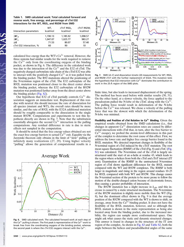
calculated free energy than the WT’s Ca2+ removal. However, thethree systems had similar results for the work required to removethe Ca2+ only from the coordinating oxygens of the bindingpocket, as shown in Fig. 6. The difference in the calculated workwas due to the interaction of the Ca2+ and the E32 of cTnI. Thenegatively charged carboxylate side chain of the cTnI E32 was ableto interact with the positively charged Ca2+ as it was pulled fromthe binding pocket. The R92 mutations altered the positioning ofthe N-terminus region of the cTnI. The E32 carboxylate of theR92L mutation was positioned closer to the direct center abovethe binding pocket, whereas the E32 carboxylate of the R92Wmutation was positioned farther away from the direct center abovethe binding pocket (Fig. S2).Our hypothesis that E32 of cTnI partially controls Ca2+ dis-
sociation suggests an immediate test. Replacement of this resi-due with neutral Ala should increase the rate of dissociation forall species (mutant and WT), the overall rates should be moresimilar, and the rate of R92L with the E32A additional mutationshould be roughly comparable to the dissociation in the singlemutant R92W. Computations and experiments to test this hy-pothesis directly are shown in Fig. 7. Note that the substitutionessentially abrogates the second Ca2+ interaction in the pullingcomputations. Any residual difference in rate is due to stericeffects not sampled in the computation.It should be noted that the free energy values obtained are not
the exact free energy barriers to actual Ca2+ exit. Equality via theJarzynski theorem only obtains in the limit of “slow” pulling ofinfinitely many realizations (27, 29). Using higher velocity“pulling” allows the generation of computational results in a
finite time, but also leads to increased displacement of the spring.The method has been used before with similar results (30, 31).On the other hand, at a slower velocity, the force applied to thepseudoatom pulled the N-lobe of the cTnC along with the Ca2+.The pulling force would result in deformation of the N-lobebefore the Ca2+ was released. We chose a velocity of the pullingforce that was the slowest with which the deformation of theN-lobe was minimal.
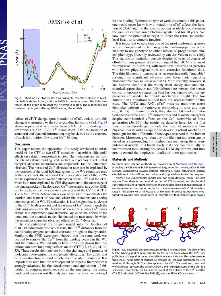
Flexibility and Position of cTnI Relative to Ca2+ Binding. Given theempirical results obtained from the SMD calculations (i.e., thatchanges in apparent Ca2+ dissociation rates are caused by differ-ential interactions with cTnI that, in turn, alter the free barrier toCa2+ escape), we probed the atomic-level differences in this partof the complex to determine the root cause of this effect. First, weaddress the lower rate and higher free energy barrier found in theR92L mutation. We detected important changes in flexibility of theN-terminal region of cTnI induced by the cTnT mutation. The rootmean square fluctuation (RMSF) of the cTnI (Fig. 8) and cTnC (Fig.S1) was calculated. The N-terminus end of the cTnI is largely un-structured until the start of an α-helix at residue 43, which leads tothe region where α-helices from both the cTnI and cTnT interact (ITarm). Examination of the RMSF in the unstructured N-terminusregion of cTnI shows significant differences for the R92L mutantcompared with the WT and R92W complexes. The RMSF is bothlarger in magnitude and rising in the region around residues 10–25for R92L compared with both WT and R92W. This change causesthe N-terminal section of the protein to be more prone to follow themotion of the double-charged cation because of electrostatic forces,thus causing a greater probability of interaction.The R92W mutation has a slight increase in koff, and this in-
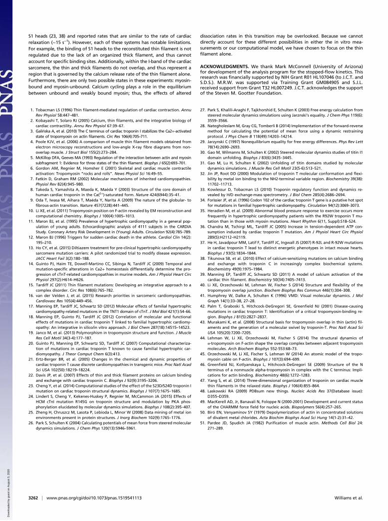
crease is caused by a static structural mechanism. The N-terminusof the R92W mutation is slightly more mobile than the WT (Fig.8), but the dominant effect shown in Fig. 9 is that the averageposition of the R92W compared with the WT is shown to shift, onaverage, away from the Ca2+ binding pocket. It does not have theflexibility of the R92L mutation, however, and so although theR92L mutant is shown in the static representation as being alsolocated away from the binding pocket, due to the increased mo-bility, the region can sample more conformational space. Onemight ask what causes the static and dynamic structural changes.This answer is found in changes in the structure of the IT armregion of the complex. As shown in Fig. S3 and Table S2, both theangle between the helices and pseudodihedral angles of the same
Table 1. SMD calculated work: Total calculated forward andreverse work, free energy, and percentage of cTnI E32interactions for the WT, R92L, and R92W from SMD
Interaction parametersWT,
kcal/molcTnT R92L,kcal/mol
cTnT R92W,kcal/mol
hWif 1,190.13 1,185.92 1,098.57hWir 1,047.07 1,025.77 983.27ΔG 71.53 80.07 57.40cTnI E32 interaction, % 60 90 10
Fig. 6. SMD calculated work. The calculated forward work at each step ofthe Ca2+ pulling is shown. The first peak is representative of the Ca2+ work tobreak free from the coordinating oxygens of the binding pocket, whereasthe second peak is where the cTnI E32 oxygens interact with the Ca2+.
Fig. 7. SMD (A–C) and dissociation kinetic (D) measurements for WT, R92L,and R92W cTnT with the further replacement of E32A. This mutation teststhe hypothesis that E32 interaction with Ca2+ dominates the contribution towork in the 25-Å region of the SMD plots.
3260 | www.pnas.org/cgi/doi/10.1073/pnas.1519541113 Williams et al.
Dow
nloa
ded
by g
uest
on
Aug
ust 3
, 202
0
helices of cTnT change upon mutation of cTnT, and, in turn, thischange is transmitted to the corresponding helices of cTnI. Fig. S4shows representative results from SMD, demonstrating thedifferences in cTnI E32 Ca2+ association. This transmission ofstructural and dynamic information may be viewed as the converseof such information flow upon Ca2+ binding.
DiscussionThis paper reports the application of a newly developed atomisticmodel of the CTF to two cTnT mutations that exhibit differentialeffects on calcium homeostasis in vivo. The mutations are far fromthe site of calcium binding, and, in fact, our primary result is thatcomplex allosteric interactions are responsible for the differencesobserved. The measured Ca2+ dissociation rates can be explained bythe variation of the cTnI E32 interaction. If the WT results are usedas the benchmark, the increased Ca2+ dissociation rate of the R92Wcan be explained by the weaker interaction of the Ca2+ and the cTnI,which is, in turn, explained by cTnI displacement to the outer edge ofthe binding pocket. The decreased Ca2+ dissociation rate of the R92Lcan be explained by the increased interaction of the Ca2+ and cTnI.The RMSF of the N-terminus region of the cTnI demonstrates thelocation and manner of how and where the mutations are alteringinteractions of the WT. This alteration is in a location that is relevantto the Ca2+ binding pocket and the release of Ca2+, even though themutations occur over 100 Å away. Whereas the in vitro Ca2+ disso-ciation rate experiment gave numerical values to the effects of themutations, the atomistic model illuminated the mechanism by whichthe mutations cause the observed effects on Ca2+ dynamics.The computational results give insight into the function of
cTnC. In simulation production runs, the Ca2+ distances from thecoordinating oxygens remained constant throughout the dynamics.Similarly, the SMD experiment showed that the same work wasneeded to remove the Ca2+ from the binding pocket in the WTand the mutants. We and others have previously shown that mu-tations can have long-range effects on the CTF (17, 18, 20, 21, 32,33). These results demonstrate both the challenge and promise ofmolecular intervention in such genetic alterations. The effect thatcauses dysfunction is found remote from the site of mutation. It isimportant to note that the development of targeted therapeutics isstrongly advanced by the development and application of thismodel. In complex machines, such as the sarcomere, the strongbinding of agents is not the only goal; one needs to have a target
for the binding. Without the type of work presented in this paper,one would never know how a mutation in cTnT affects the func-tion of cTnC, and the therapeutic options available would remainthe same calcium-channel blocking agents used for 50 years. Wenow have the potential to begin to target the actual molecular-level insult to sarcomeric function.It is important to note that one of the most confounding issues
in the management of human genetic cardiomyopathies is theinability to use genotype to either inform or prognosticate clin-ical phenotype [recently reviewed by van der Velden et al. (16)].This significant limitation persists despite 20 years of concertedefforts by many groups. It has been argued that HCM is the most“biophysical” of disorders, with mutations occurring in proteinswith known physiological roles and extensive functional data.The thin filament, in particular, is an experimentally “tractable”system; thus, significant advances have been made regardingmolecular mechanism (reviewed in 5). More recently, however, ithas become clear that the widely used steady-state and bio-chemical approaches do not fully differentiate between the knownclinical phenotypes, suggesting that further, high-resolution ap-proaches are needed to provide mechanistic insight. The twohuman cTnT mutations studied here are representative of thisissue. The R92W and R92L cTnT missense mutations causedissimilar patterns of ventricular remodeling in mice and men(21, 34, 35). In animal models, they also exhibit marked muta-tion-specific effects on Ca2+ homeostasis and myocyte energeticsdespite near-identical effects on the Ca2+ sensitivity of forcegeneration (36, 37). The results we describe here, for the firsttime to our knowledge, provide the level of molecular bio-physical understanding required to develop a robust mechanisticparadigm for the differential phenotypes observed in the humandisorder. Moreover, given that any thin filament mutation can betested in a rigorous, high-throughput manner using these com-putational models, it is highly likely that they can eventually beincorporated into existing predictive HCM algorithms, and thusgreatly extend the biophysical quality of these approaches.
Materials and MethodsDetailed materials and methods are provided in SI Materials and Methods,including the CTF model building methodology, mutation models, MD and SMDsettings, coordinating oxygen distance calculation, RMSF calculations, energycalculations, in vitro CTF reconstruction, and stopped-flow kinetics techniques.
Neither our experimental model nor our computational model includesmyosin. Thus, there is no chemical reaction of ATP hydrolysis, and this level ofcontrol is simply not present. Although the physiological role ofmyosin heads incardiac relaxation is an important factor, the measurement of Ca2+ dissociationrates in the presence of S1 heads is challenging. Previous groups have mea-sured the calcium dissociation rates in reconstituted thin filaments plus myosin
Fig. 8. RMSF of the cTnI for the 1-ns ensemble. The WT is shown in black,the R92L is shown in red, and the R92W is shown in green. The light blueregion of the graph represents the N-terminus region. The N-terminus endcontains the largest differing RMSF among the models.
Fig. 9. MD ensemble average structures of the 1-ns simulations. The view of theN-lobe binding pocket perpendicular to the vector from which the Ca2+ waspulled out of the pocket during the SMD simulations is shown. The red representsthe cTnC EF-hand motif of residues 53 through 88. The blue represents the cTnIresidues 12 through 38. The silver sphere is the Ca2+. The small red, cyan, andwhite spheres represent the oxygen, carbon, and hydrogen atoms of the cTnI E32side chain, respectively. The black arrows point at the distance of the Ca2+ and thecTnI E32 side chain. WT (A), the R92L (B), and the R92W (C) are shown.
Williams et al. PNAS | March 22, 2016 | vol. 113 | no. 12 | 3261
BIOPH
YSICSAND
COMPU
TATIONALBIOLO
GY
Dow
nloa
ded
by g
uest
on
Aug
ust 3
, 202
0
S1 heads (23, 38) and reported rates that are similar to the rate of cardiacrelaxation (∼15 s−1). However, each of these systems has notable limitations.For example, the binding of S1 heads to the reconstituted thin filament is notregulated due to the lack of an organized thick filament, and thus cannotaccount for specific binding sites. Additionally, within the I-band of the cardiacsarcomere, the thin and thick filaments do not overlap, and thus represent aregion that is governed by the calcium release rate of the thin filament alone.Furthermore, there are only two possible states in these experiments: myosin-bound and myosin-unbound. Calcium cycling plays a role in the equilibriumbetween unbound and weakly bound myosin; thus, the effects of altered
dissociation rates in this transition may be overlooked. Because we cannotdirectly account for these different possibilities in either the in vitro mea-surements or our computational model, we have chosen to focus on the thinfilament alone.
ACKNOWLEDGMENTS. We thank Mark McConnell (University of Arizona)for development of the analysis program for the stopped-flow kinetics. Thisresearch was financially supported by NIH Grant R01 HL107046 (to J.C.T. andS.D.S.). M.R.W. was supported via Training Grant GM084905 and S.J.L.received support from Grant T32 HL007249. J.C.T. acknowledges the supportof the Steven M. Gootter Foundation.
1. Tobacman LS (1996) Thin filament-mediated regulation of cardiac contraction. AnnuRev Physiol 58:447–481.
2. Kobayashi T, Solaro RJ (2005) Calcium, thin filaments, and the integrative biology ofcardiac contractility. Annu Rev Physiol 67:39–67.
3. Gali�nska A, et al. (2010) The C terminus of cardiac troponin I stabilizes the Ca2+-activatedstate of tropomyosin on actin filaments. Circ Res 106(4):705–711.
4. Poole KJV, et al. (2006) A comparison of muscle thin filament models obtained fromelectron microscopy reconstructions and low-angle X-ray fibre diagrams from non-overlap muscle. J Struct Biol 155(2):273–284.
5. McKillop DFA, Geeves MA (1993) Regulation of the interaction between actin and myosinsubfragment 1: Evidence for three states of the thin filament. Biophys J 65(2):693–701.
6. Gordon AM, Regnier M, Homsher E (2001) Skeletal and cardiac muscle contractileactivation: Tropomyosin “rocks and rolls”. News Physiol Sci 16:49–55.
7. Fatkin D, Graham RM (2002) Molecular mechanisms of inherited cardiomyopathies.Physiol Rev 82(4):945–980.
8. Takeda S, Yamashita A, Maeda K, Maéda Y (2003) Structure of the core domain ofhuman cardiac troponin in the Ca(2+)-saturated form. Nature 424(6944):35–41.
9. Oda T, Iwasa M, Aihara T, Maéda Y, Narita A (2009) The nature of the globular- tofibrous-actin transition. Nature 457(7228):441–445.
10. Li XE, et al. (2011) Tropomyosin position on F-actin revealed by EM reconstruction andcomputational chemistry. Biophys J 100(4):1005–1013.
11. Maron BJ, et al. (1995) Prevalence of hypertrophic cardiomyopathy in a general pop-ulation of young adults. Echocardiographic analysis of 4111 subjects in the CARDIAStudy. Coronary Artery Risk Development in (Young) Adults. Circulation 92(4):785–789.
12. Maron BJ (1996) Triggers for sudden cardiac death in the athlete. Cardiol Clin 14(2):195–210.
13. Ho CY, et al. (2015) Diltiazem treatment for pre-clinical hypertrophic cardiomyopathysarcomere mutation carriers: A pilot randomized trial to modify disease expression.JACC Heart Fail 3(2):180–188.
14. Guinto PJ, Haim TE, Dowell-Martino CC, Sibinga N, Tardiff JC (2009) Temporal andmutation-specific alterations in Ca2+ homeostasis differentially determine the pro-gression of cTnT-related cardiomyopathies in murine models. Am J Physiol Heart CircPhysiol 297(2):H614–H626.
15. Tardiff JC (2011) Thin filament mutations: Developing an integrative approach to acomplex disorder. Circ Res 108(6):765–782.
16. van der Velden J, et al. (2015) Research priorities in sarcomeric cardiomyopathies.Cardiovasc Res 105(4):449–456.
17. Manning EP, Tardiff JC, Schwartz SD (2012) Molecular effects of familial hypertrophiccardiomyopathy-related mutations in the TNT1 domain of cTnT. J Mol Biol 421(1):54–66.
18. Manning EP, Guinto PJ, Tardiff JC (2012) Correlation of molecular and functionaleffects of mutations in cardiac troponin T linked to familial hypertrophic cardiomy-opathy: An integrative in silico/in vitro approach. J Biol Chem 287(18):14515–14523.
19. Janco M, et al. (2013) Polymorphism in tropomyosin structure and function. J MuscleRes Cell Motil 34(3-4):177–187.
20. Guinto PJ, Manning EP, Schwartz SD, Tardiff JC (2007) Computational characteriza-tion of mutations in cardiac troponin T known to cause familial hypertrophic car-diomyopathy. J Theor Comput Chem 6(3):413.
21. Ertz-Berger BR, et al. (2005) Changes in the chemical and dynamic properties ofcardiac troponin T cause discrete cardiomyopathies in transgenic mice. Proc Natl AcadSci USA 102(50):18219–18224.
22. Davis JP, et al. (2007) Effects of thin and thick filament proteins on calcium bindingand exchange with cardiac troponin C. Biophys J 92(9):3195–3206.
23. Cheng Y, et al. (2014) Computational studies of the effect of the S23D/S24D troponin Imutation on cardiac troponin structural dynamics. Biophys J 107(7):1675–1685.
24. Lindert S, Cheng Y, Kekenes-Huskey P, Regnier M, McCammon JA (2015) Effects ofHCM cTnI mutation R145G on troponin structure and modulation by PKA phos-phorylation elucidated by molecular dynamics simulations. Biophys J 108(2):395–407.
25. Zheng H, Chruszcz M, Lasota P, Lebioda L, Minor W (2008) Data mining of metal ionenvironments present in protein structures. J Inorg Biochem 102(9):1765–1776.
26. Park S, Schulten K (2004) Calculating potentials of mean force from steered moleculardynamics simulations. J Chem Phys 120(13):5946–5961.
27. Park S, Khalili-Araghi F, Tajkhorshid E, Schulten K (2003) Free energy calculation fromsteered molecular dynamics simulations using Jarznski’s equality. J Chem Phys 119(6):3559–3566.
28. NategholeslamM, Gray CG, Tomberli B (2014) Implementation of the forward-reversemethod for calculating the potential of mean force using a dynamic restrainingprotocol. J Phys Chem B 118(49):14203–14214.
29. Jarzynski C (1997) Nonequilibrium equality for free energy differences. Phys Rev Lett78(14):2690–2693.
30. Gao M, Wilmanns M, Schulten K (2002) Steered molecular dynamics studies of titin I1domain unfolding. Biophys J 83(6):3435–3445.
31. Gao M, Lu H, Schulten K (2002) Unfolding of titin domains studied by moleculardynamics simulations. J Muscle Res Cell Motil 23(5-6):513–521.
32. Jin JP, Root DD (2000) Modulation of troponin T molecular conformation and flexi-bility by metal ion binding to the NH2-terminal variable region. Biochemistry 39(38):11702–11713.
33. Kowlessur D, Tobacman LS (2010) Troponin regulatory function and dynamics re-vealed by H/D exchange-mass spectrometry. J Biol Chem 285(4):2686–2694.
34. Forissier JF, et al. (1996) Codon 102 of the cardiac troponin T gene is a putative hot spotfor mutations in familial hypertrophic cardiomyopathy. Circulation 94(12):3069–3073.
35. Heradien M, et al. (2009) Abnormal blood pressure response to exercise occurs morefrequently in hypertrophic cardiomyopathy patients with the R92W troponin T mu-tation than in those with myosin mutations. Heart Rhythm 6(11, Suppl):S18–S24.
36. Chandra M, Tschirgi ML, Tardiff JC (2005) Increase in tension-dependent ATP con-sumption induced by cardiac troponin T mutation. Am J Physiol Heart Circ Physiol289(5):H2112–H2119.
37. He H, Javadpour MM, Latif F, Tardiff JC, Ingwall JS (2007) R-92L and R-92W mutationsin cardiac troponin T lead to distinct energetic phenotypes in intact mouse hearts.Biophys J 93(5):1834–1844.
38. Tikunova SB, et al. (2010) Effect of calcium-sensitizing mutations on calcium bindingand exchange with troponin C in increasingly complex biochemical systems.Biochemistry 49(9):1975–1984.
39. Manning EP, Tardiff JC, Schwartz SD (2011) A model of calcium activation of thecardiac thin filament. Biochemistry 50(34):7405–7413.
40. Li XE, Orzechowski M, Lehman W, Fischer S (2014) Structure and flexibility of thetropomyosin overlap junction. Biochem Biophys Res Commun 446(1):304–308.
41. Humphrey W, Dalke A, Schulten K (1996) VMD: Visual molecular dynamics. J MolGraph 14(1):33–38, 27–28.
42. Palm T, Graboski S, Hitchcock-DeGregori SE, Greenfield NJ (2001) Disease-causingmutations in cardiac troponin T: Identification of a critical tropomyosin-binding re-gion. Biophys J 81(5):2827–2837.
43. Murakami K, et al. (2008) Structural basis for tropomyosin overlap in thin (actin) fil-aments and the generation of a molecular swivel by troponin-T. Proc Natl Acad SciUSA 105(20):7200–7205.
44. Lehman W, Li XE, Orzechowski M, Fischer S (2014) The structural dynamics ofα-tropomyosin on F-actin shape the overlap complex between adjacent tropomyosinmolecules. Arch Biochem Biophys 552-553:68–73.
45. Orzechowski M, Li XE, Fischer S, Lehman W (2014) An atomic model of the tropo-myosin cable on F-actin. Biophys J 107(3):694–699.
46. Greenfield NJ, Kotlyanskaya L, Hitchcock-DeGregori SE (2009) Structure of the Nterminus of a nonmuscle alpha-tropomyosin in complex with the C terminus: Impli-cations for actin binding. Biochemistry 48(6):1272–1283.
47. Yang S, et al. (2014) Three-dimensional organization of troponin on cardiac musclethin filaments in the relaxed state. Biophys J 106(4):855–864.
48. Laskowski RA (2009) PDBsum new things. Nucleic Acids Res 37(Database issue):D355–D359.
49. MacKerell AD, Jr, Banavali N, Foloppe N (2000-2001) Development and current statusof the CHARMM force field for nucleic acids. Biopolymers 56(4):257–265.
50. Bíró EN, Venyaminov SY (1979) Depolymerization of actin in concentrated solutionsof divalent metal chlorides. Acta Biochim Biophys Acad Sci Hung 14(1-2):31–42.
51. Pardee JD, Spudich JA (1982) Purification of muscle actin. Methods Cell Biol 24:271–289.
3262 | www.pnas.org/cgi/doi/10.1073/pnas.1519541113 Williams et al.
Dow
nloa
ded
by g
uest
on
Aug
ust 3
, 202
0