augmentation of tibial plateau fractures with an injectable bone ... · background the goal of...
TRANSCRIPT

Iundusi et al. BMC Musculoskeletal Disorders (2015) 16:115 DOI 10.1186/s12891-015-0574-6
RESEARCH ARTICLE Open Access
Augmentation of tibial plateau fractures withan injectable bone substitute: CERAMENT™.Three year follow-up from a prospective study
Riccardo Iundusi1, Elena Gasbarra1, Michele D’Arienzo2, Andrea Piccioli3 and Umberto Tarantino1*Abstract
Background: Reduction of tibial plateau fractures and maintain a level of well aligned congruent joint is key toa satisfactory clinical outcome and is important for the return to pre-trauma level of activity. Stable internal fixationsupport early mobility and weight bearing. The augmentation with bone graft substitute is often required to supportthe fixation to mantain reduction. For these reasons there has been development of novel bone graft substitutes fortrauma applications and in particular synthetic materials based on calcium phosphates and/or apatite combined withcalcium sulfates. Injectable bone substitutes can optimize the filling of irregular bone defects. The purpose of this studywas to assess the potential of a novel injectable bone substitute CERAMENT™|BONE VOID FILLER in supporting theinitial reduction and preserving alignment of the joint surface until fracture healing.
Methods: From June 2010 through May 2011 adult patients presenting with acute, closed and unstable tibial plateaufractures which required both grafting and internal fixation, were included in a prospective study with percutaneous oropen reduction and internal fixation (ORIF) augmented with an injectable ceramic biphasic bone substituteCERAMENT™|BONE VOID FILLER (BONESUPPORT™, Lund, Sweden) to fill residual voids. Clinical follow up was performedat 1, 3, 9 and 12 months and any subsequent year; including radiographic analysis and Rasmussen system for kneefunctional grading.
Results: Twenty four patients, balanced male-to-female, with a mean age of 47 years, were included and followedwith an average of 44 months (range 41–52 months). Both Schatzker and Müller classifications were used and was typeII or 41-B3 in 7 patients, type III or 41-B2 in 12 patients, type IV or 41-C1 in 2 patients and type VI or 41-C3 in 3 patients,respectively. The joint alignement was satisfactory and manteined within a range of 2 mm, with an average of1.18 mm. The mean Rasmussen knee function score was 26.5, with 14 patients having an excellent result and theremaining 10 with a good result.
Conclusion: It can be concluded that radiological and clinical outcome was satisfactory and obtained in all caseswithout complications. This injectable novel biphasic hydroxyapatite and calcium sulfate ceramic material is a valuablearmamentarium in the treatment of trauma where bone graft is required.
Keywords: Tibial plateau fracture, Surgical treatment, Bone graft, Ceramic injectable biphasic bone substitute, Clinicaland radiographic outcome
* Correspondence: [email protected] of Orthopedics and Traumatology, University “Tor Vergata”,“Policlinico Tor Vergata” Foundation, Viale Oxford 81, 00133 Rome, ItalyFull list of author information is available at the end of the article
© 2015 Iundusi et al. This is an Open Access article distributed under the terms of the Creative Commons AttributionLicense (http://creativecommons.org/licenses/by/4.0), which permits unrestricted use, distribution, and reproduction in anymedium, provided the original work is properly credited. The Creative Commons Public Domain Dedication waiver (http://creativecommons.org/publicdomain/zero/1.0/) applies to the data made available in this article, unless otherwise stated.
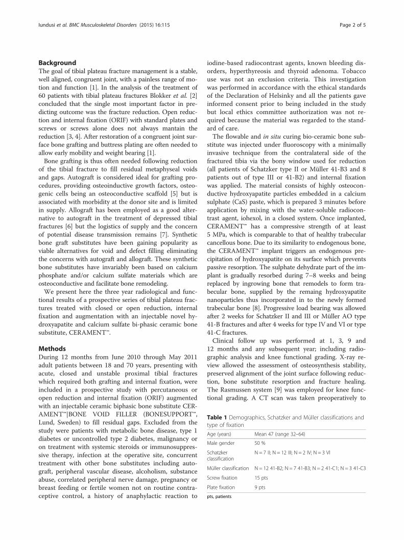
Table 1 Demographics, Schatzker and Müller classifications andtype of fixation
Age (years) Mean 47 (range 32–64)
Male gender 50 %
Schatzkerclassification
N = 7 II; N = 12 III; N = 2 IV; N = 3 VI
Müller classification N = 12 41-B2; N = 7 41-B3; N = 2 41-C1; N = 3 41-C3
Screw fixation 15 pts
Plate fixation 9 pts
pts, patients
Iundusi et al. BMC Musculoskeletal Disorders (2015) 16:115 Page 2 of 5
BackgroundThe goal of tibial plateau fracture management is a stable,well aligned, congruent joint, with a painless range of mo-tion and function [1]. In the analysis of the treatment of60 patients with tibial plateau fractures Blokker et al. [2]concluded that the single most important factor in pre-dicting outcome was the fracture reduction. Open reduc-tion and internal fixation (ORIF) with standard plates andscrews or screws alone does not always mantain thereduction [3, 4]. After restoration of a congruent joint sur-face bone grafting and buttress plating are often needed toallow early mobility and weight bearing [1].Bone grafting is thus often needed following reduction
of the tibial fracture to fill residual metaphyseal voidsand gaps. Autograft is considered ideal for grafting pro-cedures, providing osteoinductive growth factors, osteo-genic cells being an osteoconductive scaffold [5] but isassociated with morbidity at the donor site and is limitedin supply. Allograft has been employed as a good alter-native to autograft in the treatment of depressed tibialfractures [6] but the logistics of supply and the concernof potential disease transmission remains [7]. Syntheticbone graft substitutes have been gaining popularity asviable alternatives for void and defect filling eliminatingthe concerns with autograft and allograft. These syntheticbone substitutes have invariably been based on calciumphosphate and/or calcium sulfate materials which areosteoconductive and facilitate bone remodeling.We present here the three year radiological and func-
tional results of a prospective series of tibial plateau frac-tures treated with closed or open reduction, internalfixation and augmentation with an injectable novel hy-droxyapatite and calcium sulfate bi-phasic ceramic bonesubstitute, CERAMENT™.
MethodsDuring 12 months from June 2010 through May 2011adult patients between 18 and 70 years, presenting withacute, closed and unstable proximal tibial fractureswhich required both grafting and internal fixation, wereincluded in a prospective study with percutaneous oropen reduction and internal fixation (ORIF) augmentedwith an injectable ceramic biphasic bone substitute CER-AMENT™|BONE VOID FILLER (BONESUPPORT™,Lund, Sweden) to fill residual gaps. Excluded from thestudy were patients with metabolic bone disease, type 1diabetes or uncontrolled type 2 diabetes, malignancy oron treatment with systemic steroids or immunosuppres-sive therapy, infection at the operative site, concurrenttreatment with other bone substitutes including auto-graft, peripheral vascular disease, alcoholism, substanceabuse, correlated peripheral nerve damage, pregnancy orbreast feeding or fertile women not on routine contra-ceptive control, a history of anaphylactic reaction to
iodine-based radiocontrast agents, known bleeding dis-orders, hyperthyreosis and thyroid adenoma. Tobaccouse was not an exclusion criteria. This investigationwas performed in accordance with the ethical standardsof the Declaration of Helsinky and all the patients gaveinformed consent prior to being included in the studybut local ethics committee authorization was not re-quired because the material was regarded to the stand-ard of care.The flowable and in situ curing bio-ceramic bone sub-
stitute was injected under fluoroscopy with a minimallyinvasive technique from the contralateral side of thefractured tibia via the bony window used for reduction(all patients of Schatzker type II or Müller 41-B3 and 8patients out of type III or 41-B2) and internal fixationwas applied. The material consists of highly osteocon-ductive hydroxyapatite particles embedded in a calciumsulphate (CaS) paste, which is prepared 3 minutes beforeapplication by mixing with the water-soluble radiocon-trast agent, iohexol, in a closed system. Once implanted,CERAMENT™ has a compressive strength of at least5 MPa, which is comparable to that of healthy trabecularcancellous bone. Due to its similarity to endogenous bone,the CERAMENT™ implant triggers an endogenous pre-cipitation of hydroxyapatite on its surface which preventspassive resorption. The sulphate dehydrate part of the im-plant is gradually resorbed during 7–8 weeks and beingreplaced by ingrowing bone that remodels to form tra-becular bone, supplied by the remaing hydroxyapatitenanoparticles thus incorporated in to the newly formedtrabecular bone [8]. Progressive load bearing was allowedafter 2 weeks for Schatzker II and III or Müller AO type41-B fractures and after 4 weeks for type IV and VI or type41-C fractures.Clinical follow up was performed at 1, 3, 9 and
12 months and any subsequent year; including radio-graphic analysis and knee functional grading. X-ray re-view allowed the assessment of osteosynthesis stability,preserved alignment of the joint surface following reduc-tion, bone substitute resorption and fracture healing.The Rasmussen system [9] was employed for knee func-tional grading. A CT scan was taken preoperatively to
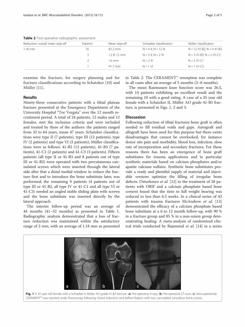
Table 2 Post-operative radiographic assessment
Reduction: overall mean step-off Patients Mean step-off Schatzker classification Müller classification
1.18 mm 16 ≤1.2 mm N = 4 II; N = 12 III N = 12 41-B2; N = 4 41-B3
5 1.2≤ 1.5 mm N = 3 II; N = 2 VI N = 3 41-B3; N = 2 41-C3
2 1.6 mm N = 2 IV N = 2 41-C1
1 ≈1.7 mm N = 1 VI N = 1 41-C3
Iundusi et al. BMC Musculoskeletal Disorders (2015) 16:115 Page 3 of 5
examine the fracture, for surgery planning and forfracture classifications according to Schatzker [10] andMüller [11].
ResultsNinety-three consecutive patients with a tibial plateaufracture presented at the Emergency Department of theUniversity Hospital “Tor Vergata” over the 12 month re-cruitment period. A total of 24 patients, 12 males and 12females, met the inclusion criteria and were includedand treated by three of the authors: the patients rangedfrom 32 to 64 years, mean 47 years. Schatzker classifica-tions were type II (7 patients), type III (12 patients), typeIV (2 patients) and type VI (3 patients); Müller classifica-tions were as follows: 41-B2 (12 patients), 41-B3 (7 pa-tients), 41-C1 (2 patients) and 41-C3 (3 patients). Fifteenpatients (all type II or 41-B3 and 8 patients out of typeIII or 41-B2) were operated with two percutaneous can-nulated screws which were inserted through the lateralside after that a distal medial window to reduce the frac-ture first and to introduce the bone substitute later, wasperformed; the remaining 9 patients (4 patients out oftype III or 41-B2, all type IV or 41-C1 and all type VI or41-C3) needed an angled stable sliding plate with screwsand the bone substitute was inserted directly by thelateral approach.The interim follow-up period was an average of
44 months (41–52 months) as presented in Table 1.Radiographic analysis demonstrated that a loss of frac-ture reduction was maintained within the satisfactoryrange of 2 mm, with an average of 1.18 mm as presented
Fig. 1 A 35 year old female with a Schatzker II, Müller AO grade 41-B3 fracturCERAMENT™ was injected under fluoroscopy following closed reduction and
in Table 2. The CERAMENT™ resorption was completein all cases after an average of 5 months (3–8 months).The mean Rasmussen knee function score was 26.5,
with 14 patients exhibiting an excellent result and theremaining 10 with a good rating. A case of a 35 year oldfemale with a Schatzker II, Müller AO grade 41-B3 frac-ture, is presented in Figs. 1, 2 and 3.
DiscussionFollowing reduction of tibial fractures bone graft is oftenneeded to fill residual voids and gaps. Autograft andallograft have been used for this purpose but there existsdisadvantages that cannot be overlooked, for instancedonor site pain and morbidity, blood loss, infection, slowrate of incorporation and secondary fractures. For thesereasons there has been an emergence of bone graftsubstitutes for trauma applications and in particularsynthetic materials based on calcium phosphates and/orapatite calcium sulfates. Synthetic bone substitutes pro-vide a ready and plentiful supply of material and inject-able versions optimize the filling of irregular bonedefects. Ozturkmen et al. [12] in the treatment of 28 pa-tients with ORIF and a calcium phosphate based bonecement found that the time to full weight bearing wasreduced to less than 6.5 weeks. In a clinical series of 43patients with trauma fractures McAndrew et al. [13]demonstrated the efficacy of a calcium phosphate basedbone substitute at a 6 to 12 month follow-up, with 90 %in a fracture group and 85 % in a non-union group dem-onstrating healing. A meta-analysis of randomized clin-ical trials conducted by Bajammal et al. [14] in a series
e. (a) Pre-operative X-rays; (b) Pre-operative CT-scan; (c) Intra-operativelybefore fixation with two cannulated cancellous bone screws

Fig. 3 Excellent bone regeneration with the removal of thehardware after 9 months since osteosynthesis
Fig. 2 X-rays follow-up at different steps. (a, b) The positioning of the bioceramic may been seen on the immediate post-operative frontal andsagittal x-rays; (c) On the same views at 1 month post-op there is no further evidence of radiocontrast of the bone cement indicating thewashout of iohexol; (d) The remodeling process is now progressing. New bone appears well consolidated at 3 months
Iundusi et al. BMC Musculoskeletal Disorders (2015) 16:115 Page 4 of 5
of metaphyseal fractures involving tibial plateau, femoralneck, intertrochanteric femoral and calcaneal indica-tions, concluded that loss of fracture reduction was lesswith a calcium phosphate based bone filler in compari-son with autogenous bone graft. Bucholz et al. [15]found no significant differences in radiographic and clin-ical assessments between a calcium phosphate basedbone cement and autograft for residual void and gap ina series of 40 patients with displaced tibial fracture.In cadaveric pre-clinical investigations [16–18] calcium
phosphate based cements used to fill proximal tibial de-fects following trauma were found to be stronger thanautologous graft filling with greater stiffness, higherfatigue strength and a greater ultimate load supportingcapacity. This is supported clinically by Simpson andKeating [19] at 1 year in 26 patients in an equally bal-anced study with the use of autograft and a calciumphosphate based bone substitute for the treatment ofproximal tibial fractures. The mean residual plateau de-pression was 4 mm in the autograft group (coupled withbuttress plating) and 0.7 mm in the calcium phosphatebased group (with minimal internal fixation) at 1 yearfollow-up. They also were able to use less hardware withthe calcium phosphate group and had a corresponding40 % reduction in operative time on average (from amean of 101 min to 55 min). Larsson and Hammick [20]in their analysis of randomized clinical trauma studiessupport the benefits of injectable calcium phosphatebased bone substitutes as suitable alternatives to auto-graft, with the flexibility to be applied around the hard-ware once in position and to augment weaker bonearound the screws as well as replace autograft in defectsand voids.It can be concluded from our preliminary review that
radiological and clinical outcome was satisfactorily ob-tained in all cases without complications. The enhancedstability with the application of CERAMENT™ supportedand maintained fracture reduction with a mean settingof 1.18 mm at an average of 9 months in the current
clinical series, well within the satisfactory range of 2 mmand in line with the findings of Simpson and Keating[19]. The Rasmussen knee functional score had all pa-tients in the good and excellent categories demonstrat-ing results at least as good as those for Ozturkmen et al.[12] who employed calcium phosphate cement augmen-tation in their treatment of depressed tibial fractures andreturned 78 % of patients back to their pre- operativelevel of activity.CERAMENT™ is designed to remodel in tune with the
natural bone remodelling process. Due to the microporos-ity through the release of the CaS component, an immedi-ate flow of tissue fluids with nutrients and growth factorsare allowed to penetrate the implant, which promotesosteoclasts and macrophages to enter the material andcreate macropores resulting in a widespread ingrowth ofearly bone. The end result is full transformation andremodeling into mature bone after 9–12 months [21, 22].This interim study at an average of 44 months follow-upof the bone remodeling process shows that new normalbone is formed and maintained which is important forclinical performance in the longer term.This investigation has some limitations. The study
population is relatively small and doesn’t effectively

Iundusi et al. BMC Musculoskeletal Disorders (2015) 16:115 Page 5 of 5
represent all types of tibial plateau fractures. Three outof the authors performed the surgical procedures. Next,a control group using other bone substitutes or graftingwas not enrolled. Lastly, patients were followed for amid-period, so further studies will be required to verifythe long-term clinical and radiological outcomes.
ConclusionCERAMENT™|BONE VOID FILLER has demonstratedthat it is a viable replacement to bone grafting in fillingvoids and gaps following fracture of the tibial plateau. Itprovides a material, being injectable which optimizes thefilling of irregular bone defects. This study has demon-strated that CERAMENT™ has supported the reductionand preservation of alignment of the joint surface untilfracture healing. This makes the novel biphasic bio-ceramic material an attractive solution in the treatmentof trauma situations where bone graft is required.
Competing interestsThe authors declare that they have no competing interests.
Authors’ contributionsRI conceived the study, collaborated on the study design, patientrecruitment, data analysis and manuscript preparation and review. EGcollaborated on the study design, patient recruitment, data analysis andmanuscript preparation and review. AP collaborated on data analysis,manuscript preparation and review. MDA collaborated on data analysis,manuscript preparation and review. UT conceived the study, participated inits design and coordination and collaborated on patient recruitment, dataanalysis and manuscript preparation and review. All authors read andapproved the final manuscript.
AcknowledgementsAll authors have no financial or personal relationships with other people ororganizations that could inappropriately influence this work.
Author details1Department of Orthopedics and Traumatology, University “Tor Vergata”,“Policlinico Tor Vergata” Foundation, Viale Oxford 81, 00133 Rome, Italy.2Orthopaedic Department, University of Palermo, Via del Vespro, 90100Palermo, Italy. 3Oncologic Center, “Palazzo Baleani”, Azienda PoliclinicoUmberto I, Corso Vittorio Emanuele II 244, Rome, Italy.
Received: 17 December 2014 Accepted: 1 May 2015
References1. Koval KJ, Helfet DL. Tibial plateau fractures: evaluation and treatment. J Am
Acad Orthop Surg. 1995;3:86–94.2. Blokker CP, Rorabeck CH, Bourne RB. Tibial plateau fractures: an analysis of the
results of treatment in 60 patients. Clin Orthop Relat Res. 1984;182:193–9.3. Stevens DG, Beharry R, McKee MD, Waddell JP, Schemitsch EH. The long-
term functional outcome of operatively treated tibial plateau fractures.J Orthop Trauma. 2001;15:312–20.
4. Russell TA, Leighton RK. Comparison of autogenous bone graft andendothermic calcium phosphate cement for augmentation in tibial plateaufractures: a multicenter, prospective, randomized study. J Bone Joint SurgAm. 2008;90:2057–61.
5. Faour O, Dimitriou R, Cousins CA, Giannoudis PV. The use of bone graftsubstitutes in large cancellous voids: any specific needs? Injury.2011;42 Suppl 2:87–90.
6. Segur JM, Torner P, Garcia S, Combalía A, Suso S, Ramón R. Use of boneallograft in tibial plateau fractures. Arch Orthop Trauma Surg. 1998;117:357–9.
7. Ong JC, Kennedy MT, Mitra A, Harty JA. Fixation of tibial plateau fractureswith synthetic bone graft versus natural bone graft: a comparison study.Ir J Med Sci. 2012;181:247–52.
8. Truedsson A, Wang JS, Lindberg P, Gordh M, Sunzel B, Warfvinge G. Bonesubstitute as an on-lay graft on rat tibia. Clin Oral Implants Res.2010;21:424–9.
9. Rasmussen PS. Tibial condylar fractures: impairment of knee joint stability asan indication for surgical treatment. J Bone Joint Surg Am. 1973;55:1331–50.
10. Schatzker J, McBroom R, Bruce D. The tibial plateau fracture. The Torontoexperience 1968–1975. Clin Orthop Relat Res. 1979;138:94–104.
11. Kellam JF, Audige L. Fracture Classification. In: Ruedi JP, editor. AO Principles ofFracture Management. 2nd ed. Switzerland: AO Publishing; 2007. p. 69–86.
12. Ozturkman Y, Caniklioglu M, Karamehmetoglu M, Sükür E. Calciumphosphate augmentation in the treatment of depressed tibial plateaufractures with open reduction and internal fixation. Acta Orthop TraumatolTurc. 2010;44:262–9.
13. McAndrew MP, Gorman PW, Lange TA. Tricalcium phosphate as a bone graftsubstitute in trauma: preliminary report. J Orthop Trauma. 1988;2:333–9.
14. Bajammal SS, Zlowodzki M, Lelwica A, Tornetta 3rd P, Einhorn TA, Buckley R,et al. The use of calcium phosphate bone cement in fracture treatment: ameta-analysis of randomized trials. J Bone Joint Surg Am. 2008;90:1186–96.
15. Bucholz RW, Carlton A, Holmes R. Interporous hydroxyapatite as a bonegraft substitute in tibial plateau fractures. Clin Orthop Relat Res.1989;240:53–62.
16. McDonald E, Chu T, Tufaga M, Marmor M, Singh R, Yetkinler D, et al. Tibialplateau fracture repairs augmented with calcium phosphate cement havehigher in situ fatigue strength than those with autograft. J Orthop Trauma.2011;25:90–5.
17. Yetkinler DN, McClellan RT, Reindel ES, Carter D, Poser RD. Biomechanicalcomparison of conventional open reduction and internal fixation versuscalcium phosphate cement fixation of a central depressed tibial plateaufracture. J Orthop Trauma. 2001;15:197–206.
18. Trenholm A, Landry S, McLaughlin K, Deluzio KJ, Leighton J, Trask K, et al.Comparative fixation of tibial plateau fractures using alpha-BSM, a calciumphosphate cement, versus cancellous bone graft. J Orthop Trauma.2005;19:698–702.
19. Simpson D, Keating JF. Outcome of tibial plateau fractures managed withcalcium phosphate cement. Injury. 2004;35:913–8.
20. Larsson S, Hannink G. Injectable bone-graft substitutes: current products,their characteristics and indications and new developments. Injury.2011;42 Suppl 2:30–4.
21. Abramo A, Geijer M, Kopylov P, Tägil M. Osteotomy of distal radius fracturemalunion using a fast remodeling bone substitute consisting of calciumsulfate and calcium phosphate. J Biomed Mat Res B Applied Biomaterials.2010;92:281–6.
22. Hatten Jr HP, Voor MJ. Bone healing using a bi-phasic ceramic bone substitutedemonstrated in human vertebroplasty and with histology in a rabbitcancellous bone defect model. Interv Neuroradiol. 2012;18:105–13.
Submit your next manuscript to BioMed Centraland take full advantage of:
• Convenient online submission
• Thorough peer review
• No space constraints or color figure charges
• Immediate publication on acceptance
• Inclusion in PubMed, CAS, Scopus and Google Scholar
• Research which is freely available for redistribution
Submit your manuscript at www.biomedcentral.com/submit