autologous bone graft versus pekk cage for vertebral
TRANSCRIPT

J Neurosurg Spine Volume 24 • February 2016 309
cliNical articleJ Neurosurg Spine 24:309–314, 2016
Anterior cervical corpectomy followed by anterior fusion (ACCF) has become a widely used proce-dure for the treatment of multilevel degenerative
cervical stenosis. After the pioneering work of Smith and Robinson12 on an anterior approach to the cervical spine, removal of the intervertebral disc, and interbody fusion in 1958, the implantation of an autologous bone graft, most frequently taken from the iliac crest, combined with
an anterior plate-screw osteosynthesis remained the gold standard of vertebral replacement and has been reported to result in fusion rates of 70%–100%.3,13 In recent years, nu-merous nonbiological implants have been developed and introduced for clinical use to avoid donor-site morbidities such as additional pain, delayed mobilization, and iliac fracture.1,10 Titanium cages are the most widely used xeno-grafts for use in ACCF, and several groups have reported
abbreviatioNS ACCF = anterior cervical corpectomy with fusion; EMS = European Myelopathy Scale; PEKK = polyetherketoneketone; VAS = visual analog scale.Submitted Augst 27, 2014. accepted May 12, 2015.iNclude wheN citiNg Published online October 16, 2015; DOI: 10.3171/2015.5.SPINE14887.
Autologous bone graft versus PEKK cage for vertebral replacement after 1- or 2-level anterior median corpectomyStefan Koehler, md,1 Furat raslan, md,2 christian Stetter, md,1 Stefan mark rueckriegel, md,1 ralf-ingo ernestus, md,1 and thomas westermaier, md1
1Department of Neurosurgery, University Hospital Wuerzburg; and 2Department of Neurosurgery, Klinikum Hohe Warte, Bayreuth, Germany
obJective Anterior cervical corpectomy with fusion has become the most widely used procedure for the treatment of multilevel cervical stenosis. Although an autologous bone graft is the gold standard for vertebral replacement after corpectomy, industrial implants have become popular because they result in no donor-site morbidity. In this study, the authors compared clinical and radiological results of autologous iliac grafts versus those of bone-filled polyetherketoneketone (PEKK) cage implants.methodS The clinical and radiological data of 46 patients with degenerative multilevel cervical stenosis and who un-derwent 1- or 2-level anterior median corpectomy between 2004 and 2012 were analyzed. The patients in Group 1 were treated with vertebral replacement with an autologous iliac graft, and those in Group 2 were treated with a PEKK cage implant. Each patient also underwent osteosynthesis with an anterior plate-screw system. Visual analog scale (VAS) and European Myelopathy Scale scores, loss of height and regional cervical lordosis angle, and complication rates of the 2 groups were compared.reSultS The mean follow-up time was 20 months. In both groups, the VAS and European Myelopathy Scale scores improved significantly. The loss of height was 3.7% in patients with iliac grafts and 5.3% in patients with PEKK implants. The rates of osseous fusion were similar in Groups 1 and 2 (94.7% and 91.3%, respectively). At the end of the follow-up period, none of the patients complained about donor-site pain. One patient in Group 1 suffered a fracture of the iliac bone that required osteosynthesis. Four patients in Group 2 had to receive revision surgery for cage and/or plate-screw dislocation and new neurological deficit or intractable pain.coNcluSioNS Preoperative pain and radicular and myelopathic symptoms improve after decompression irrespective of the material used for vertebral replacement. The use of PEKK cages for vertebral replacement seems to result in a higher risk of implant-related complications. A prospective randomized study is necessary to supply evidence for the use of autografts and artificial implants after anterior cervical corpectomy with fusion.http://thejns.org/doi/abs/10.3171/2015.5.SPINE14887Key wordS corpectomy; ACCF; autologous bone graft; PEEK; PEKK; cage; cervical
©AANS, 2016
Unauthenticated | Downloaded 01/27/22 06:31 AM UTC

S. Koehler et al.
J Neurosurg Spine Volume 24 • February 2016310
good clinical and radiological results with them,14–16 but significant numbers of implant-related complications, such as implant failure and subsidence, have been men-tioned.4,5 Polyetherketone cages have the theoretical ad-vantage of being less rigid than titanium cages and, there-fore, might result in reduced cage subsidence and a lower rate of dislocation.
To the best of our knowledge, there has been no system-atic analysis to directly compare the clinical and radio-logical courses after ACCF using autologous bone grafts or polyetherketoneketone (PEKK) cages. It was the aim of this study to analyze the clinical and radiological out-comes of patients with autologous bone graft or a PEKK cage for vertebral replacement after ACCF of the cervical spine.
methodspatient collective
Between 2004 and 2012, 137 patients underwent a 1- or 2-level ACCF in the Department of Neurosurgery, Univer-sity Hospital Wuerzburg. Forty patients were treated for trauma, infection, or tumor, 12 received an implant other than a PEKK cage, and 15 simultaneously underwent ad-ditional posterior instrumentation and were excluded from further investigation. Of the remaining 70 patients, 24 did not continue follow-up for the minimum of 12 months and were excluded from further analysis. The clinical and ra-diological data of the remaining 46 patients (mean age 67 ± 10 years) with degenerative multilevel cervical steno-sis who underwent a 1- or 2-level ACCF between 2004 and 2012 were analyzed retrospectively. In our depart-ment, PEKK cages (Invadur-Oxpekk cage, PINA Med-izintechnik Vertriebs AG) were introduced for vertebral replacement in 2007. Each patient in whom surgery was performed between 2004 and 2007 underwent vertebral replacement with an autologous iliac graft (Group 1, n = 19), and those who underwent surgery between 2007 and 2012 received a PEKK cage (Group 2, n = 27). All of the patients also underwent osteosynthesis with an anterior plate-screw system (cervical spine locking plate [CSLP] system, Synthes). Decompression in both groups was per-formed in an identical way. ACCF was performed using a common anterior approach via a right-sided skin inci-sion. Foraminal decompression was performed bilater-ally with excision of the posterior longitudinal ligament to ensure adequate neural decompression. In Group 1, a large tricortical bone graft was harvested from the right iliac crest and fit into the vertebrectomy space. In Group 2, the PEKK cages were filled with spongious bone obtained from the osseus decompression.
All patients had radiological signs of a multilevel de-generative spinal stenosis with clinical and electrophysio-logical (somatosensory and motor evoked potentials) signs of cervical myelopathy and/or radiculopathy.
complicationsSurgical complications were recorded to differentiate
implant-related and general complications. The numbers of surgical revisions in both groups were compared.
radiological assessmentFor each patient, preoperative CT scans, MRI scans,
and digitalized lateral and anteroposterior radiographs of the cervical spine were obtained. A digital lateral radio-graph or CT scan of every patient was obtained on Day 1 after surgery. Radiographs were taken periodically until at least 12 months after the operation. A CT scan of the cervical spine was obtained at the end of follow-up. For this purpose, a thin-slice (1.5-mm) CT scan (SOMATOM 4+ Volume Zoom CT Scanner, Siemens Healthcare Di-agnostics GmbH), which included the disc space above and below the fused vertebrae, and triplanar reconstruc-tions were performed. Measurements of the height of the spondylodesis made from sagittal reconstructions of the CT images and from lateral radiographs on Day 1 after surgery and at the end of follow-up were compared and used for the radiological follow-up.
The loss of height during the follow-up period was de-termined by comparing the total height of the spondylode-sis between the first postoperative image on Day 1 after surgery and that at the end of follow-up. The total height of the spondylodesis was measured from the superior end-plate of the cranial vertebra to the inferior endplate of the caudal vertebra (Fig. 1). Digital measurements for use in our analyses were made by using the Picture Archiving and Communication System (syngo.plaza PACS, Siemens).
The relative height of the implant was determined by measuring the height of the bone graft or cage and divid-
Fig. 1. The total height of the spondylodesis was measured as the distance from the superior endplate of the cranial vertebra to the inferior endplate of the caudal vertebra. For these measurements, digital Picture Archiving and Communication System (PACS) tools were used. Figure is available in color online only.
Unauthenticated | Downloaded 01/27/22 06:31 AM UTC
bone graft vs peKK cage for vertebral replacement after accF
J Neurosurg Spine Volume 24 • February 2016 311
ing it by the total height of the spondylodesis to obtain information about the extent and manner of graft subsid-ence in the 2 groups.
The local cervical lordosis angle over the fused ver-tebrae was measured on Day 1 after surgery and at the end of follow-up. If there was a kyphotic alignment of the vertebrae, the angles were defined as negative.
The fusion rates in both groups were determined. Osse-ous fusion was considered if a bony bridge without inter-ruption was found in the images at the end of follow-up.
clinical assessmentAll patients underwent standardized neurological as-
sessments preoperatively and periodically postoperatively until the end of follow-up. Pain levels were assessed by using visual analog scale (VAS) scores, and functional outcomes were classified by using European Myelopathy Scale (EMS) scores.
Statistical analysisResults are presented as means ± SD. Normally distrib-
uted parametric data were compared by using the Student t-test to compare the 2 groups. Incidences were compared by using the Fisher exact test. Statistical analysis was per-formed by using GraphPad Prism statistical software. A p value of < 0.05 was considered statistically significant.
resultspatient collective
Forty-six patients met the inclusion criteria. Twenty-eight patients were male, and 18 were female. Nineteen patients underwent vertebral replacement with an autolo-gous iliac crest bone graft (Group 1), and 27 were treated with a PEKK-cage implant (Group 2). The mean ages at the time of surgery were 66 ± 11 years [Group 1] and 67 ± 10 years [Group 2]. The mean follow-up times were 19.2 ± 15.2 months [Group 1] and 21.9 ± 13.6 months [Group 2]. In Group 1, 14 (73.7%) patients underwent a 1-level ACCF, and 5 (26.3%) patients underwent a 2-level ACCF. In Group 2, 19 (70.4%) patients underwent a 1-level ACCF, and 8 (29.6%) patients underwent a 2-level ACCF.
general complicationsSignificant rebleeding into the cervical soft tissue, lead-
ing to early revision surgery, occurred in 2 (4.3%) patients, 1 in each group. One patient in Group 2 required early revision surgery as a result of CSF leakage. None of the patients had a persistent new neurological deficit at the end of follow-up.
implant-related complicationsOne patient in Group 1 experienced a fracture of the ili-
ac crest that required surgical revision by an osteosynthesis of the iliac bone. No other implant-related complications occurred in this group. In particular, there was no disloca-tion of the bone graft or the anterior plate–screw system.
In Group 2, a minor dislocation of the plate-screw sys-tem in 1 patient was recorded, and in another patient, a mi-nor loosening of the screws occurred, but revision surgery
was not needed. In 4 patients of Group 2, a dislocation of the PEKK cage and loosening of the plate-screw sys-tem with a new neurological deficit and/or intractable pain made revision surgery necessary. In 2 of these patients, anterior revision of the PEKK cage and plate–screw sys-tem was performed after 3 and 4 months, respectively. In another patient, a 2-level corpectomy had to be extended to a 3-level corpectomy with replacement of the PEKK cage by an autologous bone graft and subsequent poste-rior instrumentation 1 week after the first operation. In the fourth case, an anterior revision procedure followed by posterior instrumentation was performed because of persistent pain and failed bony fusion (Table 1).
radiological assessmentPatients who underwent cervical revision surgery and/
or secondary posterior instrumentation were excluded from further radiological assessment. Thus, 19 patients in Group 1 and 23 patients in Group 2 were investigated.
In Group 1, the mean total height of the spondylodesis was 48.0 ± 9.0 mm on Day 1 after surgery and decreased to 46.3 ± 9.1 mm (96.3% ± 4.1%) at the end of follow-up. In Group 2, the total height of the spondylodesis was 51.7 ± 9.7 mm on Day 1 after surgery and decreased to 48.9 ± 8.8 mm (94.7% ± 3.6%) at the end of follow-up (Fig. 2). The differences did not reach the level of significance (p = 0.17 for loss of height during follow-up).
In Group 1, the mean ratio between the height of the cage and the height of the fused vertebrae on Day 1 after surgery was 0.48 ± 0.09; at the end of follow-up, the mean ratio decreased to 0.47 ± 0.09. In Group 2, the mean ratio was 0.52 ± 0.07 on Day 1 and increased to 0.54 ± 0.06 at the end of follow-up (Fig. 3).
In Group 1, the mean lordosis angle over the fused ver-tebrae decreased significantly (p < 0.05) from 5.9° ± 5.2° at Day 1 after surgery to 4.1° ± 4.4° at the end of follow-up. In Group 2, the mean lordosis angle decreased signifi-cantly (p < 0.05) from 6.6° ± 7.0° to 3.5° ± 6.0°. The differ-ences between the groups were not statistically significant.
Osseous fusion at the end of follow-up was achieved in 18 (94.7%) of 19 patients in Group 1 and in 21 (91.3%) of 23 patients in Group 2. The difference between the groups was not statistically significant.
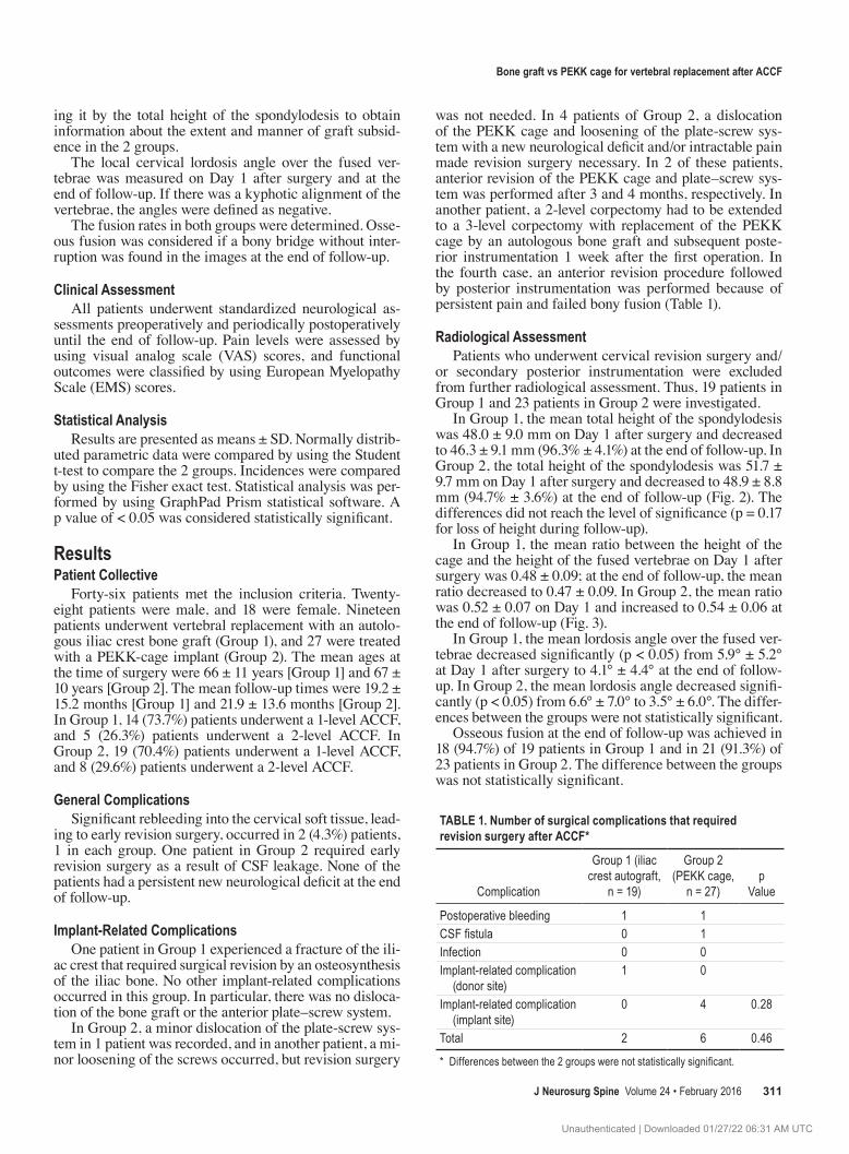
table 1. Number of surgical complications that required revision surgery after accF*
Complication
Group 1 (iliac crest autograft,
n = 19)
Group 2 (PEKK cage,
n = 27)p
Value
Postoperative bleeding 1 1CSF fistula 0 1Infection 0 0Implant-related complication
(donor site) 1 0
Implant-related complication (implant site)
0 4 0.28
Total 2 6 0.46
* Differences between the 2 groups were not statistically significant.
Unauthenticated | Downloaded 01/27/22 06:31 AM UTC

S. Koehler et al.
J Neurosurg Spine Volume 24 • February 2016312
clinical assessmentThe mean reported pain VAS scores at the end of fol-
low-up had improved significantly (p < 0.01) over those reported in the preoperative assessments in Group 1 (from 2.26 ± 1.79 to 1.11 ± 0.99) and in Group 2 (from 2.63 ± 1.63 to 1.19 ± 0.88). The differences in the mean preop-erative VAS scores and the mean improvement of pain according to the VAS scores (1.15 in Group 1 vs 1.44 in Group 2) were not statistically significant (Fig. 4 upper).
The EMS scores improved significantly (p < 0.001) from 14.74 ± 1.56 preoperatively to 16.05 ± 1.18 at the end of follow-up in Group 1 and from 14.15 ± 2.49 to 15.63 ± 1.94 in Group 2. The differences in the mean preopera-tive EMS scores and those at the end of follow-up and the mean improvement of EMS scores (1.31 [Group 1] vs 1.48 [Group 2]) were not statistically significant between the 2 groups (Fig. 4 lower).
discussionIn this study, we retrospectively analyzed 46 patients
who underwent a 1- or 2-level ACCF performed to treat degenerative disorders of the cervical spine. Nineteen pa-tients underwent vertebral replacement with an autologous iliac crest bone graft, and 27 patients were treated by us-ing a PEKK cage. Until 2007, we exclusively used bone grafts for vertebral replacement in our department for this group of patients. After 2007, only PEKK cages were used in the treatment of degenerative diseases of the cervical spine. Because there were no criteria for the choice of the implant other than the year in which the patient under-went surgery, this retrospective analysis has the charac-teristics of a quasi-randomized trial; this is confirmed by the patient data, which are nearly identical in both groups concerning age, sex, number of fused vertebrae, and pre-operative radiological and clinical presentations.
To our knowledge, there have been no previous reports of studies in the literature that assessed the fusion and complication rates of PEKK cages used for ACCF. Titani-um grafts are the most widely used implants for this pur-pose and have been shown to result in fusion rates of 70%–100%.14–16 In some of these reports, however, a remarkable rate of implant-related complications were mentioned.2 In our series, general complications occurred in both groups at comparable frequencies. This result is plausible because CSF leaks and hematomas are complications that are in-
Fig. 4. In both groups, preoperative pain and signs of cervical myelopa-thy, as determined by VAS (upper) and EMS (lower) scores, showed long-term decreases.
Fig. 3. The mean loss of height in Group 1 was caused by impaction of the bony implant, as depicted by the mean decrease of the relative height of the implant related to the total height of the spondylodesis. In Group 2, the mean relative height of the implants increased, which suggests that the implants impressed the adjacent vertebrae and their endplates.
Fig. 2. The mean losses of height were 3.6% in Group 1 and 5.3% in Group 2. The difference between the 2 groups did not reach the level of significance (p = 0.17).
Unauthenticated | Downloaded 01/27/22 06:31 AM UTC

bone graft vs peKK cage for vertebral replacement after accF
J Neurosurg Spine Volume 24 • February 2016 313
dependent of the fusion materials used. However, there was an obvious difference in the incidence of implant-related complications between the 2 groups. There was 1 patient with significant donor-site morbidity (fracture of the iliac crest) that required iliac osteosynthesis. Avoiding donor-site morbidity is the most striking argument for the use of artificial implants,1,7 and this case in our series also illustrates the problem. Donor-site morbidity may include pain, hematoma, fracture, or meralgia paresthetica and has been reported to occur in up to 25% of patients. Some au-thors reported that most of these complaints resolved over time, and others observed that significant pain at the donor site could persist for more than 24 months after surgery or that chronic pain and functional impairment could persist even longer after vertebral replacement by an iliac crest autograft.1,10,11 With the exception of this single case, we were unable to reproduce these results; no patient in our study reported long-term problems concerning the donor site at the end of follow-up period.
Concerning the local implant failure that necessitated revision surgery, our study showed a considerable accumu-lation of these cases in the PEKK cage group. There was no dislocation of an iliac crest bone graft or adjacent os-teosynthetic material noted, which is in contrast to 4 cases of dislocation of the PEKK cage and adjacent plate-screw system in the cage group. This difference is remarkable and suggests that the fusion was more stable with the use of the bone graft. However, our finding that the relative height of the implant in relation to the total height of the spon-dylodesis decreased in the bone-graft group and increased in the cage group suggests different mechanisms of cage impaction. In the bone-graft group, the loss of height was caused by impaction of the bone graft itself, and in the cage group, the loss of height was caused by impaction of the rigid cage into the adjacent endplates, which may explain the greater loss of height in the cage group. Once the end-plate—the strongest, weight-loading part of the vertebral body—is weakened, the resistance decreases and the con-struction might become more unstable and prone to lose height.9 Thus, this pathomechanism might be determined to be the reason for the loosening of screws and the break-out of implants. An implant-related strain on the endplates is a well known phenomenon after anterior cervical discec-tomy with fusion (ACDF) involving polyetheretherketone (PEEK) cages or polymethylmethacrylate (PMMA) substi-tutes.6,8 After ACDF, however, this problem is obviously less eminent because of the shorter distance and the lower degree of instability caused by the decompressive proce-dure than those in ACCF procedures. A certain degree of impaction of the implant is normal and probably promotes osseous fusion. This is underlined by the postoperative re-duction of the local lordosis angle and by the fact that the loss of height in the patients who did not undergo revision surgery had no correlation with the clinical outcomes mea-sured by VAS and EMS scores. The clinical results were good, and there was a significant improvement of both pa-rameters and an improvement in all patients.
conclusionsIn this series, the rate of implant failures that necessi-
tated revision surgery was higher in the group of patients who had undergone a PEKK cage implant for spondylode-sis than in the group of patients who had undergone ACCF with an autologous bone graft. The radiological data sug-gest that intrusion of the cage into the adjacent vertebrae might contribute to a higher rate of implant and screw–plate dislocation. The drawbacks of this analysis are that the data originated from a relatively small number of pa-tients and, although the setting was quasi-randomized, from a retrospective analysis. A prospective randomized trial to compare the clinical and radiological outcomes after vertebral body replacement with autologous bone grafts and those after various artificial implants are used instead could provide more evidence for or against their use in ACCF.
references 1. Banwart JC, Asher MA, Hassanein RS: Iliac crest bone graft
harvest donor site morbidity. A statistical evaluation. Spine (Phila Pa 1976) 20:1055–1060, 1995
2. Boakye M, Patil CG, Ho C, Lad SP: Cervical corpectomy: complications and outcomes. Neurosurgery 63 (4 Suppl 2):295–302, 2008
3. Brown JA, Havel P, Ebraheim N, Greenblatt SH, Jackson WT: Cervical stabilization by plate and bone fusion. Spine (Phila Pa 1976) 13:236–240, 1988
4. Castellvi AE, Castellvi A, Clabeaux DH: Corpectomy with titanium cage reconstruction in the cervical spine. J Clin Neurosci 19:517–521, 2012
5. Daubs MD: Early failures following cervical corpectomy reconstruction with titanium mesh cages and anterior plating. Spine (Phila Pa 1976) 30:1402–1406, 2005
6. Gebremariam L, Koes BW, Peul WC, Huisstede BM: Evalua-tion of treatment effectiveness for the herniated cervical disc: a systematic review. Spine (Phila Pa 1976) 37:E109–E118, 2012
7. Kim MK, Kim SM, Jeon KM, Kim TS: Radiographic com-parison of four anterior fusion methods in two level cervical disc diseases: autograft plate fixation versus cage plate fixa-tion versus stand-alone cage fusion versus corpectomy and plate fixation. J Korean Neurosurg Soc 51:135–140, 2012
8. Park JI, Cho DC, Kim KT, Sung JK: Anterior cervical disc-ectomy and fusion using a stand-alone polyetheretherketone cage packed with local autobone: assessment of bone fusion and subsidence. J Korean Neurosurg Soc 54:189–193, 2013
9. Porto Filho MR, Pastorello MT, Defino HL: Experimental study of the participation of the vertebral endplate in the in-tegration of bone grafts. Eur Spine J 14:965–970, 2005
10. Sawin PD, Traynelis VC, Menezes AH: A comparative anal-ysis of fusion rates and donor-site morbidity for autogeneic rib and iliac crest bone grafts in posterior cervical fusions. J Neurosurg 88:255–265, 1998
11. Silber JS, Anderson DG, Daffner SD, Brislin BT, Leland JM, Hilibrand AS, et al: Donor site morbidity after anterior iliac crest bone harvest for single-level anterior cervical discecto-my and fusion. Spine (Phila Pa 1976) 28:134–139, 2003
12. Smith GW, Robinson RA: The treatment of certain cervical-spine disorders by anterior removal of the intervertebral disc and interbody fusion. J Bone Joint Surg Am 40-A:607–624, 1958
13. Swank ML, Lowery GL, Bhat AL, McDonough RF: Anterior cervical allograft arthrodesis and instrumentation: multilevel interbody grafting or strut graft reconstruction. Eur Spine J 6:138–143, 1997
14. Uribe JS, Sangala JR, Duckworth EA, Vale FL: Comparison between anterior cervical discectomy fusion and cervical
Unauthenticated | Downloaded 01/27/22 06:31 AM UTC

S. Koehler et al.
J Neurosurg Spine Volume 24 • February 2016314
corpectomy fusion using titanium cages for reconstruction: analysis of outcome and long-term follow-up. Eur Spine J 18:654–662, 2009
15. Waschke A, Kaczor S, Walter J, Duenisch P, Kalff R, Ewald C: Expandable titanium cages for anterior column cervical reconstruction and their effect on sagittal profile: a review of 48 cases. Acta Neurochir (Wien) 155:801–807, 2013
16. Woiciechowsky C: Distractable vertebral cages for recon-struction after cervical corpectomy. Spine (Phila Pa 1976) 30:1736–1741, 2005
disclosureDr. Westermaier received payments from Johnson & Johnson for teaching activities (spinal neurosurgery).
author contributionsConception and design: Koehler, Westermaier. Acquisition of
data: Koehler, Raslan, Stetter, Westermaier. Analysis and inter-pretation of data: Koehler, Westermaier. Drafting the article: Koehler, Westermaier. Critically revising the article: Ernestus, Westermaier. Reviewed submitted version of manuscript: Raslan, Stetter, Rueckriegel, Ernestus, Westermaier. Approved the final version of the manuscript on behalf of all authors: Koehler. Sta-tistical analysis: Westermaier. Administrative/technical/material support: Ernestus. Study supervision: Westermaier.
Supplemental informationCurrent AffiliationDr. Raslan: Department of Neurosurgery, BG University Hospital Bergmannsheil, Bochum, Germany.
correspondenceStefan Koehler, Department of Neurosurgery, University Hospital Wuerzburg, Josef-Schneider-Strasse 11, Wuerzburg 97080, Ger-many. email: [email protected].
Unauthenticated | Downloaded 01/27/22 06:31 AM UTC