available online at sciencedirect sciencedirectlbmd.coe.pku.edu.cn/pdf/675-685.pdf · major...
TRANSCRIPT
Available online at ScienceDirect
ScienceDirectJ. Mater. Sci. Technol., 2014, 30(7), 675e685
In Vitro Study on MgeSneMn Alloy as Biodegradable Metals
Zhen Zhen1), Tingfei Xi1,2)*, Yufeng Zheng1,3)**, Li Li4), Lugee Li5)
1) Center for Biomedical Materials and Tissue Engineering, Academy for Advanced Interdisciplinary Studies,Peking University, Beijing 100871, China
2) Biomedical Engineering Research Center, Shenzhen Institute, Peking University, Shenzhen 518057, China3) Department of Materials Science and Engineering, College of Engineering, Peking University,
Beijing 100871, China4) Center for Biomedical Materials and Engineering, Harbin Engineering University, Harbin 150001, China5) Dongguan EONTEC Company Ltd, Dongguan 523662, China
[Manuscript received October 15, 2013, in revised form November 22, 2013, Available online 21 April 2014]
* Corresaddress:** CorreE-mail a1005-03JournalLimited.http://dx
The mechanical properties, chemical properties and biocompatibility of Mge3Sne0.5Mn alloy were tested.A series of in vitro evaluations such as tensile test, static and dynamic immersion test, hemocompatibilitytest as well as cytotoxicity test were presented, with commercial magnesium alloy WE43 as the control.Mge3Sne0.5Mn alloy possesses suitable strength and superior ductility compared with WE43 and AZ31.Static immersion and dynamic degradation tests showed more uniform degradation with a more moderaterate for Mge3Sne0.5Mn alloy (0.34 mm/y in static condition and 0.25 mm/y in dynamic condition)compared with WE43 alloy (0.42 mm/y in static condition and 0.33 mm/y in dynamic condition) in Hank’ssolution. Blood compatibility evaluation suggested that Mge3Sne0.5Mn alloy had no destructive effect onerythrocyte and showed excellent anti-thrombogenicity to blood system. Besides, Mge3Sne0.5Mn alloyshowed no inhibition effect to L929 metabolic activity and mild toxicity to vascular smooth muscle cell(VSMC) in preliminary cell viability assessment. By considering its excellent mechanical strength, corrosionresistance, low ion release rate and good biocompatibility, Mge3Sne0.5Mn alloy may be a promisingeconomical candidate as biomedical implant material for load-bearing clinical applications in the future.
KEY WORDS: Mge3Sne0.5Mn alloy; Corrosion; Dynamic immersion; Static immersion; Biodegradable material
1. Introduction
Magnesium and its alloys, have shown great potential to serveas biocompatible, osteoconductive, cardiovascular and biode-gradable implants for load-bearing applications[1]. Magnesium isa light weight metal with mechanical properties similar to that ofnatural bone, a natural ionic presence with significant functionalroles in biological systems, and can be degraded in vivo viacorrosion in the electrolytic environment of the body. Eventhough it seems that magnesium alloys are safe and effective inclinical trials and animal experiments, their high corrosion rate isunsatisfactory and has been a great concern in clinical applica-tion[2e9]. Recently, several approaches have been adopted to
ponding author. Prof.; Tel./Fax: þ86 10 62753404; [email protected] (T. Xi).sponding author. Prof., Ph.D.; Tel./Fax: þ86 10 62757411;ddress: [email protected] (Y. Zheng).02/$e see front matter Copyright� 2014, The editorial office ofof Materials Science & Technology. Published by ElsevierAll rights reserved..doi.org/10.1016/j.jmst.2014.04.005
improve the corrosion resistance of magnesium, such as ele-ments alloying[10e12], surface treatment[13e16], etc. Pure Mg,MgeCa based[11], MgeZn based[17], MgeSi based[18], MgeSrbased[19] and MgeRE based alloys[20] are mainly series mag-nesium alloys developed for bioapplications[21].In this work, MgeSneMn alloys are comprehensively studied
as a biodegradable biomaterial for the first time. Sn is an efficientaging strengthening element of magnesium alloys due to theprecipitates of Mg2Sn and is much more economical for clinicalapplications compared to rare earth elements. When used as amajor alloying element in magnesium, Sn can improve thecorrosion resistance, for its high hydrogen overvoltage[22]. Inphysiology, Sn is a trace element in human nutrition and healthand its toxicity also modifies the activities of several enzymes. Itis believed that Sn can interfere with the metabolism of Zn, Cuand Ca and alter the tissue concentrations of these elements[23].As for Mn, it has been chosen as a ternary alloying elementbecause of its influence on the improvement of mechanical andcorrosion properties of magnesium alloys due to the formation ofintermetallics[24]. Though Mn have no much effect on tensilestrength, it increases yield strength slightly and improves thesaltwater resistance of magnesium alloy by removing iron and

676 Z. Zhen et al.: J. Mater. Sci. Technol., 2014, 30(7), 675e685
other heavy-metal elements[25]. In addition, Mn has no toxiceffect only after extreme occupational exposure and plays aprimary role in the activation of multiple enzyme system, i.e.,hydrolases, kinases, transferases, decarboxylases and mitochon-drial respiration[25].The feasibility of as-extruded Mge3Sne0.5Mn alloy for
biomedical applications was investigated through mechanicalproperty, biocompatibility and corrosion property analysis withas-extruded WE43 as control.
2. Materials and Methods
2.1. Material preparation
As-extruded Mge3Sne0.5Mn alloy (extrusion ratio ¼ 11.1:1)was studied in this work, with as-extruded WE43 as control. Theworking surface was ground with SiC emery papers up to 2000grit. All the samples were then ultrasonically cleaned in absoluteethanol for 15 min. After being dried at room temperature, theywere weighed by a precision balance (AL204, Mettle Toledo).Hank’s solution[26] (NaCl 8.0 g, CaCl2 0.14 g, KCl 0.4 g,NaHCO3 0.35 g, glucose 1.0 g, MgCl2$6H2O 0.1 g, Na2H-PO4$12H2O 0.06 g, KH2PO4 0.06 g, MgSO4$7H2O 0.06 gdissolved in 1 L deionized water) was prepared and adjusted topH 7.4 for immersion test and electrochemical analysis.
2.2. Microstructure characterization and mechanical test
The microstructure was observed by optical microscopy(OM, Olympus BX51 M) and environmental scanning electronmicroscopy (SEM, Quanta-200FEG) in backscattered electronmode (BSE), equipped with an energy dispersive spectrometerattachment used for chemical composition analysis. The tensilesamples of Mge3Sne0.5Mn alloy were machined according toASTM-E8-04[27]. Tensile tests were carried out at a displace-ment rate of 1 mm/min with an Instron 3365 universal testmachine.
2.3. Electrochemical tests
Electrochemical tests were carried out at room temperature ina beaker containing 150 mL Hank’s solution on an electro-chemical workstation (CHI604D, Chenhua) using a standardthree-electrode configuration, with saturated calomel as a refer-ence and a platinum electrode as a counter and the sample as aworking electrode. Each sample was firstly used to monitor opencircuit potential (OCP) up to 3600 s and then potentiodynamicpolarization curve was measured on the sample with a scanningrate of 0.5 mV/s[28].
2.4. Immersion test
Each specimen was put into a tightly closed tube and the ratio ofsolution volume to specimen surface is 20 mL/cm2 according toASTM-G31-72[29] and ISO 10993-15[30]. The static immersiontest was carried out in water bath, while the dynamic immersiontest was in the air bath shaker at a rotation speed of 60 r/min[31].After static and dynamic immersion at 37 �C for 5 days, 10 daysand 30 days separately, the samples were removed from the so-lution, gently washed by deionized water and dried at room tem-perature. The pH value of the immersion solution was measuredby pH meter (PHS-3C, Lei-ci), and the specimens were weighed
by a precision balance (AL204, Mettle Toledo). Changes on thesurface morphology and microstructure of the samples after im-mersion were characterized by SEM (S-4800, Hitachi), and sur-face phase was identified on X-ray diffraction (XRD, RigakuSmartLab) using CuKa radiation. After that, the specimens werewashed in a boiling 200 g/L chromic acid solution to remove thesurface corrosion products, and thenweighed and characterized bySEM again. Inductively coupled plasma atomic emission spec-trometry (ICP, Profile ICP-AES, Leeman) was employed tomeasure the ion concentration of magnesium and alloying elementreleased from the alloy specimens. An average of three measure-ments was taken for each group.
2.5. Cytotoxicity test
CCK-8 assay of human vascular smooth muscle cell(VSMC) line and murine fibroblast cell line (L929) were usedto test the cell toxicity following ISO 10993-5 standard[32]. Thecells were first cultured in Dulbecco’s modified Eagle’s me-dium (DMEM, Gibco) supplemented with 10% fetal bovineserum (FBS, Gibco), 50 mg/mL L-glutamine, 100 IU/L peni-cillin and 100 mg/L streptomycin. An extract of the alloys wasprepared according to ISO 10993-12[33], in which magnesiumsamples were immersed in the culture medium with serum at37 � 1 �C for 24 h in humidified 5% CO2 atmosphere in anincubator. The ratio of the surface area of the magnesiumsample to the volume of the medium was 1.25 cm2/mL. Thecells were seeded in 96-well culture plates at a density of5000 cells/well, and incubated at 37 �C in humidified 5% CO2
atmosphere for 24 h. Then the medium was discarded. 100 mLof the extract, 100 mL of a negative control (medium alone),and 100 mL of a positive control (10% DMSO (dimethylsulf-oxide)) were added in the wells, respectively. Plates wereincubated for 24, 48, and 72 h at 37 �C in an incubator. At theend of each incubation time, 10 mL WST-8 solution was addedto each well and incubated for 2 h. Then the absorbance or theoptical density (OD) was measured at a wavelength of 450 nmwith a refereed wavelength of 630 nm using an enzyme-linkedimmunoassay (ELISA) microplate reader. The cell viabilitieswere expressed as relative growth rate (RGR) determined byRGR (%) ¼ (OD (sample)/OD (negative control)) � 100%.The data were expressed in mean � S.D.
2.6. Hemocompatibility evaluation
Hemolysis rate and platelet adhesion were tested in this sec-tion. Healthy human blood from a volunteer containing sodiumcitrate (3.8 wt%) in the ratio of 9:1 was taken and diluted withPBS (4:5 ratio by volume). Samples were dipped in a standardtube containing 10 mL of PBS which has been previouslyincubated at 37 �C for 30 min. Then 0.2 mL of diluted blood wasadded to this standard tube and the mixtures were incubated for60 min at 37 �C. Similarly, PBS solution was used as a blankcontrol and deionized water as a positive control. After that, allthe tubes were centrifuged for 5 min at 3000 r/min and the su-pernatant was carefully removed and transferred to the 96-wellplates for spectroscopic analysis at 545 nm. In addition, thehemolysis was calculated using an ultraviolet spectrophotometer(UNIC-7200, China) following Eq. (1). The hemolysis wascalculated based on the average of three replicates.OD stands for Optical Density. OD (test) stands for the optical
density of test group, OD (negative control) stands for that of

hemolysis ¼ OD ðtestÞ � OD ð negative control ÞOD ð positive control Þ � OD ð negative control Þ � 100% (1)
Z. Zhen et al.: J. Mater. Sci. Technol., 2014, 30(7), 675e685 677
PBS control group, and OD (positive control) stands for that ofdeionized water group.For platelet adhesion, platelet-rich plasma (PRP) was har-
vested by centrifugation of whole human blood at 180 g for20 min at room temperature. The PRP was overlaid the surfaceof the experimental samples and incubated at 37 �C for 1 h. Afterthat the specimens were gently rinsed with PBS to remove non-adherent platelets. The specimens were then fixed in 2.5%glutaraldehyde solutions for 1 h at room temperature, followedby dehydration in a gradient ethanol/distilled water mixture from50% to 100% in 10% increment for 10 min each and finally driedat room temperature. The surfaces of platelet attached sampleswere observed by SEM.Statistical analysis was performed by GraphPa Prism 5 and
differences were analyzed using the two-way analysis of vari-ance (ANOVA) technique, where statistical significance wasdefined as P < 0.05.
3. Results
3.1. Microstructure and mechanical properties of Mge3Sne0.5Mn alloy
The microstructures of Mge3Sne0.5Mn alloy and WE43alloy in BSE and OM mode are shown in Fig. 1. Highlightedinterphases are precipitated in both alloys, which contain high
Fig. 1 SEM micrographs in BSE mode and optical micrographs of WE43 ancircled highlighted particles A and B in WE43 and Mge3Sne0.5Mnmental alloys observed by optical microscopy.
concentration of Sn and Nd, respectively, as revealed by theenergy dispersive spectroscopy (EDS) results shown in the in-sets. XRD results in Fig. 2 indicate that the phases in the originalsamples are composed of a-Mg with Mg2Sn precipitates in Mge3Sne0.5Mn alloy and Mg41Nd5 in WE43 alloy. Combined withthe results of BSE, EDS and XRD, it could be concluded that thehighlighted interphases are Mg2Sn and Mg41Nd5 in MgeSneMn alloy and WE43, respectively. Fig. 3 illustrates the tensilecurve of Mge3Sne0.5Mn alloy. Strain work-hardening isobserved when the tensile strength reaches about 240 MPa, whilethe elongation of Mge3Sne0.5Mn alloy is greatly improved to11% compared to that of the commercial magnesium alloysAZ31, which is 2%, as shown in Table 1.
3.2. Electrochemical test
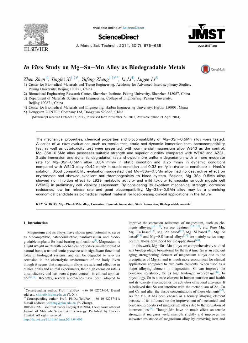
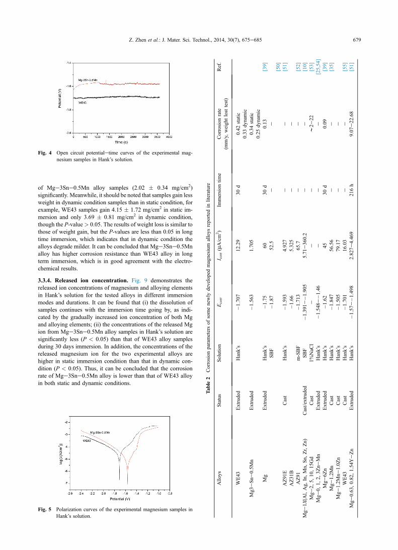
Fig. 4 displays the open circuit potentialetime curves of theMge3Sne0.5Mn alloy and WE43 alloy samples in Hank’s so-lution. A nobler balance potential is observed for Mge3Sne0.5Mn alloy as compared to WE43 alloy, which indicates thatMge3Sne0.5Mn alloy is more thermodynamically stable thanWE43 alloy in Hank’s solution.Fig. 5 presents the potentiodynamic polarization results of the
specimens and the average electrochemical parameters calcu-lated for the alloys according to ASTM G102[34] are listed inTable 2. It is clear that the corrosion potential (Ecorr) of Mge
d Mge3Sne0.5Mn alloys: (a) and (b) BSE micrographs and EDS of thealloys, respectively; (c) and (d) metallographic structures of the experi-

Fig. 2 X-ray diffraction patterns of WE43 (a) and Mge3Sne0.5Mn (b) alloys before and after 30 days immersion in Hank’s solution.
678 Z. Zhen et al.: J. Mater. Sci. Technol., 2014, 30(7), 675e685
3Sne0.5Mn alloy (�1.563 V) is superior to that of WE43 alloy(�1.707 V), and the result of corrosion current (Icorr) indicatesthat Mge3Sne0.5Mn alloy (1.705 mA/cm2) is more resistant tocorrosion than WE43 alloy (12.29 mA/cm2).
3.3. Immersion test
3.3.1. PH value changes. Fig. 6(a) and (b) demonstrates the pHvalues of the Hank’s solution after Mge3Sne0.5Mn alloy andWE43 alloy are immersed in static and dynamic condition fordifferent durations, respectively. Since OH� is produced duringthe corrosion reaction of magnesium in Hank’s solution, the pHvalues increase during the immersion test. After 5 days staticimmersion, the pH values of Mge3Sne0.5Mn alloy solutionincrease from 7.4 to nearly 11 as shown in Fig. 6(a), andmaintain at approximately 11 from 10 days to 30 days during thelong time immersion. As for the dynamic condition (Fig. 6(b)),the change of pH values of the two kinds of solutions shows asimilar trend: the pH ascends to 10 in the first 5 days and keepsconstant at about 11 in the following 20 days. There is no sig-nificant difference between pH value of Hank’s solutions withWE43 alloy and Mge3Sne0.5Mn alloy in long time immersion,and so does that between static immersion and dynamicimmersion.
3.3.2. Change of surface composition and morphology. Thephase compositions of WE43 alloy and Mge3Sne0.5Mn alloy
Fig. 3 Tensile curve of Mge3Sne0.5Mn alloy.
are characterized by XRD before and after immersion test inHank’s solution for 30 days, respectively (Fig. 1), which in-dicates that the corrosion products of the two magnesium alloysare mainly Mg(OH)2, with a little CaCO3 attached on the surfacein both static and dynamic conditions.Fig. 7(a) displays the surface morphologies of the experi-
mental specimens after being immersed in Hank’s solution for10 days. Localized corrosion can be observed on all the spec-imens after immersion in different environments, and samplesin dynamic condition suffer less corrosion attacks than that instatic condition. Besides, relatively dense corrosion productslayer can be observed in dynamic condition. Meanwhile, Ca/Pcompounds precipitate within the corrosion layer, as indicatedby the EDS results of the corrosion layer. After the corrosionproducts are removed by chromic acid, the matrix becomesvisible (Fig. 7(b)) and the dynamically immersed samplesexperience less corrosion attack than statically immersed onesas well.
3.3.3. Weight gain and loss. Due to the increased pH values andthe super saturation of Hank’s solution, more corrosion productsare precipitated and accumulated on the surface, with the exten-sion of immersion time. The weight changes before and after thecorrosion products washed off by chromic acid in immersion testsare shown in Fig. 8. Fig. 8(a) and (b) presents the increasedweight of magnesium alloys after static and dynamic immersionfor different durations. It seems that there is no much differencebetween weight gains of the two alloys after short time immer-sion. However, after 30 days immersion, the weights of WE43alloy samples increase muchmore (4.15� 1.72mg/cm2) than that
Table 1 Comparison on the mechanical properties of Mge3Sne0.5Mnalloy and several commercial magnesium alloys
Materials Status Tensilestrength(MPa)
Yieldstrength(MPa)
Elongation(%)
Ref.
AZ31 Extruded 241e260 165e200 12e16 [36]AZ31 Sheet 255e290 150e220 15e21 [36]
LAE442 247 162 2 [36]WE43 Extruded 277 198 17 [36]WE43 Tube 260 170 25 [36]
Mge3Sne0.5Mn Extruded 240 150 23
ble
2Corrosion
parametersof
somenewly
developedmagnesium
alloys
reported
inliterature
Solution
Ecorr
I corr(mA/cm
2)
Immersion
time
Corrosion
rate
(mm/y,weigh
tlosttest)
Ref.
Hank’s
�1.707
12.29
30d
0.42
static
0.33
dynamic
�1.563
1.705
0.34
static
0.25
dynamic
Hank’s
�1.75
6030
d0.13
[39]
SBF
�1.87
52.5
ee
[50]
Hank’s
�1.593
4.927
ee
[51]
�1.66
5.325
ee
m-SBF
�1.713
65.7
ee
[52]
dSBF
�1.391e�1
.905
5.71e36
0.2
ee
[10]
1%NaC
le
ee
w2e
22[53]
Hank’s
�1.548e�1
.46
ee
e[25,54
]Hank’s
�1.62
4530
d0.09
[39]
Hank’s
�1.847
56.56
ee
[35]
Hank’s
�1.505
79.17
ee
Hank’s
�1.701
16.03
ee
[55]
Hank’s
�1.57e
�1.498
2.827e
4.46
921
6h
9.07
e22
.68
[51]
Fig. 4 Open circuit potentialetime curves of the experimental mag-nesium samples in Hank’s solution.
Z. Zhen et al.: J. Mater. Sci. Technol., 2014, 30(7), 675e685 679
of Mge3Sne0.5Mn alloy samples (2.02 � 0.34 mg/cm2)significantly. Meanwhile, it should be noted that samples gain lessweight in dynamic condition samples than in static condition, forexample, WE43 samples gain 4.15 � 1.72 mg/cm2 in static im-mersion and only 3.69 � 0.81 mg/cm2 in dynamic condition,though the P-value> 0.05. The results of weight loss is similar tothose of weight gain, but the P-values are less than 0.05 in longtime immersion, which indicates that in dynamic condition thealloys degrade milder. It can be concluded that Mge3Sne0.5Mnalloy has higher corrosion resistance than WE43 alloy in longterm immersion, which is in good agreement with the electro-chemical results.
3.3.4. Released ion concentration. Fig. 9 demonstrates thereleased ion concentrations of magnesium and alloying elementsin Hank’s solution for the tested alloys in different immersionmodes and durations. It can be found that (i) the dissolution ofsamples continues with the immersion time going by, as indi-cated by the gradually increased ion concentration of both Mgand alloying elements; (ii) the concentrations of the released Mgion from Mge3Sne0.5Mn alloy samples in Hank’s solution aresignificantly less (P < 0.05) than that of WE43 alloy samplesduring 30 days immersion. In addition, the concentrations of thereleased magnesium ion for the two experimental alloys arehigher in static immersion condition than that in dynamic con-dition (P < 0.05). Thus, it can be concluded that the corrosionrate of Mge3Sne0.5Mn alloy is lower than that of WE43 alloyin both static and dynamic conditions.
Ta
Allo
ysStatus
WE43
Extruded
Mg3
eSne
0.5M
nExtruded
Mg
Extruded
AZ91E
Cast
AZ31
BAZ91
Mge
1X(A
l,Ag,
In,Mn,
Sn,
Zr,Zn)
Cast/extrude
Mge
2,5,
10,15
Gd
Cast
Mge
0,1,
2,3Z
neMn
Extruded
Mge
6Zn
Extruded
Mge
1.2M
nCast
Mge
1.2M
n e1.0Z
nCast
WE43
Cast
Mge
0.63,0.82,1.54YeZn
Extruded
Fig. 5 Polarization curves of the experimental magnesium samples inHank’s solution.
Fig. 6 pH value of the Hank’s solution after different immersion durations: (a) static immersion test; (b) dynamic immersion test. Where * indicatesP < 0.05 in the comparison between Mge3Sne0.5Mn alloy and WE43 alloy.
680 Z. Zhen et al.: J. Mater. Sci. Technol., 2014, 30(7), 675e685
3.4. Biocompatibility
3.4.1. Cytotoxicity. Fig. 10 illustrates the cell viability of L929(a) and VSMC (b), expressed as a percentage of the viability ofcells cultured in the negative control after 24, 48 and 72 h in-cubation in WE43 alloy and Mge3Sne0.5Mn alloy extractionmedia, with the optical micrographs of cell morphology at 72 h(c). It can be seen that the L929 cells show a higher viabilitycultured with Mge3Sne0.5Mn alloy extracts compared withnegative control, and there is no significant difference ofL929 cell viability after cultured in Mge3Sne0.5Mn and WE43alloy extracts. For VSMC, the extracts of both Mge3Sne0.5Mnalloy and WE43 alloy lead to significantly reduced cell viabil-ities, which drop to 65% on the third day, in comparison with thenegative control. And the shape of the VSMC cells changes intoround in the extraction media, as shown in Fig. 10(c). Still nosignificant difference (P < 0.05) of VSMC cell viability afterbeing cultured in Mge3Sne0.5Mn and WE43 alloy extracts isfound.The magnesium ion concentration of WE43 alloy extraction is
three times higher than that of Mge3Sne0.5Mn alloy extraction(125 mg/mL), as shown in Fig. 11, which is an another proof ofthe better corrosion resistance of Mge3Sne0.5Mn alloy. Inaddition, the ion concentrations of other alloying elements in theextracts are less than 5 mg/mL.
3.4.2. Hemocompatibility test. Fig. 12 displays the hemolysisof as-extruded Mge3Sne0.5Mn alloy and WE43 alloy samples.The hemolysis rate of Mge3Sne0.5Mn alloy (0.128 � 0.461%)shows no significant difference from that of WE43 alloy(0.171 � 0.552%), both of which are far below 5%, meaning thatthe alloys will not lead to severe hemolysis according to ISO10993-4:2002[35]. Therefore, it is suggested that the in vitrodegradation of Mge3Sne0.5Mn has no destructive effect onerythrocyte.Typical SEM images of the Mge3Sne0.5Mn alloy and WE43
alloy samples with the adhesion of platelets after incubation inPRP for 1 h are shown in the inset of Fig. 12. It is easy to findthat the number of platelets adhered on both the Mge3Sne0.5Mn alloy and WE43 alloy samples are at the same level. Theplatelets on the surface of experimental specimens are round,meaning that no signs of thrombogenicity of Mge3Sne0.5Mnalloy samples are found. Thus, it suggests that Mge3Sne0.5Mnalloy possesses the same good hemocompatiblity as WE43 alloy,the commonly investigated magnesium alloy as coronary stentmaterial.
4. Discussion
4.1. Mechanical properties
Table 1 lists the mechanical properties of several commercialmagnesium alloys, which have been studied for biomedical ap-plications. Compared with these commercial alloys[36] such asWE43 andAZ31, the most commonmagnesium alloys applied forstents, the tensile strength of Mge3Sne0.5Mn alloy stands atalmost the same level (Mge3Sne0.5Mn 240 MPa, AZ31 240e290 MPa, WE43 260e277 MPa), while its elongation is greatlyenhanced to 23% compared with AZ31, which is of great impor-tance for stent materials as large plastic deformation happenswhena stent is deployed in the blood vessel. SinceWE43 alloy stent hasshown its feasibility to bear the load in artery[2e5,37], brightprospect could be seen for the implant application of Mge3Sne0.5Mn alloy. The excellent mechanical property of Mge3Sne0.5Mn alloy may be ascribed to the strengthening mechanism ofthe secondary phase particles Mg2Sn which is hard (w119 HV)and brittle[22]. In addition, Sn has a reputation of enhancing cast-ability and is beneficial to providing corrosion resistance aswell[38].
4.2. Corrosion properties
Hydrogen evolution reaction is considered as one of the majorcorrosion mechanism for magnesium, which is shown as fol-lows[39]:
Anodic reaction : MgðsÞ/Mg2þðaqÞ þ 2e
Cathodic reaction : 2H2OðlÞ þ 2e/H2ðgÞ þ 2OH�ðaqÞ
Mg2þðaqÞ þ 2OH�ðaqÞ/MgðOHÞ2ðsÞ
According to the EDS and XRD results in Figs. 1 and 7, thecorrosion products are mainly Mg(OH)2 with calcium andphosphorus salts precipitated, which indicates that Ca2þ,H2PO4
� and HPO42� groups in the solution are also involved in
the surface reaction on the magnesium samples. As reported byGu et al.[40], the formation of this phosphate coating protects themagnesium alloy from fast dissolution. Both immersion tests and

Fig. 7 OM and SEM images from: (a) surface morphology of the experimental alloys after 10 days immersion; (b) matrix of the WE43 specimen afterremoving the corrosion products.
Z. Zhen et al.: J. Mater. Sci. Technol., 2014, 30(7), 675e685 681
polarization measurement clearly prove that Mge3Sne0.5Mnalloy exhibits quite higher corrosion resistance than WE43 alloyin Hank’s solution. Park et al.[24] reported that the presence of theSn stabilized the Mg(OH)2 layers and elevated the resistance tohydrogen evolution in the salt solution, which resulted indecreased anodic current density. As for alloying element Mn, itimproves the saltwater resistance of magnesium alloy by
removing iron and other heavy-metal elements into relativelyharmless intermetallic compounds, some of which will beseparated out during melting[17], thus Mn also plays a beneficialrole in improving the corrosion resistance of Mg alloys. Anotherreason for the high corrosion resistance may be ascribed to thepresence of less secondary phase particles Mg2Sn in Mge3Sne0.5Mn alloy than Mg41Nd5 in WE43 alloy, as revealed by SEM,

Fig. 8 Weight gain of the experimental alloys after immersed for different time in static (a) and dynamic (b) conditions before corrosion products wereremoved; weight loss of experimental alloys after immersed for different time in static (c) and dynamic (d) conditions after corrosion productswere removed. *S stands for static condition, and *D for dynamic condition. Above the bars * indicates P < 0.05 in the comparison betweenMge3Sne0.5Mn alloy and WE43 alloy and þ indicates P < 0.05 in the comparison between dynamic condition and static condition.
682 Z. Zhen et al.: J. Mater. Sci. Technol., 2014, 30(7), 675e685
which leads to micro galvanic corrosion between secondaryphases and Mg matrix.Comparing the corrosion parameters of some newly devel-
oped magnesium alloys recommended by other researchers inTable 2, it could be concluded that the corrosion resistance ofmagnesium alloy is greatly dependent on the corrosion envi-ronment, alloying elements, and the immersion time. Accordingto the electrochemical results listed in the table, Mge3Sne0.5Mn alloy exhibits excellent anticorrosion properties, with thelowest level of corrosion current (Icorr ¼ 1.705 mA/cm2) and thehighest level of corrosion potential (Ecorr ¼ �1.563 V) overthese magnesium alloys developed for biomedical applications.From the perspective of immersion condition, the magnesium
alloys show more uniform corrosion mode and slower
Fig. 9 Released ion concentrations of magnesium and alloying elements oimmersion conditions in Hank’s solution: magnesium concentration (0.5Mn (c) extractions, respectively. *S stands for static condition, andcomparison between Mge3Sne0.5Mn alloy and WE43 alloy and þ incondition.
degradation rate in dynamic condition than in static condition inthe present study. More similar to the in vivo implant condition,dynamic immersion test is preferred than static immersion testnowadays to simulate in vivo corrosion environment. Slowerdegradation rate in dynamic immersion test may be due to thefollowing two reasons: (i) the flowing solution brings the loosecorrosion products off the surface but leaves the uniform denseproducts, which prevents the corrosion from going deep into thematrix and avoids the galvanic corrosion between corrosionproducts and matrix; (ii) the dynamic condition balances the pHvalue and Cl� of flowing solution in the pitting and the surface,which blocks the corrosion. In static immersion the solutionwould be stored in the hole once a pitting is formed, and themetal in the pitting will be activated, while the surface metal is
f experimental alloys after different immersion durations with differenta), ion concentration of alloying elements in WE43 (b) and Mge3Sne*D for dynamic condition. Above the bars * indicates P < 0.05 in the
dicates P < 0.05 in the comparison between dynamic condition and static

Fig. 10 Cell viability of L929 (a) and VSMC (b) after 24, 48 and 72 h incubation in WE43 and Mge3Sne0.5Mn extraction media, and representativeoptical micrographs of the two cell lines after 72 h incubation in WE43 and Mge3Sne0.5Mn extraction media with negative and positivecontrols (c).
Z. Zhen et al.: J. Mater. Sci. Technol., 2014, 30(7), 675e685 683
still at passivation state. In the pitting, O2 concentration isreduced by reaction with Mg. Therefore, the difference in O2
concentration between outside and inside the hole forms anoxygen concentration cell, which induces the corrosion of Mgand increases Mg2þ concentration in the hole. To keep electricneutrality, the Cl� ion moves into the hole from outside. Due tothe chloride concentration and hydrolysis of Mg(OH)2, the pHvalue in the pitting declines, resulting in autocatalytic pittingcorrosion. On the contrary, in dynamic immersion condition, theflowing solution keeps the pH value and concentration of Cl�
consistent between inside and outside the pitting hole, preventingthe enlargement of the pitting corrosion.
4.3. Biocompatibility
Cells culture is a very useful in vitro method to examine thecell/biomaterial interactions. The cytotoxicity test indicates that
Fig. 11 Released ion concentrations of magnesium (a)
Mge3Sne0.5Mn alloy possesses similar cytocompatibility toWE43 alloy. The extract of Mge3Sne0.5Mn alloy showsdifferent influence on L929 and VSMC cell lines. In general, thesensitivity of cells to inorganic stimuli is different for differentcell species, and the maximal stimulation occurs at differentionic concentrations[41]. Though Mge3Sne0.5Mn alloy extractleads to an undiminished viability of L929 cells, it may causesignificant cytotoxicity to VSMC, which may be favorable forstent applications, since previous work[42] has reported that thereduction of the VSMC proliferation rate might play a beneficialrole in antagonizing restenosis in vivo. High cell viability ofL929 is observed for solutions with 1000 mg/mL Mg2þ ionconcentration after 72 h incubation, and the IC50 (mol/L) forL929 of Sn4þ, Sn2þ, Y3þ, Zr2þ, Mn2þ are 6.65 � 10�2,1.41 � 10�4, 2.54 � 10�4, 4.59 � 10�5[43], respectively, whichare much higher than the ion concentrations in the extracts. Forhuman vascular smooth cell, Mg2þ is well tolerated and does not
and alloying elements (b) in the extraction media.

Fig. 12 Hemolysis ratio and SEM images of platelets adhering toWE43 (a) and Mge3Sne0.5Mn (b) alloys.
684 Z. Zhen et al.: J. Mater. Sci. Technol., 2014, 30(7), 675e685
significantly reduce cell survival after a 24 h exposure at con-centrations of up to 10 mmol/L[44] and the rare earth metal ionsY3þ, Nd3þ exhibit no change in metabolic activity over a wideconcentration range up to 20 mg/mL[45]. Therefore, it is supposedthat the decline of the cell viability might be caused by thechange of pH value rather than the released ions in the corrosionprocedure.According to ISO 10993-4, hemolysis rate is used to assess
the destructive effect of medical materials to the erythrocyte,which should be less than 5% for materials used in bloodenvironment. In current study, PBS was used as negative controlin order to maintain the pH following ASTM F 756-00 stan-dard[46]. Other studies[10,25,39] show that the hemolysis rates ofdifferent magnesium alloys are much higher than 5%, which maybe ascribed to the fact that normal saline is chosen as the extract.Because the high concentration of Cl� in normal saline causesserious corrosive reaction with magnesium[47] and there is nobuffering in the solution, the pH value reaches a much higherlevel, which could be the main reason for the high hemolysisratio of magnesium alloys. In the real-life situation in vivo, thebody fluid is a kind of strong buffer solution; therefore the in-crease in pH is limited. The addition of magnesium ions duringhypotonic hemolysis prevents the disruption of adult cattleerythrocyte membranes which is accompanied by slightlyincreased hemoglobin retention[48]. Therefore, the hemolysisresults in this article are more representative of in vivo situation,and it is suggested that in vitro degradation of Mge3Sne0.5Mnalloy has no destructive effect on erythrocyte.The change of platelet shape can be used to evaluate the
thrombogenicity of materials, for the initial adherence and acti-vation of platelets on a foreign surface are believed to be majordeterminant of the thrombogenicity[49]. The round shapedplatelets adhered on the surface of Mge3Sne0.5Mn and WE43alloys demonstrate that the two alloys have excellent anti-thrombogenicity. The results of the hemolysis test and bloodplatelets adhesion test suggest that Mge3Sne0.5Mn alloy hassuperior blood compatibility and its hemolytic properties andhemolysis property are comparable to the commercial WE43alloy.
5. Conclusion
The feasibility of as-extruded Mge3Sne0.5Mn alloyfor biomedical applications has been investigated through me-chanical property, biocompatibility and corrosion propertyanalysis with as-extruded WE43 as control. Tensile test shows
that Mge3Sne0.5Mn alloy has excellent ductility (23%) andhigh tensile strength (240 MPa) compared with commercialmagnesium alloys. Results of polarization test indicate superiorcorrosion potential of Mge3Sne0.5Mn alloy (�1.563 V) to thatof WE43 alloy (�1.707 V), and lower corrosion current of Mge3Sne0.5Mn alloy (1.705 mA/cm2) than that of WE43 alloy(12.29 mA/cm2). In the immersion test, the changes of sampleweight, surface morphology, pH value of the immersion solutionand the released ion concentrations suggest higher corrosionresistance for Mge3Sne0.5Mn alloy than WE43 alloy againstHank’s solution. In addition, slower degradation rate was foundin dynamic immersion test than in static condition due to thebalance of the pH value and Cl� in the solution. For thebiocompatibility, this novel magnesium alloy displays similarbiocompatibility to WE43 alloy, which has no cell toxicity toL929, but mild toxicity to VSMC. Mge3Sne0.5Mn alloy pre-sents superb compatibility to blood system in hemocompatibilitytest, of which the hemolysis rate is only 0.128% and nothrombogenicity was found in the thrombotest. Meanwhile it ismuch lower cost than WE43 alloys, and avoids the application ofrare earth element. Our study suggests that Mge3Sne0.5Mnalloy could be a promising candidate for biodegradable implants,and the in vivo degradation behavior will be investigated further.
AcknowledgmentsWe are grateful for the financial support of the National
Basic Research Program of China (Grant No. 2012CB619102),National High Technology Research and Development Programof China (Grant No. 2011AA030103), National Science andTechnology Support Program (Grant No. 2012BAI18B01), andGuangdong Innovation R&D Team Project (Grant No.201001C0104669453). The authors thank Jianxia Xu and Shu-qin Wang (National Institutes for Food and Drug Control, China)for their assistance in the cytotoxicity and hemocompatibilitytests. We also thank Xiaoli Liu (University of Science andTechnology Beijing) and Jian Cheng (Peking University) foruseful advices.
REFERENCES
[1] M.P. Staiger, A.M. Pietak, J. Huadmai, G. Dias, Biomaterials 27(2006) 1728e1734.
[2] B. Heublein, R. Rohde, V. Kaese, M. Niemeyer, W. Hartung, A.Haverich, Heart 89 (2003) 651e656.
[3] C. Di Mario, H. Griffiths, O. Goktekin, N. Peeters, J. Verbist, M.Bosiers, K. Deloose, B. Heublein, R. Rohde, V. Kasese, J. Interv.Cardiol. 17 (2004) 391e395.
[4] P. Zartner, R. Cesnjevar, H. Singer, M. Weyand, Catheter. Car-diovasc. Interv. 66 (2005) 590e594.
[5] D. Schranz, P. Zartner, I. Michel-Behnke, H. Akintürk, Catheter.Cardiovasc. Interv. 67 (2006) 671e673.
[6] R. Waksman, R. Pakala, P.K. Kuchulakanti, R. Baffour, D. Hel-linga, R. Seabron, F.O. Tio, E. Wittchow, S. Hartwig, C. Harder,Catheter. Cardiovasc. Interv. 68 (2006) 607e617.
[7] F. Witte, V. Kaese, H. Haferkamp, E. Switzer, A. Meyer-Linden-berg, C. Wirth, H. Windhagen, Biomaterials 26 (2005) 3557e3563.
[8] F. Witte, J. Fischer, J. Nellesen, C. Vogt, J. Vogt, T. Donath, F.Beckmann, Acta Biomater. 6 (2010) 1792e1799.
[9] D. Xue, Y. Yun, Z. Tan, Z. Dong, M.J. Schulz, J. Mater. Sci.Technol. 28 (2012) 261e267.
[10] X. Gu, Y. Zheng, Y. Cheng, S. Zhong, T. Xi, Biomaterials 30(2009) 484e498.
[11] Z. Li, X. Gu, S. Lou, Y. Zheng, Biomaterials 29 (2008) 1329e1344.
Z. Zhen et al.: J. Mater. Sci. Technol., 2014, 30(7), 675e685 685
[12] X. Gu, N. Li, Y. Zheng, L. Ruan, Mater. Sci. Eng. B 176 (2011)1778e1784.
[13] Y. Song, S. Zhang, J. Li, C. Zhao, X. Zhang, Acta Biomater. 6(2010) 1736e1742.
[14] J. Yang, F. Cui, I.S. Lee, Ann. Biomed. Eng. (2011) 1e15.[15] S. Shadanbaz, J. Walker, M.P. Staiger, G.J. Dias, A. Pietak, J.
Biomed. Mater. Res. B-Appl. Biomater. 101 (2013) 162e172.[16] S. Kunjukunju, A. Roy, M. Ramanathan, B. Lee, J.E. Candiello, P.
N. Kumta, Acta Biomater. 9 (2013) 8690e8703.[17] S. Zhang, X. Zhang, C. Zhao, J. Li, Y. Song, C. Xie, H. Tao, Y.
Zhang, Y. He, Y. Jiang, Acta Biomater. 6 (2010) 626e640.[18] E. Zhang, L. Yang, J. Xu, H. Chen, Acta Biomater. 6 (2010) 1756e
1762.[19] X. Gu, X. Xie, N. Li, Y. Zheng, L. Qin, Acta Biomater. 8 (2012)
2360e2374.[20] X. Zhao, L.L. Shi, J. Xu, J. Mater. Sci. Technol. 29 (2013) 781e787.[21] N. Li, Y. Zheng, J. Mater. Sci. Technol. 29 (2013) 489e502.[22] H. Liu, Y. Chen, Y. Tang, S. Wei, G. Niu, J. Alloy. Compd. 440
(2007) 122e126.[23] World Health Organization, Trace Elements in Human Nutrition
and Health, Geneva, 1996.[24] K.C. Park, B.H. Kim, H. Kimura, Y.H. Park, I.M. Park, Mater.
Trans. 51 (2010) 472e476.[25] E. Zhang, D. Yin, L. Xu, L. Yang, K. Yang, Mater. Sci. Eng. C 29
(2009) 987e993.[26] E. Chang, T. Lee, Biomaterials 23 (2002) 2917e2925.[27] ASTM, E8-04, Standard Test Methods for Tension Testing of
Metallic Materials, Annual Book of ASTM Standards, AmericanSociety of Testing and Materials, West Conshohocken, PA, 2004.
[28] L. Yang, E. Zhang, Mater. Sci. Eng. C 29 (2009) 1691e1696.[29] ASTM, G31-72, Standard Practice for Laboratory Immersion
Corrosion Testing of Metals, Annual Book of ASTM Standards,American Society of Testing and Materials, West Conshohocken,PA, 2004.
[30] E. ISO, 10993-15, Biological Evaluation of Medical Devices-Part15: Identification and Quantification of Degradation Productsfrom Metals and Alloys, 2000.
[31] K. Hai, T. Sawase, H. Matsumura, M. Atsuta, K. Baba, R. Hatada,J. Oral Rehabil. 27 (2000) 361e366.
[32] E. ISO, 10993-5, Biological Evaluation of Medical Devices-Part 5:Tests for in vitro Cytotoxicity, 1999.
[33] E. ISO, 10993-12, Biological Evaluation of Medical Devices-Part12, Sample Preparation and Reference Materials, British Stan-dards Institution, UK, 2004.
[34] ASTM, G102-89, Standard Practice for Calculation of CorrosionRates and Related Information from Electrochemical
Measurements, American Society for Testing and Materials, WestConshohocken, PA, 1994.
[35] E. ISO, 10993-4, Biological Evaluation of Medical Devices-Part 4,Selection of Tests for Interactions with Blood, 2006.
[36] F. Witte, N. Hort, C. Vogt, S. Cohen, K.U. Kainer, R. Willumeit, F.Feyerabend, Curr. Opin. Solid State Mater. Sci. 12 (2008) 63e72.
[37] R. Erbel, C. Di Mario, J. Bartunek, J. Bonnier, B. de Bruyne, F.R.Eberli, P. Erne, M. Haude, B. Heublein, M. Horrigan, The Lancet369 (2007) 1869e1875.
[38] T. Abu Leil, N. Hort, W. Dietzel, C. Blawert, Y. Huang, K. Kainer,K. Rao, Trans. Nonferrous Met. Soc. China 19 (2009) 40e44.
[39] S. Zhang, J. Li, Y. Song, C. Zhao, X. Zhang, C. Xie, Y. Zhang, H.Tao, Y. He, Y. Jiang, Mater. Sci. Eng. C 29 (2009) 1907e1912.
[40] X. Gu, N. Li, W. Zhou, Y. Zheng, X. Zhao, Q. Cai, et al., ActaBiomater. 7 (2011) 1880e1889.
[41] C. Wu, J. Chang, S. Ni, J. Wang, J. Biomed. Mater. Res. Part A 76(2005) 73e80.
[42] S. Zhu, N. Huang, L. Xu, Y. Zhang, H. Liu, H. Sun, Y. Leng, Mater.Sci. Eng. C 29 (2009) 1589e1592.
[43] A. Yamamoto, R. Honma, M. Sumita, J. Biomed. Mater. Res. 39(1998) 331e340.
[44] J. Schaffer, E. Nauman, L. Stanciu, Acta Biomater. 10 (2013)8574e8584.
[45] A. Drynda, N. Deinet, N. Braun, M. Peuster, J. Biomed. Mater. Res.Part A 91 (2009) 360e369.
[46] ASTM, F 756-00, Standard Practices for Assessment of HemolyticProperties of Materials, Annual Book of ASTM Standards, Amer-ican Society of Testing and Materials, West Conshohocken, PA,2000.
[47] Z. Zhen, T. Xi, Y. Zheng, Trans. Nonferrous Met. Soc. China 23(2013) 2283e2293.
[48] S. Imre, J. Plotkin, O. Thiele, Ann. Hematol. 37 (1978) 201e209.[49] S. Goodman, M. Lelah, L. Lambrecht, S. Cooper, R. Albrecht,
Scanning Electron Microsc. 1 (1984) 279e290.[50] Y. Wang, F. Wang, M. Xu, B. Zhao, L. Guo, J. Ouyang, Appl. Surf.
Sci. 255 (2009) 9124e9131.[51] E. Zhang, W. He, H. Du, K. Yang, Mater. Sci. Eng. A 488 (2008)
102e111.[52] M.B. Kannan, R. Raman, Biomaterials 29 (2008) 2306e2314.[53] N. Hort, Y. Huang, D. Fechner, M. Störmer, C. Blawert, F. Witte,
C. Vogt, H. Drücker, R. Willumeit, K. Kainer, Acta Biomater. 6(2010) 1714e1725.
[54] D.S. Yin, E.L. Zhang, S.Y. Zeng, Trans. Nonferrous Met. Soc.China 18 (2008) 763e768.
[55] L. Xu, E. Zhang, D. Yin, S. Zeng, K. Yang, J. Mater. Sci.-Mater.Med. 19 (2008) 1017e1025.