avaliação do preparo de canais radiculares ovalados...
TRANSCRIPT

UNIVERSIDADE ESTADUAL DE CAMPINAS Faculdade de Odontologia de Piracicaba
MARIO LUIS ZUOLO
Avaliação do preparo de canais radiculares ovalados realizados com instrumentos mecanizados de diferentes cinemáticas. Análise
por meio da microtomografia computadorizada
Evaluation of the preparation of oval shaped root canals performed with mecanic instruments with different kinematics.
Analysis by means of computed micro tomography
Piracicaba 2017

MARIO LUIS ZUOLO
Avaliação do preparo de canais radiculares ovalados realizados com instrumentos mecanizados de diferentes cinemáticas. Análise
por meio da microtomografia computadorizada
Evaluation of the preparation of oval shaped root canals performed with mecanic instruments with different kinematics.
Analysis by means of computed micro tomography
Tese apresentada à Faculdade de Odontologia de Piracicaba da Universidade Estadual de Campinas como parte dos requisitos exigidos para obtenção do título de Doutor em Clínica Odontológica, na área de Endodontia. Thesis presented to the Piracicaba Dental School of the University of Campinas in partial fulfillment of the requirements for the degree of Doctor in Clinical Dentistry, in Endodontic Area.
Orientador: Prof. Dr. Alexandre Augusto Zaia Este exemplar corresponde à versão final da tese defendida pelo aluno Mario Luis Zuolo e orientada pelo Prof. Dr. Alexandre augusto Zaia
Piracicaba 2017
Agência(s) de fomento e nº(s) de processo(s): Não se aplica.
Ficha catalográfica Universidade Estadual de Campinas
Biblioteca da Faculdade de Odontologia de Piracicaba Marilene Girello - CRB 8/6159
Informações para Biblioteca Digital Título em outro idioma: Evaluation of the preparation of oval shaped root canals performed with mecanic instruments with different kinematics. Analysis by means of computed micro tomography Palavras-chave em inglês: Endodontics Root canal preparation Área de concentração: Endodontia Titulação: Doutor em Clínica Odontológica Banca examinadora: Alexandre Augusto Zaia [Orientador] Carlos Eduardo da Silveira Bueno Emmanuel João Nogueira Leal da Silva Adriana de Jesus Soares Renato Miotto Palo Data de defesa: 13-02-2017 Programa de Pós-Graduação: Clínica Odontológica
Zuolo, Mario Luis, 1959- Z87a Avaliação do preparo de canais radiculares ovalados realizados com
instrumentos mecanizados de diferentes cinemáticas. Análise por meio de microtomografia computadorizada / Mario Luis Zuolo. – Piracicaba, SP : [s.n.], 2017.
Orientador: Alexandre Augusto Zaia. Tese (doutorado) – Universidade Estadual de Campinas, Faculdade de Odontologia de Piracicaba.
1. Endodontia. 2. Preparo de canal radicular. I. Zaia, Alexandre Augusto,1968-. II. Universidade Estadual de Campinas. Faculdade de Odontologia de Piracicaba. III. Título.
DEDICATÓRIA
Ao professor Francisco José de Souza Filho (in memorian),
Como eu gostaria que meu amigo CHICO estivesse aqui comigo, seu falecimento
precoce deixou inúmeros “órfãos endodônticos”, entre os quais me incluo. Sem o incentivo do
Chico jamais conseguiria alcançar este estágio em minha carreira profissional. Vou ser
eternamente grato pela oportunidade única de poder ter participado do grupo de doutorado
orientado por ele. Mas quem conviveu com o Chico sabe que ao seu lado não se aprende
apenas endodontia. Seu lado humano e sua vontade de compartilhar sempre foram fonte de
inspiração

AGRADECIMENTOS
A Deus.
Por ter me dado saúde e uma vida cheia de oportunidades. A Fé em seu nome sempre
me amparou nos bons e maus momentos
Ao Arthur de Siqueira Zuolo. Meu filho escolheu como profissão Odontologia e Endodontia como especialidade. Eu
digo sempre – quer coisa melhor que trabalhar em harmonia com o filho todo dia? Não
poderia ter pedido a Deus um presente melhor. Meu filho sempre amado e meu amigo que
respeito muito.
Aos meus Pais.
O velho Gino e a Mary – meus exemplos de vida – Amor
À minha esposa. Maria Cristina Carvalho, a Cris: minha companheira de todo dia. Meu amor que
sempre está comigo. Divido com você essa conquista, não só porque você me ajudou na parte
experimental, mas porque esta tese foi escrita a seu lado, nos nossos dias de “folga” entre o
consultório e as aulas. Agora espero poder estar a seu lado no seu doutorado, como sempre
juntos.
Ao meu orientador Prof. Dr. Alexandre Augusto Zaia.
Admiração sempre foi meu sentimento em relação ao Zaia e quis o destino que eu me
tornasse seu orientado. Adotou-me. Esse relacionamento mais estreito me impactou ainda
mais e o respeito mais ainda agora. Queria agradecer muito toda sua dedicação e apoio. Sem
sua ajuda não conseguiria acabar de percorrer este percurso.
Aos meus amigos:
Gustavo de Deus, Erick Souza, Marco Versiani, Emmanuel Nogueira da Silva e Felipe
Belladona. Que time. Vocês me estenderam a mão e me ajudaram. Sem vocês seria
impossível.
Ao meu “filho primo”: José Eduardo de Mello Jr, o Edu: outro filho que convivo diariamente no consultório,
sempre a meu lado, sempre me apoiando, um amigo sempre.
Ao meu amigo: Daniel Kherlakian, Dani: meu ex-aluno e agora meu sócio. Uma longa jornada de
mais de 30 anos. Amizade de uma vida.

Aos meus mestres: Jesus Djalma Pecora, Noboru Imura, Richard Walton: figuras marcantes na minha
carreira profissional e na minha vida pessoal.
A minha equipe Endogroup:
Maria Inês R. C. Fagundes, Iracema Ehrhardt, Cristiane Galdeano, Gisele Krenke,
Priscila Fernandes, além do Edu, Dani, Cris e Arthur. Na verdade, uma família. Longevidade
é nossa marca e eu adoro vocês. Minha equipe é show.
Aos amigos do doutorado:
Jefferson, Elilton, Daniel, Thaís, Fred, grato por me receberem tão bem e pela
parceria.
Aos professores da banca de qualificação e defesa:
Prof. Matheus Lima de Oliveira, Prof. Daniel Rodrigo Herrera Morante, Profa. Fernanda Graziela Correa Signoretti, Prof. Alexandre Augusto Zaia, Profa. Adriana de Jesus Soares, Prof. Emmanuel João Nogueira Leal da Silva, Prof. Carlos Eduardo da Silveira Bueno e Prof. Renato Miotto Palo, pela colaboração para a melhoria deste
trabalho.
À Faculdade de Odontologia de Piracicaba, FOP/UNICAMP. Especialmente a todos os professores e alunos do Departamento de Endodontia , sempre
me senti acolhido por vocês. Aos amigos da Endodontia:
Impossível lembrar-me de todos: professores, inspiradores, alunos... tanta gente que
pude encontrar nesses 35 anos de profissão. Vamos seguir nos encontrando.

RESUMO
Um dos principais objetivos do preparo químico-mecânico (PQM) durante o tratamento endodôntico é limpar e modelar o sistema de canais radiculares. Embora distintos, esses objetivos são alcançados simultaneamente durante o preparo do sistema de canais pelo uso de instrumentos e irrigantes. Este estudo avaliou a porcentagem de acúmulo de debris, áreas de canal não tocadas, dentina excisada e a frequência de microfraturas dentinárias após o preparo de canais com os sistemas BioRace, Reciproc, Self-Adjusting File (SAF) and TRUShape, por meio do uso de microtomografia computadorizada. Materiais e Métodos: Quarenta incisivos inferiores com canais únicos e ovalados anatomicamente pareados foram escaneados em um microtomógrafo de raios-X (SkyScan 1.173) com resolução de 14.25 μm e divididos em 4 grupos experimentais (n=10) de acordo com o sistema utilizado para o PQM: BioRace, Reciproc, SAF and TRUShape. Após o PQM os espécimes foram então escaneados novamente e os dados de registro em µCT pré e pós- PQM foram avaliados com relação : i) porcentagem de acúmulo de debris, ii) áreas de canal não tocadas, iii) quantidade de dentina excisada e iiii) frequência de microfraturas dentinárias. Após a verificação da não aderência à curva Gaussiana, os testes de Kruskal-Wallis e Mann-Whitney com correção de Bonferroni foram usados para comparar os sistemas de preparo de acordo com as variáveis descritas, com nível de significância de 5%. Resultados: As técnicas de preparo não influenciaram a porcentagem de acúmulo de debris, uma vez que nenhuma diferença estatisticamente significante foi observada entre os grupos (P > .05). O percentual de áreas do canal não tocadas foi significantemente mais alto para o sistema BioRace (32.38%), enquanto os sistemas Reciproc (18.95%) e SAF (16.08%) apresentaram os menores resultados (P<.05). O sistema TRUShape (19.20%) apresentou resultados intermediários, não sendo significativamente diferente dos sistemas BioRace, Reciproc ou SAF (P>.05). Quanto à remoção de dentina, o sistema Reciproc apresentou os maiores percentuais (4.18%), sendo significativamente diferente dos sistemas BioRace (2.21%) e SAF (2.56%) (P<.05). O sistema TRUShape (3.77%) novamente apresentou resultados intermediários, não sendo significativamente diferente dos sistemas BioRace, Reciproc ou SAF (P>.05). Todos os defeitos dentinários observados nas imagens de µCT pós-operatórias estavam presentes nas imagens correspondentes pré-operatórias, independentemente do grupo testado, demonstrando não haver correlação entre o PQM e a formação de defeitos. Conclusões: As técnicas de preparo avaliadas não influenciaram a porcentagem de debris acumulados e não induziram à formação de defeitos dentinários. O uso do sistema BioRace resulta em maior quantidade de paredes não tocadas, enquanto o sistema Reciproc produz o maior percentual de remoção de dentina. O sistema SAF resulta em maior percentual de paredes tocadas, porém em menor quantidade de dentina excisada. O sistema TRUShape apresenta resultados intermediários em relação às paredes não tocadas e remoção de dentina. Todos os sistemas testados deixaram paredes não tocadas após o preparo de canais ovalados. Palavras-chave: Preparo de canais. Remoção de dentina. Área não tocada. Debris. Micro CT. Microfraturas. Defeitos dentinários.
ABSTRACT
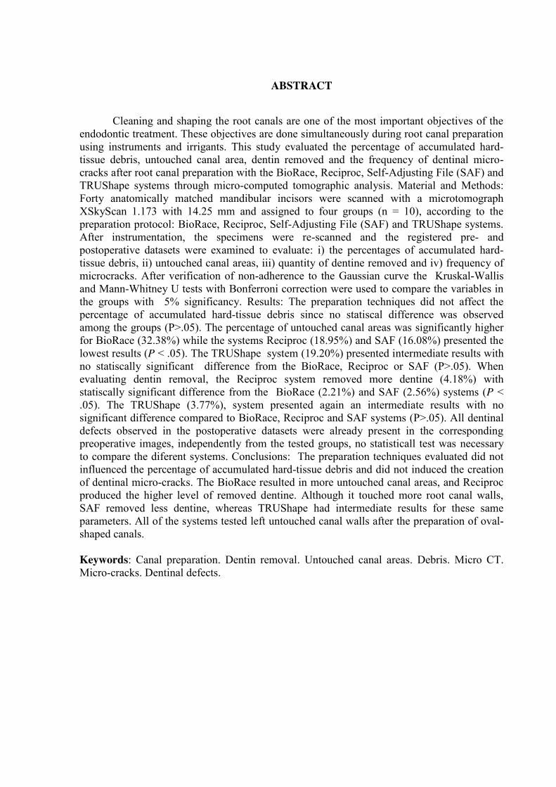
Cleaning and shaping the root canals are one of the most important objectives of the endodontic treatment. These objectives are done simultaneously during root canal preparation using instruments and irrigants. This study evaluated the percentage of accumulated hard-tissue debris, untouched canal area, dentin removed and the frequency of dentinal micro-cracks after root canal preparation with the BioRace, Reciproc, Self-Adjusting File (SAF) and TRUShape systems through micro-computed tomographic analysis. Material and Methods: Forty anatomically matched mandibular incisors were scanned with a microtomograph XSkyScan 1.173 with 14.25 mm and assigned to four groups (n = 10), according to the preparation protocol: BioRace, Reciproc, Self-Adjusting File (SAF) and TRUShape systems. After instrumentation, the specimens were re-scanned and the registered pre- and postoperative datasets were examined to evaluate: i) the percentages of accumulated hard-tissue debris, ii) untouched canal areas, iii) quantity of dentine removed and iv) frequency of microcracks. After verification of non-adherence to the Gaussian curve the Kruskal-Wallis and Mann-Whitney U tests with Bonferroni correction were used to compare the variables in the groups with 5% significancy. Results: The preparation techniques did not affect the percentage of accumulated hard-tissue debris since no statiscal difference was observed among the groups (P>.05). The percentage of untouched canal areas was significantly higher for BioRace (32.38%) while the systems Reciproc (18.95%) and SAF (16.08%) presented the lowest results (P < .05). The TRUShape system (19.20%) presented intermediate results with no statiscally significant difference from the BioRace, Reciproc or SAF (P>.05). When evaluating dentin removal, the Reciproc system removed more dentine (4.18%) with statiscally significant difference from the BioRace (2.21%) and SAF (2.56%) systems (P < .05). The TRUShape (3.77%), system presented again an intermediate results with no significant difference compared to BioRace, Reciproc and SAF systems (P>.05). All dentinal defects observed in the postoperative datasets were already present in the corresponding preoperative images, independently from the tested groups, no statisticall test was necessary to compare the diferent systems. Conclusions: The preparation techniques evaluated did not influenced the percentage of accumulated hard-tissue debris and did not induced the creation of dentinal micro-cracks. The BioRace resulted in more untouched canal areas, and Reciproc produced the higher level of removed dentine. Although it touched more root canal walls, SAF removed less dentine, whereas TRUShape had intermediate results for these same parameters. All of the systems tested left untouched canal walls after the preparation of oval-shaped canals.
Keywords: Canal preparation. Dentin removal. Untouched canal areas. Debris. Micro CT. Micro-cracks. Dentinal defects.

SUMÁRIO 1 INTRODUÇÃO .................................................................................................................... 11
2 ARTIGOS ............................................................................................................................. 15
2.1 Artigo: Challenging the shaping ability of four root canal systems in oval-shaped
canals ........................................................................................................................ 15
2.2 Artigo: Micro-CT assessment of dentinal micro-cracks after root canal preparation
with TRUShape and Self-Adjusting File systems.................................................... 28
3 DISCUSSÃO ........................................................................................................................ 38
3.1 Justificativa da pesquisa ............................................................................................. 38
3.2 Debris acumulados ..................................................................................................... 39
3.3 Áreas de canal não tocadas / Quantidade de dentina excisada .................................. 40
3.4 Frequência de microfraturas dentinárias .................................................................... 41
4 CONCLUSÃO ...................................................................................................................... 43
REFERÊNCIAS ..................................................................................................................... 44
ANEXOS ................................................................................................................................. 51 ANEXO 1: COMITÊ DE ÉTICA.......................................................................................... 51
ANEXO 2: SUBMISSÃO PARA INTERNATIONAL ENDODONTIC JOURNAL ....... 52
ANEXO 3 : APROVAÇÃO PARA PUBLICAÇÃO NO JOURNAL OF ENDODONTICS .................................................................................................................... 53
ANEXO 4: MATERIAIS E METODOS .............................................................................. 55

11
1 INTRODUÇÃO
A base da terapia endodôntica consiste em tratar dentes comprometidos por
patologias pulpares e periapicais, para que o paciente possa recuperar sua estética e
função naturais (Peters et al., 2004). Embora o sucesso do tratamento endodôntico
dependa de múltiplas variáveis, é reconhecido que um dos fatores mais importantes é o
preparo químico mecânico (PQM) do canal radicular, pois este está intimamente ligado
com a limpeza, desinfecção e posterior obturação do sistema de canais radiculares (SCR),
(Schilder, 1974).
O PQM do canal é influenciado negativamente pela anatomia muito variável dos
canais radiculares (Nagy et al., 2003). Detalhes anatômicos, como curvaturas,
achatamentos e irregularidades das paredes dos canais, posição espacial do forame,
número de canais, dureza variável da dentina, entre outros, representam um desafio para
os operadores, visto que os instrumentos endodônticos são manufaturados a partir de
fragmentos de metal retos. O resultado é uma distribuição desigual das forças em áreas de
contato e a tendência de o instrumento retificar o canal pode resultar em desvios e
transporte do canal original, bem como as áreas não tocadas nas paredes radiculares
(Roane et al., 1985; Wildey et al., 1992).
Vários instrumentos e procedimentos técnicos têm sido introduzidos com objetivo
de contornar os erros de procedimento durante o PQM (Peters e Peters, 2006). Dentre
eles, pode ser destacado a introdução de instrumentos fabricados com ligas de Níquel-
Titânio (NiTi) e acionados por meio de motores elétricos. O desenvolvimento de sistemas
de NiTi para o preparo de canais radiculares se baseia em mudanças no design do
instrumento, no tratamento da liga NiTi e na cinemática de uso, resultando em avanços
no campo da instrumentação de canais (Hülsmann et al., 2005)
Com a evolução das ligas de NiTi foram lançados no mercado vários sistemas que
diferem em conicidade, tipo e tamanho da ponta, secção transversal, ângulo de corte e
área de escape. As propriedades mecânicas e o comportamento dos instrumentos variam
de acordo com a composição química da liga metálica e características de produção, bem
como do seu modo de uso durante a instrumentação dos canais (Peters e Peters, 2006).
Dentre os vários instrumentos rotatórios disponíveis para o preparo de canais, o
sistema BioRace (FKG Dentaire, La Chaux-de-Fonds, Switzerland) pode ser citado. A
sequência BioRace foi especialmente desenvolvida para promover preparo de canais com
uso de poucos instrumentos de uso sequencial. O sistema apresenta as seguintes

12
características: a) ponta de segurança não ativa, b) secção transversal triangular sem guia
radial, c) ângulos helicoidais variáveis para evitar rosqueamento durante o uso e d)
acabamento eletroquímico de superfície. O conjunto básico é composto por 5
instrumentos BRO - 25/08, BR1 - 15/05, BR2 - 25/04, BR3 - 25/06, BR4 - 35/04 e BR5 -
40/04. Os instrumentos BioRace utilizam movimento rotatório contínuo em micromotor
elétrico com a velocidade recomendada entre 500-600 rpm com 1Ncm de torque. Existem
ainda limas complementares BR4C - 35/02, BR5C - 40/02, BR6 - 50/04 e BR7 - 60/02
(BioRace / FKG website).
O sistema Self-Adjusting File (SAF) (ReDent-Nova, Ra’anana, Israel) é um
exemplo de instrumento modificado que foi desenhado com objetivo de se adaptar às
características anatômicas de cada canal a ser preparado. SAF é um instrumento oco e
flexível composto por fios entrelaçados compressíveis que promovem o corte de dentina
por abrasão com movimento de vai e vem (Metzger et al., 2010a). Esses instrumentos
modificados têm a habilidade de se adaptar às paredes dos canais de forma tridimensional
e são usados em conjunção com uma bomba peristáltica que fornece irrigação contínua
durante todo o preparo, por isso a solução irrigante é constantemente agitada pela
vibração do próprio instrumento (Metzger et al., 2010b). O sistema é composto por
apenas 2 instrumentos: SAF 1.5 mm, indicado para canais com diâmetro apical inicial,
correspondente a limas ISO 20-35 e SAF 2.0 mm para canais mais amplos com diâmetro
inicial apical ISO 35-60. Os instrumentos são fabricados em Niti e foram desenhados
para serem utilizados com motor elétrico EndoStation™, contra ângulo de redução RDT3
e bomba peristáltica VATEA (website ReDentNova).
O conceito de preparo de canais com instrumento único tem chamado a atenção de
inúmeros clínicos e pesquisadores. Dentre os sistemas disponíveis, o Reciproc (VDW,
Munich, German) utiliza o princípio do movimento rotatório alternado, isto é, o
instrumento é movimentado em uma direção de corte, e depois é solto girando na direção
oposta. Os valores de rotação no sentido anti-horário e horário são diferentes e permitem
o avanço dos instrumentos com pouca pressão apical. O movimento, assim denominado,
recíproco reduz o estresse torsional sobre o instrumento, pois reverte periodicamente a
rotação deste, diminuindo dessa forma o risco de fratura (Yared, 2011; Kim et al.,
2012). O instrumento Reciproc (VDW, Munich, German) tem secção transversal em
forma de S, ponta inativa e é confeccionado com a liga M-Wire. São oferecidos em 3
tamanhos: R 25 (ponta 0,25 mm e conicidade 0,8 mm – 25/0.8), R40 (40/0.6) e R50
(50/0.5). Cumpre ressaltar que essa é a conicidade nos primeiros 3 mm dos instrumentos.

13
Em D 16, os instrumentos apresentam conicidade: 1.05, 1,10 e 1,17 mm,
respectivamente. Os instrumentos são oferecidos em blisters estéreis e devem ser
descartados após uso único, evitando fratura e contaminação cruzada (VDW website).
Yared (2011) preconiza que o preparo de canais com Reciproc pode ser realizado sem a
necessidade de glide path e também não necessita de pré-alargamento cervical, dois
conceitos amplamente difundidos e aceitos para instrumentação de canais com
instrumentos rotatórios. O instrumento Reciproc, devido ao seu design e ao tipo de
movimento, pode seguir a via de menor resistência, que é o canal radicular, sem alteração
em sua anatomia original e ser utilizado também em movimento de pincelamento,
promovendo o pré-alargamento do canal.
Recentemente um novo sistema de instrumentos foi introduzido no mercado, o
TRUShape 3D Conforming Files system (Dentsply Tulsa Dental Specialties, Tulsa, OK).
Esse novo sistema de limas de NiTi rotatórias tratadas termicamente consiste de um
alargador de orifício, Orifice Modifier (20/.08) e 4 limas de conformação Conforming
Files (20/.06v, 25/.06v, 30/.06v and 40/.06v) com secção transversal triangular e ponta
inativa. A conicidade na porção apical do instrumento é de 0.06 mm, porém devido à sua
cinemática especial em forma de varredura, a conicidade geral do instrumento é variável
e, por conseguinte, é designada como .06v. De acordo com o fabricante, o design do
instrumento e sua cinemática permitem que esses instrumentos contatem mais de 75%
das paredes dos canais, porém com menos desvios e transporte dos canais e pouca
remoção de estrutura dental (Tulsa website). Poucos estudos (Bortoluzzi et al., 2015;
Peters et al., 2015) ainda foram publicados sobre esse novo sistema. Bortoluzzi et al.
(2015), mostraram que o preparo de canais com TRUShape removeu significantemente
mais bactérias em canais ovalados, quando comparado às limas Twisted-File
(SybronEndo, Orange, CA), porém bactérias estavam presentes nos túbulos dentinários
após instrumentação com os dois sistemas.
No entanto, o PQM de canais com formato ovalado ainda representa um desafio
para os clínicos, pois nenhuma técnica de preparo tem promovido uma adequada limpeza
e desinfecção dos canais, deixando áreas não tocadas nas paredes vestibular e lingual
(Wu et al., 2003; Versiani et al., 2011; Versiani et al., 2013; Busquim et al., 2015 BR; De
Deus et al., 2010; De Deus et al., 2011; Taha et al., 2010). Esses espaços intocados
podem abrigar bactérias, biofilmes e detritos e servir como uma causa potencial de
insucesso do tratamento endodôntico (Siqueira et al., 2010; Alves et al., 2011; Paque et
al., 2011; De Deus et al., 2015).

14
Muitas metodologias foram propostas para avaliar as mudanças morfológicas na
anatomia do canal após preparo endodôntico, como injeção de silicone (Davis et al.,
1968), canais simulados em blocos de resina (Weine et al., 1975), cortes transversais em
dentes extraídos (Bramante et al., 1987), análises histológicas (Walton et al., 1976),
radiográficas (Southard et al., 1987) e métodos computadorizados (Berutti, 1993).
Apesar de serem extensamente utilizadas ao longo dos anos, essas metodologias
apresentam limitações, sendo a principal delas a falta de um grupo controle adequado. A
procura por metodologias menos invasivas e mais confiáveis levou à utilização da
microtomografia computadorizada (µCT). Essa metodologia possibilita ao pesquisador
uma avaliação completa e detalhada da anatomia dental, além disso, possibilita a
comparação do mesmo canal radicular antes e depois da instrumentação, sendo cada
canal seu próprio controle (Nielsen et al, 1995). O uso dessa tecnologia para avaliação de
diferentes parâmetros geométricos e a direção de transporte antes e depois do preparo de
canais têm sido cada vez mais utilizados e diferentes pesquisadores atestam sua precisão
(Rhodes et al., 2000; Peters et al., 2000, 2001; Bergmans et al., 2001; You et al., 2011;
Paqué et al., 2011).
Diante do exposto acima, o objetivo desta pesquisa foi avaliar com auxílio de
microtomografia computadorizada (µCT) a qualidade do preparo mecânico de canais
ovalados, comparando distintos parâmetros : i) porcentagem de acúmulo de debris, ii)
áreas de canal não tocadas, iii) dentina excisada e iiii) frequência de microfraturas
dentinárias, após o preparo de canais com os sistemas BioRace, Reciproc, Self-Adjusting
File (SAF) and TRUShape.
15
2 ARTIGOS
2.1 Challenging the shaping ability of four root canal systems in oval-shaped canals * Artigo submetido ao periódico International Endodontic Journal (Anexo 2)
Abstract Aim To challenge the shaping ability of TRUShape and three other instrumentation
systems in oval-shaped canals using micro-computed tomographic analysis.
Methodology Forty anatomically matched mandibular incisors were scanned and
assigned to four groups (n = 10), according to the preparation protocol: BioRace,
Reciproc, Self-Adjusting File (SAF) and TRUShape systems. After instrumentation, the
specimens were re-scanned and the registered pre- and postoperative datasets were
examined to evaluate the percentages of accumulated hard-tissue debris, untouched canal
areas and dentine removed. Kruskal-Wallis and Mann-Whitney U tests with Bonferroni
correction were used to compare the variables in the groups (α = 5%).
Results The preparation techniques did not affect the percentage of accumulated hard-
tissue debris (P = .126). The percentage of untouched canal areas was significantly higher
for BioRace (32.38%), and Reciproc (18.95%) and SAF (16.08%) presented the lowest
results (P < .05). The Reciproc system removed significantly more dentine (4.18%)
compared to the BioRace (2.21%) and SAF (2.56%) systems (P < .05). The TRUShape
system presented intermediate results for both untouched canal areas (19.20%) and
removed dentine (3.77%), with no significant difference compared to BioRace, Reciproc
and SAF systems.
Conclusions The preparation techniques resulted in the same level of accumulated hard-
tissue debris. Compared to the other tested systems, BioRace resulted in more untouched
canal areas, and Reciproc produced the higher level of removed dentine. Although it
touched more root canal walls, SAF removed less dentine, whereas TRUShape had
intermediate results for these same parameters. None of the systems tested were able to
provide optimal shaping ability in oval-shaped canals.
Keywords: Dentin removal. Hard-tissue debris. Micro-CT. Shaping ability. Untouched
canal areas.
16
Introduction
The development of a new generation of nickel-titanium (NiTi) systems for canal
preparation relies on changes in instrument design, alloy and kinematics, aiming to
optimise the mechanical instrumentation of the root canals (Peters 2004, Hülsmann et al.
2005). Most of the available rotary and reciprocating systems have failed to improve
debridement of oval-shaped canals (Versiani et al. 2013), leaving large areas of
untouched canal wall (Peters et al. 2001, Versiani et al. 2013, De-Deus et al. 2015a) and
accumulated hard-tissue debris in fins, isthmuses and irregularities within the root canal
space (Paqué et al. 2009, De-Deus et al. 2015b, Versiani et al. 2016). Bacteria located in
these areas have the potential to remain unaffected and might be responsible for persistent
periapical inflammation (Versiani et al. 2016).
Recently, a novel heat-treated NiTi rotary system, the TRUShape 3D Conforming
Files system (Dentsply Tulsa Dental Specialties, Tulsa, OK, USA) was introduced to the
market. It is advertised by the manufacturer as a set of instruments that enables greater
dentinal preservation than conventional files, while making contact with nearly 75% of
the canal walls (Dentsply Tulsa Dental Specialties website), due to the triangular cross
section, a noncutting tip, and a sweeping S-shape along its long axis, resulting in a 0.06
taper in the apical 2 mm and an increasing variable taper from this level denoted as a .06v
taper. A recent study showed that root canal preparation with TRUShape instruments
removed significantly more bacteria from oval-shaped root canals than the Twisted File
rotary system (SybronEndo, Orange, CA, USA) (Bortoluzzi et al. 2015), however, to the
best of our knowledge, no study has attempted to challenge the shaping ability of this
novel system regarding the accumulation of hard-tissue debris, untouched root canal
walls and the amount of dentine removed from oval-shaped root canals compared to other
preparation systems.
The aim of this study was therefore to compare the percentages of accumulated
hard-tissue debris, untouched canal areas and dentine removed after root canal
preparation with the BioRace (FKG Dentaire, La-Chaux-de-Fonds, Switzerland),
Reciproc (VDW, Munich, Germany), Self-Adjusting File (SAF; ReDent-Nova, Ra’anana,
Israel) and TRUShape systems through micro-computed tomographic (micro-CT)
analysis. The null hypothesis tested was that there would be no significant difference in
shaping outcomes among these four preparation systems.

17
Materials and methods
Sample size estimation
Based on the results of a previous study (De-Deus et al. 2015b), an effect size of
0.9 was estimated and input, together with the parameter alpha-type error of 0.05 and
power beta of 0.95, into a one-way ANOVA procedure (G*Power 3.1 for Macintosh;
Heinrich Heine, Universität Düsseldorf, Dusseldorf, Germany). A sample size of 28 teeth
(7 per group) was indicated as the minimum to reveal statistical significance among
groups.
Sample selection and preparation
After local research ethics committee approval, 127 human mandibular incisors
were obtained from a pool of teeth. Each tooth was radiographed in both buccolingual
and mesiodistal directions. The inclusion criteria was only teeth presenting straight roots
(< 5º) (Schneider 1971), a canal ratio of long to short diameter more than 2.5 at the 5 mm
level from the root apex and an initial apical size equivalent to a size 10 K-file (Dentsply
Maillefer, Ballaigues, Switzerland). As a result, 63 teeth were selected and scanned in a
micro-CT device (SkyScan 1173; Bruker-microCT, Kontich, Belgium) operated at 70 kV
and 114 mA, using a low resolution (70 µm) to obtain an outline of the root canals. The
acquired projection images were reconstructed (NRecon v.1.6.10; Bruker-microCT)
providing axial cross sections of their inner structure, and 40 mandibular incisors with
similar canal configuration were selected and scanned again at an increased resolution
(14.25 µm) with 360° rotation around the vertical axis, a rotation step of 0.5°, a camera
exposure time of 7000 milliseconds and frame averaging 5, using a 1.0 mm-thick
aluminium filter. Images of each specimen were reconstructed using standardised
parameters for beam hardening (40%), ring artefact correction (10) and similar contrast
limits. The volume of interest was selected to extend from the cemento-enamel junction
to the apex of the root, resulting in the acquisition of 800-900 transverse cross-sections
per tooth. The apexes of the teeth were then sealed with hot glue and embedded in
polyvinyl siloxane to create a closed-end system (Susin et al. 2010).
After access cavity preparation, a glide path was created by scouting a stainless
steel size 20 K-file (Dentsply Maillefer) up to the working length (WL), which was
established by deducting 1 mm from the canal length. Canals were then matched to create
ten groups of four based on morphological elements of the canal (volume, surface area

18
and configuration), and 1 root from each group was randomly assigned to one of the four
experimental groups (n = 10) according to the preparation protocol:
BioRace system. BR0 (25/.08), BR1 (15/.05), BR2 (25/.04) and BR3 (25/.06) NiTi rotary
instruments were used at 500-600 rpm and 1 N.cm in a crown-down manner (VDW
Silver motor; VDW) up to the WL, using a gentle in-and-out pecking motion. After three
steady strokes, the file was removed from the canal and cleaned.
Reciproc system. An R25 instrument (25/.08) was moved in the apical direction using a
slow in-and-out pecking motion of about 3 mm in amplitude with a light apical pressure
in a reciprocating motion (‘RECIPROC ALL’) powered by an electric motor (VDW
Silver) until the WL was reached. After three pecking motions, the instrument was
removed from the canal and cleaned. The WL was reached in the third wave of
instrumentation for all teeth.
SAF system. A 1.5 mm diameter SAF instrument was operated to the WL with an in-
and-out motion using an RDT3 head (ReDent-Nova) adapted to a vibrating handpiece
(GentlePower Lux 20LP; KaVo, Biberach, Germany). Continuous irrigation with 5.25%
NaOCl was applied throughout the procedure at a flow rate of 5 mL/min using a special
irrigation apparatus (VATEA; ReDent-Nova).
TRUShape system. Using an electric motor (VDW Silver) pre-set at 300 rpm and 3
N.cm, TRUShape instrument (20/.08) was used with a gentle 2-5 mm in-and-out motion
to shape the middle third. TRUShape 20/.06v and 25/.06v instruments were then used
with a further 2 to 3 mm amplitude towards the WL. Each tooth was shaped as two canals
due to its larger buccal-lingual dimension, as recommended by the manufacturer.
In all groups, the total preparation time was 4 min and included only active
instrumentation. Irrigation was performed with a NaviTip needle (Ultradent Products
Inc., South Jordan, UT, USA) using 25 mL of 5.25% NaOCl per tooth. At the end of the
preparation, passive ultrasonic irrigation was performed for 20 s at 2 mm short of the WL
using a size 15 K-file (Dentsply Maillefer). Canals were flushed with 3 mL of 17%
EDTA for 5 min and 2 mL of bi-distilled water for 1 min. Aspiration of the irrigant
solution was performed at the canal orifice with a SurgiTip (Ultradent Products Inc.)

19
attached to a high-speed suction pump. All preparation procedures were conducted by an
experienced operator, after substantial training with all systems. Root canals were dried
with absorbent paper points (Dentsply Maillefer) and the specimens submitted to a
postoperative scan and reconstruction, applying the aforementioned parameters.
Micro-CT evaluation
The image stacks of the specimens after preparation were rendered and co-
registered with their respective preoperative datasets using an affine algorithm of the 3D
Slicer 4.5.0 software (available from http://www.slicer.org) (Fedorov et al. 2012). The
quantification of accumulated hard-tissue debris was expressed as the percentage of the
total canal system volume after preparation for each specimen, and undertaken as
described elsewhere (De-Deus et al. 2014, De-Deus et al. 2015b, Neves et al. 2015). The
volume of the dentine removed after preparation was calculated by subtracting pre- and
postoperative segmented root dentine using morphological operations (Fiji v.1.47n;
Madison, WI, USA). The area of untouched canal surface was determined by calculating
the number of static voxels (voxels present in the same position on the canal surface
before and after instrumentation). The untouched area was expressed as a percentage of
the total number of voxels present on the canal surface (Paqué & Peters 2011), according
to the formula:
𝑛𝑢𝑚𝑏𝑒𝑟 𝑜𝑓 𝑠𝑡𝑎𝑡𝑖𝑐 𝑣𝑜𝑥𝑒𝑙𝑠 × 100𝑡𝑜𝑡𝑎𝑙 𝑛𝑢𝑚𝑏𝑒𝑟 𝑜𝑓 𝑠𝑢𝑟𝑓𝑎𝑐𝑒 𝑣𝑜𝑥𝑒𝑙𝑠
Statistical analysis
The degree of homogeneity (baseline) of the groups regarding root canal volume
(mm3) and surface area (mm2) was statistically compared using a one-way ANOVA test.
Because normality assumptions of the percentages of accumulated hard-tissue debris,
untouched canal area and dentine removed after root canal preparation could not be
verified (Shapiro-Wilk test; P < .05), the results were expressed as medians and
compared between groups by Kruskal-Wallis and Mann-Whitney U tests with Bonferroni
correction (SPSS v.17; SPSS Inc., Chicago, IL, USA). Significance was set at α = 5%.

20
Results Figure 1 shows representative images of the internal anatomy of four mandibular
incisors before and after canal preparation with the tested systems.
The degree of homogeneity (baseline) of the groups regarding canal volume and
surface area was confirmed (P > .05). There was no statistically significant difference in
the mean and interquartile ranges (IQR) regarding the percentage of accumulated hard-
tissue debris among TRUShape (0.00%, IQR 0.23), BioRace (0.00%, IQR 0.59),
Reciproc (0.01%, IQR 0.49), or SAF (0.00%, IQR 0.17) groups (P > .05). A significantly
higher percentage of untouched canal area was observed after preparation with the
BioRace system (32.38%) compared to the Reciproc (18.95%) and SAF (16.08%)
systems (P < .05). Reciproc removed significantly more dentine (4.18%) than BioRace
(2.21%) and SAF (2.56%) (P < .05). The TRUShape system showed intermediate results
regarding the untouched canal area (19.20%) and the amount of dentine removal (3.77%)
with no significant difference compared to the other tested systems (P > .05) (Fig. 1).
Figure 2 shows a boxplot representation of the median percentages and IQR of the
parameters tested (untouched canal areas [a] and removed dentine [b]) after root canal
preparation with the BioRace, Reciproc, SAF and TRUShape systems.
Discussion The current investigation was designed to evaluate the percentages of
accumulated hard-tissue debris, untouched canal areas and dentine removed after the
preparation of oval-shaped canals of mandibular incisors with BioRace, Reciproc, SAF
and TRUShape systems, using micro-CT analysis. Despite natural variations in the
morphology of teeth, attempts were made to ensure comparability of the groups regarding
root canal morphology. As oval-shaped canals present a challenge to the clinician (Peters
2004, De-Deus et al. 2010, Versiani et al. 2011), this type of canal configuration was
selected. The specimens were balanced with respect to the canal anatomy, volume and
surface area through micro-CT screening, enhancing the internal validity of the study and
potentially eliminating significant anatomic biases that may confound the outcomes
(Peters et al. 2001, Versiani et al. 2016).
Recently, 3-dimensional non-destructive imaging micro-CT technology has also
been used for the quantitative evaluation of hard-tissue debris packed into recesses during
canal preparation (Paqué et al. 2009, Paqué et al. 2011, Paqué et al. 2012, Robinson et al.
2013, De-Deus et al. 2014, De-Deus et al. 2015b, Neves et al. 2015, Versiani et al.

21
2016). Evidence from these studies indicates that dentine particles cut from the canal
walls by endodontic instruments can be actively packed into the anatomical complexities
of the canal system, becoming more resistant to removal. In the present study,
accumulation of hard-tissue debris occurred regardless of system design and kinematics,
in accordance with De-Deus et al. (2015b). On the other hand, the present findings
disagree with other micro-CT studies, in which preparation with the SAF system resulted
in less debris accumulation (Paqué et al. 2012), and with a reciprocating system that left
significantly more debris within the root canals than a multi-file rotary system (Robinson
et al. 2013). These contradictory results may be explained by differences in the
methodological design. The oval-shaped canals of mandibular incisors were used here,
and in those studies (Paqué et al. 2012, Robinson et al. 2013) a more complex
preoperative root canal configuration (mesial canals of mandibular molars) was used.
Passive ultrasonic irrigation was also used as a supplementary irrigation protocol in the
present study. According to recent research, the activation of the irrigant solution with an
oscillating ultrasonic tip after root canal preparation is more likely to remove hard-tissue
debris from root canals with a single anatomy (Versiani et al. 2016), which may also help
to explain the present results.
It is well established that untouched canal areas may be colonised by biofilms and
serve as a potential cause of persistent infection, which may compromise the treatment
outcome (Alves et al. 2011, Dietrich et al. 2012). In the current study, the percentage of
untouched canal areas and dentine removed were significantly affected by the preparation
protocols. Consequently, the null hypothesis tested was rejected. The median percentage
of untouched canal walls ranged from 16.08% to 32.38% and none of the tested systems
were able to completely debride the dentinal walls, which is in agreement with previous
reports (Peters et al. 2001, Paqué & Peters 2011, Versiani et al. 2013, Bortoluzzi et al.
2015, De-Deus et al. 2015a). Amongst the tested systems, the SAF and Reciproc systems
showed the lowest percentage of untouched canal area. As previously demonstrated
(Metzger et al. 2010a, Paqué & Peters 2011, Versiani et al. 2011, Versiani et al. 2013),
results from SAF system can be explained as due to its hollow NiTi lattice-like form,
which adapts itself to the shape of the root canal, enabling a higher percentage of root
canal walls to be touched. The back-and-forth grinding motion of the SAF instrument
enables a circumferential removal of only a thin layer of dentine from most of the canal
walls (Metzger et al. 2010b), which explains the lowest percentage of dentine removed
by the SAF system in this study. On the other hand, the low percentage of untouched

22
canal walls and high amount of removed dentine observed after canal preparation with
the Reciproc system may be explained by the combination of its reciprocating kinematics,
larger taper size (0.08 in the first 3 mm) and design (sharp cutting edges and smaller
cross-sectional area), which effect its flexibility and increase its cutting efficiency in a
brushing motion (Plotino et al. 2014). Similarly, the lower dimensions and cutting
efficiency of BioRace instruments compared to Reciproc explain its higher percentage of
untouched canal walls and lower amount of dentine removal (Lopes et al. 2010).
Root canal preparation with the TRUShape system showed intermediate median
results regarding untouched canal areas and dentine removed. Peters et al. (2015)
reported that TRUShape enables dentine preservation during root canal shaping, and
Elnaghy et al. (2016) reported a mean percentage of dentine removed of about 2.77%,
which contrasts with the 3.77% observed herein. The asymmetric cutting motion of the
TRUShape files, which can reach a fluted diameter up to 0.80 mm, might be the basis on
which to explain the lack of significance compared to the other groups studied here (Fig.
2).
Like other studies using the non-destructive micro-CT approach (Peters et al.
2001, Paqué et al. 2012, De-Deus et al. 2015a, De-Deus et al. 2015b, Versiani et al.
2016), the current results highlight the less than ideal ability of currently available
preparation systems to prepare this type of root canal configuration. These findings
emphasise the importance of irrigation and intracanal dressing procedures in an attempt
to compensate for the suboptimal status of the mechanical debridement (Versiani et al.
2011, 2013).
Conclusions Under the conditions of the present study, none of the tested systems were able to
provide optimal shaping of oval-shaped canals. Root canal preparation with the BioRace,
Reciproc, SAF and TRUShape systems resulted in similar amounts of accumulated hard-
tissue debris. More untouched canal areas and dentine removed were observed after
preparation with BioRace and Reciproc systems, respectively. SAF touched more root
canal walls and removed the least dentine, and TRUShape had intermediate results for
these same parameters.

23
REFERENCES 1. Alves FR, Almeida BM, Neves MA, Moreno JO, Rôças IN, Siqueira JF Jr. (2011)
Disinfecting oval-shaped root canals: effectiveness of different supplementary
approaches. Journal of Endodontics 37, 496-501.
2. Bortoluzzi EA, Carlon D Jr, Meghil MM et al. (2015) Efficacy of 3D conforming
nickel titanium rotary instruments in eliminating canal wall bacteria from oval-shaped
root canals. Journal of Dentistry 43, 597-604.
3. De-Deus G, Barino B, Zamolyi RQ et al. (2010) Suboptimal debridement quality
produced by the single-file F2 ProTaper technique in oval-shaped canals. Journal of
Endodontics 36, 1897-1900.
4. De-Deus G, Belladonna FG, Silva EJ et al. (2015a) Micro-CT evaluation of non-
instrumented canal areas with different enlargements performed by NiTi systems.
Brazilian Dental Journal 26, 624-29.
5. De-Deus G, Marins J, Neves Ade A et al. (2014) Assessing accumulated hard-tissue
debris using micro-computed tomography and free software for image processing and
analysis. Journal of Endodontics 40, 271-76.
6. De-Deus G, Marins J, Silva EJ et al. (2015b) Accumulated hard tissue debris
produced during reciprocating and rotary nickel-titanium canal preparation. Journal
of Endodontics 41, 676-81.
7. Dietrich MA, Kirkpatrick TC, Yaccino JM (2012) In vitro canal and isthmus debris
removal of the self-adjusting file, K3, and WaveOne files in the mesial root of human
mandibular molars. Journal of Endodontics 38, 1140-44.
8. Elnaghy AM, Al-Dharrab AA, Abbas HM, Elsaka SE. Evaluation of root canal
transportation, centering ratio, and remaining dentin thickness of TRUShape and
ProTaper Next systems in curved root canals using micro-computed tomography.
Quintessence International [Epub ahead of print].
9. Fedorov A, Beichel R, Kalpathy-Cramer J et al. (2012) 3D Slicer as an image
computing platform for the Quantitative Imaging Network. Magnetic Resonance
Imaging 30, 1323-41.
10. Hülsmann M, Peters OA, Dummer PMH (2005) Mechanical preparation of root
canals: shaping goals, techniques and means. Endodontic Topics 10, 30-76.

24
11. Lopes HP, Elias CN, Vieira VT et al. (2010) Effects of electropolishing surface
treatment on the cyclic fatigue resistance of BioRace nickel-titanium rotary
instruments. Journal of Endodontics 36, 1653-57.
12. Metzger Z, Teperovich E, Zary R, Cohen R, Hof R (2010b) The self-adjusting file
(SAF). Part 1: respecting the root canal anatomy-a new concept of endodontic files
and its implementation. Journal of Endodontics 36, 679-90.
13. Metzger Z, Zary R, Cohen R, Teperovich E, Paqué F (2010a) The quality of root
canal preparation and root canal obturation in canals treated with rotary versus self-
adjusting files: a three-dimensional micro-computed tomographic study. Journal of
Endodontics 36, 1569-73.
14. Neves AA, Silva EJ, Roter JM et al. (2015) Exploiting the potential of free software
to evaluate root canal biomechanical preparation outcomes through micro-CT images.
International Endodontic Journal 48, 1033-42.
15. Paqué F, Al-Jadaa A, Kfir A (2012) Hard-tissue debris accumulation created by
conventional rotary versus self-adjusting file instrumentation in mesial root canal
systems of mandibular molars. International Endodontic Journal 45, 413-18.
16. Paqué F, Boessler C, Zehnder M (2011) Accumulated hard tissue debris levels in
mesial roots of mandibular molars after sequential irrigation steps. International
Endodontic Journal 44, 148-53.
17. Paqué F, Laib A, Gautschi H, Zehnder M (2009) Hard-tissue debris accumulation
analysis by high-resolution computed tomography scans. Journal of Endodontics 35,
1044-47.
18. Paqué F, Peters OA (2011) Micro-computed tomography evaluation of the
preparation of long oval root canals in mandibular molars with the self-adjusting file.
Journal of Endodontics 37, 517-21.
19. Peters OA (2004) Current challenges and concepts in the preparation of root canal
systems: a review. Journal of Endodontics 30, 559-67.
20. Peters OA, Arias A, Paqué F (2015) A micro-computed tomographic assessment of
root canal preparation with a novel instrument, TRUShape, in mesial roots of
mandibular molars. Journal of Endodontics 41, 1545-50.
21. Peters OA, Schönenberger K, Laib A (2001) Effects of four Ni-Ti preparation
techniques on root canal geometry assessed by micro computed tomography.
International Endodontic Journal 34, 221-30.

25
22. Plotino G, Giansiracusa Rubini A, Grande NM, Testarelli L, Gambarini G (2014)
Cutting efficiency of Reciproc and WaveOne reciprocating instruments. Journal of
Endodontics 40, 1228-30.
23. Robinson JP, Lumley PJ, Cooper PR, Grover LM, Walmsley AD (2013)
Reciprocating root canal technique induces greater debris accumulation than a
continuous rotary technique as assessed by 3-dimensional micro-computed
tomography. Journal of Endodontics 39, 1067-70.
24. Schneider SW (1971) A comparison of canal preparations in straight and curved root
canals. Oral Surgery, Oral Medicine, Oral Pathology and Endodontics 32, 271-5.
25. Siqueira Jr. JF, Alves FRF, Versiani MA et al. (2013) Correlative bacteriologic and
micro-computed tomographic analysis of mandibular molar mesial canals prepared by
Self-Adjusting File, Reciproc and Twisted File systems. Journal of Endodontics 39,
1044-50.
26. Susin L, Liu Y, Yoon JC et al. (2010) Canal and isthmus debridement efficacies of
two irrigant agitation techniques in a closed system. International Endodontic Journal
43, 1077-90.
27. TRUShape® 3D Conforming Files. Dentsply Tulsa Dental Specialties website.
Available at: http://www.tulsadentalspecialties.com/default/endodontics/TRUShape.a
spx. Accessed September 9, 2016.
28. Versiani MA, Alves FR, Andrade-Junior CV et al. (2016) Micro-CT evaluation of the
efficacy of hard-tissue removal from the root canal and isthmus area by positive and
negative pressure irrigation systems. International Endodontic Journal 49, 1079-87.
29. Versiani MA, Leoni GB, Steier L et al. (2013) Micro-computed tomography study of
oval-shaped canals prepared with the self-adjusting file, Reciproc, WaveOne, and
ProTaper universal systems. Journal of Endodontics 39, 1060-66.
30. Versiani MA, Pécora JD, de Sousa-Neto MD (2011) Flat-oval root canal preparation
with self-adjusting file instrument: a micro-computed tomography study. Journal of
Endodontics 37, 1002-07.

26
Figure 1: Representative 2D and 3D reconstructions of the anatomy of mandibular incisors from each
experimental group, before and after root canal preparation. (a and b) Buccal and (c and d) lateral views of superimposed specimens before (green) and after (red) root canal preparation in each experimental group; (e) representative cross-sections of the superimposed root canals before (green) and after (red) preparation at the coronal (c), middle (m) and apical (a) thirds.

27
Figure 2: Representative boxplot with the medians (interquartile range) for the percentages of (a) untouched canal areas and (b) dentine removed by each root canal system.
The different letters in each graph indicate statistical significance between groups (Mann-Whitney test with a Bonferroni correction; P < .05).

28
2.2 Micro-CT assessment of dentinal micro-cracks after root canal preparation with TRUShape and Self-Adjusting File systems
*Artigo aceito para publicação no periódico Journal of Endodontics (anexo 3)
Abstract Introduction: The aim of the present study was to evaluate the percentage frequency of
dentinal micro-cracks observed after root canal preparation with TRUShape and Self-
Adjusting File (SAF) systems by means of micro-CT imaging analysis. A conventional full-
sequence rotary system (BioRace) and a single-file reciprocation system (Reciproc) were used
as reference techniques for comparison due to their known assertive cutting efficiency.
Methods: Forty anatomically-matched mandibular incisors were selected, scanned at a
resolution of 14.25 µm, and assigned to 4 experimental groups (n = 10), according to the
preparation protocol: TRUShape, SAF, BioRace and Reciproc systems. After the
experimental procedures, the specimens were scanned again and the registered pre- and
postoperative cross-section images of the roots (n = 70,030) were screened to identify the
presence of dentinal micro-cracks. Results: Overall, dentinal defects were observed in 28,790
cross-section images (41.11%). In the TRUShape, Self-Adjusting File, BioRace and Reciproc
groups, dentinal micro-cracks were visualized in 56.47% (n = 9,842), 42.38% (n = 7,450),
32.90% (n = 5,826) and 32.77% (n = 5,672) of the slices, respectively. All dentinal defects
observed in the postoperative datasets were already present in the corresponding preoperative
images. Conclusion: None of the preparation systems induced the formation of new dentinal
micro-cracks.
Keywords: dentinal defects, instrumentation, micro-cracks, micro-CT, root canal preparation.

29
Introduction Vertical root fracture (VRF) is a clinical complication that may lead to tooth extraction
(1) and have been described in either treated or non-endodontically treated teeth (2, 3). Over
the last few years, several studies have reported a causal relationship between mechanical
preparation of the root canal with nickel-titanium (NiTi) instruments and the formation of
dentinal micro-cracks (4-8), which may potentially develop into VRF (9).
It has been speculated that the design and the hard-hitting cutting ability of the
preparation systems are the main reasons associated with the development of dentinal defects,
as they might generate damaging forces towards dentine (10). Recently, a novel heat-treated
NiTi system named TRUShape 3D Conforming Files (Dentsply Tulsa Dental Specialties,
Tulsa, OK) has been introduced onto the market, claiming to preserve more dentinal structure
while providing an optimized canal debridement. The TRUShape system uses the same
symmetric triangular cross-section, but displays a proprietary file design which resembles an
S-shape configuration, providing an ability to flex within the canal, creating an envelope of
motion kinematics (11-13). Another system, which is able to preserve more dentine, is the
Self-Adjusting File (SAF; ReDent-Nova, Ra’anana, Israel). The SAF is a hollow file designed
as a compressible cylinder composed of a thin NiTi lattice with an abrasive surface. It has the
ability to adapt its shape to the root canal anatomy, applying a constant and delicate pressure
on the canal walls, which might help to reduce the incidence of dentinal defects (4, 5). This
system operates with a continuous flow of irrigant running through the instrument, allowing
continuous replacement (8, 14, 15).
No studies have evaluated the incidence of dentinal micro-cracks resulting from the
use of the claimed less aggressive cutting instruments (TRUShape and SAF systems) using
micro-computed tomographic (micro-CT) imaging technology to date. Therefore, the aim of
the present study was to evaluate the frequency of dentinal micro-cracks observed after root
canal preparation with TRUShape and SAF systems through micro-CT imaging analysis. A
conventional full-sequence rotary system (BioRace; FKG Dentaire, La-Chaux-de-Fonds,
Switzerland) and a single-file reciprocation system (Reciproc; VDW, Munich, Germany)
were used as reference techniques for comparison due to their known assertive cutting
efficiency. The hypothesis tested was that there would be differences in the frequency of
dentinal micro-cracks generation between the groups.

30
Material And Methods
Sample size estimation
Sample size was derived from the effect size of dentinal defects promoted by rotary
and reciprocating systems by Bürklein et al. (16), in which the percentage sum of the sample
with complete and incomplete dentinal defects varied from 18.3% to 51.6%. Chi-square test
family and variance statistical test (G*Power 3.1 for Macintosh; Heinrich Heine, Universität
Düsseldorf, Düsseldorf, Germany), with α = 0.05 and β = 0.95, output 8 specimens as the
minimum ideal size required for observing the same frequency of instruments-induced defects
over dentine.
Sample selection and scanning
After approval of the local Ethics Committee, one hundred and twenty-seven straight
mandibular incisors were obtained from a pool of teeth. The specimens were initially
inspected with the aid of a stereomicroscope under 12X magnification. The exclusion criteria
comprised teeth with pre-existing cracks or not patent to the canal length with a size 10 K-file
(Dentsply Maillefer, Baillagues, Switzerland). As a result, 102 specimens were selected and
scanned in a micro-CT device (SkyScan 1173; Bruker-microCT, Kontich, Belgium) operated
at 70 kV and 114 mA, using a low resolution (70 µm). Then, forty mandibular incisors with a
canal ratio of long to short diameter of more than 2.5 at 5-mm level from the root apex were
selected and stored in 0.1% thymol solution at 5ºC. These specimens were scanned again at an
increased resolution (14.25 µm) performed by 360° rotation around the vertical axis, rotation
step of 0.5°, camera exposure time of 7000 milliseconds, and frame averaging of 5, using a
1.0-mm-thick aluminum filter. Images of each specimen were reconstructed (NRecon
v.1.6.10, Bruker-microCT) providing axial cross-sections of their inner structure using
standardized parameters for beam hardening (40%), ring artefact correction (10) and similar
contrast limits. The volume of interest was selected to extend from the cementoenamel
junction to the apex of the root, resulting in the acquisition of 800-900 transverse cross-
sections per tooth.
Root canal preparation
The apexes were sealed with hot glue and embedded into polyvinyl siloxane to create
a closed-end system (17). After access cavity preparation, a glide path was created by
scouting a stainless steel size 20 K-file (Dentsply Maillefer) up to the working length (WL),
31
which was established by deducting 1 mm from the canal length. Then, the specimens were
randomly assigned to 4 experimental groups (n = 10), according to the following protocols:
TRUShape. Using an electric motor (VDW Silver; VDW) preset at 300 rpm and 3 Ncm,
TRUShape instruments were used with a gentle in-and-out motion in the following sequence:
20/.08v (two thirds of the WL), 20/.06v (full WL) and 25/.06v (full WL). The instruments
were advanced to the midroot in 2 to 5 mm and then in further 2 to 3 mm amplitudes toward
the WL.
SAF. A 1.5-mm diameter SAF instrument was operated to the WL with an in-and-out motion
using an RDT3 head (ReDent-Nova) adapted to a vibrating handpiece (GentlePower Lux
20LP; KaVo, Biberach, Germany). Continuous irrigation with 5.25% of NaOCl was applied
throughout the procedure at a flow rate of 5 mL/min using a special irrigation apparatus
(VATEA; ReDent-Nova).
BioRace. BR0 (25/.08), BR1 (15/.05), BR2 (25/.04) and BR3 (25/.06) NiTi rotary
instruments (FKG) were used at 500-600 rpm and 1 Ncm in a crown-down manner (VDW
Silver) up to the WL, using a gentle in-and-out pecking motion. After 3 steady strokes, the
file was removed from the canal and cleaned.
Reciproc. R25 instrument (25/.08) was moved in the apical direction using a slow in-and-out
pecking motion of about 3 mm in amplitude with a light apical pressure in a reciprocating
motion (‘RECIPROC ALL’) powered by an electric motor (VDW Silver) until the WL was
reached. After 3 pecking motions, the instrument was removed from the canal and cleaned.
The WL was reached in the third wave of instrumentation for all teeth.
All experimental procedures were performed by an experienced operator after training
with the systems. Irrigation was performed using a total of 40 mL of 5.25% NaOCl per tooth.
Apical patency was confirmed with a size 10 K-file after each file use. After preparation, a
postoperative micro-CT scan of each specimen was performed using the aforementioned
parameters.
32
Dentinal micro-cracks evaluation
The pre- and postoperative image stacks of the specimens were co-registered using the
affine algorithm of the 3D Slicer v.4.5.0 software (available from http://www.slicer.org) (18).
Then, the cross-section images of the mandibular incisors were screened from the
cementoenamel junction to the apex (n = 70,030) by 3 pre-calibrated examiners. Firstly,
postoperative images were analyzed, and the cross-section number in which a dentinal micro-
crack had been observed was recorded. Afterward, the preoperative corresponding cross-
section image was also examined to verify the existence of the defect. To validate the
screening process, image analyses were repeated twice at 2-week intervals; in the case of
divergence, the images were examined together until an agreement was reached (19, 20). In
this study, dentinal micro-cracks or dentinal defects were defined as all lines observed on the
cross-section slice that extended either from the outer root surface into the dentine or from the
root canal lumen to the dentine.
Results Overall, dentinal defects were observed in 28,790 cross-section images (41.11%). In
the TRUShape, SAF, BioRace and Reciproc groups, dentinal micro-cracks were visualized in
56.47% (n = 9,842), 42.38% (n = 7,450), 32.90% (n = 5,826) and 32.77% (n = 5,672) of the
slices, respectively. All dentinal defects observed in the postoperative datasets were already
present in the corresponding preoperative images (Figure 1), indicating that no new micro-
cracks were observed after root canal preparation with the tested systems.
Discussion This in vitro study evaluated the incidence of dentinal micro-cracks after root canal
preparation with TRUShape, SAF, BioRace and Reciproc systems. To the best of the authors’
knowledge, this is the first study assessing the potential correlation between the use of
TRUShape and SAF systems and dentinal micro-cracks using a non-destructive imaging
method. All of the systems tested herein, which includes a wide range of dentinal cutting
ability, did not create any new dentinal micro-cracks; this means that all dentinal micro-cracks
appear not to be induced by the mechanical instrumentation of the root canals, regardless of
the file design or its cutting efficiency. Therefore, the hypothesis tested was accepted.
The present results markedly contrast with several previous studies that have shown an
association between mechanical canal preparation and the creation of dentinal micro-cracks
(4, 5, 7, 21, 22). Hin et al (4) and Saha et al (22) showed that SAF caused cracks in 10% of
33
the specimens. Liu and colleagues (5) observed the presence of a new defect in 5% of
mandibular incisors prepared with Reciproc system, whereas Karataş et al (7) observed cracks
in an increased rate of 11% after instrumentation with Reciproc system. Similarly, Saber &
Schäfer (21) found the incidence of dentinal defects to be 26% in the group instrumented with
Reciproc system. This discrepancy in the results may be explained by an essential difference
in the analytical method used. The current body of evidence correlating mechanical
preparation and the development of dentinal micro-cracks is mainly based on root sectioning
methods and direct observation by some sort of optical microscopy (4, 5, 7, 21, 22). As
previously stated (19, 20), these methods present a meaningful disadvantage related to its own
destructive nature, which is probably the main cause of the reported outcomes. Although the
control groups using non-prepared teeth in the sectioning studies seemed to validate the
experimental design as no dentinal defects were detected, they do not take into consideration
the potential damage to dentin produced by the interplay of the mechanical preparation, the
chemical attack of the NaOCl-based irrigation and the sectioning procedures (10).
Recently, De-Deus et al (19, 20) reported that there was no causal relationship
between canal preparation with rotary/reciprocating systems and micro-cracks formation,
which is in accordance with the results presented in the current study. The similarities
between these studies are related to the use of micro-CT imaging as an assessment tool.
Micro-CT non-destructive technology provides the possibility to examine the dentinal tissue
before any root canal procedure, which is indeed a very suitable and important feature. It also
presents several advantages over the well-established root sectioning approach. While the
latter allows the analysis of only a few slices per sample, which may result in a loss of
information, the highly accurate micro-CT method permits the evaluation of hundreds of
slices per tooth (19, 20). This explains the lower frequency of dentinal micro-cracks observed
in the control groups of root-sectioning models, which usually investigate a few slices
compared to micro-CT studies (19, 20). Besides, this method enables not only the
visualization of pre-existing dentinal defects but also their precise location throughout the
root, before and after root canal preparation, improving the internal validity of the experiment
as each specimen acts as its own control. In addition, micro-CT technology admits
overlapping further experiments on the same specimens, tracking the development of dentinal
defects after root canal filling, canal retreatment, post-space preparation and post-removal
procedures.

34
Conclusion Under the conditions of the current study, it can be concluded that none of the
preparation systems induced the formation of new dentinal micro-cracks.

35
REFERENCES
Ashwinkumar V, Krithikadatta J, Surendran S, Velmurugan N. Effect of reciprocating file motion on microcrack formation in root canals: an SEM study. Int Endod J. 2014; 47: 622-7.
Bonessio N, Arias A, Lomiento G, Peters OA. Effect of root canal treatment procedures with a novel rotary nickel titanium instrument (TRUShape) on stress in mandibular molars: a comparative finite element analysis. Odontology [Epub ahead of print].
Bortoluzzi EA, Carlon D Jr, Meghil MM, El-Awady AR, Niu L, Bergeron BE, et al. Efficacy of 3D conforming nickel titanium rotary instruments in eliminating canal wall bacteria from oval-shaped root canals. J Dent. 2015; 43: 597-604.
Bürklein S, Tsotsis P, Schäfer E. Incidence of dentinal defects after root canal preparation: reciprocating versus rotary instrumentation. J Endod. 2013; 39: 501-4.
Chan CP, Lin CP, Tseng SC, Jeng JH. Vertical root fracture in endodontically versus nonendodontically treated teeth: a survey of 315 cases in Chinese patients. Oral Surg Oral Med Oral Pathol Oral Radiol Endod. 1999; 87: 504-7. Chan CP, Tseng SC, Lin CP, Huang CC, Tsai TP, Chen CC. Vertical root fracture in non-endodontically treated teeth-a clinical report of 64 cases in Chinese patients. J Endod. 1998; 24: 678-81.
De-Deus G, Belladonna FG, Souza EM, Silva EJ, Neves AA, Alves H, et al. Microcomputed tomographic assessment on the effect of ProTaper Next and Twisted File Adaptive systems on dentinal cracks. J Endod. 2015; 41: 1116-9.
De-Deus G, Silva EJ, Marins J, Souza E, Neves AA, Belladonna FG, et al. Lack of causal relationship between dentinal microcracks and root canal preparation with reciprocation systems. J Endod. 2014; 40:1 447-50.
Fedorov A, Beichel R, Kalpathy-Cramer J, Finet J, Fillion-Robin JC, Pujol S, et al. 3D Slicer as an image computing platform for the Quantitative Imaging Network. Magn Reson Imaging. 2012; 30: 1323-41.
Hin ES, Wu MK, Wesselink PR, Shemesh H. Effects of Self-Adjusting File, Mtwo, and ProTaper on the root canal wall. J Endod. 2013; 39: 262-4.
Hof R, Perevalov V, Eltanani M, Zary R, Metzger Z. The self-adjusting file (SAF). Part 2: mechanical analysis. J Endod. 2010; 36: 691-6.
Karataş E, Gündüz HA, Kırıcı DÖ, Arslan H. Incidence of dentinal cracks after root canal preparation with ProTaper Gold, Profile Vortex, F360, Reciproc and ProTaper Universal instruments. Int Endod J [Epub ahead of print].
36
Kfir A, Elkes D, Pawar A, Weissman A, Tsesis I. Incidence of microcracks in maxillary first premolars after instrumentation with three different mechanized file systems: a comparative ex vivo study. Clin Oral Investig [Epub ahead of print]. Liu R, Hou BX, Wesselink PR, Wu MK, Shemesh H. The incidence of root microcracks caused by 3 different single-file systems versus the ProTaper system. J Endod. 2013; 39: 1054-6.
Liu R, Kaiwar A, Shemesh H, Wesselink PR, Hou B, Wu MK. Incidence of apical root cracks and apical dentinal detachments after canal preparation with hand and rotary files at different instrumentation lengths. J Endod. 2013; 39: 129-32.
Metzger Z, Teperovich E, Zary R, Cohen R, Hof R. The self-adjusting file (SAF). Part 1: respecting the root canal anatomy-a new concept of endodontic files and its implementation. J Endod. 2010; 36: 679-90.
Peters OA, Arias A, Paqué F. A micro-computed tomographic assessment of root canal preparation with a novel instrument, TRUShape, in Mesial Roots of Mandibular Molars. J Endod. 2015; 41: 1545-50.
Saber SE, Schäfer E. Incidence of dentinal defects after preparation of severely curved root canals using the Reciproc single-file system with and without prior creation of a glide path. Int Endod J [Epub ahead of print].
Saha SG, Vijaywargiya N, Dubey S, Saxena D, Kala S. Evaluation of the incidence of microcracks caused by Mtwo and ProTaper NEXT rotary file systems versus the Self Adjusting File: A scanning electron microscopic study. Int Endod J [Epub ahead of print].
Versiani MA, Souza E, De-Deus G. Critical appraisal of studies on dentinal radicular microcracks in endodontics: methodological issues, contemporary concepts, and future perspectives. Endod Topics. 2015; 33: 87-156. Wilcox LR, Roskelley C, Sutton T. The relationship of root canal enlargement to finger-spreader induced vertical root fracture. J Endod. 1997; 23: 533-4. Zadik Y, Sandler V, Bechor R, Salehrabi R. Analysis of factors related to extraction of endodontically treated teeth. Oral Surg Oral Med Oral Pathol Oral Radiol Endod. 2008; 106: e31-5.

37
Figure 1. Representative cross-section images of 4 mandibular incisors showing the presence of dentinal micro-
cracks (yellow arrows) before and after root canal preparation with TRUShape, BioRace, SAF and
Reciproc systems.
38
3 DISCUSSÃO
3.1 Justificativa da pesquisa
O desenvolvimento de diversos novos instrumentos de NiTi e sua introdução no
mercado têm como objetivo principal contornar os erros e limitações durante o preparo de
canais e promover avanços nesse campo de conhecimento. Entretanto, mesmo essas novas
tecnologias têm se mostrado insuficientes para o correto PQM de canais ovalados (Wu et al.,
2003; Versiani et al., 2011; Versiani et al., 2013; Busquim et al., 2015; DeDeus et al., 2010;
DeDeus et al., 2011; DeDeus et al., 2015; Taha et al., 2010). O PQM de canais ovalados tem
se mostrado parcial, pois fica evidente nas pesquisas que há paredes não tocadas após a
instrumentação e remoção incompleta de debris. Bactérias e biofilme bacteriano localizados
em áreas não tocadas pelos instrumentos resultam em infecção intracanal persistente e podem
influenciar negativamente o índice de sucesso do tratamento endodôntico. (Siqueira et al.,
2010; Alves et al., 2011).
Nesse estudo foram selecionados incisivos inferiores com raízes retas e achatadas no
sentido próximo proximal, apresentando canais com mais de 2,5 x de diâmetro no sentido
vestíbulo lingual, quando comparados ao sentido mésio distal nos últimos 5 mm a partir do
vértice radiográfico. Portanto, os dentes dessa amostra podem ser considerados como canais
ovalados. (Wu et al., 2003; Versiani et al., 2011; Versiani et al., 2013)
A microtomografia computadorizada (µCT) tem sido amplamente utilizada para
avaliação da qualidade do preparo de canais. Essa metodologia não invasiva possibilita a
comparação do mesmo canal radicular antes e depois da instrumentação, de maneira
tridimensional, permitindo o estudo de diferentes parâmetros anatômicos e geométricos, como
por exemplo, a porcentagem de debris acumulados, variações de área e volume dos canais e
presença de microfraturas. (Nielsen et al., 1995; DeDeus et al., 2014; DeDeus et al., 2015).
Uma importante característica dessa metodologia explorada nesse estudo foi a possibilidade
de utilização de espécimes pareados em termos de anatomia interna, volume e área do espaço
intracanal. Esse processo de triagem para escolha de espécimes morfologicamente
semelhantes e sua distribuição igualitária entre os grupos teve como objetivo melhorar a
validade interna do estudo e eliminar o possível viés anatômico, que poderia exercer
influência negativa nos resultados finais (Peters at al., 2001; De Deus, 2015).
Dentre os vários sistemas de limas rotatórias de NiTi lançados para a especialidade, o
sistema TRUShape 3D Conforming Files system (Dentsply Tulsa Dental Specialties, Tulsa,
39
OK) é um dos mais recentes. Segundo o fabricante, o design do instrumento aliado à sua
cinemática de trabalho resulta em preparos de canais com menor incidência de desvios
apicais, remove uma menor quantidade de dentina, embora contatando mais de 75% das
paredes dos canais, sendo indicado especialmente para canais com geometrias irregulares.
Poucos estudos até agora têm avaliado essas propriedades e comparado a performance desses
novos instrumentos com outros disponíveis no mercado. (Peters et al., 2015; Bortoluzzi et al.,
2015; Shen et al., 2016; Coelho et al., 2016). Portanto, um estudo avaliando a porcentagem de
debris acumulados, áreas de canais não tocadas, dentina excisada e incidência de
microfraturas, durante o preparo de canais ovalados, comparando esse novo instrumento
TRUShape com os sistemas BioRace, Reciproc e SAF, parece estar justificado.
3.2 Debris acumulados
Vários estudos atestam que partículas de dentina excisadas por meio de instrumentos
endodônticos durante o preparo de canais podem ficar impactadas nas irregularidades
anatômicas das paredes do canal, tornando difícil sua remoção (Paqué et al., 2009; De Deus,
2015; De Deus, 2014; Paqué et al., 2012; Paqué et al., 2011; Robinson et al., 2013). Nesse
estudo, o acúmulo de debris ocorreu nos quatro grupos estudados, independentemente do
modo de atuação dos instrumentos, sem diferenças estatisticamente significantes entre os
grupos. De Deus et al. (2015) também reportaram os mesmos resultados, quando comparando
dois sistemas de lima única, WaveOne (Dentsply Maillefer, Baillaigues, Switzerland) e
Reciproc (VDW, Munich, Germany) com um sistema de limas múltiplas, BioRaCe (FKG
Dentaire, La-Chaux-de-Fonds, Switzerland).
Entretanto, outros estudos utilizando microtomografia reportaram resultados
diferentes. Paqué et al. (2012) concluíram que canais preparados com o sistema SAF
resultaram em menor acúmulo de debris, quando comparados ao sistema ProTaper Universal
(Dentsply Maillefer, Ballaigues, Switzerland). Robinson et al. (2013) também reportaram que
o sistema de lima única WaveOne deixou mais debris acumulados em paredes de dentina,
quando comparado ao sistema de limas múltiplas Pro Taper Universal. Os resultados
contraditórios desses estudos podem ser explicados por diferenças metodológicas. Este estudo
utilizou canais ovalados em incisivos inferiores, enquanto os estudos mencionados utilizaram
canais mesiais de molares inferiores. As diferenças anatômicas entre esses distintos grupos de
dentes impedem, dessa forma, uma comparação direta de resultados.

40
Outra diferença importante que pode justificar os resultados contraditórios é que, neste
estudo, utilizou-se irrigação ultrassônica passiva ao final do protocolo de preparo, tal
procedimento pode ter facilitado a remoção de debris como demonstrado em outros trabalhos
(Lee et al., 2004; Gutarts, 2005). Além disso, chama a atenção o pequeno volume de solução
irrigante utilizada por Robinson (et al., 2013) em sua pesquisa, um a dois ml a cada troca de
instrumento, enquanto neste estudo utilizou-se um volume total de 25 ml de NaOCl, seguido
de irrigação ultrassônica passiva por 20 segundos e depois 3 ml de EDTA por 5 minutos e
como irrigação final de 2 ml de água bidestilada.
3.3 Áreas de canal não tocadas / Quantidade de dentina excisada
Está bem estabelecido na literatura que áreas de canais que não foram tocadas durante
o preparo químico mecânico podem servir de abrigo para colonização bacteriana e contribuir
para a persistência da infecção endodôntica. (Alves et al., 2011; Dietrich et al., 2012). Nesse
estudo todos os grupos exibiram áreas de canal não tocadas, indicando que nenhum sistema
estudado foi capaz de promover uma limpeza completa das paredes dentinárias, o que está de
acordo com outros estudos previamente publicados (Versiani et al., 2013; De Deus, 2015;
Bortoluzzi et al., 2015; Siqueira et al., 2010; Paqué e Peters, 2011).
Entre os sistemas testados, os grupos SAF e Reciproc foram os que exibiram uma
porcentagem mais baixa de paredes não tocadas, 16.08% e 18.95%, respectivamente, quando
comparados aos grupos BioRace (32.38%), com diferença estatisticamente significante. O
sistema TRUShape (19.20%) apresentou resultados intermediários, não sendo
significantemente diferente dos sistemas BioRace, SAF e Reciproc
O sistema Reciproc removeu mais dentina (4.18%) em comparação com os outros
sistemas avaliados, sendo significativamente diferente dos sistemas BioRace (2.21%) e SAF
(2.56%) (P<.05). O sistema TRUShape (3.77%) novamente apresentou resultados
intermediários, não sendo significativamente diferente dos sistemas BioRace, Reciproc ou
SAF (P>.05).
Como demonstrado em outros estudos (Paque e Peters, 2011; Versiani et al., 2011;
Versiani et al., 2013; Metzger, 2010), os resultados superiores com o sistema SAF podem ser
explicados por causa do design do instrumento constituído por uma trama de metal oca, que
tem a capacidade de se adaptar às paredes do canal. Além disso, sua cinemática que utiliza
movimentos de raspagem no sentido vai e vem pode facilitar o corte de dentina
circunferencial mesmo em canais mais irregulares (Metzger, 2010a).
41
Com respeito ao sistema Reciproc, a baixa porcentagem de paredes não tocadas e uma
maior quantidade de dentina removida observada após o preparo de canais pode ser explicada
pela cinemática do instrumento que corta dentina com movimentos alternados reciprocantes e
pelo design do instrumento que apresenta duas características distintas: a) grande conicidade
(0.08 mm) nos 3 mm finais do instrumento, e b) arestas com ângulos agudos de corte e
pequena área de secção transversal em forma de S. Essas características de design influenciam
na flexibilidade do instrumento e aumentam sua eficiência de corte, quando utilizadas em
movimentos de pincelamento contra as paredes dos canais (Plotino et al., 2014).
Da mesma maneira, as pequenas dimensões e a menor capacidade de corte dos
instrumentos Biorace, quando comparados aos do sistema Reciproc, podem explicar os
resultados desse estudo, que apontam diferenças entre os dois grupos, em que o grupo
BioRace deixou uma maior porcentagem de paredes não tocadas e removeu uma menor
quantidade de dentina após a instrumentação (Lopes et al., 2010).
Preparos de canais com o sistema TRUShape apresentaram resultados intermediários
com respeito a paredes não tocadas e remoção de dentina sem diferenças estatiscamente
significante, quando comparados aos outros grupos. Em estudo recente, Peters (et al., 2015)
preparando canais mesiais de molares inferiores reportaram que o uso do instrumento
TRUShape promoveu boa conservação de dentina e ausência de transporte do canal principal.
Elnaghy et al. (2016) reportaram uma porcentagem de 2,77% de dentina removida durante o
preparo de canais radiculares curvos, porém comparações com esse estudo não podem ser
feitas devido às diferenças na metodologia empregada.
3.4 Frequência de microfraturas dentinárias
Os resultados desta pesquisa estão em marcante contraste com inúmeros artigos, que
têm demonstrado uma associação positiva entre o preparo mecanizado de canais e o
aparecimento de microfraturas dentinárias (Hin et al., 2013; Liu et al., 2013; Karatas et al.,
2016; Saber e Schafer, 2016; Saha et al., 2016) Neste estudo, as mesmas microfraturas
observadas após a instrumentação dos canais já haviam sido previamente detectadas nas
tomadas de micro Ct pré-operatórias, portanto, nenhuma nova microfratura foi induzida após
o preparo de canais com os sistemas testados.
As discrepâncias entre os estudos de Hin (et al., 2013) e Saha (et al., 2016), que
apontam um aumento na incidência de microfraturas na casa de 10% nos casos de uso da
SAF, dos estudos de Liu et al. 2013, Karatas (et al., 2016), Saber e Schafer (2016), que
42
relatam um aumento entre 5% a 26% na incidência de microfratura após uso de limas
Reciproc, e os resultados dessa pesquisa podem ser explicados pelo tipo de metodologia
utilizada. Nos estudos citados acima, a correlação positiva entre preparo mecanizado e
aparecimento de microfraturas está baseada em metodologias que empregam o corte de raízes
e a observação com diversos tipos de microscópios ópticos. Esses métodos empregados têm
como principal desvantagem seu caráter destrutivo durante o processo de corte dos dentes, o
que também pode influenciar no aparecimento de microfraturas (De Deus, 2014; De Deus,
2015).
Os resultados deste trabalho estão em concordância com os de De Deus et al. (2014) e
De Deus et al. (2015), que não observaram uma relação de causa e efeito entre preparos
mecanizados rotatórios ou reciprocantes e o aparecimento de microfraturas. A similaridade de
resultados entre os estudos está relacionada ao uso de microtomografia para análise dos
espécimes. Essa tecnologia permite a avaliação tridimensional dos espécimes antes e depois
do preparo dos canais, utilizando uma metodologia não invasiva, sem necessidade de cortes
de dentes. Além disso, a tecnologia de micro Ct permite a avaliação de centenas de cortes por
dente. Neste estudo foram analisados entre 800-900 cortes transversais de cada dente tratado.
De maneira geral, defeitos dentinários foram observados em 28.790 imagens transversais, o
que corresponde a 41,11% da amostra. Não houve diferença entre os grupos TRUShape, SAF,
BioRace e Reciproc, onde microfraturas foram observadas em 56.47% (n = 9,842), 42.38% (n
= 7,450), 32.90% (n = 5,826) e 32.77% (n = 5,672) dos cortes respectivamente. Todos os
defeitos dentinários observados nas imagens pós-operatórias já estavam presentes nas
imagens pré-operatórias correspondentes.

43
4 CONCLUSÕES
De acordo com a metodologia utilizada neste estudo, pode-se concluir que:
1) As técnicas de preparo avaliadas não influenciaram a porcentagem de debris
acumulados.
2) Todos os sistemas testados deixaram paredes não tocadas após o preparo de canais
ovalados. O uso do sistema BioRace resulta em maior quantidade de paredes não
tocadas, enquanto o sistema Reciproc produz o maior percentual de remoção de
dentina. O sistema SAF resulta em maior percentual de paredes tocadas, porém
em menor quantidade de dentina excisada. O sistema TRUShape apresenta
resultados intermediários em relação às paredes não tocadas e remoção de dentina.
3) Nenhum sistema testado induziu a formação de microfraturas dentinárias.
44
REFERÊNCIAS
Alves FR, Almeida BM, Neves MA, Rôças IN, Siqueira JF Jr. Time-dependent antibacterial
effects of the self-adjusting file used with two sodium hypochlorite concentrations. J Endod.
2011 Oct; 37(10): 1451-5.
Alves FR, Almeida BM, Neves MA, Moreno JO, Rôças IN, Siqueira JF Jr. Disinfecting oval-
shaped root canals: effectiveness of different supplementary approaches. J Endod. 2011 Apr;
37(4):496-501.
Bergmans L, Van Cleynenbreugel J, Wevers M, Lambrechts P. A methodology for
quantitative avaluation of root canal instrumentation using microcomputed tomography. Int
Endod J. 2001 Jul; 34(5):390-8.
Berutti E. Computerized analysis of instrumentation of the root canal system. J Endod. 1993
May; 19(5):236-8.
FKG Dentair. BioRace safe and biological sequence. [Internet]. 2016 [acesso em 2016 aug
21]. Disponível em:http://www.fkg.ch/products/root-canal-preparationandretreatment/biorace.
Bortoluzzi EA, Carlon D Jr, Meghil MM, El-Awady AR, Niu L, Bergeron BE, et al. Efficacy
of 3D conforming nickel titanium rotary instruments in eliminating canal wall bacteria from
oval-shaped root canals. J Dent. 2015 May; 43(5):597-604.
Bramante CM, Berbert A, Borges RP. A methodology for evaluation of root canal
instrumentation. J Endod. 1987 May; 13(5): 243-5.
Busquim S, Cunha RS, Freire L, Gavini G, Machado ME, Santos M. A micro-computed
tomography evaluation of long-oval canal preparation using reciprocating or rotary systems.
Int Endod J. 2015 Oct; 48(10): 1001-6.
Coelho MS, Card SJ, DDS, MS, Tawil PZ. Light-emitting Diode Assessment of Dentinal
Defects after Root Canal Preparation with Profile, TRUShape, and WaveOne Gold Systems. J
Endod. 2016 Sep; 42(9): 1393-6.
Davis SR, Brayton SM, Goldman M. The morphology of the prepared root canal: a study
utilizing injectable silicone. Oral Surg Oral Med Oral Pathol. 1972 Oct;34(4):642-8.

45
De-Deus G, Barino B, Zamolyi RQ, Souza E, Fonseca A Jr, Fidel S, et al. Suboptimal
debridement quality produced by the single-file F2 ProTaper technique in oval-shaped canals.
J Endod. 2010 Nov; 36(11): 1897-900.
De-Deus G, Belladonna FG, Silva EJ, Marins JR, Souza EM, Perez R, et al. Micro-CT
evaluation of non-instrumented canal areas with different enlargements performed by NiTi
systems. Braz Dent J. 2015 Nov-Dec; 26(6): 624-9.
De-Deus G, Belladonna FG, Souza EM, Silva EJ, Neves AA, Alves H, et al. Microcomputed
tomographic assessment on the effect of ProTaper Next and Twisted File Adaptive systems
on dentinal cracks. J Endod. 2015 Jul; 41(7): 1116-9.
De-Deus G, Marins J, Neves AA, Reis C, Fidel S, Versiani MA, et al. Assessing accumulated
hard-tissue debris using micro-computed tomography and free software for image processing
and analysis. J Endod. 2014 Feb; 40(2): 271-6.
De-Deus G, Marins J, Silva EJ, Souza E, Belladonna FG, Reis C, et al. Accumulated hard
tissue debris produced during reciprocating and rotary nickel-titanium canal preparation. J
Endod. 2015 May; 41(5): 676-81.
De-Deus G, Silva EJ, Marins J, Souza E, Neves AA, Belladonna FG, et al. Lack of causal
relationship between dentinal microcracks and root canal preparation with reciprocation
systems. J Endod. 2014 Sep; 40(9): 1447-50.
DeDeus G, Souza EM, Barino B, et al. The self adjusting file optimizes debridement quality
in oval-shaped root canals. J Endod. 2011 May; 37(5): 701-5.
Dietrich MA, Kirkpatrick TC, Yaccino JM. In vitro canal and isthmus debris removal of the
self-adjusting file, K3, and WaveOne files in the mesial root of human mandibular molars. J
Endod. 2012 Aug; 38(8): 1140-4.
Elnaghy AM, Al-Dharrab AA, Abbas HM, Elsaka SE. Evaluation of root canal transportation,
centering ratio, and remaining dentin thickness of TRUShape and ProTaper Next systems in
curved root canals using micro-computed tomography. Quintessence Int. 2016 Sep 26. [Epub
ahead of print].

46
Gutarts R, Nusstein J, Reader A, Beck M. In vivo debridement efficacy of ultrasonic
irrigation following hand-rotary instrumentation in human mandibular molars. J Endod. 2005
Mar; 31(3): 166-70.
Hin ES, Wu MK, Wesselink PR, Shemesh H. Effects of Self-Adjusting File, Mtwo, and
ProTaper on the root canal wall. J Endod. 2013 Feb; 39(2): 262-4.
Hülsmann M, Peters OA, Dummer PMH. Mechanical preparation of root canals: shaping
goals, techniques and means. Endod Topics. 2005 Mar; 10(1): 30-76.
Karataş E, Gündüz HA, Kırıcı DÖ, Arslan H. Incidence of dentinal cracks after root canal
preparation with ProTaper Gold, Profile Vortex, F360, Reciproc and ProTaper Universal
instruments. Int Endod J. 2015 Sep 3. [Epub ahead of print].
Kim HC, Kwak SD, Cheung GSP, Ko DH, Chung SM, Lee WC. Cyclic Fatigue and
Torsional Resistance of Two New Nickel-Titanium Instruments Used in Reciprocation
Motion: Reciproc Versus WaveOne. J Endod. 2012 Apr; 38(4): 541-4.
Lee JS, Wu MK, Wessenlink PR. The effectiveness of syringe irrigation and ultrasonics to
remove debris from simulated irregularities within prepared root canal walls. Int Endod J.
2004 Oct; 37(10): 672-8.
Liu R, Hou BX, Wesselink PR, Wu MK, Shemesh H. The incidence of root microcracks
caused by 3 different single-file systems versus the ProTaper system. J Endod. 2013 Aug;
39(8): 1054-6.
Lopes HP, Elias CN, Vieira VT, Moreira EJ, Marques RV, de Oliveira JC, et al. Effects of
electropolishing surface treatment on the cyclic fatigue resistance of BioRace nickel-titanium
rotary instruments. J Endod. 2010 Oct; 36(10): 1653-7.
Metzger Z, Teperovich E, Cohen R, Zary R, Paqu e F, H€ulsmann M. The self-adjusting file
(SAF): part 3 – removal of debris and smear layer: a scanning electron micro- scope study. J
Endod. 2010 Apr; 36(4): 697-702.
Metzger Z, Teperovich E, Zary R, Cohen R, Hof R. The self-adjusting file (SAF): Part 1 –
respecting the root canal anatomy: A new concept of endodontic files and its implementation.
J Endod. 2010 Apr; 36(4): 679-90.
47
Metzger Z, Zary R, Cohen R, Teperovich E, Paqué F. The quality of root canal preparation
and root canal obturation in canals treated with rotary versus self-adjusting files: a three-
dimensional micro-computed tomographic study. J Endod. 2010 Sep; 36(9): 1569-73.
Nagy CD, Bartha K, Bernath M, Verdes E, Szabo J. The effect of root canal morphology on
canal shape following instrumentation using different techniques. Int Endod J. 1997 Mar;
30(2): 133-40.
Nielsen RD, Alyassin AM, Peters DD, Carnes DL, Lancaster J. Microcomputed Tomography:
an advanced system to detailed endodontic research. J Endod. 1995 Nov; 21(11): 561-8.
Paqué F, Al-Jadaa A, Kfir A. Hard-tissue debris accumulation created by conventional rotary
versus self-adjusting file instrumentation in mesial root canal systems of mandibular molars.
Int Endod J. 2012 May; 45(5): 413-8.
Paque ́ F, Boessler C, Zehnder M. Accumulated hard tissue debris levels in mesial roots of
mandibular molars after sequential irrigation steps. Int Endod J. 2011 Feb; 44(2): 148-53.
Paqué F, Laib A, Gautschi H, Zehnder M. Hard-tissue debris accumulation analysis by high-
resolution computed tomography scans. J Endod. 2009 Jul; 35(7): 1044-7.
Paqué F, Peters OA. Micro-computed tomography evaluation of the preparation of long oval
root canals in mandibular molars with the self-adjusting file. J Endod. 2011 Apr; 37(4): 517-
21.
Paqué F, Zehnder M, De-Deus G. Microtomography-based Comparison of Reciprocating
Single-File F2 ProTaper Technique versus Rotary Full Sequence. J Endod. 2011 Oct; 37(10):
1394-7.
Peters OA. Current challenges and concepts in the preparation of root canal systems: a
review. J Endod. 2004 Aug; 30(8): 559-67.
Peters OA, Arias A, Paqué F. A Micro-computed Tomographic Assessment of Root Canal
preparation with a novel instrument, TRUShape, in mesial roots of mandibular molars. J
Endod. 2015 Sep;41(9):1545-50.
Peters OA, Laib A, Ruegsegger P, Barbakow F. Three-dimensional analysis of root canal
geometry by high-resolution computed tomography. J J Dent Res. 2000 Jun; 79(6): 1405-9.

48
Peters OA, Peters CI. Cleaning and shaping of the root canal system. In: Cohen S, Hargreaves
KM, eds. Pathways of the pulp. 9th ed. St. Louis, MO: Mosby; 2006. p. 283-348.
Peters OA, Schonenberger K, Laib A. Effects of four NiTi preparation techniques on root
canal geometry assessed by micro computed tomography. Int Endod J. 2001 Apr; 34(3):221-
30.
Plotino G, Giansiracusa Rubini A, Grande NM, Testarelli L, Gambarini G. Cutting efficiency
of Reciproc and WaveOne reciprocating instruments. J Endod. 2014 Aug; 40(8): 1228-30.
Reciproc System. VDWwebsite. [Internet]. 2016 [acesso em 2016 aug 21]. Disponível em:
http://www.vdwdental.com/en/products/reciprocating-preparation.html
Rhodes JS, Pitt Ford TR, Lynch JA, Liepins PJ, Curtis RV. A comparison of two nickel-
titanium instrumentation techniques in teeth using microcomputed tomography. Int Endod J.
2000 May;33(3):279-85.
Roane JB, Sabala CL, Duncanson MG Jr. The “balanced force” concept for instrumentation
of curved canals. J Endod. 1985 May; 11(5) :203-11.
Robinson JP, Lumley PJ, Cooper PR, Grover LM, Walmsley AD. Reciprocating root canal
technique induces greater debris accumulation than a continuous rotary technique as assessed
by 3-dimensional micro-computed tomography. J Endod. 2013 Aug; 39(8): 1067-70.
Saber SE, Schäfer E. Incidence of dentinal defects after preparation of severely curved root
canals using the Reciproc single-file system with and without prior creation of a glide path.
Int Endod J. 2016 Nov; 49(11): 1057-1064.
Saha SG, Vijaywargiya N, Dubey S, Saxena D, Kala S. Evaluation of the incidence of
microcracks caused by Mtwo and ProTaper NEXT rotary file systems versus the Self
Adjusting File: A scanning electron microscopic study. Int Endod J. 2015 Nov 24. [Epub
ahead of print].
Schilder H. Cleaning and shaping the root canal. Dent Clin North Am. 1974 Apr; 18(2): 269-
96.
Shen Y, Hieawy A, Huang X, Wang ZJ, Maezono H, Haapasalo M. Fatigue Resistance of a 3-
dimensional Conforming Nickel-Titanium Rotary Instrument in Double Curvatures. J Endod.
49
2016 Jun; 42(6): 961-4.
Siqueira JF Jr, Alves FR, Almeida BM, Oliveira JCM, Rôças IN. Ability of chemomechanical
preparation with either rotary instruments or self-adjusting file to disinfect oval-shaped root
canals. J Endod. 2010 Nov; 36(11): 1860-5.
Southard DW, Oswald RJ, Natkin E. Instrumentation of curved molar root canals with the
Roane Technique. J Endod. 1987 Oct; 13(10): 479-89.
Taha NA, Ozawa T, Messer HH. Comparison of three techniques for preparing oval- shaped
root canals. J Endod. 2010 Mar; 36(3): 532-5.
SAF System. The self-adjusting system (SAF). [Internet]. 2016 [acesso em 2016 aug 21].
Disponível em: http://www.redentnova.com/index.php/what-is-saf/description.
TRUShape 3D Conforming Files. Dentsply Tulsa Dental Specialties website. [Internet]. 2016
[acesso em 2016 aug 21]. Disponível em: http://www.tulsadentalspecialties.com/default/
endodontics/TRUShape. aspx
Versiani MA, Leoni GB, Steier L, De-Deus G, Tassani S, Pécora JD, et al. Micro-computed
tomography study of oval-shaped canals prepared with the self-adjusting file, Reciproc,
WaveOne, and ProTaper universal systems. J Endod. 2013 Aug; 39(8): 1060-6.
Versiani MA, Pecora JD, de Sousa-Neto MD. Flat-oval root canal preparation with self-
adjusting file instrument: a micro-computed tomography study. J Endod. 2011
Jul;37(7):1002-7.
Walton RE. Histologic evaluation of different methods f enlarging the pulp canal space. J
Endod. 1976 Oct;2(10):304-11.
Weine FS, Kelly RF, Lio PJ. The effect of preparation procedures on original canal shape ad
on apical foramen shape. J Endod. 1975 Aug;1(8):255-62.
Wildey WL, Senia ES, Montgomery S. Another look at root canal instrumentation. Oral Surg
Oral Med Oral Pathol. 1992 Oct; 74(4): 499-507.
Wu MK, van der Sluis LW, Wesselink PR. The capability of two hand instrumentation
techniques to remove the inner layer of dentine in oval canals. Int Endod J. 2003 Mar; 36(3):
50
218-24.
Yared G. Canal preparation with only one reciprocating instrument without prior handfiling:
A new concept. Australasian Dental Practice. 2011 May;22(3):178. [Internet]. 2016 [acesso
em 2016 Aug 21]. Disponível em:http://www.vdwdental.com/fileadmin/redaktion/downloads/
presse/yared_reciproc_concept_en.pdf.
You SY, Kim HC, Bae KS, Baek SH, Kum KY, Lee W. Shaping ability of reciprocating
motion in curved root canals: a comparative study with micro-computed tomography. J
Endod. 2011 Sep;37(9):1296-300.
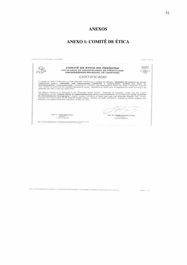
51
ANEXOS
ANEXO 1: COMITÊ DE ÉTICA

52
ANEXO 2: SUBMISSÃO PARA INTERNATIONAL ENDODONTIC JOURNAL
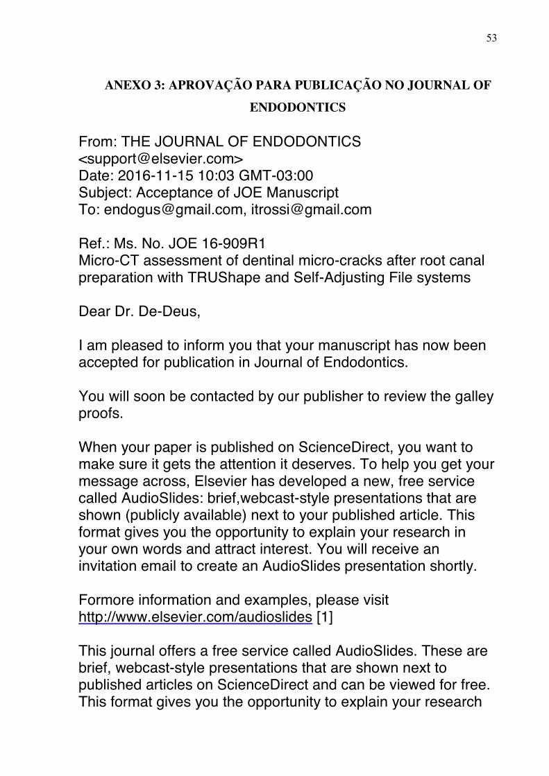
53
ANEXO 3: APROVAÇÃO PARA PUBLICAÇÃO NO JOURNAL OF ENDODONTICS
From: THE JOURNAL OF ENDODONTICS <[email protected]> Date: 2016-11-15 10:03 GMT-03:00 Subject: Acceptance of JOE Manuscript To: [email protected], [email protected] Ref.: Ms. No. JOE 16-909R1 Micro-CT assessment of dentinal micro-cracks after root canal preparation with TRUShape and Self-Adjusting File systems Dear Dr. De-Deus, I am pleased to inform you that your manuscript has now been accepted for publication in Journal of Endodontics. You will soon be contacted by our publisher to review the galley proofs. When your paper is published on ScienceDirect, you want to make sure it gets the attention it deserves. To help you get your message across, Elsevier has developed a new, free service called AudioSlides: brief,webcast-style presentations that are shown (publicly available) next to your published article. This format gives you the opportunity to explain your research in your own words and attract interest. You will receive an invitation email to create an AudioSlides presentation shortly. Formore information and examples, please visit http://www.elsevier.com/audioslides [1] This journal offers a free service called AudioSlides. These are brief, webcast-style presentations that are shown next to published articles on ScienceDirect and can be viewed for free. This format gives you the opportunity to explain your research

54
in your own words and promote your work by embedding it on your own website and sharing in social media. For more information and examples, please visit http://www.elsevier.com/audioslides [1] or watch our webinar: https://www.publishingcampus.elsevier.com/pages/206/Tell-your-research-story-with-AudioSlides.html [2] Thank you for submitting this manuscript. I look forward to seeing it published soon. With kind regards, Ken Hargreaves Editor Journal of Endodontics

55
ANEXO 4: MATERIAIS E METODOS Estimação do tamanho da amostra
Baseado em estudo prévio (De-Deus et al. 2015b) uma amostra composta por 28
dentes (7 por grupo) foi indicado como minima para revelar diferenças estatisticamente
significantes entre os grupos (tamanho de efeito de 0,9 foi estimado e computado juntamente
com o parâmetro de erro tipo alfa de 0,05 e potência beta de 0,95, utilizando ANOVA) com
auxilio do software específico (G*Power 3.1 for Macintosh; Heinrich Heine, Universität
Düsseldorf, Dusseldorf, Germany)
Seleção dos espécimes e preparo
Após aprovação do conselho de ética, 127 incisivos mandibulares humanos foram
selecionados de uma amostra de dentes extaidos . Cada dente foi radiografado nos sentidos
mésio-distal (MD) e vestibulo-lingual (VL). O critério de inclusão inicial foi: dentes com
raizes retas e canais únicos (< 5º) (Schneider 1971), mais de 2.5 mm de diferença entre os
sentidos MD e VL a partir de 5 mm antes do apice e após acesso endodontico um diametro
apical mínimo equivalente a uma lima 10 tipo – K.

56
Como resultado 63 dentes foram selecionados e escaneados no micro-CT (SkyScan
1173; Bruker-microCT, Kontich, Belgium) usando baixa resolução (70 kV 114 mA, 70 µm)
para obter um contorno dos canais radiculares. As imagens obtidas foram reconstruidas
(NRecon v.1.6.10; Bruker-microCT) proporcionando secções transversais axiais da estrutura
interna dental.

57
Depois disso foram selecionados 40 incisivos mandibulares com canais de
configurações similares. Esses dentes foram então novamente escaneados nas mesmas
condições anteriores agora utilizando alta resolução (14.25 µm com rotação de 360° 0.5°/,
7000 milisegundos utilizando filtro de aluminio 1.0 mm). As imagens de cada espécime
foram reconstruídas usando parâmetros padronizados: 40% feixe duro, correção de artefato
anelar e limites de contraste semelhantes. Os volumes de interesse foram selecionados desde
a extensão do limite cemento - esmalte até a porção apical, resultando na aquisição de 800-
900 secções trasversais por dente .
Os apices dos dentes foram então selados com cola e embebidos em silano polivinílico
criando dessa forma um sistema fechado (Susin et al. 2010). Foi feito então em cada dente
glide path com limas manuais tipo K até que uma lima 20 tipo K pudesse alacançar o
comprimento de trabalho (CT) estabelecido a 1 mm aquém do comprimento do canal. Os
especimes foram então pareados em quadruplicata tendo como base as caracteristicas
morfologicas do canal (volume, área e configuração) e divididos aleatoriamente em 4 grupos
experimentais de 10 dentes (n = 10). Cada grupo foi então preparado utilizando os sistemas :
BioRace (FKG Dentaire, La-Chaux-de-Fonds, Switzerland), Reciproc (VDW, Munich,
Germany), Self-Adjusting File (SAF; ReDent-Nova, Ra’anana, Israel) e TRUShape 3D
Conforming Files system (Dentsply Tulsa Dental Specialties, Tulsa, OK, USA), segundo as
recomendações de cada fabricante .

58
Em todos os grupos o tempo total de preparo e irrigação foi de 4 minutos. A irrigação
foi realizada utilizando agulha NaviTip (Ultradent Products Inc., South Jordan, UT, USA) e
25 mL of 5.25% NaOCl por dente. Após o final do preparo, irrigação ultrasonica passiva foi
realizada com insertos de limas # 15 tipo K , por 20 segundos a 2 mm co CT. Os canais
foram então irrigados com 3 mL de 17% EDTA por 5 min. seguidos por irrigação final
utilizando 2 mL de agua bi-destilada por 1 min. Aspiração da soluções irrigantes foi feita
com a ponta SurgiTip (Ultradent Products Inc.) no orificio de entrada dos canais. Todos os
preparos foram realizados por operador experiente treinado com todos os sistemas . Os canais
foram então secos com pontas de papel (Dentsply Maillefer). Os espécimes foram então
submetidos a escaneamento pós operatório e reconstruções seguindo os parametros já
mencionados
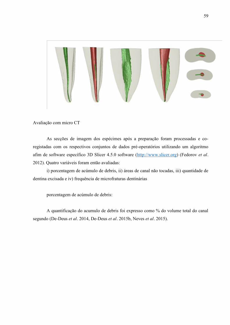
59
Avaliação com micro CT
As secções de imagem dos espécimes após a preparação foram processadas e co-
registadas com os respectivos conjuntos de dados pré-operatórios utilizando um algoritmo
afim de software específico 3D Slicer 4.5.0 software (http://www.slicer.org) (Fedorov et al.
2012). Quatro variáveis foram então avaliadas:
i) porcentagem de acúmulo de debris, ii) áreas de canal não tocadas, iii) quantidade de
dentina excisada e iv) frequência de microfraturas dentinárias
porcentagem de acúmulo de debris:
A quantificação do acumulo de debris foi expresso como % do volume total do canal
segundo (De-Deus et al. 2014, De-Deus et al. 2015b, Neves et al. 2015).
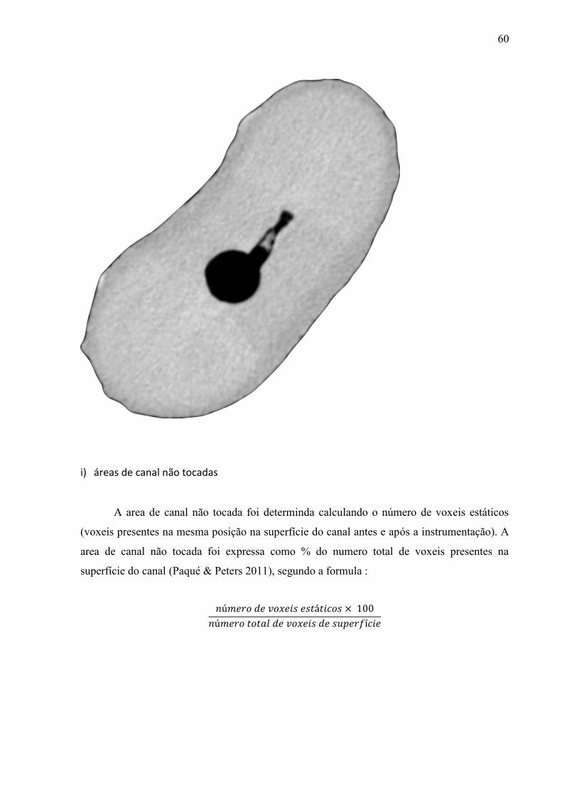
60
i) áreas de canal não tocadas
A area de canal não tocada foi determinda calculando o número de voxeis estáticos
(voxeis presentes na mesma posição na superfície do canal antes e após a instrumentação). A
area de canal não tocada foi expressa como % do numero total de voxeis presentes na
superfície do canal (Paqué & Peters 2011), segundo a formula :
𝑛ú𝑚𝑒𝑟𝑜 𝑑𝑒 𝑣𝑜𝑥𝑒𝑖𝑠 𝑒𝑠𝑡á𝑡𝑖𝑐𝑜𝑠 × 100
𝑛ú𝑚𝑒𝑟𝑜 𝑡𝑜𝑡𝑎𝑙 𝑑𝑒 𝑣𝑜𝑥𝑒𝑖𝑠 𝑑𝑒 𝑠𝑢𝑝𝑒𝑟𝑓í𝑐𝑖𝑒
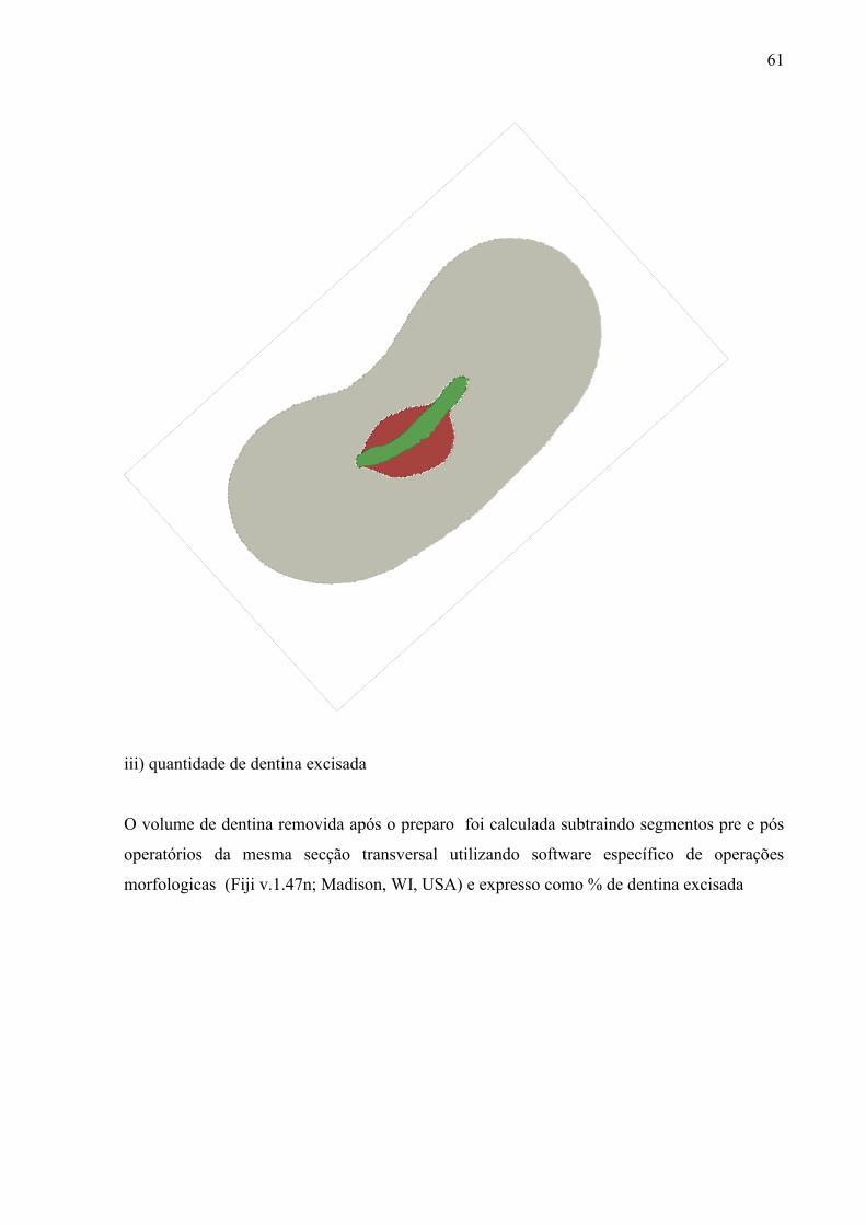
61
iii) quantidade de dentina excisada
O volume de dentina removida após o preparo foi calculada subtraindo segmentos pre e pós
operatórios da mesma secção transversal utilizando software específico de operações
morfologicas (Fiji v.1.47n; Madison, WI, USA) e expresso como % de dentina excisada

62
iv) frequência de microfraturas dentinárias
Inicialmente secções pós operatórias foram examinadas por 3 operadores (n =
70,030). As secções que apresentavam microfraturas dentinarias foram anotadas. Depois disso
as secções pré operatórias correspondentes foram verificadas para observar se essas fraturas
já estavam presentes ou não. Para validação do processo todas as imagens foram novamente
analisadas por duas vezes com intervalos de duas semanas. Em caso de divergência, as
imagens foram então analisadas conjuntamente até que um consenso fosse estabelecido.

63