avoid insect defenses a pathogenic nematode targets recognition proteins to · a pathogenic...
TRANSCRIPT
A Pathogenic Nematode Targets Recognition Proteins toAvoid Insect DefensesDuarte Toubarro1, Mónica Martinez Avila1, Rafael Montiel2, Nelson Simões1*
1 IBB/CBA and CIRN/Departamento de Biologia, Universidade dos Açores, Ponta Delgada, Portugal, 2 Laboratorio Nacional de Genómica para laBiodiversidad, Centro de Investigación y de Estudios Avanzados del Instituto Politécnico Nacional (CINVESTAV-IPN), Irapuato, Guanajuato, Mexico
Abstract
Steinernema carpocapsae is a nematode pathogenic in a wide variety of insect species. The great pathogenicity ofthis nematode has been ascribed to its ability to overcome the host immune response; however, little is known aboutthe mechanisms involved in this process. The analysis of an expressed sequence tags (EST) library in the nematodeduring the infective phase was performed and a highly abundant contig homologous to serine protease inhibitors wasidentified. In this work, we show that this contig is part of a 641-bp cDNA that encodes a BPTI-Kunitz family inhibitor(Sc-KU-4), which is up-regulated in the parasite during invasion and installation. Recombinant Sc-KU-4 protein wasproduced in Escherichia coli and shown to inhibit chymotrypsin and elastase activities in a dose-dependent mannerby a competitive mechanism with Ki values of 1.8 nM and 2.6 nM, respectively. Sc-KU-4 also inhibited trypsin andthrombin activities to a lesser extent. Studies of the mode of action of Sc-KU-4 and its effects on insect defensessuggest that although Sc-KU-4 did not inhibit the activation of hemocytes or the formation of clotting fibers, it didinhibit hemocyte aggregation and the entrapment of foreign particles by fibers. Moreover, Sc-KU-4 avoidedencapsulation and the deposition of clotting materials, which usually occurs in response to foreign particles. We showby protein-protein interaction that Sc-KU-4 targets recognition proteins of insect immune system such asmasquerade-like and serine protease-like homologs. The interaction of Sc-KU-4 with these proteins explains theability of the nematode to overcome host reactions and its large pathogenic spectrum, once these immune proteinsare well conserved in insects. The discovery of this inhibitor targeting insect recognition proteins opens new avenuesfor the development of S. carpocapsae as a biological control agent and provides a new tool to study host-pathogeninteractions.
Citation: Toubarro D, Avila MM, Montiel R, Simões N (2013) A Pathogenic Nematode Targets Recognition Proteins to Avoid Insect Defenses. PLoS ONE8(9): e75691. doi:10.1371/journal.pone.0075691
Editor: Stefan Pöhlmann, German Primate Center, Germany
Received May 22, 2013; Accepted August 20, 2013; Published September 30, 2013
Copyright: © 2013 Toubarro et al. This is an open-access article distributed under the terms of the Creative Commons Attribution License, which permitsunrestricted use, distribution, and reproduction in any medium, provided the original author and source are credited.
Funding: This research was supported by Fundação para a Ciência e Tecnologia (FCT) (PDCT/AGR/AAM/104487/2008) and by FLAD (Luso-AmericanFoundation) (Proj. 223/2006) awarded to NS. DT received a fellowship from FCT (PDCT/AGR/AAM/104487/2008), and MMA was a recipient of afellowship from the Regional Government of Açores (RGA) (M3.1.2/F/005/2007). The funders had no role in study design, data collection and analysis,decision to publish, or preparation of the manuscript.
Competing interests: The authors have declared that no competing interests exist.
* E-mail: [email protected]
Introduction
Steinernema carpocapsae is an entomopathogenicnematode (EPN) that is currently used to control insect pests,owing to its high virulence against a wide variety of insects [1].The virulence of S. carpocapsae is mainly attributed to theability the infective juvenile has to overcome insect defensesand to the symbiotic bacteria it carries into the parasitizedinsect, which releases toxic factors [2,3].
Insects are equipped with a system of pathogen recognitionreceptors and effectors that enables them to resist a widevariety of pathogens [4]. Pathogen receptors are found assoluble proteins in body fluids and on the cellular surface, likeToll receptor, and the effectors are composed of cellular andhumoral components that cooperate to neutralize invasive
organisms [5,6]. A complex reaction of encapsulation takesplace when large foreign bodies such as EPNs areencountered [7]. In the encapsulation are participating solubleproteins from the haemocoel, proteins released from activatedhemocytes and the hemocytes themselves [4]. This processinvolves three main events: cell activation, clot formation andactivation of phenoloxidase [4,8]. Hemocytes activation istriggered within minutes of pathogen exposure with cellsbecoming adherent to each other and to the foreign surface[9,10]. The clot formation involves the activation of solubleproteins in the hemocoel, such as transglutaminase, lipophorin,hexamerins, and fondue and proteins derived from hemocytes,for instance hemolectin and tiggrin, that lead to the clotting ofhemolymph forming a network of fibers that bind together toisolate the foreign body [11,12]. In the presence of foreign
PLOS ONE | www.plosone.org 1 September 2013 | Volume 8 | Issue 9 | e75691
agents a series of proteolytic enzymes are activated leading tothe processing of the zymogen prophenoloxidase (PPO) into itsactive form phenoloxidase (PO). Phenoloxidase producesindole groups, which are polymerized to melanin andsubsequently deposited in entrapped foreign body [13]. Thethree systems work together leading to the formation of hardclots that efficiently protect from invasive pathogens [14].
To escape host defenses EPNs have developed passive andactive mechanisms. The passive mechanisms usually mimicthe host components to evade detection, whereas in the activeprocess the pathogen actively destroys the host defenseeffectors [7]. Surface coating proteins that participate in theevasion of the host immune system were identified in S.glaseri, S. feltiae and H. bacteriophora [15-17] and S.carpocapsae and S. feltiae were shown to destroy insectimmune effectors namely antibacterial peptides [18,19].Despite the ability of these nematodes to counteract insectdefenses, the pathogenicity of EPNs against a particular insectis usually thought to result from an “arms race” between theEPN and the insect [20,21]. Moreover, there are severalreports describing the immune reactions of insects againstEPNs [22]. For example, a small part of the Pseudaletiaunipuncta larvae infected with S. carpocapsae reacts through acellular encapsulation mechanism [23], the diptera Tipulaoleracea reacts via a humoral encapsulation mechanismagainst S. feltiae [24], and Drosophila melanogaster recognizesH. bacteriophora through transglutaminase, an importantcomponent of clot system [25].
The pathogenicity of parasitic nematodes is known to beessentially modulated by the nematode’s secreted andexcreted products (ESPs), which are active against effectors ofthe host’s immune system [26]. Many of these active ESPs areproteases and protease inhibitors. For example, the nematodeAncylostoma ceylanicum produces a serine protease inhibitorthat prevents the activation of neutrophils [27], the infectivestages of Schistosoma mansoni and Brugia malayi releaseserpins that protect the parasites during inflammation byinhibiting neutrophil elastases [28,29], and Trichuris suisreleases an inhibitor of trypsin and chymotrypsin thatmodulates neutrophil proteases [30].
In ESPs of S. carpocapsae, a few serine proteases wereshown to participate in host immune evasion by destroying theinsect’s cellular and humoral defenses [31-34]. Furthermore,the analysis of an EST library obtained from S. carpocapsaeduring the parasitic phase allowed the detection of a highlyabundant contig with homology to the serine protease inhibitorsof parasitic nematodes, a few of them shown to participate inhost immune modulation [35]. To understand the role of thisputative protease inhibitor, we cloned and sequenced the fullcDNA and show that it encodes a BPTI-Kunitz family inhibitor.Recombinant protein produced in E. coli was characterized,and the effects on insect defenses were determined. Ourresults suggest that this inhibitor binds to key recognitionproteins in the insect, thereby impairing cellular aggregation,modifying clotting fiber arrangements and partially preventingPPO activation, all of them allowing the EPN to avoidencapsulation.
Materials and Methods
Nematodes and insectsInfective juveniles of the S. carpocapsae Breton strain were
produced in the larvae of the moth Galleria mellonella [36],harvested in a White trap [37] and stored in tap water at 10°Cfor 1–2 months before use.
Full-length cDNA and genomic DNAThe full-length Sc-KU-4 cDNA was generated using the
primers KU-4'Rf (5'-TTTGGTTCCATCTGGGCAAGTGTC-3')and KU-4'Rr (5'-CGCTGTATTGATCGTTTGCTG-3') designedbased on an EST expressed in the parasitic stage of S.carpocapsae (GenBank accession number GR977821). RNAwas isolated from nematodes in the parasitic stage asdescribed [35], and the first-strand cDNA was synthesizedusing a Superscript III reverse transcriptase kit (Invitrogen) andan oligo (dT) primer. The full-length cDNA was produced byrapid amplification of cDNA ends (RACE) using the SMARTRACE cDNA Amplification Kit (Clontech-Takara) and theKU-4'Rf and KU-4'Rr primers. The reaction mixture contained50 ng of cDNA, 2.5 µl of 10x PCR buffer, 10 mM of each dNTP,0.4 µM of each primer, 1.6 mM MgCl2 and 0.2 µl of Taq DNApolymerase in a final reaction volume of 25 µl. Theamplification protocol consisted of an initial heating step at94°C for 3 min, followed by 30 cycles of 94°C for 30 s, 55°C for30 s and 72°C for 30 s, with a final extension cycle at 72°C for3 min. Amplified cDNA fragments were TA-cloned into thevector pCR4-TOPO using the TOPO TA Cloning Kit(Invitrogen) and sequenced by Stabvida (Oeiras, Portugal).Full-length cDNA was obtained by aligning the fragments usingthe Bioedit software package.
Genomic DNA was extracted from nematode parasitic stageand amplified by PCR using flanking primers (Fwd5'CGCTGTATTGATCGTTTGCTG3' and Rev 5’CAATCACCTCTGACTTCCAC3’) designed to target the full-length cDNA sequence. The amplification conditions were asfollows: 94°C for 3 min, followed by 30 cycles of 94°C for 30 s,55°C for 30 s and 72°C for 30 s, with a final extension at 72°Cfor 3 min. The 741-bp PCR product was isolated, cloned intothe aforementioned TOPO-TA vector (Promega) andtransformed into E. coli DH5α cells, and DNA inserts of positiveclones were confirmed by sequencing. The exon-intronboundaries were determined by comparing cDNA with gDNA.
In silico analysis of sc-KU-4Sc-KU-4 full-length cDNA was used as a BLAST search
query (http://ncbi.nlm.nih.gov/blast), and sequence alignmentswere created with ClustalW (http://www.ebi.ac.uk/clustalw).Protein motifs were predicted using SMART (http://smart.emblheidelberg.de), and the signal peptide was identifiedby SignalP 3.0 (http://www.cbs.dtu.dk/services/SignalP). Forphylogenetic reconstruction, sequences were aligned withMega5 (http://www.megasoftware.net/mega4/index.html), andthe phylogenetic tree was constructed with maximum likelihoodusing PhyML (www.atgc-montpellier.fr/phyml/). Robustnesswas assessed by the bootstrap method (100 pseudoreplicates).The 3D structure prediction was obtained using the on-line
A Nematode Targets Insect Recognition Proteins
PLOS ONE | www.plosone.org 2 September 2013 | Volume 8 | Issue 9 | e75691

platform I-TASSER (http://zhanglab.ccmb.med.umich.edu/I-TASSER).
Analysis of gene expressionSc-KU-4 mRNA levels were determined by quantitative RT-
PCR during different stages of nematode development in aparasitized insect. For this purpose, parasitism was generatedby exposing G. mellonella larvae to nematode infectivejuveniles (IJs) in Petri dishes padded with wet filter paper. Thedifferent nematode stages were collected from parasitizedinsects after inspection for the desired stages. L3 stagenematodes were isolated from the gut and the hemocoel, L4stage nematodes were isolated from the hemocoel, and adultsand L1/L2 stage nematodes were isolated from the bodytissues. Approximately 20 specimens were collected from eachstage in three separate exposures and homogenized in liquidnitrogen; total RNA was extracted using the RNeasy Micro Kit(Qiagen). Reverse transcription was performed using theSuperscript III reverse transcriptase kit (Invitrogen) and anoligo (dT) primer. Data were normalized to 18S rRNA. Sc-KU-4was amplified with the primers Ku-4Fg (5'- TAC AAC GGGACT GGA GGC AA -3') and KU-4Rg (5'- TGG TTC CAT CTGGGC AAG TG -3'), and the 18S rRNA with primers 18SF (5'-GCTAATCGGAAACGAAAGTC-3') and 18SR (5'-CATCCACCGAATCAAGAAAG-3') using an ABI 7900 HT RealTime PCR thermocycler (Applied Biosystems). Each reactionconsisted of an initial heating step at 95°C for 10 min, followedby 60 cycles of 95°C for 15 s and 60°C for 60 s. Amplificationswere performed in triplicate. RT-PCR data were analyzed usingthe comparative CT method according to the manufacturer’srecommendations. Data were expressed as means ± standarderrors. Statistical analysis was carried out by one-way ANOVAfollowed by Bonferroni multiple comparison tests (SPSSsoftware, version 13.0). Differences were considered significantat P values < 0.05.
Construction of the expression vectorThe full-length sc-ku-4 coding sequence was amplified using
the forward primer containing an NdeI restriction site (5'-CATATGATGTTTCCACTTTTCGAGTTTAAC -3'
) and the reverse primer containing an XhoI site (5'-CTCGAGTTCCTTCGAGCAGCATTTAGC -3'). The 0.8 kbproduct was purified from a 1.5% agarose gel using the QIAEXII Gel Extraction Kit (Qiagen) and digested with NdeI and XhoI.The fragment was then inserted into the expression vectorpET23a (+) to produce the recombinant vector pET23a (+)-sc-ku-4 in frame with a C-terminal His6X-tag. The vector wasintroduced into E. coli C41 (DE3) and Rosetta™ II (DE3)strains. Identity and integrity of the inserts were confirmed bysequencing.
Expression of recombinant Sc-KU-4A single colony of each transformed E. coli strain was
incubated in 5 ml Luria broth (LB) containing 100 µg/mlampicillin and 34 µg/ml chloramphenicol at 37°C and 250 rpmovernight. These cultures were used to inoculate 20 ml LB withantibiotics as above and were incubated at 37°C until the OD600
reached 0.6. Sc-KU-4 expression in both strains was induced
under the following conditions: 0.2 mM isopropyl β-D-1-thiogalactopyranoside (IPTG) at 37°C for 3 h, 1 mM IPTG at37°C for 3 h, 0.2 mM IPTG at 20°C overnight and 1 mM IPTGat 20°C overnight. Cells from the different induction conditionswere harvested by centrifugation at 6,500 x g for 15 min, andthe pellet was resuspended in 2 ml 0.05 M Tris-HCl (pH 7.4)containing 0.5 M NaCl (TN) and 1 mg/ml lysozyme and frozenovernight at −20°C. After thawing, 1 ml 1 M MgCl2 and 600 µl 1mg/ml DNAse were added and the solution was incubated for30 min at RT under orbital agitation. Cellular debris wasremoved by centrifugation at 12,000 x g for 20 min, and thesupernatant was collected and analyzed for recombinantprotein expression.
SDS-PAGE and Western blotSuspensions with recombinant Sc-KU-4 were run on a 10%
SDS-PAGE in a Mini-PROTEAN II gel system (Bio-Rad), andproteins were stained with colloidal Coomassie. For Westernblot analysis, proteins in the gel were electro-blotted onto aPVDF membrane using a mini Trans-Blot Cell (Bio-Rad). Themembrane was blocked with 0.01 M Tris-HCl (pH 7.5)containing 0.1 M NaCl, 5% (w/v) BSA and 0.05% (v/v)Tween-20 for 30 min at RT, incubated with anti-His6X-tagperoxidase-conjugated mouse antibody diluted in blockingsolution (1:5,000) for 2 h at RT and then washed 3 times for 10min with 0.01 M Tris-HCl (pH 7.5), 0.1 M NaCl. Antibodies weredetected by incubation with the tetramethylbenzidineperoxidase substrate (Sigma-Aldrich) according to themanufacturer’s instructions.
Production, refolding and purification of Sc-KU-4Recombinant Sc-KU-4 was produced in transformed E. coli
Rosetta (DE3) cells in 2 L of LB medium, and expression wasinduced with 0.2 mM IPTG followed by a 3 h incubation at37°C. Cells were recovered after centrifugation at 6,500 x g for15 min in 100 ml of TN and 1 mg/ml lysozyme, incubated for 30min at RT and then frozen overnight. After thawing, the solutionwas adjusted to 500 ml with TN and centrifuged at 10,000 x gfor 15 min. The pellet was resuspended in 1 L of TN plus 1%Triton X-100 and incubated under agitation for at least 4 h at4°C.
Inclusion bodies were retrieved by centrifugation at 10,000 xg for 20 min at 4°C, followed by solubilization in 6 M ureacontaining 0.02% β-mercaptoethanol overnight with orbitalagitation. Solubilized protein was dialyzed in a cellulosemembrane tube with 20 mM Tris-HCl (pH 8) for 48 h at 4°C,followed by two changes in the same buffer. Recombinantprotein was captured by nickel chelate affinity chromatography(His-Tag, GE) under the recommended conditions in an AKTAFPLC system (GE Healthcare). Purified recombinant proteinswere refolded for 3 h in 50 mM Tris (pH 8) with 300 mM L-arginine, 50 mM NaCl and 5 mM DTT. Folded Sc-KU-4 wasapplied to a MonoQ column (GE) equilibrated with 20 mM Tris-HCl (pH 8) and eluted on a linear gradient of 1 M NaCl toeliminate minor contaminants. Purity was assessed by 12%SDS-PAGE. The protein concentration was determined usingthe BCA Kit (Thermo Scientific).
A Nematode Targets Insect Recognition Proteins
PLOS ONE | www.plosone.org 3 September 2013 | Volume 8 | Issue 9 | e75691

Sc-KU-4 specificityThe inhibitory activity of Sc-KU-4 was tested against trypsin,
α-chymotrypsin and elastase from bovine pancreas andthrombin from bovine plasma (Sigma).
Ten micrograms of purified Sc-KU-4 was added to 2 µg ofeach enzyme, and the final volume was adjusted to 50 µl with0.1 M Tris-HCl (pH 7.5) containing 0.1 M NaCl and 1 mMCaCl2. After incubation for 15 min at 37°C, the remaininghydrolytic activity was quantified using 1 mM of each specificchromogenic substrate: BApNA, Suc-AAPFpNA, Suc-AAPLpNA and BPVA-pNA (Sigma) for trypsin, α chymotrypsin,elastase and thrombin, respectively. Sc-KU-4 was replacedwith buffer in control reactions. The formation of p-nitroanilinewas monitored at 405 nm in an ELISA microplate reader (Bio-Rad). The percentage of enzyme inhibition was calculated asfollows: % inhibition = [
(total enzyme activity units - residual enzyme activity units)/total enzyme activity units] X 100. IC50 values were calculatedusing Probit (v1.4) based on the percentage of inhibition ofenzymatic activity after incubation with increasingconcentrations of recombinant Sc-KU-4 (0, 5, 11 and 16 µM)under the conditions described above.
Kinetic analysisThe fractional activity (velocity of enzyme with Sc-KU-4 /
velocity of enzyme without Sc-KU-4) was plotted against theratio of inhibitor / enzyme concentrations, and the x-axisintercept as a value for the stoichiometry of inhibition (SI) wasdetermined by linear regression analysis. The inhibitionconstant (Ki) of Sc-KU-4 for α-chymotrypsin and elastase wasestimated using a Dixon plot. Recombinant Sc-KU-4 (0, 5, 11and 16 µM) was incubated with 5 µM of each enzyme for 10min at 37°C; then, 0.05, 1 and 2 mM of Suc-AAPFpNA or Suc-AAPLpNA was added to the α-chymotrypsin and elastasereactions, respectively. The remaining hydrolytic activity wasmonitored as described above. A single regression line foreach concentration of substrate was obtained, and the Ki wascalculated from the intersection of the three lines. The inhibitorymechanism was determined using Lineweaver-Burk plots.Regression lines for α-chymotrypsin and elastase wereproduced using the inverse of the initial rate for each Sc-KU-4concentration plotted against the inverse of the concentrationof the substrate.
Isolation of plasma proteins interacting with Sc-KU-4G. mellonella hemolymph was collected by a ventral pro-leg
puncture of the 4th instar larvae and was drained into ananticoagulant solution (Ringer without CaCl2 and with added 20mM EDTA). Hemocytes were removed by centrifugation at1,500 x g for 3 min at 4°C, and plasma was recovered anddiluted (5:1) in the anticoagulant solution plus 0.1%polyvinylpyrrolidone.
Sc-KU-4 was immobilized on CNBr-activated Sepharosebeads 4B (Sigma) according to the manufacturer’s instructionsand then incubated with 50 mg of plasma proteins in 2 ml of 20mM Tris-HCl (pH 8) for 3 h at 4°C with orbital agitation. BSA-coated beads were used as controls. After the reaction, beadswere packed onto a 0.7 × 0.5 cm column coupled to an AKTA
FPLC system (GE Healthcare). Unbound material was washedthrough the column with binding buffer, followed by 5 columnvolumes of 20 mM Tris-HCl(pH 8), 0.5 M NaCl. The remainingbound proteins were eluted with 50 mM glycine (pH 3). Elutedproteins were dialyzed with a centricon (MWCO 10 kDa,Millipore), suspended in rehydration buffer (9.8 M Urea, 4%(w/v) CHAPS, 2 mM TBP, 1% (v/v) ampholytes pH 3-10) andapplied to a 7 cm IPG strip (Bio-Rad) for 2DE. After activerehydration for 12 h at 20°C, proteins were focused in a ProteinIEF Cell (Bio-Rad). Prior to the second dimension, the IPG stripwas equilibrated with 130 mM DTT and 135 mMiodoacetamide. SDS-PAGE and protein staining wereperformed as above.
MALDI-MS/MSFor MALDI-MS/MS analysis, proteins were digested in-gel
with trypsin, purified and concentrated as previously described[33]. The m/z spectra were acquired in a 4700 ProteomicsAnalyzer MALDI-TOF/TOF (Applied Biosystems) in both MSand MS/MS mode. Protein identification was achieved usingMASCOT (www.matrixscience.com) to search the UniProtKBdatabase (downloaded on 10/06/2012).
Analysis of phenoloxidase activityPlasma was prepared as described above and PO activity
quantified as previously described [38] with minormodifications. Forty microliters of plasma solution was added to10 µl of Sc-KU-4 (3 µM/µl) and incubated for 5 min on ice. Incontrols, Sc-KU-4 was replaced with anticoagulant solution.The samples were then activated with 5.5 µl (10 µg/ml) oflipopolysaccharide (LPS E. coli O55:B5, Sigma - Aldrich)dissolved in 50 mM Tris-HCl (pH 7.5), 0.1 M NaCl andincubated 15 min at 25°C. Activated plasma was centrifuged at5,000 x g for 5 min at RT, the supernatant was recovered andsupplemented with 20 µl 15 mM L-DOPA (Sigma) dissolved in20 mM Tris-HCl (pH 7.5), and the reaction was incubated for15 min at 37°C. The reaction was monitored at 490 nm usingan ELISA microplate reader (Bio-Rad). PO activity wasmeasured as the relative change in OD at reaction time pointsof 0 and 15 min. Experiments were carried out in triplicate.
Analysis of hemolymph coagulation and encapsulationassay
The analysis of clotting fibers was carried out on a slide byadding Sc-KU-4 (30 µM) to 200 µl of plasma, incubated for 30min in a humidified chamber at RT and then activated with LPS(10 µg/ml). In the controls, Sc-KU-4 was replaced with theanticoagulant solution. The incorporation of melanin into clotswas tested by carefully aspirating the supernatant with a finetip. Adherent materials were then visualized and photographedby light microscopy. To check for the formation of clottingfibers, plasma drops were prepared and incubated asdescribed and were fixed with 2% glutaraldehyde in 10 mMphosphate buffer (PB) (pH 7) at 4°C for 15 min. The fixativewas removed gently by washing with 10 mM PB and replacedwith 70% ethanol. Samples of the floating clot strands werecarefully transferred to two new slides, one for phase contrastobservation and the other for peanut agglutinin-FITC
A Nematode Targets Insect Recognition Proteins
PLOS ONE | www.plosone.org 4 September 2013 | Volume 8 | Issue 9 | e75691

conjugated (PNA-FITC) staining (Sigma) (10 mg/ml) in PB andobserved at 480 nm by a fluorescence microscope (Zeiss).Hemocyte aggregation and activation were produced in elicitedhemolymph according to a previously described method [10].Hemocyte aggregates were inspected using an opticalmicroscope at 50X magnification and data was obtained fromfive optical fields of three replicates. Statistical analyzed wasperformed using SPSS software, version 13.0. In theencapsulation assay, beads of Sephadex G200 were soakedwith Sc-KU-4 for 10 min, added to plasma activated with LPS,incubated for 30 min in a humidified chamber at RT and thenfixed with 2% glutaraldehyde. In a second assay ofencapsulation, the plasma was first incubated with Sc-KU-4 for30 min at RT, then mixed with Sephadex G200 beads,incubated for 30 min and fixed with 2% glutaraldehyde asdescribed above. Fixed materials were processed for SEM asdescribed elsewhere [34]. Experiments were carried out intriplicate.
Results
Molecular Characterization of Sc-KU-4A full-length 641 bp cDNA was produced using primers
designed based in the EST sequence predicted for a serineprotease inhibitor of S. carpocapsae. After cloning andsequencing, a single open reading frame of 555 bp wasidentified and submitted to NCBI with the accession numberHM586101. This ORF was denoted Sc-KU-4, and has a 5´UTR with an “ATG” start codon and an 86-bp 3´ UTR in whicha stop codon is followed by a non-canonical polyadenylationsignal “CATAAA” and a poly (A) tail. This cDNA codes for aprotein with 184 amino acids, including a signal peptide of 19residues and a single Kunitz domain beginning at aa 27 andending at aa 77 (NCBI accession ADQ44000). The putativemature protein had a MM of 20,418.16 Da and a pI of 5.07.
Genomic organizationA single 741 bp fragment corresponding to the genomic DNA
of Sc-KU-4 was amplified using primers flanking the 5’ and 3’regions of the ORF and was sequenced and submitted to NCBI(Accession number - HQ328060). The alignment of gDNA andcDNA suggests that the sc-ku-4 gene has five exons containing43, 72, 123, 140, and 175 bp, which are coded for by residues1-43, 92-163, 211-333, 380-520 and 566-741, respectively, allflanked by the typical splice signals AG/GT (see hyperlinkhttp://www.ncbi.nlm.nih.gov/nuccore/312192649?report=graph). The first exon encodes the signal peptide andexons II and III encode the Kunitz domain.
Sequence homology, phylogeny and predictedstructure
The Sc-KU-4 sequence has identity with secreted Kunitz-likeserine protease inhibitors of the parasitic nematodes Ascarissuum (45%), Loa loa (35%) and Anisakis simplex (30%) andalso has homology with Kunitz domains from the free-livingnematodes Caenorhabditis remanei (32%), C. briggsae (30%)and C. elegans (30%). All of these nematode Kunitz inhibitors
shared a similar molecular architecture with an N-terminalsignal peptide followed by a single Kunitz domain, except forthe inhibitor from A. suum, which presented two Kunitzdomains in tandem. Multiple sequence alignment showed thatSc-KU-4 has a BPTI-Kunitz family domain with 60 amino-acidresidues located at the NH terminus (Figure 1A). The Sc-KU-4Kunitz domain contained the conserved residues Gly34, Tyr57,Phe55 and Gly59 and four of the six well-conserved cysteineresidues. Therefore, it was possible that the CysI - CysVI andCysIII – CysV disulfide bridges were formed (Cys27-Cys77 andCys52-Cys73) but that the CysII – CysIV bridge was missing.The positions of CysII and CysIV in the canonical Kunitzdomain were occupied by Gly36 and Gly61 in Sc-KU-4. Theputative primary binding site (P1 residue) in Sc-KU-4 waspredicted to be occupied by a serine (Ser37), as in the A.suum, C. remanei, C. elegans and C. briggsae homologs.
The phylogenetic analysis revealed that Sc-KU-4 clusteredwith other nematode Kunitz-like inhibitors, which diverged earlyfrom the canonical bovine BPTI-Kunitz inhibitor. In thenematode cluster, Sc-KU-4 formed a group with the parasiticnematode A. suum, which was separated from the groupformed by the Caenorhabditis free-living species and theparasites A. simplex and L. loa. These conclusions aresupported by high bootstrap values (100%) in a Nearest-Neighbor-Interchange tree (Figure 1B).
A predicted 3D model for Sc-KU-4 was generated using theI-TASSER server, with an estimated model accuracy of 0.72 ±0.10 (TM-score). Under this model, the active domain of Sc-KU-4 was predicted to preserve the three antiparallel β-strandsfollowed by one α-helix, the characteristic tertiary fold of theKunitz domain. The preservation of such structure is probablydue to the Van Der Waals interactions established among theglycine residues that replaced CysII and CysIV, thus allowingfor the stabilization of the recognition loops (Figure 1C). Thetwo loops originated by β-strand twist are particularly importantfor inhibitor recognition. The first recognition loop containedresidues 32-GSGSNVTV-38 (P4 - P4´), and the secondcontained residues 57-YNGTG-61. In this model, the scissilebond site was predicted to be exposed on the protein surface.
Bacterial transformation and production ofrecombinant Sc-KU-4
The Sc-ku-4 coding sequence was inserted into theexpression vector pET23a in frame with a C-terminal His6X-tag. Recombinant Sc-KU-4 was produced only in inclusionbodies, and the highest recombinant concentration (74 mg/L)was obtained in the E. coli host strain Rosetta (DE3) inducedwith 0.5 and 1 mM of IPTG for 3 h at 37°C (Figure S1). Sc-KU-4 was isolated in a single step by Ni2+ affinitychromatography, folded and purified using Mono Qchromatography. After purification, one band with the expectedmass of approximately 20 kDa appeared on an SDS-PAGE geland was detected with an anti-His(C-term)-HRP antibody.MS/MS spectrometry confirmed that this protein was Sc-KU-4with a significant score of 306 (protein score probability limitwith P < 0.05 is 85) and 34% coverage.
A Nematode Targets Insect Recognition Proteins
PLOS ONE | www.plosone.org 5 September 2013 | Volume 8 | Issue 9 | e75691

Sc-KU-4 exhibits a competitive inhibitory mechanismRecombinant Sc-KU-4 strongly inhibited the hydrolytic
activity of α-chymotrypsin and elastase with IC50 of 6.3 nM and8.6 nM. A weak inhibitory effect on the activity of thrombin andtrypsin was also detected. The stoichiometry of inhibition (SI)for Sc-KU-4 was 14.26, 16.94, 29.57 and 35.25 for α-chymotrypsin elastase, thrombin and trypsin, respectively
(Figure 2A). Sc-KU-4 displayed a competitive inhibitorymechanism, as shown by Lineweaver-Burk reciprocal plots(Figure 2B). Ki values of 1.8 nM and 2.6 nM were determinedagainst α-chymotrypsin and elastase, respectively (Figure 2C).
Figure 1. Sequence, phylogeny and structural analysis of Sc-KU-4. A: Amino acid sequence alignment of the Kunitz domain inSc-KU-4 with the homologous domains of other nematode Kunitz-like inhibitors. Conserved positions are shaded; the solid linesindicate the conserved disulfide bridges, and the discontinuous line indicates the missing CysII–CysIV bridge in Sc-KU-4. B:Phylogenetic analysis showing Sc-KU-4 closes to other nematode Kunitz-like inhibitors forming a separate cluster with A. suum. C:Sc-KU-4 structure prediction based on multiple-threading alignments showing the Kunitz domain exposed on the molecular surfacein a characteristic tertiary fold of three antiparallel β-strands followed by one α-helix. Cysteine residues responsible for reactive loopstabilization and glycine residues that replace CysII–CysIV are shown in gray. The discontinuous line box highlights the amino acidspresent in reactive loops. The P1 position is shown in green. Asi: Anisakis simplex; Asu: Ascaris suum; Cbn: Caenorhabditisbrenneri; Cbr: C. briggsae; Cel: C. elegans; Cre: C. remanei; Llo: Loa loa; Sca: S. carpocapsae.doi: 10.1371/journal.pone.0075691.g001
Figure 2. Recombinant Sc-KU-4 inhibits a broad range of trypsin-like enzymes. A: Stoichiometry of inhibition of Sc-KU-4determined against α-chymotrypsin, elastase, thrombin and trypsin. B: The competitive inhibitory mechanism of Sc-KU-4 revealedby Lineweaver-Burk reciprocal plots. C: The highest Sc-KU-4 Ki of 2.6 nM was obtained for α-chymotrypsin.doi: 10.1371/journal.pone.0075691.g002
A Nematode Targets Insect Recognition Proteins
PLOS ONE | www.plosone.org 6 September 2013 | Volume 8 | Issue 9 | e75691

Invasive nematodes express the highest levels of sc-ku-4
Transcript levels of sc-ku-4 were measured in all of thenematode stages of development in parasitized G. mellonellalarvae, and in the L3 at two time points. L3 nematodescollected in the digestive tract of parasitized insects and L4nematodes already in the hemocoel of living insects expressedthe highest levels of sc-ku-4 mRNA. Remarkably, significantlyless (P < 0.05) sc-ku-4 mRNA was expressed by L3nematodes just after hemocoel invasion. Adult nematodescollected from insect cadavers (approximately 56 HPE)expressed significantly lower levels of sc-ku-4 than the larvalstages. L1/L2 stage nematodes collected from cadavers(approximately 72 HPE) did not express sc-ku-4 (Figure 3).These data indicate that nematodes (L3 and L4) present in liveinsects expressed the highest levels of sc-ku-4.
Sc-KU-4 does not impair hemocyte activation butimpairs hemocyte aggregation
Sequence homologies and expression profiles of Sc-KU-4suggest that it should play a role in modulating the hostdefenses. Therefore, we analyzed the effects of Sc-KU-4 inhemocyte aggregation using the hemolymph of G. mellonella
Figure 3. Transcript quantification of Sc-KU-4 duringsuccessive stages of the S. carpocapsae life cycle revealsan up-regulation during the invasive phases. Sc-KU-4relative expression was determined by quantitative RT-PCRusing 18S as an endogenous control. Nematodes in eachphase were collected from parasitized G. mellonella larvae: L3-gut – third stage juveniles collected inside the gut lumen at 6 hpost-exposition (HPE); L3-hem – third stage juveniles collectedin the hemocoel at 12 HPE; L4 – fourth stage juvenilescollected in the hemocoel 36 HPE; Adults – males and femalescollected in the hemocoel at 56 HPE; L1/L2 – a pool of first andsecond stage juveniles collected in the hemocoel at 72 HPE.Bars represent standard deviations from three independentreplicates. Different letters indicate significant differences (p <0.05).doi: 10.1371/journal.pone.0075691.g003
elicited with LPS. Significant differences (p < 0.05) wereobserved between the number of hemocyte aggregates formedin the hemolymph treated with Sc-KU-4(5.3 ± 1.5) and in thecontrol (14.5 + 4.4) (Figure 4A). The size of aggregates(measured in the larger diameter) was also significantlydifferent (p < 0.05) in samples treated with Sc-KU-4(66 µm ±11) and in controls (280 µm ± 27). Because the aggregation ofhemocytes requires its activation, we used a previouslydescribed method [10] to investigate whether Sc—KU-4interferes with the activation. In this experiment activatedhemocytes were observed in hemolymph treated with Sc-KU-4,similarly to the control, thus indicating that Sc-KU-4 impairedthe aggregation of hemocytes but did not impair its activation.
Detailed inspections by SEM consistently showed that thelarge aggregates of hemocytes formed in the control weretrapped in a newly formed matrix whereas they were not inhemolymph treated with Sc-KU-4 (Figure 4B).
Sc-KU-4 does not impair clot activation but modifiesclotting fibers
Clotting fibers were visualized in plasma treated with Sc-KU-4 just as they were in the plasma control after activationwith LPS. However, the clotting fibers formed in plasma treatedwith Sc-KU-4 were less expanded than those formed in thecontrol (Figure 5). A more detailed inspection by SEM showedthat clotting fibers in plasma treated with Sc-KU-4 appearedmore striated, exhibiting fibbers more individualized than thoseobserved in non-treated plasma.
Sc-KU-4 does not inhibit PPO activation but slightlydecreases PO activity
Our assays showed that Sc-KU-4 did not inhibit PPOactivation; however, the kinetics of PO activity suggested thatthe Vmax was slightly lower in plasma treated with Sc-KU-4than in non-treated plasma. Furthermore, the final product ofthe reaction did not differ significantly (P > 0.05) in both assays(Figure 6).
Sc-KU-4 impairs the aggregation of foreign bodies andthe deposition of clotting materials
To determine if Sc-KU-4 might protect a foreign body fromthe host response, we added beads soaked in Sc-KU-4 to G.mellonella plasma activated with LPS. Non-treated beadsaggregated as expected but beads treated with Sc-KU-4 didnot aggregate and remained individualized. Moreover, treatedbeads were surrounded by a halo free of clotting mesh,whereas non-treated beads were entrapped by clottingmaterials. The relevance of Sc-KU-4 in avoiding the recognitionof foreign bodies was evidenced also by the distribution of thehalos around the beads. The halo was localized in the side ofthe bead the liquid runoff, whereas in the opposite the beadwas covered by clotting material (Figure 7B).
In another assay, plasma was incubated with Sc-KU-4 for afew minutes and beads were subsequently added. Despite theformation of a light mesh of fibers in this assay, the beadsremained individualized with a completely polished surface.These results contrasted with those observed for the controls in
A Nematode Targets Insect Recognition Proteins
PLOS ONE | www.plosone.org 7 September 2013 | Volume 8 | Issue 9 | e75691

which beads were aggregated and enclosed in newly formedclotting materials (Figure 7C).
Sc-KU-4 targets serine protease homologs of G.mellonella plasma
To determine which proteins in insect plasma were targetedby Sc-KU-4, we conducted a pull-down protein/protein assayusing recombinant Sc-KU-4 as bait. A differential elution wasperformed to recover proteins according to binding forces. Twoproteins strongly bounded to Sc-KU-4-coupled beads wereeluted using a low pH (pH 3) buffer, detected by 2DE andidentified by MS/MS with significant scores in Lepidoptera(Figure 8 and Table S1). One was identified as a homolog ofthe masquerade-like serine protease (gi:270298184) of Pierisrapae and another as the serine protease-like 1b (gi:242351233) of Manduca sexta, both of which belong to thefamily of serine protease homologs (SPHs). Among theproteins weakly bounded with Sc-KU-4-coupled beads, elutedby ionic strength, four displayed significant scores by MS/MS: ahomolog of a serine proteinase-like protein (gi:114052256) ofBombyx mori; a homolog of apolipophorin-3 (gi:5915688) of G.mellonella; a homolog of immulectin-2 (gi:259493819) of M.sexta; and a homolog of imaginal disc growth factor (gi:308512729) of Biston betularia. Three other proteins eluted byionic strength had no homology to any known proteins in NCBI.
Discussion
S. carpocapsae is an entomopathogenic nematode that canovercome host defenses by mechanisms that are not wellunderstood [19]. In this work, we show that the nematodeexpresses a serine protease inhibitor during the invasive stagethat is capable of targeting recognition proteins, thus impairinghost defenses.
In previous work conducted by our laboratory, S.carpocapsae virulence factors were identified from an ESTlibrary of the nematode during the parasitic stage and a set ofserine protease inhibitor genes was detected [35]. ESTsencoding serine protease inhibitors are frequently identifiedduring the infective stage of parasitic animal and plantnematodes, and this has led to the proposal that serineprotease inhibitors participate in the host-pathogen arms race[39]. These data encouraged us to study the serine proteaseinhibitor Sc-KU-4, which was the most represented contig inthe S. carpocapsae EST library. The full Sc-KU-4 cDNA sharedidentity with the secreted Kunitz inhibitors of the vertebrateparasitic nematodes A. simplex, L. loa and A. suum and wasphylogenetically closest to that of A. suum. Sc-KU-4 belongs tothe I2 BPTI–Kunitz family, as shown by full cDNA alignment inthe MEROPS database. The Kunitz domain of Sc-KU-4contains two conserved disulfide bridges (CysI - CysVI andCysIII - CysV) but lacks the third bridge (CysII - CysIV). Themissing disulfide bridge was probably substituted by van-der-Waals interactions established between Gly36 and Gly61.
Figure 4. Sc-KU-4 impairs hemocyte aggregation. A: Hemolymph stained with peanut agglutinin-FITC conjugated (PNA-FITC)reveal a significant (p < 0.05) less number of aggregates on samples treated with Sc-KU-4 that in control. Also the size ofaggregates was significantly (p < 0.05) smaller in treated samples. Magnified boxes highlight activated hemocytes (arrows). B: SEMshowing hemocytes aggregated in hemolymph activated with LPS whereas in hemolymph treated with Sc-KU-4, the huge majorityof hemocytes remained dispersed. Magnified boxes highlight that hemocytes aggregates formed in the control were trapped in anewly formed matrix but in treated samples were not (arrows). Three independent replicates were done.doi: 10.1371/journal.pone.0075691.g004
A Nematode Targets Insect Recognition Proteins
PLOS ONE | www.plosone.org 8 September 2013 | Volume 8 | Issue 9 | e75691

These interactions might be important for the stabilization ofthe Sc-KU-4 binding site, owing to its allosteric properties [40].The binding site residues in Sc-KU-4 differed from other Kunitzdomains in particular hits, namely the predominance of polarresidues (Gly, Ser, Thr, Asn), which may confer stability towarda broad range of trypsin-like enzymes. Furthermore, Sc-KU-4presented a polar amino acid serine in the P1 binding site thatis the pocket residue for binding trypsin, α-chymotrypsin andelastase-like enzymes [41,42]. In fact, assays performed withrecombinant Sc-KU-4 showed that it effectively inhibited α-
chymotrypsin and elastase through a competitive inhibitorymechanism, characteristic of the BPTI-kunitz family [43]. Anidentical spectrum of inhibition was observed in a Kunitzinhibitor released by the vertebrate parasitic nematode A.ceylanicum, which is proposed to participate in the depletion ofthe host immune system [44]. Interestingly, Sc-KU-4 exhibits20% homology and shares structural features with the humanbikunin (PDB 1BIK) protein, which inhibits leukocyte elastases,thereby modulating the activity of the inflammatory serine
Figure 5. Sc-KU-4 prevents the clotting fibers from forming a stretched mesh. A: Clotting fibers observed in activatedplasma, after stained with PNA-FITC. In the control a large mesh of fibers was formed whereas in plasma treated with Sc-KU-4, themesh was less expanded. B: Fibers observed by SEM appeared compacted in the control, whereas in treatments with Sc-KU-4 theywere individualized. Magnified boxes highlight the individualized fibers (arrows) present in samples treated with Sc-KU-4, in SEMand PNA-FITC.doi: 10.1371/journal.pone.0075691.g005
Figure 7. Sc-KU-4 treatment prevents encapsulation of foreign particles. A: Non treated Sephadex beads were attached andentrapped by clotting fibers forming aggregates, as observed in the scanning electron micrographs. B: Sephadex beads soaked withSc-KU-4 remained individualized in the plasma and were not fully encapsulated, forming halos (arrows). C: A general inhibition ofclotting reaction was observed in plasma treated with Sc-KU-4 with a reduction in the number of fibers formed (head arrows) andbeads not displaying adhesion of clotting material but instead staying with a slick surface.doi: 10.1371/journal.pone.0075691.g007
A Nematode Targets Insect Recognition Proteins
PLOS ONE | www.plosone.org 9 September 2013 | Volume 8 | Issue 9 | e75691

protease cascade and consequently impairing host defenses[45].
Figure 6. Sc-KU-4 does not inhibit PPO activation, despitethe slightly lowered Vmax of PO activity. Vmax of PO in Sc-KU-4-treated plasma was lower than in non-treated plasma, butthe final product formed did not differ significantly (P > 0.05)from the control. Each point was plotted from a mean of threereplicates. Error bars are shown indicating standard errors.doi: 10.1371/journal.pone.0075691.g006
Figure 8. Sc-KU-4 targeted host pattern recognitionproteins. Insect plasma proteins were captured byrecombinant Sc-KU-4 after performing a pull-down protein,eluted and analyzed by 2DE. A: Two proteins were detected inthe eluate obtained from the Sc-KU-4-coupled beads with anacidic gradient. B: Seven proteins were detected in the eluateobtained with a salt gradient. Isofocalization were performed on3-10 NL IPG strips, and the second dimension was run on a12% SDS-PAGE gel. Proteins were stained with colloidalCoomassie blue. MM is shown in kDa. C: Sc-KU-4 targetedproteins identified by MS/MS.a: Protein score probability limit (P < 0.05) of 85; b: Onlypeptides with confidence intervals above 95% wereconsidered; c: Sequence coverage of identified peptides; d:Number of peptides identified by MS/MS.doi: 10.1371/journal.pone.0075691.g008
The analysis of Sc-KU-4 transcripts in the stages of thenematode life cycle and at different phases of parasitismshowed that it was up-regulated during the invasion andinstallation of the parasite (L3 and L4), both of which occurwhile the insect is still alive, thus suggesting that theexpression of Sc-KU-4 is somehow triggered in response to thehost.
Serine proteases and serine protease inhibitors mediatedifferent pathways of cellular and humoral responses in insects[4]. Recognition of invaders can activate intermediate stepsconsisting of serine proteases and their inhibitors that modulatethe signal before activating the effectors, such as the Tollpathway [46], or can directly activate effectors of defensemechanisms, such as the induction of melanization [47].Modulation of these recognition mechanisms is a strategy usedby pathogens to impair host defenses. For example, theendoparasitoid Venturia canescens inhibits melanization inhost hemolymph by releasing a trypsin-like serine proteaseinhibitor [48].
Our experiments demonstrate that Sc-KU-4 was able todeplete insect defenses by modulating the cellular and humoralimmune reactions of G. mellonella through severalmechanisms. First, Sc-KU-4 inhibited hemocyte aggregationthat is the primary step in cellular encapsulation and requiresthe activation of granular cells and plasmatocytes and results inthe entrapment of the invading pathogen [49]. In the presenceof Sc-KU-4, despite the activation of hemocytes to an adherentstate, few and small hemocyte aggregates were observed.Second, Sc-KU-4 did not interfere with clotting fiber activationbut interferes with its structure, preventing the clot fibers toentrap foreign particles (beads), thus resulting in the reduceddeposition of clotting materials. Third, Sc-KU-4 delayed PPOactivation, despite the formation of final products was identicalin the treated and non-treated assays. Taken together, theseobservations suggested that Sc-KU-4 may be interacting withproteins in the plasma.
Strongly bounded with Sc-KU-4, two plasmatic proteins wereidentified: a homolog of a masquerade-like protein (MSPH) anda homolog of a serine protease-like 1b (SPH-1). MSPH is aserine protease homolog that contains a trypsin-like domain atthe C-terminal end in which the Ser in the catalytic site wasreplaced by a Gly, thus conferring the loss of its proteolyticactivity. MSPH was demonstrated to be up-regulated in theplasma of Lepidoptera after parasitism [50] and to bind LPSand β-1,3-glucan in pathogens, promoting hemocyteadhesiveness [51]. SPH-1 has an identical mutation to that ofMSPH in the catalytic triad [52]. SPH-1 was shown to form acomplex in hemolymph with immulectin-2, prophenoloxidaseand prophenoloxidase activating proteases, thus localizing thephenoloxidase activity in the surface of pathogens [53].Interestingly, the clip-domain present in the N-terminal regionof MSPH and SPH-1 was shown to play a role in the activationof proPO [54-56], whereas the C-terminal trypsin-like domainwas involved in granulocyte adhesion, pattern recognition, andopsonization [51,52,57]. In accordance with our data, it isrational to hypothesize that Sc-KU-4 should target the catalyticsite of SPHs, leaving the N-terminal region free, thus explainingwhy hemocyte adhesion and encapsulation were attained,
A Nematode Targets Insect Recognition Proteins
PLOS ONE | www.plosone.org 10 September 2013 | Volume 8 | Issue 9 | e75691
while the activation of PO remained unaffected. Remarkably,two other PRP homologs, hemolin and apolipophorin III wereinteracting with Sc-KU-4.
MSPH and SPH targeted by Sc-KU-4 participate in therecognition of invading organisms as non-self thus wereconsidered recognition proteins [51,58]. Recent investigationsbased on RNAi studies of genes encoding defense effectorproteins and genes encoding the PRPs suggest that the PRPsignaling pathway has a much greater impact on an insect’ssurvival after infection than does the effector gene pathway[58,59]. Thus, it is reasonable to assume that the ability of S.carpocapsae to evade host defenses in a wide number ofinsects [60] can be due to its ability to destroy a key pathway ofinvader recognition.
The identification of a virulence factor in a beneficialpathogen such as the EPN S. carpocapsae opens newavenues into the genetic improvement of this organism.Furthermore, it may be a useful tool in the study of host-pathogen interaction, using S. carpocapsae as a model, asrecently proposed [61].
Supporting Information
Table S1. Insect plasma proteins targeted by Sc-KU-4 andidentified by MS/MS.(DOCX)
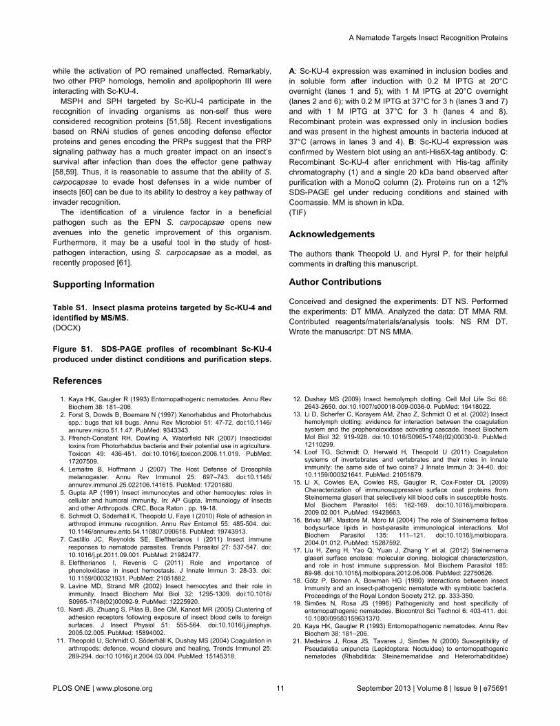
Figure S1. SDS-PAGE profiles of recombinant Sc-KU-4produced under distinct conditions and purification steps.
A: Sc-KU-4 expression was examined in inclusion bodies andin soluble form after induction with 0.2 M IPTG at 20°Covernight (lanes 1 and 5); with 1 M IPTG at 20°C overnight(lanes 2 and 6); with 0.2 M IPTG at 37°C for 3 h (lanes 3 and 7)and with 1 M IPTG at 37°C for 3 h (lanes 4 and 8).Recombinant protein was expressed only in inclusion bodiesand was present in the highest amounts in bacteria induced at37°C (arrows in lanes 3 and 4). B: Sc-KU-4 expression wasconfirmed by Western blot using an anti-His6X-tag antibody. C:Recombinant Sc-KU-4 after enrichment with His-tag affinitychromatography (1) and a single 20 kDa band observed afterpurification with a MonoQ column (2). Proteins run on a 12%SDS-PAGE gel under reducing conditions and stained withCoomassie. MM is shown in kDa.(TIF)
Acknowledgements
The authors thank Theopold U. and Hyrsl P. for their helpfulcomments in drafting this manuscript.
Author Contributions
Conceived and designed the experiments: DT NS. Performedthe experiments: DT MMA. Analyzed the data: DT MMA RM.Contributed reagents/materials/analysis tools: NS RM DT.Wrote the manuscript: DT NS MMA.
References
1. Kaya HK, Gaugler R (1993) Entomopathogenic nematodes. Annu RevBiochem 38: 181–206.
2. Forst S, Dowds B, Boemare N (1997) Xenorhabdus and Photorhabdusspp.: bugs that kill bugs. Annu Rev Microbiol 51: 47-72. doi:10.1146/annurev.micro.51.1.47. PubMed: 9343343.
3. Ffrench-Constant RH, Dowling A, Waterfield NR (2007) Insecticidaltoxins from Photorhabdus bacteria and their potential use in agriculture.Toxicon 49: 436-451. doi:10.1016/j.toxicon.2006.11.019. PubMed:17207509.
4. Lemaitre B, Hoffmann J (2007) The Host Defense of Drosophilamelanogaster. Annu Rev Immunol 25: 697–743. doi:10.1146/annurev.immunol.25.022106.141615. PubMed: 17201680.
5. Gupta AP (1991) Insect immunocytes and other hemocytes: roles incellular and humoral immunity. In: AP Gupta. Immunology of Insectsand other Arthropods. CRC, Boca Raton . pp. 19-18.
6. Schmidt O, Söderhäll K, Theopold U, Faye I (2010) Role of adhesion inarthropod immune recognition. Annu Rev Entomol 55: 485-504. doi:10.1146/annurev.ento.54.110807.090618. PubMed: 19743913.
7. Castillo JC, Reynolds SE, Eleftherianos I (2011) Insect immuneresponses to nematode parasites. Trends Parasitol 27: 537-547. doi:10.1016/j.pt.2011.09.001. PubMed: 21982477.
8. Eleftherianos I, Revenis C (2011) Role and importance ofphenoloxidase in insect hemostasis. J Innate Immun 3: 28-33. doi:10.1159/000321931. PubMed: 21051882.
9. Lavine MD, Strand MR (2002) Insect hemocytes and their role inimmunity. Insect Biochem Mol Biol 32: 1295-1309. doi:10.1016/S0965-1748(02)00092-9. PubMed: 12225920.
10. Nardi JB, Zhuang S, Pilas B, Bee CM, Kanost MR (2005) Clustering ofadhesion receptors following exposure of insect blood cells to foreignsurfaces. J Insect Physiol 51: 555-564. doi:10.1016/j.jinsphys.2005.02.005. PubMed: 15894002.
11. Theopold U, Schmidt O, Söderhäll K, Dushay MS (2004) Coagulation inarthropods: defence, wound closure and healing. Trends Immunol 25:289-294. doi:10.1016/j.it.2004.03.004. PubMed: 15145318.
12. Dushay MS (2009) Insect hemolymph clotting. Cell Mol Life Sci 66:2643-2650. doi:10.1007/s00018-009-0036-0. PubMed: 19418022.
13. Li D, Scherfer C, Korayem AM, Zhao Z, Schmidt O et al. (2002) Insecthemolymph clotting: evidence for interaction between the coagulationsystem and the prophenoloxidase activating cascade. Insect BiochemMol Biol 32: 919-928. doi:10.1016/S0965-1748(02)00030-9. PubMed:12110299.
14. Loof TG, Schmidt O, Herwald H, Theopold U (2011) Coagulationsystems of invertebrates and vertebrates and their roles in innateimmunity: the same side of two coins? J Innate Immun 3: 34-40. doi:10.1159/000321641. PubMed: 21051879.
15. Li X, Cowles EA, Cowles RS, Gaugler R, Cox-Foster DL (2009)Characterization of immunosuppressive surface coat proteins fromSteinernema glaseri that selectively kill blood cells in susceptible hosts.Mol Biochem Parasitol 165: 162-169. doi:10.1016/j.molbiopara.2009.02.001. PubMed: 19428663.
16. Brivio MF, Mastore M, Moro M (2004) The role of Steinernema feltiaebodysurface lipids in host-parasite immunological interactions. MolBiochem Parasitol 135: 111–121. doi:10.1016/j.molbiopara.2004.01.012. PubMed: 15287592.
17. Liu H, Zeng H, Yao Q, Yuan J, Zhang Y et al. (2012) Steinernemaglaseri surface enolase: molecular cloning, biological characterization,and role in host immune suppression. Mol Biochem Parasitol 185:89-98. doi:10.1016/j.molbiopara.2012.06.006. PubMed: 22750626.
18. Götz P, Boman A, Bowman HG (1980) Interactions between insectimmunity and an insect-pathogenic nematode with symbiotic bacteria.Proceedings of the Royal London Society 212. pp. 333-350.
19. Simões N, Rosa JS (1996) Pathogenicity and host specificity ofentomopathogenic nematodes. Biocontrol Sci Technol 6: 403-411. doi:10.1080/09583159631370.
20. Kaya HK, Gaugler R (1993) Entomopathogenic nematodes. Annu RevBiochem 38: 181–206.
21. Medeiros J, Rosa JS, Tavares J, Simões N (2000) Susceptibility ofPseudaletia unipuncta (Lepidoptera: Noctuidae) to entomopathogenicnematodes (Rhabditida: Steinernematidae and Heterorhabditidae)
A Nematode Targets Insect Recognition Proteins
PLOS ONE | www.plosone.org 11 September 2013 | Volume 8 | Issue 9 | e75691

isolated in the Azores: effect of nematode strain and host age. J EconEntomol 93: 1403-1408. doi:10.1603/0022-0493-93.5.1403. PubMed:11057710.
22. Dowds BCA, Peters A (2002) Virulence mechanisms.Entomopathogenic Nematol: 79–98.
23. Cruz N, Rosa JS, Simões N (2001) Encapsulation response of 6thinstar of Pseudaletia unipuncta (Lepidoptera: Noctuidae) toSteinernema carpocapsae (Nematoda: Steinernematidae). J InvertebrPathol 78: 272-274. doi:10.1006/jipa.2001.5033. PubMed: 12009810.
24. Peters A, Ehlers RU (1997) Encapsulation of the entomopathogenicnematode Steinernema feltiae in Tipula oleracea. J Invertebr Pathol 69:218-222. doi:10.1006/jipa.1996.4648. PubMed: 9170347.
25. Wang Z, Wilhelmsson C, Hyrsl P, Loof TG, Dobes P (2010) Pathogenentrapment by transglutaminase - a conserved early innate immunemechanism. PLOS Pathog 6:e1000763.
26. Zang X, Maizels RM (2001) Serine proteinase inhibitors fromnematodes and the arms race between host and pathogen. TrendsBiochem Sci 26: 191-197. doi:10.1016/S0968-0004(00)01761-8.PubMed: 11246026.
27. Milstone AM, Harrison LM, Bungiro RD, Kuzmic P, Cappello M (2000)A broad spectrum Kunitz type serine protease inhibitor secreted by thehookworm Ancylostoma ceylanicum. J Biol Chem 275: 29391-29399.doi:10.1074/jbc.M002715200. PubMed: 10893410.
28. Zang X, Maizels RM (2001) Serine proteinase inhibitors fromnematodes and the arms race between host and pathogen. TrendsBiochem Sci 26: 191-197. doi:10.1016/S0968-0004(00)01761-8.PubMed: 11246026.
29. Ghendler Y, Arnon R, Fishelson Z (1994) Schistosoma mansoni:isolation and characterization of Smpi56, a novel serine proteaseinhibitor. Exp Parasitol 78: 121-131. doi:10.1006/expr.1994.1013.PubMed: 8119369.
30. Rhoads ML, Fetterer RH, Hill DE, Urban JF (2000) Trichuris suis: asecretory chymotrypsin/elastase inhibitor with potential as animmunomodulator. Exp Parasitol 95: 36-44. doi:10.1006/expr.2000.4502. PubMed: 10864516.
31. Balasubramanian N, Hao YJ, Toubarro D, Nascimento G, Simões N(2009) Purification, biochemical and molecular analysis of achymotrypsin protease with prophenoloxidase suppression activity fromthe entomopathogenic nematode Steinernema carpocapsae. Int JParasitol 39: 975–984. doi:10.1016/j.ijpara.2009.01.012. PubMed:19249304.
32. Balasubramanian N, Toubarro D, Simões N (2010) Biochemical studyand in vitro insect immune suppression by a trypsin-like secretedprotease from the nematode Steinernema carpocapsae. ParasiteImmunol 32: 165-175. doi:10.1111/j.1365-3024.2009.01172.x. PubMed:20398179.
33. Toubarro D, Lucena-Robles M, Nascimento G, Santos R, Montiel R etal. (2010) Serine protease-mediated host invasion by the parasiticnematode Steinernema carpocapsae. J Biol Chem 285: 30666-30675.doi:10.1074/jbc.M110.129346. PubMed: 20656686.
34. Toubarro D, Lucena-Robles M, Nascimento G, Costa G, Montiel R etal. (2009) An apoptosis-inducing serine protease secreted by theentomopathogenic nematode Steinernema carpocapsae. Int J Parasitol39: 1319-1330. doi:10.1016/j.ijpara.2009.04.013. PubMed: 19481087.
35. Hao YJ, Montiel R, Abubucker S, Mitreva M, Simões N (2010)Transcripts analysis of the entomopathogenic nematode Steinernemacarpocapsae induced in vitro with insect haemolymph. Mol BiochemParasitol 169: 79-86. doi:10.1016/j.molbiopara.2009.10.002. PubMed:19836423.
36. Dutky SR, Thompson JV, Cantwell GE (1964) A technique for massproduction of the DD-136 nematode. J Insect Pathol 6: 417-422.
37. White GF (1927) A method for obtaining infective nematode larvae fromcultures. Science 66: 302-303. doi:10.1126/science.66.1709.302-a.PubMed: 17749713.
38. Bidla G, Lindgren M, Theopold U, Dushay MS (2005) Hemolymphcoagulation and phenoloxidase in Drosophila larvae. Dev CompImmunol 29: 669–679. doi:10.1016/j.dci.2004.11.007. PubMed:15854679.
39. Zang X, Maizels RM (2001) Serine proteinase inhibitors fromnematodes and the arms race between host and pathogen. TrendsBiochem Sci 26: 191-197. doi:10.1016/S0968-0004(00)01761-8.PubMed: 11246026.
40. Dy CY, Buczek P, Imperial JS, Bulaj G, Horvath MP (2006) Structure ofconkunitzin-S1, a neurotoxin and Kunitz-fold disulphide variant fromcone snail. Acta Crystallogr D Biol Crystallogr 62: 980-990. doi:10.1107/S1744309106033884. PubMed: 16929098.
41. Helland R, Berglund GI, Otlewski J, Apostoluk W, Andersen OA et al.(1999) High-resolution structures of three new trypsin-squash-inhibitorcomplexes: a detailed comparison with other trypsins and their
complexes. Acta Crystallogr D Biol Crystallogr 55: 139-148. doi:10.1107/S090744499801052X. PubMed: 10089404.
42. Laskowski M Jr, Kato I (1980) Protein inhibitors of proteinases. AnnuRev Biochem 49: 593-626. doi:10.1146/annurev.bi.49.070180.003113.PubMed: 6996568.
43. Zhou JM (1989) Kinetics of trypsin inhibition by its specific inhibitors.Biochemistry 3: 1070-1076. PubMed: 2713358.
44. Milstone AM, Harrison LM, Bungiro RD, Kuzmic P, Cappello M (2000)A broad spectrum Kunitz type serine protease inhibitor secreted by thehookworm Ancylostoma ceylanicum. J Biol Chem 275: 29391-29399.doi:10.1074/jbc.M002715200. PubMed: 10893410.
45. Pugia MJ, Valdes R Jr, Jortani SA (2007) Bikunin (urinary trypsininhibitor): structure, biological relevance, and measurement. Adv ClinChem 44: 223-245. doi:10.1016/S0065-2423(07)44007-0. PubMed:17682344.
46. Gobert V, Gottar M, Matskevich AA, Rutschmann S, Royet J et al.(2003) Dual activation of the Drosophila toll pathway by two patternrecognition receptors. Science 302: 2126-2130. doi:10.1126/science.1085432. PubMed: 14684822.
47. Söderhäll K, Cerenius L (1998) Role of the prophenoloxidase-activatingsystem in invertebrate immunity. Curr Opin Immunol 10: 23-28. doi:10.1016/S0952-7915(98)80026-5. PubMed: 9523106.
48. Beck M, Theopold U, Schmidt O (2000) Evidence for serine proteaseinhibitor activity in the ovarian calyx fluid of the endoparasitoid Venturiacanescens. J Insect Physiol 46: 1275-1283. doi:10.1016/S0022-1910(00)00048-2. PubMed: 10844146.
49. Lavine MD, Strand MR (2002) Insect hemocytes and their role inimmunity. Insect Biochem Mol Biol 32: 1295-1309. doi:10.1016/S0965-1748(02)00092-9. PubMed: 12225920.
50. Zhu JY, Fang Q, Ye GY, Hu C (2011) Proteome changes in the plasmaof Pieris rapae parasitized by the endoparasitoid wasp Pteromaluspuparum. J Zhejiang Univ Sci B 12: 93-102. doi:10.1631/jzus.B1000158. PubMed: 21265041.
51. Lee SY, Söderhäll K (2001) Characterization of a pattern recognitionprotein, a masquerade-like protein, in the freshwater crayfishPacifastacus leniusculus. J Immunol 166: 7319-7326. PubMed:11390482.
52. Lu Z, Jiang H (2008) Expression of Manduca sexta serine proteinasehomolog precursors in insect cells and their proteolytic activation.Insect Biochem Mol Biol 38: 89-98. doi:10.1016/j.ibmb.2007.09.011.PubMed: 18070668.
53. Yu XQ, Jiang H, Wang Y, Kanost MR (2003) Nonproteolytic serineproteinase homologs are involved in prophenoloxidase activation in thetobacco hornworm, Manduca sexta. Insect Biochem Mol Biol 33:197-208. doi:10.1016/S0965-1748(02)00191-1. PubMed: 12535678.
54. Gupta S, Wang Y, Jiang H (2005) Manduca sexta prophenoloxidase(proPO) activation requires proPO-activating proteinase (PAP) andserine proteinase homologs (SPHs) simultaneously. Insect BiochemMol Biol 35: 241–248. doi:10.1016/j.ibmb.2004.12.003. PubMed:15705503.
55. Jitvaropas R, Amparyup P, Gross PS, Tassanakajon A (2009)Functional characterization of a masquerade-like serine proteinasehomologue from the black tiger shrimp Penaeus monodon. CompBiochem Physiol B Biochem Mol Biol 153: 236-243. doi:10.1016/j.cbpb.2009.03.007. PubMed: 19328243.
56. Yu XQ, Kanost MR (2002) Binding of hemolin to bacteriallipopolysaccharide and lipoteichoic acid. An immunoglobulinsuperfamily member from insects as a pattern-recognition receptor. EurJ Biochem 269: 1827-1834. doi:10.1046/j.1432-1033.2002.02830.x.PubMed: 11952784.
57. Huang TS, Wang H, Lee SY, Johansson MW (2000) A cell adhesionprotein from the crayfish Pacifastacus leniusculus, a serine proteinasehomologue similar to Drosophila masquerade. J Biol Chem 275:9996-10001. doi:10.1074/jbc.275.14.9996. PubMed: 10744675.
58. Eleftherianos I, Gökçen F, Felföldi G, Millichap PJ, Trenczek TE et al.(2007) The immunoglobulin family protein Hemolin mediates cellularimmune responses to bacteria in the insect Manduca sexta. CellMicrobiol 9: 1137-1147. doi:10.1111/j.1462-5822.2006.00855.x.PubMed: 17166232.
59. Felföldi G, Eleftherianos I, Ffrench-Constant RH, Venekei I (2011) Aserine proteinase homologue, SPH-3, plays a central role in insectimmunity. J Immunol 186: 4828-4834. doi:10.4049/jimmunol.1003246.PubMed: 21398604.
60. Laumond C, Mauleon H, Kermarrec A (1979) Données nouvelles sur lespectre d’hôtes et le parasitisme du nématode entomophageNeoaplectana carpocapsae. Entomophaga 24: 13–27. doi:10.1007/BF02377505.
61. Hallem EA, Rengarajan M, Ciche TA, Sternberg PW (2007)Nematodes, bacteria, and flies: a tripartite model for nematode
A Nematode Targets Insect Recognition Proteins
PLOS ONE | www.plosone.org 12 September 2013 | Volume 8 | Issue 9 | e75691

parasitism. Curr Biol 17: 898-904. doi:10.1016/j.cub.2007.04.027.PubMed: 17475494.
A Nematode Targets Insect Recognition Proteins
PLOS ONE | www.plosone.org 13 September 2013 | Volume 8 | Issue 9 | e75691