back to basics review: respirology in under two hours andrew burkett md frcpc respirology...
TRANSCRIPT
Back to Basics Review:Respirology in Under Two Hours
Andrew Burkett MD FRCPC RespirologyInterventional Pulmonology Fellow
April 1, 2015

The Plan...
• Pulmonary Function Testing
• Blood Gases
• Asthma
• COPD
• Sleep Apnea
• Pleural Effusion
• Lung Cancer

Pulmonary Function tests
1. Flows: Volume-Time Curve Flow-Volume Loop2. Lung Volumes: Body Box (plesmography)
vs. Helium Dilution

Spirometry: Measurement of Airflow
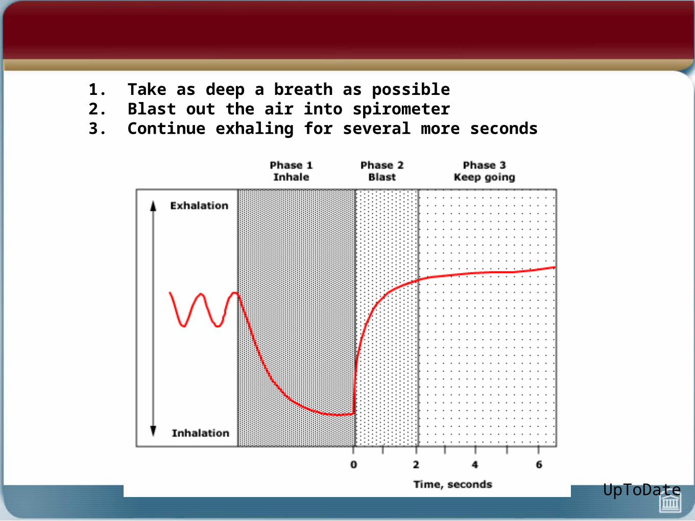
1. Take as deep a breath as possible2. Blast out the air into spirometer3. Continue exhaling for several more seconds
UpToDate

RVTLC
Flow Volume Loop
Interpretation
• Upper Airway Abnormalities
• Obstructive Lung Disease
• Restrictive Lung Disease

Interpretation
• Look at ID• Flow volume loop• FEV1/FVC ratio• FEV1

Upper Airway Abnormalities
• Variable extrathoracic obstruction impairs inspiratory flow more than expiratory flow -- negative pressure during inspiratory “sucks in” (narrows) airway
• Variable intrathoracic obstruction impairs expiratory flow more than inspiratory flow -- positive intrathoracic pressure compresses in airway
ERJ 2005; 26: 948-968
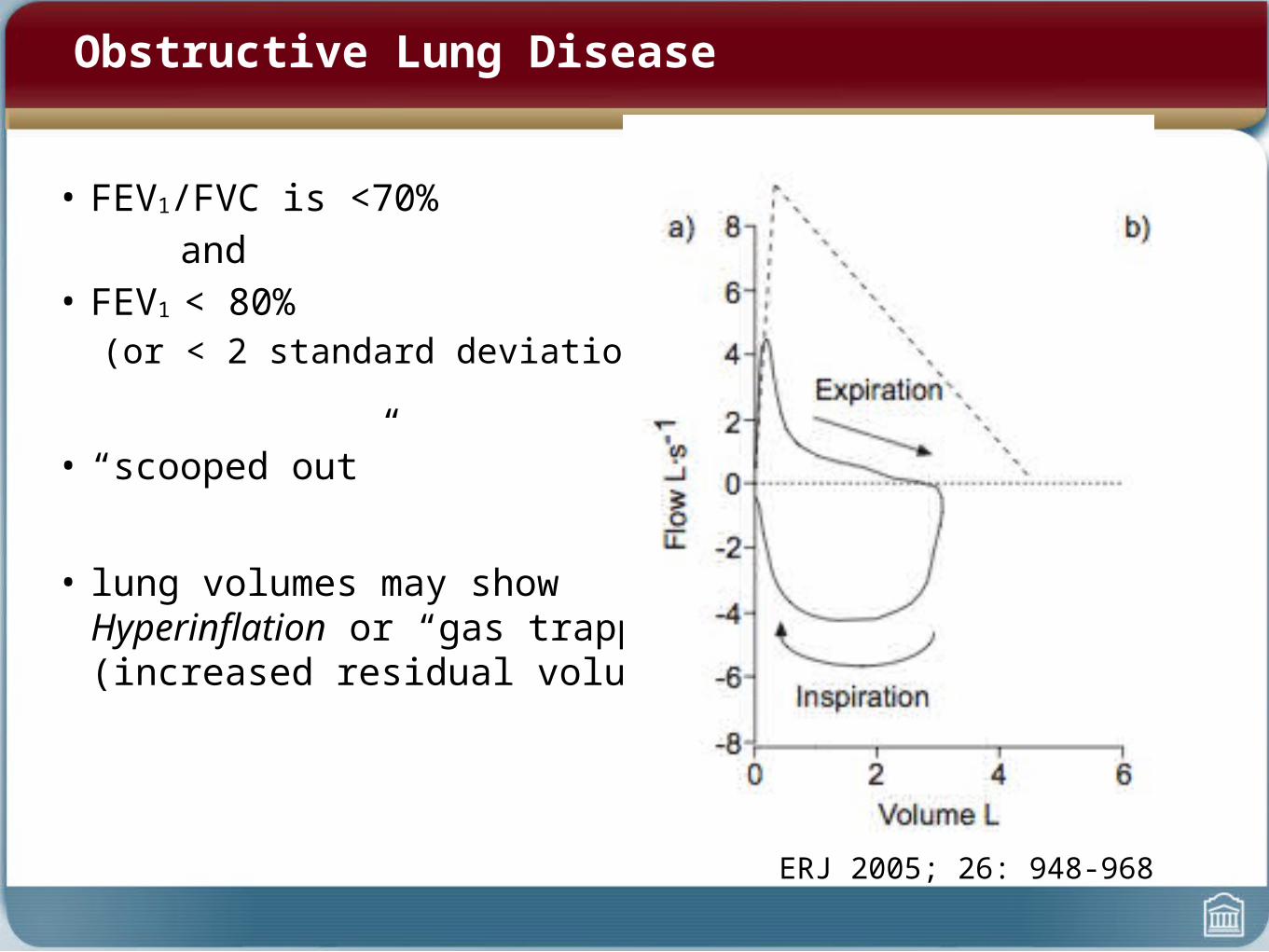
Obstructive Lung Disease
• FEV1/FVC is <70%and
• FEV1 < 80%(or < 2 standard deviations)
• “scooped out”
• lung volumes may show Hyperinflation or “gas trapping”(increased residual volume)
ERJ 2005; 26: 948-968
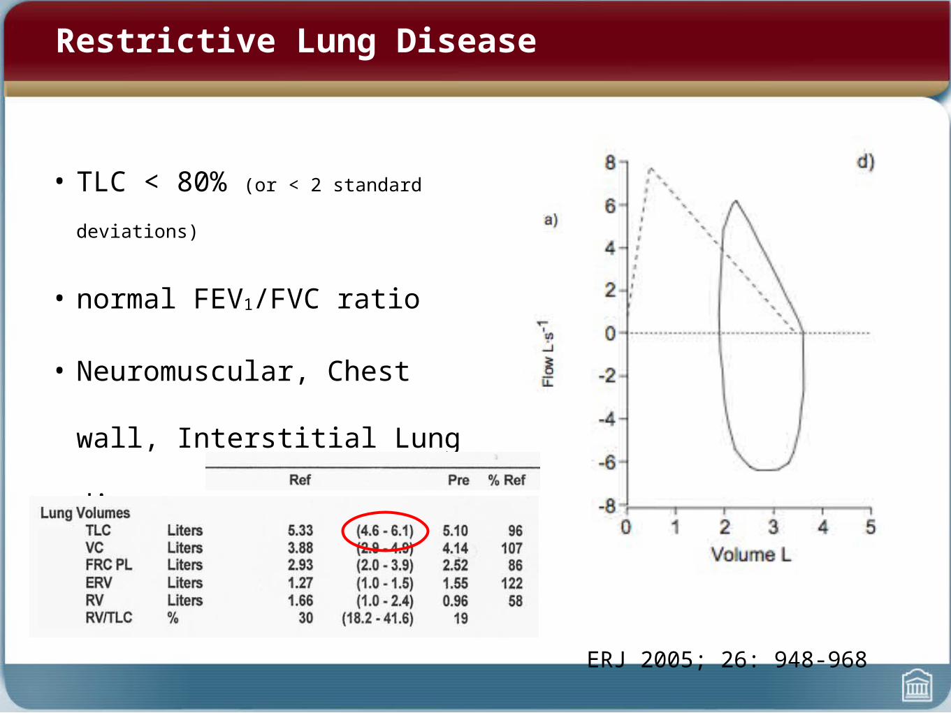
Restrictive Lung Disease
• TLC < 80% (or < 2 standard deviations)
• normal FEV1/FVC ratio
• Neuromuscular, Chest wall,
Interstitial Lung disease
ERJ 2005; 26: 948-968
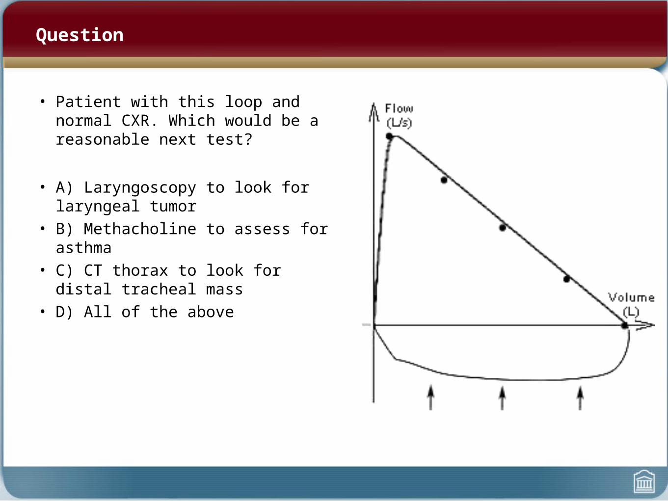
Question
• Patient with this loop and normal CXR. Which would be a reasonable next test?
• A) Laryngoscopy to look for laryngeal tumor• B) Methacholine to assess for asthma• C) CT thorax to look for distal tracheal mass• D) All of the above
Blood gases
• Stable patient. Compensated or uncompensated?
• ABG 7.23/80/60/30• pH = 7.23• PO2 = 80• PCO2 = 60• Bicarb = 30

Blood gases
• ABG 7.25/80/80/36• pH = 7.25• PO2 = 80• PCO2 = 80• Bicarb = 36
• This patient has had this stable ABG for the last 48 hours. The correct interpretation if this is:
• A) Compensated Respiratory Acidosis• B) Uncompensated Respiratory Acidosis• C) Compensated Metabolic Acidosis• D) Uncompensated Metabolic Acidosis

Respiratory Acidosis

Blood gases
• ABG. Compensated or Uncompensated?
• ABG 7.23/80/25/9• pH = 7.23• PO2 = 80• PCO2 = 25• Bicarb = 9
Metabolic Acidosis/Alkalosis
• Compensation rules– For every drop of 1 by Bicarb (met acidosis), PC02 should drop by approx 1– For every raise of 1 by Bicarb (met alkalosis), PCO2 should rise by 0.5-0.7 (about 2/3)– “Left hand rule” or “rule of thumb”
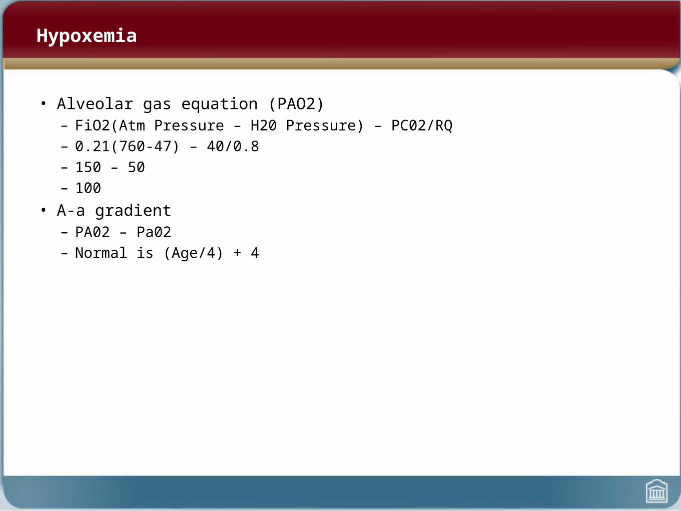
Hypoxemia
• Alveolar gas equation (PAO2)– FiO2(Atm Pressure – H20 Pressure) – PC02/RQ– 0.21(760-47) – 40/0.8– 150 – 50– 100
• A-a gradient– PA02 – Pa02– Normal is (Age/4) + 4

Causes of Hypoxia
• Impaired ability for tissues to utilize oxygen• All the causes of hypoxemia and
– Problems carrying oxygen to cells (CO poisoning, metHb)– Problems with cells using oygen (Sepsis, genetic mitochondrial
conditions, cyanide poisoning)

What causes a low DLCO?
The life of a red blood cell (and where it could all go wrong)Propelled forward out of RV (RV failure)Should have friends to come along with him (Anemia)No blockages along the way causing deadspace (PE)Thin walled capillary that CO can diffuse through (Pulmonary Hypertension)No reduced total area for gas exchange (Emphysema, ILD)Doesn’t already have CO bound to him (Smoker)

Question
• Knowing that, what could cause an abnormally high DLCO?
Question
• Called to see a 40 year old patient post Op for hypoxia with the following ABG on room air:
• pH = 7.25• PO2 = 55• PCO2 = 68• Bicarb = 27
• Her hypoxemia is caused by:• A) Bilateral basal atelectasis from obesity• B) Narcotic use• C) Acute aspiration pneumonitis• D) Massive pulmonary embolism
( Hint: 68/0.8 = 85)
Asthma
• Pathophysiology
• Diagnosis
• Chronic Management
• Acute Management
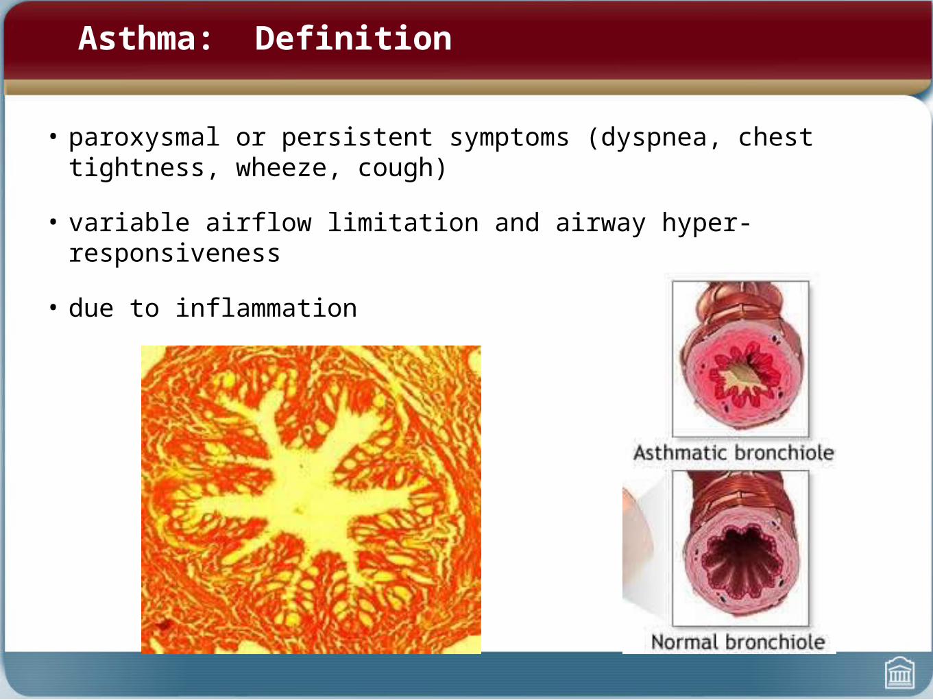
Asthma: Definition
• paroxysmal or persistent symptoms (dyspnea, chest tightness, wheeze, cough)
• variable airflow limitation and airway hyper-responsiveness
• due to inflammation

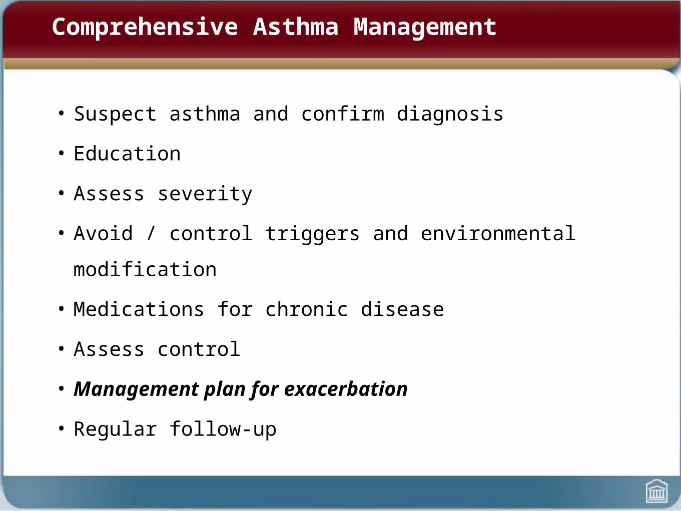
Comprehensive Asthma Management
• Suspect asthma and confirm diagnosis
• Education
• Assess severity
• Avoid / control triggers and environmental modification
• Medications for chronic disease
• Assess control
• Management plan for exacerbation
• Regular follow-up
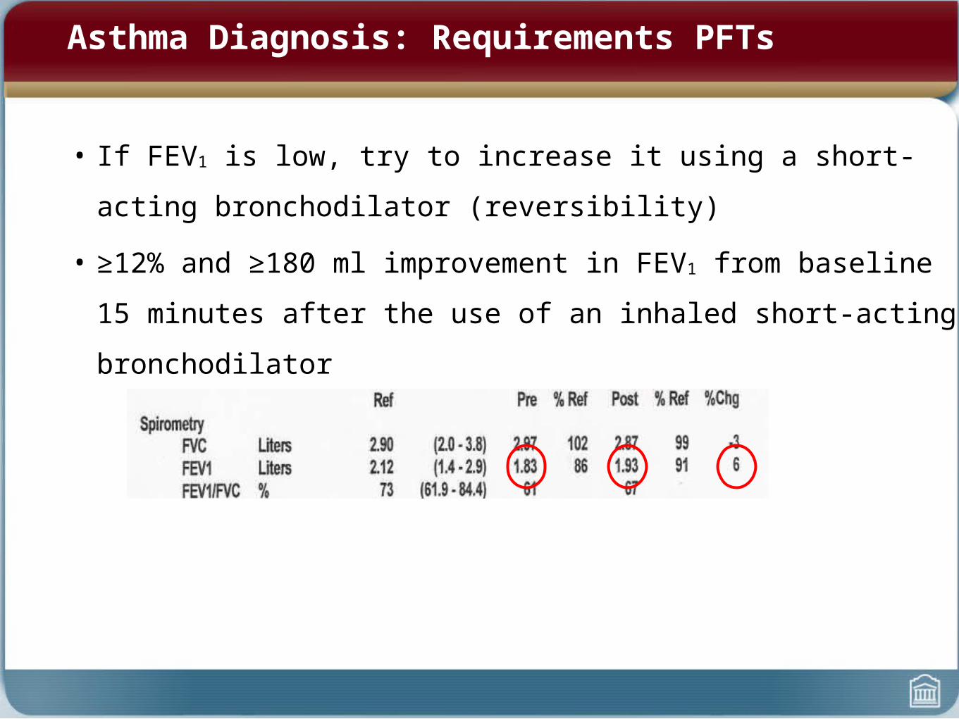
• If FEV1 is low, try to increase it using a short-acting bronchodilator
(reversibility)
• ≥12% and ≥180 ml improvement in FEV1 from baseline 15 minutes after the
use of an inhaled short-acting bronchodilator
Asthma Diagnosis: Requirements PFTs

Asthma Diagnosis

• If FEV1 is normal, try to see if airways are hyperresponsive by giving an
irritant (methacholine challenge)
Asthma Diagnosis

Comprehensive Asthma Management
• Suspect asthma and confirm diagnosis
• Education
• Assess severity
• Avoid / control triggers and environmental modification
• Medications for chronic disease
• Assess control
• Management plan for exacerbation
• Regular follow-up

Asthma Management
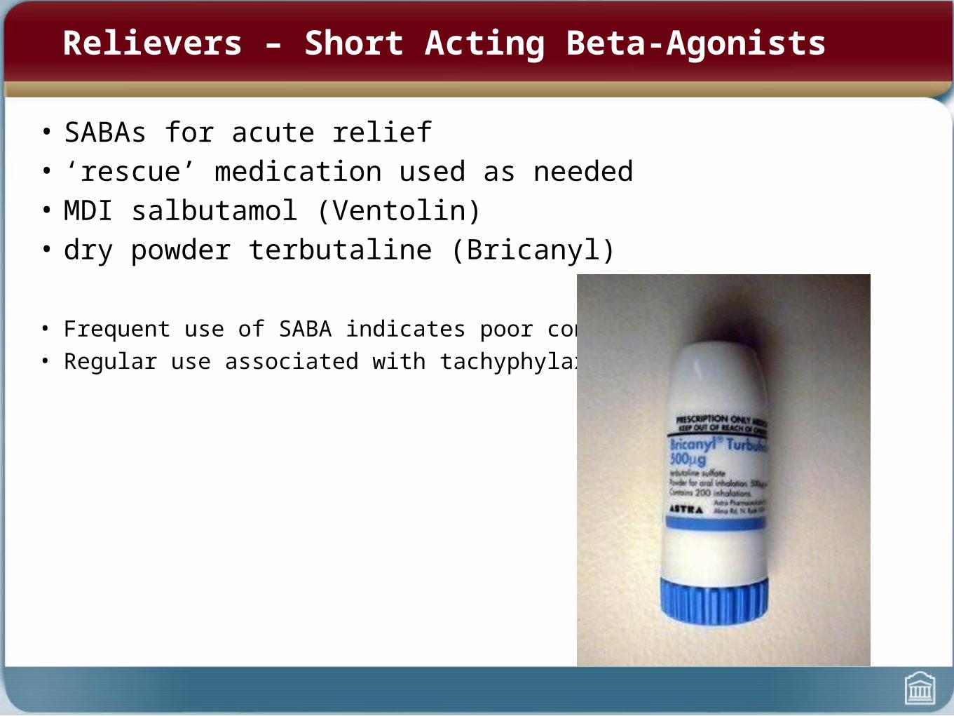
Relievers – Short Acting Beta-Agonists
• SABAs for acute relief• ‘rescue’ medication used as needed• MDI salbutamol (Ventolin) • dry powder terbutaline (Bricanyl)
• Frequent use of SABA indicates poor control• Regular use associated with tachyphylaxis

Inhaled Corticosteroids (ICS)
• Anti-inflammatory ICS mainstay of therapy
– Prevent symptoms, improve PFTs, decrease hyper-responsiveness, reduce morbidity

Inhaled Corticosteroids – How do they work?
• Like steroids produced endogenously by adrenal cortex
• Anti-inflammatory – inhibit production of cytokines, which:
– reduces eosinophil infiltration– inhibits macrophage function– reduces production of leukotrienes
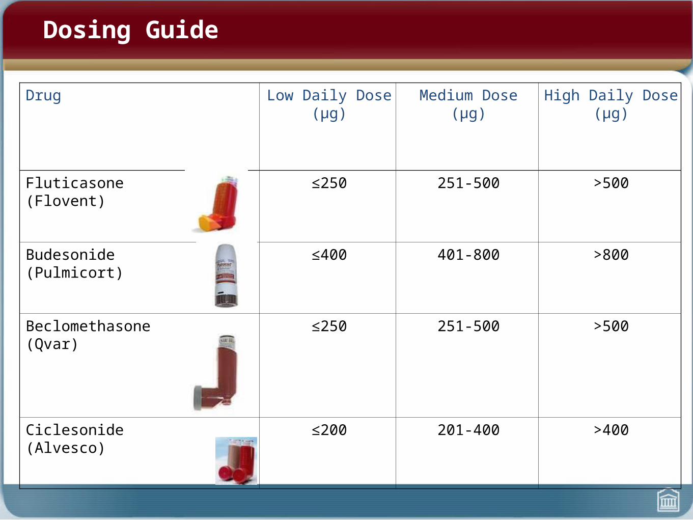
Dosing Guide
Drug Low Daily Dose (μg) Medium Dose (μg) High Daily Dose (μg)
Fluticasone(Flovent)
≤250 251-500 >500
Budesonide(Pulmicort)
≤400 401-800 >800
Beclomethasone(Qvar)
≤250 251-500 >500
Ciclesonide(Alvesco)
≤200 201-400 >400

ICS Adverse Effects
• thrush• dysphonia
• osteoporosis• decreasedgrowth velocity (?)• glaucoma • cataracts• adrenal insufficiency
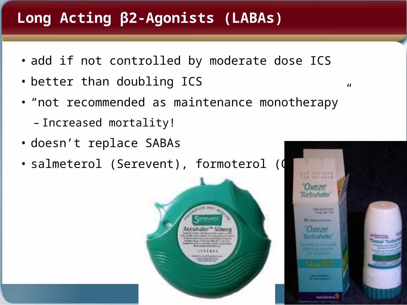
Long Acting β2-Agonists (LABAs)
• add if not controlled by moderate dose ICS
• better than doubling ICS
• “not recommended as maintenance monotherapy”
– Increased mortality!
• doesn’t replace SABAs
• salmeterol (Serevent), formoterol (Oxeze)
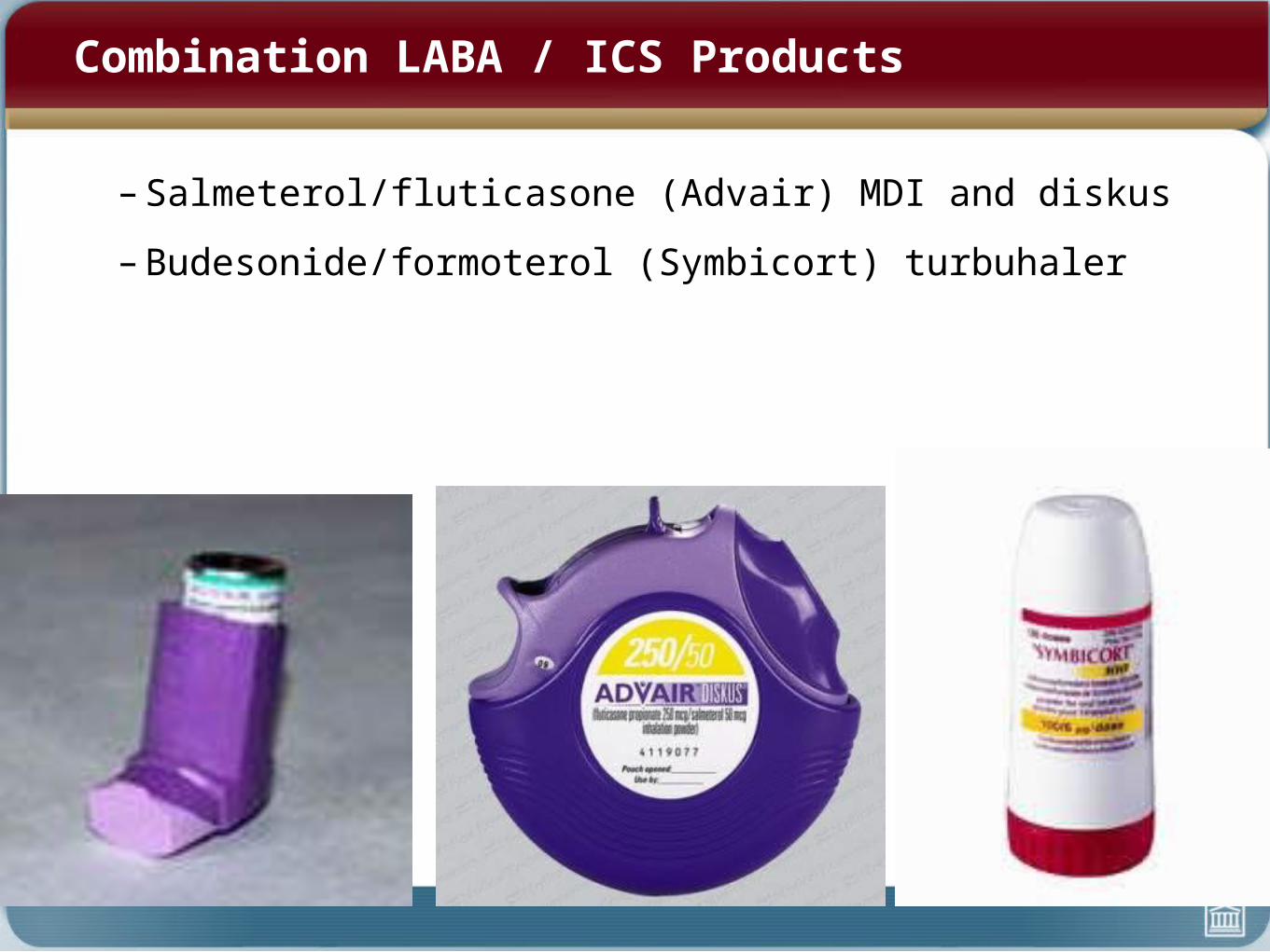
Combination LABA / ICS Products
– Salmeterol/fluticasone (Advair) MDI and diskus
– Budesonide/formoterol (Symbicort) turbuhaler
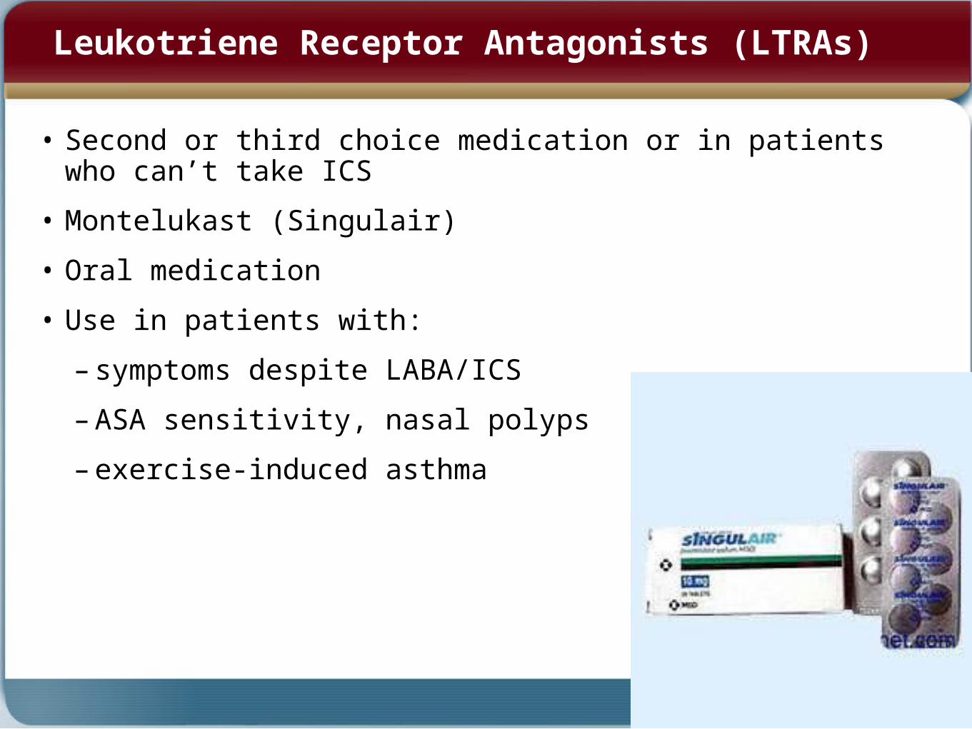
Leukotriene Receptor Antagonists (LTRAs)
• Second or third choice medication or in patients who can’t take ICS
• Montelukast (Singulair)
• Oral medication
• Use in patients with:
– symptoms despite LABA/ICS
– ASA sensitivity, nasal polyps
– exercise-induced asthma

IgE Antagonists: Omalizumab (Xolair)
• Monoclonal antibodies block action of IgE on mast cell
• Effective if IgE levels are only slightly elevated (500-1200)
• Monthly injection
• Extremely expensive
• Use if frequent need for oral steroids despite optimum conventional Rx and patient has drug plan or $$$
Comprehensive Asthma Management
• Suspect asthma and confirm diagnosis
• Education
• Assess severity
• Avoid / control triggers and environmental modification
• Medications for chronic disease
• Assess control
• Management plan for exacerbation
• Regular follow-up
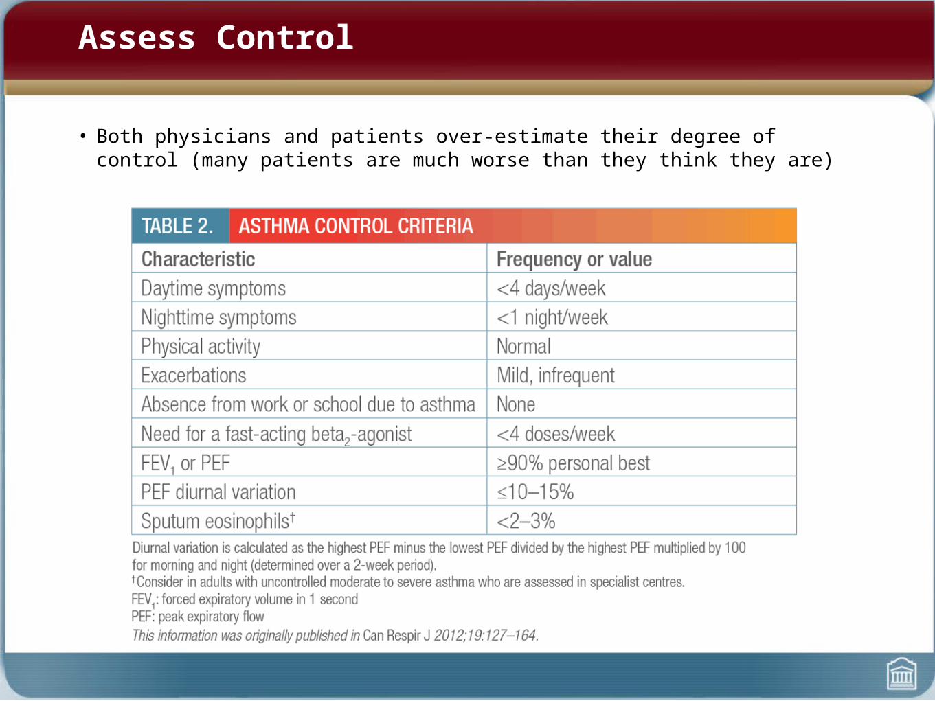
Assess Control
• Both physicians and patients over-estimate their degree of control (many patients are much worse than they think they are)
Comprehensive Asthma Management
• Suspect asthma and confirm diagnosis
• Education
• Assess severity
• Avoid / control triggers and environmental modification
• Medications for chronic disease
• Assess control
• Management plan for exacerbation
• Regular follow-up
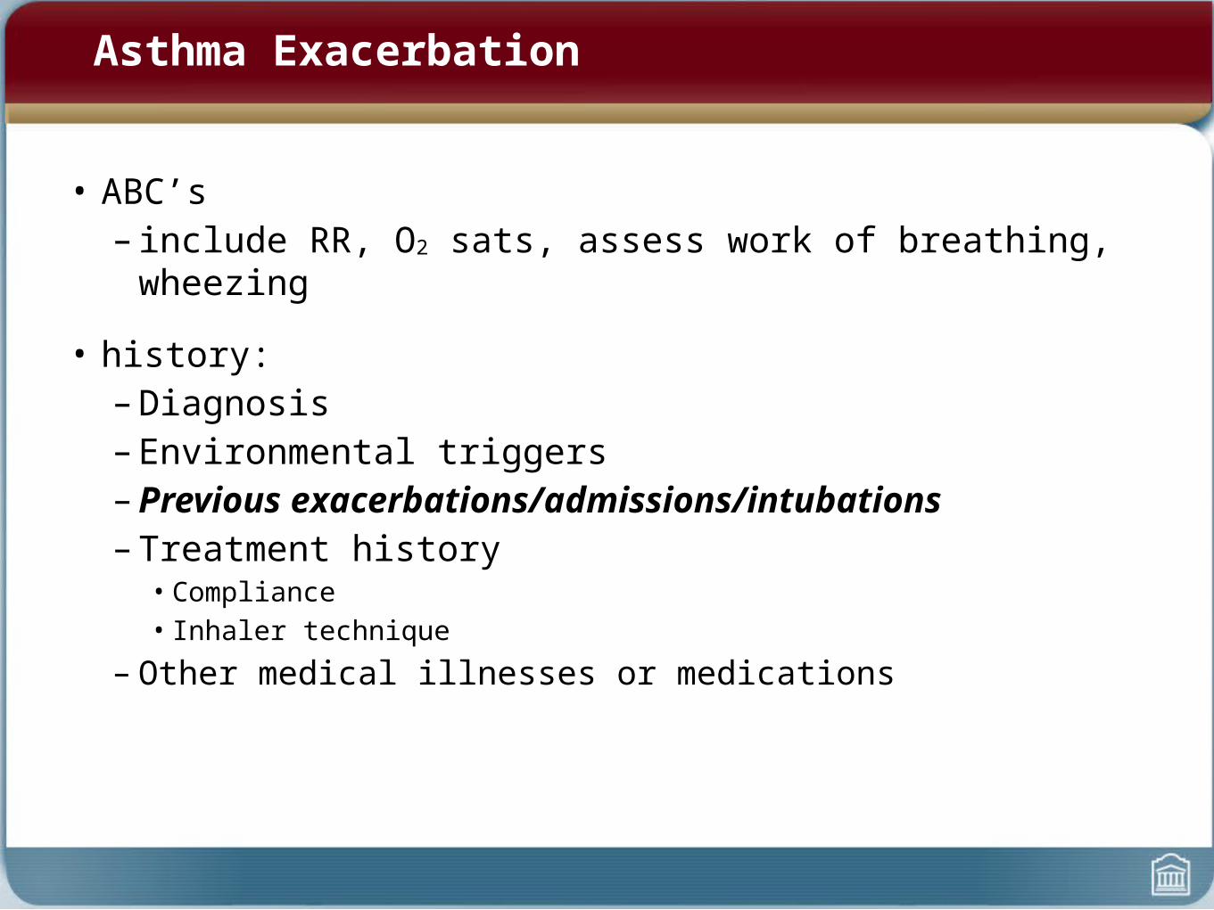
Asthma Exacerbation
• ABC’s– include RR, O2 sats, assess work of breathing, wheezing
• history: – Diagnosis– Environmental triggers– Previous exacerbations/admissions/intubations– Treatment history
• Compliance• Inhaler technique
– Other medical illnesses or medications
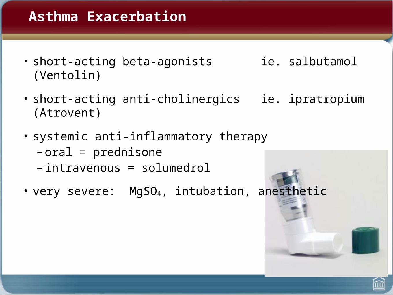
• short-acting beta-agonists ie. salbutamol (Ventolin)
• short-acting anti-cholinergics ie. ipratropium (Atrovent)
• systemic anti-inflammatory therapy– oral = prednisone– intravenous = solumedrol
• very severe: MgSO4, intubation, anesthetic
Asthma Exacerbation
COPD
• Definition
• Constrast from asthma
• Pathophysiology
• Diagnosis
• Chronic Management
• Acute Management
COPD Definition
• respiratory disorder largely caused by smoking characterized by:
- progressive, partially reversible airway obstruction
- hyperinflation
- systemic manifestations
- increasing frequency and severity of exacerbations

COPD vs. Asthma
COPD Risk Factors
• Host Factors:
- genetics (alpha-1-antitrypsin deficiency)
- bronchial hyper-responsiveness
• Environmental Factors:
- smoking
- childhood viral infections
- occupational & environmental exposures
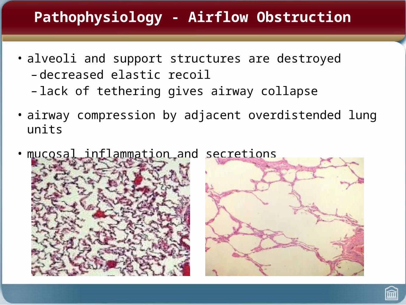
Pathophysiology - Airflow Obstruction
• alveoli and support structures are destroyed– decreased elastic recoil– lack of tethering gives airway collapse
• airway compression by adjacent overdistended lung units
• mucosal inflammation and secretions
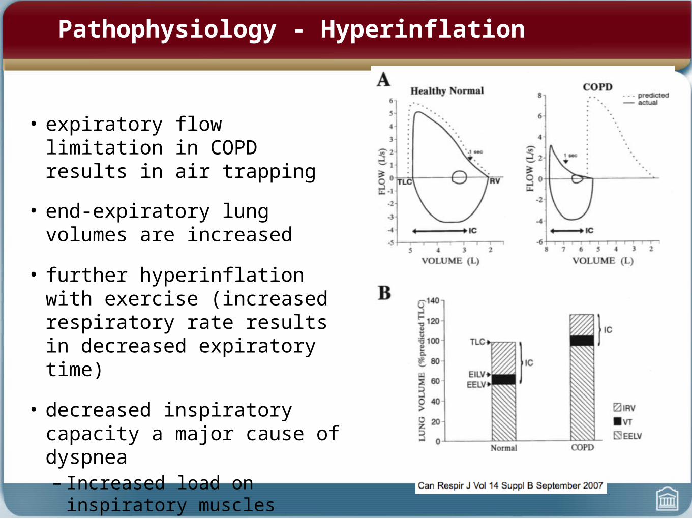
Pathophysiology - Hyperinflation
• expiratory flow limitation in COPD results in air trapping
• end-expiratory lung volumes are increased
• further hyperinflation with exercise (increased respiratory rate results in decreased expiratory time)
• decreased inspiratory capacity a major cause of dyspnea– Increased load on inspiratory
muscles
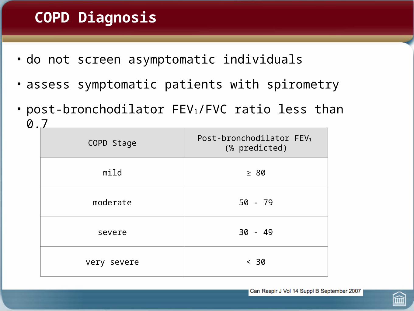
COPD Diagnosis
• do not screen asymptomatic individuals
• assess symptomatic patients with spirometry
• post-bronchodilator FEV1/FVC ratio less than 0.7
COPD Stage Post-bronchodilator FEV1
(% predicted)
mild ≥ 80
moderate 50 - 79
severe 30 - 49
very severe < 30
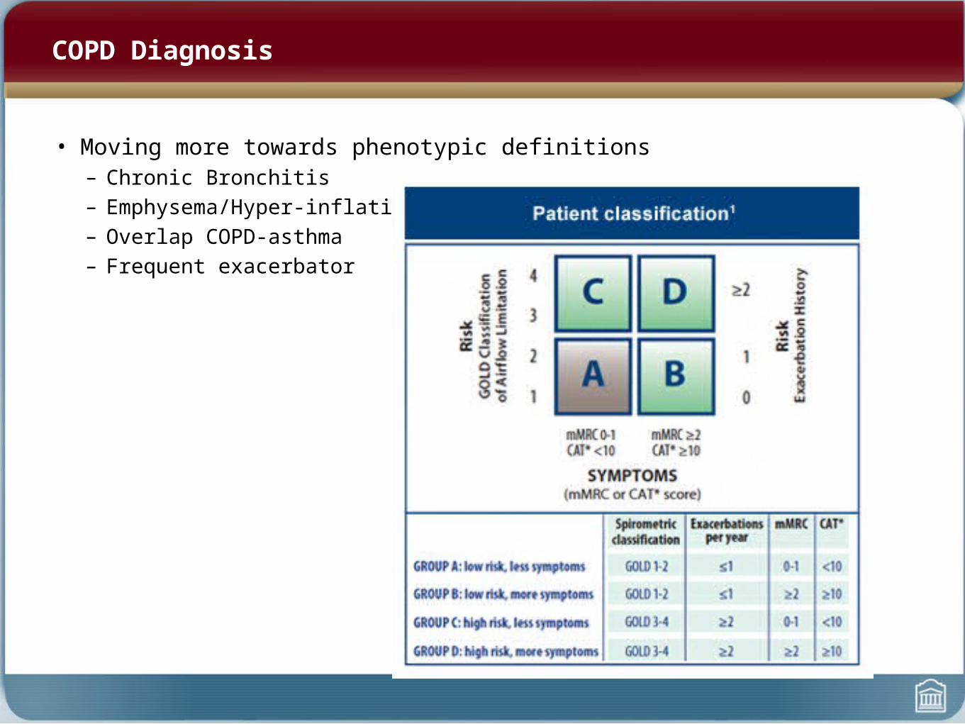
COPD Diagnosis
• Moving more towards phenotypic definitions– Chronic Bronchitis– Emphysema/Hyper-inflation– Overlap COPD-asthma– Frequent exacerbator

COPD Management


BMJ 2008; 336: 598-600.
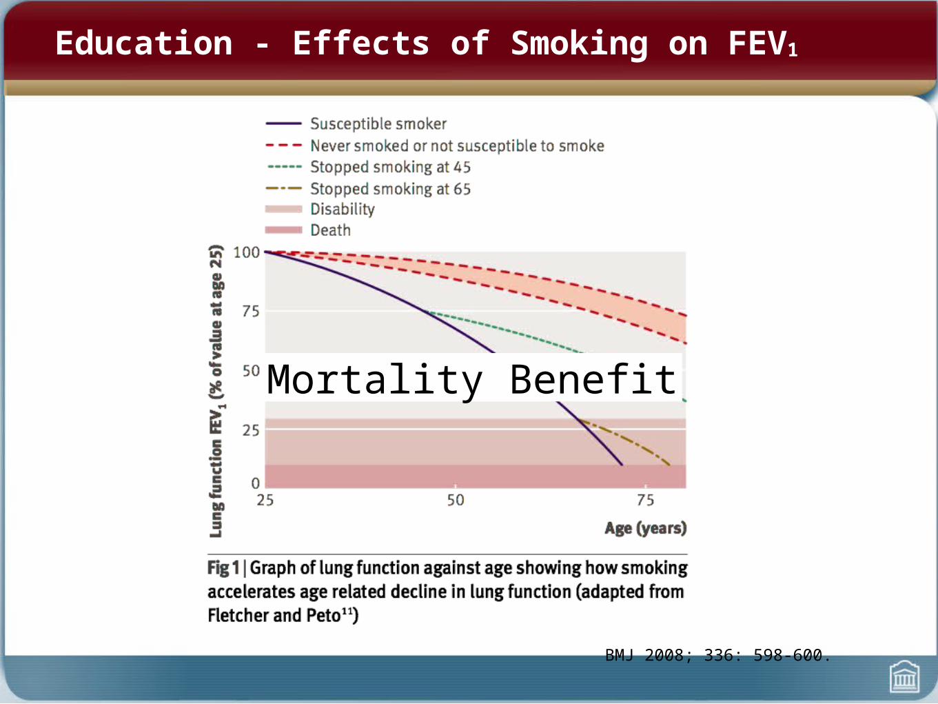
Education - Effects of Smoking on FEV1
Mortality Benefit
What Can You Do?
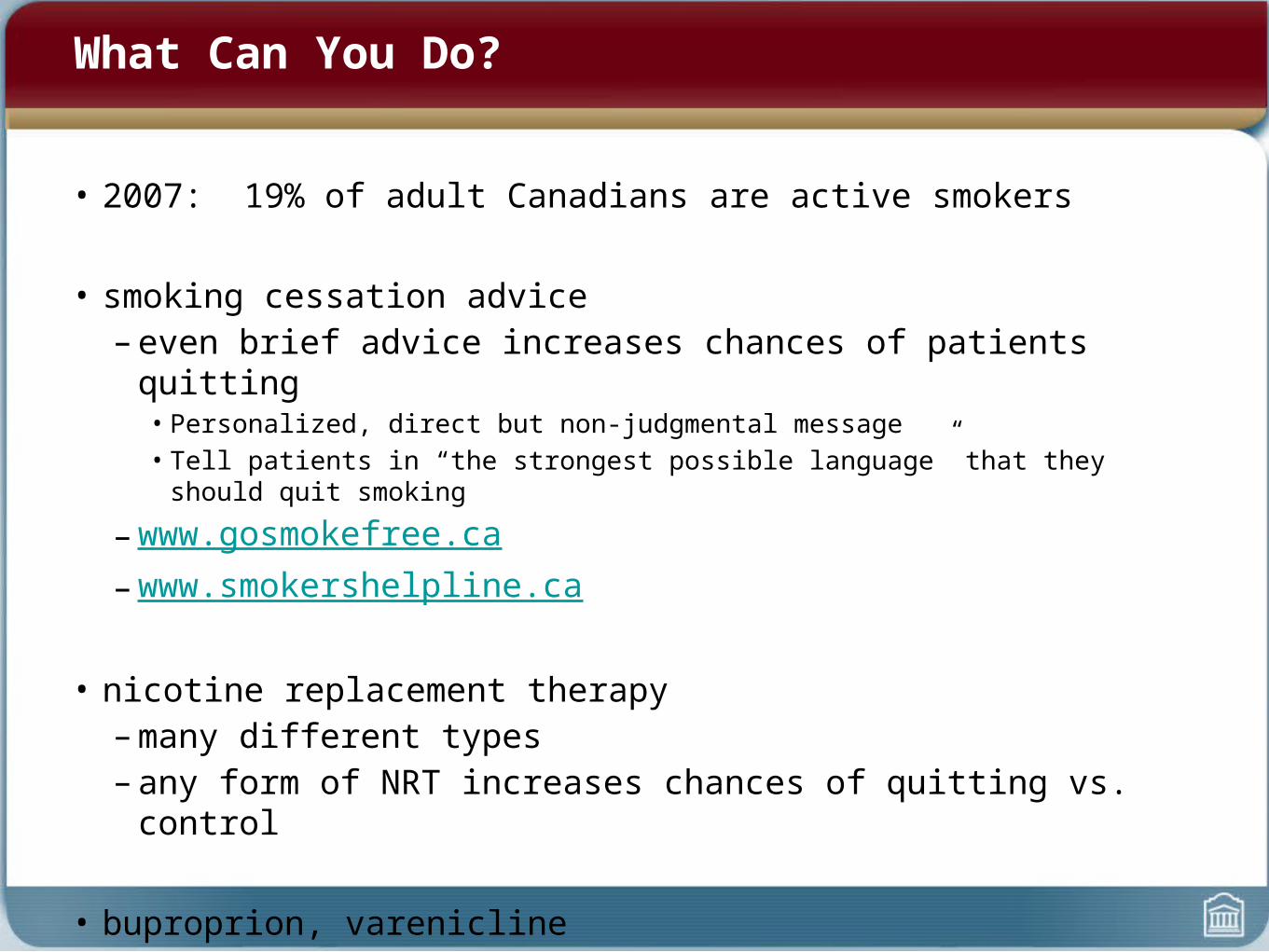
• 2007: 19% of adult Canadians are active smokers
• smoking cessation advice– even brief advice increases chances of patients quitting
• Personalized, direct but non-judgmental message• Tell patients in “the strongest possible language” that they should quit smoking
– www.gosmokefree.ca– www.smokershelpline.ca
• nicotine replacement therapy– many different types– any form of NRT increases chances of quitting vs. control
• buproprion, varenicline
Other Prevention
• vaccination:– flu vaccine yearly– pneumococcal vaccine q5years
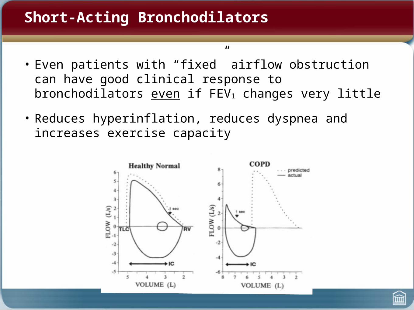
Short-Acting Bronchodilators
• Even patients with “fixed” airflow obstruction can have good clinical response to bronchodilators even if FEV1 changes very little
• Reduces hyperinflation, reduces dyspnea and increases exercise capacity
Short-Acting Bronchodilators
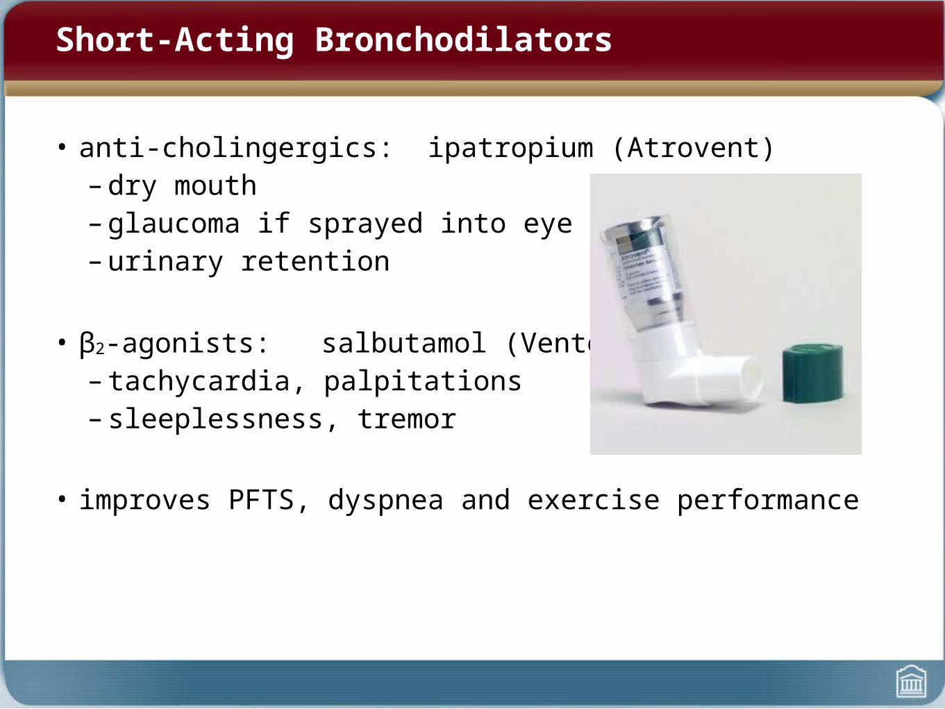
• anti-cholingergics: ipatropium (Atrovent)– dry mouth– glaucoma if sprayed into eye– urinary retention
• β2-agonists: salbutamol (Ventolin)– tachycardia, palpitations– sleeplessness, tremor
• improves PFTS, dyspnea and exercise performance
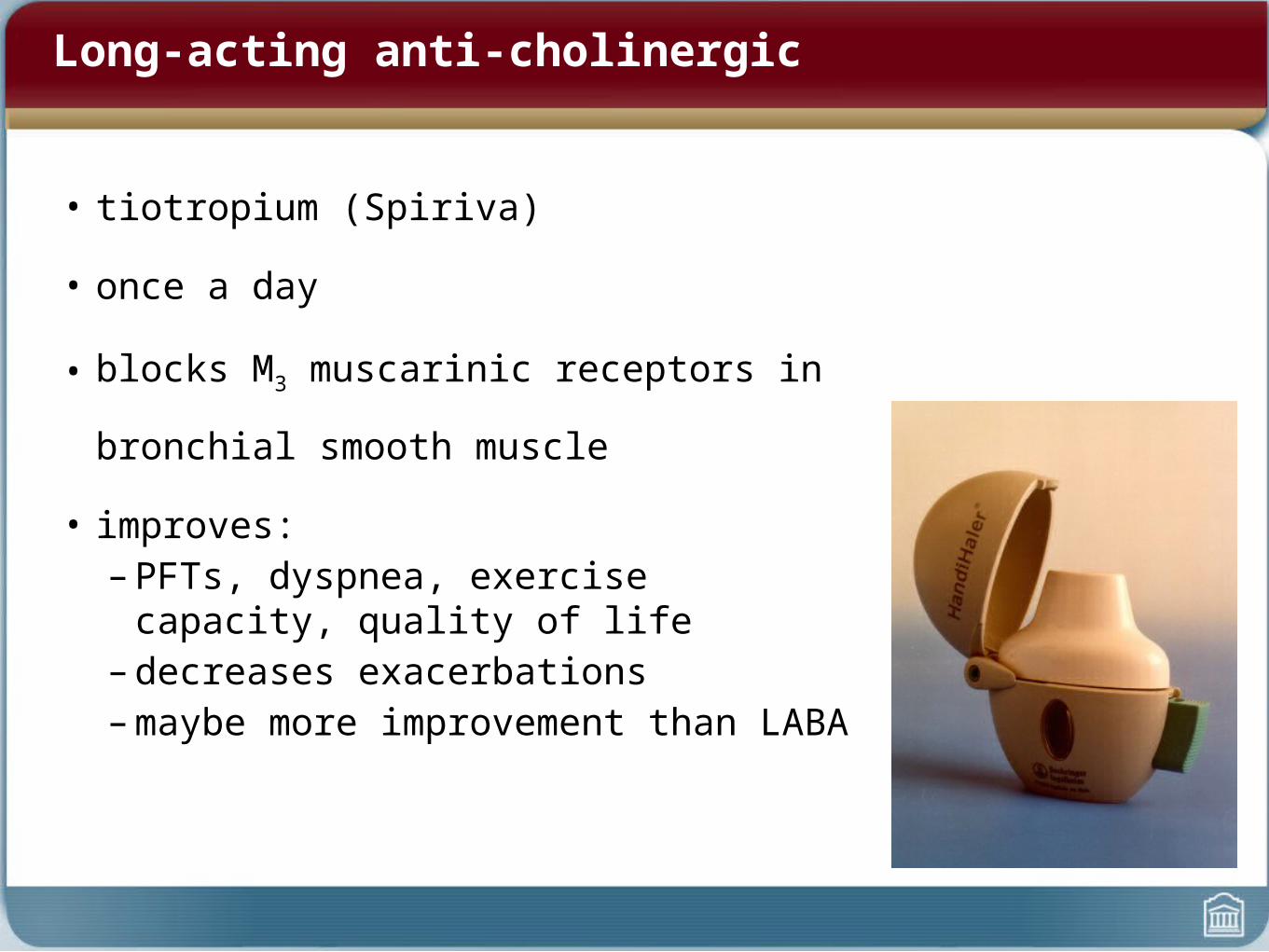
Long-acting anti-cholinergic
• tiotropium (Spiriva)
• once a day
• blocks M3 muscarinic receptors in bronchial
smooth muscle
• improves:– PFTs, dyspnea, exercise capacity, quality of life– decreases exacerbations– maybe more improvement than LABA
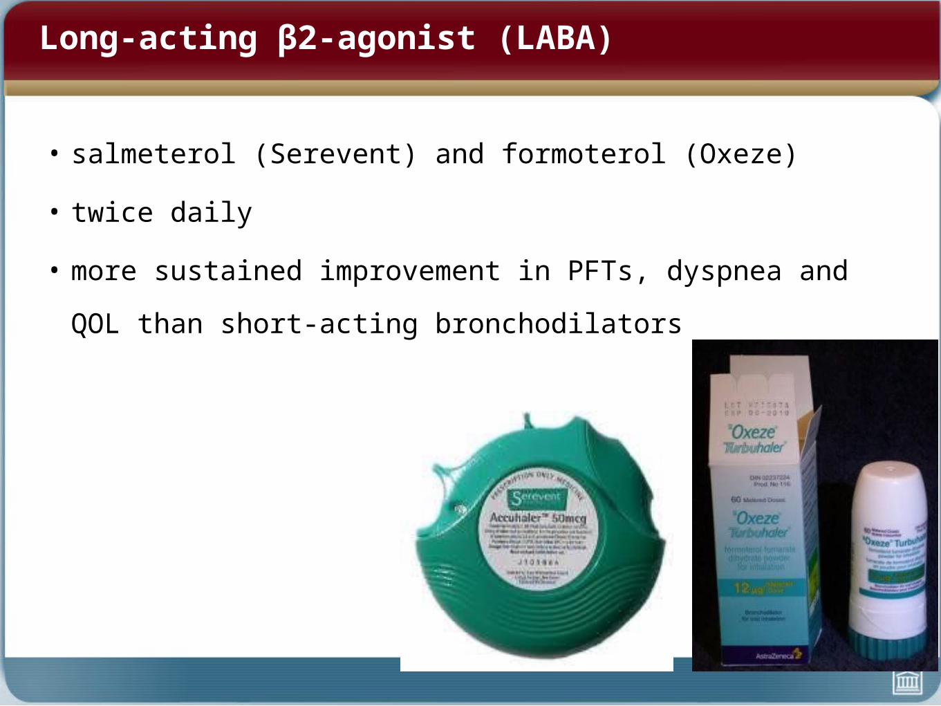
Long-acting β2-agonist (LABA)
• salmeterol (Serevent) and formoterol (Oxeze)
• twice daily
• more sustained improvement in PFTs, dyspnea and QOL than short-
acting bronchodilators
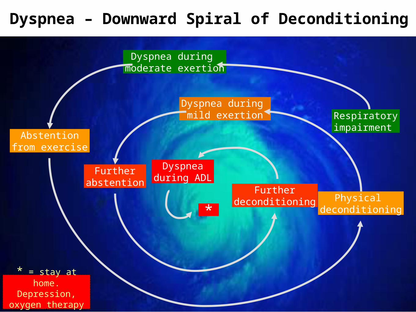
Dyspnea – Downward Spiral of Deconditioning
Respiratoryimpairment
Dyspnea during moderate exertion
Abstentionfrom exercise
Physical deconditioning
Dyspnea during mild exertion
Furtherabstention
Furtherdeconditioning
Dyspneaduring ADL
*
* = stay at home. Depression, oxygen
therapy etc.
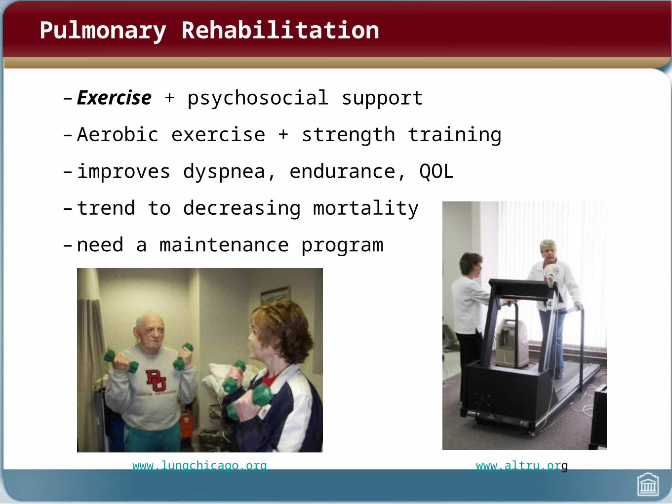
Pulmonary Rehabilitation
– Exercise + psychosocial support
– Aerobic exercise + strength training
– improves dyspnea, endurance, QOL
– trend to decreasing mortality
– need a maintenance program
www.lungchicago.org www.altru.org
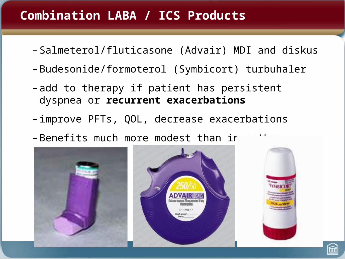
Combination LABA / ICS Products
– Salmeterol/fluticasone (Advair) MDI and diskus
– Budesonide/formoterol (Symbicort) turbuhaler
– add to therapy if patient has persistent dyspnea or recurrent exacerbations
– improve PFTs, QOL, decrease exacerbations
– Benefits much more modest than in asthma
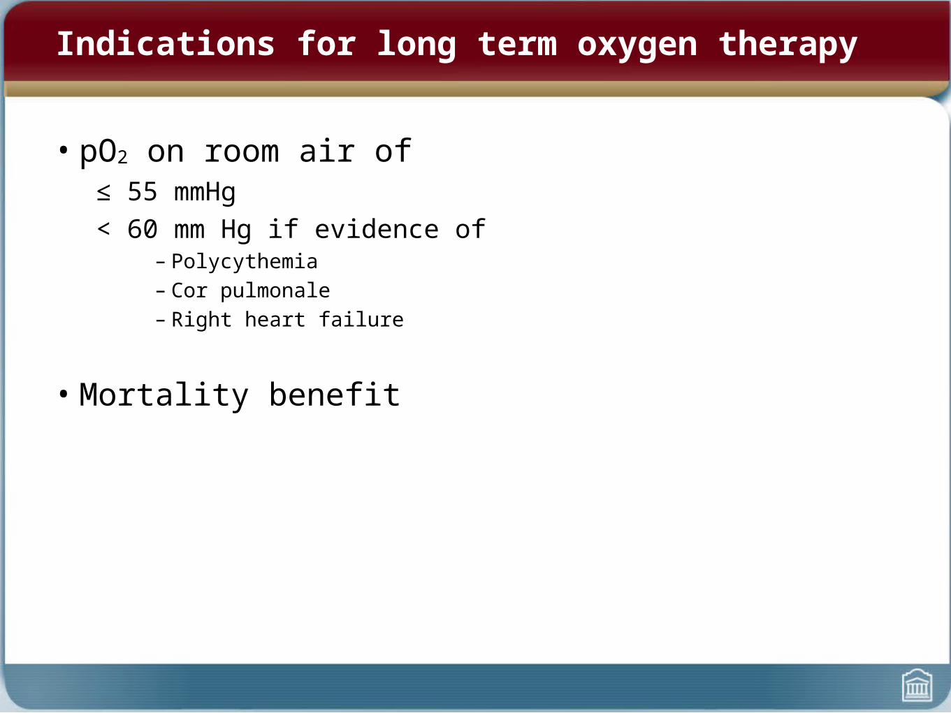
Indications for long term oxygen therapy
• pO2 on room air of≤ 55 mmHg< 60 mm Hg if evidence of
– Polycythemia– Cor pulmonale– Right heart failure
• Mortality benefit

Surgery
• Lung Volume Reduction Surgery- benefits patients with upper lobe (heterogenous) emphysema and
poor exercise capacity
• Lung Transplantation- single or double lung- non-smoker (must have quit smoking)- generally age< 65 without significant cardiac, renal, hepatic disease- post-transplant survival is 5-6 years on average- death from infection (early) and chronic rejection (later)
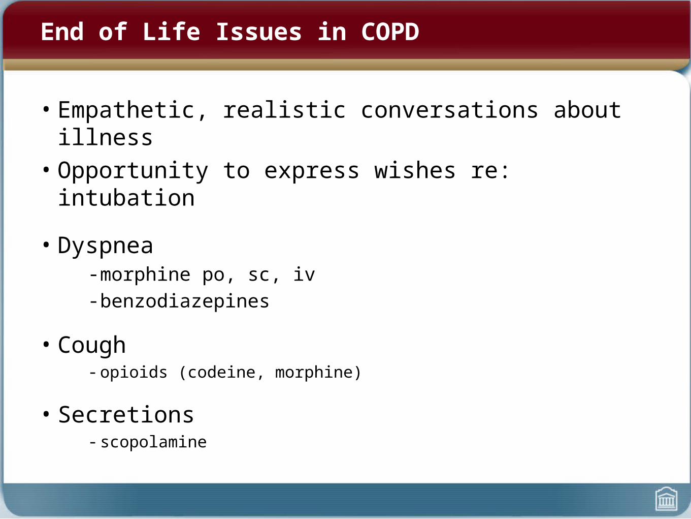
End of Life Issues in COPD
• Empathetic, realistic conversations about illness• Opportunity to express wishes re: intubation
• Dyspnea- morphine po, sc, iv- benzodiazepines
• Cough- opioids (codeine, morphine)
• Secretions- scopolamine
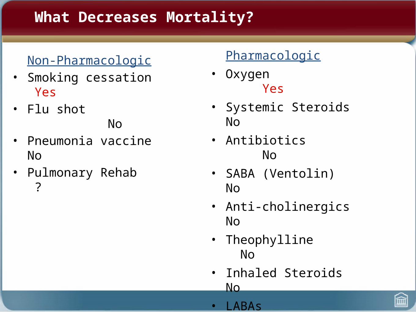
What Decreases Mortality?
Non-Pharmacologic• Smoking cessation Yes• Flu shot No• Pneumonia vaccine No• Pulmonary Rehab ?
Pharmacologic• Oxygen Yes• Systemic Steroids No• Antibiotics No• SABA (Ventolin) No• Anti-cholinergics No• Theophylline No• Inhaled Steroids No• LABAs No• Combo ICS/LABA No

COPD
• Contrast from asthma
• Definition
• Pathophysiology
• Diagnosis
• Chronic Management
• Acute Management
Acute exacerbations of COPD
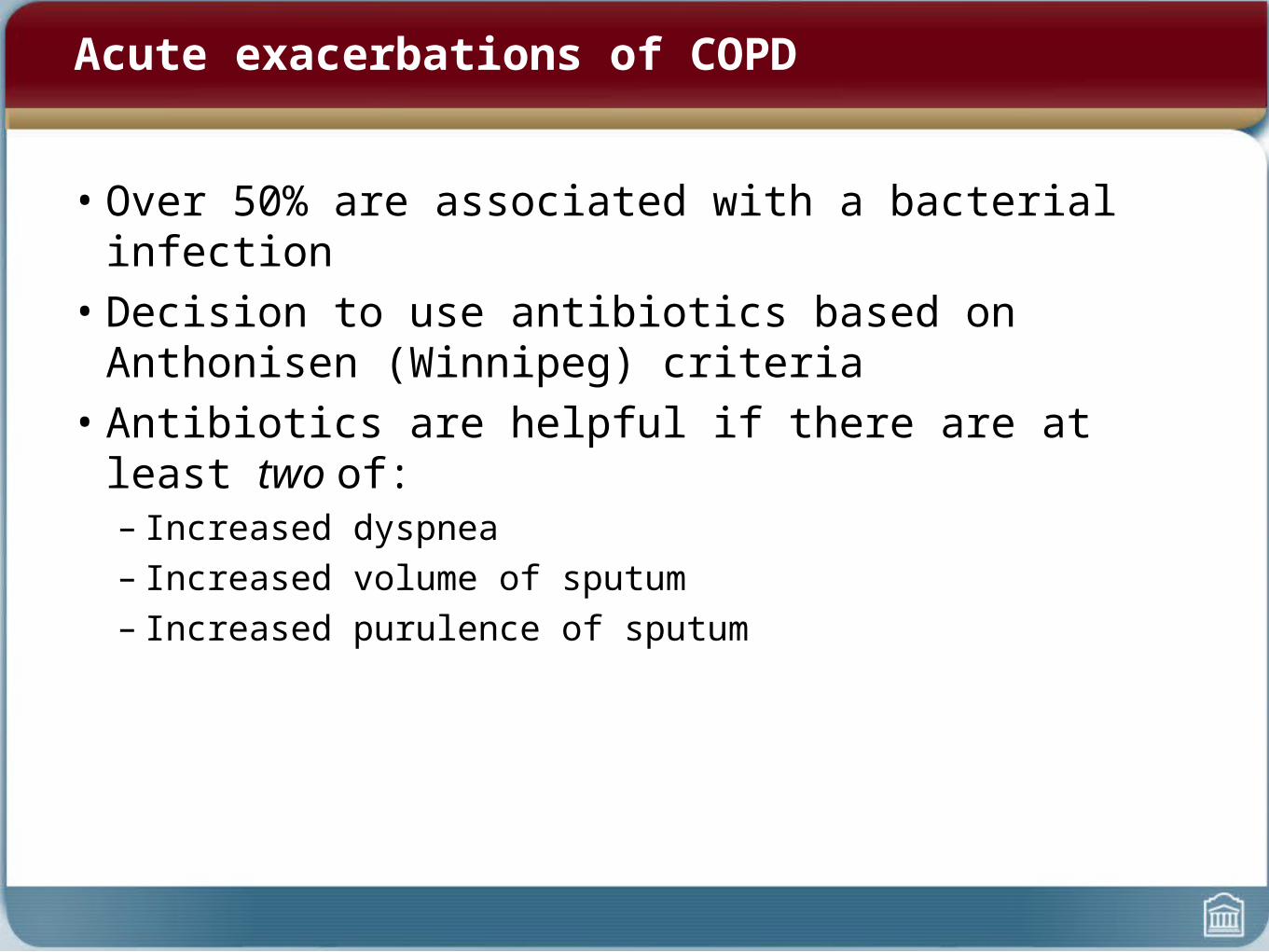
• Over 50% are associated with a bacterial infection• Decision to use antibiotics based on Anthonisen (Winnipeg)
criteria• Antibiotics are helpful if there are at least two of:
– Increased dyspnea– Increased volume of sputum– Increased purulence of sputum

Acute exacerbations of COPD
• Treatment:
– ABCs
– O2 sat monitoring and oxygen prn
– history and p/e to rule out other causes of dyspnea
– CXR, ABG, sputum C&S
– Bronchodilators
– systemic steroids: prednisone 50 mg/d x 10-14 days (?)
– antibiotics if purulent sputum
– NIPPV
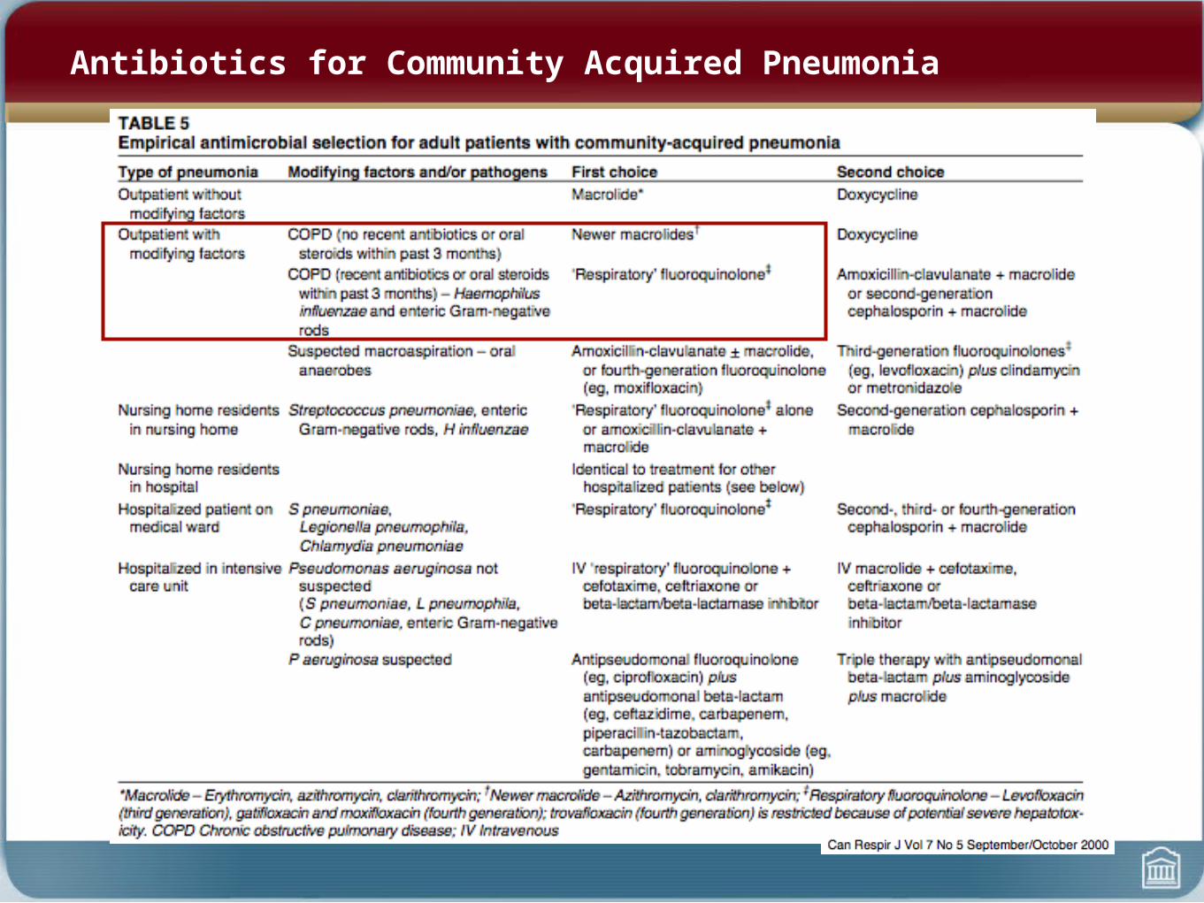
Antibiotics for Community Acquired Pneumonia

Obstructive Sleep Apnea Syndrome
• Elevated Apnea-Hypopnea Index on Sleep study (Polysomnography)
AND
• Nighttime Symptoms: Snoring, witnessed apneas
OR
• Daytime Symptoms: Morning headache, daytime sleepiness
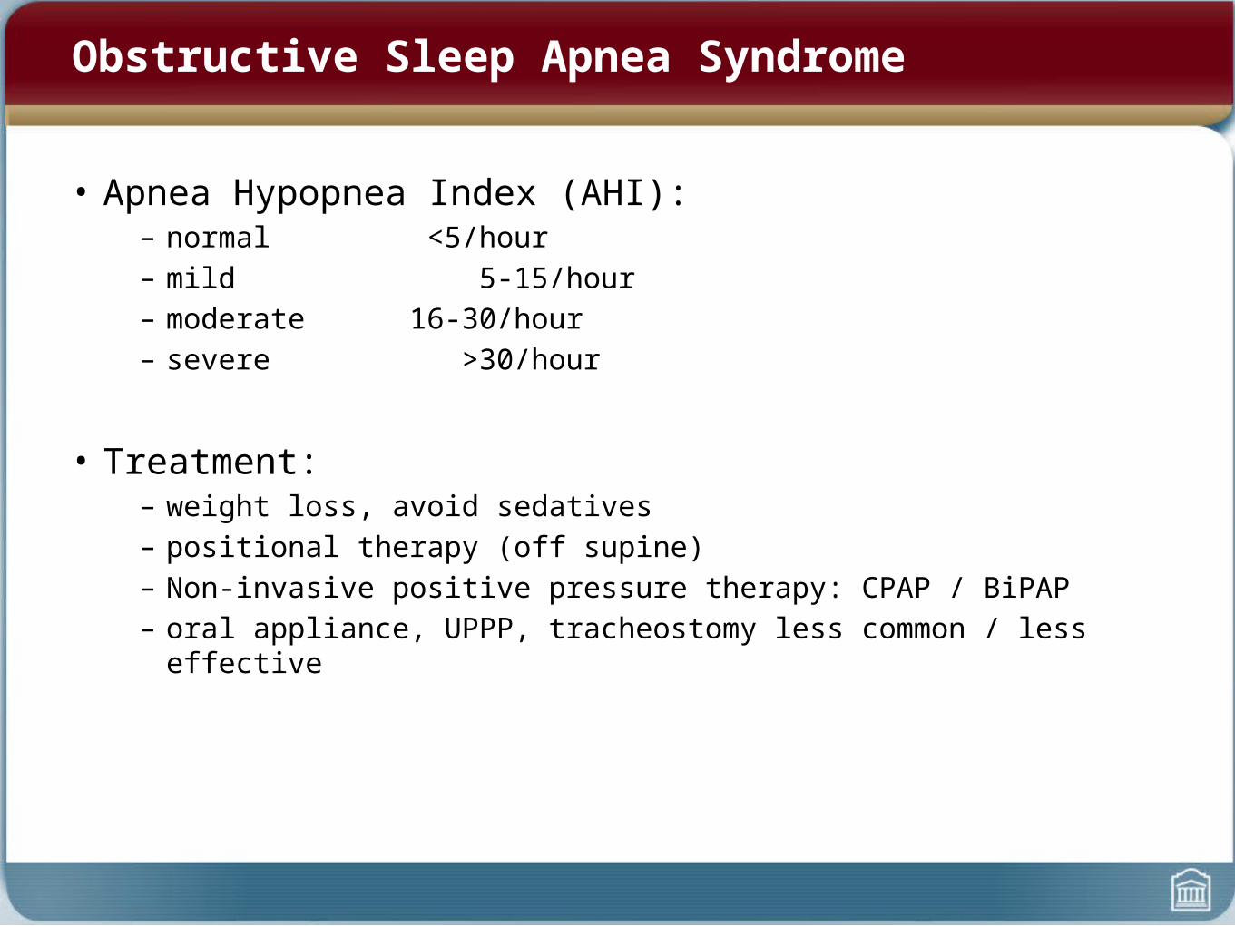
Obstructive Sleep Apnea Syndrome
• Apnea Hypopnea Index (AHI):– normal <5/hour– mild 5-15/hour– moderate 16-30/hour– severe >30/hour
• Treatment:– weight loss, avoid sedatives– positional therapy (off supine)– Non-invasive positive pressure therapy: CPAP / BiPAP– oral appliance, UPPP, tracheostomy less common / less effective
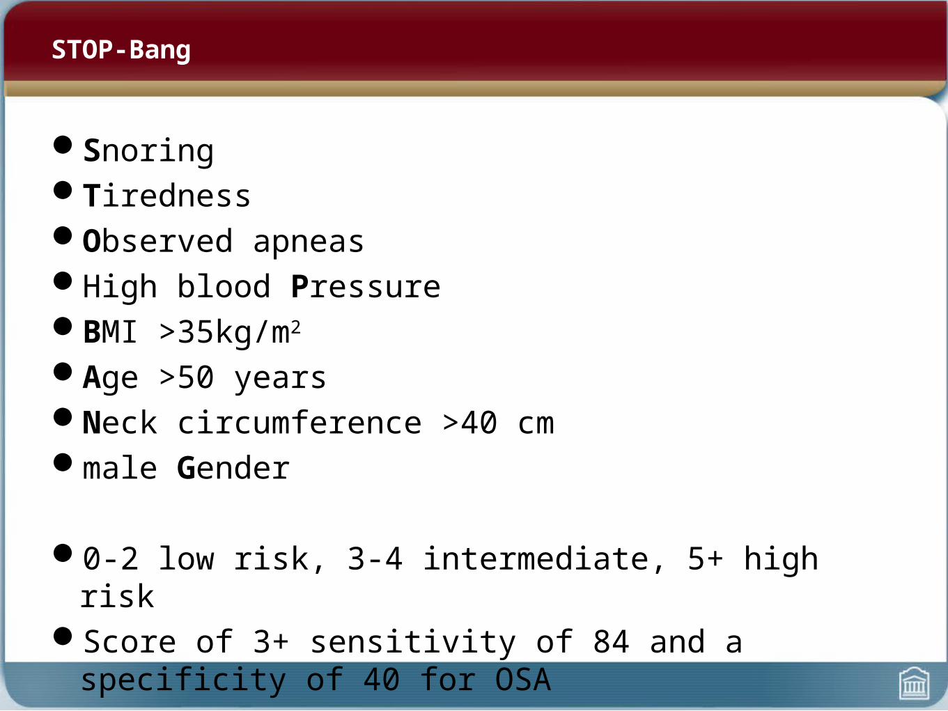
STOP-Bang
SnoringTirednessObserved apneasHigh blood PressureBMI >35kg/m2
Age >50 yearsNeck circumference >40 cmmale Gender
0-2 low risk, 3-4 intermediate, 5+ high riskScore of 3+ sensitivity of 84 and a specificity of 40 for OSA
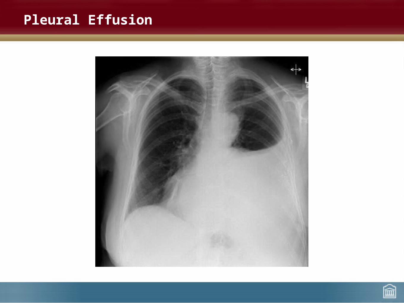
Pleural Effusion

Pleural Fluid Accumulation
• In normal pleural space, the rate of fluid formation is balanced by the rate of removal
• Rate of fluid formation is determined by the Starling equation
– hydrostatic forces push water out of vessel
– osmotic forces pull water back into vessel
• Pleural effusion is due to abnormalities in one of these processes

• Cell count and differential• Gram stain• Culture• AFB• Cytology
• LDH• Total protein• Glucose • pH
Pleural Effusion Evaluation
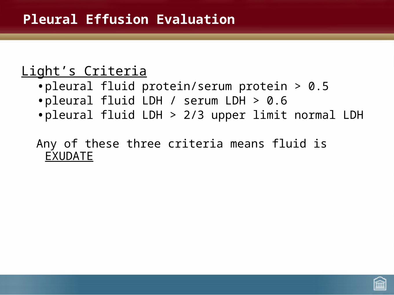
Light’s Criteria• pleural fluid protein/serum protein > 0.5• pleural fluid LDH / serum LDH > 0.6• pleural fluid LDH > 2/3 upper limit normal LDH
Any of these three criteria means fluid is EXUDATE
Pleural Effusion Evaluation
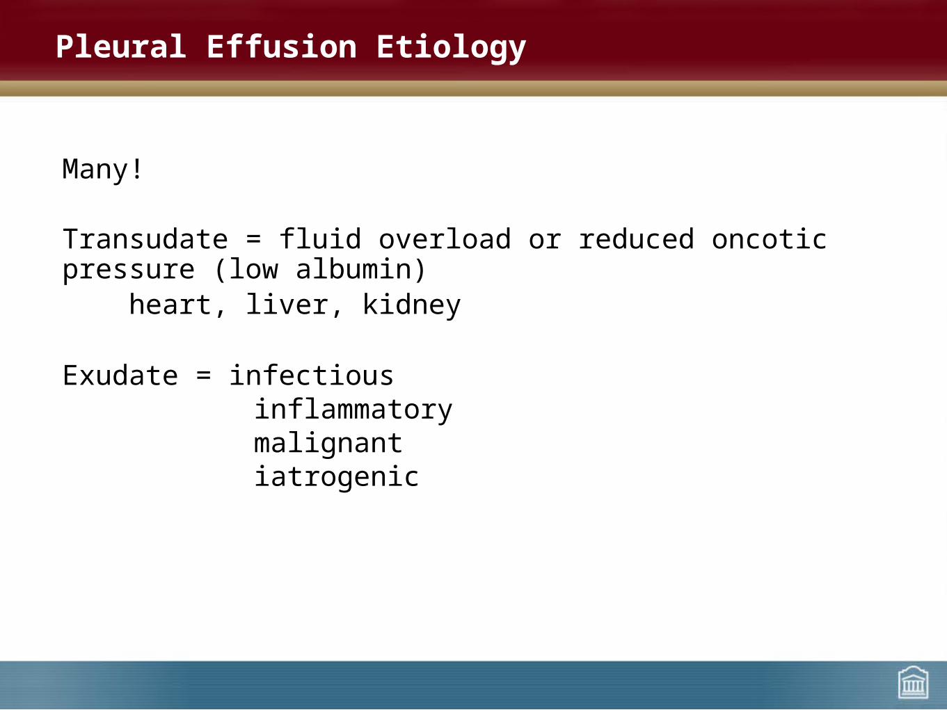
Many!
Transudate = fluid overload or reduced oncotic pressure (low albumin)heart, liver, kidney
Exudate = infectious inflammatory malignant iatrogenic
Pleural Effusion Etiology
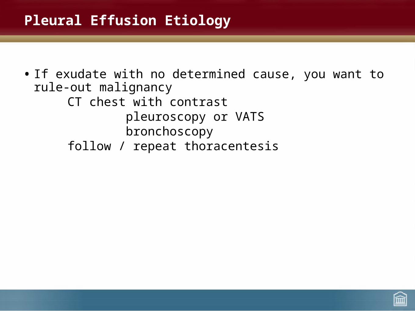
• If exudate with no determined cause, you want to rule-out malignancyCT chest with contrast
pleuroscopy or VATSbronchoscopy
follow / repeat thoracentesis
Pleural Effusion Etiology

Treatment is palliative (reduce symptoms associated with effusion)Cure generally not possibleIn most cases, effusion will persist despite chemotherapy
Most places, patients admitted for symptomatic thoracentesis +/- tube drainage and pleurodesis (talc)
In Ottawa, patients mostly receiving PleurX (indwelling) catheters to allow home drainage
Malignant Pleural Effusions
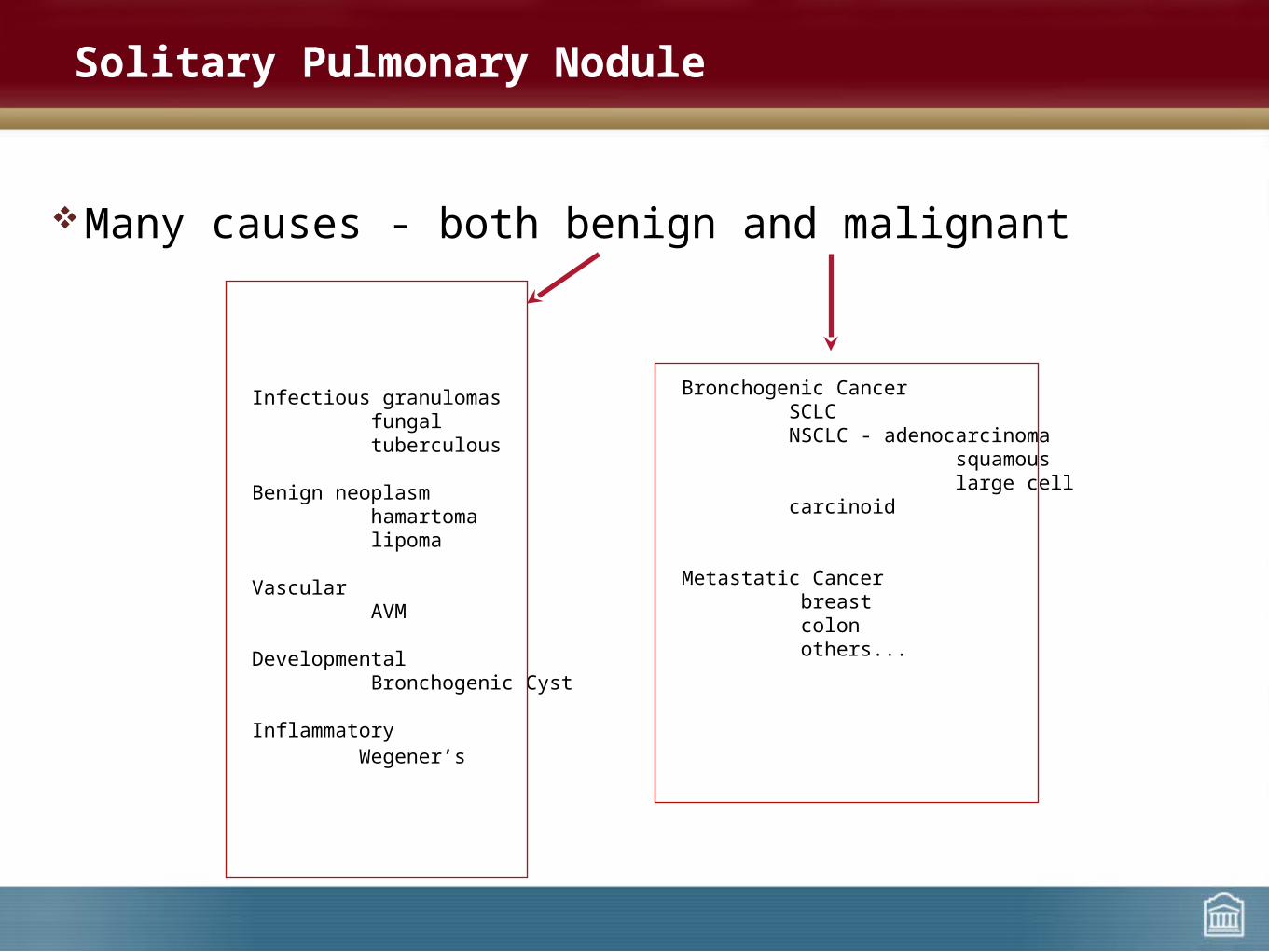
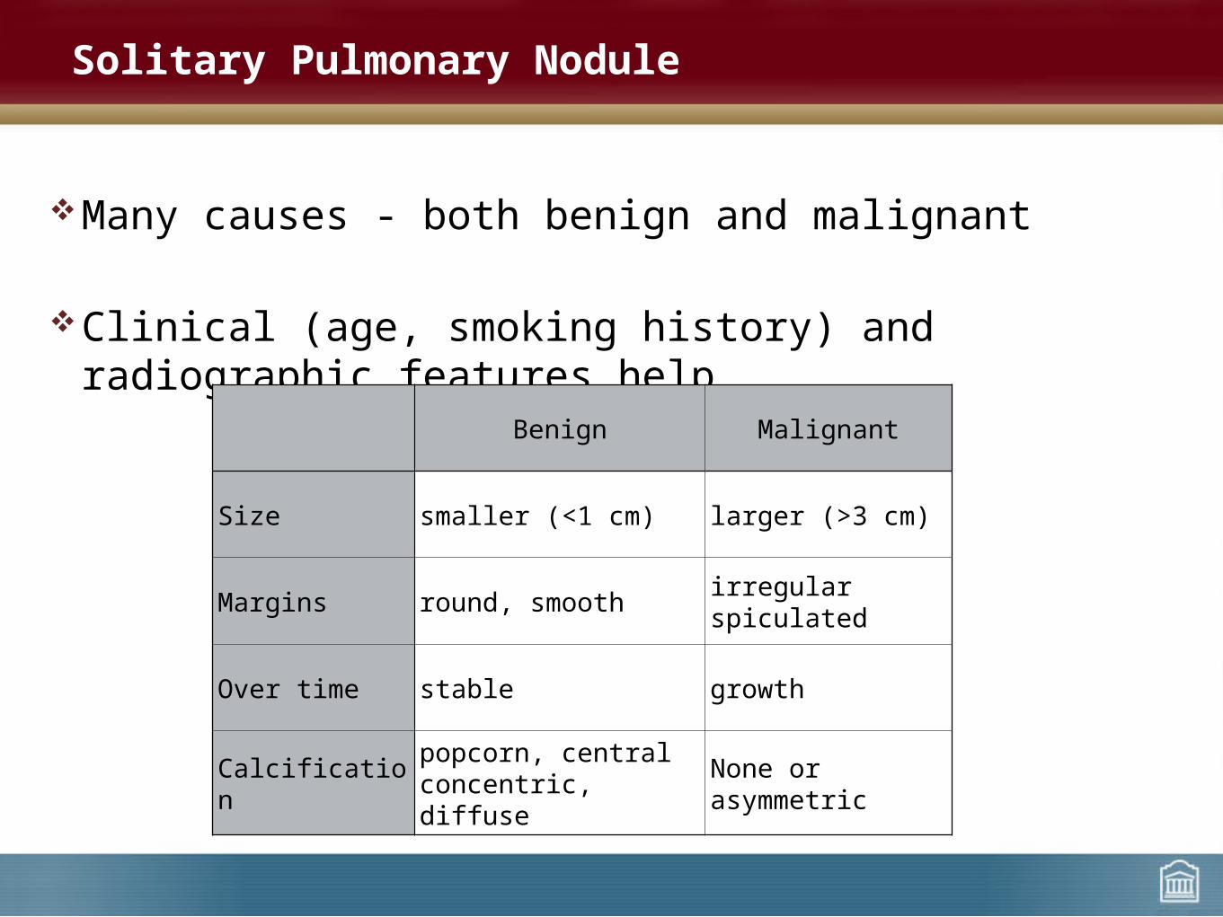
Many causes - both benign and malignant
Solitary Pulmonary Nodule
Infectious granulomas fungal tuberculous
Benign neoplasm hamartoma lipoma
Vascular AVM
Developmental Bronchogenic Cyst
Inflammatory Wegener’s
Bronchogenic Cancer SCLC NSCLC - adenocarcinoma squamous large cell carcinoid
Metastatic Cancer breast colon others...
Many causes - both benign and malignant
Clinical (age, smoking history) and radiographic features help
Solitary Pulmonary Nodule
Benign Malignant
Size smaller (<1 cm) larger (>3 cm)
Margins round, smooth irregularspiculated
Over time stable growth
Calcification popcorn, centralconcentric, diffuse None or asymmetric
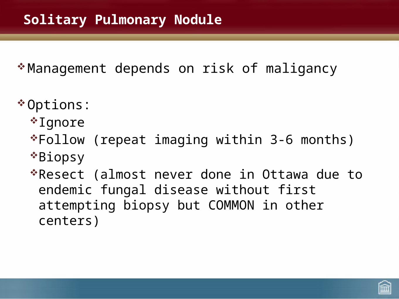
Management depends on risk of maligancy
Options:IgnoreFollow (repeat imaging within 3-6 months)BiopsyResect (almost never done in Ottawa due to endemic fungal
disease without first attempting biopsy but COMMON in other centers)
Solitary Pulmonary Nodule
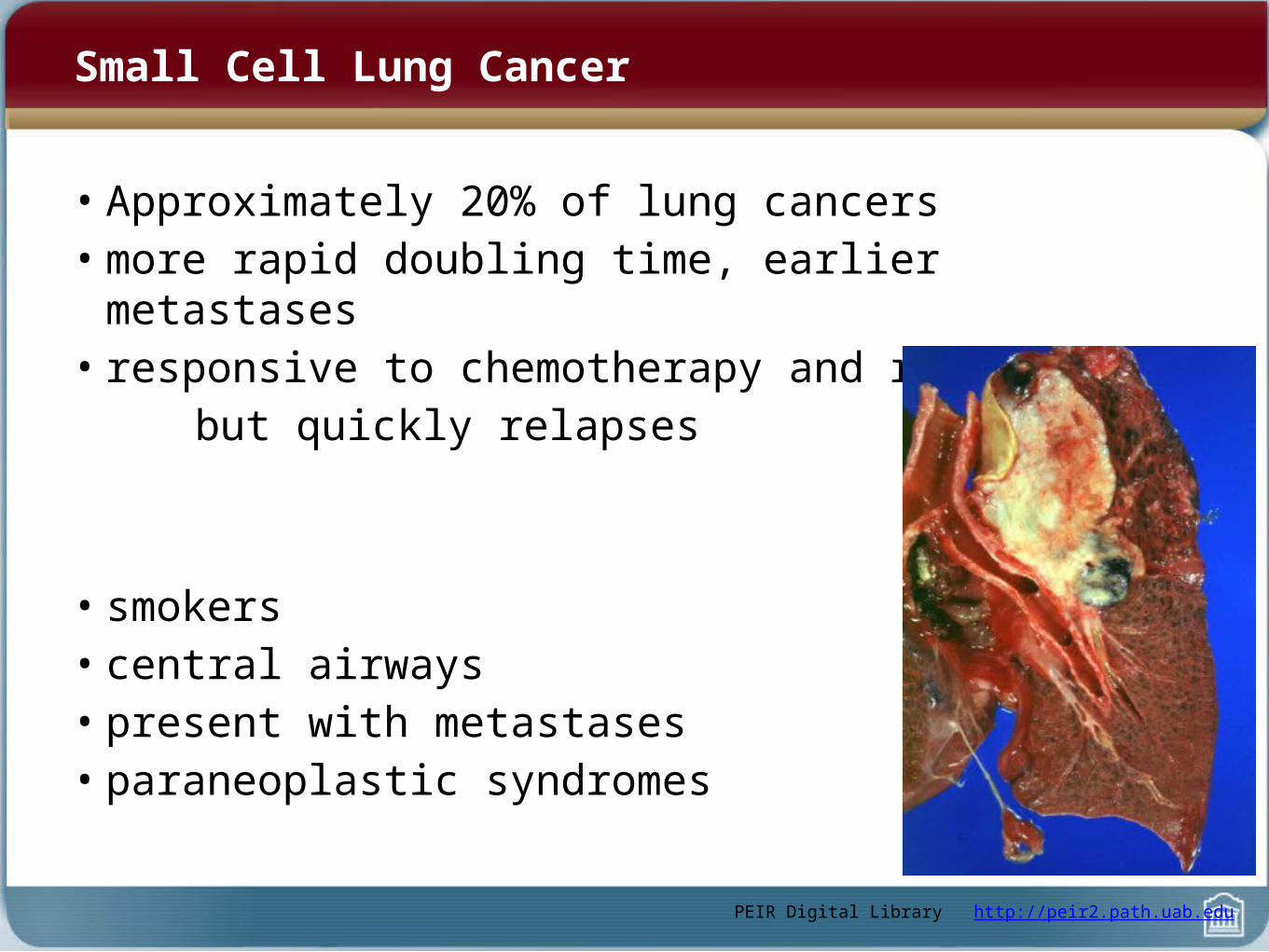
Small Cell Lung Cancer
• Approximately 20% of lung cancers• more rapid doubling time, earlier metastases• responsive to chemotherapy and radiation but quickly relapses
• smokers• central airways• present with metastases• paraneoplastic syndromes
PEIR Digital Library http://peir2.path.uab.edu

• Limited (minority): involves only one hemithorax (maximum allowable for radiation portal)
- concurrent chemo + radiation- median survival 15-20 months
- (very small chance of cure)
• Extensive (majority): extends beyond hemithorax- Chemotherapy only- median 8-13 months
- (cure not possible)
Small Cell Lung Cancer
PEIR Digital Library http://peir2.path.uab.edu
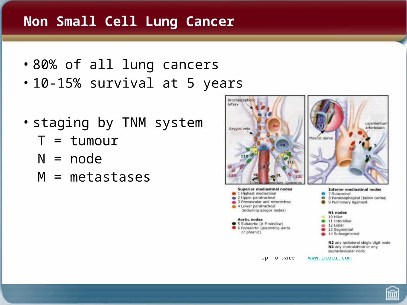
Non Small Cell Lung Cancer
• 80% of all lung cancers• 10-15% survival at 5 years
• staging by TNM systemT = tumourN = nodeM = metastases
Up To Date www.utdol.com
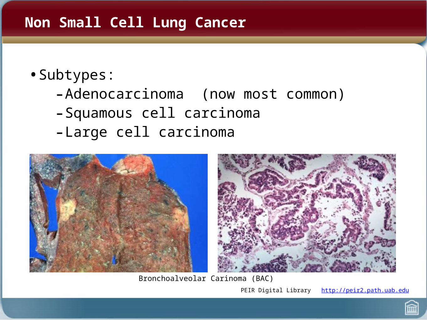
Non Small Cell Lung Cancer
• Subtypes:- Adenocarcinoma (now most common)- Squamous cell carcinoma - Large cell carcinoma
PEIR Digital Library http://peir2.path.uab.edu
Bronchoalveolar Carinoma (BAC)

Non Small Cell Lung Cancer
• Treatment and prognosis depend on stage
• Early Stage (1 or 2)–Surgical resection if tolerated–Adjuvant chemotherapy to reduce risk of recurrence
• Later Stage (3B or 4)–Chemotherapy if performance status is reasonable–Palliative Radiotherapy for symptoms
• Majority of NSCLC will not be resectable and/or operable
u
The Plan...
• Spirometry
• Blood gases
• Asthma
• COPD
• Sleep Apnea
• Pleural Effusion
• Lung Cancer

Good Luck
• Questions?