bacteroides: the good, the bad, and the nitty-gritty · bacteroides: the good, the bad, and the...
TRANSCRIPT

CLINICAL MICROBIOLOGY REVIEWS, Oct. 2007, p. 593–621 Vol. 20, No. 40893-8512/07/$08.00�0 doi:10.1128/CMR.00008-07
Bacteroides: the Good, the Bad, and the Nitty-GrittyHannah M. Wexler*
Wadsworth Anaerobe Laboratory, Greater Los Angeles VA Healthcare Systems, and Department of Medicine,University of California, Los Angeles, California
INTRODUCTION .......................................................................................................................................................594THE GOOD .................................................................................................................................................................595
Bacteroides as Friendly Commensals....................................................................................................................595Appearance in the GI tract ...............................................................................................................................595Nutrient sources for intestinal bacteria ..........................................................................................................595Adaptive survival in the GI tract......................................................................................................................596Carbohydrate metabolism in B. thetaiotaomicron ...........................................................................................596Carbohydrate metabolism in B. fragilis ...........................................................................................................596Miscellaneous enzymes used in sugar transport or utilization....................................................................596Association of levels of intestinal Bacteroides with obesity ...........................................................................596Adaptation of B. thetaiotomicron and B. fragilis to their respective microenvironments ..........................597
Environmental Sensing Systems...........................................................................................................................597ECF �-factors......................................................................................................................................................597Two-component signal transduction systems..................................................................................................598Cross talk between Bacteroides and the intestinal cells.................................................................................598
Interactions with the Immune System.................................................................................................................598Polysaccharides produced by B. fragilis are important in the activation of the T-cell-dependent
immune response. ..........................................................................................................................................598Gut bacteria and the “hygiene hypothesis” ....................................................................................................599Bacteroides can affect expression of Paneth cell proteins..............................................................................599
Limiting Colonization of the GI Tract by Pathogens ........................................................................................599TRANSITION: FROM COMMENSAL TO PATHOGEN.....................................................................................599THE BAD .....................................................................................................................................................................599
Virulence ..................................................................................................................................................................599The bacterial capsule .........................................................................................................................................601Evasion of host immune response ....................................................................................................................601Enzymes implicated in virulence ......................................................................................................................601Enterotoxin ..........................................................................................................................................................602Endotoxin/LPS.....................................................................................................................................................602Aerotolerance of Bacteroides ..............................................................................................................................602
Infections in Adults ................................................................................................................................................602Intra-abdominal sepsis.......................................................................................................................................602Perforated and gangrenous appendicitis .........................................................................................................602Gynecological infections.....................................................................................................................................602Skin and soft tissue infections ..........................................................................................................................603Endocarditis and pericarditis ...........................................................................................................................603Bacteremia ...........................................................................................................................................................603Septic arthritis ....................................................................................................................................................603Brain abscess and meningitis ...........................................................................................................................603IBD........................................................................................................................................................................603
Anaerobes in Pediatric Infections ........................................................................................................................604Infections with intra-abdominal origin............................................................................................................604Bone and joint infections in children ...................................................................................................604Bacteremia in children.......................................................................................................................................604Infections in newborns .......................................................................................................................................604
Bacteroides as a Reservoir of Resistance Determinants ....................................................................................604THE NITTY-GRITTY.................................................................................................................................................605
Taxonomy .................................................................................................................................................................605Isolation and Identification ...................................................................................................................................605Physiology ................................................................................................................................................................606Cell Surface Structures..........................................................................................................................................606
* Mailing address: Wadsworth Anaerobe Laboratory, GLAVAHCS691/151J, 11301 Wilshire Blvd., Los Angeles, CA 90073. Phone: (310)268-3404. Fax: (310) 268-4458. E-mail: [email protected].
593
on January 12, 2020 by guesthttp://cm
r.asm.org/
Dow
nloaded from
Pili, fimbriae, and adhesins...............................................................................................................................606Fibrils ...................................................................................................................................................................606LPS .......................................................................................................................................................................606Outer membrane proteins .................................................................................................................................606Outer membrane vesicles...................................................................................................................................606Export systems ....................................................................................................................................................607Bacteriocins .........................................................................................................................................................607Resistance to bile ................................................................................................................................................607Oxidative stress response ..................................................................................................................................607
Susceptibility Patterns of Bacteroides ...................................................................................................................607Mechanisms of Antimicrobial Resistance ...........................................................................................................608
�-Lactam agents .................................................................................................................................................608Carbapenems .......................................................................................................................................................608PBPs......................................................................................................................................................................608Outer membrane proteins .................................................................................................................................608Aminoglycosides ..................................................................................................................................................611Macrolides, lincosamides, and chloramphenicol............................................................................................611Tetracycline..........................................................................................................................................................611Nitroimidazoles ...................................................................................................................................................611Quinolones ...........................................................................................................................................................612
IS Elements..............................................................................................................................................................612Mobile Genetic Elements in Bacteroides ..............................................................................................................612
Plasmids ...............................................................................................................................................................612Conjugative transposons....................................................................................................................................612Mobilizable transposons ....................................................................................................................................613
Multidrug Resistance .............................................................................................................................................613RND efflux systems in B. fragilis.......................................................................................................................613Characterization of bmeB gene function..........................................................................................................613Selection of multidrug-resistant mutants........................................................................................................613Multidrug resistance in clinical isolates .........................................................................................................614
IMPORTANT FUTURE RESEARCH AVENUES..................................................................................................615ACKNOWLEDGMENT..............................................................................................................................................615REFERENCES ............................................................................................................................................................615
INTRODUCTION
By a variety of measures, the species Homo sapiens is moremicrobial than human. Microorganisms comprise only a small,albeit significant, percentage of the body weight (between 2and 5 pounds of live bacteria). However, in terms of cell num-bers, we are about 10% human and 90% bacterial (308)! Fur-ther, the number of genes in our microbiome may exceed thenumber of human genes by two orders of magnitude (264,308), making us genetically 1% human and 99% bacterial!Consequently, bacteria play a major role in bodily functions,including immunity, digestion, and protection against disease(208). Colonization of the human body by microorganismsoccurs at the very beginning of human life (208), and many ofthese organisms become truly indigenous to the host.
The human colon has the largest population of bacteria inthe body (in excess of 1011 organisms per gram of wet weight),and the majority of these organisms are anaerobes; of these,�25% are species of Bacteroides (226), the bacterial genus thatis focus of this review. This review will summarize the currentstate of knowledge about Bacteroides species, the most pre-dominant anaerobes in the gut. The aspects of these organismsthat will be covered will include their role as commensal or-ganisms (The Good); their involvement in human disease (TheBad); and information about their physiology, metabolism, andresistance mechanisms as well as a brief overview of clinicalcharacteristics (The Nitty-Gritty).
Bacteroidetes is one of the major lineages of bacteria and
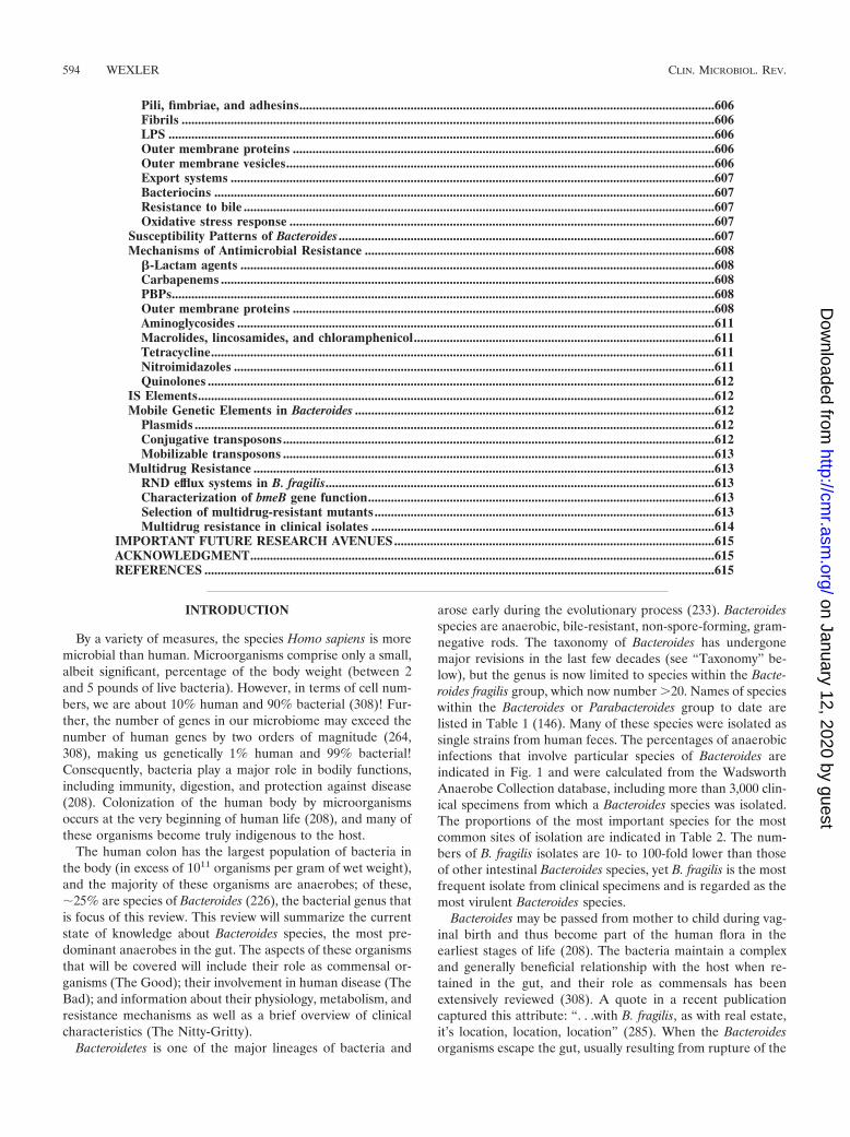
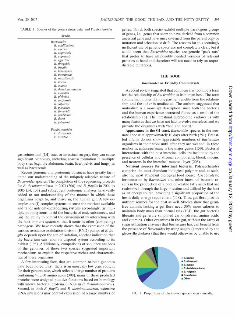
arose early during the evolutionary process (233). Bacteroidesspecies are anaerobic, bile-resistant, non-spore-forming, gram-negative rods. The taxonomy of Bacteroides has undergonemajor revisions in the last few decades (see “Taxonomy” be-low), but the genus is now limited to species within the Bacte-roides fragilis group, which now number �20. Names of specieswithin the Bacteroides or Parabacteroides group to date arelisted in Table 1 (146). Many of these species were isolated assingle strains from human feces. The percentages of anaerobicinfections that involve particular species of Bacteroides areindicated in Fig. 1 and were calculated from the WadsworthAnaerobe Collection database, including more than 3,000 clin-ical specimens from which a Bacteroides species was isolated.The proportions of the most important species for the mostcommon sites of isolation are indicated in Table 2. The num-bers of B. fragilis isolates are 10- to 100-fold lower than thoseof other intestinal Bacteroides species, yet B. fragilis is the mostfrequent isolate from clinical specimens and is regarded as themost virulent Bacteroides species.
Bacteroides may be passed from mother to child during vag-inal birth and thus become part of the human flora in theearliest stages of life (208). The bacteria maintain a complexand generally beneficial relationship with the host when re-tained in the gut, and their role as commensals has beenextensively reviewed (308). A quote in a recent publicationcaptured this attribute: “. . .with B. fragilis, as with real estate,it’s location, location, location” (285). When the Bacteroidesorganisms escape the gut, usually resulting from rupture of the
594 WEXLER CLIN. MICROBIOL. REV.
on January 12, 2020 by guesthttp://cm
r.asm.org/
Dow
nloaded from
gastrointestinal (GI) tract or intestinal surgery, they can causesignificant pathology, including abscess formation in multiplebody sites (e.g., the abdomen, brain, liver, pelvis, and lungs) aswell as bacteremia.
Recent genomic and proteomic advances have greatly facil-itated our understanding of the uniquely adaptive nature ofBacteroides species. The completion of the sequencing projectsfor B. thetaiotaomicron in 2003 (306) and B. fragilis in 2004 to2005 (54, 138) and subsequent proteomic analyses have vastlyadded to our understanding of the manner in which theseorganisms adapt to, and thrive in, the human gut. A few ex-amples are (i) complex systems to sense the nutrient availableand tailor nutrient-metabolizing systems accordingly, (ii) mul-tiple pump systems to rid the bacteria of toxic substances, and(iii) the ability to control the environment by interacting withthe host immune system so that it controls other (competing)pathogens. We have recently shown that the expression of thevarious resistance-nodulation-division (RND) pumps of B. fra-gilis depends upon the site of isolation, another indication thatthe bacterium can tailor its disposal system according to itshabitat (198). Additionally, comparisons of sequence analysesof the genomes of these two species suggested importantmechanisms to explain the respective niches and characteris-tics of these organisms.
A few interesting facts that are common to both genomeshave been noted. First, there is an unusually low gene contentfor their genome size, which reflects a large number of proteinscontaining �1,000 amino acids (308); many of these predictedproteins were assigned putative functions based on homologywith known bacterial proteins (�60% in B. thetaiotaomicron).Second, in both B. fragilis and B. thetaiotaomicron, extensiveDNA inversions may control expression of a large number of
genes. Third, both species exhibit multiple paralogous groupsof genes, i.e., genes that seem to have derived from a commonancestral gene and have since diverged from the parent copy bymutation and selection or drift. The reasons for this seeminglyinefficient use of genetic space are not completely clear, but itwould seem that Bacteroides species are genetic “pack rats”that prefer to have all possibly needed versions of relevantproteins at hand and therefore will not need to rely on unpre-dictable mutations.
THE GOOD
Bacteroides as Friendly Commensals
A recent review suggested that commensal is too mild a termfor the relationship of Bacteroides to its human host. The termcommensal implies that one partner benefits from the relation-ship and the other is unaffected. The authors suggested thatmutualism is a more apt description, since both the bacteriaand the human experience increased fitness as a result of therelationship (8). The intestinal microbiome endows us withmany features that we have not had to evolve ourselves, and weprovide the organisms with “bed and board.”
Appearance in the GI tract. Bacteroides species in the neo-nate appear at approximately 10 days after birth (251). Breast-fed infants do not show appreciable numbers of Bacteroidesorganisms in their stool until after they are weaned; in thesenewborns, Bifidobacterium is the major genus (150). Bacterialinteractions with the host intestinal cells are facilitated by thepresence of cellular and stromal components, blood, mucins,and neurons in the intestinal mucosal layer (208).
Nutrient sources for intestinal bacteria. Polysaccharidescomprise the most abundant biological polymer and, as such,also the most abundant biological food source. Carbohydratefermentation by Bacteroides and other intestinal bacteria re-sults in the production of a pool of volatile fatty acids that arereabsorbed through the large intestine and utilized by the hostas an energy source, providing a significant proportion of thehost’s daily energy requirement (118). Thus, gut flora providenutrient sources for the host as well. Studies show that germ-free animals lacking a gut flora need 30% more calories tomaintain body mass than normal rats (104); the gut bacterialiberate and generate simplified carbohydrates, amino acids,and vitamins. Other organisms in the gut, without the array ofsugar utilization enzymes that Bacteroides has, can benefit fromthe presence of Bacteroides by using sugars (generated by theglycosylhydrolases) that they would otherwise be unable to use
FIG. 1. Proportions of Bacteroides species seen clinically.
TABLE 1. Species of the genera Bacteroides and Parabacteroides
Species
BacteroidesB. acidifaciensB. caccaeB. coprocolaB. coprosuisB. eggerthiiB. finegoldiiB. fragilisB. helcogenesB. intestinalisB. massiliensisB. nordiiB. ovatusB. thetaiotaomicronB. vulgatusB. plebeiusB. uniformisB. salyersaiB. pyogenesB. finegoldiiB. goldsteiniiB. doreiB. johnsonii
ParabacteroidesP. distasonisP. merdae
VOL. 20, 2007 BACTEROIDES: THE GOOD, THE BAD, AND THE NITTY-GRITTY 595
on January 12, 2020 by guesthttp://cm
r.asm.org/
Dow
nloaded from

(264). For example, Bifidobacterium longum has a better sys-tem for importing simple sugars than does B. thetaiotaomicron,but B. thetaiotaomicron can break down a large variety ofglycosidic bonds, providing nutrients that B. longum can thenuse. Also, studies with mice indicate that B. thetaiotaomicroncan redirect its carbohydrate-utilizing capability from dietaryto host polysaccharides according to nutrient availability (265).In another study, the adaptation of B. thetaiotomicron to utilizedifferent nutrients during the suckling and weaning periodswas investigated (27). Transcriptome analysis indicated that B.thetaiotaomicron harvested from the ceca of suckling mice hasincreased expression of enzymes that can utilize host-derivedpolysaccharides (host glycans, hexoseamines, and sialic acidsthat are present in mucus and the underlying gut epithelium),as well as enzymes to aid in the catabolism of mono-and oli-gosaccharides present in mother’s milk. After weaning, therepertoire of sugar-digesting metabolic enzymes was expandedso that plant-derived polysaccharides (which would now bepresent in the gut) could be utilized (27).
Adaptive survival in the GI tract. Bacteroides species have asuperb ability to utilize the nutrients at hand. In the largeintestine, these bacteria utilize simple and complex sugars andpolysaccharides for growth (118). At sites of infection, B. fra-gilis may utilize host cell surface glycoproteins and glycolipidsas a nutrient source; these may include simple sugars such asgalactose and mannose and more complex compounds (e.g.,N-acetyl-D-glucosamine [NAG]) and N-acetylneuraminic ac-ids). Indeed, the largest paralogous group of proteins in B.thetaiotaomicron are those involved in oligo- and polysaccha-ride uptake and degradation (2, 3, 58, 59, 72, 73, 156, 206, 207,229, 247, 248, 276), capsular biosynthesis, and environmentalsensing/signal transduction/DNA mobilization (306). The au-thors of the B. thetaiotaomicron genome sequence publicationsuggest that these expansions “reveal strategies used by B.thetaiotaomicron to survive and to dominate in the denselypopulated intestinal system” (306). The coupling of these para-logs with a variety of regulatory apparatus may explain theexquisitely tuned ability of Bacteroides to sense and adapt toenvironmental changes and stresses, such as would normally beencountered in the gut. Another system used by Bacteroides toadapt to the human gut is its ability to modulate its surfacepolysaccharides by “flipping” the promoters needed for theirexpression to an “on” or “off” position (137); this ability mayallow it to evade a host immune response.
Carbohydrate metabolism in B. thetaiotaomicron. B. theta-iotaomicron has an extensive starch utilization system and multi-ple genes (sus genes) that are involved in starch binding andutilization. One hundred seventy-two glycosylhydrolases and163 homologs of starch binding proteins (106 members homol-ogous to SusC and 57 members homologous to SusD [306,308]) enable the organisms to use the wide variety of dietarycarbohydrates that might be available in the gut. Nearly half ofthe genes encoding the starch binding proteins (SusC ho-mologs) are located next to glycosylhydrolase genes. In all, B.thetaiotaomicron contains more glycosylhydrolases than anysequenced prokaryote and appears to be able to cleave most ofthe glycosidic bonds found in nature (307). This ability to adaptto the use of different nutrient sources undoubtedly gives it an“edge” in its intestinal environment. These proteins may also
be important in the attachment of the organism to mucusglycans.
Carbohydrate metabolism in B. fragilis. The polysaccharide-utilizing ability of B. fragilis has not been as extensively studied,although analysis of the proteome of B. fragilis and comparisonwith B. thetaiotaomicron also suggests a tremendous capacity touse a wide range of dietary polysaccharides. A few years ago,we characterized a 200-kDa two-component protein (Omp200[composed of Omp120 and Omp70, corresponding to theirrespective apparent molecular masses]). The intact two-com-ponent system had pore-forming ability in liposomes and blacklipid bilayer membranes (two artificial systems that mimic theouter membrane of the cell and can measure pore formation).The 120-kDa component of this porin had significant homol-ogy to B. thetaiotaomicron SusC proteins (301). While Omp71had no detectable similarity to SusD, it had homologs in the B.thetaiotomicron genome that are positioned next to a SusChomolog. Xu and Gordon speculated that the B. fragilis SusC-like component may be a conserved component of multifunc-tional outer membrane proteins. These multifunctional com-plexes may be divided into two groups: those with adownstream susD homolog that may affect acquisition/utiliza-tion of polysaccharides and those with homologs of omp71,encoding a protein whose function has not yet been defined(308).
The B. fragilis neuraminidase enzyme (product of the nanHgene) catalyzes the removal of terminal sialic acid from surfacepolysaccharides (105), and nanH mutants are often growthdeficient. Because NAG is used in cell wall production, theability to use extracytoplasmic NAG facilitates cell growth(181). Possibly, neuraminidase activity may render other car-bon sources available when glucose levels are reduced (105),thus serving the nutritional requirements of the bacterium.
Miscellaneous enzymes used in sugar transport or utiliza-tion. Many bacteria have transport-linked phosphorylation sys-tems that allow sugars transported into the cells to be imme-diately utilized in pathways for energy metabolism orbiosynthesis; any sugar transported across the cell membraneby these phosphotransfer systems can immediately enter met-abolic or biosynthetic pathways. Genes for these systems werenot found in the genome of either B. fragilis or B. thetaiotaomi-cron. Thus, they must have alternate ways of transporting sug-ars into the cell and attaching an active phosphate moiety.Recently, two broad-specificity hexokinases from B. fragiliswere characterized, and their roles in hexose and NAG utili-zation were studied (31). These enzymes allow utilization ofnutrients found in the gut (undigested dietary polysaccharidesand host-derived glycoproteins) and at sites of infection (hostcell surface antigens [including the Lewis antigen] and glyco-lipids) (31).
Association of levels of intestinal Bacteroides with obesity.During the last 2 years, there have been a number of reports inprominent journals pointing out that the respective levels ofthe two main intestinal phyla, the Bacteroidetes and the Firmi-cutes, are linked to obesity, both in humans and in germfreemice (102, 143, 144, 280). The authors of the studies deducethat carbohydrate metabolism is the important factor. Theyobserve that the microbiota of obese individuals are moreheavily enriched with bacteria of the phylum Firmicutes andless with Bacteroidetes, and they surmise that this bacterial mix
596 WEXLER CLIN. MICROBIOL. REV.
on January 12, 2020 by guesthttp://cm
r.asm.org/
Dow
nloaded from

may be more efficient at extracting energy from a given dietthan the microbiota of lean individuals (which have the oppo-site proportions) (280). In some studies, they found that therelative abundance of Bacteroidetes increases as obese individ-uals lose weight and, further, that when the microbiota ofobese mice are transferred to germfree mice, these mice gainmore fat than a control group that received microbiota fromlean mice (280). Until very recently, reports in the literatureagreed that B. thetaiotaomicron had more glycosylhydrolasesthan any sequenced prokaryote and appeared to be able tocleave most of the glycosidic bonds found in nature (307).However, the most recent genomic analysis found that envi-ronmental gene tags coding for many enzymes involved in theinitial steps in breaking down otherwise indigestible dietarypolysaccharides were enriched in obese mice (note that thesewere ob/ob homozygous mice with a defective leptin gene aswell). The genome of Eubacterium rectale (a member of theFirmicutes division), which has not been completed, is signifi-cantly enriched for glycoside hydrolases compared to severalcompleted genomes of Bacteroides species (280).
While it is not completely clear how significant these differ-ences are or how well they will translate into human equiva-lents (9), they have, in fact, been extended to the human diet.A very recent study found that diets that are based on a highintake of protein but a low intake of fermentable carbohydrate(e.g., many of the popular diets, including Atkins, SouthBeach, etc.) may alter the gut flora. These workers found thatproportions of Bacteroides and several clusters of Clostridiawere not altered but that numbers of Roseburia, Eubacteriumrectale, and bifidobacteria decreased significantly as carbohy-drate intake decreased (79). Furthermore, human colonic bu-tyrate-producing organisms that are related to Roseburia spp.and Butyrivibrio fibrisolvens showed an increased ability to usea variety of starches for growth compared to B. thetaiota-omicron (202).
Adaptation of B. thetaiotomicron and B. fragilis to their re-spective microenvironments. While both B. thetaiotaomicronand B. fragilis contain large numbers of paralogous genes,comparison of the two genomes suggests that they are specif-ically tailored for their respective microenvironments. For ex-ample, B. fragilis has a pronounced capacity to create variablesurface antigenicities by multiple DNA inversion systems(138). This surface-altering capability is more developed in B.fragilis, which is more frequently found at the mucosal surface(i.e., often the site of attack by host defenses) than is B. theta-iotaomicron. Also, the ability of B. fragilis to tolerate and useoxygen may account for the observation that it is found ingreatest numbers at the mucosal surface, where the PO2 shouldbe higher than it is within the intestinal lumen (13). The im-pressive capacity to utilize polysaccharides is more pronouncedin B. thetaiotaomicron, which is more concentrated within thecolon. The multiplicity of sensing systems of B. thetaiota-omicron, discussed below, also allow fine-tuned and efficient re-cruitment of the appropriate carbohydrate utilization systems.This was aptly illustrated by a very recent study that demon-strated adaptations in B. thetaiotomicron in the guts of miceduring the suckling period and after weaning. By analyzingwhole-genome transcriptional profiles of the bacterium har-vested from the intestines of mice at different time points, theauthors demonstrated that in sucking animals, glucose/galac-
tose transporters and other glycosidases (enzymes that wouldbe important in using host glycans sources) were expressed athigher levels, whereas B. thetaiotaomicron harvested from micein the weaned stage showed increased expression of genes forenzymes that can liberate sugars from plant polysaccharides.Thus, during the suckling period, B. thetaiotomicron preferen-tially used host-derived polysaccharides as well as mono-andoligosaccharides present in mother’s milk, and after weaning,this organism expanded its metabolism to exploit abundantpolysaccharides of plant origin (27).
Environmental Sensing Systems
Beneficial symbiosis requires that the bacteria can sensechanges in the environment so that they can adapt to alter-ations in their surroundings. The genome studies of B. theta-iotaomicron reveal that they have multiple genes encoding signalsensing systems; these include �-factors and two-componentregulatory systems. The function of these systems in Bacte-roides is not understood to the extent that they are understoodin aerobic bacteria, but indications are that they serve similarfunctions.
ECF �-factors. �-factors are essential dissociable proteinsubunits of prokaryotic RNA polymerase that are necessary forinitiation of transcription. These factors provide promoter rec-ognition specificity to the polymerase and contribute to DNAstrand separation; they then dissociate from the RNA poly-merase core. In some cases the factor may regulate large num-bers of prokaryotic genes, and in some cases the genes com-prising a sigma factor regulon have a clearly defined function(131). One class of these factors, the extracytoplasmic function�-factors, known as ECF-type �-factors, are relatively smallproteins (65). They are frequently associated with specificmembrane-tethered cognates, known as anti-�-factors. Thiscognate may receive a signal causing it to release its �-factor;the released �-factor can then interact with RNA polymeraseto initiate transcription.
B. thetaiotaomicron contains the largest proportion andnumber of ECF �-factors among the species of Bacteria andArchaea for which complete genome data are available (307).Approximately half of the ECF �-factor genes are located nextto open reading frames encoding putative anti-�-factors. Fur-ther, all but one of these ECF �-factor/anti �-factor pairs islocated upstream of open reading frames encoding homologsof the polysaccharide binding susC gene products. While thestarch binding proteins are located on the cell surface, thestarch-degrading enzymes are located in the periplasm, per-haps to allow Bacteroides exclusive access to the substrate (3).However, genomic analysis of B. thetaiotomicron identified ahost of glycosyl hydrolases (�-galactosidases, �-galactosidases,�-glucosidases, �-glucosidases, �-glucuronidases, �-fructo-furanosidases, �-mannosidases, amylases, and endo-1,2-�-xylanases, plus 14 other activities); 61% of these glycosylhydro-lases are predicted to be in the periplasm or outer membrane orto be extracellular and may be important in shaping the nutri-ent availability of the intestinal ecosystem (306). Taken to-gether, these results suggest a finely tuned regulatory systemthat allows B. thetaiotaomicron to sense the nutrients at handand adjust its metabolism accordingly, thus benefiting (i) itself,(ii) other bacteria that cannot utilize the complex polysaccha-
VOL. 20, 2007 BACTEROIDES: THE GOOD, THE BAD, AND THE NITTY-GRITTY 597
on January 12, 2020 by guesthttp://cm
r.asm.org/
Dow
nloaded from
ride, and (iii) the human host, who obtains 10 to 15% of his/hercaloric intake through microbial fermentation of oligosaccha-rides (307).
Two-component signal transduction systems. The architec-ture and regulation of two component signal transduction sys-tems have been extensively studied (268). These systems alloworganisms to sense and respond to changes in many differentenvironmental conditions. The prototype structure is well con-served and includes a histidine protein kinase that is regulatedby environmental stimuli. In response to a stimulus, this pro-tein autophosphorylates at a histidine residue, creating a high-energy phosphoryl group. The phosphoryl group is subse-quently transferred to an aspartate residue in the responseregulator protein; this induces a conformational change in theregulatory domain that results in activation of an associateddownstream domain and causes the response (268). The ma-jority of response regulators are transcription factors withDNA-binding effector domains, although some have C-termi-nal domains that function as enzymes. Examples of this systeminclude regulation of the differential expression of ompF andompC by the EnvZ-OmpR system in Escherichia coli and of thecommitment to sporulation by the Spo system in Bacillus sub-tilis (268). While there are few functional studies of thesesystems in Bacteroides, it is reasonable to assume that they aresimilar to those described for other organisms. For example,expression of a two-component regulatory system gene fromBacteroides that was cloned into a multicopy plasmid vector inE. coli resulted in a decrease in the level of the outer mem-brane porin protein OmpF and an increase in the level of theouter membrane porin protein OmpC (204).
One Bacteroides two-component regulatory system whichhas been extensively studied is the RteA-RteB two-componentsystem. The tetracycline resistance gene, tetQ, is part of therteA-rteB-tetQ operon, which is located on a mobile element(CTnDot [see below]) found in many strains of Bacteroides.RteA is the sensor component, and RteB is the transcriptionalregulator that controls the expression of a third downstreamgene, rteC. The RteC product, in turn, controls the expressionof a gene cluster (orf2C) that is important for excision (andtherefore of transfer) of the CTnDot element. Tetracycline hasa stimulatory effect on expression of the RteA-RteB systemand, therefore, on both expression and transfer of tetracyclineresistance. However, the rteA gene is not directly sensing tet-racycline, and exactly what it is sensing is not yet clear (164).
In addition to the sizeable numbers of classical two-compo-nent systems (i.e., sensor kinases and response regulators),there is a family of 32 unique proteins in B. thetaiotaomicronthat incorporate all of the domains found in the classical two-component system into a single polypeptide. In one system,nutrient sensing is coupled to regulation of monosaccharidemetabolism (263). BT3172 belongs to this family of proteins,and its expression is regulated by polysaccharides in the envi-ronment. The presence of �-mannosides in the medium caninduce expression of BT3172, which, in turn, will cause upregu-lated expression of secreted �-mannosidases. This system mayalso be important for capsular polysaccharide gene expression.Typically, expression of one of the capsular polysaccharidesynthesis loci (cps3) is upregulated when polysaccharides arescarce. In a mutant deficient in BT3172, expression of cps3 isincreased even in the presence of a medium rich in polysac-
charides, suggesting that BT3172 is important for the bacte-rium to properly interpret its nutrient landscape in terms ofadequate supply of polysaccharides (263).
Another feature of the ability of BT3172 to sense the neigh-boring polysaccharide landscape is that it can modulate “mim-icry” so that the surface polysaccharide structure of the bacte-rium can be altered to match the surrounding landscape,possibly allowing the bacterium to avoid eliciting a host im-mune response (68).
Cross talk between Bacteroides and the intestinal cells. Mostof the characteristics of Bacteroides discussed to this pointpertain to the adaptability of Bacteroides to its environmentand changes that occur as a result of alterations in that envi-ronment. However, this is a two-way communication system,and studies have shown that the intestinal Bacteroides strainsdirectly modulate gut function (94). Almost a decade ago,Hooper et al. demonstrated that B. thetaiotomicron can modifyintestinal fucosylation in a complex interaction mediated by afucose repressor gene and a signaling system (121). Subse-quently, using transcriptional analysis, they demonstrated thatB. thetaiotaomicron could modulate expression of a variety ofhost genes, including those involved in nutrient absorption,mucosal barrier fortification, and production of angiogenicfactors (120). The line of communication from bacterium tointestinal cell can morph into a complete circle: B. theta-iotaomicron can stimulate production of RegIII�, a bactericidallectin, which can then bind directly to bacterial peptidoglycanin gram-positive bacteria and result in bacterial killing (53).
Interactions with the Immune System
There are numerous studies detailing the host immune re-sponse to bacterial virulence factors. However, the vast major-ity of human-bacterial interactions are benign and commensalor mutualistic in nature. Intestinal bacteria are important inthe development of gut-associated lymphoid tissues (GALT):in the absence of bacterial colonization, development ofGALT is defective (117). In rabbits, the combination of B.fragilis and Bacillus subtilis consistently promoted GALT de-velopment and led to development of the preimmune antibodyrepertoire (212). Mazmanian and Kasper reviewed the factorsthat allow the GI tract—an environment with multiple immunecapabilities—to coexist with the huge numbers of bacteriafound there (154) and proposed a model whereby the immunecapabilities of the GI tract are profoundly affected by some ofthose bacteria. The general outline of this model is describedbelow.
Polysaccharides produced by B. fragilis are important inthe activation of the T-cell-dependent immune response. Azwitterionic polysaccharide (ZPS) produced by B. fragiliscan activate CD4� T cells (i.e., T helper cells expressing theCD4 [cluster of differentiation 4] glycoprotein). Polysaccha-rides A and B (PS-A and PS-B) of the B. fragilis capsularpolysaccharide complex are both ZPSs. Normally, polysac-charides (which almost never carry a positive charge) areconsidered activators of B cells, and they promote increasedimmunoglobulin M (IgM) production but without IgG pro-duction and without a memory response. However, the un-usually structured ZPSs can bind onto the borders of thepeptide-binding groove on major histocompatibility com-
598 WEXLER CLIN. MICROBIOL. REV.
on January 12, 2020 by guesthttp://cm
r.asm.org/
Dow
nloaded from
plex class II molecules of the antigen-presenting cells(APCs) and dock in this groove, and they can thus be pre-sented to the T-cell receptors of CD4� T cells in the sameway that a peptide or glycopeptide conjugate would be pre-sented. Experimental data indicate that these ZPSs are in-ternalized by the APCs, processed by chemical oxidationinto smaller fragments, and then presented to the T cell atthe surface of the APC. Indeed, these ZPSs appear to beimportant in the development of CD4� T cells. Splenocytesfrom germfree mice showed a lower proportion of CD4�
cells than splenocytes from conventionally colonized mice,and colonization with B. fragilis (even in the absence of allthe other gut microflora) could correct this proportion inthe germfree animals. Moreover, colonization with a mutantthat could not produce PS-A was not able to correct thedefect, whereas PS-A alone could also correct the defect(154, 155).
The CD4� T cells stimulated by PS-A produce interleu-kin-10 (IL-10), which can act to prevent abscess formation andother inflammatory responses. Also, in either in vivo or in vitroexperiments, these PS-A-stimulated cells also produce gammainterferon, IL-2, and IL-12. The studies accomplished by thisgroup reveal an intricate “dance” between the microbe andcertain components of the host immune system, and the au-thors conclude that the PS-A of B. fragilis “is necessary andsufficient to mediate the generation of a normal mature im-mune system” (154).
Gut bacteria and the “hygiene hypothesis.” There have beenreports that modulation of the immune system by the com-mensal gut bacteria is important in allergy development. Onescenario is that increases in vaccination, antibiotic usage, anddisinfectant use decrease the gut flora at an important point inthe development of the immune system, which results in askewing of the immune system toward TH2 cell response andoverproduction of TH2 cytokines and IgE; the innate immunemechanisms and TH1, TH2, and regulatory T cells are all partof this fine balancing act (210). The specific bacterial determi-nants of this phenomenon are not clear. Mazmanian andKasper suggest that B. fragilis PS-A may be involved (154).Others suggest that lipopolysaccharide (LPS) (not necessarilyBacteroides LPS), which at low levels is an inducer of IL-12 andgamma interferon (cytokines that stimulate TH1-mediated im-munity and decreases the production of TH2 inflammatorycytokines such as IL-4, IL-5, and IL-13), is part of this process(293).
Bacteroides can affect expression of Paneth cell proteins.Small intestinal crypts house stem cells that serve to constantlyreplenish epithelial cells that die and are lost from the villi.Paneth cells (immune systems cells similar to neutrophils),located adjacent to these stem cells, protect them against mi-crobes by secreting a number of antimicrobial molecules (de-fensins) into the lumen of the crypt (97), and it is possible thattheir protective effect even extends to the mature cells thathave migrated onto the villi (97). In animal models, B. theta-iotaomicron can stimulate production of an antibiotic Panethcell protein (Ang4) that can kill certain pathogenic organisms(e.g., Listeria monocytogenes) (119). In newborn mice, B. theta-iotaomicron promotes angiogenesis and postnatal development(266).
Limiting Colonization of the GI Tract by Pathogens
Studies by Wells and colleagues indicate that anaerobic bac-teria play a pivotal role in limiting the translocation of normalintestinal bacteria but that other bacterial groups also have arole in preventing the intestinal colonization and translocationof potential pathogens (295). Recent studies suggest that Bac-teroides, and possibly specific species of Bacteroides, have a rolein preventing infection with Clostridium difficile (122, 123). Asdetailed above, the development of the immune response thatlimits entry and proliferation of potential pathogens is pro-foundly dependent upon B. fragilis. Also as mentioned, Panethcell proteins may produce antibacterial peptides in response tostimulation by B. thetaiotomicron (119), and these moleculesmay prevent pathogens from colonizing the space. In addition,B. thetaiotomicron can induce Paneth cells to produce a bac-tericidal lectin, RegIII�, which exerts its antimicrobial effect bybinding to the peptidoglycan of gram-positive organisms (53).
TRANSITION: FROM COMMENSAL TO PATHOGEN
As outlined above, Bacteroides species are normally com-mensals in the gut flora. However, these organisms can also beresponsible for infections with significant morbidity and mor-tality. A similar scenario is found with the “commensal gonebad” (186), i.e., Enterococcus faecalis. E. faecalis is normally abenign resident of the gut flora. However, the first vancomycin-resistant enterococcal strain had a number of DNA elements,apparently of foreign origin, which comprised a quarter of thegenome of that strain (186). These elements included a varietyof resistance determinants as well as a pathogenicity islandcarrying a number of virulence-associated genes. Thus, theremay be considerable “sharing” of genes within the crowdedneighborhood of the gut flora. Acquiring genes that favor the“new” resident (e.g., genes that code for improved adhesion,new nutrition pathways, antibiotic resistance, and inhibition ofhost defenses) will give these organisms an edge in establishinga niche for themselves. Indeed, some bacteria may not evenneed to acquire new genes. Organisms such as Bacteroides withsuch a large genome bank at their disposal may simply need toturn on certain genes (such as those involving new nutritionpathways, efflux pumps to rid the cell of toxic substrates, ornew surface epitopes) to change from friendly commensal todangerous threat (104).
Additionally, the capsular polysaccharide of B. fragilis, whichis so important in development of the host immune system, isalso responsible for abscess formation; it is thus one of themost important virulence determinants in this bacterium and isthe most obvious bacterial element that is both “friend andfoe.”
The proportions of various species of the B. fragilis groupfound in anaerobic infections are given in Table 2.
THE BAD
Virulence
Although B. fragilis accounts for only 0.5% of the humancolonic flora (190), it is the most commonly isolated anaerobicpathogen, due in part to its potent virulence factors. Virulencefactors can generally be subdivided into three broad categories:
VOL. 20, 2007 BACTEROIDES: THE GOOD, THE BAD, AND THE NITTY-GRITTY 599
on January 12, 2020 by guesthttp://cm
r.asm.org/
Dow
nloaded from

TA
BL
E2.
Prop
ortio
nsof
vari
ous
spec
ies
ofth
eB
.fra
gilis
grou
pfo
und
inan
aero
bic
infe
ctio
ns
Site
/spe
cim
enN
o.of
spec
imen
s
%of
isol
ates
a
B.c
acca
eB
.cap
illos
usB
.dis
taso
nis
B.f
ragi
lisB
.mer
dae
B.o
vatu
sB
.put
redi
nis
B.s
terc
oris
B.t
heta
iota
omic
ron
B.u
nifo
rmis
B.v
ulga
tus
B.f
ragi
lisgr
oup
Tot
alb
Abd
omin
al(p
erito
neal
)71
32.
80.
810
.031
.00.
410
.90.
41.
815
.44.
99.
47.
095
.0
App
endi
x49
03.
50.
410
.226
.70.
611
.40.
62.
016
.34.
59.
211
.096
.5B
lood
286
4.9
61.2
5.2
12.2
5.2
2.4
91.3
But
tock
,hip
,or
groi
nre
ctal
195
8.7
35.9
9.2
15.4
10.3
5.1
84.6
Mis
cella
neou
sw
ound
ulce
r18
244
.011
.014
.88.
211
.089
.0
Foo
tab
sces
sor
ulce
r17
941
.38.
99.
55.
622
.387
.7
Leg
862.
35.
836
.04.
723
.32.
37.
017
.498
.8B
one
728.
32.
833
.312
.519
.46.
912
.595
.8B
ronc
hial
spec
imen
(inc
ludi
ngT
TA
)c66
4.5
39.4
3.0
7.6
7.6
31.8
93.9
Che
st63
3.2
11.1
9.5
4.8
61.9
90.5
Abo
veth
ene
ck53
3.8
3.8
22.6
3.8
13.2
5.7
39.6
92.5
Uri
nary
trac
tin
fect
ion
21
Obs
tetr
ic/
gyne
colo
gica
l19
Liv
er19
Bile
duct
14St
ump
wou
nd14
Peni
s11
Bra
in8
Skin
7B
lind
loop
5H
and
5O
ral
4C
ereb
rosp
inal
fluid
3H
eart
3A
rm3
Hem
ovac
tube
3V
erte
brae
2Pr
ostr
ate
2L
ymph
1
Tot
al2,
529
aPr
opor
tions
ofsp
ecie
sw
ere
calc
ulat
edfo
rsi
tes
for
whi
ch�
50sp
ecim
ens
wer
edo
cum
ente
d.b
The
isol
ates
enum
erat
edac
coun
tfo
r�
80%
ofin
fect
ions
.c
TT
A,t
rans
trac
heal
aspi
rate
.
600 WEXLER CLIN. MICROBIOL. REV.
on January 12, 2020 by guesthttp://cm
r.asm.org/
Dow
nloaded from
those involved in (i) adherence to tissues, (ii) protection fromthe host immune response (such as oxygen toxicity and phago-cytosis), or (iii) destruction of tissues. Bacteroides strains maypossess all of these characteristics. The fimbriae and aggluti-nins of B. fragilis function as adhesins, allowing them to beestablished in the host tissue. The polysaccharide capsule, LPS,and a variety of enzymes protect it from the host immuneresponse. The capsule is responsible for abscess formation, andhistolytic enzymes found in B. fragilis can mediate tissue de-struction.
The bacterial capsule. The capsule of B. fragilis initiates aunique immune response in the host: abscess formation. Theactual formation of the abscess is an example of a pathologicalhost response to the invading bacterium: a fibrous membranelocalizes invading bacteria and surrounds a mass of cellulardebris, dead polymorphonuclear leukocytes, and a mixed pop-ulation of bacteria (282). Abscesses left untreated can expandand can even cause intestinal obstruction, erosion of residentblood vessels, and ultimately fistula formation. Abscesses mayalso rupture and result in bacteremia and disseminatedinfection.
B. fragilis is the only bacterium that has been shown toinduce abscess formation as the sole infecting organism. Ab-scess formation has been clearly linked to the B. fragilis capsulein an animal model (282). Injection of capsules alone wassufficient to induce abscess formation (67), while systemic in-jection prevented abscess formation in rats, presumably due toantibody development and subsequent protection (281). Re-sponses to most other polysaccharide antigens are T-cell inde-pendent, but abscess formation induced by B. fragilis is depen-dent on T cells (243–245, 310).
The B. fragilis capsule was first analyzed with a prototypestrain. Two distinct high-molecular-weight polysaccharides(PS-A and PS-B) that are coexpressed were described (179,283), and the structures of these two polysaccharides wereelucidated (14). PS-A is made up of repeating tetrasaccharideunits, and PS-B is made up of repeating hexasaccharide units(283, 289). Other strains of B. fragilis were subsequently ana-lyzed, and all possessed a complex capsular polysaccharidecomposed of at least two different polysaccharides; these poly-saccharides were antigenically diverse, although some cross-reactivity with the prototype capsular polysaccharide was seen(180). A third capsular polysaccharide (PS-C) was also found,and the biosynthetic loci involved were cloned and sequenced(67, 129).
The assignment of specific biosynthetic loci (involving up to22 genes/locus) to specific polysaccharides has been amendedsince first described (67), but the basic features of the complexpolysaccharides remain. The most predominant feature ofthese polysaccharides is the presence of both positively andnegatively charged groups on each repeating unit. The twopolysaccharides have very different net charges at physiologicalpH and exhibit variable expression on the bacterial cell surface(180). The zwitterionic motif is necessary for the activities ofthis group of molecules, including promoting the formation ofabscesses (282). The structural basis of the abscess-modulatingactivity of the polysaccharide has been extensively studied; onemodel suggests that grooves in the polysaccharide may serve as“docking sites” for �-helices of specific molecules (e.g., immu-
nomodulating molecules such as major histocompatibility/an-tigen molecular complexes) and thus trigger specific T-cellresponses which then lead to abscess formation (289).
There are various opinions concerning the prevalence ofcapsule among clinical isolates of B. fragilis (48, 180, 193); onepossible explanation is that different staining techniques willdetect capsule to different extents (180). Electron micrographsreveal that even within an individual strain of B. fragilis, onemight observe a large capsule, a small capsule, and noncapsu-late variants. The large capsule and unencapsulated strainsshare antigenic epitopes, but the bacteria with small capsulesare different. Intra- and interstrain antigenic variation wasnoted (184), and this variation has been observed in clinicalisolates from a variety of anatomical sites and different geo-graphical locations and also in bacteria grown in an in vivomodel of peritoneal infection (184). Expression of the differentcapsular types is inheritable, since populations can be enrichedfor their particular type by subculture from different layers ofdensity gradients. In some bacteria that appear noncapsulate,an additional electron-dense layer might be visible adjacent tothe outer membrane.
Evasion of host immune response. The ability to evade thehost immune response certainly contributes to the virulence ofa bacterium. The B. fragilis capsule can mediate resistance tocomplement-mediated killing and to phagocytic uptake andkilling (98, 209, 252). Recent studies indicate that B. fragilismay interfere with the peritoneal macrophages, the first hostimmunologic defense response to rupture of the intestine orother compromise of the peritoneal cavity (287). Macrophagesare important for early immune responses to invading micro-organisms, and the production of nitric oxide (NO) is central tothis function. NO is generated by inducible nitric oxide syn-thase (iNOS) following exposure to certain cytokines (e.g.,gamma interferon). These cytotoxic radicals enhance microbi-cidal function but can also act on host cells to produce cellnecrosis or death. In the study mentioned above, macrophagesactivated by interaction with B. fragilis showed decreased NOproduction, decreased iNOS activity, and colocalization ofiNOS and actin filaments in the macrophage cytoskeleton,along with pore formations not seen in the control cells. Theauthors concluded that the infection of macrophages with B.fragilis leads to actin filaments and iNOS extrusion through thepore formations, thus allowing the bacteria to evade killing bythe macrophages.
Another remarkable feature of Bacteroides is its ability tomodulate its surface polysaccharides. The production of thesepolysaccharides is regulated by a reversible inversion of theDNA segment containing the promoter needed for their ex-pression to an “on” or “off” position (137). These inversionsare mediated by invertase genes; mpi, the best known of thesegenes, codes for a global DNA invertase that is involved ininverting 13 distinct DNA regions, including the promoters ofseven of the capsular polysaccharide biosynthesis regions (69).By changing its surface architecture, Bacteroides may avoid thehost immune response; other potential effects of surface mod-ification would include the ability to colonize host tissue orform biofilms.
Enzymes implicated in virulence. Proteases of B. fragilishave been implicated in destroying brush border enzymes(214); these enzymes on the microvillus membranes aid in
VOL. 20, 2007 BACTEROIDES: THE GOOD, THE BAD, AND THE NITTY-GRITTY 601
on January 12, 2020 by guesthttp://cm
r.asm.org/
Dow
nloaded from

the final digestion of food and in mechanisms that providefor the selective absorption of nutrients. The most wide-spread histolytic enzymes in B. fragilis include hyaluronidaseand chondroitin sulfatase, which attack the host extracellu-lar matrix (222). Some strains produce other histolytic (e.g.,fibrinogenolytic) enzymes (57). Two hemolysins (HlyA andHlyB) have been characterized in B. fragilis; these are two-component cytolysins that act together in hemolysis oferythrocytes (216).
Neuraminidase, the product of the nanH gene in Bacteroidesspecies, cleaves mucin polysaccharides and enhances growth ofthe bacterium by generating available glucose (105). This en-zyme is found in many pathogenic bacteria and is generallyconsidered a virulence factor (223), and many strains of B.fragilis produce neuraminidase (22, 274). Neuraminidase cancatalyze the removal of the sialic acid from host cell surfacesand from important immunoactive proteins such as IgG andsome components of complement and may consequently dis-rupt important host functions (237).
Enterotoxin. The B. fragilis enterotoxin (BFT) is a zinc met-alloprotease (136, 162) and may destroy the zonula adherenstight junctions in intestinal epithelium by cleaving E-cadherin(303), resulting in rearrangements of the actin cytoskeleton ofthe epithelial cells and loss of tight junctions. The result is thatthis barrier leaks and results in diarrhea (303). More recentevidence indicates that this action is initiated when BFT bindsto a specific receptor other than E-cadherin (305). BFT issecreted by enterotoxigenic B. fragilis (ETBF) strains, whichencode three isotypes of BFT on distinct bft loci, carried on a6-kb genome segment unique to these strains, called the B.fragilis pathogenicity island (238).
There is evidence that the enterotoxin pathogenicity island iscontained within a novel conjugative transposon (91). Thispathogenicity island is flanked by genes encoding mobilizationproteins (92) and may thus be transmissible to nontoxigenicstrains. A recent study found that 57% of blood culture isolatescontained the pathogenicity island and/or its flanking segments(19% had both and 38% had just the flanking segments). Com-paratively, in B. fragilis isolates from other clinical sources,10% had both the pathogenicity island and flanking segments,43% had only the flanking segments, and 47% had neither. Theauthors deduced that the pathogenicity island and the flankingelements may be general virulence factors of B. fragilis (61).BFT also induces cyclooxygenase 2 and fluid secretion in in-testinal epithelial cells (133). Finally, BFT has a possible roleas a carcinogen in colorectal cancer (277).
The presence of the BFT gene is generally detected by PCRtechniques (246). In a study of strains from Germany and fromsouthern California, blood culture isolates were more likely tocarry the enterotoxin gene than were other isolates (62). Thereis some association of ETBF and inflammatory bowel disease(IBD), although rigorous, clear-cut correlation has not beendemonstrated (12, 192). While the enterotoxin gene was notfound in patients with inactive IBD, 13% of patients with IBDand 19% of patients with active disease were toxin positive. Foran exhaustive description of ETBF, see the review by Sears(238).
Endotoxin/LPS. LPS in B. fragilis has an unusual structure(291) and is 10 to 1,000 times less toxic than that of E. coli.Thus, it is generally not referred to as “endotoxin,” although it
does have a demonstrable toxicity (71). The induction of en-dotoxin liberation on exposure to antibiotics was many timeshigher with B. fragilis than with the other species of the B.fragilis group, which may also help to explain why this speciesis particularly associated with clinical infections and highermortality (221). Both LPS and capsule may also function asadhesins that allow the bacterium to become established at thesite of infection (15).
Aerotolerance of Bacteroides. Aerotolerance is not an obvi-ous virulence factor, but it is likely that the ability to surviveoxidative stresses plays a role in its ability to initiate or persistin infection (253). Further discussion of the oxidative stressresponse in Bacteroides is found later in this review.
Infections in Adults
Anaerobic infections are usually polymicrobial, and Bacte-roides fragilis is found in most of these infections, with anassociated mortality of more than 19% (107). If a documentedB. fragilis infection is left untreated, the mortality rate is re-ported to be about 60% (107). This mortality rate can begreatly improved, however, with use of appropriate antimicro-bial therapy (107). Therefore, therapeutic regimens are nor-mally designed to cover this species.
Intra-abdominal sepsis. Intra-abdominal sepsis is the mostcommon infection caused by Bacteroides. After disruption ofthe intestinal wall, rupture of the diverticula, or other perfo-rations due to a surgical wound, malignancies, or appendicitis,members of the normal flora infiltrate the normally sterileperitoneal cavity, and the resultant infections reflect the gutflora composition. During the early, acute stage of infection(approximately 20 h), the aerobes, such as E. coli, are the mostactive members of infection, establishing preliminary tissuedestruction and reducing the oxidation-reduction potential ofthe oxygenated tissue. Once sufficient oxygen has been re-moved to allow the anaerobic Bacteroides species to replicate,these bacteria begin to predominate during the second, chronicstage of infection.
Perforated and gangrenous appendicitis. Detailed bacterio-logic studies performed in our laboratory recovered more than20 genera and 40 species of organisms from specimens takenfrom patients with gangrenous and perforated appendicitis(21); B. fragilis and E. coli were the most frequently recoveredanaerobic and aerobic species, respectively. B. thetaiotaomicronwas also frequently recovered (in more than 70% of the speci-mens). The other species of the B. fragilis group were also foundbut in lower percentages of specimens.
Gynecological infections. Bacteroides species are not part ofthe normal flora of the vagina but are occasionally isolatedfrom vaginal cultures. The rates of vaginal carriage of Bacte-roides in healthy women (both pregnant and nonpregnant)were estimated to be between 0 and 6% (142, 145), but thisrose to 16% in women in labor (141) and to 27 to 28% inpatients with cervicitis (145). In a study of 120 pregnant womenattending a hospital in Warsaw, Poland, several distinct sub-groups of B. fragilis were found, including one ETBF strain,which was genetically different than ETBF strains obtainedfrom other sources (142).
Pelvic infections in which B. fragilis is likely to be involvedare often characterized by the presence of an abscess (175); B.
602 WEXLER CLIN. MICROBIOL. REV.
on January 12, 2020 by guesthttp://cm
r.asm.org/
Dow
nloaded from

fragilis has been isolated from Bartholin’s abscess (an abscessin the glands at the side of the vaginal opening) (45) as well asabscesses in the ovaries or fallopian tubes (23). In a large studyassessing risk factors for intrauterine growth retardation, col-onization of the cervix and/or vagina with Bacteroides, Porphy-romonas, and Prevotella was significantly associated with intra-uterine growth retardation (100). In a study of 39 women withmild to severe pelvic inflammatory disease, B. thetaiotaomicronwas recovered from the endometria or Fallopian tubes of sev-eral women with moderate to severe pelvic inflammatory dis-ease (116).
Skin and soft tissue infections. Necrotizing soft tissue infec-tions are typically polymicrobic. In one retrospective study of196 patients, nearly half had mixed aerobic and anaerobicgrowth, and Bacteroides species were the most common organ-isms isolated (84). In our studies, Bacteroides was not the mostcommon anaerobic organism isolated; nevertheless, 7% of theanaerobes belonged to the B. fragilis group (299). In general,the organisms found in soft tissue infections reflect the normalflora found in the adjacent region. A comprehensive study ofthe bacteriology of human bite wounds (which includeclenched fist injuries) included multiple anaerobes, basicallyreflecting the oral flora (e.g., Prevotella, Fusobacterium, andPeptostreptococcus); Bacteroides was not reported in thesespecimens (158). In a study of infected dog and cat bites, 56%of specimens (50 dog bites and 57 cat bites) yielded bothaerobes and anaerobes. B. fragilis was isolated from one patientwith a dog bite and from one patient with a cat bite, and B.ovatus was isolated from one patient with a dog bite (272).
Endocarditis and pericarditis. Involvement of anaerobicbacteria in endocarditis is unusual (26), but when it does occurit may have serious consequences (including valvular destruc-tion, dysrhythmias, and cardiogenic shock), with a mortalityrate of 21 to 43% (42). The predominant anaerobes in peri-carditis are the B. fragilis group and probably occur from he-matogenous spread (40). If B. fragilis is found, the most likelysource is the GI tract; a literature review of endocarditis due toanaerobes reported 53 cases, and a variety of sources of theinfecting organism, including a GI malignancy, liver abscess,ruptured appendix, and decubitus ulcer (26).
Bacteremia. The incidence of anaerobic bacteremia de-creased in the 1980s but has been steadily increasing since theearly 1990s. At the Mayo Clinic, 91 cases/year during 2001 to2004 were seen (a 74% increase over that in the 1980s) (139).Increasing numbers of compromised and/or elderly patientsmay be one reason for this increase; also, improved survivalrates among cancer patients may be another reason: chemo-therapy may cause damage of GI mucosal barriers, allowinganaerobic bacteria to pass through and ultimately enter thebloodstream (139).
In one study of Bacteroides bacteremia, 44% of 128 patientshad a surgical procedure within 4 weeks of the bacteremia and30% had a malignancy (169). Twenty-eight percent of thepatients had a polymicrobial bacteremia, and nine of the pa-tients were infected with two different Bacteroides species. Themortality rate for all patients was 16% if active therapy wasinstituted and 45% if inappropriate therapy was given. In an-other study, bacteremia occurred in 27% of patients with ne-crotizing soft tissue infections; Bacteroides isolates were themost common species recovered (84). In this study, the pres-
ence of bacteremia was the only microbiological variableknown to affect mortality. In a medical center setting in Tai-wan, 48% of the systemic infections could be traced to a GIsource, and 27% of patients with community-acquired anaer-obic bacteria had an underlying malignancy. Strains of theBacteroides fragilis group species were the most common an-aerobic isolates, occurring in 45% of the cases (124).
According to a review by Brook, the mortality rate for Bac-teroides bacteremia is up to 50% and is somewhat dependenton the species recovered (B. thetaiotaomicron � B. distasonis �B. fragilis); whether this is due to differences in virulence fac-tors or to differences in antimicrobial susceptibility is notknown (44).
Septic arthritis. Bacteroides fragilis is a rare cause of septicarthritis. Most patients with B. fragilis septic arthritis have achronic joint disease, particularly rheumatoid arthritis, andsources of infection include lesions of the GI tract and the skin(1, 29, 76, 112, 157, 220, 309). There is one report of a case ofhip septic arthritis in an alcoholic patient (157). In that paper,the authors reported that about 9% of cases of anaerobicseptic arthritis are attributed to B. fragilis, but the referencesfor these data were two decades old, and the taxonomicchanges that have occurred since then would render that datamisleading. A 2006 report reviewed cases of infection in pros-thetic joints and did find reports of infection with anaerobes,including Clostridium difficile, Clostridium perfringens, Porphy-romonas melaninogenica, and Veilonella species, but not Bac-teroides species (151). However, one recent report of an im-proved method of culturing these infections found one isolateof B. fragilis in intraoperative specimens from 72 patients withprosthetic joint revision (a total of 155 isolates were recovered)(239).
Brain abscess and meningitis. While not common, cases ofmeningitis due to B. fragilis have been reported (4, 88, 93, 163,168, 172, 183). If these organisms are isolated, a predisposingsource of infection should be sought (168). Ventriculoperito-neal shunts that perforate the gut may lead to a shunt infectionwith Bacteroides and ultimately to meningitis (50).
IBD. ETBF has been implicated in IBD, but the correlationis not straightforward (12). In one study, the rate of ETBF washigh in patients with or without disease. The prevalence of theenterotoxin gene was higher in luminal washings of patientswith diarrhea in the control group than in patients withoutdiarrhea, but overall no difference was seen in the prevalencesof the toxin gene in patients with and IBD than in the controlgroup. One hypothesis to explain a potential pathogenic mech-anism is that colonization with ETBF leads to acute or chronicintestinal inflammation (304); also, the enterotoxin may cleaveE-cadherin, an intercellular adhesion protein forming thezonula adherens of intestinal epithelial cells (303) (which limitsthe ability of water or larger molecules to pass between cells),thus leading to the increased permeability of intestinal epithe-lial cells. In polarized cell monolayers, BFT alters the apicalF-actin structure, resulting in disruption of the epithelial bar-rier function (39), which may consequently contribute to thediarrhea1 disease associated with B. fragilis infection (28).
Gut bacteria have been implicated as environmental factorsin the inflammatory process of ulcerative colitis, a chronicinflammatory mucosal disease. The pANCA (perinuclear an-tineutrophil cytoplasmic antibody) autoantibody, which is di-
VOL. 20, 2007 BACTEROIDES: THE GOOD, THE BAD, AND THE NITTY-GRITTY 603
on January 12, 2020 by guesthttp://cm
r.asm.org/
Dow
nloaded from
rected against neutrophil proteins, cross-reacted with a micro-bial antigen epitope in E. coli and Bacteroides (64). In E. coli,the epitope was located on the OmpC protein, one of thewell-characterized porin proteins. In B. thetaiotaomicron andB. caccae, the epitope was found on �80-kDa and �100-kDaproteins, respectively.
Crohn’s disease is a subacute or chronic inflammation of theGI tract that may include ulcers and granulomas (95). The roleof the commensal bacteria in Crohn’s disease and ulcerativecolitis is currently being studied intensively (11, 269, 270).Some studies have implicated E. coli and B. vulgatus in thedevelopment of this disease. High titers of antisera to a 26-kDaantigen on the surface of B. vulgatus was found in patients withCrohn’s disease (10), but the association of these bacteria withthe disease process remains somewhat unclear (95). In a veryrecent report, two hypotheses of the nature of the bacterialrole in IBD (including ulcerative colitis and Crohn’s disease)are discussed (270). One theory attributes an excessive immu-nologic response to normal microflora to a malfunction in theimmune system. The second theory suggests that changes inthe composition of gut microflora and/or deranged epithelialbarrier function elicits pathological responses from the normalmucosal immune system. The authors conclude that IBD ischaracterized by an abnormal mucosal immune response butthat microbial factors and epithelial cell abnormalities are im-plicated in this response. Paneth cells, for example, producedefensins, which are small cationic peptides with antimicrobialactivity. Lack of production of these peptides may allow higherbacterial concentrations in the ileal intestinal crypt, whichcould ultimately contribute to inflammation. Several studiesindicated that the gut flora could drive mucosal inflammation,perhaps due to a lack of immune tolerance to the antigens inthis flora.
Anaerobes in Pediatric Infections
Infections with intra-abdominal origin. Sites of Bacteroidesinfections in children mirror those found in adults. As inadults, Bacteroides isolates are most predominant in infectionsthat have an intra-abdominal origin; normally present in theGI tract, these organisms may enter the peritoneal cavity dueto a disturbance such as perforation, obstruction, or directtrauma. A few studies evaluating the microbiology of the peri-toneal cavity and postoperative wounds in children followingperforated appendix in pediatric patients found that Bacte-roides species were recovered from 93% of peritoneal fluids,along with enteric gram-negative bacteria and enterococci(38). Complications following peritonitis may include sub-phrenic, hepatic, splenic, and retroperitoneal abscesses (39)(which may occur secondary to appendicitis), necrotizing en-terocolitis, pelvic inflammatory disease, tubo-ovarian infection,surgery, or trauma (39). B. fragilis was the most common anaer-obe found in postsurgical wound infections in wounds relatingto the gut flora (37). As expected, wounds and other subcuta-neous tissue infections in the rectal area, or that otherwiseoriginated from the gut flora, are typically polymicrobial andoften included Bacteroides species. A few studies found thatETBF was associated with diarrheal disease in young children1 to 5 years of age (235).
Bone and joint infections in children. Anaerobes haverarely been reported as a cause of joint and bone infectionsin children. If found, anaerobic infections in arthritis typi-cally involve a single isolate; the isolates found include an-aerobic gram-negative bacilli (both B. fragilis group andFusobacterium species), Clostridium spp., and Peptostrepto-coccus spp. Anaerobic arthritis is generally secondary tohematogenous spread. Anaerobic osteomyelitis will usuallyoccur due to an anaerobic infection elsewhere in the bodyand may involve more than one organism (36). Some ofthese infections may result in positive blood cultures, andthe organisms recovered are similar to those from the in-fected sites (46). Osteomyelitis involving long bones, whichmay occur after trauma or fracture, may also involve Bac-teroides (35). A fairly recent review by Brook comments onthe reports of the recovery of anaerobic organisms frominfected bones in children (36).
Bacteremia in children. The incidence of anaerobic bacte-remia in children appears to be lower than it is in adults (�1 to8% of blood cultures) The particular anaerobe implicated inthe bacteremia depends on the portal of entry and the under-lying disease, and while other anaerobes are found, Bacteroidesfragilis is the isolate most often recovered (36 to 64% of an-aerobic blood culture isolates) (44). These infections are morelikely to be found in children with predisposing conditions(e.g., malignancies, immunodeficiencies, renal insufficiency, orpolymicrobial sepsis).
Infections in newborns. The involvement of anaerobes ininfections in newborns is lower than that in older children.In newborns, anaerobes may be involved in cellulitis, aspi-ration pneumonia, infant botulism, conjunctivitis, andomphalitis (35). Bacteroides may be involved in cellulitis atthe site of fetal monitoring (49). The fact that ETBF-asso-ciated diarrhea can be seen in children of 1 to 5 years butnot in neonates suggests the possibility of maternal protec-tion (192). In neonatal bacteremia, anaerobes are recoveredin 2 to 12% of cultures, and close to half of those areBacteroides species (47). The overall mortality noted was26%, and mortality was highest with the B. fragilis group(34%). Inappropriate antimicrobial therapy was often a con-tributory factor for the high mortality (47). As mentionedabove, ETBF is rarely found earlier than the age of 1 year;however, an ETBF strain was the sole organism isolatedfrom the cerebrospinal fluid of a newborn with a complexcongenital medullary-colonic fistula (4). No antibodies tothe enterotoxin were found in the patient’s serum.
Bacteroides as a Reservoir of Resistance Determinants
Clearly, human intestinal bacteria can have neutral or ben-eficial effects on human health and are, in fact, essential for theproper functioning of the digestive system. The other side ofthe coin is, in the words of Salyers et al., their “sinister role inhuman health as reservoirs for antibiotic resistance genes”(228). Thus, Bacteroides isolates, dwelling as seemingly innoc-uous members of the human colon, can serve as reservoirs ofresistance determinants which they can pass on to much morevirulent bacteria that move through the gut only periodically,even respiratory bacteria that are inhaled, swallowed, and pass
604 WEXLER CLIN. MICROBIOL. REV.
on January 12, 2020 by guesthttp://cm
r.asm.org/
Dow
nloaded from
through the gut in 24 to 48 h. “Viewed in this way, the humancolon is the bacterial equivalent of eBay,” says Abigail Salyers,an expert on Bacteroides resistance and resistance transfer.“Instead of creating a new gene the hard way—through muta-tion and natural selection—you can just stop by and obtain aresistance gene that has been created by some other bacte-rium” (224).
This model suggests that human intestinal bacteria carry avariety of resistance genes that they can share among them-selves (250). This genetic “elasticity” has permitted an un-anticipated degree of transfer of resistance genes betweenspecies and suggests that multidrug resistance will continueto increase (108). The studies to support this model areretrospective in nature, comparing the DNA sequences ofresistance genes found in different bacterial species of thehuman colon. Carriage of the tetracycline resistance gene(tetQ) in Bacteroides has increased from about 30% to morethan 80%, and alleles of tetQ in different Bacteroides specieswere 96 to 100% identical at the DNA sequence level, whichis what would be expected from horizontal gene transfer.Similarly, carriage of the erm gene rose from �2 to 23%.Furthermore, carriage of the tetQ and erm genes was alsofound in healthy people. If the genes found in differentspecies are �95% identical, it is assumed that the gene wastransferred horizontally from one species to the other (asopposed to two functionally similar proteins that evolvedseparately—in that case the DNA sequences can differ bymore than 90%). Comparison of erm gene sequences foundin other species that either do not normally reside in thehuman colon (Streptococcus pneumoniae) or reside there inlow numbers (Clostridium perfringens and Enterococcus fae-calis) with those in Bacteroides indicate that some transfer(direct or indirect) occurred between the species (250).
The elements containing resistance genes are remarkablystable, even in the absence of antibiotic pressure (227). Onemechanism by which their stability is maintained may be theorganization of genes into an integron, where the genes forantibiotic resistance are maintained in the same integron asenzymes that provide a benefit for the bacterium (e.g., theability to colonize efficiently). Also, the ability to transfer theseelements, coupled with the ability of tetracycline to inducetransfer of these elements, makes it likely that bacteria exposedto low levels of tetracycline will have a tendency to transferthese elements to other bacteria that may have lost these genes(227).
Thus, aside from the danger posed by increasingly resistantB. fragilis, the possibility exists that even respiratory organismssuch as Klebsiella pneumoniae (173) and Acinetobacter bauma-nii (60, 106, 125) may acquire resistance determinants fromtheir temporary neighbors as they pass through the gut.Equally disturbing is the recent evidence that innocuous intes-tinal bacteria in cattle may be reservoirs for resistance and thatmobile DNA elements (e.g., plasmids) were responsible for therapid spread of drug resistance on farms whether or not ther-apeutic antibiotic use was involved (234). The likelihood of ameteoric rise in drug resistance is crystal clear; a strategy tohalt or delay this potentially catastrophic development is, un-fortunately, less obvious.
THE NITTY-GRITTY
Taxonomy
The genus Bacteroides has undergone major revisions in thepast 15 years. The inclusion or exclusion of species withinthe genus Bacteroides changes frequently, and keeping up withthe taxonomic revisions is a major undertaking. However,these changes are of importance both to clinicians and toclinical microbiologists, since taxonomic placement is a usefultool that can be an indicator of virulence potential or antimi-crobial resistance. Familiarity with Bacteroides taxonomy canalso influence the evaluation of published susceptibility assaysand aid in predicting susceptibility of a clinical isolate. B. theta-iotaomicron, for example, is much more resistant to many an-timicrobials than is B. fragilis, and omission of the more resis-tant species in a published antibiogram may give misleadingresults.
In 1989 to 1990, the species within Bacteroides were re-stricted to members of the B. fragilis group (241), and most ofthe clinically relevant species that were not retained in thegenus Bacteroides were placed in the genus Porphyromonas(242) or Prevotella (240). Bacteroides gracilis, which is ofteninvolved in deep-seated infections (126), was moved to thegenus Campylobacter and renamed Campylobacter gracilis(286). More recently, a host of other genera were described forBacteroides species, including, among others, Dialister, Mega-monas, Mitsuokella, Tannerella, Tissierella, and Alistipes (260).
Often by using culture-independent approaches such as 16SrRNA gene sequencing, a variety of new species have added tothe total number of Bacteroides species (now �20). In the fallof 2005, several species were added to the genus Bacteroides,including Bacteroides goldsteinii (261), Bacteroides nordii andBacteroides salyersai (262), Bacteroides plebeius and Bacteroidescoprocola from human feces (135), and Bacteroides massiliensisisolated from the blood culture of a newborn (86). Recently, B.goldsteinii, along with Bacteroides distasonis and Bacteroidesmerdae, were moved to a new genus, Parabacteroides (225).
Isolation and Identification
Laboratories experienced in processing specimens for anaer-obic bacteria will be familiar with the principles used in isolat-ing and identifying Bacteroides strains, and the reader is re-ferred to the Wadsworth-KTL Anaerobic Bacteriology Manual(127) and the Manual of Clinical Microbiology (128). A verybrief summary of points to be aware of in processing thespecimen is as follows: (i) collect the specimen in a manner toavoid contamination with normal flora; (ii) use an oxygen-freetransport medium system and avoid drying; (iii) Bacteroidesspp. grow relatively rapidly compared to most other anaerobes,and their growth on selective medium (e.g., Bacteroides bileesculin agar) is quite distinctive; and (iv) B. fragilis is resistantto kanamycin, vancomycin, and colistin and is stimulated by20% bile (which is inhibitory for most other anaerobic organ-isms [except Bilophila]). Other tests to identify the species ofBacteroides isolates are listed in the Wadsworth-KTL AnaerobicBacteriology Manual (127).
Several PCR schemes to identify Bacteroides species to thegenus and/or species level have been developed. One reportdeveloped group-specific primers to the �-isopropylmalate de-
VOL. 20, 2007 BACTEROIDES: THE GOOD, THE BAD, AND THE NITTY-GRITTY 605
on January 12, 2020 by guesthttp://cm
r.asm.org/
Dow
nloaded from
hydrogenase gene leuB and found that this was a useful tool forrapid diagnosis of Bacteroides infections (159). Our laboratorydeveloped a multiplex PCR system with group- and species-specific primers to rapidly identify species of the B. fragilisgroup (146). Using the latter technique, 10 species of the B.fragilis group could be identified.
Physiology
Bacteroides species are a pleomorphic group of non-spore-forming gram-negative anaerobic bacteria. The cell envelopeof B. fragilis is a particularly complex structure consisting ofmultiple layers, with subunits of one layer protruding throughanother. Descriptions of these layers come both from struc-tural and functional studies, but results of these studies havenot necessarily provided consistent descriptions either of themakeup, function, or relationship to each other of these layers.We have recently published a detailed review of the cell sur-face structures of Bacteroides (199), and they will be describedbriefly here. The B. fragilis capsule has already been discussedat length in a previous section (“The bacterial capsule”).
Cell Surface Structures
Pili, fimbriae, and adhesins. The terms pili, fimbriae, andadhesins are not very distinctly defined. Although adhesins (forexample, pili and fimbriae used in adhesion) are often protein,the term is not restricted to protein adhesins, and other struc-tures may be implicated. B. fragilis may possess peritrichousfimbriae (178). These fimbriae have been implicated in adhe-sion; in one study, trypsin treatment of clinical isolates of B.fragilis inhibited both hemagglutination and adhesion to a hu-man intestinal cell line, suggesting that the responsible ad-hesins were proteins (87). In various studies, pili have alsobeen implicated in hemagglutination and adhesion (51, 193).Other studies, however, implicated the polysaccharide capsulerather than the protein appendages (177). Recent functionalgenomic studies classifying databases of specific moleculesnote that members of the Bacteroides secrete large numbers oflipoproteins with an N-terminal beta-propeller domain, whichmay form a specialized adhesion module (7, 20). Discrepanciesamong the various studies may be due to differences in the celllines used, differences in the pilus type studied, or differencesin the capsule characteristics of the particular strains.
Lectin-like adhesins have been demonstrated in B. fragilis(218); correspondingly, sialic acid and other sugars, as well asmacromolecules rich in sialic acid, have been identified as thereceptors for these adhesins (77). In some cases, the adhesinwill bind to the receptor residue only after neuraminidasetreatment (110). Indeed, many B. fragilis strains have neur-aminidase activity, and Guzman et al. (110) suggest that thebacterial removal of the terminal sialic acid may serve as amechanism to expose the adhesion sites in a two-step adhesionprocess. Others found that adherence to WiDr cells and hem-agglutination were not affected by neuraminidase activity(167). Hemagglutination appears to be caused by more thanone adhesin, at least one of which is a carbohydrate (probablythe capsule), with the pili assuming this role in noncapsulatedstrains (15). Recently, a surface glycoprotein of B. fragilis wasimplicated in binding to one of the laminin proteins that make
part of the extracellular matrix that underlies epithelial, endo-thelial, and surrounding connective tissue cells (74).
Fibrils. Fibrils are a class of bacterial appendage consistingof polysaccharide and associated proteins and are much finerand often much shorter than pili. In fact, they may be impos-sible to distinguish from long LPS side chains in transmissionelectron microscopy, since the lengths quoted for these struc-tures overlap. However, peritrichous fibrils were reported inonly one out of 19 B. fragilis strains studied (178), and thesewere distinguished from the capsule, pili, and ruthenium redstaining layer (probably composed mostly of LPS side chains).Again, not all cells of a given population exhibited these fibrils.Little is known about the function of these fibrils, and theirrole in adhesion and biofilm formation remains to be deter-mined.
LPS. The LPS side chains project from the lipid moiety thatis anchored in the outer membrane, forming a visible fringe intransmission electron microscopy (the exact nature of this layerremains to be determined, but most authors assume that thefringe overlying the outer membrane comprises the LPS sidechains.) In our own recent studies, we noted significant varia-tion in the height and density of the LPS fringe between indi-vidual colonies within the same cell population grown underthe same conditions (199).
Outer membrane proteins. Several membrane proteins haveso far been characterized in B. fragilis. We demonstrated thatOmpA was the major outer membrane protein in B. fragilis(302), and our studies further suggest that B. fragilis OmpA1 isimportant in maintaining cell structure (unpublished data). Weidentified four distinct genes that encode OmpA homologs andfound that all four ompA genes are transcribed in B. fragilis. Inother bacteria OmpA has been shown to be associated withvirulence (292), but we have not yet studied this in B. fragilis.
Two porin proteins, B. distasonis HMP-1 and B. fragilisOmp200 (a two-component porin protein composed ofOmp121 and Omp71), were described by our laboratory (297,301). The Omp121 component of Omp200 of B. fragilis hadsome homology to B. thetaiotaomicron SusC; genomic analysishas indicated that one group of SusC homologs in this speciesare positioned upstream of SusD homologs that may be in-volved in acquisition or utilization of polysaccharides (308).The other group of the SusC homologs (including Omp121)are positioned upstream of homologs of Omp71, a proteinwhose function is not yet defined. Xu and Gordon speculatedthat many of the organism’s SusC homologs are conservedcomponents of a series of multifunctional outer membraneporins (308). Researchers in other laboratories have describeda 45-kDa porin-like protein and 51-, 92-, and 125-kDa porin-like proteins (130, 171). Two large porins of 210 and 135 kDathat include the 45-kDa outer membrane protein were alsodescribed (170).
The membrane also contains an iron-regulated outer mem-brane protein involved in heme uptake (176). Iron starvationtriggers the expression of this heme importer. Since iron islimiting in the host, iron uptake mechanisms are undoubtedlyan important colonization factor. There is apparently no evi-dence for the secretion of iron chelators by B. fragilis.
Outer membrane vesicles. Bacteroides fragilis characteristi-cally produces numerous outer membrane vesicles. These ves-icles appear in transmission electron microscopy as surface
606 WEXLER CLIN. MICROBIOL. REV.
on January 12, 2020 by guesthttp://cm
r.asm.org/
Dow
nloaded from
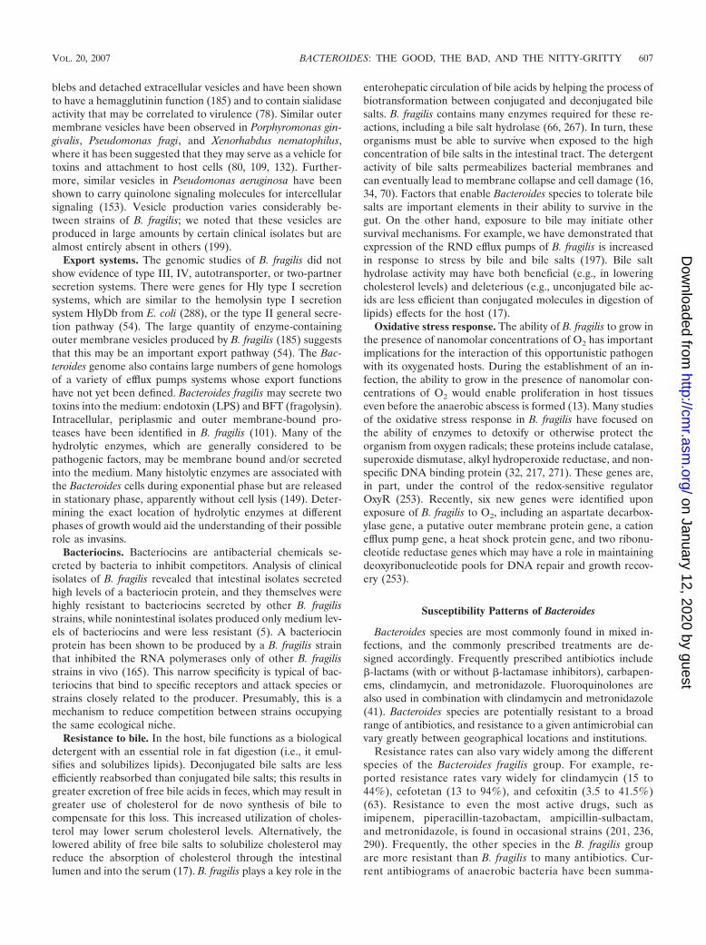
blebs and detached extracellular vesicles and have been shownto have a hemagglutinin function (185) and to contain sialidaseactivity that may be correlated to virulence (78). Similar outermembrane vesicles have been observed in Porphyromonas gin-givalis, Pseudomonas fragi, and Xenorhabdus nematophilus,where it has been suggested that they may serve as a vehicle fortoxins and attachment to host cells (80, 109, 132). Further-more, similar vesicles in Pseudomonas aeruginosa have beenshown to carry quinolone signaling molecules for intercellularsignaling (153). Vesicle production varies considerably be-tween strains of B. fragilis; we noted that these vesicles areproduced in large amounts by certain clinical isolates but arealmost entirely absent in others (199).
Export systems. The genomic studies of B. fragilis did notshow evidence of type III, IV, autotransporter, or two-partnersecretion systems. There were genes for Hly type I secretionsystems, which are similar to the hemolysin type I secretionsystem HlyDb from E. coli (288), or the type II general secre-tion pathway (54). The large quantity of enzyme-containingouter membrane vesicles produced by B. fragilis (185) suggeststhat this may be an important export pathway (54). The Bac-teroides genome also contains large numbers of gene homologsof a variety of efflux pumps systems whose export functionshave not yet been defined. Bacteroides fragilis may secrete twotoxins into the medium: endotoxin (LPS) and BFT (fragolysin).Intracellular, periplasmic and outer membrane-bound pro-teases have been identified in B. fragilis (101). Many of thehydrolytic enzymes, which are generally considered to bepathogenic factors, may be membrane bound and/or secretedinto the medium. Many histolytic enzymes are associated withthe Bacteroides cells during exponential phase but are releasedin stationary phase, apparently without cell lysis (149). Deter-mining the exact location of hydrolytic enzymes at differentphases of growth would aid the understanding of their possiblerole as invasins.
Bacteriocins. Bacteriocins are antibacterial chemicals se-creted by bacteria to inhibit competitors. Analysis of clinicalisolates of B. fragilis revealed that intestinal isolates secretedhigh levels of a bacteriocin protein, and they themselves werehighly resistant to bacteriocins secreted by other B. fragilisstrains, while nonintestinal isolates produced only medium lev-els of bacteriocins and were less resistant (5). A bacteriocinprotein has been shown to be produced by a B. fragilis strainthat inhibited the RNA polymerases only of other B. fragilisstrains in vivo (165). This narrow specificity is typical of bac-teriocins that bind to specific receptors and attack species orstrains closely related to the producer. Presumably, this is amechanism to reduce competition between strains occupyingthe same ecological niche.
Resistance to bile. In the host, bile functions as a biologicaldetergent with an essential role in fat digestion (i.e., it emul-sifies and solubilizes lipids). Deconjugated bile salts are lessefficiently reabsorbed than conjugated bile salts; this results ingreater excretion of free bile acids in feces, which may result ingreater use of cholesterol for de novo synthesis of bile tocompensate for this loss. This increased utilization of choles-terol may lower serum cholesterol levels. Alternatively, thelowered ability of free bile salts to solubilize cholesterol mayreduce the absorption of cholesterol through the intestinallumen and into the serum (17). B. fragilis plays a key role in the
enterohepatic circulation of bile acids by helping the process ofbiotransformation between conjugated and deconjugated bilesalts. B. fragilis contains many enzymes required for these re-actions, including a bile salt hydrolase (66, 267). In turn, theseorganisms must be able to survive when exposed to the highconcentration of bile salts in the intestinal tract. The detergentactivity of bile salts permeabilizes bacterial membranes andcan eventually lead to membrane collapse and cell damage (16,34, 70). Factors that enable Bacteroides species to tolerate bilesalts are important elements in their ability to survive in thegut. On the other hand, exposure to bile may initiate othersurvival mechanisms. For example, we have demonstrated thatexpression of the RND efflux pumps of B. fragilis is increasedin response to stress by bile and bile salts (197). Bile salthydrolase activity may have both beneficial (e.g., in loweringcholesterol levels) and deleterious (e.g., unconjugated bile ac-ids are less efficient than conjugated molecules in digestion oflipids) effects for the host (17).
Oxidative stress response. The ability of B. fragilis to grow inthe presence of nanomolar concentrations of O2 has importantimplications for the interaction of this opportunistic pathogenwith its oxygenated hosts. During the establishment of an in-fection, the ability to grow in the presence of nanomolar con-centrations of O2 would enable proliferation in host tissueseven before the anaerobic abscess is formed (13). Many studiesof the oxidative stress response in B. fragilis have focused onthe ability of enzymes to detoxify or otherwise protect theorganism from oxygen radicals; these proteins include catalase,superoxide dismutase, alkyl hydroperoxide reductase, and non-specific DNA binding protein (32, 217, 271). These genes are,in part, under the control of the redox-sensitive regulatorOxyR (253). Recently, six new genes were identified uponexposure of B. fragilis to O2, including an aspartate decarbox-ylase gene, a putative outer membrane protein gene, a cationefflux pump gene, a heat shock protein gene, and two ribonu-cleotide reductase genes which may have a role in maintainingdeoxyribonucleotide pools for DNA repair and growth recov-ery (253).
Susceptibility Patterns of Bacteroides
Bacteroides species are most commonly found in mixed in-fections, and the commonly prescribed treatments are de-signed accordingly. Frequently prescribed antibiotics include�-lactams (with or without �-lactamase inhibitors), carbapen-ems, clindamycin, and metronidazole. Fluoroquinolones arealso used in combination with clindamycin and metronidazole(41). Bacteroides species are potentially resistant to a broadrange of antibiotics, and resistance to a given antimicrobial canvary greatly between geographical locations and institutions.
Resistance rates can also vary widely among the differentspecies of the Bacteroides fragilis group. For example, re-ported resistance rates vary widely for clindamycin (15 to44%), cefotetan (13 to 94%), and cefoxitin (3.5 to 41.5%)(63). Resistance to even the most active drugs, such asimipenem, piperacillin-tazobactam, ampicillin-sulbactam,and metronidazole, is found in occasional strains (201, 236,290). Frequently, the other species in the B. fragilis groupare more resistant than B. fragilis to many antibiotics. Cur-rent antibiograms of anaerobic bacteria have been summa-
VOL. 20, 2007 BACTEROIDES: THE GOOD, THE BAD, AND THE NITTY-GRITTY 607
on January 12, 2020 by guesthttp://cm
r.asm.org/
Dow
nloaded from

rized and reviewed recently (114, 115, 256, 258) and resultsof several studies have been collated in Table 3.
The most recent multicenter study by Snydman and col-leagues reviewed results for 11 antimicrobials and more than5,000 isolates referred by 10 medical centers (256) and pre-sented the results as trends of antimicrobial susceptibility overan 8-year time period. Resistance to carbapenems was rarelyseen in this study (�1.5%). The trends in resistance to pip-eracillin-tazobactam, ampicillin-sulbactam, and cefoxitin werespecies dependent and did not necessarily correlate with eachother. A worrisome development was the increase in resistanceof B. distasonis (which is no longer technically in the genusBacteroides [225]) to ampicillin-sulbactam in 2002 and 2004(�20%). Resistance of B. fragilis, B. ovatus, and B. thetaiota-omicron to clindamycin increased significantly, as did the geo-metric mean MICs of these agents; similar results were seenfor moxifloxacin. Resistance rates for tigecycline were low andstable during the 5-year period during which this agent wasstudied.
Mechanisms of Antimicrobial Resistance
Species of the genus Bacteroides have the most antibioticresistance mechanisms and the highest resistance rates of allanaerobic pathogens (259). B. fragilis is intrinsically resistant toseveral classes of structurally unrelated antibiotics (114), andthe mechanisms are often poorly understood. Logically, resis-tance to any antimicrobial agent should be due to one of threemechanisms: altered target binding affinity, decreased penetra-tion for the antibiotic due to permeability or efflux changes, orthe presence of an inactivating enzyme. Clinically, Bacteroidesspecies have exhibited increasing resistance to many antibiot-ics, including cefoxitin, clindamycin, metronidazole, carbapen-ems, and fluoroquinolones (e.g., gatifloxacin, levofloxacin, andmoxifloxacin). The newer fluoroquinolones, including sita-floxacin, clinafloxacin, and garenoxacin, are generally moreactive against Bacteroides species (257). Bacteroides speciesare adept at antimicrobial evasion and may use any or all ofthe above-mentioned mechanisms to thwart effective clinicaltherapy.
�-Lactam agents. The most common mechanism of resis-tance to �-lactam agents is �-lactamase (product of the cepAgene), a chromosomally encoded class 2e cephalosporinase(113), which is found in nearly all Bacteroides species (219).This enzyme is inhibited by the most commonly used �-lacta-mase inhibitors (sulbactam, clavulanic acid, and tazobactam),and agents containing these inhibitors are active against strainsthat produce the �-lactamase enzyme. The cefoxitin resistancegene, cfxA (182), has been shown to be distantly related to theB. fragilis endogenous cepA (219).
Carbapenems. Resistance to carbapenems remains rare.Genes for two enzymes that can degrade these agents havebeen identified: cfiA and ccrA, both encoding class B metallo-�-lactamases (147). These enzymes confer resistance to car-bapenems, �-lactams, and �-lactamase inhibitor combinationagents. Some strains of Bacteroides contain “silent” carbapen-emase genes, and expression levels are dependent on promoter-containing insertion sequences (IS) inserted upstream of thecfiA/ccrA gene sequence. Changes in penicillin binding pro-teins (PBPs) or changes in porins that allow permeation of the
carbapenems are other theoretically possible carbapenem re-sistance mechanisms but have only rarely been described inclinical isolates (e.g., PBP2Bfr, a homolog of E. coli PBP3, wasimplicated in imipenem resistance in one strain [6]). On theother hand, we have recently studied multidrug-resistantstrains with increased efflux pump activity, and we have foundclinical isolates that are resistant to carbapenems because ofincreased efflux activity (194, 201).
PBPs. Penicillin and other �-lactams are cell wall-activedrugs that interfere with the final transpeptidation step in thesynthesis of the peptidoglycan. The structure of the �-lactamantibiotics facilitates their binding to the active site of PBPs;this irreversible inhibition of the PBPs prevents the final cross-linking (transpeptidation) of the nascent peptidoglycan layer.The correlation of �-lactam activity and PBP affinity in B.fragilis has not been straightforward, partly because there areconflicting results from investigations into the numbering andmolecular masses of PBPs from this bacterium. Most studieswere done by labeling cells or cell extracts with a labeledantimicrobial and analyzing the results by sodium dodecyl sul-fate (SDS)-polyacrylamide gel electrophoresis (82, 83, 298).The completion of the genome sequence of B. fragilis alloweda more complete analysis, and seven putative PBP genes wereidentified. The gene sequences for the closest homologs to theE. coli PBPs in the B. fragilis genome (pbp1abBfr, pbp1cBfr,pbpABfr, pbpBBfr, pbp4Bfr, and pbp7Bfr) were deduced asthe orthologs for the E. coli genes ponBEco (PBP1bEco),pbpCEco (PBP1cEco), pbpAEco (PBP2Eco), pbpBEco(PBP3Eco), dacBEco (PBP4Eco), and pbpGEco (PBP7Eco),respectively (188). In any case, as the authors finally conclude,it is very difficult to correlate the 50% inhibitory concentrationaffinity of a particular PBP for an antibiotic with the MIC, sincethis is not generally the only mechanism responsible for theresistance.
In spite of this difficulty, several workers have reported anassociation between the reduced affinity of �-lactam com-pounds for the PBPs of Bacteroides species and resistance tothese agents. Reduced affinity of an 80-kDa PBP for imipenem,piperacillin, cefoperazone, cefotaxime, and ceftazidime wasobserved in a resistant strain of B. fragilis, although no precise50% inhibitory concentration calculations were performed(99). Alterations in PBPs, mainly changes in PBP1 and/orPBP2, have been implicated in non-�-lactamase-mediated ce-foxitin resistance in cefoxitin-resistant mutants of B. fragilisgroup species (187, 298). The affinities of the PBP1 complex(86 kDa) of B. thetaiotaomicron 238m for cefoxitin and pip-eracillin were �100-fold and �70-fold reduced, respectively,compared with those of its parent strain (85). The ortholog ofthe E. coli PBP3 gene (pbpBBfr, which encodes the proteinPBP2Bfr) was implicated in binding to imipenem (6, 188).
Outer membrane proteins. There are some data correlatingchanges in outer membrane porin proteins and antimicrobialresistance in Bacteroides. A 45-kDa porin-like protein from B.fragilis was potentially correlated with antibiotic resistance inB. fragilis (171), and a putative porin of �70 kDa of B. theta-iotaomicron was potentially associated with resistance to �-lac-tam agents (18). To date, however, the relationships betweenouter membrane (porin) proteins and antimicrobial resistancehave been associative, and no definitive correlation has beenshown.
608 WEXLER CLIN. MICROBIOL. REV.
on January 12, 2020 by guesthttp://cm
r.asm.org/
Dow
nloaded from
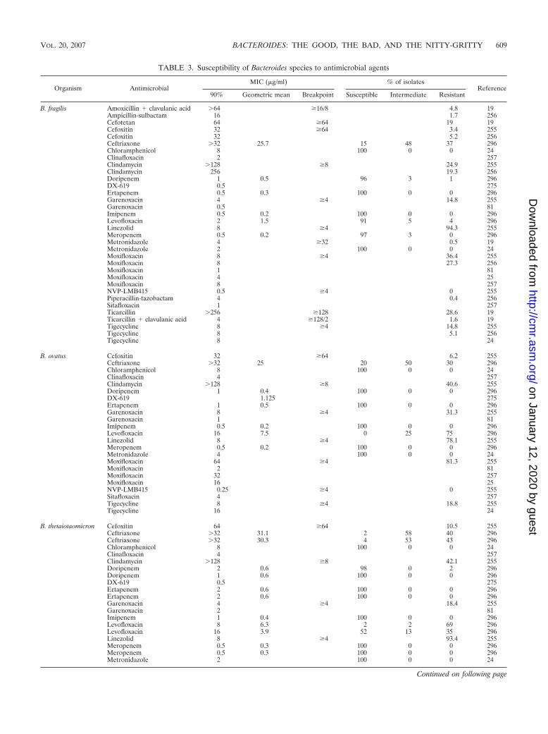
TABLE 3. Susceptibility of Bacteroides species to antimicrobial agents
Organism AntimicrobialMIC (g/ml) % of isolates
Reference90% Geometric mean Breakpoint Susceptible Intermediate Resistant
B. fragilis Amoxicillin � clavulanic acid �64 �16/8 4.8 19Ampicillin-sulbactam 16 1.7 256Cefotetan 64 �64 19 19Cefoxitin 32 �64 3.4 255Cefoxitin 32 5.2 256Ceftriaxone �32 25.7 15 48 37 296Chloramphenicol 8 100 0 0 24Clinafloxacin 2 257Clindamycin �128 �8 24.9 255Clindamycin 256 19.3 256Doripenem 1 0.5 96 3 1 296DX-619 0.5 275Ertapenem 0.5 0.3 100 0 0 296Garenoxacin 4 �4 14.8 255Garenoxacin 0.5 81Imipenem 0.5 0.2 100 0 0 296Levofloxacin 2 1.5 91 5 4 296Linezolid 8 �4 94.3 255Meropenem 0.5 0.2 97 3 0 296Metronidazole 4 �32 0.5 19Metronidazole 2 100 0 0 24Moxifloxacin 8 �4 36.4 255Moxifloxacin 8 27.3 256Moxifloxacin 1 81Moxifloxacin 4 25Moxifloxacin 8 257NVP-LMB415 0.5 �4 0 255Piperacillin-tazobactam 4 0.4 256Sitafloxacin 1 257Ticarcillin �256 �128 28.6 19Ticarcillin � clavulanic acid 4 �128/2 1.6 19Tigecycline 8 �4 14.8 255Tigecycline 8 5.1 256Tigecycline 8 24
B. ovatus Cefoxitin 32 �64 6.2 255Ceftriaxone �32 25 20 50 30 296Chloramphenicol 8 100 0 0 24Clinafloxacin 4 257Clindamycin �128 �8 40.6 255Doripenem 1 0.4 100 0 0 296DX-619 1.125 275Ertapenem 1 0.5 100 0 0 296Garenoxacin 8 �4 31.3 255Garenoxacin 1 81Imipenem 0.5 0.2 100 0 0 296Levofloxacin 16 7.5 0 25 75 296Linezolid 8 �4 78.1 255Meropenem 0.5 0.2 100 0 0 296Metronidazole 4 100 0 0 24Moxifloxacin 64 �4 81.3 255Moxifloxacin 2 81Moxifloxacin 32 257Moxifloxacin 16 25NVP-LMB415 0.25 �4 0 255Sitafloxacin 4 257Tigecycline 8 �4 18.8 255Tigecycline 16 24
B. thetaiotaomicron Cefoxitin 64 �64 10.5 255Ceftriaxone �32 31.1 2 58 40 296Ceftriaxone �32 30.3 4 53 43 296Chloramphenicol 8 100 0 0 24Clinafloxacin 4 257Clindamycin �128 �8 42.1 255Doripenem 2 0.6 98 0 2 296Doripenem 1 0.6 100 0 0 296DX-619 0.5 275Ertapenem 2 0.6 100 0 0 296Ertapenem 2 0.6 100 0 0 296Garenoxacin 4 �4 18.4 255Garenoxacin 2 81Imipenem 1 0.4 100 0 0 296Levofloxacin 8 6.3 2 2 69 296Levofloxacin 16 3.9 52 13 35 296Linezolid 8 �4 93.4 255Meropenem 0.5 0.3 100 0 0 296Meropenem 0.5 0.3 100 0 0 296Metronidazole 2 100 0 0 24
Continued on following page
VOL. 20, 2007 BACTEROIDES: THE GOOD, THE BAD, AND THE NITTY-GRITTY 609
on January 12, 2020 by guesthttp://cm
r.asm.org/
Dow
nloaded from
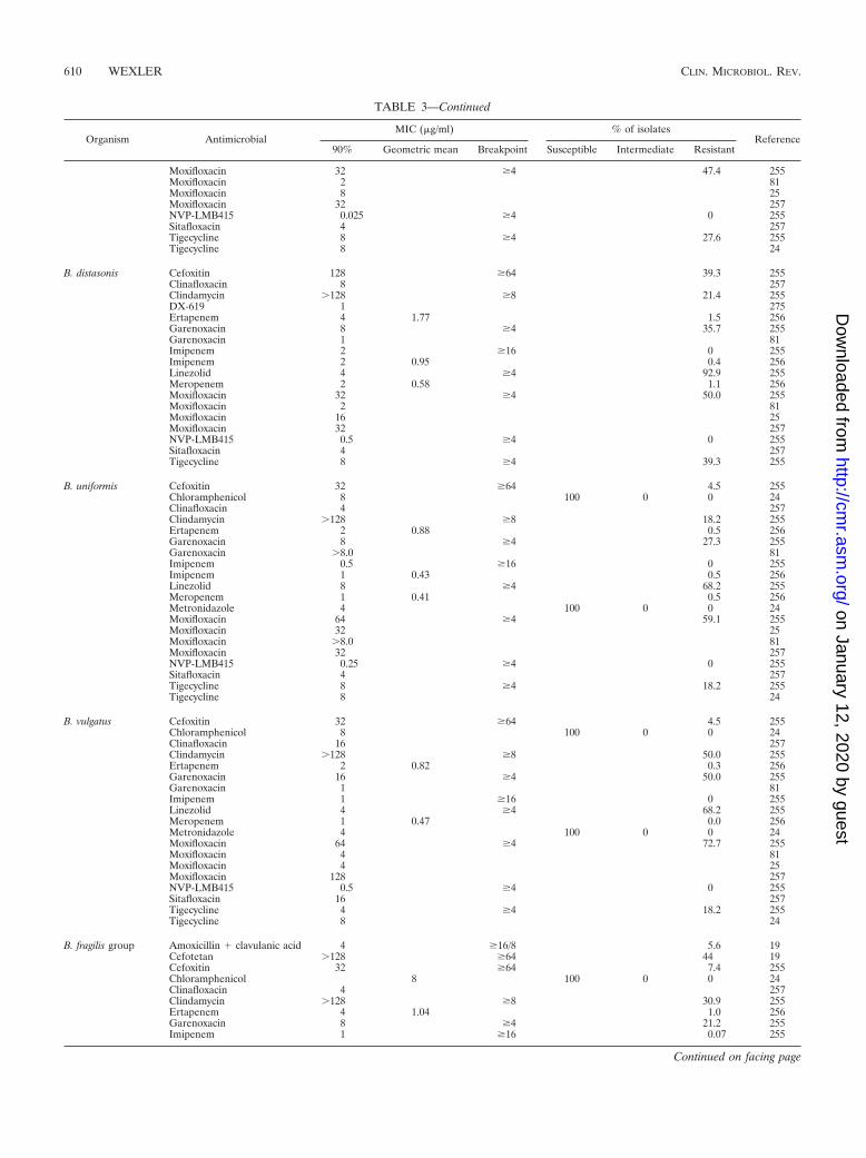
TABLE 3—Continued
Organism AntimicrobialMIC (g/ml) % of isolates
Reference90% Geometric mean Breakpoint Susceptible Intermediate Resistant
Moxifloxacin 32 �4 47.4 255Moxifloxacin 2 81Moxifloxacin 8 25Moxifloxacin 32 257NVP-LMB415 0.025 �4 0 255Sitafloxacin 4 257Tigecycline 8 �4 27.6 255Tigecycline 8 24
B. distasonis Cefoxitin 128 �64 39.3 255Clinafloxacin 8 257Clindamycin �128 �8 21.4 255DX-619 1 275Ertapenem 4 1.77 1.5 256Garenoxacin 8 �4 35.7 255Garenoxacin 1 81Imipenem 2 �16 0 255Imipenem 2 0.95 0.4 256Linezolid 4 �4 92.9 255Meropenem 2 0.58 1.1 256Moxifloxacin 32 �4 50.0 255Moxifloxacin 2 81Moxifloxacin 16 25Moxifloxacin 32 257NVP-LMB415 0.5 �4 0 255Sitafloxacin 4 257Tigecycline 8 �4 39.3 255
B. uniformis Cefoxitin 32 �64 4.5 255Chloramphenicol 8 100 0 0 24Clinafloxacin 4 257Clindamycin �128 �8 18.2 255Ertapenem 2 0.88 0.5 256Garenoxacin 8 �4 27.3 255Garenoxacin �8.0 81Imipenem 0.5 �16 0 255Imipenem 1 0.43 0.5 256Linezolid 8 �4 68.2 255Meropenem 1 0.41 0.5 256Metronidazole 4 100 0 0 24Moxifloxacin 64 �4 59.1 255Moxifloxacin 32 25Moxifloxacin �8.0 81Moxifloxacin 32 257NVP-LMB415 0.25 �4 0 255Sitafloxacin 4 257Tigecycline 8 �4 18.2 255Tigecycline 8 24
B. vulgatus Cefoxitin 32 �64 4.5 255Chloramphenicol 8 100 0 0 24Clinafloxacin 16 257Clindamycin �128 �8 50.0 255Ertapenem 2 0.82 0.3 256Garenoxacin 16 �4 50.0 255Garenoxacin 1 81Imipenem 1 �16 0 255Linezolid 4 �4 68.2 255Meropenem 1 0.47 0.0 256Metronidazole 4 100 0 0 24Moxifloxacin 64 �4 72.7 255Moxifloxacin 4 81Moxifloxacin 4 25Moxifloxacin 128 257NVP-LMB415 0.5 �4 0 255Sitafloxacin 16 257Tigecycline 4 �4 18.2 255Tigecycline 8 24
B. fragilis group Amoxicillin � clavulanic acid 4 �16/8 5.6 19Cefotetan �128 �64 44 19Cefoxitin 32 �64 7.4 255Chloramphenicol 8 100 0 0 24Clinafloxacin 4 257Clindamycin �128 �8 30.9 255Ertapenem 4 1.04 1.0 256Garenoxacin 8 �4 21.2 255Imipenem 1 �16 0.07 255
Continued on facing page
610 WEXLER CLIN. MICROBIOL. REV.
on January 12, 2020 by guesthttp://cm
r.asm.org/
Dow
nloaded from
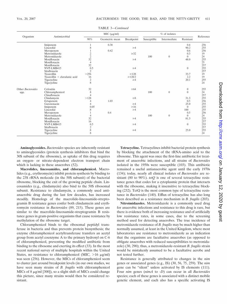
Aminoglycosides. Bacteroides species are inherently resistantto aminoglycosides (protein synthesis inhibitors that bind the30S subunit of the ribosome), as uptake of this drug requiresan oxygen- or nitrate-dependent electron transport chainwhich is lacking in these anaerobes (52).
Macrolides, lincosamides, and chloramphenicol. Macro-lides (e.g., erythromycin) inhibit protein synthesis by binding tothe 23S rRNA molecule (in the 50S subunit) of the bacterialribosome, blocking the exit of the growing peptide chain. Lin-cosamides (e.g., clindamycin) also bind to the 50S ribosomalsubunit. Resistance to clindamycin, a commonly used anti-anaerobic drug during the last few decades, has increasedsteadily. Homologs of the macrolide-lincosamide-strepto-gramin B resistance genes confer both clindamycin and eryth-romycin resistance in Bacteroides (89, 215). These genes aresimilar to the macrolide-lincosamide-streptogramin B resis-tance genes in gram-positive organisms that cause resistance bymethylation of the ribosome target.
Chloramphenicol binds to the ribosomal peptidyl trans-ferase in bacteria and thus prevents protein biosynthesis; theenzyme chloramphenicol acetyltransferase transfers an acetylgroup from acetyl coenzyme A to the primary hydroxyl on C-6of chloramphenicol, preventing the modified antibiotic frombinding to the ribosome and exerting its effect (33). In the mostrecent national survey of multiple hospitals within the UnitedStates, no resistance to chloramphenicol (MIC, �16 g/ml)was seen (256). However, the MICs of chloramphenicol seemto cluster just around breakpoint levels (in our own studies, wehave seen many strains of B. fragilis with chloramphenicolMICs of 8 g/ml [300]), so a slight shift of MICs could changethis picture, since many strains would then be considered re-sistant.
Tetracycline. Tetracyclines inhibit bacterial protein synthesisby blocking the attachment of the tRNA-amino acid to theribosome. This agent was once the first-line antibiotic for treat-ment of anaerobic infections, and all strains of Bacteroidesisolated in the 1950s were susceptible (103). This antibioticremained a useful antianaerobic agent until the early 1970s(134); today, nearly all clinical isolates of Bacteroides are re-sistant (80 to 90%). tetQ is one of several tetracycline resis-tance genes that codes for a cytoplasmic protein that interactswith the ribosome, making it insensitive to tetracycline block-ing (232). TetQ is the most common type of tetracycline resis-tance in Bacteroides (140). Efflux of tetracycline has also longbeen described as a resistance mechanism in B. fragilis (203).
Nitroimidazoles. Metronidazole is a commonly used drugfor anaerobic infections and resistance to this drug is rare, butthere is evidence both of increasing resistance and of artificiallylow resistance rates, in some cases, due to the screeningmethod used for detecting anaerobes. The true incidence ofmetronidazole resistance of B. fragilis may be much higher thannormally assumed, at least in the United Kingdom, where mostlaboratories use resistance to metronidazole as an indicationthat the organisms are facultative anaerobes (as opposed toobligate anaerobes with reduced susceptibilities to metronida-zole) (30, 290); thus, a metronidazole-resistant B. fragilis strainwould be mistakenly assumed to be a facultative aerobe andnot tested further.
Resistance is generally attributed to changes in the nimgenes or associated genes (e.g., IS) (30, 56, 75, 259). The nimgene can be “silent” unless activated by IS elements (111).Four nim genes (nimA to -D) can occur in all Bacteroidesspecies; each of these genes is associated with a distinct mobilegenetic element, and each also has a specific activating IS
TABLE 3—Continued
Organism AntimicrobialMIC (g/ml) % of isolates
Reference90% Geometric mean Breakpoint Susceptible Intermediate Resistant
Imipenem 1 0.38 0.4 256Linezolid 8 �4 90.2 255Meropenem 1 0.42 0.6 256Metronidazole 2 �32 0.3 19Metronidazole 2 100 0 0 24Moxifloxacin 32 �4 48.0 255Moxifloxacin 4 25Moxifloxacin 32 257NVP-LMB415 0.5 �4 0 255Sitafloxacin 2 257Ticarcillin �256 �128 33.7 19Ticarcillin � clavulanic acid 16 �128/2 2.2 19Tigecycline 2 �4 20.1 255Tigecycline 8 24
Other Bacteroidesspp.
Cefoxitin 32 �64 0 255Chloramphenicol 8 100 0 0 24Clinafloxacin 4 257Clindamycin �128 �8 31.3 255Ertapenem 4 1.03 0.5 256Garenoxacin 4 �4 25.0 255Imipenem 0.05 �16 0 255Linezolid 8 �4 100 255Meropenem 1 0.4 0.5 256Metronidazole 4 100 0 0 24Moxifloxacin 32 �4 81.3 255Moxifloxacin 16 257NVP-LMB415 0.5 �4 0 255Sitafloxacin 2 257Tigecycline 8 �4 25.0 255Tigecycline 8 24
VOL. 20, 2007 BACTEROIDES: THE GOOD, THE BAD, AND THE NITTY-GRITTY 611
on January 12, 2020 by guesthttp://cm
r.asm.org/
Dow
nloaded from
element (111, 148, 211, 278). The presence of any one of thesegenes can be detected by PCR (279). Three additional nimgenes (nimE, nimF, and nimG) have also been described (148),but less is known about their mobility and association with ISelements. However, the presence of a nim gene does not nec-essarily confer clinical levels of metronidazole resistance; insome cases, the gene may not be expressed or may be ex-pressed only at very low levels (96).
Quinolones. Quinolones inhibit two specific enzymes, DNAgyrase and DNA topoisomerase IV, which aid in bacterialDNA replication, and mutations in these enzymes are the mostcommon causes of quinolone resistance. Mutations in GyrAcausing fluoroquinolone resistance in B. fragilis have beenidentified at hot-spot positions 82 and 86 (equivalent to posi-tions 83 and 87 in E. coli) (174, 213). Substitutions in GyrB,ParC, and ParE have so far proven uncommon and are not wellestablished in B. fragilis. Bacteria may also become resistant byincreased export of quinolones out of the cell, and Miyamae etal. found active efflux of norfloxacin by B. fragilis and suggestedthat the efflux is catalyzed by a pump similar to that of NorA/Bmr (160). Also, B. thetaiotaomicron possesses a MATE (mul-tidrug and toxic compound extrusion) family multidrug effluxexporter (BexA) that pumps out fluoroquinolones (161). Wehave also demonstrated quinolone resistance in clinical isolatesand in laboratory mutants that is due to increased efflux activityof pumps of the RND family (194, 196, 200, 201).
IS Elements
IS typically contain DNA coding for a transposase that allowthis element to generate mutations and genome rearrange-ments, as well as sometimes providing efficient promoters, ei-ther carried entirely by the IS element or created as a resultof the hybrid sequence between the IS and the target se-quence. This is the case for the IS-borne promoters that canbe found upstream of the cfiA or ccrA gene (189), macrolideresistance genes (205), and metronidazole resistance nimgenes (259, 278).
Mobile Genetic Elements in Bacteroides
Bacteroides species, like other bacteria, have a variety ofmechanisms for exchanging genetic information. The frequentpresence of mobile genetic elements in Bacteroides is notmerely an interesting topic for molecular biologists but criti-cally relevant to both the clinical microbiologist and the infec-tious disease practitioner. These elements are vitally importantin the spread of antibiotic resistance genes. Mobile elementsfound in Bacteroides include plasmids, transposons, conjuga-tive transposons, and bacteriophages (254), and all but bacte-riophages have been implicated in transfer of antimicrobialresistance genes.
Plasmids. Plasmids are very common in Bacteroides speciesand are found in �20 to 50% of strains (254). Plasmids typi-cally replicate as separate elements in the host cell, althoughthere are some plasmids in other genera that can either inte-grate into the chromosome or replicate as plasmids, dependingon the host (231). Many plasmids possess oriT and a trans-acting mobilization gene, which allow them to be transferredby conjugation (note that trans-acting factors may act on enti-
ties that are not in close proximity.) There are also plasmidsthat do not self-transfer but may be transferred via mobiliza-tion by other chromosomal elements (e.g., pBFTM10) (254).
Genes conferring resistance to many different classes ofantibiotics have been found on plasmids in Bacteroides. Resis-tance genes nimA to -F, which have been implicated in spo-radic cases of metronidazole resistance in several hospitalsworldwide, have been identified on transferable plasmids (56,148, 279). Transferable plasmid-linked chloramphenicolacetyltransferase conferring high-level resistance was observedin a clinical isolate of Bacteroides uniformis (152). The cfiAgene, conferring resistance to carbapenems, has also beenfound on a 6.4-kb plasmid in clinical isolates (166). Clindamy-cin and erythromycin resistance can be transferable betweenBacteroides species, either via a chromosomal element (273) orin association with a conjugative plasmid (249, 294).
Conjugative transposons. Transposons, both mobilizableand conjugative, do not replicate independently; rather, theyexcise from and integrate into chromosomal DNA and arecopied along with the chromosomal DNA. Conjugative trans-posons have a mechanism of excision and integration thatresemble some features of both plasmids and bacteriophage(231). Conjugative transposons are practically ubiquitousamong the Bacteroides: over 80% of Bacteroides strains containat least one conjugative transposon (250).
The particular attributes of conjugative transposons havebeen elegantly summarized by Salyers et al. (231). Conjugativetransposons have certain characteristics of plasmids, of trans-posons, and of phages. Often, but not always, they are desig-nated by CTn (conjugative transposon) as opposed to Tn(transposon). The conjugative transposons of Bacteroides be-long to at least two families; CTnDot is the best described (231,254). Often, the name of the strain in which they are found isadded to the designation (e.g., CTnDot, found in the DOTstrain of B. thetaiotaomicron). In addition to being able toinsert into the chromosome, Bacteroides conjugative trans-posons can insert into coresident plasmids and mobilizethem in cis (i.e., they can act on entities that are physicallyadjacent) by integrating themselves into the plasmid andfacilitating transfer of the plasmid-conjugative transposonhybrid into another cell (230). They can also mobilizecoresident plasmids “in trans” by supplying factors neededto facilitate transfer of the plasmid, while remaining physi-cally separate from the plasmid.
The conjugative transposons do not exclude each other as doplasmids, so a strain can accumulate more than one conjuga-tive transposon. Furthermore, there is some evidence that thepresence of more than one copy of the conjugative transposonin the strain results in a stimulation of transposition (transac-tivation) (231). Theoretically, this suggests that as more con-jugative transposons with antibiotic resistance genes accumu-late in the environment, the transfer of these genes to otherbacteria will also increase, and there will be a significant up-ward spiraling of antibiotic resistance.
Conjugative transposons have been largely responsible forthe spread of tetracycline and erythromycin resistance in clin-ical isolates of Bacteroides (250). Fifty years ago, tetracyclineresistance was almost nonexistent in this genus (103). By the1980s a majority of strains were resistant, and today almost all
612 WEXLER CLIN. MICROBIOL. REV.
on January 12, 2020 by guesthttp://cm
r.asm.org/
Dow
nloaded from
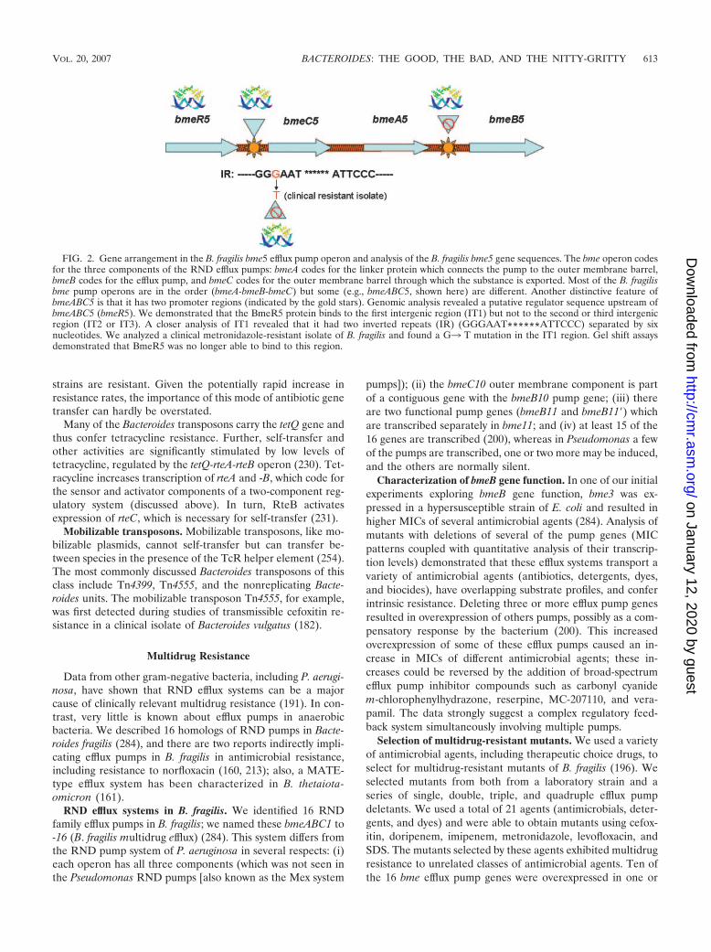
strains are resistant. Given the potentially rapid increase inresistance rates, the importance of this mode of antibiotic genetransfer can hardly be overstated.
Many of the Bacteroides transposons carry the tetQ gene andthus confer tetracycline resistance. Further, self-transfer andother activities are significantly stimulated by low levels oftetracycline, regulated by the tetQ-rteA-rteB operon (230). Tet-racycline increases transcription of rteA and -B, which code forthe sensor and activator components of a two-component reg-ulatory system (discussed above). In turn, RteB activatesexpression of rteC, which is necessary for self-transfer (231).
Mobilizable transposons. Mobilizable transposons, like mo-bilizable plasmids, cannot self-transfer but can transfer be-tween species in the presence of the TcR helper element (254).The most commonly discussed Bacteroides transposons of thisclass include Tn4399, Tn4555, and the nonreplicating Bacte-roides units. The mobilizable transposon Tn4555, for example,was first detected during studies of transmissible cefoxitin re-sistance in a clinical isolate of Bacteroides vulgatus (182).
Multidrug Resistance
Data from other gram-negative bacteria, including P. aerugi-nosa, have shown that RND efflux systems can be a majorcause of clinically relevant multidrug resistance (191). In con-trast, very little is known about efflux pumps in anaerobicbacteria. We described 16 homologs of RND pumps in Bacte-roides fragilis (284), and there are two reports indirectly impli-cating efflux pumps in B. fragilis in antimicrobial resistance,including resistance to norfloxacin (160, 213); also, a MATE-type efflux system has been characterized in B. thetaiota-omicron (161).
RND efflux systems in B. fragilis. We identified 16 RNDfamily efflux pumps in B. fragilis; we named these bmeABC1 to-16 (B. fragilis multidrug efflux) (284). This system differs fromthe RND pump system of P. aeruginosa in several respects: (i)each operon has all three components (which was not seen inthe Pseudomonas RND pumps [also known as the Mex system
pumps]); (ii) the bmeC10 outer membrane component is partof a contiguous gene with the bmeB10 pump gene; (iii) thereare two functional pump genes (bmeB11 and bmeB11) whichare transcribed separately in bme11; and (iv) at least 15 of the16 genes are transcribed (200), whereas in Pseudomonas a fewof the pumps are transcribed, one or two more may be induced,and the others are normally silent.
Characterization of bmeB gene function. In one of our initialexperiments exploring bmeB gene function, bme3 was ex-pressed in a hypersusceptible strain of E. coli and resulted inhigher MICs of several antimicrobial agents (284). Analysis ofmutants with deletions of several of the pump genes (MICpatterns coupled with quantitative analysis of their transcrip-tion levels) demonstrated that these efflux systems transport avariety of antimicrobial agents (antibiotics, detergents, dyes,and biocides), have overlapping substrate profiles, and conferintrinsic resistance. Deleting three or more efflux pump genesresulted in overexpression of others pumps, possibly as a com-pensatory response by the bacterium (200). This increasedoverexpression of some of these efflux pumps caused an in-crease in MICs of different antimicrobial agents; these in-creases could be reversed by the addition of broad-spectrumefflux pump inhibitor compounds such as carbonyl cyanidem-chlorophenylhydrazone, reserpine, MC-207110, and vera-pamil. The data strongly suggest a complex regulatory feed-back system simultaneously involving multiple pumps.
Selection of multidrug-resistant mutants. We used a varietyof antimicrobial agents, including therapeutic choice drugs, toselect for multidrug-resistant mutants of B. fragilis (196). Weselected mutants from both from a laboratory strain and aseries of single, double, triple, and quadruple efflux pumpdeletants. We used a total of 21 agents (antimicrobials, deter-gents, and dyes) and were able to obtain mutants using cefox-itin, doripenem, imipenem, metronidazole, levofloxacin, andSDS. The mutants selected by these agents exhibited multidrugresistance to unrelated classes of antimicrobial agents. Ten ofthe 16 bme efflux pump genes were overexpressed in one or
FIG. 2. Gene arrangement in the B. fragilis bme5 efflux pump operon and analysis of the B. fragilis bme5 gene sequences. The bme operon codesfor the three components of the RND efflux pumps: bmeA codes for the linker protein which connects the pump to the outer membrane barrel,bmeB codes for the efflux pump, and bmeC codes for the outer membrane barrel through which the substance is exported. Most of the B. fragilisbme pump operons are in the order (bmeA-bmeB-bmeC) but some (e.g., bmeABC5, shown here) are different. Another distinctive feature ofbmeABC5 is that it has two promoter regions (indicated by the gold stars). Genomic analysis revealed a putative regulator sequence upstream ofbmeABC5 (bmeR5). We demonstrated that the BmeR5 protein binds to the first intergenic region (IT1) but not to the second or third intergenicregion (IT2 or IT3). A closer analysis of IT1 revealed that it had two inverted repeats (IR) (GGGAAT******ATTCCC) separated by sixnucleotides. We analyzed a clinical metronidazole-resistant isolate of B. fragilis and found a G3 T mutation in the IT1 region. Gel shift assaysdemonstrated that BmeR5 was no longer able to bind to this region.
VOL. 20, 2007 BACTEROIDES: THE GOOD, THE BAD, AND THE NITTY-GRITTY 613
on January 12, 2020 by guesthttp://cm
r.asm.org/
Dow
nloaded from
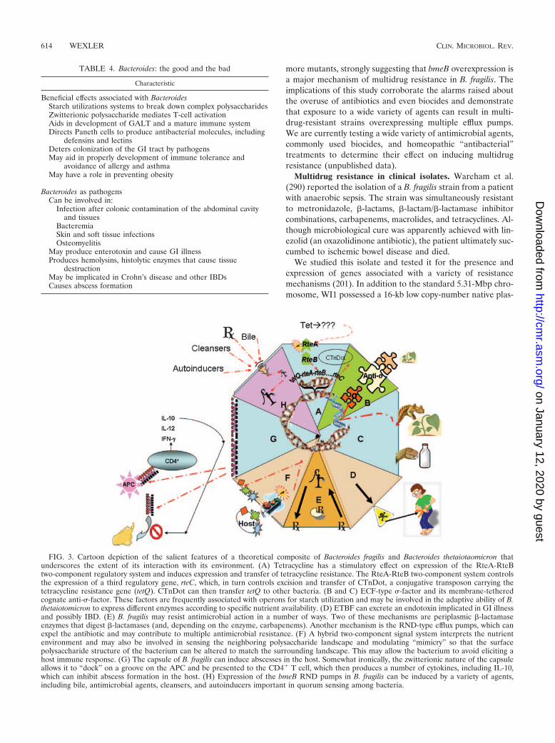
more mutants, strongly suggesting that bmeB overexpression isa major mechanism of multidrug resistance in B. fragilis. Theimplications of this study corroborate the alarms raised aboutthe overuse of antibiotics and even biocides and demonstratethat exposure to a wide variety of agents can result in multi-drug-resistant strains overexpressing multiple efflux pumps.We are currently testing a wide variety of antimicrobial agents,commonly used biocides, and homeopathic “antibacterial”treatments to determine their effect on inducing multidrugresistance (unpublished data).
Multidrug resistance in clinical isolates. Wareham et al.(290) reported the isolation of a B. fragilis strain from a patientwith anaerobic sepsis. The strain was simultaneously resistantto metronidazole, �-lactams, �-lactam/�-lactamase inhibitorcombinations, carbapenems, macrolides, and tetracyclines. Al-though microbiological cure was apparently achieved with lin-ezolid (an oxazolidinone antibiotic), the patient ultimately suc-cumbed to ischemic bowel disease and died.
We studied this isolate and tested it for the presence andexpression of genes associated with a variety of resistancemechanisms (201). In addition to the standard 5.31-Mbp chro-mosome, WI1 possessed a 16-kb low copy-number native plas-
FIG. 3. Cartoon depiction of the salient features of a theoretical composite of Bacteroides fragilis and Bacteroides thetaiotaomicron thatunderscores the extent of its interaction with its environment. (A) Tetracycline has a stimulatory effect on expression of the RteA-RteBtwo-component regulatory system and induces expression and transfer of tetracycline resistance. The RteA-RteB two-component system controlsthe expression of a third regulatory gene, rteC, which, in turn controls excision and transfer of CTnDot, a conjugative transposon carrying thetetracycline resistance gene (tetQ). CTnDot can then transfer tetQ to other bacteria. (B and C) ECF-type �-factor and its membrane-tetheredcognate anti-�-factor. These factors are frequently associated with operons for starch utilization and may be involved in the adaptive ability of B.thetaiotomicron to express different enzymes according to specific nutrient availability. (D) ETBF can excrete an endotoxin implicated in GI illnessand possibly IBD. (E) B. fragilis may resist antimicrobial action in a number of ways. Two of these mechanisms are periplasmic �-lactamaseenzymes that digest �-lactamases (and, depending on the enzyme, carbapenems). Another mechanism is the RND-type efflux pumps, which canexpel the antibiotic and may contribute to multiple antimicrobial resistance. (F) A hybrid two-component signal system interprets the nutrientenvironment and may also be involved in sensing the neighboring polysaccharide landscape and modulating “mimicry” so that the surfacepolysaccharide structure of the bacterium can be altered to match the surrounding landscape. This may allow the bacterium to avoid eliciting ahost immune response. (G) The capsule of B. fragilis can induce abscesses in the host. Somewhat ironically, the zwitterionic nature of the capsuleallows it to “dock” on a groove on the APC and be presented to the CD4� T cell, which then produces a number of cytokines, including IL-10,which can inhibit abscess formation in the host. (H) Expression of the bmeB RND pumps in B. fragilis can be induced by a variety of agents,including bile, antimicrobial agents, cleansers, and autoinducers important in quorum sensing among bacteria.
TABLE 4. Bacteroides: the good and the bad
Characteristic
Beneficial effects associated with BacteroidesStarch utilizations systems to break down complex polysaccharidesZwitterionic polysaccharide mediates T-cell activationAids in development of GALT and a mature immune systemDirects Paneth cells to produce antibacterial molecules, including
defensins and lectinsDeters colonization of the GI tract by pathogensMay aid in properly development of immune tolerance and
avoidance of allergy and asthmaMay have a role in preventing obesity
Bacteroides as pathogensCan be involved in:
Infection after colonic contamination of the abdominal cavityand tissues
BacteremiaSkin and soft tissue infectionsOsteomyelitis
May produce enterotoxin and cause GI illnessProduces hemolysins, histolytic enzymes that cause tissue
destructionMay be implicated in Crohn’s disease and other IBDsCauses abscess formation
614 WEXLER CLIN. MICROBIOL. REV.
on January 12, 2020 by guesthttp://cm
r.asm.org/
Dow
nloaded from
mid, pBHag, which was absent in the B. fragilis type strain;sequencing of this plasmid revealed that this plasmid containedthe gene for tetQ as well as a number of genes that normallyreside on the conjugative transposon CTn341. Failed curingattempts suggested that it was stable without obvious selection.We found that the cfiA, ermF, tetQ2, and tetQ3 genes wereexpressed in the total cellular RNA; neither tetX nor nim geneswere detected. Sequencing of the gyrA quinolone resistance-determining region revealed a mutation causing a Ser833Ilesubstitution. The efflux pump genes bmeB9 and bmeB15 weresignificantly overexpressed, and addition of efflux pump inhib-itors significantly increased susceptibility of the isolate to sev-eral unrelated antibiotics. These data suggest that this isolatewas highly multidrug resistant due to additive effects of chro-mosomally and plasmid-encoded resistance determinants.
We also studied recent multidrug-resistant clinical isolatesand found that they overexpressed several bmeB efflux pumps.The data suggest that bmeB efflux pump overexpression cancause low- to intermediate-level clinically relevant fluoroquin-olone resistance and can contribute to high-level clinically rel-evant resistance to �-lactams; moreover, it can be coupled withGyrA substitutions to cause high-level fluoroquinolone resis-tance (194).
We demonstrated that bmeB5 was overexpressed in metron-idazole-resistant laboratory mutants of Bacteroides fragilis, andwe identified an upstream putative TetR family regulator gene(bmeR5). Deletant strains lacking bmeB5 or bmeR5 were con-structed and characterized. The MICs of ampicillin, cefopera-zone, metronidazole, and SDS were reduced by approximatelytwofold in ADB77 �bmeB5. Deletion of bmeR5 (ADB77�bmeR5) resulted in a significant (P � 0.05) increase in ex-pression of bmeA5, bmeB5, and bmeC5 and a �2-fold increasein MICs of ampicillin, cefoxitin, cefoperazone, ciprofloxacin,imipenem, metronidazole, ethidium bromide, and SDS. Wefound that BmeR5-His6 bound specifically to the bmeR5-bmeA5 intergenic region (IT1). A multidrug (metronidazole)-resistant, nim-negative B. fragilis clinical isolate overexpressedbmeABC5 genes, had a G-T point mutation in IT1, and signif-icantly reduced binding to BmeR5-His6 (195). A schematicdescription of bmeRABC5 is depicted in Fig. 2.
IMPORTANT FUTURE RESEARCH AVENUES
With the adaptability of a chameleon, the Bacteroides spe-cies can function as an integral partner in the human metabolicsystem and yet can be the cause of serious, life-threateningdisease (Table 4). A cartoon description of the some of themost interesting features of a theoretical composite strain of B.fragilis and B. distasonis is depicted in Fig. 3. Despite its pre-dominance as the most numerous organism in the human host,progress in understanding Bacteroides has consistently laggedbehind that for aerobic bacteria. However, with the publicationof the genome sequences of B. fragilis and B. thetaiotaomicron,several tantalizing potential avenues of research are nowwithin reach. Constructing microarrays for Bacteroides to mea-sure expression of various genes would be a major step towardunraveling some of the complex regulatory relationships foundin this organism. For example, the regulation of efflux pumpexpression appears to be an important factor in multidrugresistance. The multiplicity of the pumps, coupled with their
overlapping substrate profiles, suggests that inhibiting theiraction may need to happen at a regulatory level. These effluxpumps may also be involved in the ability of the organism tothrive in the GI tract. Maintaining the proper balance of GImicrobes has been long recognized as an important mechanismto avoid colonization by dangerous pathogens; it is now alsorecognized as a potentially important factor in obesity. A re-cent report suggested that understanding the manner by whichbacteria adapt to specific niches may aid in designing site-directed bacterial vehicles for delivering beneficial moleculesto the human GI tract (90). Happily, many of these long-rangeresearch goals appear to be intertwined: efflux pumps thataffect antimicrobial resistance may be involved in virulence andadhesion, sugar-acquiring pathways and metabolism that allowadaptation to factors in the GI tract may be important inobesity control, and understanding the regulatory factors re-sponsible for controlling the complex face of the Bacteroidesouter membrane may ultimately allow manipulation to reduceresistance and pathogenicity and to exploit its particular char-acteristics for beneficial purposes.
ACKNOWLEDGMENT
H.M.W. is supported by Merit Review Funds from the Departmentof Veterans Affairs.
REFERENCES
1. Abi, K. G., H. Awada, and R. Nasnas. 2001. Isolated septic arthritis of alumbar facet joint. J. Med. Liban. 49:228–230.
2. Anderson, K. L., and A. A. Salyers. 1989. Genetic evidence that outermembrane binding of starch is required for starch utilization by Bacteroidesthetaiotaomicron. J. Bacteriol. 171:3199–3204.
3. Anderson, K. L., and A. A. Salyers. 1989. Biochemical evidence that starchbreakdown by Bacteroides thetaiotaomicron involves outer membranestarch-binding sites and periplasmic starch-degrading enzymes. J. Bacteriol.171:3192–3198.
4. Aucher, P., J. P. Saunier, G. Grollier, M. Sebald, and J. L. Fauchere. 1996.Meningitis due to enterotoxigenic Bacteroides fragilis. Eur. J. Clin. Micro-biol. Infect. Dis. 15:820–823.
5. Avelar, K. E., L. J. Pinto, L. C. Antunes, L. A. Lobo, M. C. Bastos, R. M.Domingues, and M. C. Ferreira. 1999. Production of bacteriocin by Bacte-riodes fragilis and partial characterization. Lett. Appl. Microbiol. 29:264–268.
6. Ayala, J., A. Quesada, S. Vadillo, J. Criado, and S. Piriz. 2005. Penicillin-binding proteins of Bacteroides fragilis and their role in the resistance toimipenem of clinical isolates. J. Med. Microbiol. 54:1055–1064.
7. Babu, M. M., M. L. Priya, A. T. Selvan, M. Madera, J. Gough, L. Aravind,and K. Sankaran. 2006. A database of bacterial lipoproteins (DOLOP)with functional assignments to predicted lipoproteins. J. Bacteriol. 188:2761–2773.
8. Backhed, F., R. E. Ley, J. L. Sonnenburg, D. A. Peterson, and J. I. Gordon.2005. Host-bacterial mutualism in the human intestine. Science 307:1915–1920.
9. Bajzer, M., and R. J. Seeley. 2006. Physiology: obesity and gut flora. Nature444:1009–1010.
10. Bamba, T., H. Matsuda, M. Endo, and Y. Fujiyama. 1995. The pathogenicrole of Bacteroides vulgatus in patients with ulcerative colitis. J. Gastroen-terol. 30(Suppl. 8):45–47.
11. Bamias, G., A. Okazawa, J. Rivera-Nieves, K. O. Arseneau, S. A. De La Rue,T. T. Pizarro, and F. Cominelli. 2007. Commensal bacteria exacerbateintestinal inflammation but are not essential for the development of murineileitis. J. Immunol. 178:1809–1818.
12. Basset, C., J. Holton, A. Bazeos, D. Vaira, and S. Bloom. 2004. Are Heli-cobacter species and enterotoxigenic Bacteroides fragilis involved in inflam-matory bowel disease? Dig. Dis. Sci. 49:1425–1432.
13. Baughn, A. D., and M. H. Malamy. 2004. The strict anaerobe Bacteroidesfragilis grows in and benefits from nanomolar concentrations of oxygen.Nature 427:441–444.
14. Baumann, H., A. O. Tzianabos, J. R. Brisson, D. L. Kasper, and H. J.Jennings. 1992. Structural elucidation of two capsular polysaccharides fromone strain of Bacteroides fragilis using high-resolution NMR spectroscopy.Biochemistry 31:4081–4089.
15. Beena, V. K., and P. G. Shivananda. 1997. In vitro adhesiveness of Bacte-
VOL. 20, 2007 BACTEROIDES: THE GOOD, THE BAD, AND THE NITTY-GRITTY 615
on January 12, 2020 by guesthttp://cm
r.asm.org/
Dow
nloaded from

roides fragilis group in relation to encapsulation. Indian J. Med. Res. 105:258–261.
16. Begley, M., C. G. Gahan, and C. Hill. 2005. The interaction betweenbacteria and bile. FEMS Microbiol. Rev. 29:625–651.
17. Begley, M., C. Hill, and C. G. Gahan. 2006. Bile salt hydrolase activity inprobiotics. Appl. Environ. Microbiol. 72:1729–1738.
18. Behra-Miellet, J., L. Calvet, and L. Dubreuil. 2004. A Bacteroides thetaiota-micron porin that could take part in resistance to beta-lactams. Int. J.Antimicrob. Agents 24:135–143.
19. Behra-Miellet, J., L. Calvet, F. Mory, C. Muller, M. Chomarat, M. C.Bezian, S. Bland, M. E. Juvenin, T. Fosse, F. Goldstein, B. Jaulhac, and L.Dubreuil. 2003. Antibiotic resistance among anaerobic Gram-negative ba-cilli: lessons from a French multicentric survey. Anaerobe 9:105–111.
20. Bendtsen, J. D., T. T. Binnewies, P. F. Hallin, T. Sicheritz-Ponten, andD. W. Ussery. 2005. Genome update: prediction of secreted proteins in 225bacterial proteomes. Microbiology 151:1725–1727.
21. Bennion, R. S., E. J. Baron, J. E. Thompson, Jr., J. Downes, P. Summanen,D. A. Talan, and S. M. Finegold. 1990. The bacteriology of gangrenous andperforated appendicitis—revisited. Ann. Surg. 211:165–171.
22. Berg, J. O., L. Lindqvist, G. Andersson, and C. E. Nord. 1983. Neuramin-idase in Bacteroides fragilis. Appl. Environ. Microbiol. 46:75–80.
23. Bergan, T. 1983. Anaerobic bacteria as cause of infections in female genitalorgans. Scand. J. Gastroenterol. Suppl. 85:37–47.
24. Betriu, C., E. Culebras, M. Gomez, I. Rodriguez-Avial, and J. J. Picazo.2005. In vitro activity of tigecycline against Bacteroides species. J. Antimi-crob. Chemother. 56:349–352.
25. Betriu, C., I. Rodriguez-Avial, M. Gomez, E. Culebras, and J. J. Picazo.2005. Changing patterns of fluoroquinolone resistance among Bacteroidesfragilis group organisms over a 6-year period (1997–2002). Diagn. Micro-biol. Infect. Dis. 53:221–223.
26. Bisharat, N., L. Goldstein, R. Raz, and M. Elias. 2001. Gram-negativeanaerobic endocarditis: two case reports and review of the literature. Eur.J. Clin. Microbiol. Infect. Dis. 20:651–654.
27. Bjursell, M. K., E. C. Martens, and J. I. Gordon. 2006. Functional genomicand metabolic studies of the adaptations of a prominent adult human gutsymbiont, Bacteroides thetaiotaomicron, to the suckling period. J. Biol.Chem. 281:36269–36279.
28. Boquet, P., P. Munro, C. Fiorentini, and I. Just. 1998. Toxins from anaer-obic bacteria: specificity and molecular mechanisms of action. Curr. Opin.Microbiol. 1:66–74.
29. Borer, A., G. Weber, K. Riesenberg, F. Schlaeffer, and J. Horowitz. 1997.Septic arthritis due to Bacteroides fragilis after pilonidal sinus resection in apatient with rheumatoid arthritis. Clin. Rheumatol. 16:632–634.
30. Brazier, J. S., S. L. Stubbs, and B. I. Duerden. 1999. Metronidazole resis-tance among clinical isolates belonging to the Bacteroides fragilis group:time to be concerned? J. Antimicrob. Chemother. 44:580–581. (Letter.)
31. Brigham, C. J., and M. H. Malamy. 2005. Characterization of the RokAand HexA broad-substrate-specificity hexokinases from Bacteroides fragilisand their role in hexose and N-acetylglucosamine utilization. J. Bacteriol.187:890–901.
32. Brioukhanov, A. L., and A. I. Netrusov. 2004. Catalase and superoxidedismutase: distribution, properties, and physiological role in cells of strictanaerobes. Biochemistry (Moscow) 69:949–962.
33. Britz, M. L., and R. G. Wilkinson. 1978. Chloramphenicol acetyl-trans-ferase of Bacteroides fragilis. Antimicrob. Agents Chemother. 14:105–111.
34. Bron, P. A., M. Marco, S. M. Hoffer, M. E. Van, W. M. de Vos, and M.Kleerebezem. 2004. Genetic characterization of the bile salt response inLactobacillus plantarum and analysis of responsive promoters in vitro and insitu in the gastrointestinal tract. J. Bacteriol. 186:7829–7835.
35. Brook, I. 2002. Anaerobic infections in children. Microbes. Infect. 4:1271–1280.
36. Brook, I. 2002. Joint and bone infections due to anaerobic bacteria inchildren. Pediatr. Rehabil. 5:11–19.
37. Brook, I. 2002. Microbiology and management of post-surgical woundsinfection in children. Pediatr. Rehabil. 5:171–176.
38. Brook, I. 2003. Microbiology and management of intra-abdominal infec-tions in children. Pediatr. Int. 45:123–129.
39. Brook, I. 2004. Intra-abdominal, retroperitoneal, and visceral abscesses inchildren. Eur. J. Pediatr. Surg. 14:265–273.
40. Brook, I. 2002. Pericarditis due to anaerobic bacteria. Cardiology 97:55–58.41. Brook, I. 2002. Microbiology of polymicrobial abscesses and implications
for therapy. J. Antimicrob. Chemother. 50:805–810.42. Brook, I. 2002. Endocarditis due to anaerobic bacteria. Cardiology 98:1–5.43. Reference deleted.44. Brook, I. 2002. Clinical review: bacteremia caused by anaerobic bacteria in
children. Crit. Care 6:205–211.45. Brook, I. 1989. Aerobic and anaerobic microbiology of Bartholin’s abscess.
Surg. Gynecol. Obstet. 169:32–34.46. Brook, I. 1986. Anaerobic osteomyelitis in children. Pediatr. Infect. Dis.
5:550–556.47. Brook, I. 1990. Bacteremia due to anaerobic bacteria in newborns. J. Peri-
natol. 10:351–356.
48. Brook, I., J. C. Coolbaugh, and R. I. Walker. 1984. Pathogenicity of piliatedand encapsulated Bacteroides fragilis. Eur. J. Clin. Microbiol. 3:207–209.
49. Brook, I., and E. H. Frazier. 1992. Microbiology of scalp abscess in new-borns. Pediatr. Infect. Dis. J. 11:766–768.
50. Brook, I., N. Johnson, G. D. Overturf, and J. Wilkins. 1977. Mixed bacterialmeningitis: a complication of ventriculo- and lumboperitoneal shunts.J. Neurosurg. 47:961–964.
51. Brook, I., and M. L. Myhal. 1991. Adherence of Bacteroides fragilis groupspecies. Infect. Immun. 59:742–744.
52. Bryan, L. E., S. K. Kowand, and H. M. Van Den Elzen. 1979. Mechanism ofaminoglycoside antibiotic resistance in anaerobic bacteria: Clostridium per-fringens and Bacteroides fragilis. Antimicrob. Agents Chemother. 15:7–13.
53. Cash, H. L., C. V. Whitham, C. L. Behrendt, and L. V. Hooper. 2006.Symbiotic bacteria direct expression of an intestinal bactericidal lectin.Science 313:1126–1130.
54. Cerdeno-Tarraga, A. M., S. Patrick, L. C. Crossman, G. Blakely, V. Abratt,N. Lennard, I. Poxton, B. Duerden, B. Harris, M. A. Quail, A. Barron, L.Clark, C. Corton, J. Doggett, M. T. Holden, N. Larke, A. Line, A. Lord, H.Norbertczak, D. Ormond, C. Price, E. Rabbinowitsch, J. Woodward, B.Barrell, and J. Parkhill. 2005. Extensive DNA inversions in the B. fragilisgenome control variable gene expression. Science 307:1463–1465.
55. Reference deleted.56. Chaudhry, R., P. Mathur, B. Dhawan, and L. Kumar. 2001. Emergence of
metronidazole-resistant Bacteroides fragilis, India. Emerg. Infect. Dis.7:485–486. (Letter.)
57. Chen, Y., T. Kinouchi, K. Kataoka, S. Akimoto, and Y. Ohnishi. 1995.Purification and characterization of a fibrinogen-degrading protease in Bac-teroides fragilis strain YCH46. Microbiol. Immunol. 39:967–977.
58. Cho, K. H., D. Cho, G. R. Wang, and A. A. Salyers. 2001. New regulatorygene that contributes to control of Bacteroides thetaiotaomicron starch uti-lization genes. J. Bacteriol. 183:7198–7205.
59. Cho, K. H., and A. A. Salyers. 2001. Biochemical analysis of interactionsbetween outer membrane proteins that contribute to starch utilization byBacteroides thetaiotaomicron. J. Bacteriol. 183:7224–7230.
60. Cisneros, J. M., J. Rodriguez-Bano, F. Fernandez-Cuenca, A. Ribera, J.Vila, A. Pascual, L. Martinez-Martinez, G. Bou, and J. Pachon. 2005.Risk-factors for the acquisition of imipenem-resistant Acinetobacter bau-mannii in Spain: a nationwide study. Clin. Microbiol. Infect. 11:874–879.
61. Claros, M. C., Z. C. Claros, D. W. Hecht, D. M. Citron, E. J. Goldstein, J.Silva, Jr., Y. Tang-Feldman, and A. C. Rodloff. 2006. Characterization ofthe Bacteroides fragilis pathogenicity island in human blood culture iso-lates. Anaerobe 12:17–22.
62. Claros, M. C., Z. C. Claros, Y. J. Tang, S. H. Cohen, J. Silva, Jr., E. J.Goldstein, and A. C. Rodloff. 2000. Occurrence of Bacteroides fragilis en-terotoxin gene-carrying strains in Germany and the United States. J. Clin.Microbiol. 38:1996–1997.
63. Clinical and Laboratory Standards Institute. 2006. Methods for antimicro-bial susceptibility testing of anaerobic bacteria; approved standard, 7th ed.Clinical and Laboratory Standards Institute, Wayne, PA.
64. Cohavy, O., D. Bruckner, L. K. Gordon, R. Misra, B. Wei, M. E. Eggena,S. R. Targan, and J. Braun. 2000. Colonic bacteria express an ulcerativecolitis pANCA-related protein epitope. Infect. Immun. 68:1542–1548.
65. Comstock, L. E., and M. J. Coyne. 2003. Bacteroides thetaiotaomicron: adynamic, niche-adapted human symbiont. Bioessays 25:926–929.
66. Corzo, G., and S. E. Gilliland. 1999. Measurement of bile salt hydrolaseactivity from Lactobacillus acidophilus based on disappearance of conju-gated bile salts. J. Dairy Sci. 82:466–471.
67. Coyne, M. J., W. Kalka-Moll, A. O. Tzianabos, D. L. Kasper, and L. E.Comstock. 2000. Bacteroides fragilis NCTC9343 produces at least threedistinct capsular polysaccharides: cloning, characterization, and reassign-ment of polysaccharide B and C biosynthesis loci. Infect. Immun. 68:6176–6181.
68. Coyne, M. J., B. Reinap, M. M. Lee, and L. E. Comstock. 2005. Humansymbionts use a host-like pathway for surface fucosylation. Science 307:1778–1781.
69. Coyne, M. J., K. G. Weinacht, C. M. Krinos, and L. E. Comstock. 2003. Mpirecombinase globally modulates the surface architecture of a human com-mensal bacterium. Proc. Natl. Acad. Sci. USA 100:10446–10451.
70. De Boever, P., and W. Verstraete. 1999. Bile salt deconjugation by Lacto-bacillus plantarum 80 and its implication for bacterial toxicity. J. Appl.Microbiol. 87:345–352.
71. Delahooke, D. M., G. R. Barclay, and I. R. Poxton. 1995. A re-appraisal ofthe biological activity of bacteroides [sic] LPS. J. Med. Microbiol. 42:102–112.
72. D’Elia, J. N., and A. A. Salyers. 1996. Contribution of a neopullulanase, apullulanase, and an alpha-glucosidase to growth of Bacteroides theta-iotaomicron on starch. J. Bacteriol. 178:7173–7179.
73. D’Elia, J. N., and A. A. Salyers. 1996. Effect of regulatory protein levels onutilization of starch by Bacteroides thetaiotaomicron. J. Bacteriol. 178:7180–7186.
74. de O. Ferreira, E., L. Araujo Lobo, D. Barreiros Petropolis, K. dos S.Avelar, M. C. Ferreira, F. Costa e Silva Filho, and R. M. Domingues. 2006.
616 WEXLER CLIN. MICROBIOL. REV.
on January 12, 2020 by guesthttp://cm
r.asm.org/
Dow
nloaded from

A Bacteroides fragilis surface glycoprotein mediates the interaction betweenthe bacterium and the extracellular matrix component laminin-1. Res. Mi-crobiol. 157:960–966.
75. Diniz, C. G., L. M. Farias, M. A. Carvalho, E. R. Rocha, and C. J. Smith.2004. Differential gene expression in a Bacteroides fragilis metronidazole-resistant mutant. J. Antimicrob. Chemother. 54:100–108.
76. Dodd, M. J., I. D. Griffiths, and R. Freeman. 1982. Pyogenic arthritis due tobacteroides [sic] complicating rheumatoid arthritis. Ann. Rheum. Dis. 41:248–249.
77. Domingues, R. M., S. M. Cavalcanti, A. F. Andrade, and M. C. Ferreira.1992. Sialic acid as receptor of Bacteroides fragilis lectin-like adhesin.Zentbl. Bakteriol. 277:340–344.
78. Domingues, R. M., W. das G. Silva e Souza, S. R. Moraes, K. E. Avelar,J. R. Hirata, M. E. Fonseca, and M. C. Ferreira. 1997. Surface vesicles: apossible function in commensal relations of Bacteroides fragilis. Zentbl.Bakteriol. 285:509–517.
79. Duncan, S. H., A. Belenguer, G. Holtrop, A. M. Johnstone, H. J. Flint, andG. E. Lobley. 2007. Reduced dietary intake of carbohydrates by obesesubjects results in decreased concentrations of butyrate and butyrate-pro-ducing bacteria in feces. Appl. Environ. Microbiol. 73:1073–1078.
80. Dutson, T. R., A. M. Pearson, J. F. Price, G. C. Spink, and P. J. Tarrant.1971. Observations by electron microscopy on pig muscle inoculated andincubated with Pseudomonas fragi. Appl. Microbiol. 22:1152–1158.
81. Edmiston, C. E., Jr., C. J. Krepel, K. S. Kehl, G. R. Seabrook, L. B.Somberg, G. H. Almassi, T. L. Smith, T. A. Loehrl, K. R. Brown, B. D.Lewis, and J. B. Towne. 2005. Comparative in vitro antimicrobial activity ofa novel quinolone, garenoxacin, against aerobic and anaerobic microbialisolates recovered from general, vascular, cardiothoracic and otolaryngo-logic surgical patients. J. Antimicrob. Chemother. 56:872–878.
82. Edwards, R. 1997. Resistance to beta-lactam antibiotics in Bacteroides spp.J. Med. Microbiol. 46:979–986.
83. Edwards, R., and D. Greenwood. 1996. Mechanisms responsible for re-duced susceptibility to imipenem in Bacteroides fragilis. J. Antimicrob. Che-mother. 38:941–951.
84. Elliott, D., J. A. Kufera, and R. A. Myers. 2000. The microbiology ofnecrotizing soft tissue infections. Am. J. Surg. 179:361–366.
85. Fang, H., C. Edlund, C. E. Nord, and M. Hedberg. 2002. Selection ofcefoxitin-resistant Bacteroides thetaiotaomicron mutants and mechanismsinvolved in �-lactam resistance. Clin. Infect. Dis. 35:S47–S53.
86. Fenner, L., V. Roux, M. N. Mallet, and D. Raoult. 2005. Bacteroides mas-siliensis sp. nov., isolated from blood culture of a newborn. Int. J. Syst. Evol.Microbiol. 55:1335–1337.
87. Ferreira, R., M. C. Alexandre, E. N. Antunes, A. T. Pinhao, S. R. Moraes,M. C. Ferreira, and R. M. Domingues. 1999. Expression of Bacteroidesfragilis virulence markers in vitro. J. Med. Microbiol. 48:999–1004.
88. Feuillet, L., J. Carvajal, I. Sudre, J. Pelletier, J. M. Thomassin, M. Dran-court, and A. A. Cherif. 2005. First isolation of Bacteroides thetaiotaomicronfrom a patient with a cholesteatoma and experiencing meningitis. J. Clin.Microbiol. 43:1467–1469.
89. Fletcher, H. M., and F. L. Macrina. 1991. Molecular survey of clindamycinand tetracycline resistance determinants in Bacteroides species. Antimicrob.Agents Chemother. 35:2415–2418.
90. Forum on Microbial Threats. 2006. Manipulating host-microbe interac-tions: probiotic research and regulations, p. 207. In Ending the war meta-phor: the changing agenda for unraveling the host-microbe relationship.The National Academies Press, Washington, DC.
91. Franco, A. A. 2004. The Bacteroides fragilis pathogenicity island is containedin a putative novel conjugative transposon. J. Bacteriol. 186:6077–6092.
92. Franco, A. A., R. K. Cheng, G. T. Chung, S. Wu, H. B. Oh, and C. L. Sears.1999. Molecular evolution of the pathogenicity island of enterotoxigenicBacteroides fragilis strains. J. Bacteriol. 181:6623–6633.
93. Frank, G. R., and L. G. Rubin. 1989. Mixed meningitis with Bacteroidesovatus caused by an occult congenital dermal sinus. Pediatr. Infect. Dis. J.8:401–403.
94. Freitas, M., E. Tavan, C. Cayuela, L. Diop, C. Sapin, and G. Trugnan. 2003.Host-pathogens cross-talk. Indigenous bacteria and probiotics also play thegame. Biol. Cell 95:503–506.
95. Fujita, H., Y. Eishi, I. Ishige, K. Saitoh, T. Takizawa, T. Arima, and M.Koike. 2002. Quantitative analysis of bacterial DNA from Mycobacteriaspp., Bacteroides vulgatus, and Escherichia coli in tissue samples from pa-tients with inflammatory bowel diseases. J. Gastroenterol. 37:509–516.
96. Gal, M., and J. S. Brazier. 2004. Metronidazole resistance in Bacteroidesspp. carrying nim genes and the selection of slow-growing metronidazole-resistant mutants. J. Antimicrob. Chemother. 54:109–116.
97. Ganz, T. 2000. Paneth cells–guardians of the gut cell hatchery. Nat. Immu-nol. 1:99–100.
98. Gemmell, C. G., P. K. Peterson, D. Schmeling, J. Mathews, and P. G. Quie.1983. Antibiotic-induced modification of Bacteroides fragilis and its suscep-tibility to phagocytosis by human polymorphonuclear leukocytes. Eur.J. Clin. Microbiol. 2:327–334.
99. Georgopapadakou, N. H., S. A. Smith, and R. B. Sykes. 1983. Penicillin-binding proteins in Bacteroides fragilis. J. Antibiot. (Tokyo) 36:907–910.
100. Germain, M., M. A. Krohn, S. L. Hillier, and D. A. Eschenbach. 1994.Genital flora in pregnancy and its association with intrauterine growthretardation. J. Clin. Microbiol. 32:2162–2168.
101. Gibson, S. A., and G. T. Macfarlane. 1988. Characterization of proteasesformed by Bacteroides fragilis. J. Gen. Microbiol. 134:2231–2240.
102. Gill, S. R., M. Pop, R. T. DeBoy, P. B. Eckburg, P. J. Turnbaugh, B. S.Samuel, J. I. Gordon, D. A. Relman, C. M. Fraser-Liggett, and K. E.Nelson. 2006. Metagenomic analysis of the human distal gut microbiome.Science 312:1355–1359.
103. Gillespie, W. A., and J. Guy. 1956. Bacteroides in intra-abdominal sepsis.Lancet i:1039–1041.
104. Gilmore, M. S., and J. J. Ferretti. 2003. The thin line between gut com-mensal and pathogen. Science 299:1999–2002.
105. Godoy, V. G., M. M. Dallas, T. A. Russo, and M. H. Malamy. 1993. A rolefor Bacteroides fragilis neuraminidase in bacterial growth in two modelsystems. Infect. Immun. 61:4415–4426.
106. Goh, B. K., G. Alkouder, T. K. Lama, and C. E. Tan. 2005. Multi-drug-resistant Acinetobacter baumannii intra-abdominal abscess. Surg. Infect.6:345–347.
107. Goldstein, E. J. 1996. Anaerobic bacteremia. Clin. Infect. Dis. 23(Suppl.1):S97–S101.
108. Gootz, T. D. 2006. The forgotten Gram-negative bacilli: what genetic de-terminants are telling us about the spread of antibiotic resistance. Biochem.Pharmacol. 71:1073–1084.
109. Grenier, D., and D. Mayrand. 1987. Functional characterization of extra-cellular vesicles produced by Bacteroides gingivalis. Infect. Immun. 55:111–117.
110. Guzman, C. A., M. Plate, and C. Pruzzo. 1990. Role of neuraminidase-dependent adherence in Bacteroides fragilis attachment to human epithelialcells. FEMS Microbiol. Lett. 59:187–192.
111. Haggoud, A., G. Reysset, H. Azeddoug, and M. Sebald. 1994. Nucleotidesequence analysis of two 5-nitroimidazole resistance determinants fromBacteroides strains and of a new insertion sequence upstream of the twogenes. Antimicrob. Agents Chemother. 38:1047–1051.
112. Hart, C. A., V. M. Godfrey, J. C. Woodrow, and A. Percival. 1982. Septicarthritis due to Bacteroides fragilis in a wrist affected by rheumatoid arthri-tis. Ann. Rheum. Dis. 41:623–624.
113. Hecht, D. W. 2006. Anaerobes: antibiotic resistance, clinical significance,and the role of susceptibility testing. Anaerobe 12:115–121.
114. Hecht, D. W. 2004. Prevalence of antibiotic resistance in anaerobic bacteria:worrisome developments. Clin. Infect. Dis. 39:92–97.
115. Hedberg, M., and C. E. Nord. 2003. Antimicrobial susceptibility of Bacte-roides fragilis group isolates in Europe. Clin. Microbiol. Infect. 9:475–488.
116. Heinonen, P. K., and M. Leinonen. 2003. Fecundity and morbidity followingacute pelvic inflammatory disease treated with doxycycline and metronida-zole. Arch. Gynecol. Obstet. 268:284–288.
117. Hooper, L. V., and J. I. Gordon. 2001. Commensal host-bacterial relation-ships in the gut. Science 292:1115–1118.
118. Hooper, L. V., T. Midtvedt, and J. I. Gordon. 2002. How host-microbialinteractions shape the nutrient environment of the mammalian intestine.Annu. Rev. Nutr. 22:283–307.
119. Hooper, L. V., T. S. Stappenbeck, C. V. Hong, and J. I. Gordon. 2003.Angiogenins: a new class of microbicidal proteins involved in innate immu-nity. Nat. Immunol. 4:269–273.
120. Hooper, L. V., M. H. Wong, A. Thelin, L. Hansson, P. G. Falk, and J. I.Gordon. 2001. Molecular analysis of commensal host-microbial relation-ships in the intestine. Science 291:881–884.
121. Hooper, L. V., J. Xu, P. G. Falk, T. Midtvedt, and J. I. Gordon. 1999. Amolecular sensor that allows a gut commensal to control its nutrient foun-dation in a competitive ecosystem. Proc. Natl. Acad. Sci. USA 96:9833–9838.
122. Hopkins, M. J., and G. T. Macfarlane. 2002. Changes in predominantbacterial populations in human faeces with age and with Clostridium dif-ficile infection. J. Med. Microbiol. 51:448–454.
123. Hopkins, M. J., and G. T. Macfarlane. 2003. Nondigestible oligosaccharidesenhance bacterial colonization resistance against Clostridium difficile invitro. Appl. Environ. Microbiol. 69:1920–1927.
124. Hung, M. N., S. Y. Chen, J. L. Wang, S. C. Chang, P. R. Hsueh, C. H. Liao,and Y. C. Chen. 2005. Community-acquired anaerobic bacteremia in adults:one-year experience in a medical center. J. Microbiol. Immunol. Infect.38:436–443.
125. Jain, R., and L. H. Danziger. 2004. Multidrug-resistant Acinetobacter infec-tions: an emerging challenge to clinicians. Ann. Pharmacother. 38:1449–1459.
126. Johnson, C. C., J. F. Reinhardt, M. A. Edelstein, M. E. Mulligan, W. L.George, and S. M. Finegold. 1985. Bacteroides gracilis, an important anaer-obic bacterial pathogen. J. Clin. Microbiol. 22:799–802.
127. Jousimies-Somer, H., P. Summanen, D. M. Citron, E. J. Baron, H. M.Wexler, and S. M. Finegold. 2002. Wadsworth-KTL anaerobic bacteriologymanual. Star Publishing Company, Belmont, CA.
128. Jousimies-Somer, H., P. Summanen, and S. M. Finegold. 2003. Bacteroides,Porphyromonas, Prevotella, Fusobacterium and other anaerobic gram-nega-
VOL. 20, 2007 BACTEROIDES: THE GOOD, THE BAD, AND THE NITTY-GRITTY 617
on January 12, 2020 by guesthttp://cm
r.asm.org/
Dow
nloaded from

tive rods and cocci, p. 690–711. In P. R. Murray, E. J. Baron, J. H. P. M. A.Jorgensen, and R. H. Yolken (ed.), Manual of clinical microbiology, 8th ed.ASM Press, Washington, DC.
129. Kalka-Moll, W. M., Y. Wang, L. E. Comstock, S. E. Gonzalez, A. O. Tzi-anabos, and D. L. Kasper. 2001. Immunochemical and biological charac-terization of three capsular polysaccharides from a single Bacteroides fragilisstrain. Infect. Immun. 69:2339–2344.
130. Kanazawa, K., Y. Kobayashi, H. Nakano, M. Sakurai, N. Gotoh, and T.Nishino. 1995. Identification of three porins in the outer membrane ofBacteroides fragilis. FEMS Microbiol. Lett. 127:181–186.
131. Kazmierczak, M. J., M. Wiedmann, and K. J. Boor. 2005. Alternative sigmafactors and their roles in bacterial virulence. Microbiol. Mol. Biol. Rev.69:527–543.
132. Khandelwal, P., D. Choudhury, A. Birah, M. K. Reddy, G. P. Gupta, and N.Banerjee. 2004. Insecticidal pilin subunit from the insect pathogen Xenorh-abdus nematophila. J. Bacteriol. 186:6465–6476.
133. Kim, J. M., J. Y. Lee, Y. M. Yoon, Y. K. Oh, J. S. Kang, Y. J. Kim, and K. H.Kim. 2006. Bacteroides fragilis enterotoxin induces cyclooxygenase-2 andfluid secretion in intestinal epithelial cells through NF-kappaB activation.Eur. J. Immunol. 36:2446–2456.
134. Kislak, J. W. 1972. The susceptibility of Bacteroides fragilis to 24 antibiotics.J. Infect. Dis. 125:295–298.
135. Kitahara, M., M. Sakamoto, M. Ike, S. Sakata, and Y. Benno. 2005. Bac-teroides plebeius sp. nov. and Bacteroides coprocola sp. nov., isolated fromhuman faeces. Int. J. Syst. Evol. Microbiol. 55:2143–2147.
136. Kling, J. J., R. L. Wright, J. S. Moncrief, and T. D. Wilkins. 1997. Cloningand characterization of the gene for the metalloprotease enterotoxin ofBacteroides fragilis. FEMS Microbiol. Lett. 146:279–284.
137. Krinos, C. M., M. J. Coyne, K. G. Weinacht, A. O. Tzianabos, D. L. Kasper,and L. E. Comstock. 2001. Extensive surface diversity of a commensalmicroorganism by multiple DNA inversions. Nature 414:555–558.
138. Kuwahara, T., A. Yamashita, H. Hirakawa, H. Nakayama, H. Toh, N.Okada, S. Kuhara, M. Hattori, T. Hayashi, and Y. Ohnishi. 2004. Genomicanalysis of Bacteroides fragilis reveals extensive DNA inversions regulatingcell surface adaptation. Proc. Natl. Acad. Sci. USA 101:14919–14924.
139. Lassmann, B., D. R. Gustafson, C. M. Wood, and J. E. Rosenblatt. 2007.Reemergence of anaerobic bacteremia. Clin. Infect. Dis. 44:895–900.
140. Leng, Z., D. E. Riley, R. E. Berger, J. N. Krieger, and M. C. Roberts. 1997.Distribution and mobility of the tetracycline resistance determinant tetQ. J.Antimicrob. Chemother. 40:551–559.
141. Leszczynski, P., F. Meisel-Mikolajczyk, M. Dworczynska, L. Cwyl-Zembr-zuska, and L. Marianowski. 1995. Occurrence of Bacteroides fragilis strainsin full term and post term pregnancies. Ginekol. Pol. 66:324–329.
142. Leszczynski, P., B. A. Van, H. Pituch, H. Verbrugh, and F. Meisel-Miko-lajczyk. 1997. Vaginal carriage of enterotoxigenic Bacteroides fragilis inpregnant women. J. Clin. Microbiol. 35:2899–2903.
143. Ley, R. E., F. Backhed, P. Turnbaugh, C. A. Lozupone, R. D. Knight, andJ. I. Gordon. 2005. Obesity alters gut microbial ecology. Proc. Natl. Acad.Sci. USA 102:11070–11075.
144. Ley, R. E., P. J. Turnbaugh, S. Klein, and J. I. Gordon. 2006. Microbialecology: human gut microbes associated with obesity. Nature 444:1022–1023.
145. Lindner, J. G., F. H. Plantema, and J. A. Hoogkamp-Korstanje. 1978.Quantitative studies of the vaginal flora of healthy women and of obstetricand gynaecological patients. J. Med. Microbiol. 11:233–241.
146. Liu, C., Y. Song, M. McTeague, A. W. Vu, H. Wexler, and S. M. Finegold.2003. Rapid identification of the species of the Bacteroides fragilis group bymultiplex PCR assays using group- and species-specific primers. FEMSMicrobiol. Lett. 222:9–16.
147. Livermore, D. M., and N. Woodford. 2000. Carbapenemases: a problem inwaiting? Curr. Opin. Microbiol. 3:489–495.
148. Lofmark, S., H. Fang, M. Hedberg, and C. Edlund. 2005. Inducible met-ronidazole resistance and nim genes in clinical Bacteroides fragilis groupisolates. Antimicrob. Agents Chemother. 49:1253–1256.
149. Macfarlane, G. T., S. Macfarlane, and G. R. Gibson. 1992. Synthesis andrelease of proteases by Bacteroides fragilis. Curr. Microbiol. 24:55–59.
150. Mackie, R. I., A. Sghir, and H. R. Gaskins. 1999. Developmental microbialecology of the neonatal gastrointestinal tract. Am. J. Clin. Nutr. 69:1035S–1045S.
151. Marculescu, C. E., E. F. Berbari, F. R. Cockerill III, and D. R. Osmon.2006. Unusual aerobic and anaerobic bacteria associated with prostheticjoint infections. Clin. Orthop. Relat. Res. 451:55–63.
152. Martinez-Suarez, J. V., F. Baquero, M. Reig, and J. C. Perez-Diaz. 1985.Transferable plasmid-linked chloramphenicol acetyltransferase conferringhigh-level resistance in Bacteroides uniformis. Antimicrob. Agents Che-mother. 28:113–117.
153. Mashburn, L. M., and M. Whiteley. 2005. Membrane vesicles traffic signalsand facilitate group activities in a prokaryote. Nature 437:422–425.
154. Mazmanian, S. K., and D. L. Kasper. 2006. The love-hate relationshipbetween bacterial polysaccharides and the host immune system. Nat. Rev.Immunol. 6:849–858.
155. Mazmanian, S. K., C. H. Liu, A. O. Tzianabos, and D. L. Kasper. 2005. An
immunomodulatory molecule of symbiotic bacteria directs maturation ofthe host immune system. Cell 122:107–118. (Comment.).
156. McCarthy, R. E., M. Pajeau, and A. A. Salyers. 1988. Role of starch as asubstrate for Bacteroides vulgatus growing in the human colon. Appl. Envi-ron. Microbiol. 54:1911–1916.
157. Merle-Melet, M., D. Mainard, D. Regent, C. Dopff, J. N. Tamisier, P. Ross,J. P. Delagoutte, and A. Gerard. 1994. An unusual case of hip septicarthritis due to Bacteroides fragilis in an alcoholic patient. Infection 22:353–355.
158. Merriam, C. V., H. T. Fernandez, D. M. Citron, K. L. Tyrrell, Y. A. Warren,and E. J. Goldstein. 2003. Bacteriology of human bite wound infections.Anaerobe 9:83–86.
159. Miki, T., T. Kuwahara, H. Nakayama, N. Okada, K. Kataoka, H. Arimochi,and Y. Ohnishi. 2005. Simultaneous detection of Bacteroides fragilis groupspecies by leuB-directed PCR. J. Med. Investig. 52:101–108.
160. Miyamae, S., H. Nikaido, Y. Tanaka, and F. Yoshimura. 1998. Active effluxof norfloxacin by Bacteroides fragilis. Antimicrob. Agents Chemother. 42:2119–2121.
161. Miyamae, S., O. Ueda, F. Yoshimura, J. Hwang, Y. Tanaka, and H. Nikaido.2001. A MATE family multidrug efflux transporter pumps out fluoroquino-lones in Bacteroides thetaiotaomicron. Antimicrob. Agents Chemother. 45:3341–3346.
162. Moncrief, J. S., R. Obiso, Jr., L. A. Barroso, J. J. Kling, R. L. Wright, R. L.Van Tassell, D. M. Lyerly, and T. D. Wilkins. 1995. The enterotoxin ofBacteroides fragilis is a metalloprotease. Infect. Immun. 63:175–181.
163. Montejo, B. M., A. P. Gonzalez de Zarate, F. J. Barron, and E. C. Aguirre.1984. Meningitis caused by Bacteroides fragilis. Can. Med. Assoc. J. 131:184–186.
164. Moon, K., N. B. Shoemaker, J. F. Gardner, and A. A. Salyers. 2005. Reg-ulation of excision genes of the Bacteroides conjugative transposonCTnDOT. J. Bacteriol. 187:5732–5741.
165. Mossie, K. G., F. T. Robb, D. T. Jones, and D. R. Woods. 1981. Inhibitionof ribonucleic acid polymerase by a bacteriocin from Bacteroides fragilis.Antimicrob. Agents Chemother. 20:437–442.
166. Nakano, V., G. Padilla, M. M. do Valle, and M. J. Vila-Campos. 2004.Plasmid-related beta-lactamase production in Bacteroides fragilis strains.Res. Microbiol. 155:843–846.
167. Namavar, F., M. W. Van der Bijl, B. J. Appelmelk, J. de Graaff, and D. M.Maclaren. 1994. The role of neuraminidase in haemagglutination and ad-herence to colon WiDr cells by Bacteroides fragilis. J. Med. Microbiol.40:393–396.
168. Ngan, C. C., and A. L. Tan. 1994. Bacteroides fragilis meningitis. SingaporeMed. J. 35:283–285.
169. Nguyen, M. H., V. L. Yu, A. J. Morris, L. McDermott, M. W. Wagener, L.Harrell, and D. R. Snydman. 2000. Antimicrobial resistance and clinicaloutcome of Bacteroides bacteremia: findings of a multicenter prospectiveobservational trial. Clin. Infect. Dis. 30:870–876.
170. Odou, M. F., E. Singer, and L. Dubreuil. 2001. Description of complexforms of a porin in Bacteroides fragilis and possible implication of thisprotein in antibiotic resistance. Anaerobe 7:219–225.
171. Odou, M. F., E. Singer, M. B. Romond, and L. Dubreuil. 1998. Isolation andcharacterization of a porin-like protein of 45 kilodaltons from Bacteroidesfragilis. FEMS Microbiol. Lett. 166:347–354.
172. Odugbemi, T., S. A. Jatto, and K. Afolabi. 1985. Bacteroides fragilis menin-gitis. J. Clin. Microbiol. 21:282–283.
173. Ogawa, W., D. W. Li, P. Yu, A. Begum, T. Mizushima, T. Kuroda, and T.Tsuchiya. 2005. Multidrug resistance in Klebsiella pneumoniae MGH78578and cloning of genes responsible for the resistance. Biol. Pharm. Bull.28:1505–1508.
174. Oh, H., N. El Amin, T. Davies, P. C. Appelbaum, and C. Edlund. 2001. gyrAmutations associated with quinolone resistance in Bacteroides fragilis groupstrains. Antimicrob. Agents Chemother. 45:1977–1981.
175. Osborne, N. G. 2006. The role of Bacteroides fragilis in pelvic infections. J.Gynecol. Surg. 22:81–82.
176. Otto, B. R., M. Sparrius, A. M. Verweij-van Vught, and D. M. Maclaren.1990. Iron-regulated outer membrane protein of Bacteroides fragilis in-volved in heme uptake. Infect. Immun. 58:3954–3958.
177. Oyston, P. C., and P. S. Handley. 1990. Surface structures, haemagglutina-tion and cell surface hydrophobicity of Bacteroides fragilis strains. J. Gen.Microbiol. 136:941–948.
178. Oyston, P. C., and P. S. Handley. 1991. Surface components of Bacteroidesfragilis involved in adhesion and haemagglutination. J. Med. Microbiol.34:51–55.
179. Pantosti, A., A. O. Tzianabos, A. B. Onderdonk, and D. L. Kasper. 1991.Immunochemical characterization of two surface polysaccharides of Bacte-roides fragilis. Infect. Immun. 59:2075–2082.
180. Pantosti, A., A. O. Tzianabos, B. G. Reinap, A. B. Onderdonk, and D. L.Kasper. 1993. Bacteroides fragilis strains express multiple capsular polysac-charides. J. Clin. Microbiol. 31:1850–1855.
181. Park, J. T. 2001. Identification of a dedicated recycling pathway for anhy-dro-N-acetylmuramic acid and N-acetylglucosamine derived from Esche-richia coli cell wall murein. J. Bacteriol. 183:3842–3847.
618 WEXLER CLIN. MICROBIOL. REV.
on January 12, 2020 by guesthttp://cm
r.asm.org/
Dow
nloaded from

182. Parker, A. C., and C. J. Smith. 1993. Genetic and biochemical analysis of anovel Ambler class A beta-lactamase responsible for cefoxitin resistance inBacteroides species. Antimicrob. Agents Chemother. 37:1028–1036.
183. Patey, O., J. Breuil, A. Fisch, C. Burnat, F. Vincent-Ballereau, and A.Dublanchet. 1990. Bacteroides fragilis meningitis: report of two cases. Rev.Infect. Dis. 12:364–365.
184. Patrick, S., D. Gilpin, and L. Stevenson. 1999. Detection of intrastrainantigenic variation of Bacteroides fragilis surface polysaccharides by mono-clonal antibody labelling. Infect. Immun. 67:4346–4351.
185. Patrick, S., J. P. McKenna, S. O’Hagan, and E. Dermott. 1996. A compar-ison of the haemagglutinating and enzymic activities of Bacteroides fragiliswhole cells and outer membrane vesicles. Microb. Pathog. 20:191–202.
186. Paulsen, I. T., L. Banerjei, G. S. Myers, K. E. Nelson, R. Seshadri, T. D.Read, D. E. Fouts, J. A. Eisen, S. R. Gill, J. F. Heidelberg, H. Tettelin, R. J.Dodson, L. Umayam, L. Brinkac, M. Beanan, S. Daugherty, R. T. DeBoy, S.Durkin, J. Kolonay, R. Madupu, W. Nelson, J. Vamathevan, B. Tran, J.Upton, T. Hansen, J. Shetty, H. Khouri, T. Utterback, D. Radune, K. A.Ketchum, B. A. Dougherty, and C. M. Fraser. 2003. Role of mobile DNAin the evolution of vancomycin-resistant Enterococcus faecalis. Science 299:2071–2074.
187. Piddock, L. J. V., and R. Wise. 1987. Cefoxitin resistance in Bacteroidesspecies: evidence indicating two mechanisms causing decreased susceptibil-ity. J. Antimicrob. Chemother. 19:161–170.
188. Piriz, S., S. Vadillo, A. Quesada, J. Criado, R. Cerrato, and J. Ayala. 2004.Relationship between penicillin-binding protein patterns and beta-lactama-ses in clinical isolates of Bacteroides fragilis with different susceptibility tobeta-lactam antibiotics. J. Med. Microbiol. 53:213–221.
189. Podglajen, I., J. Breuil, and E. Collatz. 1994. Insertion of a novel DNAsequence, IS1186, upstream of the silent carbapenemase gene cfiA, pro-motes expression of carbapenem resistance in clinical isolates of Bacteroidesfragilis. Mol. Microbiol. 12:105–114.
190. Polk, B. F., and D. L. Kasper. 1977. Bacteroides fragilis subspecies in clinicalisolates. Ann. Intern. Med. 86:569–571.
191. Poole, K., and R. Srikumar. 2001. Multidrug efflux in Pseudomonas aerugi-nosa: components, mechanisms and clinical significance. Curr. Top. Med.Chem. 1:59–71.
192. Prindiville, T. P., R. A. Sheikh, S. H. Cohen, Y. J. Tang, M. C. Cantrell, andJ. Silva, Jr. 2000. Bacteroides fragilis enterotoxin gene sequences in patientswith inflammatory bowel disease. Emerg. Infect. Dis. 6:171–174.
193. Pruzzo, C., B. Dainelli, and M. Ricchetti. 1984. Piliated Bacteroides fragilisstrains adhere to epithelial cells and are more sensitive to phagocytosis byhuman neutrophils than nonpiliated strains. Infect. Immun. 43:189–194.
194. Pumbwe, L., A. Chang, R. L. Smith, and H. M. Wexler. 2006. Clinicalsignificance of overexpression of multiple RND-family efflux pumps inBacteroides fragilis isolates. J. Antimicrob. Chemother. 58:543–548.
195. Pumbwe, L., A. Chang, and H. M. Wexler. 2007. The RND-family transportsystem BmeRABC-5 is a metronidazole efflux pump in Bacteroides fragilis.Microb. Drug Resist. 13:96–101.
196. Pumbwe, L., D. Glass, and H. M. Wexler. 2006. Efflux pump overexpressionin multiple antibiotic resistant mutants of Bacteroides fragilis. Antimicrob.Agents Chemother. 50:3150–3153.
197. Pumbwe, L., C. Skilbeck, A. Oren, and H. M. Wexler. 2007. Bile saltsenhance bacterial co-aggregation, bacterial-intestinal epithelial cell adhe-sion, biofilm formation and antimicrobial resistance of Bacteroides fragilis.Microb. Pathog. 43:78–87.
198. Pumbwe, L., C. Skilbeck, and H. M. Wexler. Impact of anatomic site ongrowth, efflux pump expression, cell structure and stress responsiveness ofBacteroides fragilis. Curr. Microbiol., in press.
199. Pumbwe, L., C. A. Skilbeck, and H. M. Wexler. 2006. The Bacteroides fragiliscell envelope: quarterback, linebacker, coach—or all three? Anaerobe 12:211–220.
200. Pumbwe, L., O. Ueda, F. Yoshimura, A. Chang, R. Smith, and H. M.Wexler. 2006. Bacteroides fragilis BmeABC efflux systems additively conferintrinsic antimicrobial resistance. J. Antimicrob. Chemother. 58:37–46.
201. Pumbwe, L., D. W. Wareham, J. Aduse-Opoku, J. S. Brazier, and H. M.Wexler. 2007. Genetic analysis of mechanisms of multidrug resistance in aclinical isolate of Bacteroides fragilis. Clin. Microbiol. Infect. 13:183–189.
202. Ramsay, A. G., K. P. Scott, J. C. Martin, M. T. Rincon, and H. J. Flint.2006. Cell-associated alpha-amylases of butyrate-producing Firmicute bac-teria from the human colon. Microbiology 152:3281–3290.
203. Rasmussen, B. A., K. Bush, and F. P. Tally. 1993. Antimicrobial resistancein Bacteroides. Clin. Infect. Dis. 16(Suppl. 4):S390–S400.
204. Rasmussen, B. A., and E. Kovacs. 1993. Cloning and identification of atwo-component signal-transducing regulatory system from Bacteroides fra-gilis. Mol. Microbiol. 7:765–776.
205. Rasmussen, J. L., D. A. Odelson, and F. L. Macrina. 1987. Completenucleotide sequence of insertion element IS4351 from Bacteroides fragilis. J.Bacteriol. 169:3573–3580.
206. Reeves, A. R., J. N. D’Elia, J. Frias, and A. A. Salyers. 1996. A Bacteroidesthetaiotaomicron outer membrane protein that is essential for utilization ofmaltooligosaccharides and starch. J. Bacteriol. 178:823–830.
207. Reeves, A. R., G. R. Wang, and A. A. Salyers. 1997. Characterization of four
outer membrane proteins that play a role in utilization of starch by Bacte-roides thetaiotaomicron. J. Bacteriol. 179:643–649.
208. Reid, G. 2004. When microbe meets human. Clin. Infect. Dis. 39:827–830.209. Reid, J. H., and S. Patrick. 1984. Phagocytic and serum killing of capsulate
and non-capsulate Bacteroides fragilis. J. Med. Microbiol. 17:247–257.210. Renz, H., N. Blumer, S. Virna, S. Sel, and H. Garn. 2006. The immuno-
logical basis of the hygiene hypothesis. Chem. Immunol. Allergy 91:30–48.211. Reysset, G. 1996. Genetics of 5-nitroimidazole resistance in Bacteroides
species. Anaerobe 2:59–69.212. Rhee, K. J., P. Sethupathi, A. Driks, D. K. Lanning, and K. L. Knight. 2004.
Role of commensal bacteria in development of gut-associated lymphoidtissues and preimmune antibody repertoire. J. Immunol. 172:1118–1124.
213. Ricci, V., M. L. Peterson, J. C. Rotschafer, H. Wexler, and L. J. Piddock.2004. Role of topoisomerase mutations and efflux in fluoroquinolone resis-tance of Bacteroides fragilis clinical isolates and laboratory mutants. Anti-microb. Agents Chemother. 48:1344–1346.
214. Riepe, S. P., J. Goldstein, and D. H. Alpers. 1980. Effect of secretedBacteroides proteases on human intestinal brush border hydrolases. J. Clin.Investig. 66:314–322.
215. Roberts, M. C. 2003. Acquired tetracycline and/or macrolide-lincosamides-streptogramin resistance in anaerobes. Anaerobe 9:63–69.
216. Robertson, K. P., C. J. Smith, A. M. Gough, and E. R. Rocha. 2006.Characterization of Bacteroides fragilis hemolysins and regulation and syn-ergistic interactions of HlyA and HlyB. Infect. Immun. 74:2304–2316.
217. Rocha, E. R., T. Selby, J. P. Coleman, and C. J. Smith. 1996. Oxidativestress response in an anaerobe, Bacteroides fragilis: a role for catalase inprotection against hydrogen peroxide. J. Bacteriol. 178:6895–6903.
218. Rogemond, V., and R. M. Guinet. 1986. Lectinlike adhesins in the Bacte-roides fragilis group. Infect. Immun. 53:99–102.
219. Rogers, M. B., A. C. Parker, and C. J. Smith. 1993. Cloning and charac-terization of the endogenous cephalosporinase gene, cepA, from Bacteroidesfragilis reveals a new subgroup of Ambler class A beta-lactamases. Antimi-crob. Agents Chemother. 37:2391–2400.
220. Rosenkranz, P., M. M. Lederman, K. V. Gopalakrishna, and J. J. Ell-ner. 1990. Septic arthritis caused by Bacteroides fragilis. Rev. Infect. Dis.12:20–30.
221. Rotimi, V. O., T. L. Verghese, N. Al-Sweih, F. B. Khodakhast, and K.Ahmed. 2000. Influence of five antianaerobic antibiotics on endotoxin lib-eration by gram-negative anaerobes. J. Chemother. 12:40–47.
222. Rudek, W., and R. U. Haque. 1976. Extracellular enzymes of the genusBacteroides. J. Clin. Microbiol. 4:458–460.
223. Russo, T. A., J. S. Thompson, V. G. Godoy, and M. H. Malamy. 1990.Cloning and expression of the Bacteroides fragilis TAL2480 neuraminidasegene, nanH, in Escherichia coli. J. Bacteriol. 172:2594–2600.
224. Sachs, J. 2005. Are antibiotics killing us? Discover 26:36.225. Sakamoto, M., and Y. Benno. 2006. Reclassification of Bacteroides distaso-
nis, Bacteroides goldsteinii and Bacteroides merdae as Parabacteroides dista-sonis gen.nov, comb. nov., Parabacteroides goldsteinii comb. nov. and Para-bacteroides merdae comb. nov. Int. J. Syst. Bacteriol. 56:1599–1605.
226. Salyers, A. A. 1984. Bacteroides of the human lower intestinal tract. Annu.Rev. Microbiol. 38:293–313.
227. Salyers, A. A., and C. F. Amabile-Cuevas. 1997. Why are antibiotic resis-tance genes so resistant to elimination? Antimicrob. Agents Chemother.41:2321–2325.
228. Salyers, A. A., A. Gupta, and Y. Wang. 2004. Human intestinal bacteria asreservoirs for antibiotic resistance genes. Trends Microbiol. 12:412–416.
229. Salyers, A. A., and M. Pajeau. 1989. Competitiveness of different polysac-charide utilization mutants of Bacteroides thetaiotaomicron in the intestinaltracts of germfree mice. Appl. Environ. Microbiol. 55:2572–2578.
230. Salyers, A. A., N. B. Shoemaker, and L. Y. Li. 1995. In the driver’s seat: theBacteroides conjugative transposons and the elements they mobilize. J.Bacteriol. 177:5727–5731.
231. Salyers, A. A., N. B. Shoemaker, A. M. Stevens, and L. Y. Li. 1995. Con-jugative transposons: an unusual and diverse set of integrated gene transferelements. Microbiol. Rev. 59:579–590.
232. Salyers, A. A., B. S. Speer, and N. B. Shoemaker. 1990. New perspectives intetracycline resistance. Mol. Microbiol. 4:151–156.
233. Salyers, A. A., P. Valentine, and V. Hwa. 1993. Genetics of polysaccharideutilization pathways of colonic Bacteroides species, p. 505–516. In M. Sebald(ed.), Genetics and molecular biology of anaerobic bacteria. Springer-Ver-lag, New York, NY.
234. Sanchez, S. 2006. Where do antimicrobial resistances reside on the farm?,abstr. A-108. Abstr. 105th Gen. Meet. Am. Soc. Microbiol. American So-ciety for Microbiology, Washington, DC.
235. San Joaquin, V., J. C. Griffis, C. Lee, and C. L. Sears. 1995. Association ofBacteroides fragilis with childhood diarrhea. Scand. J. Infect. Dis. 27:211–215.
236. Schapiro, J. M., R. Gupta, E. Stefansson, F. C. Fang, and A. P. Limaye.2004. Isolation of metronidazole-resistant Bacteroides fragilis carrying thenimA nitroreductase gene from a patient in Washington State. J. Clin.Microbiol. 42:4127–4129.
VOL. 20, 2007 BACTEROIDES: THE GOOD, THE BAD, AND THE NITTY-GRITTY 619
on January 12, 2020 by guesthttp://cm
r.asm.org/
Dow
nloaded from
237. Schauer, R. 2004. Sialic acids: fascinating sugars in higher animals and man.Zoology (Jena) 107:49–64.
238. Sears, C. L. 2001. The toxins of Bacteroides fragilis. Toxicon 39:1737–1746.239. Senneville, E., C. Savage, I. Nallet, Y. Yazdanpanah, F. Giraud, H. Migaud,
L. Dubreuil, R. Courcol, and Y. Mouton. 2006. Improved aero-anaeroberecovery from infected prosthetic joint samples taken from 72 patients andcollected intraoperatively in Rosenow’s broth. Acta Orthop. 77:120–124.
240. Shah, H. N., and D. M. Collins. 1990. Prevotella, a new genus to includeBacteroides melaninogenicus and related species formerly classified in thegenus Bacteroides. Int. J. Syst. Bacteriol. 40:205–208.
241. Shah, H. N., and M. D. Collins. 1989. Proposal to restrict the genus Bac-teroides (Castellani and Chalmers) to Bacteroides fragilis and closely relatedspecies. Int. J. Syst. Bacteriol. 39:85–87.
242. Shah, H. N., and M. D. Collins. 1988. Proposal for reclassfication of Bac-teroides asaccharolyticus, Bacteroides gingivalis, and Bacteroides endodontalisin a new genus, Porphyromonas. Int. J. Syst. Bacteriol. 38:128–131.
243. Shapiro, M. E., D. L. Kasper, D. F. Zaleznik, S. Spriggs, A. B. Onderdonk,and R. W. Finberg. 1986. Cellular control of abscess formation: role of Tcells in the regulation of abscesses formed in response to Bacteroides fragilis.J. Immunol. 137:341–346.
244. Shapiro, M. E., A. B. Onderdonk, D. L. Kasper, and R. W. Finberg. 1982.Cellular immunity to Bacteroides fragilis capsular polysaccharide. J. Exp.Med. 155:1188–1197.
245. Shapiro, M. E., A. B. Onderdonk, D. L. Kasper, and R. W. Finberg. 1983.Immune T cells prevent Bacteroides fragilis abscesses. Curr. Surg. 40:123–126.
246. Shetab, R., S. H. Cohen, T. Prindiville, Y. J. Tang, M. Cantrell, D. Rah-mani, and J. Silva, Jr. 1998. Detection of Bacteroides fragilis enterotoxingene by PCR. J. Clin. Microbiol. 36:1729–1732.
247. Shipman, J. A., J. E. Berleman, and A. A. Salyers. 2000. Characterization offour outer membrane proteins involved in binding starch to the cell surfaceof Bacteroides thetaiotaomicron. J. Bacteriol. 182:5365–5372.
248. Shipman, J. A., K. H. Cho, H. A. Siegel, and A. A. Salyers. 1999. Physio-logical characterization of SusG, an outer membrane protein essential forstarch utilization by Bacteroides thetaiotaomicron. J. Bacteriol. 181:7206–7211.
249. Shoemaker, N. B., R. D. Barber, and A. A. Salyers. 1989. Cloning andcharacterization of a Bacteroides conjugal tetracycline-erythromycin resis-tance element by using a shuttle cosmid vector. J. Bacteriol. 171:1294–1302.
250. Shoemaker, N. B., H. Vlamakis, K. Hayes, and A. A. Salyers. 2001. Evi-dence for extensive resistance gene transfer among Bacteroides spp. andamong Bacteroides and other genera in the human colon. Appl. Environ.Microbiol. 67:561–568.
251. Simon, G. L., and S. L. Gorbach. 1984. Intestinal flora in health and disease.Gastroenterology 86:174–193.
252. Simon, G. L., M. S. Klempner, D. L. Kasper, and S. L. Gorbach. 1982.Alterations in opsonophagocytic killing by neutrophils of Bacteroides fragilisassociated with animal and laboratory passage: effect of capsular polysac-charide. J. Infect. Dis. 145:72–77.
253. Smalley, D., E. R. Rocha, and C. J. Smith. 2002. Aerobic-type ribonucle-otide reductase in the anaerobe Bacteroides fragilis. J. Bacteriol. 184:895–903.
254. Smith, C. J., G. D. Tribble, and D. P. Bayley. 1998. Genetic elements ofBacteroides species: a moving story. Plasmid 40:12–29.
255. Snydman, D. R., N. V. Jacobus, and L. A. McDermott. 2005. Evaluation ofthe in vitro activity of NVP-LMB415 against clinical anaerobic isolates withemphasis on the Bacteroides fragilis group. J. Antimicrob. Chemother.55:1024–1028.
256. Snydman, D. R., N. V. Jacobus, L. A. McDermott, R. Ruthazer, Y. Golan,E. J. Goldstein, S. M. Finegold, L. J. Harrell, D. W. Hecht, S. G. Jenkins,C. Pierson, R. Venezia, V. Yu, J. Rihs, and S. L. Gorbach. 2007. Nationalsurvey on the susceptibility of Bacteroides fragilis group: report and analysisof trends in the United States from 1997 to 2004. Antimicrob. AgentsChemother. 51:1649–1655.
257. Snydman, D. R., N. V. Jacobus, L. A. McDermott, R. Ruthazer, E. Gold-stein, S. Finegold, L. Harrell, D. W. Hecht, S. Jenkins, C. Pierson, R.Venezia, J. Rihs, and S. L. Gorbach. 2002. In vitro activities of newerquinolones against Bacteroides group organisms. Antimicrob. Agents Che-mother. 46:3276–3279.
258. Snydman, D. R., N. V. Jacobus, L. A. McDermott, R. Ruthazer, E. J.Goldstein, S. M. Finegold, L. J. Harrell, D. W. Hecht, S. G. Jenkins, C.Pierson, R. Venezia, J. Rihs, and S. L. Gorbach. 2002. National survey onthe susceptibility of Bacteroides fragilis group: report and analysis of trendsfor 1997-2000. Clin. Infect. Dis. 35:S126–S134.
259. Soki, J., M. Gal, J. S. Brazier, V. O. Rotimi, E. Urban, E. Nagy, and B. I.Duerden. 2006. Molecular investigation of genetic elements contributing tometronidazole resistance in Bacteroides strains. J. Antimicrob. Chemother.57:212–220.
260. Song, Y., C. Liu, M. Bolanos, J. Lee, M. McTeague, and S. M. Finegold.2005. Evaluation of 16S rRNA sequencing and reevaluation of a shortbiochemical scheme for identification of clinically significant Bacteroidesspecies. J. Clin. Microbiol. 43:1531–1537.
261. Song, Y., C. Liu, J. Lee, M. Bolanos, M. L. Vaisanen, and S. M. Finegold.2005. “Bacteroides goldsteinii sp. nov.” isolated from clinical specimens ofhuman intestinal origin. J. Clin. Microbiol. 43:4522–4527.
262. Song, Y. L., C. X. Liu, M. McTeague, and S. M. Finegold. 2004. “Bacteroidesnordii” sp. nov. and “Bacteroides salyersae” sp. nov. isolated from clinicalspecimens of human intestinal origin. J. Clin. Microbiol. 42:5565–5570.
263. Sonnenburg, E. D., J. L. Sonnenburg, J. K. Manchester, E. E. Hansen,H. C. Chiang, and J. I. Gordon. 2006. A hybrid two-component systemprotein of a prominent human gut symbiont couples glycan sensing in vivoto carbohydrate metabolism. Proc. Natl. Acad. Sci. USA 103:8834–8839.
264. Sonnenburg, J. L., L. T. Angenent, and J. I. Gordon. 2004. Getting a gripon things: how do communities of bacterial symbionts become establishedin our intestine? Nat. Immunol. 5:569–573.
265. Sonnenburg, J. L., J. Xu, D. D. Leip, C. H. Chen, B. P. Westover, J.Weatherford, J. D. Buhler, and J. I. Gordon. 2005. Glycan foraging in vivoby an intestine-adapted bacterial symbiont. Science 307:1955–1959.
266. Stappenbeck, T. S., L. V. Hooper, and J. I. Gordon. 2002. Developmentalregulation of intestinal angiogenesis by indigenous microbes via Panethcells. Proc. Natl. Acad. Sci. USA 99:15451–15455.
267. Stellwag, E. J., and P. B. Hylemon. 1976. Purification and characterizationof bile salt hydrolase from Bacteroides fragilis subsp. fragilis. Biochim. Bio-phys. Acta 452:165–176.
268. Stock, A. M., V. L. Robinson, and P. N. Goudreau. 2000. Two-componentsignal transduction. Annu. Rev. Biochem. 69:183–215.
269. Strober, W. 2006. Unraveling gut inflammation. Science 313:1052–1054.270. Strober, W., I. Fuss, and P. Mannon. 2007. The fundamental basis of
inflammatory bowel disease. J. Clin. Investig. 117:514–521.271. Takeuchi, T., Y. Nakaya, N. Kato, K. Watanabe, and K. Morimoto. 1999.
Induction of oxidative DNA damage in anaerobes. FEBS Lett. 450:178–180.
272. Talan, D. A., D. M. Citron, F. M. Abrahamian, G. J. Moran, E. J. Goldstein,et al. 1999. Bacteriologic analysis of infected dog and cat bites. N. Engl.J. Med. 340:85–92.
273. Tally, F. P., G. J. Cuchural, Jr., and M. H. Malamy. 1984. Mechanisms ofresistance and resistance transfer in anaerobic bacteria: factors influencingantimicrobial therapy. Rev. Infect. Dis. 6(Suppl. 1):S260–S269.
274. Tanaka, H., F. Ito, and T. Iwasaki. 1992. Purification and characterizationof a sialidase from Bacteroides fragilis SBT3182. Biochem. Biophys. Res.Commun. 189:524–529.
275. Tanaka, K., H. Mikamo, K. Nakao, and K. Watanabe. 2006. In vitro anti-anaerobic activity of DX-619, a new des-fluoro(6) quinolone. Antimicrob.Agents Chemother. 50:3908–3913.
276. Tancula, E., M. J. Feldhaus, L. A. Bedzyk, and A. A. Salyers. 1992. Locationand characterization of genes involved in binding of starch to the surface ofBacteroides thetaiotaomicron. J. Bacteriol. 174:5609–5616.
277. Toprak, N. U., A. Yagci, B. M. Gulluoglu, M. L. Akin, P. Demirkalem, T.Celenk, and G. Soyletir. 2006. A possible role of Bacteroides fragilis entero-toxin in the aetiology of colorectal cancer. Clin. Microbiol. Infect. 12:782–786.
278. Trinh, S., A. Haggoud, G. Reysset, and M. Sebald. 1995. Plasmids pIP419and pIP421 from Bacteroides: 5-nitroimidazole resistance genes and theirupstream insertion sequence elements. Microbiology 141:927–935.
279. Trinh, S., and G. Reysset. 1996. Detection by PCR of the nim genesencoding 5-nitroimidazole resistance in Bacteroides spp. J. Clin. Microbiol.34:2078–2084.
280. Turnbaugh, P. J., R. E. Ley, M. A. Mahowald, V. Magrini, E. R. Mardis,and J. I. Gordon. 2006. An obesity-associated gut microbiome with in-creased capacity for energy harvest. Nature 444:1027–1131.
281. Tzianabos, A. O., D. L. Kasper, and A. B. Onderdonk. 1995. Structure andfunction of Bacteroides fragilis capsular polysaccharides: relationship toinduction and prevention of abscesses. Clin. Infect. Dis. 20(Suppl. 2):S132–S140.
282. Tzianabos, A. O., A. B. Onderdonk, B. Rosner, R. L. Cisneros, and D. L.Kasper. 1993. Structural features of polysaccharides that induce intra-abdominal abscesses. Science 262:416–419.
283. Tzianabos, A. O., A. Pantosti, H. Baumann, J. R. Brisson, H. J. Jennings,and D. L. Kasper. 1992. The capsular polysaccharide of Bacteroides fragiliscomprises two ionically linked polysaccharides. J. Biol. Chem. 267:18230–18235.
284. Ueda, O., H. M. Wexler, K. Hirai, Y. Shibata, F. Yoshimura, and S. Fu-jimura. 2005. Sixteen homologs of the Mex-type multidrug resistance effluxpump in Bacteroides fragilis. Antimicrob. Agents Chemother. 49:2807–2815.
285. University of Capetown. 11 April 2005, posting date. The enemy inside. InUCT Online–Monday paper. University of Capetown, Capetown, SouthAfrica. http://www.news.uct.ac.za/mondaypaper/archives/?id�5053.
286. Vandamme, P., M. I. Daneshvar, F. E. Dewhirst, B. J. Paster, K. Kersters,H. Goossens, and C. W. Moss. 1995. Chemotaxonomic analyses of Bacte-roides gracilis and Bacteroides ureolyticus and reclassification of B. gracilis asCampylobacter gracilis comb. nov. Int. J. Syst. Bacteriol. 45:145–152.
287. Vieira, J. M., D. C. Vallim, E. O. Ferreira, S. H. Seabra, R. C. Vommaro,K. E. Avelar, S. W. De, M. C. Ferreira, and R. M. Domingues. 2005.
620 WEXLER CLIN. MICROBIOL. REV.
on January 12, 2020 by guesthttp://cm
r.asm.org/
Dow
nloaded from
Bacteroides fragilis interferes with iNOS activity and leads to pore formationin macrophage surface. Biochem. Biophys. Res. Commun. 326:607–613.
288. Wang, R. C., S. J. Seror, M. Blight, J. M. Pratt, J. K. Broome-Smith, andI. B. Holland. 1991. Analysis of the membrane organization of an Esche-richia coli protein translocator, HlyB, a member of a large family of pro-karyote and eukaryote surface transport proteins. J. Mol. Biol. 217:441–454.
289. Wang, Y., W. M. Kalka-Moll, M. H. Roehrl, and D. L. Kasper. 2000.Structural basis of the abscess-modulating polysaccharide A2 from Bacte-roides fragilis. Proc. Natl. Acad. Sci. USA 97:13478–13483.
290. Wareham, D. W., M. Wilks, D. Ahmed, J. S. Brazier, and M. Millar. 2005.Anaerobic sepsis due to multidrug-resistant Bacteroides fragilis: microbio-logical cure and clinical response with linezolid therapy. Clin. Infect. Dis.40:e67–e68.
291. Weintraub, A., B. E. Larsson, and A. A. Lindberg. 1985. Chemical andimmunochemical analyses of Bacteroides fragilis lipopolysaccharides. Infect.Immun. 49:197–201.
292. Weiser, J. N., and E. C. Gotschlich. 1991. Outer membrane protein A(OmpA) contributes to serum resistance and pathogenicity of Escherichiacoli K-1. Infect. Immun. 59:2252–2258.
293. Weiss, S. T. 2002. Eat dirt: the hygiene hypothesis and allergic diseases.N. Engl. J. Med. 347:930–931.
294. Welch, R. A., K. R. Jones, and F. L. Macrina. 1979. Transferable lincos-amide-macrolide resistance in Bacteroides. Plasmid 2:261–268.
295. Wells, C. L., M. A. Maddaus, R. P. Jechorek, and R. L. Simmons. 1988.Role of intestinal anaerobic bacteria in colonization resistance. Eur. J. Clin.Microbiol. Infect. Dis. 7:107–113.
296. Wexler, H. M., A. E. Engel, D. Glass, and C. Li. 2005. In vitro activities ofdoripenem and comparator agents against 364 anaerobic clinical isolates.Antimicrob. Agents Chemother. 49:4413–4417.
297. Wexler, H. M., C. Getty, and G. Fisher. 1992. The isolation and characteri-sation of a major outer-membrane protein from Bacteroides distasonis.J. Med. Microbiol. 37:165–175.
298. Wexler, H. M., and S. Halebian. 1990. Alterations to the penicillin-bindingproteins in the Bacteroides fragilis group: a mechanism for non-�-lactamasemediated cefoxitin resistance. J. Antimicrob. Chemother. 26:7–20.
299. Wexler, H. M., E. Molitoris, D. Molitoris, and S. M. Finegold. 1998. In vitroactivity of levofloxacin against a selected group of anaerobic bacteria iso-
lated from skin and soft tissue infections. Antimicrob. Agents Chemother.42:984–986.
300. Wexler, H. M., E. Molitoris, D. Reeves, and S. M. Finegold. 1994. In vitroactivity of DU-6859a against anaerobic bacteria. Antimicrob. Agents Che-mother. 38:2504–2509.
301. Wexler, H. M., E. K. Read, and T. J. Tomzynski. 2002. Characterization ofomp200, a porin gene complex from Bacteroides fragilis: omp121 and omp71,gene sequence, deduced amino acid sequence and predictions of porinstructure. Gene 283:95–105.
302. Wexler, H. M., E. K. Read, and T. J. Tomzynski. 2002. Identification of anOmpA protein from Bacteroides fragilis: ompA gene sequence, OmpAamino acid sequence and predictions of protein structure. Anaerobe 8:180–191.
303. Wu, S., K. C. Lim, J. Huang, R. F. Saidi, and C. L. Sears. 1998. Bacteroidesfragilis enterotoxin cleaves the zonula adherens protein, E-cadherin. Proc.Natl. Acad. Sci. USA 95:14979–14984.
304. Wu, S., J. Powell, N. Mathioudakis, S. Kane, E. Fernandez, and C. L. Sears.2004. Bacteroides fragilis enterotoxin induces intestinal epithelial cell secre-tion of interleukin-8 through mitogen-activated protein kinases and a ty-rosine kinase-regulated nuclear factor- B pathway. Infect. Immun.72:5832–5839.
305. Wu, S., J. Shin, G. Zhang, M. Cohen, A. Franco, and C. L. Sears. 2006. TheBacteroides fragilis toxin binds to a specific intestinal epithelial cell receptor.Infect. Immun. 74:5382–5390.
306. Xu, J., M. K. Bjursell, J. Himrod, S. Deng, L. K. Carmichael, H. C. Chiang,L. V. Hooper, and J. I. Gordon. 2003. A genomic view of the human-Bacteroides thetaiotaomicron symbiosis. Science 299:2074–2076.
307. Xu, J., H. C. Chiang, M. K. Bjursell, and J. I. Gordon. 2004. Message froma human gut symbiont: sensitivity is a prerequisite for sharing. TrendsMicrobiol. 12:21–28.
308. Xu, J., and J. I. Gordon. 2003. Honor thy symbionts. Proc. Natl. Acad. Sci.USA 100:10452–10459.
309. Yang, S. H., R. S. Yang, and C. L. Tsai. 2001. Septic arthritis of the hip jointin cervical cancer patients after radiotherapy: three case reports. J. Orthop.Surg. (Hong Kong) 9:41–45.
310. Zaleznik, D. F., R. W. Finberg, M. E. Shapiro, A. B. Onderdonk, and D. L.Kasper. 1985. A soluble suppressor T cell factor protects against experi-mental intraabdominal abscesses. J. Clin. Investig. 75:1023–1027.
VOL. 20, 2007 BACTEROIDES: THE GOOD, THE BAD, AND THE NITTY-GRITTY 621
on January 12, 2020 by guesthttp://cm
r.asm.org/
Dow
nloaded from