basic fetal brain - goho memorial foundation · basic fetal brain: what is normal and what is not....
TRANSCRIPT

11/17/2019
1
Basic Fetal Brain:What is normal and what is not.
When do I call for help?
Ilan E Timor-Tritsch -- Ana Monteagudo
Gottesfeld-Hohler Memorial Ultrasound Conference 2019
What is unique about fetal neuroscan?
• The brain is the only fetal organ that constantly develops & changes during fetal life
• The largest changes occur within the 1st 20 wks, when most scans
are done• Scanning the fetal brain without
knowledge of its developmental milestones is like going to the
battlefield without a gun
Facts• CNS anomalies are among the most
common fetal anomalies (2ND or 3RD
after cardiac anomalies)• The fetal CNS develops slowly &
continuously• To do fetal neuroscan one has to:
– know some basic embryology– Know the developmental milestones– have a high frequency transducer– understand the most frequent CNS
anomalies• Lately: do understand how 3D works
Therefore….
If you want to properly scan the fetal brain, know
developmental state of the CNS at the
gestational age at which you scan
My simple plan:• The 1st ∆ fetal brain
• Minimum requirements of evaluating the
2nd ∆ fetal brain (AIUM, ISUOG guidelines)
• Basic fetal neuroscan
– What is NORMAL and what is NOT
• Extended, targetted fetal neuroscan
– What is NORMAL and what is NOT
• If we have time: Abbreviated TVS & 3D
The 1st ∆ fetal brain
• No AIUM, ACR, SRU guidelines!
• There are ISUOG guidelines (11-14w)
• New AIUM guidelines on the way!!
• Many structures can be evaluated at the time of the 1st ∆ screening (i.e at 12-14 weeks).
1 2
3 4
5 6

11/17/2019
2
This is a “sneak preview” of the future implementation of the First Trimester
Anatomy Scan.
At the present it is in the “pipeline” and awaits FINAL approval of the involved
societies.
General
Biometry Fetal head
Fetal head continued Fetal head continued
7 8
9 10
11 12

11/17/2019
3
Fetal head continued
IMAGE WITH THE IT
Facial structures
Facial structures continued
Minimum requirements of
evaluating the 2nd ∆ fetal brain
(AIUM, ISUOG guidelines)
Anatomic Survey must include:
Biometry: BPD & HCHead & neck
Cerebellum (measure)
Choroid plexus
Cisterna magna (measure)
Lateral ventricles (measure)
Midline Falx
Cavum septi pellucidi
American Institute of Ultrasound in Medicine
AIUMInternational Society of Ultrasound in
Obstetrics and Gynecology
2007 (updated on the way!)
13 14
15 16
17 18

11/17/2019
4
These guidelines were developed under the auspices ofthe ISUOG Education Committee. Chair, Dario Paladini,
University of Naples, ItalyGustavo Malinger, MDFetal Neurology Clinic, Department of Obstetricsand Gynecology, Wolfson Medical Center, Tel-AvivUniversity, Israel
Ana Monteagudo, MDDepartment of Obstetrics and Gynecology, New YorkUniversity School of Medicine, New York, USA
Gianluigi Pilu, MDDepartment of Obstetrics and Gynecology, Universityof Bologna, Italy
Ilan Timor-Tritsch, MDDepartment of Obstetrics and Gynecology, New YorkUniversity School of Medicine, New York, USA
Ants Toi, MDDepartment of Medical Imaging, Mount Sinai Hospital,University of Toronto, Canada
Ultrasound Obstet Gynecol 2007;29:109-16
The Basic Brain Scan
What is NORMAL and what is NOT
Basic Brain Scan
– Biometry:• Biparietal diameter
• Head circumference
• Occipitofrontal diameter
• Atrium of the lateral ventricle at the level of the choroid plexus
• Transcerebellar diameter
• Depth of cisterna magna
• The brain structures:– Head shape
– Lateral ventricles
– Cavum septi pellucidi
– Thalami
– Cerebellum
– Cisterna magna
– Spine
Transabdominal sonography: Axial
Sonographic examination of the fetal central nervous system: guidelines for performing the 'basic examination' and the 'fetal neurosonogram'.
Ultrasound Obstet Gynecol 2007;29:109-16.
You can generate MANY axial sections using the
transabdominal US probe,,,,,.
Slide showing the consecutive section from top to bottom of the fetal brain in the axial planes
…..but out of the MANY axial sections the Basic Fetal Brain
Scan uses onl THREE.
19 20
21 22
23 24

11/17/2019
5
Basic Brain Scan:
The 3 compulsory Axial Planes
Plane 1º:Transventricular plane
5. Cavum septi pellucidi
If you see the midline and the brain is symmetric, you are done! Next!
If you don’t see it, or there is
asymmetry:
get help!
Think of: space occupying lesion, unilateral V-megaly, etc..
Head shape
Scroll up and down
This is normal: You are done!
These are not! Get help
Think of: Genetic diseases, trisomies, spina bifida, cephaloceles, hygromas, OI
Correct measurement of the ventricles opposite the parieto-occipital fissure
One more thing…..The choroid plexus should fill the atrium
of the lateral ventricles
This is normal! This is not!
Get help!
Think of: V-megaly = look for reasons
Dangling choroid plexus
25 26
27 28
29 30

11/17/2019
6
Shape of lateral ventricles on the axial planes
Back-to-back letters ‘C’
This is normal: you are done! Next!
This is normal! This is not!
Get help!
Think of: colpocephaly, AGCC…..
Widely separated, verically oriented lateral ventricles
(tear drop shaped)
Shape of the anterior horns of the lateral ventricles on the axial planes
This is NL: you’re done! This is not! Get help
Upward & laterally diverging anterior horns
Widely separated, verically oriented lateral ventricles
(“viking’s helmet” sign)Think of: AGCC…..
Up
Are the anterior horns of the lateral ventricles separated by septae of the cavum
pellucidi on the axial planes?
Yes: You are done!These are not! Get help
You MUST get coronal & sagittal planes!!
Think of: lobar HPE, SOD, AGS
Broad connection of anterior horns
Transthalamic plane
2
2
1
1
3
3
4
45
5
6
SF
SF
1&2- Lateral ventricles 1- Frontal horns2- Posterior horns3- Choroid plexuses of lateral ventricles4-Frontal lobes5- Thalami6- Cavum septi pellucidiSF- Future Sylvian fissures
Plane 2º: Transthalamic plane Imaging the cava
The CSP should be routinely imaged on the 3 obligatory views of the fetal head obtained during obstetric sonography.
31 32
33 34
35 36

11/17/2019
7
Imaging the cava
WE can see it reliably both by US as well as MRI.
Thomas C. Winter, MD
MRI
Warning!!!!Do not confuse the CSP with the fornices!!
They are seen several mm below the plane of the CSP
Fornices
Recap: Make sure you have the right plane. The mistake often made is that the small rectangle at the level of the fornices is mistaken as the CSP. Most of these cases occur when someone is desperate to display the CSP however the underlying diagnosis most of these cases is AGENESIS OF THE CORPUS CALLOSUM
Plane 3º: Transcerebellar plane
Imaging the posterior fossa using the axial
view approach
First: how do you get the transcerebellar plane or “tilted axial
plane”??
37 38
39 40
41 42

11/17/2019
8
Demo on bone obliterating the view
….and getting around the problem
Parieto occipital fontanelle
”
”
A “secret”
window to the
posterior fossa
A
BC
D
A
B
C
D
Low posterior axial section of the posterior fossa
Blake’s Pouch a.k.a. Cisterna Magna Septa Quick Facts
• The cisterna magna septa are the walls of Blake’s pouch
• Blake’s pouch is a normal fingerlike appendage of the 4th
ventricle.• ‘Potential marker’ for
normal development
Pretorius DH et al, JUM1992;11:125
Knutzon RK et al, Radiology 1991;190:70
Robinson AJ & Goldstein R, JUM2007;26:83
Volpe P et al. UOG 2012:39:632
43 44
45 46
47 48

11/17/2019
9
Blake’s Pouch a.k.a. Cisterna Magna Septa Normal Anatomy
The cisterna magna (cerebello-peduncular cistern)
These are normal! You are done!
5mm 6mm
If all the above were seen, documented and measured as NL:
THE BASIC SCAN IS COMPLEETED AND YOU ARE
DONE!
If an abnormality is detected during the Basic Scan, a detailed, targetted fetal
neurosonogram is recommended, or GET HELP.
49 50
51 52
53 54
11/17/2019
10
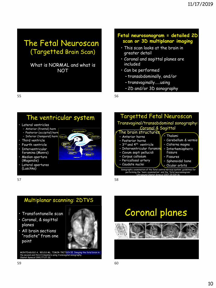
The Fetal Neuroscan (Targetted Brain Scan)
What is NORMAL and what is NOT
Fetal neurosonogram = detailed 2D scan or 3D multiplanar imaging
• This scan looks at the brain in greater detail
• Coronal and sagittal planes are included
• Can be performed
– transabdominally, and/or
– transvaginally.....using
– 2D and/or 3D sonography
• Lateral ventricles– Anterior (frontal) horn– Posterior (occipital) horn– Inferior (temporal) horn
• Third ventricle• Fourth ventricle• Interventricular
foramina (Monro)• Median aperture
(Magendie)• Lateral apertures
(Luschka)
The ventricular system Targetted Fetal Neuroscan
• The brain structures– Anterior horns– Posterior horns – 3rd and 4th ventricle – Interventricular foramina– Cavum septi pellucidi– Corpus callosum– Pericallosal artery– Caudate nuclei
– Thalami– Cerebellum & vermis– Cisterna magna– Interhemispheric
fissure – Fissures– Sphenoidal bone– Ocular orbits
Transvaginal/transabdominal sonography: Coronal & Sagittal
Sonographic examination of the fetal central nervous system: guidelines for performing the 'basic examination' and the 'fetal neurosonogram'.
Ultrasound Obstet Gynecol 2007;29:109-16.
Multiplanar scanning: 2DTVS
• Transfontanelle scan
• Coronal, & sagittal planes
• All brain sections “radiate” from one point
MONTEAGUDO A, REUSS ML, TIMOR-TRITSCH IE. Imaging the fetal brain in the second and third trimesters using transvaginal sonography. Obstet Gynecol 1991;77:27-32.
Coronal planes
55 56
57 58
59 60

11/17/2019
11
You can generate MANY coronal sections using the
transabdominal US probe,,,,,.
Tomographic coronal images of the fetal brain at 22 weeks
…..but out of the MANY axial sections the Basic Fetal Brain Scan uses only about FOUR.
The Most Useful 2D Coronal Sections
Anterior transfrontal
Mid-coronal Transcaudate
Posterior occipital transcerebellar
Mid-coronal Transthalamic
– Interhemispheric fissure
– Orbits
Ultrasound Obstet Gynecol 2007;29:109-16
The Frontal group
Anterior frontal (2) or transfrontal plane Mid-coronal-1 or transcaudate plane
– Caudate nuclei
– Genu corpus callosum
– Cavum septi pellucidi
– Frontal horns
Ultrasound Obstet Gynecol 2007;29:109-16
The Midcoronal group
- Interhemispheric fissure
61 62
63 64
65 66

11/17/2019
12
Mid-coronal-2 or transthalamic plane
– Thalami
– Interventricular foramina
– Atrium Lat Ventr
– 3rd Ventricle
– Body of C C
Ultrasound Obstet Gynecol 2007;29:109-16
The Midcoronal group
- Interhemispheric fissureOccipital-1& 2 or transcerebellar plane
– Interhemispheric fissure
– Occipital horns
– Tentorium
– Cerebellar hemispheres
– Vermis
– Cisterna Magna
Ultrasound Obstet Gynecol 2007;29:109-16
The Occipital group
Are the anterior horns of the lateral ventricles visible on this anterior coronal
section (steer’s head configuration)?Not seen!, This is
normal: You are done!Seen! This is abnormal!
Get help
Think of: Unilateral v-megaly; look for a reason
From: G Malinger
Are the anterior horns of the lateral ventricles separated by septae on this
coronal planes boxing-in the CSP?
Yes! This is normal: You are done!
No septae! Get help
Think of diff dx: HPE, SOD, ASP
Yes! This is normal: You are done!
No! Get help
Think of: lobar HPE, SOD, AGS
Are the anterior horns of the lateral ventricles separated by septae on this
coronal planes flanking the CSP?
?
67 68
69 70
71 72

11/17/2019
13
Sagittal
Upward & laterally diverging anterior horns
Yes! This is normal: You are done!
Are the anterior horns of the lateral ventricles diverging up and laterally on
this coronal plane?Not diverging?!
Get help
Straight upward pointing anterior horns
Think of: AGCC
Does the falx “stop” at the corpus callosum?
Yes! This is normal: You are done! No! Get help
Think of: AGCC
Are the lateral ventricles of normal size?
Yes! This is NL! You are done! No! They are not! Get help
This is V-megaly, determine the cause
V-megaly is NOT a diagnosis, it is a sign!
Are the lateral ventricles of the same size? Are they separated?
Yes!This is normal: You are done!
No!They are not! Get help
Think of: asymmetric v-megaly, HPE, bleed
Are the posterior horns of the lateral ventricles of the same size?
Remember, the occipital horns are most of the time slightly asymmetrical.
This is quite normal
Yes! (almost) This is normal: You are done!
73 74
75 76
77 78

11/17/2019
14
Is the cerebellum symmetrical & of NL size on the posterior coronal plane?
Yes! You are done!! No !They are small! Get help
Think of: hypoplasia, rhombencephalosynapsis
Sagittal planes
Overview of structures on the three orthogonal planes at 22 weeks Sagittal Sections
Median (mid-sagittal)Oblique or
lateral sagittal
MANY consecutive sections can be generated in the sagittal plane.
The most important one is the MEDIAN plane (Mid-sagittal)
Median or mid-sagittal plane– Corpus callosum
– Cavum septi pellucidi
– 3rd Ventr/Thalami
– Brain stem
– Pons
– Tentorium
– Vermis
– 4th Ventricle
– Cisterna Magna
Ultrasound Obstet Gynecol 2007;29:109-16
79 80
81 82
83 84

11/17/2019
15
Rostrum
Genu
Corpus
Splenium
CVCSP
B
22 w
Anatomy of the corpus callosum
Do you see the entire corpus callosum or only part of it?
Entire CC?,Yes! This is normal: You are done!
Only part or none! Get help
G. Malinger
Think: partial/total agenesis or hypogenesis of CC
Do you see a nice figure of 3 outlining the thalamus and the quadrigeminal
plate?No! Get helpYes! This is normal:
You are done!
Think: 1stor 2nd grade IVH, quadrigeminal cistern cyst
How to determine if the corpus callosum is normal: Look at the FIGURE OF 3!!
85 86
87 88
89 90

11/17/2019
16
Pericallosal arteries
• Paired vessels
• Their presence predicts a normal corpus callosum
Pericallosal arteries
Anatomy of the main arteries
seen on the median plane
Do you see the entire pericallosal artery above the CC on the
median plane?
No? Get help!
Think of: partial or total AGCC
Yes? You are done!
Do you see the normal position, size of the vermis and the
tentorium?
NL position of the tentorium
No! Get help
Think of: DWM
Yes! This is normal: You are done!
NL vermis–to-pons angle
Oblique-1 or Parasaggital plane– Entire lateral
ventricle• Anterior horn
• Posterior horn
• Inferior horn
– Choroid plexus
– Periventricular tissue
– Thalamus
Ultrasound Obstet Gynecol 2007;29:109-16
The Rt & Lt Oblique
91 92
93 94
95 96
11/17/2019
17
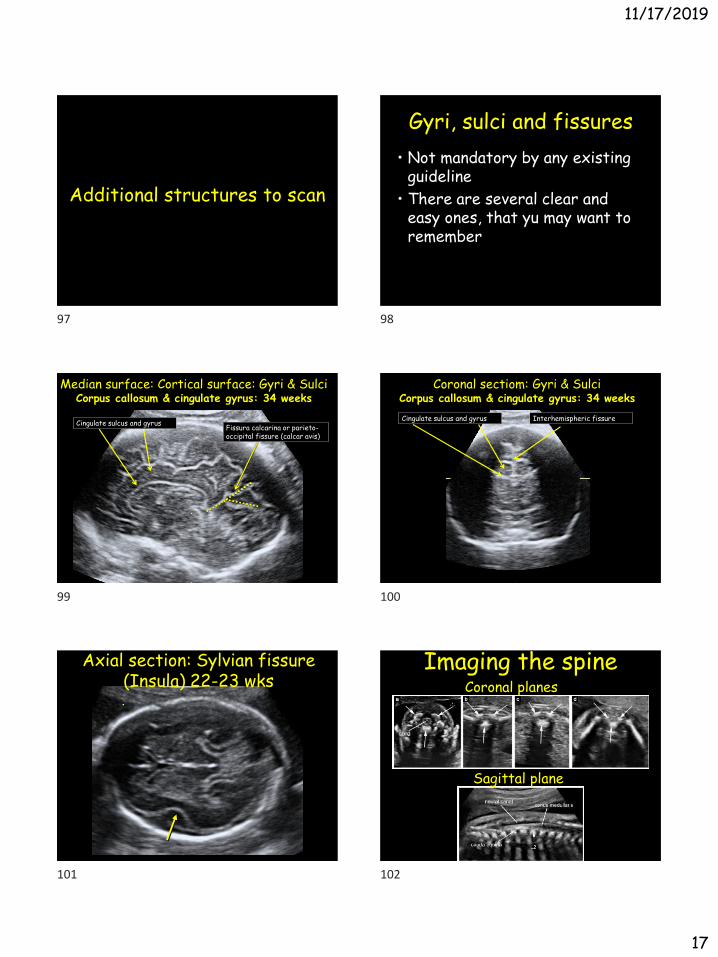
Additional structures to scan
Gyri, sulci and fissures
• Not mandatory by any existing guideline
• There are several clear and easy ones, that yu may want to remember
Median surface: Cortical surface: Gyri & SulciCorpus callosum & cingulate gyrus: 34 weeks
Fissura calcarina or parieto-occipital fissure (calcar avis)
Cingulate sulcus and gyrus
Coronal sectiom: Gyri & SulciCorpus callosum & cingulate gyrus: 34 weeks
Cingulate sulcus and gyrus Interhemispheric fissure
Axial section: Sylvian fissure (Insula) 22-23 wks
Imaging the spine
Sagittal plane
Coronal planes
97 98
99 100
101 102

11/17/2019
18
The most common pathology on the sagittal plane: Spina bifida
No? Get helpNormal? : You are
done!...
The sagittal plane of the spine Don’t Forget to Look at
•A fetal neuroscan is NOT complete unless the fetal face is thoroughly examined.
Thank you!
End
Extra slides if needed
TransvaginalSonography
Only if there is time. If not kip to Conclusions
• Imaging intracranial anatomy with
2D Transvaginal sonography was described a decade ago*
• Although this technique has gained some popularity, it is still not as widely used as it should or could be.
• It is perceived as a cumbersome and time-consuming process.
• HOWEVER: scanning the fetal brain by 2D TVS is relatively easy & fast
•Timor-Tritsch & Monteagudo 1996
103 104
105 106
107 108

11/17/2019
19
Multiplanar scanning by 2D TVS
• Coronal, & sagittal planes
• Sections “radiate” from
one point
Transvaginal Acquisition
• Our preferred mode: Excellent resolution (high frequency probe through the fontanelle / sutures
• Fetal position: Gentle manipulation of the fetus by the examiners’ free hand to perfectly align the footprint of the probe with a fontanelle / suture
• Version in selected cases
Coronal Planes
• The Frontal group
• The Midcoronal group
• The Occipital group
Anterior Posterior
Orbits
Ant. fontanelleAnt. fontanelle
AHAHAH AH
Palate
Rt. Lt.
Ant. fontanelle
Ant coronalsAnterior Posterior
Rt. Lt.
AH AHCP CP
CP CP
C C
T TCN CN
Falx Falx
109 110
112 113
114 115

11/17/2019
20
Mid-coronals
Anterior Posterior
Rt. Lt.
PH PH
C C
Post coronals Sagittal Planes
• The Median
• The Rt & Lt Oblique-1
• The Rt & Lt Oblique-2
Right Left
MedianRt. parasagittal L. parasagittal
AH AH
PH PH
CP CP
CSP
T
C
4th
C
M
Can you do more?• Yes you can!
– If you have a 3D machine
– If you are interested in advanced fetal brain scan
– If you invest the use of volume scanning
• Then you move up one step to 3D
116 117
118 119
120 121

11/17/2019
21
The many ways to display structures
The volume
Inversion
Tomography Angiography
Localize structures
3D Fetal Neuroscan• The most useful features of 3D
scanning the fetal CNS:– Orthogonal planes
– Tomographic imaging
– Angiography
– Inversion
– Thick slice/volume contrast imaging
3D Angiography
3D Angiography• Power/ Color Doppler (brain vasculature)
• Pericallosal branch of the anterior cerebral artery
• Assist to trace a deviant artery (due to a mass effect)
122 123
124 125
126 127

11/17/2019
22
Inversion Rendering
Ventriculomegaly
Ventriculomegaly
Concluding suggestions
Summary & conclusions• The basic normal and abnormal fetal
brain scan was presented having in mind its daily use
• My suggestion: basic brain scan for all fetuses regardless of risk!!
• Targetted scan for suspicious brain findings & for fetuses with any anomaly
• The median (mid-sagittal plane is essential for a complete fetal brain scan
Summary & conclusions
• If the fetus is in vertex presentation, use transvaginal neuroscan (if breech and TAS not clear, consider version)
• In tertiary centers or for an in-depth scan: 3D fetal neuroscan (if vertex: transvaginal)
128 129
130 131
132 133

11/17/2019
23
Thank you!
End
134