basic research comparison of prophylactic and ...basic research comparison of prophylactic and...
TRANSCRIPT

BASIC RESEARCH
Comparison of prophylactic and therapeutic use ofshort-chain fatty acid enemas in diversion colitis:a study in Wistar ratsAriano Jose Freitas de Oliveira,I Francisco Edilson Leite Pinto Junior,II Maria Celia Carvalho Formiga,III
Syomara Pereira da Costa Melo,IV Jose Brandao-Neto,V Ana Maria de Oliveira RamosIV
I Postgraduate Program in Health Sciences, Federal University of Rio Grande do Norte, Natal, Brazil. II Department of Surgery, Federal University of Rio
Grande do Norte, Natal, Brazil. III Department of Statistics, Federal University of Rio Grande do Norte, Natal, Brazil. IV Department of Pathology, Federal
University of Rio Grande do Norte, Natal, Brazil. V Department of Internal Medicine, Federal University of Rio Grande do Norte, Natal, Brazil.
OBJECTIVES: To study the effect of short-chain fatty-acids on atrophy and inflammation of excluded colonicsegments before and after the development of diversion colitis.
INTRODUCTION: Diversion colitis is a chronic inflammatory process affecting the dysfunctional colon, possiblyevolving with mucous and blood discharge. The most favored hypotheses to explain its development is short-chainfatty-acid deficiency in the colon lumen.
METHODS: Wistar rats were submitted to colostomy with distal colon exclusion. Two control groups (A1 and B1)received rectally administered physiological saline, whereas two experimental groups (A2 and B2) received rectallyadministered short-chain fatty-acids. The A groups were prophylactically treated (5th to 40th days postoperatively),whereas the B groups were therapeutically treated (after post-operative day 40). The mucosal thickness of theexcluded colon was measured histologically. The inflammatory reaction of the mucosal lamina propria and thelymphoid tissue response were quantified through established scores.
RESULTS: There was a significant thickness recovery of the colonic mucosa in group B2 animals (p = 0.0001), whichalso exhibited a significant reduction in the number of eosinophilic polymorphonuclear cells in the lamina propria(p = 0.0126) and in the intestinal lumen (p = 0.0256). Group A2 showed no mucosal thickness recovery andsignificant increases in the numbers of lymphocytes (p = 0.0006) and eosinophilic polymorphonuclear cells in thelamina propria of the mucosa (p = 0.0022).
CONCLUSION: Therapeutic use of short-chain fatty-acids significantly reduced eosinophilic polymorphonuclear cellnumbers in the intestinal wall and in the colonic lumen; it also reversed the atrophy of the colonic mucosa.Prophylactic use did not impede the development of mucosal atrophy.
KEYWORDS: Colostomy; Short-Chain Fatty Acids; Diversion colitis; Prophylactic; Treatment.
Oliveira AJF, Pinto Jr FEL, Formiga MCC, Melo SPC, Brandao-Neto J, Ana Ramos MO. Comparison of prophylactic and therapeutic use of short-chainfatty acid enemas in diversion colitis:?a study in Wistar rats. Clinics. 2010;65(12):1351-1356.
Received for publication on August 14, 2010; First publication on September 7, 2010; Accepted for publication on September 24, 2010
E-mail: [email protected]
Tel.: 55 84 3215-4220
INTRODUCTION
After colostomy, the nonfunctioning intestinal segmentpresents inflammatory alterations comprising a nosologicalentity known as diversion colitis (DC).1 The preferredtreatment for DC is intestinal transit reconstruction which,in most cases, resolves the inflammatory process.2
Ma et al.3 analyzed 21 cases of DC and concluded thatmoderate chronic inflammation with lymphoplasmocytaryinfiltrate in the lamina propria, vascular congestion, mini-mal alterations in crypt architecture and a slight decline intheir number were the main histopathologic alterationsfound in the disease. Additionally, these changes wereaccompanied by the presence of prominent lymphoidnodules with or without hyperplasia of the germinalcenters. Haque et al.4 observed the presence of eosinophilicpolymorphonuclear cells (EPNs) in the lamina propria andcolonic lumen of children with DC. Pinto et al.5 reportedthat the onset of significant atrophy in the colonic mucosa ofWistar rats coincides with the 40th day after surgicalexclusion of the colon.
Copyright � 2010 CLINICS – This is an Open Access article distributed underthe terms of the Creative Commons Attribution Non-Commercial License (http://creativecommons.org/licenses/by-nc/3.0/) which permits unrestricted non-commercial use, distribution, and reproduction in any medium, provided theoriginal work is properly cited.
CLINICS 2010;65(12):1351-1356 DOI:10.1590/S1807-59322010001200020
1351

Grove et al.6 demonstrated the relationship between dietand the presence of short chain fatty acids (SCFAs) in thecolonic lumen. Roediger showed that around 70% ofcolonocyte energy requirements originate in the SCFAsand, further, that 90% of these SCFAs are formed by acetic,n-propionic and n-butyric acid, the latter of which serves asthe main energy source for colonocytes.7
The SCFAs exert trophic effects on the large bowelthrough direct contact of the acids with the colonic mucosa.8
Colonic trophism mechanisms occur as a result of increasedenergetic oxygenation, stimulated flow of blood microcir-culation caused by dilated resistance arteries, enterotrophichormone production and stimulation of the enteric nervoussystem. This trophism occurs transmurally and is notrestricted to the mucosa.8,9
In addition to stimulating collagen maturation, theseaforementioned functions are fundamental in colonicphysiology and contribute to decreased bacterial transloca-tion, intestinal adaptation in short bowel syndrome,stimulation of healing and increased anastomosis resis-tance.8-10
Some authors have postulated that the use of SCFAs inthe nonfunctioning segment reverses the alteration ofDC.2,11 Other studies were not as successful, however, andtheir authors question this form of treatment.12,13
The present study aimed to assess the use of SCFAs in thenonfunctioning colonic stump of Wistar rats in order todemonstrate microscopically the existence (or lack thereof)of a prophylactic or therapeutic role.
MATERIALS AND METHODS
AnimalsForty male Wistar rats weighing between 220 g and 230 g
were housed in a room under standard conditions oftemperature, light, humidity, water and diet (Labina-Purina, Sao Paulo, Brazil) according to specificationsdescribed by Reeves et al.14 The animals were supplied bythe vivarium of the Center for Experimental Surgery,Department of Surgery of the CCS (Centro de Ciencias daSaude) (Health Science Center) of UFRN (UniversidadeFederal do Rio Grande do Norte) (Federal University of RioGrande do Norte). The animals were treated according tothe use of Nonhuman Animals in Research: a Guide forScientists.15 The surgery was performed in the Laboratory ofthe Operatory Technique discipline (UFRN) with thecollaboration of the Department of Pathology (UFRN) andthe Postgraduate Program in Health Sciences (PPGCSA(Programa de Pos-graduacao do Centro de Ciencias daSaude), UFRN). This was a prospective, analytical, experi-mental, intervention study.
Surgical StudyAll of the animals were submitted to a 12-hour fast with
the exclusive use of water. Immediately before surgery, aretrograde intestinal wash was performed with physiologi-cal saline to remove all fecal matter.5 All surgical procedureswere conducted under aseptic conditions. Anesthesia wasobtained using pentobarbital (20 mg/Kg intraperitoneally)and ketamine (50 mg/Kg intramuscularly). The animalswere allowed to breathe spontaneously throughout theexperiment. They were fixed in dorsal decubitus, andtrichotomy and asepsis using povidone-iodine were per-formed.16
The abdominal cavity was opened by a median lapar-otomy of around 5 cm to identify the cecum and proximalcolon, which was divided 5 cm from the ileocecal valve. Thedistal colon was submitted to terminal suture. After itssectioning, the distal colonic segment was kept inside theabdominal cavity. The proximal segment was colostomizedand exteriorized through the abdominal wall left of themedian incision. A single-barreled end colostomy wasperformed along with fixation of the stoma to the abdominalwall (primary maturation) with a 6-0 polypropylene thread.The abdominal wall was then closed with separate 3-0cotton sutures (Fig. 1).5
Additional InterventionsFour groups of ten adult Wistar rats submitted to
colostomy underwent additional interventions. The protocoldistributed the animals into groups A (A1 and A2) and B(B1 and B2). Two control groups (A1 and B1) receivedinfusions of physiological saline administered rectally,whereas the two experimental groups (A2 and B2) receivedshort-chain fatty-acids rectally. The A groups were prophy-lactically treated (5th to 40th post-operative days, twice aweek), whereas the B groups were therapeutically treated(after post-operative day 40, for seven days). The interven-tion posologies are described in table 1.
The SCFA solution was developed in the biochemistrylaboratory of the Bioscience Center at UFRN and wascomposed of: 75 mmol/L of sodium acetate; 35 mmol/L ofsodium propionate; 20 mmol/L of butyric-N acid;2.5 mmol/L of calcium chloride; 7.5 mmol/L of magnesiumchloride; 10 mmol of potassium chloride.17 The solutionswere elaborated in an iso-osmolar (280 mosm/L), and thepH was adjusted to seven by using appropriate amounts ofNaOH or HCI.
HistomorphometryThe mean of four microscopic measures of mucosal
thickness in the rats of each subgroup was measured usinga Nikon Lobophot 10 6microscope (Nikon, Tokyo, Japan).This equipment expresses the mean thickness in millimetersby multiplying the value obtained by the standard correc-tion factor indicated for the lens (0.0078) as a function of its
Figure 1 - The macroscopic aspect of the colon of a group B1animal submitted to intestinal derivation. Arrow 1 indicates thefunctional intestinal segment, after derivation in intestinal wallcolostomy. Arrow 2 shows the defunctionalized segment, withwhitish coloration and visibly atrophic.
SCFAs and diversion colitisOliveira AJF et al.
CLINICS 2010;65(12):1351-1356
1352

amplification and diameter. The measures were obtained foreach of the transversal and longitudinal histological sectionsof the colonic wall using the final mean for each animal fromthe values found.5
Qualitative Histological AnalysisDifferent cell type counts were performed to assess
neutrophilic polymorphonuclear cells (NPNs), lympho-cytes, and EPNs. The lymphoid follicle size of the muc-ous associated lymphoid tissue (MALT) was assessed inthe lamina propria of the mucosa as well as in the colonicwall.
The intensity of alterations was graduated on a 6-pointscale ranging from 0 to 5. These values were later convertedinto whole numbers using our modified version of Myers’index,18 which is based on variables appropriate to thestudy of DC. This procedure gave rise to a histologic score.A variable was stipulated for each type of variable analyzed,depending on whether it was favorable for DC diagnosis.This value was multiplied by the intensity of the alterationsobserved in the histologic sections.
StatisticsIn quantitative assessment, statistical analysis compared
the measures obtained in the two paired groups (A1 andA2) and (B1 and B2) using analysis of variance procedures(Student’s t-test). For qualitative assessment, exploratory
analysis was performed by building tables and thencomparing the mean intensity of the cells observed in eachgroup according to cell type. To determine the existence ofsignificant differences in cellularity between groups (A1 andA2) and (B1 and B2), the non-parametric Mann-Whitney testwas applied. A significance level of 5% (p-value # 0.05) wasset for all of the results assessed.
RESULTS
HistomorphometryValues for colonic mucosal thickness are shown in
Figure 2. No significance difference was found betweenthe control (A1) and prophylactic (A2) groups in theprevention of DC in terms of mucosal atrophy (p =0.1680). However, there was a significant difference betweenthe control group (B1) and treatment group (B2), p = 0.0001.
Qualitative Histological AnalysisTable 2 shows the different cell types, including the
presence and size of MALT lymphoid follicles (LFs), withvalues expressed in accordance with Myers’ modified score.The highest indices observed were the presence of LFs (65),mainly large size (63), and number of EPNs (63) in thecolonic lumen; these findings favored the diagnosis of DC.
The histologic sections from group A1 (control) exhibitingmucosal atrophy are shown in Figure 3(a). Figure 3(b)shows sections from group A2 (prophylactic) exhibiting
Table 1 - Experimental design. Distribution of animal groups according to type of intervention, treatment period, andposologies.
Group g Infused substance Infusion period Posology
A1 (control) 10 SS 0.9% Between the 5th and 40th POD Twice a week
A2 (prophylaxis) 10 SCFA Between the 5th and 40th POD Twice a week
B1 (control) 10 SS 0.9% After the 40th POD Twice a day for 7 days
B2 (treatment) 10 SCFA After the 40th POD Twice a day for 7 days
SS = saline solution. SCAF = short-chain fatty acids. POD = post-operatory day.
Figure 2 - Comparative box plots of the mean of mucosal thickness measures in control rats (A1), rats treated prophylactically usingSCFAs (A2), control rats (B1), and rats treated with SCFAs (B2). No statistically significant difference between A1 and A2 was noted(p = 0.1680). A statistically significant difference between B1 and B2 was observed (p = 0.0001). SCFAs, short chain fatty acids.
CLINICS 2010;65(12):1351-1356 SCFAs and diversion colitisOliveira AJF et al.
1353

mucosal atrophy and slight MALT hyperplasia, andFigure 3(c) shows sections from group B1 (control) exhibit-ing mucosal atrophy and MALT hyperplasia. Figure 3(d)shows sections from group B2 (treatment) exhibiting normalmucosal thickness and MALT hyperplasia.
The assessment of histologic scores by cell type andlymphoid follicles of MALT, which are represented by thesum of the median values in the control (A1) andprophylactic (A2) groups, is shown in Table 3. The resultsobtained in the qualitative analysis of the samples revealeda significant numerical increase in lymphocytes (p = 0.006)and EPNs in the lamina propria of the colonic mucosa (p =0.0022) in the prophylactic group (A2) compared to the
control group (A1). The prophylactic use of SCFAs (groupA2) did not significantly reduce MALT hyperplasia com-pared to the control group (A1) (p = 0.0670). With respect tototal cellularity, there were no significant differencesbetween groups A1 and A2 (p = 0.2233).
Table 4 shows a significant reduction in EPNs in thelamina propria of the treatment group (B2) compared to thecontrol group (B1) (p = 0.0126), and a reduction was alsoobserved in the intestinal lumen (p = 0.0256). We alsofound that the therapeutic use of SCFAs had little effect onthe inhibition of MALT hyperplasia when comparinggroups B2 and B1 (p = 0.5514). Total cellularity showedno significant difference between groups B1 and B2 (p =0.0781).
DISCUSSION
In clinical practice, the objective assessment of DC ismade according to three main criteria: (1) analysis ofmucosal thickness; (2) study of the role of inflammatorycells of the lamina propria and MALT and; (3) investigationof surface colonocyte alterations. The colonic mucosaexhibited reduced thickness in all of the cases studied. Thelatter parameter is therefore more reliable than the others, asit can be easily measured.19
The relevant results obtained in the present study showthat SCFAs are effective for the treatment of DC as theyreverse colonic mucosal atrophy. Therapeutic use of SCFAshas also been shown to reduce the number of EPNs in thelamina propria of the mucosa and colonic lumen, therebydecreasing the inflammatory process. In contrast, therapeu-tic SCFA use did not benefit the nonfunctioning colonictissue by reversing lymphoid follicular hyperplasia inMALT.20 SCFAs were ineffective when used as DC
Table 2 - Cell types, assessment of lymphoid follicles ofmucous associated lymphoid tissue (MALT), andrespective values proposed by the Myers index modifiedfor conversion into absolute numbers.
Histologic parameters Indices adapted to DC
NPN in the lamina propria of
the colonic mucosa
61
Lymphocytes in the lamina propria
of the colonic mucosa
62
EPN in the lamina propria of
the colonic mucosa
62
EPN in the colonic lumen 63
Presence of LF of MALT 65
LF size
Large 63
Medium 62
Small 61
MALT = mucous associated lymphoid tissue. NPN = neutrophilic
polymorphonuclear cells. EPN = eosinophilic polymorphonuclear cells. LF
= lymphoid follicles.
Figure 3 - (a) Histologic section of the colon of a group A1 animal (control) exhibiting an atrophic mucosa (H&E 6400). (b) Atrophiccolonic mucosa and slight MALT hyperplasia in a group 2 animal (prophylaxis) (H&E 6200). (c) Atrophic colonic atrophy with MALThyperplasia in a group B1 animal (control) (H&E 6100). (d) Normal mucosal thickness with MALT hyperplasia in a group B2 animal(treatment) (H&E 640). MALT, mucous associated lymphoid tissue.
SCFAs and diversion colitisOliveira AJF et al.
CLINICS 2010;65(12):1351-1356
1354

prophylaxis, as shown by the fact that they did not impedemucosal atrophy on the 40th day postoperatively.
These findings corroborate data described in the litera-ture. Harig et al.,2 for example, observed clinical, endo-scopic, and histopathologic reversal in four patients withDC who underwent SCFA treatment in the excludedsegment. Worsening resulted with treatment interruptionor when the infusion solution was replaced by physiologicalsaline.
With the exclusive use of topically infused SCFAs, Kielyet al.11 observed symptom remission and significantimprovement in the endoscopic and histopathologic DCfindings in three out of five patients studied.
Sengupta et al.21 submitted rats to a fiber-free diet toestablish DC-like intestinal atrophy. These authors observedthat the topical use of butyrate favored increased cellularityin the colonic crypt, elevated mitoses, and consequent cellproliferation and atrophy reversal. The authors alsoobserved that the effect of butyrate was dose-dependentand that its action was of short duration.
Oliveira-Neto and Aguilar-Nascimento22 assessed theinfusion effect of a solution containing fibers on thenonfunctioning colonic stump of 11 patients. These authorsobserved a significant decrease in the degree of DC, as wellas a significant increase in crypt depth after infusion. Giventhat SCFAs are a result of fiber degradation caused byanaerobic bacteria in the colonic lumen, these findingssuggested that SCFAs were directly responsible for DCremission.
Some researchers, however, found no relevance in the useof SCFAs as an active agent in DC remission. Guillemot etal. published a double-blind study with thirteen patients.12
These authors observed no significant endoscopic or
histopathologic differences in DC reversal between theSCFAs and physiological saline infusion groups.
Schauber et al.13 examined 9 patients in a double-blindstudy and only 7 of these completed the protocol. All of thesubjects had intestinal inflammatory disease and colostomywas indicated. The study showed no significant endoscopicor bacteriologic differences between the control and SCFAgroups.
The disparity in results between these authors and thosewho observed data favoring the use of SCFAs may beexplained by the differences in clinical indications forcolostomy. Recent studies have revealed the inhibitoryaction of butyrate metabolism in patients with idiopathiculcerative colitis.23
The trophic action of SCFAs on the large intestine, whichoccurs through its direct contact with the colonic mucosa, isimportant in the clinical control of DC.8 This action reducesthe signs and symptoms associated with the condition itself(e.g. mucous discharge and transrectal bleeding). Further,SCFAs also prevent the emergence of complications relatedto mucosal atrophy and colonic epithelium lesion, as well ascomplications inherent to re-establishing intestinal transit.
Neut et al.20 reported that the colonic mucosal atrophywith loss of integrity seen in DC could predispose patientsto bacterial translocation by interfering with local immunityand modifying native bacterial flora, both quantitativelyand qualitatively. The present study found that the use ofSCFAs for a short period of time not only reversed atrophybut also reduced the risk of losing mucosal integrity.Additionally, it did not interfere with MALT hyperplasiaof the nonfunctioning colon. The fact that this latter findinghas not been reported in the literature makes it scientificallyrelevant. These results, therefore, favor the preservation of
Table 3 - Results of the Mann-Whitney U test for the comparison of histologic scores by cell type and lymphoid folliclesbetween the control group (A1) and prophylactic group treated with short chain fatty acids (SCFAs) (A2).
Type of cell
Median
U-value p-value
Group size
A1 A2 A1 A2
NPN in the lamina propria 1 1 39.5 0.3222 10 10
Lymphocytes in the lamina propria 2 4 9 0.0006* 10 10
EPN in the lamina propria 2 4 13.5 0.0022* 10 10
EPN in the intestinal lumen 9 6 36 0.2296 10 10
Lymphoid follicles of MALT 5 0 27 0.0670 10 10
Final histologic scores (modified Myers index) 20.9 19.9 34 0.2233 10 10
*Significant differences between groups A1 and A2. NPN = neutrophilic polymorphonuclear cells. EPN = eosinophilic polymorphonuclear cells. MALT =
mucous associated lymphoid tissue.
Table 4 - Results of Mann-Whitney U test for comparisons between types of inflammatory cells along with the role oflymphoid follicles in the control group (B1) and group treated with short chain fatty acids (SCFAs) (B2).
Type of cell
Median
U-value p-value
Group size
B1 B2 B1 B2
NPN in the lamina propria 1 1 44.5 0.9389 9 10
Lymphocytes in the lamina propria 4 4 33 0.2796 9 10
EPN in the lamina propria 4 2 18 0.0126* 9 10
EPN in the intestinal lumen 9 6 20 0.0256* 9 10
Lymphoid follicles of MALT 0 0 39 0.5514 9 10
Final histologic scores (modified Myers index) 19.6 13.8 23.5 0.0781 9 10
*Significant differences between groups B1 and B2. NPN = neutrophilic polymorphonuclear cells. EPN = eosinophilic polymorphonuclear cells. MALT =
mucous associated lymphoid tissue.
CLINICS 2010;65(12):1351-1356 SCFAs and diversion colitisOliveira AJF et al.
1355

local immunity while avoiding the modification of nativebacterial flora. Furthermore, bacterial translocation was notobserved in the results reported by Pinto Jr. et al.,24
corroborating the results obtained in this study.Lim et al.25 raised the hypothesis that DC is a factor
predisposing patients to the emergence of idiopathiculcerative rectocolitis. This occurs through the sensitizationof leukocytes in the nonfunctioning colon and subsequentleukocyte aggression toward the endothelium of thefunctional colon as a result of the emergence of anticolonicself-antibodies. The therapeutic action of SCFAs observed inthis study would reduce the possibility of disease evolution.
Owing to their high morbidity and mortality rates,intestinal anastomosis fistulae remain the greatest threat togastrointestinal tract surgeons. Pearce et al.26 observedhigher re-anastomosis dehiscence indices when the intervalfor transit reconstruction was more than six months, whichis sufficient time for atrophy of the nonfunctioning colonwall to occur. SCFA enemas facilitated the healing processof colonic anastomosis in rats.10 Possible mechanisms thatmight mediate this effect include an increase in cellproliferation of the colonic mucosa and the acceleration ofcollagen maturation.27 The first mechanism accelerates re-epithelization and increases blood flow, with a consequentrise in oxygen supply. The topical use of SCFAs twice dailyfor 7 days (the therapeutic methodology used in this study)reversed colonic mucosal atrophy. This therapy wouldreduce the potential risk of fistula formation in patientsundergoing intestinal transit reconstruction.
The use of SCFAs is important for proper nutrition of thenonfunctioning colonocyte, because SCFAs reverse colono-cyte atrophy and therefore minimize symptoms in somepatients. This treatment may be particularly valuable forthose patients undergoing intestinal transit reconstruction,however, because the aim of atrophy reversal is to preventcomplications inherent to the surgery.
CONCLUSION
In conclusion, the prophylactic action of SCFAs on DC interms of colonic mucosal trophism was not confirmed. Thisstudy demonstrates the therapeutic use of SCFAs inexperimental DC by showing the significant effects ofSCFAs on atrophy regression and EPN reduction in theintestinal lumen and lamina propria of the colonic mucosa,despite their lack of interference with the intensity of MALThyperplasia. Thus, the therapeutic application of SCFAsmay be of great significance in clinical practice, especially inpatients without associated inflammatory disease. SCFAsmay shorten hospitalization and favor better postoperativemanagement in colostomized patients by reducing compli-cations.
ACKNOWLEDGEMENTS
This study was partially funded by a grant (no. 135226/06-6) from CNPq.
REFERENCES
1. Glotzer DJ, Glick ME, Goldman H. Proctitis and colitis followingdiversion of the fecal stream. Gastroenterology. 1981;80:438-41.
2. Harig JM, Soergel KH, Komorowski RA, Wood CM. Treatment ofdiversion colitis with short-chain-fatty acid irrigation. N Engl J Med.1989;320:23-8, doi: 10.1056/NEJM198901053200105.
3. Ma CK, Glottlieb C, Haas PA. Diversion colitis: a clinic pathologic studyof 21 cases. Hum Pathol. 1990;21:429-36, doi: 10.1016/0046-8177(90)90206-K.
4. Haque S, Eisen RN, West AB. The morphologic features of diversioncolitis – studies of a population with no other disease of the intestinalmucosa. Hum Pathol. 1993;24:211-9, doi: 10.1016/0046-8177(93)90303-X.
5. Pinto Jr FEL, Oliveira AJF, Medeiros KF, Ramos AMO, Ramos CCO,Medeiros AC. Repercussoes histopatologicas da colostomia no cotocolonico distal desfuncionalizado: estudo experimental em ratos. Rev ColBras Cir. 1999;26:327-33, doi: 10.1590/S0100-69911999000600003.
6. Grove EW, Olmsted WH, Koenig K. The effect of diet and catharsis onthe lower volatile fatty acids in stools of normal men. J Biol Chem.1929;85:115-26.
7. Roediger WE. Role of anaerobic bacteria in the metabolic welfare of thecolonic mucosa in man. Gut. 1980;21:793-8, doi: 10.1136/gut.21.9.793.
8. Frankel WL, Zhang W, Singh A, Klurfeld DM, Don S, Sakata T, et al.Mediation of the trophic effects of short-chain fatty acids on rat jejunumand colon. Gastroenterology. 1994;106:375-80.
9. Mortensen FV, Nielsen H, Mulvany MJ, Hessou I. Short chain fatty acidsdilate isolated human colonic resistance arteries. Gut. 1990;31:1391-4,doi: 10.1136/gut.31.12.1391.
10. Rolandelli RH, Koruda MJ, Settle RG, Rombeau JL. Effects ofintraluminal infusion of short-chain fatty acids on the healing of colonicanastomosis in the rat. Surgery. 1986;100:198-204.
11. Kiely EM, Ajayi NA, Wheeler RA, Malone M. Diversion procto-colitis:response to treatment with short-chain fatty acids. J Pediatr Surg.2001;36:1514-7, doi: 10.1053/jpsu.2001.27034.
12. Guillemot F, Colombel JF, Neut C, Verplanck N, Lecomte M, Romond C,et al. Treatment of diversion colitis by short-chain fatty acids. Dis ColonRectum. 1991;34:861-4, doi: 10.1007/BF02049697.
13. Schauber J, Bark T, Jaramillo E, Katouli M, Sandstedt B, Svenberg T.Local short-chain fatty acids supplementation without beneficial effecton inflammation in excluded rectum. Scand J Gastroenterol. 2000; 35:184-9, doi: 10.1080/003655200750024371.
14. Reeves PG, Nielsen FH, Fahey GC. AIN-93 Purified diets for laboratoryrodents: final report of the American Institute of Nutrition Ad HocWriting Committee on the reformulation of the AIN-76 rodent diet. J Nut.1993;123:1939-51.
15. Royal Society. The use of non-human animals in research: a guide forscientists. London, The Royal Society, 2004; pp1-17.
16. Araujo Filho I, Honorato Sobrinho AA, Rego ACM, Garcia ACMA,Fernandes DP, Cruz TM, et al. Influence of laparoscopy and laparotomyon gasometry, leukocytes and cytokines in a rat abdominal sepsis model.Acta Cir Bras. 2006;21:74-9, doi: 10.1590/S0102-86502006000200004.
17. Sakata T, von Engelhardt W. Stimulatory effect of short chain fatty acidson the epithelial cell proliferation in rat large intestine. Comp BiochemPhysiol A. 1983;74:459-62, doi: 10.1016/0300-9629(83)90631-X.
18. Myers AH, Postlethwait RW, Smith AG. Histologic grading of theexperimental healing wound. Arch Surg. 1961;83:771-4.
19. Keli E, Bouchoucha M, Devroede G, Carnot F, Ohrant T, Cugnenc PH.Diversion-related experimental colitis in rats. Dis Colon Rectum. 1997;40:222-8, doi: 10.1007/BF02054992.
20. Neut C, Colombel JF, Guillemot F, Cortot A,Gower P, Quandalle P, RibetM, et al. Impaired bacterial flora in human excluded colon. Gut.1989;30:1094-8, doi: 10.1136/gut.30.8.1094.
21. Sengupta S, Tang CL, Wong CS, Tjandra JJ, Gibson PR. Colonic epithelialatrophy induced by a fiber-free diet in rats is reversed by minimalamounts of luminal butyrate, but only in the short term. ANZ J Surg.2002;72:871-6, doi: 10.1046/j.1445-2197.2002.t01-1-02586.x.
22. Oliveira-Neto JP, Aguilar-Nascimento JE. Intraluminal irrigation withfibers improves mucosal inflammation and atrophy in diversion colitis.Nutrition. 2004;20:197-9, doi: 10.1016/j.nut.2003.10.006.
23. Roediger WE, Nance S. Metabolic induction of experimental ulcerativecolitis by inhibition of fatty acid oxidation. Br J Exp Pathol. 1986; 67:773-82.
24. Pinto Jr. FEL, Brandt CT, Medeiros AC, Oliveira AJF, Jeronimo SMB,Brito HMF. Bacterial translocation in rats nonfunctioning diverted distalcolon. Acta Cir Bras. 2007;22:195-201, doi: 10.1590/S0102-86502007000300007.
25. Lim AG, Langmead FL, Feakins RM, Rampton DS. Diversion colitis: atrigger for ulcerative colitis in the in-stream colon? Gut. 1999; 44:279-82,doi: 10.1136/gut.44.2.279.
26. Pearce NW, Scott SD, Karran SJ. Timing and method of reversal ofHartmann’s procedure. Br J Surg. 1992;79:839-41, doi: 10.1002/bjs.1800790844.
27. Jonsson K, Jiborn H, Zederfeldt B. Comparison of healing in the left colonand ileum. Changes in collagen content and collagen synthesis in theintestinal wall after ileal and colonic anastomoses in the rat. Acta ChirScand. 1985;151:537-41.
SCFAs and diversion colitisOliveira AJF et al.
CLINICS 2010;65(12):1351-1356
1356
ERRATA
CLINICS 2010;65(12):1351-1356
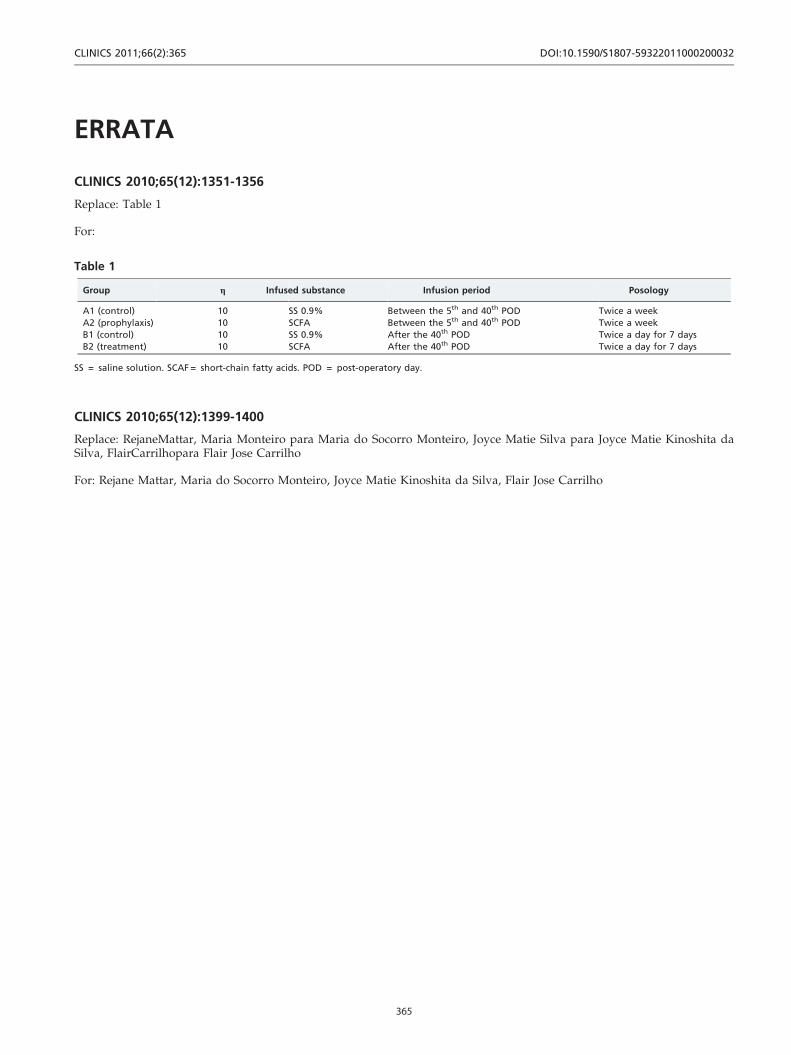
Replace: Table 1
For:
CLINICS 2010;65(12):1399-1400
Replace: RejaneMattar, Maria Monteiro para Maria do Socorro Monteiro, Joyce Matie Silva para Joyce Matie Kinoshita daSilva, FlairCarrilhopara Flair Jose Carrilho
For: Rejane Mattar, Maria do Socorro Monteiro, Joyce Matie Kinoshita da Silva, Flair Jose Carrilho
Table 1
Group g Infused substance Infusion period Posology
A1 (control) 10 SS 0.9% Between the 5th and 40th POD Twice a week
A2 (prophylaxis) 10 SCFA Between the 5th and 40th POD Twice a week
B1 (control) 10 SS 0.9% After the 40th POD Twice a day for 7 days
B2 (treatment) 10 SCFA After the 40th POD Twice a day for 7 days
SS = saline solution. SCAF = short-chain fatty acids. POD = post-operatory day.
CLINICS 2011;66(2):365 DOI:10.1590/S1807-59322011000200032
365