basic research enhancement of basolateral … · basic research enhancement of basolateral...
TRANSCRIPT
BASIC RESEARCH
Enhancement of basolateral amygdaloid neuronaldendritic arborization following Bacopa monnieraextract treatment in adult ratsVenkata Ramana Vollala,I Subramanya Upadhya,II Satheesha NayakIII
I Department of Anatomy, Rajiv Gandhi Institute of Medical Sciences (RIMS), Adilabad, India. II Department of Physiology, St. George’s University School of
Medicine, Grenada, West Indies. III Department of Anatomy, Melaka Manipal Medical College (Manipal Campus), Manipal University, India.
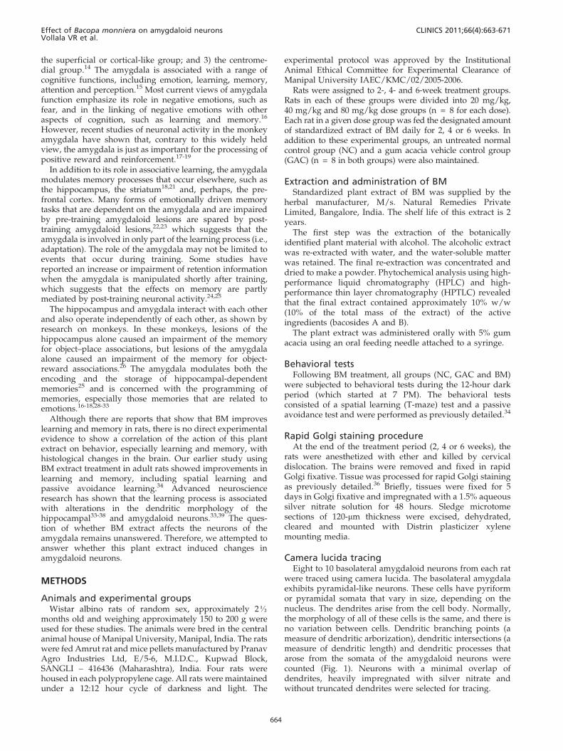
OBJECTIVE: In the ancient Indian system of medicine, Ayurveda, Bacopa monniera is classified as Medhya rasayana,which includes medicinal plants that rejuvenate intellect and memory. Here, we investigated the effect of astandardized extract of Bacopa monniera on the dendritic morphology of neurons in the basolateral amygdala, aregion that is concerned with learning and memory.
METHODS: The present study was conducted on 2K-month-old Wistar rats. The rats were divided into 2-, 4- and 6-week treatment groups. Rats in each of these groups were further divided into 20 mg/kg, 40 mg/kg and 80 mg/kgdose groups (n = 8 for each dose). After the treatment period, treated rats and age-matched control rats weresubjected to spatial learning (T-maze) and passive avoidance tests. Subsequently, these rats were killed bydecapitation, the brains were removed, and the amygdaloid neurons were impregnated with silver nitrate (Golgistaining). Basolateral amygdaloid neurons were traced using camera lucida, and dendritic branching points (ameasure of dendritic arborization) and dendritic intersections (a measure of dendritic length) were quantified.These data were compared with the data from the age-matched control rats.
RESULTS: The results showed an improvement in spatial learning performance and enhanced memory retention inrats treated with Bacopa monniera extract. Furthermore, a significant increase in dendritic length and the number ofdendritic branching points was observed along the length of the dendrites of the basolateral amygdaloid neurons ofrats treated with 40 mg/kg and 80 mg/kg of Bacopa monniera (BM) for longer periods of time (i.e., 4 and 6 weeks).
CONCLUSION: We conclude that constituents present in Bacopa monniera extract have neuronal dendritic growth-stimulating properties.
KEYWORDS: Bacopa monniera; spatial learning; passive avoidance; amygdaloid neurons; dendritic arborization;memory.
Vollala VR, Upadhya S, Nayak S. Enhancement of basolateral amygdaloid neuronal dendritic arborization following Bacopa monniera extracttreatment in adult rats. Clinics. 2011;66(4):663-671.
Received for publication on November 16, 2010; First review completed on December 8, 2010; Accepted for publication on December 29, 2010
E-mail: [email protected]
Tel.: 91 9494306083
INTRODUCTION
Bacopa monniera (BM), a traditional ayurvedic medicine, isreported to improve learning and memory behaviors inanimals and humans.1-3 The plant and plant extracts havebeen extensively investigated for their neuropharmacologi-cal effects, and studies have confirmed their nootropicaction.1-6 The active constituents of the plant facilitatelearning and memory in normal rats and inhibit the amnesiceffects of scopolamine, electroshock and immobilizationstress.7,8 BM extracts have been reported to be non-toxic,non-teratogenic and non-mutagenic in rats and monkeys;
single and multiple dosing studies in healthy humanvolunteers have not elicited adverse effects.9 In addition toits claimed memory-enhancing properties, BM has otherpotential benefits, such as a facilitatory effect on the capacityfor mental retention.10,11 BM has been used in ayurvedicmedicine and in traditional treatments for a number ofdisorders, particularly disorders that involve anxiety,intellect and poor memory.3 Significant antidepressantactivity comparable to that of imipramine has beenobserved with the Brahmi extract after five days of oraladministration, using a rodent model of depression.12
Additionally, anticholinesterase activity has been demon-strated.13
The amygdala is located deep within the medial temporallobe, anterior to the hippocampus, close to the tail of thecaudate nucleus. The amygdala is structurally diverse andcomprises many nuclei. These nuclei are further dividedinto three major groups: 1) the deep or basolateral group; 2)
Copyright � 2011 CLINICS – This is an Open Access article distributed underthe terms of the Creative Commons Attribution Non-Commercial License (http://creativecommons.org/licenses/by-nc/3.0/) which permits unrestricted non-commercial use, distribution, and reproduction in any medium, provided theoriginal work is properly cited.
CLINICS 2011;66(4):663-671 DOI:10.1590/S1807-59322011000400023
663
the superficial or cortical-like group; and 3) the centrome-dial group.14 The amygdala is associated with a range ofcognitive functions, including emotion, learning, memory,attention and perception.15 Most current views of amygdalafunction emphasize its role in negative emotions, such asfear, and in the linking of negative emotions with otheraspects of cognition, such as learning and memory.16
However, recent studies of neuronal activity in the monkeyamygdala have shown that, contrary to this widely heldview, the amygdala is just as important for the processing ofpositive reward and reinforcement.17-19
In addition to its role in associative learning, the amygdalamodulates memory processes that occur elsewhere, such asthe hippocampus, the striatum18,21 and, perhaps, the pre-frontal cortex. Many forms of emotionally driven memorytasks that are dependent on the amygdala and are impairedby pre-training amygdaloid lesions are spared by post-training amygdaloid lesions,22,23 which suggests that theamygdala is involved in only part of the learning process (i.e.,adaptation). The role of the amygdala may not be limited toevents that occur during training. Some studies havereported an increase or impairment of retention informationwhen the amygdala is manipulated shortly after training,which suggests that the effects on memory are partlymediated by post-training neuronal activity.24,25
The hippocampus and amygdala interact with each otherand also operate independently of each other, as shown byresearch on monkeys. In these monkeys, lesions of thehippocampus alone caused an impairment of the memoryfor object–place associations, but lesions of the amygdalaalone caused an impairment of the memory for object-reward associations.26 The amygdala modulates both theencoding and the storage of hippocampal-dependentmemories25 and is concerned with the programming ofmemories, especially those memories that are related toemotions.16-18,28-33
Although there are reports that show that BM improveslearning and memory in rats, there is no direct experimentalevidence to show a correlation of the action of this plantextract on behavior, especially learning and memory, withhistological changes in the brain. Our earlier study usingBM extract treatment in adult rats showed improvements inlearning and memory, including spatial learning andpassive avoidance learning.34 Advanced neuroscienceresearch has shown that the learning process is associatedwith alterations in the dendritic morphology of thehippocampal33-38 and amygdaloid neurons.33,39 The ques-tion of whether BM extract affects the neurons of theamygdala remains unanswered. Therefore, we attempted toanswer whether this plant extract induced changes inamygdaloid neurons.
METHODS
Animals and experimental groupsWistar albino rats of random sex, approximately 2K
months old and weighing approximately 150 to 200 g wereused for these studies. The animals were bred in the centralanimal house of Manipal University, Manipal, India. The ratswere fed Amrut rat and mice pellets manufactured by PranavAgro Industries Ltd, E/5-6, M.I.D.C., Kupwad Block,SANGLI – 416436 (Maharashtra), India. Four rats werehoused in each polypropylene cage. All rats were maintainedunder a 12:12 hour cycle of darkness and light. The
experimental protocol was approved by the InstitutionalAnimal Ethical Committee for Experimental Clearance ofManipal University IAEC/KMC/02/2005-2006.
Rats were assigned to 2-, 4- and 6-week treatment groups.Rats in each of these groups were divided into 20 mg/kg,40 mg/kg and 80 mg/kg dose groups (n = 8 for each dose).Each rat in a given dose group was fed the designated amountof standardized extract of BM daily for 2, 4 or 6 weeks. Inaddition to these experimental groups, an untreated normalcontrol group (NC) and a gum acacia vehicle control group(GAC) (n = 8 in both groups) were also maintained.
Extraction and administration of BMStandardized plant extract of BM was supplied by the
herbal manufacturer, M/s. Natural Remedies PrivateLimited, Bangalore, India. The shelf life of this extract is 2years.
The first step was the extraction of the botanicallyidentified plant material with alcohol. The alcoholic extractwas re-extracted with water, and the water-soluble matterwas retained. The final re-extraction was concentrated anddried to make a powder. Phytochemical analysis using high-performance liquid chromatography (HPLC) and high-performance thin layer chromatography (HPTLC) revealedthat the final extract contained approximately 10% w/w(10% of the total mass of the extract) of the activeingredients (bacosides A and B).
The plant extract was administered orally with 5% gumacacia using an oral feeding needle attached to a syringe.
Behavioral testsFollowing BM treatment, all groups (NC, GAC and BM)
were subjected to behavioral tests during the 12-hour darkperiod (which started at 7 PM). The behavioral testsconsisted of a spatial learning (T-maze) test and a passiveavoidance test and were performed as previously detailed.34
Rapid Golgi staining procedureAt the end of the treatment period (2, 4 or 6 weeks), the
rats were anesthetized with ether and killed by cervicaldislocation. The brains were removed and fixed in rapidGolgi fixative. Tissue was processed for rapid Golgi stainingas previously detailed.36 Briefly, tissues were fixed for 5days in Golgi fixative and impregnated with a 1.5% aqueoussilver nitrate solution for 48 hours. Sledge microtomesections of 120-mm thickness were excised, dehydrated,cleared and mounted with Distrin plasticizer xylenemounting media.
Camera lucida tracingEight to 10 basolateral amygdaloid neurons from each rat
were traced using camera lucida. The basolateral amygdalaexhibits pyramidal-like neurons. These cells have pyriformor pyramidal somata that vary in size, depending on thenucleus. The dendrites arise from the cell body. Normally,the morphology of all of these cells is the same, and there isno variation between cells. Dendritic branching points (ameasure of dendritic arborization), dendritic intersections (ameasure of dendritic length) and dendritic processes thatarose from the somata of the amygdaloid neurons werecounted (Fig. 1). Neurons with a minimal overlap ofdendrites, heavily impregnated with silver nitrate andwithout truncated dendrites were selected for tracing.
Effect of Bacopa monniera on amygdaloid neuronsVollala VR et al.
CLINICS 2011;66(4):663-671
664
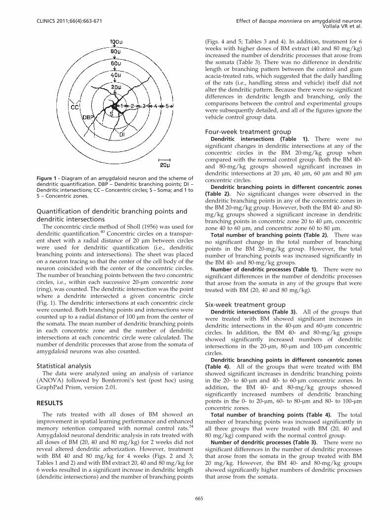
Quantification of dendritic branching points anddendritic intersections
The concentric circle method of Sholl (1956) was used fordendritic quantification.40 Concentric circles on a transpar-ent sheet with a radial distance of 20 mm between circleswere used for dendritic quantification (i.e., dendriticbranching points and intersections). The sheet was placedon a neuron tracing so that the center of the cell body of theneuron coincided with the center of the concentric circles.The number of branching points between the two concentriccircles, i.e., within each successive 20-mm concentric zone(ring), was counted. The dendritic intersection was the pointwhere a dendrite intersected a given concentric circle(Fig. 1). The dendritic intersections at each concentric circlewere counted. Both branching points and intersections werecounted up to a radial distance of 100 mm from the center ofthe somata. The mean number of dendritic branching pointsin each concentric zone and the number of dendriticintersections at each concentric circle were calculated. Thenumber of dendritic processes that arose from the somata ofamygdaloid neurons was also counted.
Statistical analysisThe data were analyzed using an analysis of variance
(ANOVA) followed by Bonferroni’s test (post hoc) usingGraphPad Prism, version 2.01.
RESULTS
The rats treated with all doses of BM showed animprovement in spatial learning performance and enhancedmemory retention compared with normal control rats.34
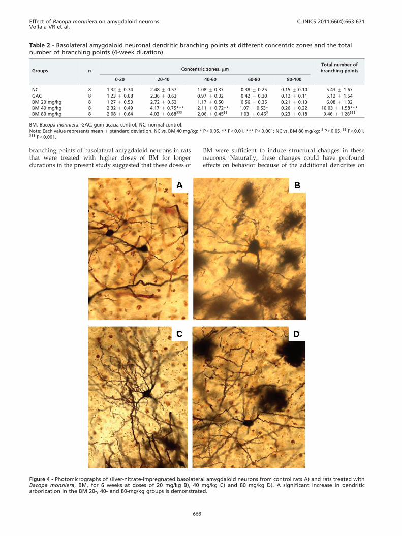
Amygdaloid neuronal dendritic analysis in rats treated withall doses of BM (20, 40 and 80 mg/kg) for 2 weeks did notreveal altered dendritic arborization. However, treatmentwith BM 40 and 80 mg/kg for 4 weeks (Figs. 2 and 3;Tables 1 and 2) and with BM extract 20, 40 and 80 mg/kg for6 weeks resulted in a significant increase in dendritic length(dendritic intersections) and the number of branching points
(Figs. 4 and 5; Tables 3 and 4). In addition, treatment for 6weeks with higher doses of BM extract (40 and 80 mg/kg)increased the number of dendritic processes that arose fromthe somata (Table 3). There was no difference in dendriticlength or branching pattern between the control and gumacacia-treated rats, which suggested that the daily handlingof the rats (i.e., handling stress and vehicle) itself did notalter the dendritic pattern. Because there were no significantdifferences in dendritic length and branching, only thecomparisons between the control and experimental groupswere subsequently detailed, and all of the figures ignore thevehicle control group data.
Four-week treatment groupDendritic intersections (Table 1). There were no
significant changes in dendritic intersections at any of theconcentric circles in the BM 20-mg/kg group whencompared with the normal control group. Both the BM 40-and 80-mg/kg groups showed significant increases indendritic intersections at 20 mm, 40 mm, 60 mm and 80 mmconcentric circles.
Dendritic branching points in different concentric zones(Table 2). No significant changes were observed in thedendritic branching points in any of the concentric zones inthe BM 20-mg/kg group. However, both the BM 40- and 80-mg/kg groups showed a significant increase in dendriticbranching points in concentric zone 20 to 40 mm, concentriczone 40 to 60 mm, and concentric zone 60 to 80 mm.
Total number of branching points (Table 2). There wasno significant change in the total number of branchingpoints in the BM 20-mg/kg group. However, the totalnumber of branching points was increased significantly inthe BM 40- and 80-mg/kg groups.
Number of dendritic processes (Table 1). There were nosignificant differences in the number of dendritic processesthat arose from the somata in any of the groups that weretreated with BM (20, 40 and 80 mg/kg).
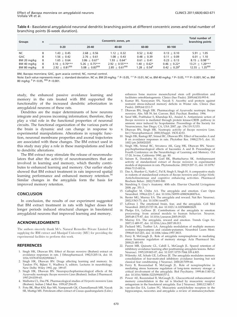
Six-week treatment groupDendritic intersections (Table 3). All of the groups that
were treated with BM showed significant increases indendritic intersections in the 40-mm and 60-mm concentriccircles. In addition, the BM 40- and 80-mg/kg groupsshowed significantly increased numbers of dendriticintersections in the 20-mm, 80-mm and 100-mm concentriccircles.
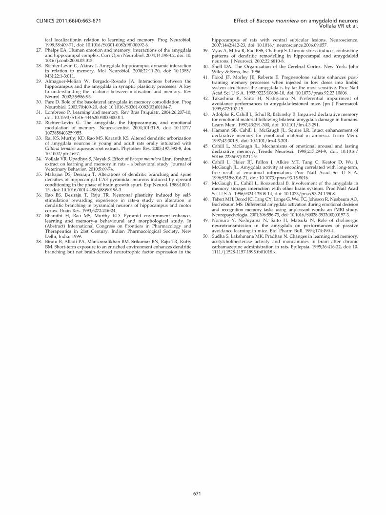
Dendritic branching points in different concentric zones(Table 4). All of the groups that were treated with BMshowed significant increases in dendritic branching pointsin the 20- to 40-mm and 40- to 60-mm concentric zones. Inaddition, the BM 40- and 80-mg/kg groups showedsignificantly increased numbers of dendritic branchingpoints in the 0- to 20-mm, 60- to 80-mm and 80- to 100-mmconcentric zones.
Total number of branching points (Table 4). The totalnumber of branching points was increased significantly inall three groups that were treated with BM (20, 40 and80 mg/kg) compared with the normal control group.
Number of dendritic processes (Table 3). There were nosignificant differences in the number of dendritic processesthat arose from the somata in the group treated with BM20 mg/kg. However, the BM 40- and 80-mg/kg groupsshowed significantly higher numbers of dendritic processesthat arose from the somata.
Figure 1 - Diagram of an amygdaloid neuron and the scheme ofdendritic quantification. DBP – Dendritic branching points; DI –Dendritic intersections; CC – Concentric circles; S – Soma; and 1 to5 – Concentric zones.
CLINICS 2011;66(4):663-671 Effect of Bacopa monniera on amygdaloid neuronsVollala VR et al.
665

DISCUSSION
Many studies have provided evidence for the involve-ment of the amygdala in different forms of memory storageand learning. Flood et al. (1995) stated that the amygdala isthe most sensitive brain region for memory enhancement.41
The amygdala plays a crucial and preferential role inavoidance learning compared with spatial learning.42 Theamygdala is involved in the formation of enhanceddeclarative memory for emotionally arousing events.43-45
According to Richter, Levin and Akirav (2000), theamygdala is involved in emotional responses and theformation of emotional memories.28 The amygdala is alsoinvolved in the formation of enhanced long-term mem-ory.46,47 This view is supported by Tabert et al. (2001), whoreported that the amygdala facilitates long-term but notshort-term memory consolidation of emotionally significantmaterial,48 and it is the cholinergic neurons that project to
the amygdala that play an important role in memoryacquisition.49
The term ‘‘passive avoidance’’ is usually employed todescribe experiments in which an animal learns to avoid anoxious event by suppressing a particular behavior. Thepassive avoidance test is a behavioral test that assessesmemory retention. Rats quickly learn to avoid noxiousstimuli, which evoke a highly emotional learning process,by suppressing an innate natural behavior, such as stayingin dark corners. The passive avoidance test is a usefulmethod for the assessment of an impairment in learning andfor the detection of both the ability to retain this learnedknowledge and its recall.50
In our earlier study, we examined the effect of BM onbehavioral changes using the same rats that were used inthe present study. The rats that were treated with all of thedoses of BM showed a significant improvement in passiveavoidance memory retention.34 This increased retention in
Figure 2 - Photomicrographs of silver-nitrate-impregnated basolateral amygdaloid neurons from control rats A) and rats treated withBacopa monniera, BM, for 4 weeks at doses of 20 mg/kg B), 40 mg/kg C) and 80 mg/kg D). A significant increase in dendriticarborization in the BM 40 and 80 mg/kg groups is demonstrated.
Effect of Bacopa monniera on amygdaloid neuronsVollala VR et al.
CLINICS 2011;66(4):663-671
666

rats treated with BM compared with normal control ratsindicated an increase in the ability to retain avoidancememories, which implied possible effects of BM onamygdaloid neurons.
The present study showed that the basolateral amygda-loid neuronal dendrites of rats treated with BM 40 and
80 mg/kg for 4 weeks and with 20, 40 and 80 mg/kg for 6weeks resulted in significant increases in dendritic lengthand number of branching points. In addition, treatmentwith higher doses of BM extract (40 and 80 mg/kg) for 6weeks increased the number of dendritic processes thatarose from the somata. Increased dendritic length and
Figure 3 - Camera lucida tracings of basolateral amygdaloid neurons from control rats A) and rats treated with Bacopa monniera for 4weeks at doses of 20 mg/kg B), 40 mg/kg C) and 80 mg/kg D).
Table 1 - Basolateral amygdaloid neuronal dendritic intersections and the number of dendritic processes arising fromthe somata (4-week duration).
Groups n Distance from soma, mmNumber of
processes
20 40 60 80 100
NC 8 5.46 ¡ 1.23 6.61 ¡ 1.03 5.66 ¡ 1.21 4.07 ¡ 1.38 2.41 ¡ 0.69 4.43 ¡ 0.68
GAC 8 5.71 ¡ 0.82 6.47 ¡ 0.95 5.39 ¡ 1.32 3.94 ¡ 1.23 2.36 ¡ 0.54 4.27 ¡ 0.50
BM 20 mg/kg 8 5.68 ¡ 0.74 7.24 ¡ 0.83 6.87 ¡ 0.80 4.68 ¡ 0.76 3.02 ¡ 0.95 4.11 ¡ 0.64
BM 40 mg/kg 8 7.25 ¡ 0.59** 10.32 ¡ 0.69*** 7.98 ¡ 1.14** 6.23 ¡ 0.90** 2.98 ¡ 1.16 4.38 ¡ 0.71
BM 80 mg/kg 8 7.08 ¡ 0.76$$ 9.86 ¡ 1.06$$$ 7.69 ¡ 0.87$$ 6.04 ¡ 0.82$$ 3.14 ¡ 0.62 4.76 ¡ 0.45
BM, Bacopa monniera; GAC, gum acacia control; NC, normal control.
Note: Each value represents mean ¡ standard deviation. NC vs. BM 40 mg/kg: ** P,0.01, *** P,0.001; NC vs. BM 80 mg/kg: $$ P,0.01, $$$ P,0.001.
CLINICS 2011;66(4):663-671 Effect of Bacopa monniera on amygdaloid neuronsVollala VR et al.
667
branching points of basolateral amygdaloid neurons in ratsthat were treated with higher doses of BM for longerdurations in the present study suggested that these doses of
BM were sufficient to induce structural changes in theseneurons. Naturally, these changes could have profoundeffects on behavior because of the additional dendrites on
Table 2 - Basolateral amygdaloid neuronal dendritic branching points at different concentric zones and the totalnumber of branching points (4-week duration).
Groups n Concentric zones, mmTotal number of
branching points
0-20 20-40 40-60 60-80 80-100
NC 8 1.32 ¡ 0.74 2.48 ¡ 0.57 1.08 ¡ 0.37 0.38 ¡ 0.25 0.15 ¡ 0.10 5.43 ¡ 1.67
GAC 8 1.23 ¡ 0.68 2.36 ¡ 0.63 0.97 ¡ 0.32 0.42 ¡ 0.30 0.12 ¡ 0.11 5.12 ¡ 1.54
BM 20 mg/kg 8 1.27 ¡ 0.53 2.72 ¡ 0.52 1.17 ¡ 0.50 0.56 ¡ 0.35 0.21 ¡ 0.13 6.08 ¡ 1.32
BM 40 mg/kg 8 2.32 ¡ 0.49 4.17 ¡ 0.75*** 2.11 ¡ 0.72** 1.07 ¡ 0.53* 0.26 ¡ 0.22 10.03 ¡ 1.58***
BM 80 mg/kg 8 2.08 ¡ 0.64 4.03 ¡ 0.68$$$ 2.06 ¡ 0.45$$ 1.03 ¡ 0.46$ 0.23 ¡ 0.18 9.46 ¡ 1.28$$$
BM, Bacopa monniera; GAC, gum acacia control; NC, normal control.
Note: Each value represents mean ¡ standard deviation. NC vs. BM 40 mg/kg: * P,0.05, ** P,0.01, *** P,0.001; NC vs. BM 80 mg/kg: $ P,0.05, $$ P,0.01,$$$ P,0.001.
Figure 4 - Photomicrographs of silver-nitrate-impregnated basolateral amygdaloid neurons from control rats A) and rats treated withBacopa monniera, BM, for 6 weeks at doses of 20 mg/kg B), 40 mg/kg C) and 80 mg/kg D). A significant increase in dendriticarborization in the BM 20-, 40- and 80-mg/kg groups is demonstrated.
Effect of Bacopa monniera on amygdaloid neuronsVollala VR et al.
CLINICS 2011;66(4):663-671
668
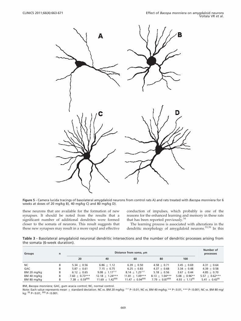
these neurons that are available for the formation of newsynapses. It should be noted from the results that asignificant number of additional dendrites were formedcloser to the somata of neurons. This result suggests thatthese new synapses may result in a more rapid and effective
conduction of impulses, which probably is one of thereasons for the enhanced learning and memory in these ratsthat has been reported previously.34
The learning process is associated with alterations in thedendritic morphology of amygdaloid neurons.33,39 In this
Figure 5 - Camera lucida tracings of basolateral amygdaloid neurons from control rats A) and rats treated with Bacopa monniera for 6weeks at doses of 20 mg/kg B), 40 mg/kg C) and 80 mg/kg D).
Table 3 - Basolateral amygdaloid neuronal dendritic intersections and the number of dendritic processes arising fromthe somata (6-week duration).
Groups n Distance from soma, mmNumber of
processes
20 40 60 80 100
NC 8 5.34 ¡ 0.56 6.86 ¡ 1.12 6.39 ¡ 0.50 4.58 ¡ 0.71 3.45 ¡ 0.69 4.31 ¡ 0.64
GAC 8 5.87 ¡ 0.61 7.15 ¡ 0.75 6.25 ¡ 0.83 4.37 ¡ 0.68 3.34 ¡ 0.48 4.39 ¡ 0.58
BM 20 mg/kg 8 6.12 ¡ 0.65 9.08 ¡ 1.17## 8.14 ¡ 1.25## 5.18 ¡ 0.56 3.67 ¡ 0.44 4.85 ¡ 0.70
BM 40 mg/kg 8 7.60 ¡ 0.73*** 12.18 ¡ 1.24*** 11.81 ¡ 1.09*** 8.13 ¡ 1.04*** 5.08 ¡ 0.96** 5.57 ¡ 0.62***
BM 80 mg/kg 8 7.38 ¡ 0.59$$$ 11.69 ¡ 1.42$$$ 11.47 ¡ 0.94$$$ 7.79 ¡ 0.87$$$ 4.93 ¡ 1.13$$ 5.41 ¡ 0.43$$
BM, Bacopa monniera; GAC, gum acacia control; NC, normal control.
Note: Each value represents mean ¡ standard deviation. NC vs. BM 20 mg/kg: ## P,0.01; NC vs. BM 40 mg/kg: ** P,0.01, *** P,0.001; NC vs. BM 80 mg/
kg: $$ P,0.01, $$$ P,0.001.
CLINICS 2011;66(4):663-671 Effect of Bacopa monniera on amygdaloid neuronsVollala VR et al.
669
study, the enhanced passive avoidance learning andmemory in the rats treated with BM supported thefunctionality of the increased dendritic arborization inamygdaloid neurons of these rats.
Dendrites are the major determinants of how neuronsintegrate and process incoming information; therefore, theyplay a vital role in the functional properties of neuronalcircuits. The functional organization of the various parts ofthe brain is dynamic and can change in response toexperimental manipulations. Alterations in synaptic func-tion, neuronal membrane properties and axonal trajectoriesare associated with these changes. The BM extract used inthis study may play a role in these manipulations and leadto dendritic alterations.
The BM extract may stimulate the release of neuromodu-lators that alter the activity of neurotransmitters that areinvolved in learning and memory, which thereby contri-butes to enhanced learning and memory. Our earlier studyshowed that BM extract treatment in rats improved spatiallearning performance and enhanced memory retention.34
Similar changes in the amygdala form the basis forimproved memory retention.
CONCLUSION
In conclusion, the results of our experiment suggestedthat BM extract treatment in rats with higher doses forlonger periods induced structural changes in basolateralamygdaloid neurons that improved learning and memory.
ACKNOWLEDGMENTS
The authors sincerely thank M/s. Natural Remedies Private Limited for
supplying the BM extract and Manipal University (MU) for providing the
experimental facilities to perform this work.
REFERENCES
1. Singh HK, Dhawan BN. Effect of Bacopa monniera (Brahmi) extract onavoidance responses in rats. J Ethnopharmacol. 1982;5:205-14, doi: 10.1016/0378-8741(82)90044-7.
2. Singh HK, Dhawan BN: Drugs affecting learning and memory. In:Tandon PN, Bijlani V, Wadhwa S, editors. Lectures in neurobiology.New Delhi: Wiley 1992, pp. 189-07.
3. Singh HK, Dhawan BN. Neuropsychopharmacological effects of theAyurvedic nootropic Bacopa monniera Linn (Brahmi). Indian J Pharmacol.1997;29:S359-65.
4. Malhotra CL, Das PK. Pharmacological studies of Herpestis monniera Linn(Brahmi). Indian J Med Res. 1959;47:294-05.
5. Potu BK, Bhat KM, Rao MS, Nampurath GK, Chamallamudi MR, NayakSR, Muttigi MS. Petroleum ether extract of Cissus quadrangularis (Linn.)
enhances bone marrow mesenchymal stem cell proliferation andfacilitates osteoblastogenesis. Clinics (Sao Paulo). 2009;64(10):993-8.
6. Kumar RS, Narayanan SN, Nayak S. Ascorbic acid protects againstrestraint stress-induced memory deficits in Wistar rats. Clinics (SaoPaulo). 2009;64:1211-7.
7. Dhawan BN, Singh HK. Pharmacology of Ayurvedic nootropic Bacopamonniera. Abs. NR 59, Int. Conven. Biol. Psychiat. Bombay. 1996.
8. Saraf MK, Prabhakar S, Khanduja KL, Anand A. Antiamnesic action ofBacopa monniera is mediated through protein kinase-CREB. pathway inamnesic mice induced by Scopolamine. Proceedings of the Society forNeuroscience, San Diego, CA, USA 2007, pp. 156.119/U153.
9. Dhawan BN, Singh HK. Nootropic activity of Bacopa monniera Linn.Int J Neuropharmacol. 2000;3(Suppl. 1SO2.4):S3.
10. Singh HK, Rastogi RP, Srimal RC, Dhawan BN. Effect of bacosides A andB on avoidance responses in rats. Phytother Res. 1988;2:70-5, doi: 10.1002/ptr.2650020205.
11. Singh HK, Srimal RC, Srivatava AK, Garg HK, Dhawan BN. Neuro-psychopharmacological effects of bacosides A and B. Proceedings ofFourth Conference on the Neurobiology of Learning and Memory. Oct17-20, Irvine, California 1990, pp. 80.
12. Sairam K, Dorababu M, Goel RK, Bhattacharya SK. Antidepressantactivity of standardized extract of Bacopa monniera in experimentalmodels of depression in rats. Phytomedicine. 2002;9:207-11, doi: 10.1078/0944-7113-00116.
13. Das A, Shanker G, Nath C, Pal R, Singh S, Singh H. A comparative studyin rodents of standardized extracts of Bacopa monniera and Ginkgo biloba.Anticholinesterase and cognitive enhancing activities. PharmacolBiochem Behav. 2002;73:893-900.
14. Standring S. Gray’s Anatomy. 40th edn. Elsevier Churchil Livingstone2008, pp. 351-3.
15. Gallagher M, Chiba AA. The amygdala and emotion. Curr OpinNeurobiol. 1996;6:221-7, doi: 10.1016/S0959-4388(96)80076-6.
16. Baxter MG, Murray EA. The amygdala and reward. Nat Rev Neurosci.2002;3:563-73, doi: 10.1038/nrn875.
17. LeDoux J. The emotional brain, fear, and the amygdala. Cell MolNeurobiol. 2003;23:727-38, doi: 10.1023/A:1025048802629.
18. Phelps EA, LeDoux JE. Contributions of the amygdala to emotionprocessing: from animal models to human behavior. Neuron.2005;48:175-87, doi: 10.1016/j.neuron.2005.09.025.
19. Murray EA. The amygdala, reward and emotion. Trends Cogn Sci.2007;11:489-97, doi: 10.1016/j.tics.2007.08.013.
20. Packard MG, Teather LA. Amygdala modulation of multiple memorysystems: hippocampus and caudate-putamen. Neurobiol Learn Mem.1998;69:163-203, doi: 10.1006/nlme.1997.3815.
21. Ferry B, McGaugh JL. Role of amygdala norepinephrine in mediatingstress hormone regulation of memory storage. Acta Pharmacol Sin.2000;21:481-93.
22. Parent MB, Quirarte GL, Cahill L, McGaugh JL. Spared retention ofinhibitory avoidance learning after posttraining amygdala lesions. BehavNeurosci. 1995;109:803-07, doi: 10.1037/0735-7044.109.4.803.
23. Wilensky AE, Schafe GE, LeDoux JE. The amygdala modulates memoryconsolidation of fear-motivated inhibitory avoidance learning but notclassical fear conditioning. J Neurosci. 2000;20:7059-66.
24. Ferry B, Roozendaal B, McGaugh JL. Role of norepinephrine inmediating stress hormone regulation of long-term memory storage: acritical involvement of the amygdala. Biol Psychiatry. 1999;46:1140-52,doi: 10.1016/S0006-3223(99)00157-2.
25. Power AE, Roozendaal B, McGaugh JL. Glucocorticoid enhancement ofmemory consolidation in the rat is blocked by muscarinic receptorantagonism in the basolateral amygdala. Eur J Neurosci. 2000;12:3481-7.
26. van-der-Zee EA, Luiten PG. Muscarinic acetylcholine receptors in thehippocampus, neocortex and amygdala: a review of immunocytochem-
Table 4 - Basolateral amygdaloid neuronal dendritic branching points at different concentric zones and total number ofbranching points (6-week duration).
Groups n Concentric zones, mmTotal number of
branching points
0-20 20-40 40-60 60-80 80-100
NC 8 1.43 ¡ 0.45 2.68 ¡ 0.56 1.12 ¡ 0.32 0.52 ¡ 0.42 0.13 ¡ 0.10 5.91 ¡ 1.05
GAC 8 1.56 ¡ 0.42 2.76 ¡ 0.61 1.08 ¡ 0.43 0.48 ¡ 0.39 0.11 ¡ 0.09 6.01 ¡ 1.10
BM 20 mg/kg 8 1.65 ¡ 0.64 3.86 ¡ 0.67# 1.93 ¡ 0.64# 0.61 ¡ 0.41 0.23 ¡ 0.13 8.15 ¡ 0.98##
BM 40 mg/kg 8 3.16 ¡ 0.70*** 5.26 ¡ 0.75*** 2.92 ¡ 0.55*** 1.40 ¡ 0.62* 0.46 ¡ 0.22* 13.21 ¡ 1.20***
BM 80 mg/kg 8 2.92 ¡ 0.81$$$ 5.08 ¡ 0.83$$$ 2.85 ¡ 0.61$$$ 1.28 ¡ 0.54$ 0.42 ¡ 0.29$ 12.55 ¡ 1.35$$$
BM, Bacopa monniera; GAC, gum acacia control; NC, normal control.
Note: Each value represents mean ¡ standard deviation. NC vs. BM 20 mg/kg: # P,0.05, ## P,0.01; NC vs. BM 40 mg/kg: * P,0.05, *** P,0.001; NC vs. BM
80 mg/kg: $ P,0.05, $$$ P,0.001.
Effect of Bacopa monniera on amygdaloid neuronsVollala VR et al.
CLINICS 2011;66(4):663-671
670
ical localizationin relation to learning and memory. Prog Neurobiol.1999;58:409-71, doi: 10.1016/S0301-0082(98)00092-6.
27. Phelps EA. Human emotion and memory: interactions of the amygdalaand hippocampal complex. Curr Opin Neurobiol. 2004;14:198-02, doi: 10.1016/j.conb.2004.03.015.
28. Richter-Levin G, Akirav I. Amygdala-hippocampus dynamic interactionin relation to memory. Mol Neurobiol. 2000;22:11-20, doi: 10.1385/MN:22:1-3:011.
29. Almaguer-Melian W, Bergado-Rosado JA. Interactions between thehippocampus and the amygdala in synaptic plasticity processes. A keyto understanding the relations between motivation and memory. RevNeurol. 2002;35:586-93.
30. Pare D. Role of the basolateral amygdala in memory consolidation. ProgNeurobiol. 2003;70:409-20, doi: 10.1016/S0301-0082(03)00104-7.
31. Lombroso P. Learning and memory. Rev Bras Psiquiatr. 2004;26:207-10,doi: 10.1590/S1516-44462004000300011.
32. Richter-Levin G. The amygdala, the hippocampus, and emotionalmodulation of memory. Neuroscientist. 2004;101:31-9, doi: 10.1177/1073858403259955.
33. Rai KS, Murthy KD, Rao MS, Karanth KS. Altered dendritic arborizationof amygdala neurons in young and adult rats orally intubated withClitoria ternatea aqueous root extract. Phytother Res. 2005;197:592-8, doi:10.1002/ptr.1657.
34. Vollala VR, Upadhya S, Nayak S. Effect of Bacopa monniera Linn. (brahmi)extract on learning and memory in rats – a behavioral study. Journal ofVeterinary Behavior. 2010;5:69-74.
35. Mahajan DS, Desiraju T. Alterations of dendritic branching and spinedensities of hippocampal CA3 pyramidal neurons induced by operantconditioning in the phase of brain growth spurt. Exp Neurol. 1988;100:1-15, doi: 10.1016/0014-4886(88)90196-3.
36. Rao BS, Desiraju T, Raju TR. Neuronal plasticity induced by self-stimulation rewarding experience in rats-a study on alteration indendritic branching in pyramidal neurons of hippocampus and motorcortex. Brain Res. 1993;6272:216-24.
37. Bharathi H, Rao MS, Murthy KD. Pyramid environment enhanceslearning and memory-a behavioural and morphological study. In(Abstract) International Congress on Frontiers in Pharmacology andTherapeutics in 21st Century. Indian Pharmacological Society, NewDelhi, India. 1999.
38. Bindu B, Alladi PA, Mansooralikhan BM, Srikumar BN, Raju TR, KuttyBM. Short-term exposure to an enriched environment enhances dendriticbranching but not brain-derived neurotrophic factor expression in the
hippocampus of rats with ventral subicular lesions. Neuroscience.2007;1442:412-23, doi: 10.1016/j.neuroscience.2006.09.057.
39. Vyas A, Mitra R, Rao BSS, Chattarji S. Chronic stress induces contrastingpatterns of dendritic remodelling in hippocampal and amygdaloidneurons. J Neurosci. 2002;22:6810-8.
40. Sholl DA. The Organization of the Cerebral Cortex. New York: JohnWiley & Sons, Inc. 1956.
41. Flood JF, Morley JE, Roberts E. Pregnenolone sulfate enhances post-training memory processes when injected in low doses into limbicsystem structures: the amygdala is by far the most sensitive. Proc NatlAcad Sci U S A. 1995;9223:10806-10, doi: 10.1073/pnas.92.23.10806.
42. Takashina K, Saito H, Nishiyama N. Preferential impairment ofavoidance performances in amygdala-lesioned mice. Jpn J Pharmacol.1995;672:107-15.
43. Adolphs R, Cahill L, Schul R, Babinsky R. Impaired declarative memoryfor emotional material following bilateral amygdala damage in humans.Learn Mem. 1997;43:291-300, doi: 10.1101/lm.4.3.291.
44. Hamann SB, Cahill L, McGaugh JL, Squire LR. Intact enhancement ofdeclarative memory for emotional material in amnesia. Learn Mem.1997;43:301-9, doi: 10.1101/lm.4.3.301.
45. Cahill L, McGaugh JL. Mechanisms of emotional arousal and lastingdeclarative memory. Trends Neurosci. 1998;217:294-9, doi: 10.1016/S0166-2236(97)01214-9.
46. Cahill L, Haier RJ, Fallon J, Alkire MT, Tang C, Keator D, Wu J,McGaugh JL. Amygdala activity at encoding correlated with long-term,free recall of emotional information. Proc Natl Acad Sci U S A.1996;9315:8016-21, doi: 10.1073/pnas.93.15.8016.
47. McGaugh JL, Cahill L, Roozendaal B. Involvement of the amygdala inmemory storage: interaction with other brain systems. Proc Natl AcadSci U S A. 1996;9324:13508-14, doi: 10.1073/pnas.93.24.13508.
48. Tabert MH, Borod JC, Tang CY, Lange G, Wei TC, Johnson R, Nusbaum AO,Buchsbaum MS. Differential amygdala activation during emotional decisionand recognition memory tasks using unpleasant words: an fMRI study.Neuropsychologia. 2001;396:556-73, doi: 10.1016/S0028-3932(00)00157-3.
49. Nomura Y, Nishiyama N, Saito H, Matsuki N. Role of cholinergicneurotransmission in the amygdala on performances of passiveavoidance learning in mice. Biol Pharm Bull. 1994;174:490-4.
50. Sudha S, Lakshmana MK, Pradhan N. Changes in learning and memory,acetylcholinesterase activity and monoamines in brain after chroniccarbamazepine administration in rats. Epilepsia. 1995;36:416-22, doi: 10.1111/j.1528-1157.1995.tb01018.x.
CLINICS 2011;66(4):663-671 Effect of Bacopa monniera on amygdaloid neuronsVollala VR et al.
671