batista, - cornell university
TRANSCRIPT

1
Investigating the role of soluble adenylyl cyclase and protein kinase A in acrosomal exocytosis in equine spermatozoa.
Honors Thesis Presented to the College of Agriculture and Life Sciences, Animal Science
Of Cornell University In Partial Fulfillment of the Requirements for the
Research Honors Program by
Michele A. Batista December 2009
S.J. Bedford‐Guaus1
1 Department of Clinical Sciences, College of Veterinary Medicine, Cornell University, Ithaca, NY 14853
2
Abstract
Capacitation involves the molecular changes that ejaculated sperm
undergo within the female reproductive tract (i.e. oviduct) to become fertilization‐
competent. Capacitation can be achieved in vitro by incubating sperm in a defined
medium that mimics the oviductal milieu. During this process, sperm gain the ability to
undergo acrosomal exocytosis whereby the release of proteolytic enzymes from the
acrosome, a lysosome‐like cap covering the anterior portion of the sperm head,
facilitates penetration of the oocyte vestments at fertilization. The ability of sperm to
undergo acrosomal exocytosis in vitro when challenged with an agonist such as calcium
ionophore (CaI) or progesterone is often used as a marker of capacitation.
This study aims to investigate the signal transduction pathway leading to
acrosomal exocytosis in stallion spermatozoa. It has been proposed that soluble
adenylyl cyclase (SACY) and protein kinase A (PKA) are two of the players of the signal
transduction pathway leading to acrosomal exocytosis. In order to clarify their role in
supporting this event in stallion sperm, we use the SACY‐specific inhibitor KH7 and the
PKA‐specific activator 3’‐5’ cyclic adenosine monophosphate (cAMP) analog SP6. In the
experiments presented herein, we add these agents to sperm incubated in capacitating
conditions and compare rates of acrosomal exocytosis with those achieved after the
addition of CaI, progesterone, or vehicle control dimethyl sulfoxide (DMSO).
In order to assess acrosomal status in these experiments, sperm are visualized
under a microscope using a fluorescent dye that binds to β‐galactosidase residues

3
within the acrosome. Based on the appearance of the acrosome, counted sperm are
classified as acrosome intact if there is no stain over the acrosomal region, or acrosome
reacted if stain is present over the acrosome. In our first set of experiments, addition of
the SACY inhibitor was used to determine the role of cAMP production in the pathways
leading to acrosomal exocytosis. There was no difference (P>0.05) in the percent of
acrosome reacted sperm between capacitated sperm challenged with progesterone
(29.62%) or CaI (30.32%). Similarly, there was no difference (P>0.05) between sperm
challenged with CaI with (29.02%) and without (30.32%) the addition of KH7, suggesting
that cAMP is not playing a role in this process when sperm are challenged with CaI.
Interestingly, there was a difference (P=0.003) when rates of acrosomal exocytosis were
compared between sperm challenged with progesterone with (22.73%) and without
(29.62%) the addition of KH7. Since progesterone is considered a more physiological
inducer of acrosomal exocytosis than CaI, it is possible that the latter is able to override
the physiological pathways that lead to acrosomal exocytosis. These results suggest that
cAMP production is required for the process of acrosomal exocytosis under
physiological conditions.
In our second set of experiments, addition of the PKA‐specific activator SP6 to
sperm incubated in capacitating conditions induced 32.97% acrosomal exocytosis, which
was higher than that resulting from the addition of DMSO (negative vehicle control,
19.37%; P=0.013). These results suggest that PKA is playing a role downstream of cAMP
in the molecular pathways leading to acrosomal exocytosis in stallion sperm. In

4
summary, this research contributes to the general knowledge in basic mammalian
reproduction and could help improve assisted reproduction techniques in the equine.
5
Acknowledgements
I would like to thank Dr. Sylvia Bedford‐Guaus, for giving me the opportunity,
resources, and direction to complete this research project; Dr. Lori McPartlin, for her
assistance, suggestions, and patience during the experiments and analysis of the results;
Mrs. Stephanie Westmiller for her guidance in my learning of laboratory protocols,
procedures, and techniques; Ms. Carol Collyer, manager of the Cornell Equine Research
Park, for handling stallions and her assistance in semen collections; and Mr. Karl Gluck,
for helping me design some of the figures presented in this study.

6
Contents Abstract ............................................................................................................................................ 2
Acknowledgements .......................................................................................................................... 5
Introduction ..................................................................................................................................... 7
Literature Review ............................................................................................................................. 9
Materials and Methods .................................................................................................................. 19
Chemicals and Reagents ............................................................................................................ 19
Media Composition .................................................................................................................... 19
Animals, Semen Collection and Processing: .............................................................................. 19
Assessment of Acrosomal Status ............................................................................................... 20
Experimental Design .................................................................................................................. 22
Experiment 1: Determining if inhibition of SACY with KH7 will inhibit acrosomal exocytosis ............................................................................................................................................... 22
Experiment 2: Determining if activation of PKA with the cAMP analog SP6 will induce acrosomal exocytosis ................................................................................................................. 23
Statistical methods..................................................................................................................... 24
Results ............................................................................................................................................ 25
Experiment 1: ............................................................................................................................. 25
Experiment 2: ............................................................................................................................. 26
Discussion ...................................................................................................................................... 27
SACY Activity may Not be Required for Acrosomal Exocytosis .................................................. 27
Acrosomal Exocytosis is a PKA‐Dependent Process ................................................................... 28
Future Directions ....................................................................................................................... 29
Conclusion .................................................................................................................................. 30
Literature Cited .............................................................................................................................. 31
7
Introduction
In mammals, spermatozoa are not capable of fertilizing an oocyte until they
undergo a series of molecular changes, collectively termed “capacitation” (Chang, 1951,
1955; Austin, 1951, 1952), that are conferred by the environment within the female
reproductive tract. In order to successfully achieve sperm capacitation in vitro, the
female milieu must be mimicked, with requirements being species‐specific. Although
molecular pathways that support sperm capacitation are still under investigation and
largely unknown, increases in protein tyrosine phosphorylation have been correlated
with capacitation and thus the ability of sperm to fertilize an oocyte (Visconti et al.,
1995a, 1995b).
While the hallmark test for sperm capacitation is the ability to fertilize an oocyte,
in the laboratory, sperm capacitation is also assessed by the ability of sperm to undergo
acrosomal exocytosis (Ward and Storey, 1984; Florman and Babcock, 1991; Kopf and
Gerton, 1991). The acrosome is a membrane‐bound, lysosome‐like structure that caps
the anterior portion of the sperm head and contains proteolytic enzymes that confer
sperm with the ability to penetrate the outer vestments of the oocyte at fertilization
(Yanagimachi, 1994). Only capacitated sperm are able to undergo acrosomal exocytosis
when challenged with an appropriate physiological stimulus. In vivo, acrosomal
exocytosis presumably occurs upon contact with the zona pellucida, a glycoprotein
matrix surrounding the oocyte (Meyers et al., 1995). In vitro, agonists such as
progesterone and ionophores can be used to induce acrosomal exocytosis in

8
capacitated sperm (Cheng et al., 1998). This is not only a marker of capacitation in vitro,
but also allows for studying the molecular pathways that control acrosomal exocytosis.
Ultimately, studying the processes of sperm capacitation and acrosomal
exocytosis allows for a better understanding of the process of fertilization, and thus its
application for the understanding of different causes of infertility, and in assisted
reproductive techniques such as in vitro fertilization (IVF). Interestingly, while IVF is
highly successful in many domestic species and in humans, it has been very difficult to
replicate in the horse and only two foals have ever been produced with this technique
(Palmer et al., 1991). Our laboratory has recently defined incubation conditions that
support equine sperm capacitation, as defined by marked increase in protein tyrosine
phosphorylation paired with high rates of acrosomal exocytosis, and IVF (McPartlin et
al., 2008, 2009). Presently we are interested in further studying the molecular pathways
that support these processes.
The overall aim of this research is to investigate the molecular pathways that
control the process of acrosomal exocytosis. To accomplish this, a SACY‐specific
inhibitor, KH7, is used to determine if acrosomal exocytosis is affected by decreased
SACY activity. Because PKA is a downstream target of cAMP, the specific analog, SP6, is
also used to determine whether PKA activation is required for acrosomal exocytosis. The
results from this research will lead to a better understanding of the molecular
pathways that support equine sperm capacitation and its associated events.

9
Literature Review
“Capacitation” was classically defined as comprising only the time dependent
acquisition of fertilization competence by ejaculated sperm (Chang 1951, 1955; Austin
1951, 1952). More recently, this definition has been revised to include the molecular
changes that render sperm with the ability to undergo acrosomal exocytosis when
challenged by a physiological stimulus (Ward and Storey, 1984; Florman and Babcock,
1991; Kopf and Gerton, 1991). Moreover, capacitation may also include the cell
membrane changes that increase permeability to calcium, and fusion of the plasma and
outer acrosomal membranes, ultimately leading to acrosomal exocytosis (Yanagimachi,
1994). Other events included in the definition of capacitation are changes in sperm
intracellular ion concentrations, metabolism, and motility (Florman and Babcock, 1991;
Yanagimachi, 1994). In this regard, hyperactivation, a distinct change in the pattern of
motility characterized by an increase in asymmetrical flagellar beating that is displayed
by capacitated sperm, is also required for fertilization competence (Quill et al., 2003;
McPartlin et al., 2009). However, this is often studied as an independent process and
thus will not be included in the discussions of this proposal.
In the laboratory, the functional endpoints of sperm capacitation that can be
readily assessed include time‐dependent increases in protein tyrosine phosphorylation
(Visconti et al., 1995a), the ability to undergo agonist‐induced acrosomal exocytosis, and
the ability to fertilize oocytes in vitro (Ward and Storey, 1984; Florman and Babcock,
1991; Kopf and Gerton, 1991). It has been demonstrated that capacitation is correlated

10
with a time‐dependent increase in the amount of tyrosine‐phosphorylated residues on a
subset of proteins in murine spermatozoa, as assessed by SDS‐PAGE and
immunoblotting (Visconti et al., 1995a). Additionally, high levels of protein tyrosine
phosphorylation are correlated with the ability of sperm to undergo solubilized zona
pellucida (ZP)‐induced acrosomal exocytosis, as well as to successfully fertilize oocytes in
vitro (Visconti et al., 1995a). These experiments provided the opportunity to study the
media components required to support these changes, or the acquisition of
capacitation, in vitro. These researchers concluded that bovine serum albumin (BSA),
sodium bicarbonate, and calcium (Yanagimachi, 1994), were required for sperm to
display increases in protein tyrosine phosphorylation, acrosomal exocytosis rates, and
thus for the acquisition of fertilization competence (Visconti et al., 1995a). It has since
been demonstrated in a variety of species that serum albumin (Go and Wolf, 1985;
Langlais and Roberts, 1985), calcium ions (Yanagimachi, 1982; Coronel and Lardy, 1987;
Fraser, 1987; Ruknudin and Silver, 1990), and sodium bicarbonate (Lee and Storey,
1986; Neill and Olds‐Clarke, 1987; Boatman and Robbins, 1991) are required for in vitro
capacitation media.
As for the postulated function of these media components, albumin is a
ubiquitous blood plasma protein that is found in all mammals; in the context of sperm
capacitation, BSA is thought to act as sterol sink, thus causing efflux of cholesterol from
the plasma membrane (Go and Wolf, 1985; Langlais and Roberts, 1985). In turn, this
efflux causes changes in membrane fluidity and the activation of certain signaling
pathways (Cross, 1998; Flesch and Gadella, 2000; Baldi, et al. 2002). Bicarbonate can

11
then enter the cell and activate the enzyme SACY (formerly abbreviated sAC) which
generates cAMP (Okamura et al., 1985; Visconti et al., 1990). Calcium ions, which also
flow into the sperm cell once the membrane fluidity is altered, are suggested to have
regulatory effects on SACY (Hyne and Garbers, 1979) and on cyclic nucleotide
phosphodiesterase (Visconti et al., 1995b). These two enzymes ensure appropriate
levels of cAMP, which in turn is presumed to activate PKA. This ultimately results in a
series of downstream protein tyrosine phosphorylation events.
The role of SACY in promoting capacitation‐driven protein tyrosine
phosphorylation has been demonstrated with SACY‐null mice (Hess et al., 2005). Sperm
from these mice have normal morphology but are immotile and unable to fertilize eggs
in vitro. Most remarkably, SACY‐null sperm display an abnormal pattern of protein
tyrosine phosphorylation that was not overcome by incubating them under capacitating
conditions with the addition of a cAMP analog or phosphodiesterase inhibitor (Hess, et
al., 2005). The requirement for cAMP during sperm capacitation is also supported by
studies showing that incubation of sperm in non‐capacitating medium but with the
addition of dbcAMP (a cAMP analog) and phosphodiesterase‐inhibitors such as IBMX
show levels of protein tyrosine phosphorylation indicative of the capacitated state
(Visconti et al., 1995b; Galantino‐Homer, 1997).
As mentioned above, PKA is considered the main downstream target of cAMP
during sperm capacitation. The role of PKA in these events has been further
demonstrated by the use of specific inhibitors. When H‐89, an agent that inhibits the

12
action of PKA, was added to the otherwise complete murine sperm capacitating
medium, inhibition of protein tyrosine phosphorylation and acrosomal exocytosis
occurred in a concentration‐dependent manner (Visconti et al., 1995b). Inhibitors of
protein kinase C (PKC) and calcium/calmodulin‐dependent protein kinase had no effect
on the levels of protein tyrosine phosphorylation in capacitated sperm (Visconti et al.,
1995b). Additionally, sperm incubated with Rp‐cAMPS, a cAMP antagonist, did not
display protein tyrosine phosphorylation when incubated under capacitating conditions
(Visconti et al., 1995b). PKA may be important not only for protein tyrosine
phosphorylation, but for acrosomal exocytosis as well.

13
As stated, capacitation prepares the sperm to undergo acrosomal exocytosis, an
exocytotic event whereby the enzymatic contents of the acrosome are released via
dissolution of the plasma and outer acrosomal membranes (Fig.1). The release of the
proteolytic enzymes contained within the acrosome facilitates penetration of the outer
vestments of the oocyte and thus successful fertilization. While capacitation prepares
sperm for acrosomal exocytosis, an external stimulus is still required to trigger this
event. In vivo, the stimulus is presumed to be the ZP (Meyers et al., 1995), and possibly
progesterone (Cheng et al., 1998a, 1998b). Therefore, in vitro, acrosomal exocytosis
may be induced by the addition of solubilized ZP (Meyers et al., 1995), progesterone
(Cheng et al., 1998a, 1998b), or CaI (Hess et al., 2005). Solubilized ZP and progesterone
are considered physiological inducers because the former surrounds the oocyte and
must be penetrated by the sperm in order for fertilization to occur, and the latter is a
hormone that is naturally present in the female mammalian oviduct. Conversely, CaI
creates pores in the sperm plasma membrane that are specifically permeable to divalent
ions such as calcium (Luckasen et al., 1974). While this is considered a non‐physiological
inducer of acrosomal exocytosis, sperm must still undergo the process of capacitation in
order to undergo acrosomal exocytosis in response to a challenge with low
concentrations of CaI. Therefore, for practical reasons, progesterone and CaI are often
used in vitro to test whether a population of sperm is capacitated. In this proposal, we
use these agents to study the pathways promoting acrosomal exocytosis in stallion
sperm.

14
Attending to what is presently known regarding the molecular pathways linked
to acrosomal exocytosis in mammalian species, challenge with CaI yields rates of
acrosomal exocytosis in SACY‐null sperm comparable to those induced in wild‐type
sperm (Hess et al., 2005). Based on these results, one may conclude that SACY activity is
not required for acrosomal exocytosis. In this regard, there is some evidence to suggest
that a transmembrane form of adenylyl cyclase (tmAC) may be an important source of
cAMP for the pathway triggering this exocytotic event, contrary to the role of SACY in
promoting protein tyrosine phosphorylation as stated above. Moreover, it has been
reported that forskolin, a specific activator of tmAC, induces acrosomal exocytosis
(Leclerc et al., 2004), and that tmAC activity is localized to the head region of
capacitated sperm (Liguori et al., 2004). Furthermore, sperm that are tmAC‐null cannot
fertilize an oocyte unless the ZP is removed (Livera et al., 2005), providing further
evidence of a role for tmAC in acrosomal exocytosis.
Recently, a compound that specifically inhibits SACY activity has been developed
(KH7; Hess et al., 2005). KH7 is a small, lipophilic molecule that selectively inhibits SACY.
It is neither competitive with the substrate for SACY (Mg2+‐ATP), nor is it competitive for
the activators calcium and bicarbonate. In addition, it does not affect the activity of
tmACs. It has been observed that at low concentrations KH7 will block cAMP increases
in sperm incubated in capacitating conditions, and thus will affect the corresponding
increases in protein tyrosine phosphorylation (Hess et al., 2005). Moreover, wild‐type
sperm treated with KH7 were unable to fertilize an oocyte, but fertilization competence
was recovered with a cAMP analog. Of importance in the context herein, however, is the
15
fact that incubation of sperm in the presence of KH7 did not prevent wild‐type sperm
from undergoing acrosomal exocytosis when challenged with solubilized ZP. While these
results are difficult to interpret, one may infer that (a) protein tyrosine phosphorylation
and acrosomal exocytosis are separable and independent events; and (b) SACY activity is
required for the former, but not for acrosomal exocytosis. This requires further
investigation.
Another important variable to consider when dissecting the molecular pathways
involved in acrosomal exocytosis is the effect of progesterone. Progesterone is present
in cumulus oophorus cells surrounding the oocyte (Osman et al., 1989) and in fluid of
pre‐ovulatory follicles (Cheng et al., 1998); moreover, the existence of a sperm plasma
membrane progesterone receptor has been reported (Cheng et al., 1998b). In fact, work
in the stallion suggests that progesterone may induce the acrosome reaction, but in a
PKA‐independent pathway (Rathi et al., 2003). In this study, the presence of a PKA
inhibitor did not abrogate rates of progesterone‐induced acrosomal exocytosis when
compared to controls challenged with progesterone. These authors proposed that
progesterone may operate through a PKC or protein tyrosine kinase (PTK) signaling
pathway, since the inhibition of these enzymes reduced the rates of progesterone‐
induced acrosomal exocytosis. Moreover, progesterone may stimulate acrosomal
exocytosis in a manner similar to a ZP protein, ZP3, which is thought to activate PTK
(Breitbart and Naor, 1999).

16
Although rates of acrosomal exocytosis are reduced when PKC or PTK are
inhibited and progesterone is used as an inducer, there is still strong evidence to
support the involvement of PKA. For instance, in the stallion, bicarbonate‐induced
acrosomal exocytosis is blocked by PKA inhibitors (Rathi et al., 2003). In the absence of
bicarbonate, acrosomal exocytosis could be induced by using a cAMP analog (but not a
cGMP analog), thus supporting a role for PKA‐related cyclonucleotide‐dependent
kinases. The cAMP analog was also found to induce the acrosome reaction more
strongly than bicarbonate, suggesting that PKA is not fully stimulated by bicarbonate.
The third line of evidence for involvement of PKA is that PKC and PTK inhibitors did not
block bicarbonate‐induced acrosomal exocytosis (Rathi et al., 2003). Therefore,
progesterone and bicarbonate may work though separate pathways, but together have
a cooperative action to induce higher rates of acrosomal exocytosis than they do
individually.
While there has been much research done on sperm capacitation in other
species, capacitation and its associated events have been far more elusive and less
researched in the horse. Medium that fully supports sperm capacitation and
fertilization‐competence has been developed for many species other than the horse,
allowing researchers to apply reproductive techniques such as in vitro fertilization in
these species. Previously, only one study had demonstrated increases in protein
tyrosine phosphorylation as one of the hallmarks of capacitation in the horse; however,
this study used dbcAMP and caffeine as activators (Pommer et al., 2003) rather than a
defined medium. These authors showed that H‐89 was able to reduce levels of time‐
17
dependent increases in protein tyrosine phosphorylation (Pommer et al., 2003);
however, since these authors did not use a defined medium to support capacitation in
stallion sperm, these results are difficult to interpret.
More recently, a complete medium that supports stallion sperm capacitation has
been defined (McPartlin et al., 2008). Equine spermatozoa incubated in a modified
Whitten’s media (Travis et al., 2004) with BSA (7 mg/mL) and sodium bicarbonate (25
mM) displayed marked increases in protein tyrosine phosphorylation levels.
Importantly, increased protein tyrosine phosphorylation levels were correlated with
significant rates of CaI‐ and progesterone‐induced acrosomal exocytosis (McPartlin et
al., 2008). Moreover, when sperm incubated in these conditions were pharmacologically
induced to display hyperactivated motility, this yielded high rates of homologous IVF
(McPartlin et al., 2008). Altogether, these results support the notion that sperm
incubated in these defined conditions were undergoing changes consistent with
capacitation.
Following up on these recent studies and using the same incubation conditions,
the aim of this research is to investigate the role of SACY and PKA in the molecular
pathways leading to acrosomal exocytosis in stallion sperm. Using a SACY specific‐
inhibitor, we will determine if rates of acrosomal exocytosis will decrease when sperm
are challenged with both a physiological and non‐physiological inducer. We will also
compare rates of acrosomal exocytosis achieved by a PKA‐specific activator to rates
attained by a non‐physiological inducer of acrosomal exocytosis. These studies will

18
enhance our knowledge in basic mammalian reproduction, as well as aid in the
improvement of assisted reproduction technologies in the horse.

19
Materials and Methods
Chemicals and Reagents
Four‐Bromo‐Calcium Ionophore A23187 was obtained from Calbiochem (San
Diego, CA, USA), and PNA‐Alexa 488 was purchased from Invitrogen Corp. (Carlsbad, CA,
USA). The acquisition of the SACY inhibitor KH7 was facilitated by Drs. Levin and Buck
(Department of Pharmacology, Weill Cornell Medical College, New York, NY (Hess et al.,
2005) and purchased from the Abby and Howard P. Milistein Synthetic Chemistry Core
Facility also at Weill Cornell Medical College. The PKA‐specific activator SP6 was
purchased from BioLog Life Sciences (Bremen, Germany). All other chemicals were
purchased from Sigma Chemical Company (St. Louis, MO, USA).
Media Composition
The non‐capacitating (NC) medium used for sperm was Modified Whitten’s (MW;
100 mM NaCl, 4.7 mM KCl, 1.2 mM MgCl2, 5.5 mM glucose (anhydrous), 22 mM HEPES,
4.8 mM lactic acid hemicalcium salt and 1.0 mM pyruvic acid). The MW medium was
always devoid of BSA and HCO3‐, and served both as transport and non‐capacitating
medium. Capacitating (Cap) conditions were achieved by adding 25 mM NaHCO3 and 7
mg/mL BSA to NC base media. For all media used, the final pH was brought to 7.25
(McPartlin et al., 2008).
Animals, Semen Collection and Processing:
Semen was collected with an artificial vagina from three adult stallions of proven
fertility at the Cornell Equine Park in compliance with IACUC protocols. After visual

20
evaluation of sperm motility under light microscopy (200x) on a heated stage and
assessment of sperm concentration using a 534B MOD1 Densimeter (Animal
Reproduction Systems; Chino, CA, USA), the sperm rich fraction was diluted 2:1 (vol:vol)
in the appropriate pre‐warmed NC medium and transported to the laboratory at 37 °C
for immediate processing. At the lab, the semen was centrifuged in 15‐mL conical tubes
at 100 x g for 1 minute (37° C) in order to remove particulate matter and dead sperm.
The supernatant from this spin was recovered, transferred to a warm, 15‐mL round‐
bottom sterilized tube and spun at 600 x g for 5 minutes (37° C). The supernatant from
this spin was disposed of and the pellet was transferred into a sterile round‐bottom
tube that contained 1.5 mL of the NC media.
Sperm concentration of the semen sample was assessed with a hemocytometer
under phase‐contrast microscopy and the sperm pellet was diluted to a final
concentration of 10x106 sperm/mL by adding the appropriate volume of Cap or NC
medium. For incubation, 500‐µL aliquots of appropriately diluted sperm were placed in
polyvinyl alcohol‐coated tubes. Incubation proceeded for 0 or 6 h at 37°C in a humidified
air atmosphere. Experiments were only performed if after washing, sperm displayed a
motility of at least 50%, which was evaluated visually under phase contrast microscopy
(200x) on a heated stage.
Assessment of Acrosomal Status
At 0 and 6 h of incubation, sperm samples in different media conditions
were further incubated for a period of time with the appropriate activators, controls, or
21
inhibitors as outlined in the following sections. At the end of each incubation period the
percentages of total and progressively motile sperm were assessed with light microscopy
on a heated stage (200x). Acrosomal status of sperm was assessed with the addition of
PNA‐Alexa 488 (final concentration 0.024 mg/mL) to a 125 μL aliquot of the sperm
suspension; 100 μL was then pipetted onto a glass slide. Sperm were allowed to settle for
5 minutes. The samples were then gently washed with warm phosphate‐buffered saline
(PBS), and saturated with a 2% paraformaldehyde solution for 10 minutes. After additional
gentle washing with PBS, a coverslip was added and the slides were scored (630x) for
acrosomal status using an upright fluorescent Zeiss Imager ZI microscope with Green
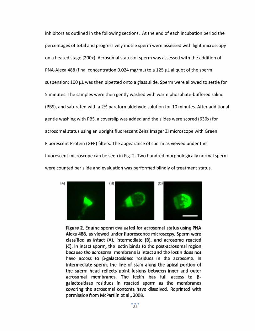
Fluorescent Protein (GFP) filters. The appearance of sperm as viewed under the
fluorescent microscope can be seen in Fig. 2. Two hundred morphologically normal sperm
were counted per slide and evaluation was performed blindly of treatment status.

22
Experimental Design
Experiment 1: Determining if inhibition of SACY with KH7 will inhibit acrosomal
exocytosis
The design of this experiment is presented in Fig. 3. Briefly, sperm were incubated in
Cap medium for 0 or 6 h. At each time period (immediately after the initiation of the
experiment for Time 0), sperm were challenged with CaI (5 µM) without and with (60
µM) the SACY inhibitor KH7; progesterone (3.2 μM) without and with (60 µM) KH7; or
vehicle control DMSO (2.5 μL; 0.5% vol:vol). KH7 was added 30 minutes before CaI,
progesterone, or DMSO, and was only used for sperm incubated 6 h under capacitating
conditions. Sperm were then incubated at 37°C in a humidified air atmosphere for an
additional 30 minutes, and then processed as outlined above for evaluation of
acrosomal status. Three experiments with each of two stallions were performed.

23
Experiment 2: Determining if activation of PKA with the cAMP analog SP6 will induce
acrosomal exocytosis
The design of this experiment is presented in Fig. 4. Briefly, sperm were
incubated in NC or Cap medium for 0 or 6 h. At each time period (immediately after the
initiation of the experiment for Time 0), sperm incubated in either condition were
challenged with CaI (5 µM), SP6 (1 µM), or DMSO (2.5 μL; 0.5% vol:vol). Sperm were
then incubated at 37°C in a humidified air atmosphere for an additional 30 minutes, and
then processed as outlined above for evaluation of acrosomal status. Two experiments
with each of two stallions were performed.

24
Statistical methods
Results for average percent acrosomal exocytosis rate in each experiment were
analyzed by two‐way repeated measures analysis of variance (ANOVA). If significant
differences were detected (P<0.05), the Student‐Newman‐Keuls test was applied to
assess all pairwise comparisons.

25
Results
Experiment 1:
There were no differences (P>0.05) in the rates of acrosomal exocytosis between
the different treatments at time 0. Conversely, at time 6, there was a decrease in the
percent of acrosome reacted sperm treated with KH7 and progesterone, versus when
treated with progesterone alone (P=0.003). However, treatment with KH7 prior to
inducing acrosomal exocytosis with CaI did not affect rates as compared to treatment
with CaI alone (P>0.05). Similarly, there was a significant difference (P=0.002) between
sperm treated with KH7 and induced with CaI, and sperm treated with KH7 and induced
with progesterone. Results are presented in Table 1.
Table 1. Experiment 1: Average percent of sperm displaying acrosomal exocytosis at 0 and 6 h, challenged with calcium ionophore (CaI) with and without the SACY inhibitor (KH7), with progesterone (P4) with and without the SACY inhibitor (KH7), and with vehicle control (DMSO).
0 h Treatment DMSO CaI P4
Percentage of acrosomal exocytosis (mean±SEM)
15.22 ±3.2 31.27 ±5.0
17.23 ±2.9
6 h Treatment DMSO CaI CaI+KH7 P4 P4+KH7
Percentage of acrosomal exocytosis (mean±SEM)
19.73 ±3.5a 30.32 ±2.2b 29.02 ±3.8b 29.62 ±2.4b 22.73 ±3.5c
a,b,c Within incubation time, different superscripts denote significant differences (P<0.05)

26
Experiment 2:
No significant differences between treatments or within ejaculates were
detected for sperm incubated in either NC or Cap conditions at time 0, or for sperm
incubated in NC conditions at time 6. After 6 hours of incubation in Cap medium, sperm
challenged with SP6, a PKA‐specific cAMP analog, displayed higher (P=0.013) rates of
acrosomal exocytosis than sperm challenged with the vehicle control DMSO, but lower
rates (P=0.021) than sperm incubated in CaI. While SP6 had effects on acrosomal
exocytosis that were intermediate between DMSO and CaI, the results sill support a role
of PKA in supporting acrosomal exocytosis in stallion sperm. Results are presented in
Table 2.
Table 2. Experiment 2: Average percent of sperm displaying acrosomal exocytosis at 0 and 6 h, incubated in both non‐capacitating and capacitation conditions, and challenged with calcium ionophore (CaI), the PKA‐specific cAMP analog (SP6), and with vehicle control (DMSO).
0 h Treatment Non‐capacitated Capacitated
DMSO CaI SP6 DMSO CaI SP6 Percentage of acrosomal exocytosis (mean±SEM)
27.43 ±3.6
38.80 ±7.3
27.40 ±7.5
23.55 ±3.4
30.10 ±10.7
25.80 ±5.9
6 h Treatment Non‐capacitated Capacitated
DMSO CaI SP6 DMSO CaI SP6 Percentage of acrosomal exocytosis (mean±SEM)
19.18 ±3.7
38.75 ±7.9
23.73 ±5.4
19.38 ±1.5a
37.90 ±2.2b
32.98 ±3.5c
a,b,c Within incubation time and condition, different superscripts denote significant differences (P<0.05)

27
Discussion
This study seeks to investigate the pathways that support acrosomal exocytosis
in stallion sperm. Therefore, using a specific inhibitor or activator we study the role of
SACY and PKA in this process, respectively. Previous studies have shown that both SACY
and PKA play a role in promoting capacitation, a prerequisite for acrosomal exocytosis;
in particular, these enzymes are important for supporting capacitation‐related protein
tyrosine phosphorylation (Visconti et al., 1995a; Visconti et al., 1995b; Pommer et al.,
2003; Hess et al., 2005). However, their roles in promoting acrosomal exocytosis have
not been clearly defined.
SACY Activity may Not be Required for Acrosomal Exocytosis
We first investigated the role of SACY by using a specific synthetic inhibitor (KH7;
Hess et al., 2005). Interestingly, KH7 was able to inhibit progesterone‐induced
acrosomal exocytosis, but did not affect rates of acrosomal exocytosis when CaI was
used as an inducer. In this regard, CaI is a compound that creates pores in the cell
membrane that are specific for divalent cations such as calcium, causing a net increase
in intracellular calcium (Luckasen et al., 1974). Calcium is required for the activation of
SACY ( Hyne and Garbers, 1979), and ultimately, for acrosomal exocytosis (Yanagimachi,
1994). We hypothesize that the inability of KH7 to abrogate rates of CaI‐induced
acrosomal exocytosis may be due to the fact that the ionophore is able to override the
requirement for SACY activation, owing to a non‐physiological role in inducing
acrosomal exocytosis. This result is consistent with studies that have shown that SACY‐

28
null sperm show rates of acrosomal exocytosis similar to wild‐type sperm when
challenged with CaI (Hess et al., 2005).
Conversely, KH7 did reduce rates of progesterone‐induced acrosomal exocytosis,
albeit not to control (DMSO‐treated) levels. Because progesterone is considered a
physiological inducer of acrosomal exocytosis, this suggests that cAMP production by
SACY is required for acrosomal exocytosis in stallion sperm. Inconsistent with this
assumption however, SACY‐null sperm achieve rates of acrosomal exocytosis
comparable to wild‐type sperm when challenged with solubilized ZP; moreover, in this
study, rates of acrosomal exocytosis in wild‐type sperm were not lessened by incubation
in the presence of KH7 (Hess et al., 2005). As a potential explanation for the
inconsistent results between this study and ours, one may argue that multiple molecular
pathways are controlling acrosomal exocytosis. For instance, a previous study shows
that greater rates of acrosomal exocytosis have been achieved when both progesterone
and bicarbonate are present in the incubating media than with either one alone (Rathi
et al., 2003). Perhaps the use of KH7 in the present study blocked acrosomal exocytosis
mediated by bicarbonate‐activated SACY, but did not completely block that induced by
progesterone. In addition, one cannot discount the possibility of inherent differences
between the species.
Acrosomal Exocytosis is a PKA‐Dependent Process
Since PKA is a logical downstream effector of SACY‐generated cAMP, we next
used the PKA‐specific activator SP6 to investigate its role in acrosomal exocytosis. In a
29
capacitating medium, the use of SP6 as a cAMP analog resulted in rates of acrosomal
exocytosis that were in between those achieved with CaI and the negative control
condition. Therefore, these results support the notion that activation of PKA is playing a
role in the induction of acrosomal exocytosis. While CaI was able to induce higher rates
of acrosomal exocytosis than those achieved with SP6, it is possible that CaI is able to
capture a higher percentage of sperm owing to its ability to induce a quasi‐non‐
physiological rise in intracellular calcium. More likely, other downstream effectors of
cAMP may be playing a role in acrosomal exocytosis in a cooperative manner with PKA.
In this regard, the recently discovered cAMP‐activated guanine nucleotide exchange
factors (EPAC) have been shown to play a role in acrosomal exocytosis in human sperm
(Branham et al., 2006). Therefore, following the argument above, it is likely that CaI is
able to overcome for the activation of both PKA and EPAC in their role supporting
acrosomal exocytosis. Ongoing work in our laboratory is investigating these possibilities.
Future Directions
Because previous research has proposed an important role for tmAC over SACY
in supporting acrosomal exocytosis (Leclerc et al., 2004; Liguori et al., 2004; Livera et al.,
2005) and in our study inhibition of SACY only partially reduced rates of acrosomal
exocytosis, this notion may require further investigation. Therefore, in future studies,
the effects of stimulating or inhibiting tmAC on acrosomal exocytosis could be compared
to the effects of stimulating or inhibiting SACY.

30
Downstream from cAMP we show evidence herein that PKA is playing a role in
the molecular pathways supporting acrosomal exocytosis. Nonetheless, owing to the
newly discovered EPAC proteins and their potential role in acrosomal exocytosis in other
species, this idea should be further explored. Therefore, future experiments should
compare the ability of EPAC‐specific cAMP analogs to those specific for SP6 in their
ability to induce acrosomal exocytosis. We suspect that EPAC and PKA are playing
cooperative roles in the pathways leading to acrosomal exocytosis.
Conclusion
The present study, in support of other research discussed along with this
proposal, strongly suggests a role for PKA in the acrosome reaction of stallion sperm,
but perhaps not for SACY. In addition, this study supports the notion that a multiple‐
pathway mechanism is required to complete acrosomal exocytosis. Future studies
should further investigate these possibilities.

31
Literature Cited
Austin, C.R. Observations on the penetration of the sperm into the mammalian egg. Aust. J. Sci. Res. 4: 581‐596 (1951) Austin, C.R. The ‘capacitation’ of the mammalian sperm. Nature 170: 326 (1952) Baldi, E., Luconi, M., Bonaccorsi, L., Forti, G. Signal transduction pathways in human spermatozoa. Journal of Reproductive Immunology 53: 121‐131 (2002) Begum, N., Grahmam, A.L., Sussman, K.E., Draznin, B. Role of cAMP in mediating effects of fasting on dephosphorylation of the insulin receptor. American Journal of Physiology 262: E142‐E149 (1992) Boatman, D.E., Bavister, B.D. Stimulation of rhesus monkey sperm capacitation by cyclic nucleotide mediators. Journal of Reproduction and Fertility 71: 357‐366 (1984) Boatman, D.E., Robbins, R.S. Bicarbonate: carbon‐dioxide regulation of sperm capacitation, hyperactivated motility, and acrosome reactions. Biology of Reproduction 44: 806‐813 (1991) Branham, M.T., Mayorga, L.S., Tomes, C.N. Calcium‐induced acrosomal exocytosis requires cAMP acting through a protein kinase A‐independent, EPAC‐mediated pathway. The Journal of Biological Chemistry 281: 8656‐8666 (2006) Brautigan, D.L., Pinault, F.M. Activation of membrane protein‐tyrosine phosphatase involving cAMP‐and Ca2+/phospholipid‐dependent protein kinases. Proceedings of the National Academy of Sciences, USA 88: 6696‐6670 (1991) Breitbart, H., Naor, Z. Protein kinases in mammalian sperm capacitation and the acrosome reaction. Reviews of Reproduction 4: 151‐159 (1999) Carrera, A., Moos, J., Ning, X.P., Gerton, G.L., Tesarik, J., Kopf, G.S., Moss, S.B. Regulation of protein tyrosine phosphorylation in human sperm by a calcium/calmodulin‐dependent mechanism: identification of A kinase anchor proteins as major substrates for tyrosine phosphorylation. Developmental Biology 180: 284‐296 (1996) Chang, M.C. Fertilizing capacity of spermatozoa deposited into the fallopian tubes. Nature 168: 697‐698 (1951) Chang, M.C. Development of fertilizing capacity of rabbit spermatozoa in the uterus. Nature 175: 1036‐1037 (1955)
32
Cheng, F.P., Fazeli, A.R., Voorhout, W.F., Tremoleda, J.L., Bevers, M.M., Colenbrander, B. Progesterone in mare follicular fluid induces the acrosome reaction in stallion spermatozoa and enhances in vitro binding to the zona pellucida. International Journal of Andrology 21: 57‐66 (1998a) Cheng, F.P., Gadella, B.M., Voorhout, W.F., Fazeli, A.R., Bevers, M.M., Colenbrander, B. Progesterone‐induced acrosome reaction in stallion spermatozoa is mediated by a plasma membrane progesterone receptor. Biology of Reproduction 59: 733‐742 (1998b) Coronel, C.E., Lardy, H.A. Characterization of Ca2+ uptake by guinea pig epididymal spermatozoa. Biology of Reproduction 37: 1097‐1107 (1987) Cross, N.L; Role of cholesterol in sperm capacitation. Biology of Reproduction 59: 7‐11 (1998) Esposito, G., Jaiswal, B.S., Xie, F., Krajnc‐Franken, M.A., Robben, T.J., Strik, A.M., Kuil, C., Philipsen, R.L., Van Duin, M., Conti, M., Gossen, J.A. Mice deficient for soluble adenylyl cyclase are infertile because of a severe sperm‐motility defect. Proceedings of the National Academy of Sciences USA 101: 2993‐2998 (2004) Flesch, F.M., Gadella, B.M. Dynamics of the mammalian sperm plasma membrane in the process of fertilization. Biochimica et Biophysica Acta 1469: 197‐235 (2000) Florman, H.M., Babcock D.F. Progress toward understanding the molecular basis of capacitation. In: Elements of Mammalian Fertilization, (ed. P.M. Wassarman), pp. 105‐132. Boca Ratan, FL: CRC Press (1991) Fraser L.R. Minimum and maximum extracellular Ca2+ requirements during mouse sperm capacitation and fertilization in vitro. Journal of Reproduction and Fertility 81: 77‐89 (1987) Galantino‐Homer, H.L., Visconti, P.E., Kopt, G.S. Regulation of protein tyrosine phosphorylation during bovine sperm capacitation by a cyclic adenosine 3’5’‐monophosphate‐dependent pathway. Biology of Reproduction 55: 707‐719 (1997) Go, K.J., Wolf D.P. Albumin‐mediated changes in sperm sterol content during capacitation. Biology of Reproduction 32: 145‐153 (1985) Hess, K.C., Jones, B.H., Marquez, B., Chen, Y., Ord, T.S., Kamenetsky, M., Miyamoto, C., Zippin, J.H., Kopf, G.S., Suarez, S.S., Levin, L.R., Williams, C.J., Buck, J., Moss, S.B. The ‘soluble’ adenylyl cyclase in sperm mediates multiple signaling events required for fertilization. Developmental Cell 9: 249‐259 (2005)

33
Hyne, R.V., Garbers, D.L. Regulation of guinea pig sperm adenylate cyclase by calcium. Biology of Reproduction 21: 1135‐1142 (1979) Kopf, G.S., Gerton, G.L. The mammalian sperm acrosome and the acrosome reaction. In: Elements of Mammalian Fertilization, (ed. P.M. Wassarman), pp. 153‐203. Boca Ratan, FL: CRC Press (1991) Langlais J., Roberts, K.D. A molecular membrane model of sperm capacitation and the acrosome reaction of mammalian spermatozoa. Gamete Research 12: 183‐224 (1985) Leclerc, P., De Lamirande, E., Gagnon, C. Cyclic adenosine 3’,5’ monophosphate‐dependent regulation of protein tyrosine phosphorylation in relation to human sperm capacitation and motility. Biology of Reproduction 55: 684‐692 (1996) Leclerc, P., De Lamirande, E., Gagnon, C. Regulation of protein‐tyrosine phosphorylation and human sperm capacitation by reactive oxygen derivatives. Free Radical Biology and Medicine 22: 643‐656 (1997) Lee, M.A., Storey, B.T. Bicarbonate is essential for fertilization of mouse eggs; Mouse sperm require it to undergo the acrosome reaction. Biology of Reproduction 34: 349‐356 (1986) Liguori, L., Rambotti, M.G., Bellezza, I., Minelli, A. Electron microscopic cytochemistry of adenylyl cyclase activity in mouse spermatozoa. Journal of Histochemistry and Cytochemistry 52: 833‐836 (2004) Livera, G., Xie, F., Garcia, M.A., Jaiswal, B., Chen, J., Law, E., Storm, D.R., Conti, M. Inactivation of the mouse adenylyl cyclase 3 gene disrupts male fertility and spermatozoon function. Molecular Endocrinology 19: 1277‐1290 (2005) Luckasen, J.R., White, J.G., Kersey, J.H. Mitogenic properties of a calcium ionophore, A23187. Proceedings of the National Academy of Sciences USA 1: 5088‐5090 (1974) Mahoney, M.C., Gwawthmey, T. Protein tyrosine phosphorylation during hyperactivated motility of cynomolgus monkey (Macaca fasicularis) spermatozoa. Biology of Reproduction 60: 1239‐1243 (1999) Mandal, A., Naaby‐Hansen, S., Wolkowicz, M.J., Klotz, K., Shetty, J., Retief, J.D., Coonrad, S.A., Kinter, M., Sherman, N., Cesar, F., Flickinger, C.J., Herr, J.C. FSP95, a testis‐specific 95‐kilodalton fibrous sheath antigen that undergoes tyrosine phosphorylation in capacitated human spermatozoa. Biology of Reproduction 61: 1184‐1197 (1999)

34
McPartlin, L. A., Littell J., Mark E., Nelson, J. L., Travis, A. J., Bedford‐Guaus, S. J. A defined medium supports changes consistent with capacitation in stallion sperm, as evidenced by increases in protein tyrosine phosphorylation and high rates of acrosomal exocytosis. Theriogenology 69:639‐650 (2008) McPartlin, L.A., Suarez, S.S., Czaya, C.A., Hinrichs, K., Bedford‐Guaus, S.J. Hyperactivation of stallion sperm is required for successful in vitro fertilization of equine oocytes. Biology of Reproduction 81: 199‐206 (2009) Meyers, S., Kiu, I., Overstreet, J., Drobins, E., Induction of acrosome reactions in stallion sperm by equine zona pellucida, porcine zona pellucida, and progesterone. Biology of Reproduction Mono;1:739‐740‐744 (1995) Nassar, A., Mahony, M., Morshedi, M., Lin, M‐H., Srisombut, C., Oehninger, S. Modulation of sperm tail protein tyrosine phosphorylation by pentaoxifylline and its correlation with hyperactivated motility. Fertility and Sterility 71: 919‐923 (1999) Naz, R.K., Ahman, K., Kumar, R. Role of membrane phosphotyrosine proteins in human spermatozoa function. Journal of Cell Science 99: 157‐166 (1991) Neill, J.M., Olds‐Clarke, P. A computer‐assisted assay for mouse sperm hyperactivation demonstrates that bicarbonate but not bovine serum albumin is required. Gamete Research 18: 121‐140 (1987) Okamura, N., Tajima, Y., Soejima, A., Masuda, H., Sugita, Y. Sodium bicarbonate in seminal plasma stimulates the motility of mammalian spermatozoa through direct activation of adenylate cyclase. Journal of Biological Chemistry 260: 9699‐9705 (1985) Osman, R.A., Andria, M.L., Jones, A.D., Meizel, S. Steroid induced exocytosis‐ the human sperm acrosome reaction. Biochemical and Biophysical Research Communications 160: 828‐833 Palmer E., Bézard J., Magistrini M., Duchamp, G. In vitro fertilization in the horse. A retrospective study. Journal of Reproduction and Fertility 44:375‐384. (1991) Parrish, J.J., Susko‐Parrish, J.L., Uguz, C., First, N.L. Differences in the role of cyclic adenosine 3’,5’‐monophosphate during capacitation of bovine sperm by heparin or oviduct fluid. Biology of Reproduction 51: 1099‐1108 (1994) Pommer, A.C., Rutllant, J., Meyers, S.A. Phosphorylation of protein tyrosine residues in fresh and cryopreserved stallion spermatozoa under capacitating conditions. Biology of Reproduction 68: 1208‐1214 (2003)
35
Quill, T.A., Sugden, S.A., Rossi, K.L., Doolittle, L.K., Hammer, R.E., Garbers, D.L. Hyperactivated sperm motility driven by CatSper2 is required for fertilization. Proceedings of the National Academy of Sciences USA 100: 14869‐14874 (2003) Rathi, R., Colenbrander, B., Stout, T.A.E., Bevers, M.M., Gadella, B.M. Progesterone induces acrosome reaction in stallion spermatozoa via a protein tyrosine kinase dependent pathway. Molecular Reproduction and Development 64: 120‐128 (2003) Ruknudin, A., Silver, I.A. Ca2+ uptake during capacitation of mouse spermatozoa and the effect of an anion transport inhibitor on Ca2+ uptake. Molecular Reproduction and Development 26: 63‐68 (1990) Travis AJ, Tutuncu L, Jorgez CJ, Ord TS, Jones BH, Kopf GS, Williams CJ. Requirements for glucose beyond sperm capacitation during in vitro fertilization in the mouse. Biology of Reproduction 71:139‐145 (2004) Vandevoort, C.A., Tollner, T.L., Overstreet, J.W. Separate effects of caffeine and dbcAMP on macaque sperm motility and interaction with the zona pellucida. Molecular Reproduction and Development 37: 299‐304 (1994) Vijayaraghaven, S., Liberty, G.A., Moham, J., Winfrey, V.P., Olson, G.E., Carr, D.W. Isolation and molecular characterization of AKAP110, a novel, sperm‐specific protein kinase A‐anchoring protein. Molecular Endocrinology 13: 705‐717 (1999) Visconti, P.E., Muschietti, J.P., Flawia, M.M., Tezon, J.G. Bicarbonate dependence of cyclic AMP accumulation induced by phorbol esters in hamster spermatozoa. Biochimica et Biophysica Acta 1054: 231‐236 (1990) Visconti, P.E., Bailey, J.L., Moore, G.D., Pan, D., Olds‐Clarke, P., Kopf, G.S. Capacitation of mouse spermatozoa I: Correlation between the capacitation state and protein tyrosine phosphorylation. Development 121: 1129‐ 1137 (1995a) Visconti, PE., Moore, G.D., Bailey, J.L., Leclerc, P., Connors, S.A., Pan, D., Olds‐Clarke, P., Kopf, G.S. Capacitation of mouse spermatozoa II: Protein tyrosine phosphorylation and capacitation are regulated by a cAMP‐dependent pathway. Development 121: 1139‐1150 (1995b) Visconti, P.E., Kopf, G.S. Regulation of protein phosphorylation during sperm capacitation. Biology of Reproduction 59: 1‐6 (1998) Ward, C.R., Kopf, G.S. Molecular events mediating sperm activation. Developmental Biology 158: 9‐34 (1993)

36
Ward, C.R., Storey, B.T. Determination of the time course of capacitation in mouse spermatozoa using a chlortetracycline fluorescence assay. Developmental Biology 104: 287‐296 (1984) Yanagimachi, R. In vitro acrosome reaction and capacitation of golden hamster spermatozoa by bovine follicular fluid and its fraction. Journal of Experimental Zoology 170:269‐280. (1969) Yanagimachi, R. Requirements of extracellular calcium ions for various stages of fertilization and fertilization‐related phenomena in the hamster. Gamete Research 5: 323‐344 (1982) Yanagimachi, R. Mammalian Fertilization. In: The Physiology of Reproduction (eds. Knobil, E., Neill, J.D.), pp. 189‐317, New York: Raven Press (1994)