bbs2710 - microbial gene research & resources...
TRANSCRIPT

BBS2710
MICROBIAL
PHYSIOLOGY
MODULE NOTES
Prepared: Semester I, 1999Modified: Semester I, 2002

Table of ContentsPROPOSED COURSE TIME TABLE ………………………………………………………….. AASSIGNMENT TOPIC ………………………………………………………………………… BMODULE 1: INTRODUCTION TO MICROBIAL PHYSIOLOGY.............................................................................10
1.1 INTRODUCTION TO MICROBIAL PHYSIOLOGY...........................................................................................11What is Microbial Physiology?...................................................................................................................................11The Importance of Microorganisms............................................................................................................................11Description of Microorganisms...................................................................................................................................11The Importance of Microorganisms in Physiology.....................................................................................................12Description of the Escherichia coli model..................................................................................................................13The Composition of Escherichia coli..........................................................................................................................12Cell Structure and Function.........................................................................................................................................13Discussion of the Bacterial Cell Structure..................................................................................................................14
1.2 MACROMOLECULAR SYNTHESIS...............................................................................................................16DNA and Replication...............................................................................................................................16Nucleoid......................................................................................................................................................................18Topoisomerases...........................................................................................................................................................18DNA Replication.........................................................................................................................................................19Initiation and Regulation.............................................................................................................................................19Elongation...................................................................................................................................................................20Termination and Partitioning......................................................................................................................................20RNA and Transcription.............................................................................................................................21RNA Polymerase.........................................................................................................................................................21Initiation......................................................................................................................................................................22Promoter Function.......................................................................................................................................................23Elongation...................................................................................................................................................................23Termination.................................................................................................................................................................24RNA Turnover.............................................................................................................................................................25RNA Processing..........................................................................................................................................................25Protein Synthesis: Translation..................................................................................................................27
1.3 STRUCTURAL ASSEMBLY..........................................................................................................................34Structures of Proteins..................................................................................................................................................34How are proteins secreted?.........................................................................................................................................35Degradation of Proteins...............................................................................................................................................35Lipids...........................................................................................................................................................................36Synthesis of the Gram Positive Cell Wall: Peptidoglycan Synthesis..........................................................................37Teichoic acids..............................................................................................................................................................38The Gram Negative Cell Wall.....................................................................................................................................38Lipopolysaccharides....................................................................................................................................................38Lipoproteins................................................................................................................................................................39Other proteins..............................................................................................................................................................39Flagella Assembly.......................................................................................................................................................39Pili and Fimbriae.........................................................................................................................................................39The Glycocalyx...........................................................................................................................................................40The Motility of Flagellated Bacteria...........................................................................................................................40Questions.....................................................................................................................................................................42
MODULE 2: BACTERIAL GROWTH, ENVIRONMENTAL EFFECT AND STRATEGIES.............44
Factors affecting bacterial growth:..............................................................................................................................45How do bacterial cells grow?......................................................................................................................................45Growth Rate (k)...........................................................................................................................................................45Measurement of growth in the Lab.............................................................................................................................46Population Growth Phases..........................................................................................................................................46Temperature as a Influential factor.............................................................................................................................47Effect of Temperature on Cell Physiology..................................................................................................................47Why does the cell pause mid-cycle?...........................................................................................................................48Upper Temperature Limits..........................................................................................................................................48Lower Temperature Limits..........................................................................................................................................48Lethal Effects of Temperature.....................................................................................................................................49Bacteria that make Ice.................................................................................................................................................49

Osmotic Pressure Effects............................................................................................................................................49Hydrostatic Pressure....................................................................................................................................................50pH................................................................................................................................................................................50Low Nutrient Levels....................................................................................................................................................51Oxygen Dependence...................................................................................................................................................51Low Water Availability...............................................................................................................................................52Light Availability........................................................................................................................................................52Questions.....................................................................................................................................................................53
MODULE 3: GENETIC ADAPTATION......................................................................................................55
3.1 GENERAL FEATURES OF THE BACTERIAL GENOME....................................................................................56Complement of Genes.................................................................................................................................................56Genetic organisation in bacteria..................................................................................................................................56Arrangement of genes on the bacterial chromosome..................................................................................................56
3.2 PLASMIDS..................................................................................................................................................58Conjugative Plasmids..................................................................................................................................................58Functions encoded by plasmids...................................................................................................................................58
3.3 MUTATIONS AND REPAIR..........................................................................................................................59The effects of mutations on phenotype.......................................................................................................................59Types of mutations...................................................................................................................................60Macrolesions...............................................................................................................................................................60Deletions......................................................................................................................................................................60Duplications................................................................................................................................................................60Inversions....................................................................................................................................................................61Insertions.....................................................................................................................................................................61Microlesions................................................................................................................................................................61Insertion and deletion of a single base pair: Frameshift mutations..........................................................61A. Wild-type................................................................................................................................................................62B. Single nucleotide-pair insertion..............................................................................................................................62Transitions and transversions......................................................................................................................................64Nonsense, missense and silent mutations....................................................................................................................64Repair Mechanisms.....................................................................................................................................................65Inducing Mutations.....................................................................................................................................................65Photoreactivation.........................................................................................................................................................66Mismatch Repair.........................................................................................................................................................66Excision Repair...........................................................................................................................................................67SOS Repair..................................................................................................................................................................70
3.4 TRANSPOSABLE ELEMENTS.......................................................................................................................70Insertion sequences (IS)..............................................................................................................................................70Composite transposons (Tn)........................................................................................................................................71Roles of transposable elements...................................................................................................................................72
3.5 EXCHANGE OF GENETIC MATERIAL BETWEEN ORGANISMS.......................................................................73Recombination............................................................................................................................................................73Generalised transduction.............................................................................................................................................76Specialised transduction..............................................................................................................................................77Questions.....................................................................................................................................................................80
MODULE 4: PHYSIOLOGICAL ADAPTATION......................................................................................82
4.1 COORDINATION OF METABOLIC REACTIONS............................................................................................834.2 REGULATION OF ENZYME ACTIVITY.........................................................................................................874.3 REGULATION OF GENE EXPRESSION.........................................................................................................894.4 SPECIFIC EXAMPLES..................................................................................................................................92
HISTIDINE BIOSYNTHESIS............................................................................................................................................92Biosynthesis of the Aspartate family of Amino Acids................................................................................................93The lac Operon............................................................................................................................................................96The trp Operon............................................................................................................................................................98Questions...................................................................................................................................................................102
MODULE 5: ENERGY AND METABOLISM....................................................................................................104
5.1 ENERGY PRODUCTION: AN OVERVIEW...................................................................................................105Oxidation and Reduction reactions...........................................................................................................................106Generation of ATP....................................................................................................................................................106Substrate-level Phosphorylation................................................................................................................................107Oxidative Phosphorylation........................................................................................................................................107Photophosphorylation................................................................................................................................................107

5.2 GLYCOLYSIS AND AEROBIC RESPIRATION..............................................................................................108Respiration..............................................................................................................................................109Gylcolysis..................................................................................................................................................................109Aerobic Respiration...................................................................................................................................................111The TCA Cycle.........................................................................................................................................................111Electron Transport Chain..........................................................................................................................................114Generation of ATP by Chemiosmosis.......................................................................................................................115
5.3 ALTERNATIVE APPROACHES TO RESPIRATION........................................................................................119Pentose-phosphate pathway......................................................................................................................................119Entner-Doudoroff Pathway.......................................................................................................................................119Anaerobic Respiration...............................................................................................................................................119
5.4 FERMENTATION.......................................................................................................................................120Lactic Acid fermentation...........................................................................................................................................121Alcohol Fermentation................................................................................................................................................122
5.5 PHOTOSYNTHESIS....................................................................................................................................123The Light Reaction....................................................................................................................................................123The Dark Reaction....................................................................................................................................................124
5.6 SUMMARY OF ENERGY PRODUCING MECHANIMS..................................................................................126Photoautotrophs.........................................................................................................................................................127Photoheterotrophs......................................................................................................................................................127Chemoautotrophs......................................................................................................................................................127Chemoheterotrophs...................................................................................................................................................127Questions...................................................................................................................................................................128

TABLE OF FIGURES
FIGURE 1:1 ELECTRON MICROGRAPH OF AN E. COLI CELL.......................................................13Figure 1:2 The eukaryotic cell..................................................................................................13Figure 1:3 The bacterial cell.....................................................................................................13Figure 1:4 The DNA double helix............................................................................................16Figure 1:5 Base pairing and anti-parallel nature of the DNA double helix..............................17Figure 1:6 A-DNA and B-DNA................................................................................................18Figure 1:7 Activities of DNA topoisomerase II........................................................................19Figure 1:8 Deoxyribonucleotides and ribonucleotides.............................................................21Figure 1:9 Stages of transcription.............................................................................................22Figure 1:10 Structure of a typical E. coli promoter..................................................................23Figure 1:11 Features of the E. coli RNA polymerase transcription site...................................24Figure 1:12 Dyad symmetry and the formation of transcription terminators...........................24Figure 1:13 The Universal Code...............................................................................................27Figure 1:14 The initiation of translation in E. coli....................................................................30Figure 1:15 Polypeptide chain elongation in E. coli.................................................................32Figure 1:16 Polypeptide chain termination in E. coli..............................................................33Figure 3:1 The universal genetic code......................................................................................63Figure 3:2 Transitions and Transversions.................................................................................64Figure 3:3 Structural elements of IS50.....................................................................................70Figure 3:4 Target site duplication following transposition.......................................................71Figure 3:5 Composite transposons............................................................................................71Figure 3:6 Homolgous recombination......................................................................................73Figure 3:7 DNA exchange following crossing-over events......................................................74Figure 3:8 Transformation........................................................................................................75Figure 3:9 Insertion of transformed DNA................................................................................75Figure 3:10 Generalised Transduction......................................................................................76Figure 3:11 Specialised Transduction.......................................................................................77Figure 3:12 Bacterial Mating....................................................................................................78Figure 3:13 Conjugation...........................................................................................................79Figure 4:1 Relationship between genotype and phenotype......................................................83Figure 4:2 Overview of pathways responsible for the synthesis of most molecules................84Figure 4:3 Enzyme catalysis.....................................................................................................87Figure 4:4 Feedback inhibition.................................................................................................88Figure 4:5 Competitive inhibition.............................................................................................89Figure 4:6 Central Dogma of Molecular Biology.....................................................................90Figure 4:7 Structural features of an operon..............................................................................91Figure 4:8 Pathway for histidine biosynthesis..........................................................................92Figure 4:9 Diaminopimelic Pathway in E. coli.........................................................................93Figure 4:10 Synthesis of Aspartic acid family amino acids in Corynebacterium....................94Figure 4:11 The lac operon.......................................................................................................96Figure 4:12 Induction of the lac operon...................................................................................97Figure 4:13 The Trp operon......................................................................................................98Figure 4:14 Trp operon: Repression.........................................................................................99Figure 4:15 Elements of the Trp attenuator............................................................................100Figure 4:16 Secondary structure formed in the Trp attenuator...............................................100Figure 4:17 Secondary structures formed in the presence of tryptophan...............................101Figure 4:18 The attenuator in the absence of tryptophan.......................................................101

Figure 5:1 Simple overview of microbial metabolism...........................................................105Figure 5:2 Generation of cellular energy................................................................................105Figure 5:3 REDOX reactions..................................................................................................106Figure 5:4 Overview of respiration and fermentation.............................................................108Figure 5:5 Glycolysis..............................................................................................................110Figure 5:6 The TCA cycle......................................................................................................112Figure 5:7 Electron transport chain.........................................................................................114Figure 5:8 Chemiosmotic generation of ATP.........................................................................115Figure 5:9 Electron transport chain.........................................................................................116Figure 5:10 Summary of respiration.......................................................................................118Figure 5:11 Overview of fermentation...................................................................................120Figure 5:12 Lactic acid fermentation......................................................................................121Figure 5:13 Alcohol fermentation...........................................................................................122Figure 5:14 Oxygenic photosynthesis.....................................................................................124Figure 5:15 Anoxygenic photosynthesis.................................................................................124Figure 5:16 The dark reaction.................................................................................................125Figure 5:17 Summary of energy producing pathways............................................................126Figure 5:18 Summary of microbial metabolisms....................................................................127

2710BBS PROPOSED COURSE TIMETABLEWeek Date Module Topic Who
1 Fri 01 March 1 Introduction to Molecular Physiology Bharat
2 Fri 08 March 1 Macromolecular Synthesis Ben
3 Fri 15 March 1 Structural Assembly Ben
4 Mon 18 March Revision Ben
4 Fri 22 March 1 Module 1 Quiz Ben
5 Fri 29 March Public holiday – Good Friday
Mid semester break
6 Fri 12 April 4 Physiological Adaptation 1 Ben
7 Fri 19 April 4 Physiological Adaptation 2 Ben
8 Fri 26 April 5 Energy and Metabolism Ben
9 Fri 3 May 4-5 Revision Ben
10 Fri 10 May 2 Bacterial Growth Bharat
11 Fri 17 May 3 Genetic Adaptation 1 Bharat
12 Mon 20 May Modules 4 & 5 Quiz 2 Bharat
12 Fri 24 May 3 Genetic Adaptation 2 (Assignment
due)
Bharat
13 Fri 31 May 2-3 Revision Bharat
14 Fri 7 June - General Revision Bharat
A

BBS 2710 Microbial Physiology AssignmentAssignment: Written 1000 words, excluding list of references
Marks: 10%
Due Date: 24th May (week 12) at 8.00 am prior to the start of Microbial Physiology Lectures
Topic: Microbes from Extreme Environments.
Summary: The past 20 years research on a diverse array of extreme environment ecosystems has lead to an explosion in our knowledge and we are now able to define the limits to the boundaries of life on our planet. Extreme environments include environments which posses extremities in heat (deep sea hydrothermal vents, terrestrial volcanic systems), ions (hypersaline lakes, soda lakes), pH (acidic or alkaline) and pressure (subsurface environments such as the deep sea ocean floor, oil fields). Oxygen free (anoxic, anaerobic) environments are also regarded as extreme environments. Cells that live in extreme environments are collectively called “extremophiles”. Extremophiles have adapted not only to cope with harshness but also thrive in these environments using different protective / adaptive mechanisms which include modifications to their cell structures and macromolecules.
As part of the assignment you are required to search the literature and provide a list of specific environments that are regarded as extreme environments. Choose one of the environments you have listed and provide information on its: (a) location and distribution (b) physicochemical properties (c) group of microbes that exist and (d) cellular mechanisms that allow them to cope with and thrive in the environment that you have chosen for the assignment. Remember to correctly cite the references in your assignment.
References: These are provided to get you started but you may need to refer to more. Journal References (Available at GU Nathan / Logan Libraries)FEMS Microbiology LettersFEMS Microbiology ReviewsInternational Journal of Systematic BacteriologyReviews in MicrobiologySystematic and Applied MicrobiologyJournal of BacteriologyApplied and Environmental MicrobiologyExtremophiles
Book References:Madigan, Matrinko and Parker. Brock Biology of Microorganisms. Prentice Hall, 9 th edition, 2000Atlas. Principles of Microbiology. WCB Publishers, 2nd edition
Web addresses:http://www.ncbi.nlm.nih.gov/Entrez/ (Search with keywords in PubMed)http://trishul.sci.gu.edu.au/sites.html#MBL (lists useful sites in Microbiology)
B

MODULE 1
INTRODUCTIONTO
MICROBIALPHYSIOLOGY

Module 1 INTRODUCTION TO MICROBIAL PHYSIOLOGY Page 10
Module 1: Introduction to Microbial Physiology
Topics1. Introduction to Microbial Physiology as a subject2. Macromolecular Synthesis3. Structural Assembly
AIMS AND OBJECTIVES* Introduce microbial physiology as a subject
* Describe the importance of microorganisms and their diversity in nature
* Describe Escherichia coli and the general molecular and structural
composition of cells
* Describe the difference between Gram-positive and Gram-negative cells
YOU SHOULD BE ABLE TO…
* discuss what microbial physiology involves
* discuss why E. coli is such a useful organism to use as a model for microbial physiology
* draw a typical prokaryotic cell, noting structures and functions
* describe the difference between Gram-positive and Gram-negative cells
* describe the difference between eukaryotic and prokaryotic cell types
* recall that all life is divided into three domains and a large diversity is present in the Bacterial and Archaeal domains
LEARNING EXERCISE revise the function of organelles in eukaryotic cells

Module 1 INTRODUCTION TO MICROBIAL PHYSIOLOGY Page 11
1.1 INTRODUCTION TO MICROBIAL PHYSIOLOGY
What is Microbial Physiology? Physiology is the understanding of the processes of life as mediated by its structures, operating together to accomplish the common tasks of life. Microbial Physiology is an understanding of cell structure, growth factors, metabolism and genetic composition of microorganisms. It introduces the inter-relatedness of Microbiology, Biochemistry, and Genetics while understanding the functioning of the bacterial cell. Microbial Physiology looks at the simpler single-cell organisms as a paradigm for trying to understand much more complex organisms. In doing this, we can understand how the cell functions in the environment, how it can alter to suit changes in the environment, and how it can produce a new cell from very simple substrates available in the environment.
The Importance of Microorganisms Microorganisms play a very important part in very nearly every environmental niche found on our planet. From under the ice at the north and south poles at -10ºC in seawater, to deep beneath the Earth's surface. They are found in both in solid rock and in volcanically heated pools that can reach temperatures over 100ºC. Bacteria can survive and reproduce in deep seas where barometric pressures can easily squash a human. Bacteria have evolved to form such a diverse group of organisms that we humans have not yet catalogued a tenth of 1% of their variety.
Not only are bacteria found in very unusual natural environments, but also bacteria with special or unusual characteristics are put to everyday use. Antibiotics from bacteria are just one important discovery. They are put to use to reduce the hazards of wastewaters created from industries. They degrade hardy and dangerous compounds (bioremediation) and ferment substrates to produce important metabolites. They are essential to element cycling on our earth, carbon and nitrogen especially. They are important in the nutrition of all organisms. The ruminant animals would not survive if it were not for the bacteria present in their guts.
The most important characteristic of microorganisms is that they have evolved as part of a microbial community. One species of bacteria may start a process, or do a particular step, but a complete community is required for nearly all life on earth. Each species is singularly different, and even within species there is variability. This is the crux of Microbial Physiology. To try to understand a part, so we can come closer to understanding the whole, both in relation to microbial communities, and complex, multicellular organisms.
Description of Microorganisms All life is divided into three domains. The domain Eukarya contains all multicellular, and some single-celled organisms. They are generally identified by the presence of a membrane-bound nucleus within the cell. The domains Bacteria and Archaea contain the single-celled organisms with no membrane-bound nucleus. They are generally much smaller and have a much simpler structure and genome than the domain Eukarya. The term "bacteria" (NB: lower case "b" in "bacteria") refer to the prokaryotes (domains Bacteria and Archaea) while "Bacteria" will only refer to the domain Bacteria.

Module 1 INTRODUCTION TO MICROBIAL PHYSIOLOGY Page 12
Microorganisms are generally described as relatively small (not visible by the naked eye), single-celled organisms and contain species from all three domains. In the context of this subject, however, Microbial Physiology, microorganisms will refer to the prokaryotic organisms. Some comparison between eukaryotes and prokaryotes will occur, and it is important to note these differences and similarities.
Microorganisms are described by their phenotype (physical characteristics). Growth optima for temperature, pH, salinity, solute availability, pressure, type of metabolism, morphological characteristics all play a part in describing bacteria. For example, Caloramator indicus is described as a gram-positive rod to filamentous non-motile cell that does not sporulate. It is chemoorganotrophic and obligately anaerobic. It is an alkalinophilic thermophile that can ferment a wide variety of carbohydrates.
Most of these terms will be explained more fully later in the course, but familiarity with many of the descriptive terms is necessary.
Some of the more commonly used terms: Temperature: Psychrophile, psychrotroph, mesophile, and moderate to
extreme thermophile
pH: Acidophile, neutrophile, alkalinophile
NaCl: Halophile
Solutes: Osmophile
Water: Xerophile
Pressure: Barophile
Metabolism: Obligate aerobe, facultative anaerobe, aerotolerant, microaerophile, obligate anaerobe. Respiration or fermentation.
Nutrition: Chemo-, organ-, litho-, photo-, auto-, hetero-troph.
The Importance of Microorganisms in Physiology Microorganisms are used to gain an understanding of physiology for many reasons. Some of the more important are:
Prokaryotes have a short generation time. Bacteria can reproduce as quickly as every twenty minutes. This allows researchers to use mutant analysis to understand the physiological effects of mutants quickly. It is very difficult to study just one cell, and because of their rapid reproduction, the ability to produce a vast array of mutants is possible and necessary in understanding physiological properties, and then to apply this knowledge to wild types or other organisms. It also allows the growth of a large number of identical cells quickly.
Prokaryotes have a small size. The small size of bacteria (down to less than 1um in diameter) ensures that the bacteria have a high surface area: volume ratio. This allows prokaryotes to uptake nutrients and expel waste products

Module 1 INTRODUCTION TO MICROBIAL PHYSIOLOGY Page 13
very quickly and efficiently (results in rapid reproduction). It also enables researchers to be able to study a large population easily.
Prokaryotes have a small genome size. Although prokaryotes have a much smaller genome than higher organisms, they are capable of much the same physiological functions that eukaryotes are. From a genome size three times smaller than the simplest eukaryotic genome (yeasts), Escherichia coli is capable of producing an identical cell from glucose, nitrate, and a variety of elemental molecules and salts (e.g. Mg, Ca, etc).
Prokaryotes have a much greater nutritional diversity. The functions of the different nutrients between eukaryotes and prokaryotes, however, are very similar. The ability to study the functions of nutrients in prokaryotes enables the carryover of much of the information to eukaryotes.
Due to the great diversity found within the microbial domains, it is necessary to concentrate on one species. By looking at one species, and then comparing between species, it is possible to understand Microbial Physiology more fully. The species that will be concentrated on is Escherichia coli.
Description of the Escherichia coli model. E. coli belongs to the Enterobacteriaceae family within the domain Bacteria. The Enteric Bacteria are described as mesophilic, neutrophilic, gram-negative rod-shaped bacteria. They are non-sporulating bacteria, but are motile by flagella. Most possess pili or fimbriae. They have a facultative anaerobic metabolism, and are generally chemoorganoheterotrophic. Enteric bacteria are commonly found as intestinal tract members (hence the name Enteric).Microorganisms were first found with the introduction of the microscope (Leeuwenhoek, 1684), however it wasn't until 1877 that a link between disease and a bacteria was shown (Koch, Bacillus anthracis, and anthrax). E. coli was isolated and characterized in 1885. It is generally found in intestinal tracts of many animals. It is a ubiquitous prokaryote, and, as such has been widely studied. Due to its non-fastidious nature, it can be grown in virtually any nutrient media. On minimal media supplemented with glucose, it has a doubling time of 40 minutes at 37ºC. E. coli is a gram-negative rod shaped organism with a temperature optimum of 37ºC (mesophile) and a pH optimum of around 7 (neutrophile). E. coli is easily studied in the laboratory as it has simple nutritional requirements, and it grows rapidly. Although haploid, sexual reproduction is known to occur. E. coli also supports a wide variety of plasmids and viruses, increasing its usage it the lab.
Figure 1:1 Electron micrograph of an E. coli cell

Module 1 INTRODUCTION TO MICROBIAL PHYSIOLOGY Page 14
http://www.indigo.com/photocd/gphpcd/em49.htmlThe Composition of Escherichia coli . To get an understanding of the number and size of bacterial cells, 1 gram of E. coli cell contain about 1012 cells. This mass will just about fill one teaspoon. This number of cells is also greater than the human population on this planet. From this, one cell has an approximate mass of 9.5x10⁻¹³g (wet), and with about 70% of each cell being water (compared to humans with 90%), the dry weight is approximately 2.8x10 -13g (280 femtograms).Atomic Composition
Major Components: 55% C, 20% O, 14% N, 8% H.
Minor Components: 3% P, 2% K, 1% S.
Trace Elements: 0.2% Fe, 0.05% each of Ca. Mg, and Cl, and 0.3% total of Mn, Co, Cu, Zn, and Mo.
Molecular Composition
(ASIDE - 1 Dalton = 1 gram/mole)
Protein: 155 fg of the cell mass is protein. In E. coli there are over 1800 detectable proteins, with an average size of 40 kDa, and an average number of 2.4x10⁶
molecules/cell. Numbers of individual proteins can vary by powers of ten.
RNA: 58 fg, and is made up of rRNA (81%), tRNA (15%), and mRNA (4%).
Lipid: 25.5 fg and is generally only found in the membrane of the cells.
Lipopolysaccharide: 9.5 fg and is only found in the outer membrane of gram-negative cells.
DNA: 9 fg and contains the genetic information of the cell. In E. coli there is around 4.6 million base pairs in its chromosome. In comparison, the human genome has approximately 3 billion base pairs. For more information on different genomes go to The Institute of Genome Research.
Murien: 6 fg and is found in the peptidoglycan of the cell wall.
Carbohydrates: 6 fg and is generally used as a storage product.
Soluble Pool: 9 fg and includes precursors, amino acids, nucleotides, sugars, fatty acids, metabolic intermediates, cofactors, polyamines, inorganic ions, etc.

Module 1 INTRODUCTION TO MICROBIAL PHYSIOLOGY Page 15
Cell Structure and Function The Eukaryotic Cell
Figure 1:2 The eukaryotic cell
The Bacterial Cell
Figure 1:3 The bacterial cellBoth above figures were taken from G.J. Tortora, B.R. Funke and C.L. Case (1997) "Microbiology: an introduction", 6th Ed, Addison Wesly Longman, Inc, USA.
Most noticeable difference between the eukaryotic and prokaryotic cells is the absence of membrane-bound organelles in the prokaryotes. These membrane organelles include the nucleus, vacuoles, chloroplasts, mitochondrion, golgi apparatus, endoplasmic reticulum and lysoszymes.

Module 1 INTRODUCTION TO MICROBIAL PHYSIOLOGY Page 16
Discussion of the Bacterial Cell Structure.
GlycocalyxThe glycocalyx of bacterial cells surrounds the true cell. It is sometimes referred to as the capsule. The glycocalyx is a gelatinous material. It is present as a means of survival. It inhibits phagocytosis, and can aid in pathogenicity by increasing the cell's adherence to surfaces. It can decrease friction, and thus increase motility of cell. It can aid in metabolism by either having an affinity for waste products (drawing them out of the cell) or substrates (accumulating from the media). The glycocalyx can be made of different material ranging from proteins or polysaccharides in both eukaryotes and prokaryotes. Depending of the attraction to the cell, the glycocalyx can be described as either a capsule (discrete) or as a slime layer (indiscrete).
Cell WallSurrounds the cytoplasmic membrane. It is important because it can directly reflect adaptive strategies involved with the uptake and excretion, movement, protection and adhesion. In some cases, more than 25% of the bacterial genome is devoted to its synthesis, regulation, and maintenance. Within the bacterial groups, the Cell Wall can be divided into two types.
Gram Positive Cell Wall. Rigid many-layered wall based on a cross-linked polymer called peptidoglycan. Gram-positive bacteria also possess teichoic acids within their cell wall. Wall Teichoic Acids are polymers bound to the wall made of ribitol and phosphate. Membrane Teichoic Acids (Lipoteichoic Acids) bind the cell wall to the cytoplasmic membrane. Membrane Teichoic Acids are polymers of glycerol and phosphate. The teichoic acids allow cation communication because of their negative charge. The Wall Teichoic Acids confer antigenic specificity to the bacteria.
Gram-Negative Outer Membrane. The gram-negative outer membrane consists of a flexible, outer phospholipid bilayer with an inner thin peptidoglycan layer. The outer membrane is much more complex than the gram-positive cell wall. The phospholipid bilayer has a strong negative charge that aids evasion of phagocytosis, as well as acting as a barrier to some antibiotics. In addition to the phospholipid bilayer, the outer membrane also has: hydrophobic lipopolysaccharides and lipoproteins, trimeric aggregates of hydrophobic proteins called porins that are involved in the transport of materials, and other proteins involved in reception and maintenance. The thin peptidoglycan layer attaches to the outer membrane by a murien lipoprotein. Lipopolysaccharides project outward from the outer membrane. The lipopolysaccharide is an important feature of gram negative bacteria. The lipopolysaccharide is comprised of three parts - Lipid A, Core sugar, and a variable polysaccharide (known as the O-antigen).
Periplasm. The periplasm is the solution found between the outer membrane and inner membrane. It contains free proteins that can be free or attached to either membrane. These proteins are usually involved in hydrolysis, reception and transport of material.
Cytoplasmic Membrane. Both gram-positive and gram-negative cells possess a cytoplasmic membrane. The cytoplasmic membrane is a phospholipid bilayer. It acts

Module 1 INTRODUCTION TO MICROBIAL PHYSIOLOGY Page 17
as a semi-solid fluid that allows membrane-components to move throughout. Peripheral or integral proteins associated with the membrane. This membrane is important in translocation of materials into and out of the cell. The prokaryotic membrane is involved in many metabolic activities: selective permeability, cell division, sporulation, electron transfer, ATP formation, DNA replication and many others.
Permeability and Transport. Simple diffusion, facilitated diffusion, osmosis, active transport and group transport (antiport, symport, and uniport).
Flagella. To make the most of any environment, organisms have to adapt to the situation. The movement of bacteria can be directed by many tactic strategies. Chemotaxis relies on chemical attractants or repellants, Phototaxis, light, Oxytaxis, oxygen, Magnetotaxis is associated with magnetic fields (and magnetosomes). The major organelle of motion in bacteria is the flagella. The arrangement of the flagella on the cell surface can be mono-, ampho- or peri-trichous. Numbers can range from one to hundreds. In prokaryotes, the flagella is made of a basal body, hook and filament. Movement of the cell is achieved by rotation of the flagella (as opposed to a wave-like motion in eukaryotes). Some prokaryotes (the spirochetes) possess axial endoflagella.
Pili and Fimbriae. Pili and fimbriae are much shorter than flagella. Pili (1-2 per cell) are involved in DNA transfer (conjugation) between bacteria. Fimbriae are much more numerous in number and are involved in attachment.
Ribosomes
Part RNA, part protein, ribosomes are the site of protein synthesis.
Nucleoid
All DNA, and contains the genetic information of the cell.
Cytoplasm
Solution found within the cell. Contains all the soluble pool: precursors, amino acids, nucleotides, sugars, fatty acids, metabolic intermediates, cofactors, polyamines, inorganic ions, etc.
Inclusions
Inclusions are found in the cytoplasm. They can be of many different types. Metachromatic inclusions are generally volutin (a polyphosphate). Glycogen is a polysaccharide, and lipid inclusions are poly-beta-hydroxybutryate. Sulfur crystals can also be found. Carboxysomes contain the enzyme ribulose-1,4-diphosphate carboxylase (essential in CO2 fixation). Aquatic microbes contain gas vacuoles that aid in buoyancy. Magnetotactic bacteria possess magnetosomes (inclusions of Fe3O4).
Endospores
Not all bacteria produce endospores. Members found within the Gram-Positive group are the only ones. The endospore is a survival mechanism. When the environment becomes hostile to the cell (temperature increases, substrates decrease or end-

Module 1 INTRODUCTION TO MICROBIAL PHYSIOLOGY Page 18
products build up, the cell undergoes a morphological change to produce an endospore. The endospore can endure harsh environments until they become more suitable.
1.2 MACROMOLECULAR SYNTHESIS
DNA AND REPLICATIONDeoxyribonucleic Acid. Composed of 4 nucleic acid bases (Adenine, Guanine, Cytosine and Thymine) covalently bonded to a deoxyribose sugar. Connecting each sugar base, there is a phosphodiester bond. Adenine and Guanine are purine-based, and Thymine and Cytosine are pyridimine based. Both purine and pyrimidine structures are planar ring structures. The structure of a DNA strand has polarity. The carbon atoms (in the deoxyribose sugar) involved in the phosphodiester bonding define this polarity. DNA sequences are usually written or spoken in the 5' to 3' polarity.
Figure 1:4 The DNA double helix(Figure 9-10, Snustad et al, 1997. Principles of Genetics)
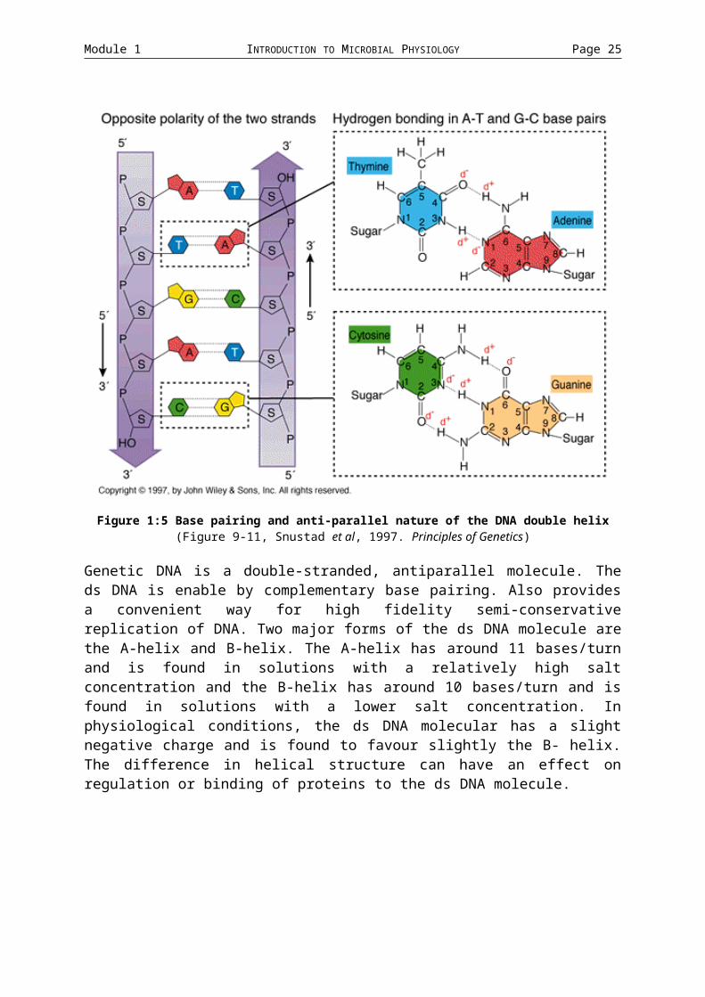
The DNA bases bind in a purine: pyrimidine structure by complementary hydrogen bonding. The position of the hydrogen bonding define which base binds to which i.e. A=T and CG.

Module 1 INTRODUCTION TO MICROBIAL PHYSIOLOGY Page 19
Figure 1:5 Base pairing and anti-parallel nature of the DNA double helix(Figure 9-11, Snustad et al, 1997. Principles of Genetics)
Genetic DNA is a double-stranded, antiparallel molecule. The ds DNA is enable by complementary base pairing. Also provides a convenient way for high fidelity semi-conservative replication of DNA. Two major forms of the ds DNA molecule are the A-helix and B-helix. The A-helix has around 11 bases/turn and is found in solutions with a relatively high salt concentration and the B-helix has around 10 bases/turn and is found in solutions with a lower salt concentration. In physiological conditions, the ds DNA molecular has a slight negative charge and is found to favour slightly the B- helix. The difference in helical structure can have an effect on regulation or binding of proteins to the ds DNA molecule.

Module 1 INTRODUCTION TO MICROBIAL PHYSIOLOGY Page 20
Figure 1:6 A-DNA and B-DNANucleoid The nucleoid in bacteria is present in a covalently closed circular structures (as well as being double-stranded and anti-parallel. When isolated the E. coli chromosome can be as long as 1 mm, but it is crammed into a cell around 6 um in length. Although it is extremely thin, the DNA molecule is compacted even further within the cell. The nucleoid is confined to ribosome-free areas within the cell. It is this compacted chromosome that is termed the nucleoid. The nucleoid structure is formed and maintained by topoisomerase. Topoisomerases Just image a rubber band, untwisted, the natural circular shape is evident. Cut it so it is linear, twist it until it bunches up, and then glue its ends together. Now the circular structure is not so evident, and the space required is much less. DNA is compacted much like this twisted rubber band. Topoisomerases supercoil the DNA into this type of structure. Positive supercoils are created when the DNA molecule is twisted in the direction of the DNA helix and negative supercoils occur when the DNA molecule is twisted in the opposite direction.
There are two forms of topoisomerases in E. coli. Type I relax negatively supercoiled DNA by breaking the phsophodiester bond on one strand, and allowing the other strand to swivel around before resealing the break. No energy is required. Type II topoisomerase (include DNA gyrase) require the input of energy to introduce negative supercoils. They relax in the absence of energy by breaking both strands, passing part of the loop through the break. The supercoiling places undue forces on the chromosome, and proteins and RNA are both used to stabilize the structure.
The E. coli chromosome is isolated as a negative supercoiled helical structure, and has between 30 and 100 negatively supercoiled loops. These loops are treated as separate topological domains, and there are sites at the end of each domain that limit the rotation and define the boundaries of each unit. Nicking one loop will relax it, but have no effect on the others. The boundary sites are formed by protein interactions with the DNA. DNA gyrase and Topoisomerase II are thought to be involved, as well as ribosomes and HU proteins (involved with transcription). Coupled transcription/translation/translocation will also tether domains (to the membrane - may be a possible mechanisms for genome division in cell replication). Supercoiling can regulate genes and gene families by modulating the state of supercoiling (less supercoiling, more transcription). Each domain can have different regulatory attributes.

Module 1 INTRODUCTION TO MICROBIAL PHYSIOLOGY Page 21
Figure 1:7 Activities of DNA topoisomerase II(Figure 10-24a, Snustad et al, 1997. Principles of Genetics)
DNA Replication. The genome of E. coli is a circular ds molecule with each strand having opposite polarity. The complementary nature of the DNA molecule allows DNA replication to proceed bidirectionally from a single starting point. Replication of DNA requires a DNA-dependent DNA polymerase, template DNA, a primer molecule with a free 3'-OH end and Mg2+. DNA is replicated in a semi-conservative manner and occurs in the 5' 3' direction. After replication the two chromosomes are linked due to topological constraints. The enzyme DNA gyrase separates the two concatamers into separate chromnosomes and occurs at a rate of 800 bp with an error rate of 1:1010
bp. In E. coli there are three types of polymerase enzymes. All synthesize DNA in the 5' 3' direction (add bases on the 3' end) and require the 3'-OH end of a primer molecule.DNA Pol I: Binds Zn2+, 3' 5' exonuclease, 5' 3' exonucleaseDNA Pol II: 3' 5' exonucleaseDNA Pol III: 3' 5' exonuclease, ss specific 5' 3' exonuclease.Replication of both strands happens simultaneously. It "appears" that one strand is synthesized 3' 5'. However, one strand is synthesized continuously, the other discontinuously (leading and lagging strands respectively).
DNA is replicated through three steps
Initation and Regulation
Elongation
Termination and Partitioning Initiation and Regulation How can a cell regulate how often its DNA is replicated??
In E. coli, it is a complicated process involving inhibitors, effectors and methylation. Occurs at 83 min on the E. coli chromosome (relative to the thr locus). This region is relatively A/T rich (requires less energy than C/G regions). It also contains RNA polymerase binding sites that allow primer synthesis. 83 min binds to membrane, but

Module 1 INTRODUCTION TO MICROBIAL PHYSIOLOGY Page 22
only transiently, so it is thought that the membrane does not play a large role in DNA replication.
Initiation starts during growth with a slow dilution of a negative inhibitor IciA (other inhibitors may be involved). The positive effector (DnaA) bind to 9-mer regions called DnaA boxes. This allows the formation of an open complex of 13 bases. Two more proteins (DnaB and DnaC) form a prepriming complex. DnaG (Primase) or RNA Pol. Then prime this site with an RNA primer. DNA Pol III binds and starts replication. The new strand is not methylated. The hemi-methylated origin associates with the membrane and allows a synthesis burst of negative inhibitor IciA (and others). The origin then detaches from the membrane, and becomes methylated by DNA methylase. Cell growth occurs, diluting the negative inhibitors.Elongation The strand separation started by the Dna molecules are aided by helix unwinding and destabilizing proteins. The helix unwinding and destabilizing proteins protect the ss DNA from intracellular nucleases. The unwinding of the DNA results in positive supercoils, so DNA gyrase relaxes the DNA by introducing negative supercoils. A protein assembly called a primosome synthesizes more primers on the ss DNA. DNA Pol III binds to the fre 3'-OH and synthesizes new DNA. DNA Pol III dissociates from the DNA when it hits a new RNA primer. DNA Pol I replaces the RNA primer with its 5' 3' exonuclease andits 5' 3' polymerase activities. DNA ligase seals the nick between the 3'-OH of the last nucleotide (made by DNA Pol I and the 5'-phosphoryl of the adjacent segment. DNA Pol III acts in a dimer form. The enzyme follows the leading strands, forming Okazaki fragments on the lagging strand. DNA Pol II can bind a primer, synthesize 1000 bp, and dissociate every second.
DNA Pol catalyse the elongation reaction
dNTP (Dna Pol, Mg2+, template, 3'OH) new DNA + P~P (pyrophosphate). The phosphodiester bond between bases is formed from the energy released by the splitting of the high-energy phosphate bonds present in the dNTP.
To ensure that this reaction continues in the forward direction, the amount of P~P in the cell must be kept as low as possible. This is accomplished by the conversion of P~P to 2Pi by pyrophosphatase.Termination and Partitioning As replication is bidirectional, termination would logically occur on the opposite side to initiation. Research has shown this to be the case. Termination occurs between the regions of 28 and 35 min. Two sites are involved in termination: T1 and T2. T1 (28 min) allows clockwise travelling forks to pass, but not counter-clockwise forks. T2 (35 min) allows counter-clockwise forks, but not clockwise forks to pass. Termination requires the TUS (termination Utilization Substance) protein that maps near T2. After replication, the chromosome is a linked concatamer, and requires the action of Type II Topoisomerase to split them into individual chromosomes.
Partitioning of the chromosomes require a protein (MukB). MukB attach to the chromosome with the aid of protein synthesis. It then "walks" along a cytoskeleton-like protein filament, separating the chromosomes.

Module 1 INTRODUCTION TO MICROBIAL PHYSIOLOGY Page 23
RNA AND TRANSCRIPTIONDNA is transcribed to RNA. RNA in cells is found as a single stranded transcript. The single strand allows for hydrogen bonding with itself, and with other RNAs, proteins and DNA. Transcription of RNA requires a DNA-dependent RNA Polymerase and uses rNTP's not dNTP's.
Figure 1:8 Deoxyribonucleotides and ribonucleotides
RNA Polymerase RNA Polymerase in E. coli consists of at least four peptide chains designated , , ' and . The RNA Pol. holoenzyme is made up of a core enzyme (2 x , and ') and the unit. The core enzyme binds randomly, and synthesizes random lengths of RNA. The holoenzyme binds at specific sequences and synthesizes specific lengths of RNA.
The sub-unit aids assembly of and ' into the core enzyme. The sub-unit is the catalytic site of RNA synthesis, and contains the binding sites for substrates and products. Rifampicin interferes with the sub-unit and stops transcription. The ' sub-unit is involved in DNA binding, and the sub-unit is involved in promoter region.
DNA is double-stranded with anti-parallel complementary strands. The positive (or coding) sense strand is the strand that ghas the identical sequence to the transcribed RNA. The negative (or antisense or anticoding) strand has the complementary sequence to the RNA. RNA is transcribed from the negative strand. Upstream and downstream refer to regions of the DNA relative to the motion of the RNA polymerase. Promoters are sequences of DNA that are generally found upstream of structural genes. The RNA Polymerase moves 3' 5' along the DNA, and synthesizes RNA 5' 3'. Upstream, therefore is 3' and downstream is 5' on the DNA molecule.The Transcription of RNA involves
Initiation
Elongation
Termination

Module 1 INTRODUCTION TO MICROBIAL PHYSIOLOGY Page 24
Figure 1:9 Stages of transcription
(Figure 9-10, Snustad et al, 1997. Principles of Genetics)
Initiation Initiation of transcription involves the recognition of a site on the DNA called the promoter region. The promoter region consists of two sequences. (ASIDE: Highly conserved sequences found in different species are generally referred to as "consensus sequences".) The consensus promoter sequences can be found at -10 and -35 from transcription start (designated +1).
The -35 region is known as the Recognition Site. 5'-TTGACA-3' 3'-AACTGT-5'The -10 is known as the Pribnow Box.5'-TATAAT-3' 3'-ATATTA-5'.Proteins and nucleic acids interact via base-specific groups that can be recognized in minor or major grooves. RNA Polymerase interacts with groups in the major grooves. It recognizes the -35 recognition site from the -10 region, and then forms a stable but closed promoter complex by moving to the -10 region.
[Recall B helix has around 10 bp/ turn]
Conversion to the open promoter complex requires unwinding of around 1 DNA helix from the middle of the Pribnow Box to just beyond the middle of the initiation site. Unwinding of the DNA allows tighter binding of the RNA Pol to the DNA, and intitiation of synthesis. Initiation ends after formation of the first internucleotide bond. The first nucleotide is generally purine. The rNTPs provide the energy for the internucleotide bonds by their high energy diphosphate bond.
Module 1 INTRODUCTION TO MICROBIAL PHYSIOLOGY Page 25
Promoter Function Negative supercoiled DNA is a much better promoter than relxed DNA. May
have something to do with the torsional stress present makes certain ares of the DNA easier to separate.
Supercoiling can affect expression. Topoisomerase I and DNA gyrase can affect supercoiling in localized areas, facilitating transcription of some genes, retarding others.
There are numerous sub-units. The different sub-units can affect the specificity of the RNA Polymerase. E.g -70 is normally present in E. coli cells, but -32 is induced and used whens cells undergo heat-shock. -32 preferentially produces RNAs for proteins that aid in surviving stress. All sub-units have four areas of a highly conserved nature.
Two classes of transcriptional activation factors. Type I bind upstream of the promoter, Type II activators overlap the promoter region.
Figure 1:10 Structure of a typical E. coli promoter(Figure 11.11, Snustad et al, 1997. Principles of Genetics)
Elongation After formation of the first transcript's first 8 or 9 bases, the RNA polymerase undergoes a conformational change, decreasing its affinity for the factor. The factor is released, allowing it to bind to free core-enzymes. Elongation occurs at rates of 30-60 bases/sec. It involves four steps: rNTP binding, bondformation, pyrophosphate release, and translocation along the DNA in the 3' 5' direction.
Movement of the RNA polymerase involves melting of the DNA ahead of the transcription bubble, as well as reformation behind. The transcription bubble generally occupies 17 base pairs. Elongation speeds can vary. Sites where the rate is low are called pausing sites. Pausing sites are generally found in regions 10 bp upstream of DNA sequences with G/C concentration or 16-20 bp upstream of regions with dyad symmetry.
ASIDE: Dyad symmetry applies to two closely spaced regions on a single strand that are capable of base pairing with each other. They can form hairpin structures.
Figure 1:11 Features of the E. coli RNA polymerase transcription site(Figure 11-12, Snustad et al, 1997. Principles of Genetics)

Module 1 INTRODUCTION TO MICROBIAL PHYSIOLOGY Page 26
Termination Termination involves cessation of elongation, transcript release from the transcription complex, and dissociation of the RNA polymerase from the template. Termination can occur in one of two ways.
Rho-Independent Termination
Regions with high G/C or dyad symmetry in the RNA allows the formation of a stem and loop structure about 20 bp upstream of the 3-OH and a stretch of 4-8 uracil bases. The RNA pairing causes pauses and disrupts the 5'-portion of the RNA-DNA hybrid helix. The remaining 3' end of the RNA-DNA hybrid containing the poly-U tail is highly unstable with poly-A in the DNA. This causes the RNA transcript to dissociate from the DNA within or distal to the poly-U string.
Figure 1:12 Dyad symmetry and the formation of transcription terminators(Figure 11-13, Snustad et al, 1997. Principles of Genetics)
Rho-Dependent Termination
Rho-dependent termination requires the protein Rho, and only occurs at strong pausing sites that are located at a distance from the initiation site. There is no consensus sequence associated with this type of termination. The Rho protein binds to a single-stranded region of the RNA. As the RNA polymerase pauses, the Rho protein translocates along the transcript. The movement of the Rho protein is dependent on ATP hydrolysis. When the Rho protein contacts the RNA polymerase, it assocaites with it. This association, coupled with the activation of a RNA/DNA helicase causes the transcript and RNA polymerase to dissociate from the template.RNA Turnover RNA is classed as either stable or unstable. Stable RNA includes rRNA and tRNA. mRNA is unstable RNA. Of its RNA, E. coli has approximately 70-80% rRNA, 15-25% tRNA, and 3-5% mRNA. Factors that contribute to the stability of RNA is ability to associate with ribosomal proteins, and extensive secondary structure. The extensive secondary structure protects the 5' terminus from ribonucleases.
Unstable RNA has an average length of 1200 bp and a life span of around 40 seconds at 37 C. Degradation of unstable RNA occurs in a 5' 3' manner, but researchshows that all exoribonuclease act 3' 5'. Degradation occurs by an initial random endonucleolytic even at the 5' end, stopping ribosomes from binding. The remaining message is progressively exposed as the ribosome move towards the 3' terminus. This exposed mRNA transcript allows further random endonucleolytic events. The small pieces can then be attacked by the 3' 5' exoribosnucleases. The stability of mRNA is enhanced by stem-loops at the end or beginning of the transcript (at 3' end they inhibit 3' 5' exonucleases, at the 5' end they inhibit endonuclease association).

Module 1 INTRODUCTION TO MICROBIAL PHYSIOLOGY Page 27
RNA Processing All stable RNA and some unstable RNA's need to be process prior to use e.g. the rRNA transcription units. In E. coli there are seven rRNA gene groups. They are always transcribed 5'-leader-16S rRNA-spacer-23S rRNA-5S rRNA-trailer-3'. The spacer always includes a tRNA gene. This type of transcript is called polycistronic as it contains more than a single transcript. The complete transcript is processed to form a mature 16S rRNA, 23S rRNA, 5S rRNA and a tRNA molecule. Most tRNAs are present in groups of up to 7 units (identical or different). To become fully functional, these transcripts need to but cut and modified.
There are four different types of RNA Processing.
The polycistronic RNA is separated into monocistronic units.
The mature 5' and 3' terminii are recognized, and the excess bases removed.
Terminal residues are added to those RNA molecules requiring them.
Bases or sugar units are modified.
There are many enzymes involved with post-transcription modification.
RNase P Removes 41 bp fragments from the 5' side of tRNA precursors. It recognizes 2 and 3 structures, not sequences. It requires a terminal CCA sequence. Catalytic RNA molecule (ribozyme).
RNase III Cleaves ds RNA and makes closely spaced ss breaks (1 per 15 bp). It recognizes ds RNA stem-loop structures.
RNase E Specifically cleaves 5S rRNA precursors from larger trasncripts. First cleaves between ss region of 5S precursor and the ds region, second cleavage between the ds region.
RNase D Involved in monomer removal distal to the CCA sequence within the precursor tRNA (mature 3'-OH terminus). The CCA-OH is required for amino acid activity on all tRNAs. It is a non-progressive 3'-exonuclease.
RNase F 3' endonuclease that exposes a 3'-OH that Rnase D acts upon.
tRNA nucelotidyl-transferase
Repairs tRNA molecules where the CCA sequence is missing. It sequenctially adds CCA to tRNAs
Others Methylases, pseudouridyllating enzymes, thiolases. Involved in the modification of the 3rd base of the anti-codon of tRNAs. Others aid in stress signals.
Module 1 INTRODUCTION TO MICROBIAL PHYSIOLOGY Page 28
PROTEIN SYNTHESIS: TRANSLATION DNA mRNA Proteins
Translation involves the three steps of
Initiation
Elongation
Termination
A given DNA sequences gives a defined protein sequence.
If there is only one base per amino acid, only four amino acids are possible. If two per amino acid, a maximum of 16 are possible (4 x 4). So three bases per amino acid is required (to give a possible 64 amino acid codes (4 x 4 x 4). This triplet code is degenerate (more than one triplet code per amino acid), but non-overlapping (only one amino acid per triplet). The degeneracy is caused by the variability in the last base differing.
Some triplet codes are important (Start: AUG-Met, Stop: UAG, UAA, UGA).
Figure 1:13 The Universal Code(Table 12-2, Snustad et al, 1997. Principles of Genetics)
tRNAIncorporation of amino acids requires the formation of an activated complex between an amino acid and a tRNA. The bonding is done by an aminoacyl tRNA synthetase (aka aminoacyl tRNA ligase).
Each amino acid has its own synthetase and specific tRNA increases specificity of overall reaction.
Each tRNA can recognize a triplet codon by its anticodon (codon recognition site), can recognize its own synthetase by its ligase recognition site. Each tRNA also consists of an amino acid attachment site, and a ribosome recognition site.
The amino acid attaches to the 3'-OH terminus (All tRNA have a 3' terminal CCA, and a 5'-G) (Recall modifying enzymes for RNA).
Each tRNA is about 80 bases long, and has a higher content of modified bases (e.g. inosine and pseudouridine) and methylated bases. These generally occupy specific positions.
Secondary structure is a cloverleaf design with three major loops and 1 minor loop. Loop 1 has a dihydroxyuridine (DHU). Loop 2 has the codon recognition site. Loop 3

Module 1 INTRODUCTION TO MICROBIAL PHYSIOLOGY Page 29
is the minor loop, and is sometimes completely lacking. Loop IV contain ribothyimylate, pseudourine and cytosine and is called the T C loop.
The recognition of tRNA to mRNA is dependent on base pairing. Some tRNA molecules have modified bases in the anticodon that can lead to sloppy or "wobble" pairing.
Charging of tRNA
Charging of tRNA is a two step process.
Amino Acid + ATP + tRNA synthetase AA-AMP-Synthetase (Aminoacyl AMP complex) + ppi
AA-AMP-Synthetase + tRNA AA-tRNA + AMP + Synthetase.
The synthetase must have two reognition properties. It must be able to differentiate between differen amino acids, and it mus be able to recognise a specific tRNA molecule. This ensures that an amino acid is charger onto its repective tRNA. tRNA must recognize the mRNA codon correctly to ensure that the amino acid is placed in its proper position in the polypeptide.
Ribosome Structure
Prokaryote ribosomes are made up of two units. The 30S SSU (Small Sub Unit) is made up of 21 ribosomal proteins and the 16S rRNA molecule. The 50S LSU (Large Sub Unit) is made up of 31 ribosomal proteins and the 5S and 23S rRNAs.
In E. coli there are seven operons that code for the rRNA molecules. This reduces the effect of possible mutation in one operon (protective strategy).
Sequence studies of rRNAs is very important for inferring phylogenetic relationships.
Translation Initiation
A complex forms between the SSU and three intitiation factors (IF-1, IF-2, and IF-3). IF-1 and IF-3 prevent association of SSU and LSU if there is no mRNA present. The mRNA and fMet-tRNA (iniator tRNA) associate with with the SSU. The mRNA associates at the site that includes the AUG codon. The mRNA has a leader seqeunce for ribosome binding (Shine-Dalgarno 35-region). The Shine-Dalgarno base pairs to the 3' region of the 16S rRNA. It positions the start so it can bind to the fMet-tRNA, and allows differentiation between internal and start Met sites. IF-2 and GTP directs binding of the fMet-tRNA to the SSU. The complexing of IF-2: GTP to the SSU increases its affinity for the LSU, while decreasing its affinity for the IF-3. This frees IF-3 from the SSU. The complex now includes the SSU, IF-1, IF-2: GTP, mRNA and fMet-tRNA. The binding of fMet-tRNA to the mRNA also determines the reading frame for the mRNA.
After the loss of IF-3, the SSU binds to the LSU. With binding, the GTP complexed to IF-2 is hydrolyzed. The binding of LSU to SSU is not dependent on the hydrolysis of GTP, but the IF-1 mediated release of IF-2 is.

Module 1 INTRODUCTION TO MICROBIAL PHYSIOLOGY Page 30
The ribosome has three sites for tRNA attachment and movement.
The A-Site (Aminoacyl-tRNA binding site) accepts the incoming AA-tRNA (decoding).
The P-Site (Peptidyl-binding site) holds the prior tRNA with the nascent polypeptide still attached.
The E-site (Deacylated tRNA molecules)
The initator tRNA, fMet-tRNA, associates directly to the P site, bypassing the A site entirely.
Figure 1:14 The initiation of translation in E. coli(Figure 12-15, Snustad et al, 1997. Principles of Genetics)
Translation Elongation
Elongation continues at a rate of 16 triplet-code directed amino acid residues / second.
The fMet-tRNA is in the P site, and the A site is free. The 5S rRNA recognizes a sequence in the TC loop of the tRNA aiding in theaa-tRNA binding to the A-site. EF-T and GTP stimulate the binding of the new AA-tRNA.
ASIDE: EF-T is made up of two proteins, EF-Tu (44kDa, unstable, 5-10% of cellular proteins) and EF-Ts (30kDa, stable)
EF-T + GTP EF-Tu: GTP and EF-Ts
EF-Tu: GTP can bind to all AA-tRNA but fMet-tRNA.
EF-Tu:GTP:AA-tRNA+A site EF-Tu:GDP+AA-tRNA:A Site.
Result of GTP hydrolysis is the release of EF-Tu from the ribosome. Peptide bond formation is not dependent on GTP, but on the release of EF-Tu from the ribosome. EF-Ts is important for the recycling of EF-Tu, but is not required for its release from the ribosome.
Peptide Bond Formation
The peptide bond formed between the amino acid group of the A site AA-tRNA and carboxyl group of the P-site aa-tRNA, catalyzed by the peptidyl transferase located in the LSU.
Translocation

Module 1 INTRODUCTION TO MICROBIAL PHYSIOLOGY Page 31
Translocation requires movement of the peptidyl-tRNA from the A to P site, and movement of the mRNA by one codon. It is facilitated by EF-G (80kDa; G=GTPase) and GTP hydrolysis. EF-G binds to the same site as EF-Tu. For one peptide bond formation, there is the hydrolysis of 2 GTP (EF-TU release, and EF-g aided translocation). Once the GTP is hydrolyzed, EF-G is released.
Summary
1. Nascent peptide at P site.
2. Incomin gAA-tRNA binds at A.
3. Peptide bond formation, nascent peptide transferred to AA-tRNA in A site
4. tRNA in P E, peptide-tRNA in A P.
5. A free, new AA-tRNA binds, reduces E site affinity for tRNA, releasing it.
Figure 1:15 Polypeptide chain elongation in E. coli(Figure 12-17, Snustad et al, 1997. Principles of Genetics)
Translation Termination
When UAA, UGA, or UAG moves into A site, two of three peptide release factors bind. RF1 or RF2 bind near the A site (RF1 recognises UAA or UGA, RF2 recognises UAG). RF3 binds elsewhere on the ribosome and hydrolyzes GTP. The hydrolysis of GTP, combined with the activity RF1 or RF2-initiated activity of a peptidyl transferase, hydrolyzes the bond between the nascent peptide and the tRNA in the P site. The peptide, tRNA, and LSU dissociate from the SSU and mRNA molecule. IF-1 and IF-3 bind to the SSU molecule, which can then either dissociate from the mRNA, or translocate and find a new start codon on the mRNA.
Figure 1:16 Polypeptide chain termination in E. coli(Figure 12-19, Snustad et al, 1997. Principles of Genetics)

Module 1 INTRODUCTION TO MICROBIAL PHYSIOLOGY Page 32
Post-Translation
Removal of the fMet start amino acid can be done in two ways. The formyl group can be removed by methionine deformylase, or the entire amino acid (fMet) can be hydrolyzed by Methionyl-Amino-Peptidase. The longer the side chain of the second amino acid, the less likely that MAP will remove the initial fMet.
Coupled Transcription-Translation
As there is no membrane separating the two processes of transcription and translation in prokaryotes (in eukaryotes the nuclear membrane separates these two process), the coupling of trasncription and translation is allowed. Coupling involves the binding of ribosomes to the incompletely transcribed mRNA. As rates of transcription are much faster than translation, long regions of unprotected mRNA can be formed. To slow down the rate of transcription so the ribosomes do not fall that far behind, puase sites are present. This ensures that no long regions of mRNA are present that endonucleases can attack.
1.3 STRUCTURAL ASSEMBLY
Structures of Proteins Amino Acids are classed according to their nature. Broken into the classes Aliphatic, Cyclic, Aromatic, Basic, Acidic and Hydroxyl or Sulfur containing side-chains.
Each amino acid is coded by a triplet code translated from RNA.
The aliphatic amino acids have hydrophobic side chains, the others are less polar in varying degrees. The basic and acidic amino acids are mostly involved with the net charge at cellular pH.
The different structural and charge properties determine the structure and activity of the final protein.
Hydrophobic amino acids are centered, while hydrophilic amino acids tend towards the surface of proteins.
The structures of proteins are dominated by the nature of the peptide bond between amino acids.
The Primary structure is the amino acid sequence of the proteins.
The Secondary structure of proteins is governed by the interactions between individual amino acids. The secondary structure is formed by the conformational entropy of the chain, charger to charge interactions, Hydrogen bonding, weak van-der Waals forces, hydrophobic effects, and disulfide bonds.
The Tertiary structure of proteins is the general structure described in gross structural terms. The chains of amino acids, after looking at their secondary structure, form recognisable shapes and structures. The most noticeable of these are -helices, -sheets (parallel or anti-parallel), and -turns. Groups of these higher-order structures determine the tertiary structure.

Module 1 INTRODUCTION TO MICROBIAL PHYSIOLOGY Page 33
The Quaternary structures describe the aggregation of more than one protein. For example, the Quaternary structure of RNA polymerase (the holoenzyme) is 2x sub-units, -unit, '-unit, -unit, and the -factor. More than one protein makes up the active enzyme. The quaternary structure of the enzyme is the different proteins that make up the active enzyme.Post-SynthesisLast Week, the modification or removal of the initial fMet amino acid was discussed, but apart from this, some proteins may need further modification to enable them to become fully active in the cell.
Some proteins will automatically adopt their active structure. Others need help. If they do not fold properly, the peptide chain remains inactive.
How do these peptide chains become active?
They need some other protein to aid them to fold properly. Known as chaperones (or chaperonins), this group of proteins is used to help folding or translocation of other proteins. Interactions including hydrophobic, charge-to-charge, etc, enable the chaperonin to bind, and then aid the folding of the protein. Chaperonins may also inhibit folding until the protein is ready to become fully functional. Chaperonins also aid in stopping protein becoming denatured when the cell enters a hostile environment (e.g. heat is raised slightly, new proteins are expressed that stop the denaturation of other proteins), How are proteins secreted? Much of the proteins produced intracellularly are involved in some way with the cell membrane or wall. Some proteins are secreted to digest large macromolecules. All secretory pathways include the use of a short leader sequence in front of the active protein primary structure. Membranous proteins that then aid in the translocation recognize this leader sequence. During translocation, the leader sequence is cleaved from the remaining protein. There are four ways that the export of proteins occurs. These are known as the General Secretory Pathways. The first two are co-translation translocation (spontaneous leader sequence association with docking proteins, or pausing of translation with Signal Recognition Particles). The other group is chaperonin-dependent with the chaperone interaction translationally linked or post-translationally linked.
Membrane proteins have a characteristic shape of hydrophilic ends with a hydrophobic torso. The hydrophobicity of the molecules is generally enough to provide the energy required for insertion.
The protein can be either inserted into, or exported past the cell membrane.
Last week, polyribosomes introduced the idea of couple transcription-translation. Now, it has been shown that translation can now be linked via translocation to the cell membrane. The genome of the cell now has a way of being adhered structurally to the cell wall. This structural adherence now enables the chromosome to be divided into separate topological unit that can supercoil independently of each other.Degradation of Proteins Abnormal Proteins: Intracellularly, abnormal proteins form aggregates (due to incorrect folding caused by incorrect primary structure). These proteins need to be

Module 1 INTRODUCTION TO MICROBIAL PHYSIOLOGY Page 34
broken down, otherwise the cell will be over-run by these abnormal proteins. An ATP-dependent endoprotease (translated from the lon gene) recognizes abnormal proteins or aggregates by their unfolded nature and catalyzes the first degradation step. The smaller peptides are then further broken down into di- and tri-peptides, and then even further to constituent amino acids. To stop the Lon protein from degrading cellular proteins, it recognizes unfolded or incorrectly folded protein structures. In addition, each degradation step results in its autoinactivation until a new substrate is found.
Normal Proteins: Some proteins are required to have a very short half-life. Many of the regulatory proteins need to be unstable to correctly and quickly react to stimuli. Certain sequences, conformations, or chemically modified amino acids are recognized, and are involved in the recognition of intracellular proteases. Lipids The lipids involved in biological systems have the general shape of a polar head with a hydrophobic hydrocarbon chain. The hydrocarbon chain is saturated (with hydrogen atoms) if there are no double bonds present between the carbon atoms. If there are double bonds present, the lipid is said to be unsaturated (e.g. mono and polyunsaturated fats in oils and margarines) Saturated chains are fairly constrained with repsect to their conformation (straight). Unsaturated chains have "kinks" caused by the double bond. The more kinks, the less structured and stable the hydrophobic tails are.
The fatty acid is the simplest biological lipid. Triglycerides are formed by the condensation of three fatty acids together, forming a polar head with three hydrophobic tails.
The main lipid-like structure found in biological systems is the phospholipid. It has two hydrophobic hydrocarbon chains connected to a phosphate poplar head.
Lipids in aqueous solutions tend to form three structures. On the surface, a monolayer is formed. Is solution, micelles are formed in low concentrations of lipids (polar heads on the outside). As the concentration increases, these micelles form bilayers. It is this phospholipid bilayer that forms the membranes of all cells.
There is a structural difference between the phospholipids of Archaeal, bacterial and eukaryotic cells. Bacterial and Eukaryotic cells have ester-linkages in the polar head, while archael cells have ether links.
In bacteria, the cell membrane forms a fluid structure with over 200 associated proteins. Many of these proteins are involved with the synthesis and maintenance of the cell wall and membrane. Others are involved in the degradation of macromolecules and transport. The export of proteins has been shown to exist, and these proteins can be targeted to the outer membrane, periplasmic space, or inner membrane. They can be integral or peripheral.
The phospholipids are produced by the addition of two fatty acid molecules (RCOOH) to gycerol-3-P. The phospholipids are inserted first into the inner leaflet, and are then transferred to the outer leaflet of the bilayer. If the phospholipid "flip-flopped" to the outer layer, it would require considerable energy to force the polar head through the

Module 1 INTRODUCTION TO MICROBIAL PHYSIOLOGY Page 35
hydrophobic core. However, the translocation process seems to be energy-independent, because it takes place in the absence of metabolic energy.
To do this, it has been proposed that the membrane is continuous via hairpin bends near transmembrane proteins.
The simple diffusion of phospholipids throughout the membrane would infer that the concentration of the different phospholipids in the inner and outer leaflet would be identical. However, this is not the case. It has been shown that different phospholipids are in different concentrations in the two leafs of the bilayer. This must mean that there is a selection process that requires specific lipid-lipid or lipid-protein interactions.Synthesis of the Gram Positive Cell Wall: Peptidoglycan Synthesis
Peptidoglycan is an alternating sugar unit motif connected by inter-peptide bridges. The two alternating sugar residues are N-acetylglucosamine and N-acetylmuramic acid. N-Acetylmuramic acid is actually a conversion product of N-acetylglucosamine. The Pentapeptide Bridge is added to N-acetylmuramic acid. Some interesting features of the interpeptide bridge is that contains naturally occurring D forms of amino acids (biological systems on the whole use L-forms of molecules). The peptide bonds formed between the amino acids are not formed by ribosomes, but by specific ligases that add the amino acids individually.
The peptidoglycan is a layer of the cell wall that interacts with the environment, and as such it needs to be hydrophilic. How does the hydrophilic peptidoglycan molecules pass the hydrophobic inner cell membrane?
The peptidoglycan unit takes advantage of a lipid carrier known as undecaprenyl phosphate or the C55 carrier. The formation of the peptidoglycan layer takes place in three phases.
Stage 1 (Occurs in the cytoplasm): N-acetylglucosamine is converted to N-acetylglucosamine: UDP (hydrolysis of UTP), and then to N-acetylmuramic acid: UDP (addition of phosphoenolpyruvate). Individual amino acids are added to N-acetylmuramic acid: UDP in the order of L-ala, D-glu, L-lys, and finally the dipeptide D-ala-D-ala. The D-isomers are formed by a racemase.
Stage 2 (In the membrane): Once the pentapeptide has been added, the N-acetylmuramic acid is bound to the carrier lipid undecaprenyl phosphate. N-acetylglucosamine is then bound to the N-acetylmuramic acid pentapeptide. Once the two sugar molecules are bound together, there are released on the other side of the membrane.
Stage 3 (Extracellular side of membrane): The individual peptidoglycan residues are then polymerized into the glycan chains, and the transpeptide bridges are formed. The formation of these transpeptide bridges releases the last D-ala residue.
The antibiotic of choice for use against gram positive bacteria are the -lactams (penicillin). This is because penicillin affects the synthesis of peptidoglycan in the cell wall. How does penicillin affect peptidoglycan synthesis? Many of the enzymes involved in the final steps of transpeptidation, glycan chain formation and undecaprenyl phosphate recycling are inhibited by penicillin. These enzymes and

Module 1 INTRODUCTION TO MICROBIAL PHYSIOLOGY Page 36
proteins are called (funnily enough) Penicillin Binding Proteins (PBP's). All PBP's are found outside the inner membrane which enable penicillin to act on them (as it cannot pass through the membrane), and why gram-negative bacteria are unaffected (the outer membrane halts the passage of penicillin).
In gram positive bacteria, the outer wall is made up of a thick many-layered peptidoglycan. The outer layers are lost, and new inner layers are made to replace them. The continual synthesis of peptidoglycan and the loss of exterior peptidoglycan gradually push the new peptidoglycan outward.
In gram-negative, the peptidoglycan layer is much thinner, and protected by an outer membrane, so the cell's peptidoglycan synthesis role is much more of a maintenance role (and in cell division), than a continual synthesis. Teichoic acids. Gram positive bacterial cells produce a characteristic wall bound acid known as teichoic acids. These teichoic acids can be wall bound (wall teichoic acids) or membrane/wall bound (Lipoteichoic acids).
Importantly to note is that the wall teichoic acids are formed by the polymerization of ribitol phosphate or glycerol phosphate molecules and are joined by a phosphodiester link. They are covalently linked to the peptidoglycan through the N-acetylmuramic acid residues. The hydroxyl group of the ribitol or glycerol sugar base can be bound to sugars, amino sugars, or amino acids allowing for a wide variety of structures. These structures then provide the antigenic specificity of individual strains.
Lipoteichoic acids are generally 16-40 phosphodiester linked glycerophosphate residues bound to a membrane anchor (usually a glycolipid or glycophospholipid).
All teichoic acids are capable of scavenging divalent cations. This is thought to be one of their major functions. The scavenging role allows them to concentrate divalent cations (Mg2+) at the cell surface, so there is a ready supply.The Gram Negative Cell Wall The peptidoglycan layer in gram-negative bacteria is produced in an identical fashion as the gram-positive bacteria. The undecaprenyl phosphate lipid carrier is used, while the N-acetylglucosamine and N-acetylmuramic acid pentapeptide is assembled in the cytoplasm. The outer membrane phospholipids are assembled in the cytoplasm and inserted first into the inner membrane. Diffusion into the outer membrane occurs via the continuity of the inner and outer membrane (Bayer Junctions). Lipopolysaccharides are an important feature of the gram-negative cell wall.Lipopolysaccharides The lipopolysaccharides of the gram-negative bacteria are made up of three units. Lipid A (the part of the molecule that embeds itself into the membrane, the core polysaccharide (essentially the same between all gram negative bacteria), and the O-antigen (a variable polysaccharide region).
Two parallel processes that occur in the cell membrane synthesize the Lipopolysaccharides (LPS). The precursors for both processes are assembled at the inner membrane. Lipid A acts as a carrier as well as the primer site for the core

Module 1 INTRODUCTION TO MICROBIAL PHYSIOLOGY Page 37
polysaccharide addition. The variable O-antigen is synthesized on the lipid carrier undecaprenyl phosphate (the same as used in peptidoglycan transport and synthesis). Once synthesized on the inner surface, there are transported across the membrane by their carriers. The PMF may drive this transfer. At the outer surface, the O-antigen is added to the Lipid A-core polysaccharide by a transfer enzyme. Individual LPS molecules then condense by mutual attraction into a two-dimensional array.
ASIDE-Proton Motive Force
The cell during metabolism creates the proton-motive force (PMF). The metabolic activity of the cell is used to pump hydrogen protons out of the cell membrane. The difference in both hydrogen proton concentration and charge generates an electrochemical gradient, and it is this electrochemical gradient that is the proton motive force. The PMF is used to transport molecules or generate ATP for the cell.Lipoproteins The outer membrane of gram negative bacteria also has a great number of small proteins called lipoproteins. The protein has a 20 amino acid leader sequence followed by a 38 amino acid peptide. Post translation of this protein includes the leader sequence being cleaved, the addition of three fatty acids, inserted into the inner leaflet of the outer membrane, and finally 1/3 of the lipoprotein molecules are bound to the peptidoglycan layer through their carboxyl terminus.Other proteins Most other proteins that are bound to or within the phospholipid bilayer undergo self-assembly after translocation past the inner membrane.Flagella Assembly. The synthesis of the bacterial flagella involves over 40 genes. The hollow structure comprised of a basal body, a hook, and a tail consisting of many protein sub-units called flagellin. The basal body consists of a number of rings (2 in gram positive, 4 in gram negative) and a rod that the flagella attach to. The assembly of the flagella is a bottom-up process. Starting with the M (motor) and S (stator) rings (found in both gram-positive and gram-negative bacteria), there are inserted into the cytoplasmic membrane. The attachment point for the flagella, the rod is added and capped. In the case of gram-negative bacteria, the P, then L rings (acts like bearings) are added. After this, the hook is formed, and flagellin proteins are added to the tail of the flagella, travelling through the incomplete structure. After the full flagella length is achieved, motor proteins are inserted into the cytoplasmic membrane. These motor proteins are driven by the PMF. Approximately 1000 protons are required per revolution, and the flagella can rotate up to 1200 rpm. This equates to 100 um/sec, or relatively speaking, twice as fast as a cheetah.Pili and Fimbriae The assembly of fimbriae and pili is less known than the assembly of flagella. Pili are straight protein rods that are involved in DNA transfer between cells. They are made of identical sub-units of the protein pilin. The proteins are capable of self-aggregation, forming a hollow tube. These rods are hollow, but the diameter of the central channel is too small for the pilin protein. The pilin proteins are synthesized in the cytoplasm and then cotranlsationally translocated across the membrane. Individual sub-units are added to the base, extending the pili from the cell.

Module 1 INTRODUCTION TO MICROBIAL PHYSIOLOGY Page 38
Even less is known about fimbriae formation. Fimbriae often have special proteins attached to the end of the rods called adhesions. These proteins aid in the attachment process. But whether they are extruded first, then pushed by the synthesis of the fimbriae, or do they attach after the fimbriae have been formed.The Glycocalyx. The glycocalyx is important physiologically speaking as it is of great importance to the survival of many bacteria. The same problem occurs with the synthesis of the glycocalyx as with the LPS. How does the cell export a large hydrophilic polymer across the membranes? With tightly associated glycocalyxes (or capsules), there is an association between the membrane and the polysaccharide. Again the same lipid carrier undecaprenyl phosphate is thought to be involved. Three repeating saccharide units are built up on the carrier in the cytoplasm. This trisaccharide is then transferred to the outer surface, then covalently bound to a nearby lipid-carrier-trisaccharide complex. In E. coli the polysaccharide is then added to a phospholipid (with the phospholipid losing a fatty acid). The polysaccharide is now associated with the outer membrane.
Another type of glycocalyx, the slime layers, are formed by extracellular enzymes. The enzymes are synthesized and translocated across the membranes. The polymers are formed without the expenditure of cellular energy or ATP.
The formation of capsules is generally continuous, and the completed polymer is sloughed off, and is repeated. The size of the capsule can vary due to the nature of the medium or temperature. Most extracellular polysaccharides are formed of carbohydrates, with the exception of some that are made of polymers of D-glutamate. The Motility of Flagellated Bacteria. Flagella can be positioned varyingly around the cell. The position of flagella can be polar, lophotrichous, and peritrichous. Bacterial with multiple flagella show the same type of movement as a single-polar flagellated bacterium. This is due to the helical structure of the flagella. There are two types of motion associated with the flagella. These movements are called the run and the tumble. When flagella are rotating counter-clockwise, their right-handed helical sense bundles them together to propel the cell in a single direction (called a run). A tumble occurs when the cell quickly and for a short period of time turns the flagella clockwise, forcing the bundle of flagella apart. In a clockwise rotation, the flagella cannot associate into a bundle, and so turn the cell about. The run can last for longer than 1 second, while tumbles are much shorter.
The question remains, how does a single cell effect these differences in rotation simultaneously, and how can it compare two spatially distant point w.r.t. attractants or repellants.
Let's start with the latter question. Looking at the size of bacteria, it would be impossible to be able to measure difference in concentration gradients using its leading and trailing poles. However, it would be able to differentiate by sensing the concentration gradient while running. And looking at the motion of flagellated bacteria, it seems this is the case. The times for runs going towards attractants are longer than runs away. Thus the cell must be able to sense concentration, and then "remember" to measure it again. The cell is responding to a change in concentration,

Module 1 INTRODUCTION TO MICROBIAL PHYSIOLOGY Page 39
not to an absolute value. So it must constantly be able to adapt to higher concentrations to be able to detect this change.
Research has shown that a group of proteins called methyl-accepting chemotaxis proteins are involved. They are able to "detect and measure" concentrations of chemotactic substrates in the medium. These proteins do this by becoming methylated in response to changes in concentration levels, and during the period of adaptation, gradually return to their unmethylated stated. A high level of methylation signals high concentrations of attractants, while low methylation means a low concentration of attractants, and a very senstive MCP to attractant concentration.
Three major MCPs are Tsr, Tar, Trg and they each respond to different attractants. A particular attract (either free or bound to a periplasmic binding protein) attaches to the periplasmic-exposed surface of the MCP. This attachment may produce a conformational change that exposes sites for methylation. Up to methyl groups can be added to each MCP. With each methylation, sensitivity to the attractant decreases (by 100-fold with full methylation). If the concentration does not increase, a chemotactic-specific methylesterase gradually removes these methyl groups (allows adaptation - desensitization).
The directional switch of the flagellar motor is controlled by the interaction of the MCP with the attractant, and a family of six proteins called Che proteins that transfer the signal from the MCP, to the flagella motor proteins.
Increase in attractant binding increases the methylation state of the MCP (caused by CheR). Increasing the methylation state has two effects on the MCP. Firstly, it becomes less sensitive to the attractant, requiring a higher concentration to enable the attractant to bind. Secondly, the higher the methylation state increases it ability to form a quaternary complex with two cellular proteins CheW and CheA.
CheA autophosphorylates when it is in the quaternary complex. After CheA is phosphorylated, it transfers the phosphate group to CheY. The phosphorylated CheY causes the flagellar motor to turn clockwise instigating a tumbling motion. CheZ dephosphorylates CheY, stopping the clockwise motion.
When the attractant binds to the MCP, it disrupts the formation of this quaternary complex, stopping the autophosphorylation of CheA, and allowing the flagella to turn counter-clockwise, and make a running motion.
Running is a competition between attractant binding the MCP and disrupting the quaternary complex, and the increase in affinity for the quaternary complex and the MCP.
The MCP is demethylated by CheB. CheB forms the feedback loop that resets the MCP for excitation after it has moved into a region of low attractant concentration.

Module 1 INTRODUCTION TO MICROBIAL PHYSIOLOGY Page 40
Questions
Short Answer Questions:1. What is an obligate aerobe?2. Distinguish between moderate and extreme thermophiles.3. Distinguish between bacteria and Bacteria4. What structures are associated with motility in bacteria?5. What does a DNA-dependent RNA polymerase do?6. Distinguish between bases, nucleosides and nucleotides.7. What is the nucleoid?8. What causes translation to terminate and give an example?9. What are the 4 groups to which secreted E. coli proteins can be placed?10.What are okazaki fragments?
Medium Answer Questions:1. Distinguish between the RNA polymerase core enzyme and holoenzyme.2. Describe two mechanisms of transcription termination.3. Diagram a Gram-negative cell wall.4. Diagram the central dogma (include the types of enzymes used).5. What is tRNA and what role does it play in translation?
Large Answer Questions:1. The E. coli chromosome, when stretched out is 1 mm long. How does it fit into a
cell less than 5 µm long?2. Outline the process of replication using a detailed diagram of the replication fork.3. Twenty percent of E. coli proteins are located outside the cytoplasm. Describe,
using suitable diagrams, the process(es) by which these proteins are secreted.4. Describe Escherichia coli and why it is used to study microbial physiology.5. Describe the process of transcription is detail.

MODULE 2
BACTERIAL GROWTH,ENVIRONMENTAL
EFFECTSAND
STRATEGIES

Module 2 BACTERIAL GROWTH, ENVIRONMENTAL EFFECTS AND STRATEGIES Page 42
Module 2: Bacterial Growth, Environmental Effect and Strategies
AIMS AND OBJECTIVES* To introduce the principles of bacterial growth and how bacterial enumeration
is determinated
* To examine environmental pressures exhibited on microorganisms and their specific strategies for dealing with these stresses
* To introduce the grouping of microorganisms based on environmental parameters
YOU SHOULD…* understand the processes of bacterial growth
* be able to describe the phases of bacterial growth
* be able to distinguish between methods of determining bacterial growth
* understand the effects of
nutrient levels temperature oxygen osmotic pressure pH
READING: Atlas, Principles of Microbiology Chapter 9
LEARNING EXERCISE find examples for each group discussed in this module

Module 2 BACTERIAL GROWTH, ENVIRONMENTAL EFFECTS AND STRATEGIES Page 43
Factors affecting bacterial growth: Temperature
pH
Avaliability of nutrients
Salt concentration
Water availability
Pressure
Bacteria are able to withstand a range of each of these factors (called a tolerance range or tolerance factor)
How do bacterial cells grow? The bacterial cell grows by binary fission, a process where one cell becomes two. It results in a large number of cells, in a relatively low number of generations.
Growth of bacteria is different to simply cell enlargement (ie caused by inclusions like poly beta hydroxybutyrate)
Binary Fission includes the stages:
Cell Elongation. Biosynthesis of new cell wall and membrane and intracellular proteins occurs.
DNA replication. A new copy of the cell's chromosome is made. Multiple initiation stages before termination leads to an effective higher copy number of genes near the origin of replication. It just so happens that many of the genes associated with cell wall/membrane synthesis and maintenance are located here - point of evolution.
Septum formation. Partitioning of the chromosomes and the formation of a cross wall between the two cells. The chromosomes are separated by membrane interactions. Septum formed by invagination of cell membrane, followed by cell wall. Division planes can be of one (leads to chains), two (leads to sheets), three (cuboidal packets) or many (grape-like formation).Growth Rate (k) The growth rate of bacterial species can be defined as the average generation time. It is noted by the lower case "k". It is the time taken for one cell to divide into two.
It can range by minutes (10 min for B. stearothermophilus at 60ºC, 20 min for E. coli at 37ºC) to days (Treponema pallidum 33 hours at 37ºC).
It is characteristic of bacterial species, and is defined by other factors, eg. temperature, media, pH.
Due to binary fission, the number of cells can increase rapidly. (2 4 8 16 32 64 )
As growth rate increases, several physiological effects can be seen. The mass of the cell increases, more cell components are required. RNA increases in cell proportion

Module 2 BACTERIAL GROWTH, ENVIRONMENTAL EFFECTS AND STRATEGIES Page 44
while protein levels decrease (due to increased protein synthesis required). Also to compensate for the faster growth rate, DNA replication needs to be initiated more frequently. Measurement of growth in the Lab. The growth of bacteria in the lab can be measured in a variety of ways, but are broken down into two groups: Direct, and Indirect.
Direct Methods.
Cell Counts (total - microscopic, or electrically)
Indirect Methods
Colony counts (viable)
Weight (wet vs dry)
Spectrophotometry
ATP measurement
DNA
RNA
Protein
Metabolic activity Population Growth Phases. The growth of bacteria can be described in couple of ways. Unrestricted growth describes the growth that occurs when there are no limiting factors of the population (nutrients, waste products, pH, etc.). Balanced growth refers to the synthesis of all cell constituents in a balanced manner.
The growth of populations of bacteria tends to follow a typical pathway in an appropriate media. It is characterised by four stages of growth.
The lag phase describes the beginning of growth after inoculation. The cells adjust to its environment and start to produce required cell constituents to adapt properly. This period is unbalanced and generally unrestricted.
After the initial period of adaptation, the cell begins to replicate in a binary fashion. This period is called the log or exponential phase due to the relative increase in cell numbers. This period is still unrestricted, but balanced.
As nutrients start to diminish, and toxic waste product build, growth becomes restricted. This is called the stationary phase. Cells adapt to lower levels of nutrients, and higher levels of toxins, causing the growth to start off unbalanced, but once adapted, become balanced. The numbers of new cells match the numbers of dying cells.

Module 2 BACTERIAL GROWTH, ENVIRONMENTAL EFFECTS AND STRATEGIES Page 45
When the toxins or waste products reach a threshold concentration, the cells enter the death phase. This phase is restricted and unbalanced, as the cells cannot obtain all their requirements to grow or replicate.
The growth of bacteria tends to follow simple curves like those represented by autocatalytic first order chemical reactions.
However if we look a little more closely at the extremes, it is noticeable that it does not follow it accurately. As enzymes and their activities govern the cell, it is reasonable to expect that the curve we see for the growth of cells is very similar to that of enzyme activity.
Temperature as a Influential factor An important factor that influences bacterial growth is temperature.
The temperature at which they are able to grow and replicate can aid in describing bacteria. Most bacteria are able to grow over a range of around 40°C.
Some terms used to describe bacteria are:
Psychrophile (Min temp less than 10°C)
Mesophile (Opt temp between 20-40°C)
Thermophile (Max temp around 70°C)
Extreme thermophile (max temp over 80°C)
(Diagrams of temp ranges of typical Psychrophile, Mesophile, Thermophile, and Extreme thermophile).
Log k vs 1/temp (Kelvin).
Above optimum temperatures, growth rate rapidly decreases. The growth rate does not simply diminish with the decrease in temperature. Below a threshold point, cells will stop growing.
Adding nutrients generally increase max temp (heat inactivates an enzyme in a particular metabolic pathway, adding the end product reduces need for that pathway), but very rarely decreases minimum temp. Effect of Temperature on Cell Physiology. Looking at the Arrhenius plot, note three areas. The area above the optimum where temp rapidly decreases, the period at the middle area where 1/k slowly decreases, and the low temp end, where 1/k rapidly decreases.
If we change the temperature within the middle region, the bacterial cells change their growth rate immediately to that of the new temperature (i.e. for E. coli from 20°C to 37°C). However, if we change from a temperature within that middle range to a temperature range outside that, the cell pauses in its growth cycle, before starting to grow again. For E.coli, a change from 37ºC to 12°C causes a 4 hour stop in its

Module 2 BACTERIAL GROWTH, ENVIRONMENTAL EFFECTS AND STRATEGIES Page 46
growth cycle. Generally at least the time of two generations expires before the population starts growing again at the new temperature.Why does the cell pause mid-cycle? The pause indicates the bacterial cell needs to adjust its physiological composition before it can function properly at the new temperature. When the temperature changes within the normal range, the cell reactions remain coordinated. With large changes in temperature, the metabolism becomes unbalanced, and the cell needs to adapt a new strategy in coping with the new temperature.
For E. coli, when the temperature is increased above the normal range, some proteins increase in levels by up 100 times. This occurs within a minute of the temperature changing, and the change in expression is variable across the protein complement (from 100 times to total repression of synthesis).
Proteins that are induced by an increase in temperature are repressed by a decrease in temperature.
The response of E. coli to high temperature is termed the Heat Shock Response, and all cells around the world possess some type of Heat Shock Response.
In E. coli, the heat shock response induces the production of 24 new proteins, 20 of which are under the control of the htpR. htpR codes for the -32 factor that alters the specificity of the RNA polymerase. Heat Shock proteins are involved in many different cellular process, stabilisation of proteins (like chaperonins), stabilisation of the cell's chromosome, cell membrane, etc.
High temperatures aid the denaturation of proteins structures.
The fatty acid composition also changes. With an increase in temperature the phospholipid bilayer of the membrane becomes more fluid, allowing leakage to occur more easily. To stop this happening, the fatty acids become more saturated, allowing to retain their fluidity. A decrease in temperature sees the increase of chain branching and more double bonds, stopping the phospholipids forming a crystalline structure. The molecular weight (or length) of the fatty acid can also be changed.Upper Temperature Limits What determines the upper limit for survival and growth? Looking at different mutations of a conserved enzyme eg. -Galactosidase from E. coli, it has been shown that mutations generally affect the thermal stability of the enzyme, rather than the catalytic activity (amino acid chain for structure, only a small number of amino acids are required for the catalytic activity, larger chance of mutation in structural amino acids). Lower Temperature Limits Lower temperature limits are brought about by a variety of processes. The temperature brings about a change in the conformation state of proteins 9temp affects H-bonding)
Proteins tend to become more sensitive to inhibition as temperature decreases (regulatory process). Assembly processes can also be affected (eg the assembly of the ribosome).

Module 2 BACTERIAL GROWTH, ENVIRONMENTAL EFFECTS AND STRATEGIES Page 47
The degree of saturation of fatty acids reach a final point as well that can affect the lower temperature limit. However, generally this is not essential, but may only add a subtle selective advantage.
Lethal Effects of Temperature Bacteria can be killed by exposure to high temperature, freezing, or sudden chilling. If kept above 0ºC, but below their minimum growth temperature, bacteria suffer a loss of viability due to the simple absence of growth.
The bacterial cell's intrinsic ability to survive, the physiological state, and the protective elements of the media all affect the rate of destruction. The rate of killing generally follows inverse exponential kinetics.
High temperatures have been used for many years in the process of sterilisation.
Bacteria are also susceptible to freeze killing. The rate at which the cell is frozen greatly affects the number of cells that can survive (from formation of ice crystals that destroy cell membranes or other proteins). The death resulting from freezing can be broken into immediate effects (occurs at the time of freezing) and storage effects (slow loss of viability).
Compounds in the media are much more protective against the cold than they can protect against heat. Glycerol is used in the lab to help store frozen stocks of cells. Others include milk proteins, meat extract, sucrose, glucose and lactose.Bacteria that make Ice The effect of freeze killing is actually caused by ice formation, not the low temperature. However, some bacteria can initiate ice formation. Water requires a template to start ice formation. Ice lattices will rapidly melt at temperatures above -40 C. Most ice formation particles (Nucleating particles) are not effective above -5 C. Some bacteria produce cell wall proteins (INA) that can cause nucleation at this temperature. Why is this physiologically important?
These ice-nucleating bacteria are epiphytes (grow on the surface of leaves). The production of these proteins may have two effects: increasing the formation of dew on the surface, providing the cells with water, and causing localised destruction of plant cells, providing the cell with nutrients.
Possible industrial use of these bacteria.
Replacing native bacteria with mutant strains that do not produce INA proteins will stop frost formation on nights that the temperature does not drop too low.
Seeding clouds may induce rain
Seeding snow machines to aid in snow production at higher temperatures.
Osmotic Pressure Effects Differences in solute concentrations between the interior and exterior of the cell produce an osmotic pressure. Water molecules will move into or out of the cell to

Module 2 BACTERIAL GROWTH, ENVIRONMENTAL EFFECTS AND STRATEGIES Page 48
equalise this pressure. This is potentially dangerous for the cell. In dilute environments, water tends to flow into the cell, forcing the cell size to increase, and possibly burst. In concentrated solutions, water will move out of the cell, cause the cell to shrink or plasmolyze. Bacterial cells have a high internal osmotic pressure, and are able to maintain a constant turgor pressure fairly easily. Because of this constant turgor pressure, the cell does not require elaborate mechanisms required to match the internal and external osmotic pressures and it also provide the driving force for division.
How does the cell maintain its turgor pressure, and what effect can a changing environment have on a cell?
With an increase in external osmotic pressure, the cell responds by increasing internal concentration of a few solutes (K+, some amino acids, and sugars). K+ is the most important. The uptake of K+ is controlled by turgor pressure. As increasing external osmotic pressure decreases the cell's turgor pressure, K+ is then pumped into the cell (along with a compatible counter ion).
As K+ is increased or decreased by transport mechanisms, what happens if K+ is not available? Synthesis of certain amino acids eg glutamate can be used to counteract outside osmotic pressure (up to 90% of free amino acids in the cell).
High osmotic pressure will eventually inhibit most enzyme activity, causing the cell to then plasmolyze.Hydrostatic Pressure Bacteria are able to withstand the highest hydrostatic pressure on this planet - at the deepest trenches in the ocean. Because water can flow easily into the cell, hydrostatic pressure cannot crush the cell. The most hydrostatic pressure can do is inhibit chemical reactions by preventing formation of the activated complex state. Directions of reactions can be reversed with high hydrostatic pressures.
However, saying this, E. coli is able to grow at pressures over 300 atm, while yeasts on the other hand rarely exceed 8 atm (allows bottled alcoholic beverages). Some bacteria grow better at high pressures (called barophiles) and others will only grow at pressures greater than 1 atm (obligate barophiles).pH Bacteria are able to grow over a range of pH values. Thos that require acidic environments are called acidophiles, neutrophiles require neutral solutions, while alkalophile require pH levels in the basic range.
While the descriptions of bacteria define which pH environments it prefers, the internal pH of the cell is kept constant. Thiobacillus ferooxidans grows at a pH of around 2, but maintains an internal pH of around 6.5. Bacillus alkalophilus survives up to a pH of around 10.5, but its internal pH is around 9. So changes in external pH do not change the internal pH by much, allowing the enzymes to remain active. The problem is how the cell maintains its internal pH?
If the cell is in an environment that has a pH lower than its internal pH, protons will be harder to bring back into the cell, reducing its available potential energy source (PMF). The PMF of a cell is derived by the electrochemical potential of the

Module 2 BACTERIAL GROWTH, ENVIRONMENTAL EFFECTS AND STRATEGIES Page 49
membrane. A difference in pH, as well as a difference in membrane potential drives the cell ATPases to produce energy in the form of ATP.
How does the cell maintain its PMF if the pH is low? It does this by the use of proton/K+ or proton/Na+ antiport systems. As protons are moved out of the cell, a greater membrane potential is formed, allowing protons to then be used to drive ATPases.
If the cell is an alkaline environment, a more complex system is involved. To counteract the alkaline environment, K+ is pumped out while protons are pumped in. However K+ is responsible for maintaining turgor pressure within the cell. The maintenance of cell growth is a complex interaction between proton pumping, cation-proton exchange, and the transport of K+ into the cell. It may also involve the synthesis of compatible solutes like glutamate.
A relative balance between the pH potential and the membrane potential maintains the PMF.Low Nutrient Levels. Apart from water, every other component needs to be taken from the environment to allow growth. Most natural ecosystems are characterised by low nutrient levels. Bacteria must be able to survive time of starvation.
When amino acid levels are reduced, most bacteria exhibit stringent response. This response reduces protein translation and other macromolecular synthesis by decreasing transcription of ribosomal RNA. If there are reduced levels of ribosomes, protein synthesis is reduced, allowing the cell to enter a hibernation-like state.
Low levels of ammonia (nitrogen) cause the synthesis of a glutamine synthetase enzyme. This enzyme catalyses and ATP-dependent assimilation of glutamine from very low levels of ammonia. This glutamine amino nitrogen group can then be transferred to other amino acids (glutamate) that supply the nitrogen-containing molecules for the cell.
If phosphate is limiting, E. coli synthesises over 100 proteins. This ultimately leads to the over production of alkaline phosphatase which enable the cell to obtain phosphate from organic sources.
General Low Nutrient Levels
Oligotrophic bacteria have specific evolutionary strategies that enable them to prosper in environments with low nutrient levels. These generally include a small cell size (increases SA:volume ratio) or production of appendages like a prosthecae that also provide the same function. The enzymes produced have a higher affinity for substrates, allowing them to uptake solutes against steep gradients.
Oxygen Dependence The different relationships between microorganisms and oxygen are due to several factors. During aerobic metabolism, oxygen radicals are formed which can destroy proteins and membranes (tea is full of anti-oxidants, oxygen radical blamed for the ageing process).

Module 2 BACTERIAL GROWTH, ENVIRONMENTAL EFFECTS AND STRATEGIES Page 50
The ability of bacteria to cope with these oxygen radicals defines the aerobic nature. Three enzymes are important in the detoxification of these oxygen radicals: catalase, peroxidase, and to a small extent, superoxide dismutase.
Obligate and facultative aerobes produce these enzymes and are able to cope with the toxic oxygen radicals. Microaerophiles have a reduced ability to detoxify these compounds, and can only survive in low oxygen concentrations (around 5%). Obligate anaerobes do not produce these enzymes, and are destroyed by oxygen.Low Water Availability Most bacteria require water to be easily available to grow and replicate. However, some bacteria have adapted to low water availability. These xerophiles are very resistant to desiccation. The most important physiological adaptation is a slow growth rate. Another is the production of capsule or slime layers that aid in protection of desiccation. Light Availability. Phototrophs all require light as part of their metabolism. Possessing a phototactic capability, flagellated bacteria are able to move towards regions of high light. Many aquatic bacteria are dependent on phototrophy, and depending on the light density, the cells will alter the production of gas vesicles that enable the cell to rise or sink in the water column.
However too much light can be dangerous. Light especially UV irradiation can be potentially dangerous to a cell. To protect the cell from irradiating light sources, they produce pigments and carotenoids that absorb the Light before it can be damage the cell.

Module 2 BACTERIAL GROWTH, ENVIRONMENTAL EFFECTS AND STRATEGIES Page 51
Questions Short Answer Questions:1. What is batch culturing?2. Compare cell reproduction in bacteria with the eukaryotic cell cycle.3. Describe the biosynthesis of new cell wall and membrane components in Gram-
positive cocci cells.4. What factors define bacterial growth rate?5. What is unrestricted growth?6. What is balanced growth?7. What is relA and what does it do?8. What is an oligotroph?9. Graph the temperature growth range for a mesophilic bacterium.10.Distinguish between halotolerant and halophilic organisms.
Medium Answer Questions:1. Describe the stringent response.2. Compare methods of measuring bacterial cell growth showing growth curves
determined using each method and explaining any discrepancies.3. Explain stationary phase.4. Discuss endospore formation.5. Discuss psychrophiles.

MODULE 3
GENETIC
ADAPTATION

Module 3 GENETIC ADAPTATION Page 53
Module 3: Genetic AdaptationThis module summarizes the dynamics of the bacterial genome by examining the changes that occur at the DNA level. It is through these changes (or mutations) that new functions arise which, ultimately, may result in the generation of new bacterial species.
Topics1. General features of the bacterial genome2. Plasmids3. Mutations and Repair4. Transposable elements5. Exchange of genetic material between organisms
AIMS AND OBJECTIVES* To introduce the features of microbial genomes
* To introduce the mechanisms involved in generation of genetic diversity and cellular processes that assist and resist genetic change
* To understand the processes used to transfer genetic information between species
YOU SHOULD…
* have an understanding of the differences between prokaryotic and eukaryotic genomes
* know about the different types of plasmids and what roles they play in microbial genetics
* understand the process of mutation and the roles they play in the generation of microbial diversity and the processes involved in resisting genetic change
* understand the processes involved in genetic exchange and the cellular mechanisms that exist to resist the introduction of foreign genetic material
READING: Atlas, Principles of Microbiology Chapter 7Snustad et al, Principles of Genetics
Chapter 13, pp 325-340Chapter 16, pp 403-411Chapter 17, pp 424-427
LEARNING EXERCISE learn about the AMES test, rolling circle replication and bacteriophage Mu

Module 3 GENETIC ADAPTATION Page 54
3.1 GENERAL FEATURES OF THE BACTERIAL GENOME
The bacterial genome, like that of all other cells, serves as a replicating repository of all the genetic information encoded within its DNA sequence. There are, however, some significant differences between the genomes of bacteria (and generally prokaryotes) and other organisms. These features are tabulated and discussed below.
Feature Bacteria EukaryoteComplement of genes Haploid DiploidRedundant DNA sequences No YesPolycistronic mRNA Yes NoColinearity between genes and proteins Yes No
Complement of Genes Bacteria have haploid genomes (i.e. only one allele for each gene) as compared to the diploid complement of genes in eukaryotes. This has several consequences on the bacterial genome. Firstly, any mutations incorporated into the genome have immediate consequences, irrespective of whether these changes are beneficial or not, and are, as such, immediately subjected to the pressures of natural selection. The effects of mutation on organisms with a diploid genome have less impact, as the non-mutant allele would provide the functionality required for normal cellular processes. Secondly, the nature of a haploid genome allows for the use of genetic switches in the promoter regions of genes as a mechanism of controlling their function.
Genetic organisation in bacteria The structure and organisation of genes in bacteria differ greatly from those in eukaryotes. The compact nature of prokaryotic genomes leaves little space for redundant DNA. This is evident both within and between genes, however, the major differences occur within the gene sequences themselves. The coding sequence of eukaryotic genes is made up of blocks (exons) which are separated by sequences (introns) which do not code for any part of the final product. The process of transcription produces a pre-mRNA from which the introns are subsequently removed (spliced) to yield the mRNA from which the protein is translated. This process is very different in bacteria. Bacterial genes exhibit a colinearity with their protein products (i.e. there are no introns in bacterial genes) so there is no need for the splicing mechanisms. Secondly, bacterial genes encoding related functions tend to be linked in such a way that they are transcribed together on a polycistronic mRNA (i.e. they are arranged in operons), a feature not seen in eukaryotes.
Arrangement of genes on the bacterial chromosome Genes with related functions tend to be arranged in operons and cotranscribed. This is just one example of the organisation of genes on the bacterial chromosome. As the sequence information of more genomes has become available, other patterns of gene organisation have been described.
For example, genes (and operons) with a high frequency of transcription tend to be oriented with respect to the chromosome replication origin so that collisions between the processes of transcription (the RNA polymerase) and replication are minimised.

Module 3 GENETIC ADAPTATION Page 55
Remember:The bacterial chromosome, like most DNA, is double stranded with the two strands being antiparallel. The coding sequence of the genes present on this chromosome is approximately equally distributed on each strand. During DNA replication the replication forks initiate DNA synthesis at the origin of replication and proceeds in opposite (clockwise and anticlockwise) directions around the chromosome until they meet at the terminus.
The coding sequences of highly expressed genes are on the clockwise strand if they are in the half of the chromosome that is replicated in the clockwise direction and on the anticlockwise strand if they map to the half of the chromosome that is replicated in the that direction.
Finally, during replication, the genes closest to the origin of replication tend to be more highly expressed (by up to 2-fold). This is due to the presence of extra copies of the genes mapping to these regions of the chromosome during the replication process.
It should also be noted that genes encoding proteins with similar functions (i.e. homologous genes) map to similar regions of chromosomes from different organisms. For example, the detailed maps of Escherichia coli and Salmonella enterica serotype Typhimurium are virtually identical except for a region (10% of the chromosome) where the genes are inverted.

Module 3 GENETIC ADAPTATION Page 56
3.2 PLASMIDS
Plasmids are circular, extrachromosomal genetic elements capable of autonomous replication. They, like chromosomes, are composed of double stranded DNA. They vary in size from 1 to 200 kb (kilobase pairs where 1 kb = 1000 base pairs), multiple copied may be present in a single cell and generally share little or no sequence homology with the chromosome. Furthermore, plasmids rarely encode for functions critical to the survival of the bacteria under laboratory conditions. In fact, under prolonged storage and propagation in a laboratory, plasmids may be lost or cured as they present a burden to the rapidly growing cells. Cells that have shed the burden of synthesising these plasmids have a selective advantage and outgrow strains still harbouring plasmids.
In nature, however, the converse is true. This is due, presumably, to advantages conferred by plasmids when conditions other than simple nutrient availability determine growth and survival.
Conjugative Plasmids All plasmids are capable of autonomous replication i.e. they, like the chromosome, have a unique origin of replication. Furthermore, if a cell carries more than one type of plasmid then each plasmid will have a unique origin (though multiple copies of the same plasmid have the same origin). In this way, plasmids are classified into incompatability groups. That is, cells can only maintain different plasmids if they are from different incompatability groups.
Some plasmids are capable of mediating their transfer from one cell to another. These are called conjugative plasmids (like the F plasmid in E.coli) and a transferred through a process called conjugation. In some cases, mobilisation of the plasmid can result in the transfer of some of the donor organism’s chromosome to the recipient cell. This transfer first requires the insertion or integration of the conjugative plasmid into the chromosome. Plasmids capable of autonomous replication or integration are known as episomes. The processes of conjugation and integration are discussed further in Topic 5.
Functions encoded by plasmids The genes carried on plasmids depend, in part, on the nature of the plasmid. All plasmids contain an origin of replication and many carry genes encoding proteins that are involved in its replication. Conjugative plasmids must also carry the genes responsible for its transfer from one cell to another.
In addition to the genes responsible for the basic propagation, plasmids can also carry a wide range of additional genetic information that may be of selective value in natural environments. These include the production of toxins, pili and other adhesions involved in pathogenicity, resistance to antibiotics (R plasmids) and the production of bacteriocins (toxic proteins that kill other bacteria; colicinogenic plasmids) to name but a few.
Plasmids are also an invaluable part of modern molecular biology and recombinant DNA techniques.

Module 3 GENETIC ADAPTATION Page 57
3.3 MUTATIONS AND REPAIR
The result of mutations in bacteria, good or bad have an immediate impact on the cell, as there is no other allele present to mask any deleterious effects. It is, however, through mutation that new functions, under the pressures of natural selection may develop and ultimately new bacterial species may arise. In this respect, mutation plays an important role in the generation of microbial biodiversity.
The genetic make-up of an organism is referred to as its genotype whilst the expressed, observable characteristics are its phenotype. This topic discusses the types of phenotypic changes that arise through mutation and examines at some length the types of mutations.
The effects of mutations on phenotype Mutations can have a variety of phenotypic effects. The most dramatic effects are caused by lethal mutations. These mutations affect the organism's ability to reproduce and, as such, result in the death of the cell. Mutations of this kind usually occur in the genes of vital cell processes. For example, mutations in RNA polymerase genes effect have a wide-ranging effect on transcription and remove the cells ability to produce proteins. Cells that can't produce proteins will not survive.
Less drastic are mutations that are lethal under certain conditions. These conditional mutations will survive in certain environments (permissive conditions) but will be lethal in others (restrictive conditions).
Mutations producing conditionally lethal phenotypes can be broadly catagorised into 3 groups: (1) auxotrophic mutants, (2) temperature-sensitive mutants and (3) suppressor-sensitive mutants.
Auxotrophic mutants result in the inability to synthesize an essential metabolite that can be synthesized by the wild-type (non-mutant or prototrophic) organism. These could include purines, pyrimidines or amino acids, for example. The organism will grow normally under permissive conditions (i.e. where the metabolite is present in the media), however in the absence of the metabolite, the organism will not grow. For example, a mutant E. coli strain unable to synthesize valine will grow normally under conditions where valine in present in the media but will not where valine is absent. The wild-type E. coli strain (capable of valine synthesis) will grow normally irrespective of weather valine is present or not.
Temperature-sensitive mutants grow at a one temperature but not at another. Most mutants of this type are heat-sensitive, but this is not universally true. The sensitivity to temperature usually results from increased temperature lability of the mutant gene product (eg. the produced protein may fold correctly at one a particular temperature, but not when the temperature is elevated). Less frequently, synthesis of the protein may be temperature sensitive but the protein product itself will function normally irrespective of temperature.

Module 3 GENETIC ADAPTATION Page 58
Suppressor-sensitive mutants are viable only when a second genetic factor (or suppressor) is present. This suppressor may either compensate for the defect that is the result of the mutation or correct it.
TYPES OF MUTATIONSMutations can be broadly classified as being either macrolesions or microlesions.
Macrolesions Macrolesions are large changes in the bacterial chromosome. Types of macrolesions are deletions, duplications, inversions and insertions.
Deletions Deletion mutants result from the loss of a segment of DNA and represent ~12% of all spontaneously occurring mutants. These deletions do not revert, as the complete loss of DNA is irreversible.
Duplications Duplications, as the name suggests, are the duplication of a segment of the chromosome and, of course, all the genes present on that segment. Following an initial duplication, a large region of homology exists and further amplification can occur through the processes of recombination. This homology, however, also means that duplications tend to be highly unstable and are lost from the genome through homologous recombination.
The formation of duplications is somewhat less obvious than the processes by which duplicated segments are amplified or lost. In some cases the initial duplication event may result from nonhomologous recombination between the replicating sister chromosomes. Most, however, are formed between regions of homology that already exist on the chromosome.
So if duplications are unstable, how are they maintained in the genome? Their retention, like most traits, is under the pressures of natural selection. If the duplicated segment contains genes whose products provide some selective advantage, then cells containing stable duplications will be selected for and will outgrown the cells that either do not contain duplications or those that have subsequently lost the duplicated regions.
Gene duplication may also play an important role in the evolution of new genes. If a duplicated gene is maintained within the genome and further mutated in such a way that it is not transcribed, its burden on the cells is greatly reduced under conditions that do not select for cells containing functional duplicates (i.e. its presence is selectively neutral). With the pressures of natural selection removed from the inactivated duplicate, it can continue to accumulate mutations until (a) it is eventually deleted from the genome, (b) it reverts to its original activity or (c) through the accumulation of mutations the gene produces a protein with new or improved activities.
Module 3 GENETIC ADAPTATION Page 59
Inversions Inversions are the reversal of the order that the genes are organised on the chromosome. An example of an inversion has previously been mentioned. The physical maps of E. coli and S. Typhimurium are nearly identical except for a region in which the order of the genes has been inverted. Only in rare circumstances will this have an effect of the transcription of these genes.
Insertions Insertions or translocations happen rarely in bacteria. These events are characterised by the movement of a DNA fragment from one region of the chromosome to another without any duplication of genetic material. However genetic elements called insertion sequences (or IS) and transposons facilitate replicative translocation whereby a copy of a segment of the chromosome is translocated to a different region. These are discussed in Topic 4.
Microlesions microlesions represent changes in a single base pair which include insertion of a single base, deletion of a single base or alteration of an existing nucleotide.
INSERTION AND DELETION OF A SINGLE BASE PAIR: FRAMESHIFT MUTATIONSOf all the microlesions, insertion or deletion of a single base pair probably has the most dramatic effect on the function of the mutated gene.

Module 3 GENETIC ADAPTATION Page 60
Remember:The decoding of the genetic code is a two step process (basically). First, the double-stranded DNA sequence of the gene is converted to a single-stranded RNA molecule (mRNA) by the process of transcription. It is this mRNA sequence that is used to produce the protein product (i.e. it is translated). During translation, the ribosome progresses along the mRNA and decodes the sequence in blocks of 3 nucleotides (codons) and the appropriate tRNA adds the correct amino acid to the elongating polypeptide chain. This has all been detailed in Module 1.
The sequence of nucleotide-pair triplets in the DNA that correspond to the codons in the mRNA is called the reading frame. Insertions or deletions within this sequence alter the reading frame of the gene resulting in a frame shift.
A. Wild-type Wild-type DNA 5’-ATG AAA ATA GGC GGC CTC ATT CAG …-3’
3’-TAC TTT TAT CCG CCG GAG TAA GTC …-5’Wild-type mRNA 5’-AUG AAA AUA GGC GGC CUC AUU CUG …-3’Protein product Met Lys Ile Gly Gly Leu Ile Leu …
B. Single nucleotide-pair insertion “Insertion” 5’-ATG AAA ATA GGG CGG CCT CAT TCA G…-3’
3’-TAC TTT TAT CCC GCC GGA GTA AGT C…-5’“Insertion” mRNA 5’-AUG AAA AUA GGG CGG CCU CAU UCU G…-3’Protein product Met Lys Ile Gly Arg Pro His Ser …
C. Single nucleotide-pair deletion A
“Deletion” DNA 5’-ATG AAA TAG GCG GCC TCA TTC AG …-3’3’-TAC TTT ATC CGC CGG AGT AAG TC …-5’
“Deletion” mRNA 5’-AUG AAA UAG GCG GCC UCA UUC UG …-3’Protein product Met Lys STP Ala Ala Ser Phe …
Figure 3:17 The universal genetic code (Table 12-2: Snustad et al, Principles of Genetics)
The panels above show the effects on insertion and deletions on a wild-type gene sequence. The regions marked in teal indicate the changes caused by the mutations.

Module 3 GENETIC ADAPTATION Page 61
Panel A show the DNA, mRNA and protein sequence (in 3-letter amino acid code) for the first 8 amino acids of this protein.
The insertion of a G at the third position of the fourth codon (Panel B) does not change the amino acid at that position as GGC and GGG both code for the amino acid glycine. However, it is readily apparent that all the amino acids following the insertion are different as a result of the alteration in the reading frame.
The effects of the deletion in Panel C are immediately obvious. The removal of the A in the first position of the third codon not only causes a frame shift (alteration of the reading frame) but generates one of the three codons that result in the cessation of translation (stop codons). With translation terminated at this point, the bulk of the mRNA will not be translated. This usually results in an inactive truncated product.
In fact, frame shift mutations can result from the addition or removal of any number of nucleotide pairs not divisible by three and usually results in inactive products.

Module 3 GENETIC ADAPTATION Page 62
Transitions and transversions Transitions and transversions are mutations where one nucleotide is substituted for another. Transitions result from the substitution of one purine or pyrimidine for another nucleotide of the same type (i.e. purine for purine, pyrimidine for pyrimidine). Conversely, transversions result from the substitution of a purine for a pyrimidine and vice versa. These mutations do not result in frame shifts.
Figure 3:18 Transitions and Transversions(Figure 13-15(a), Principles of Genetics, Snustad et al, 1997)
Nonsense, missense and silent mutations The effects of mutation (weather they arise from insertions, deletions, transitions or transversions) within a particular codon can be described as being missense, nonsense or silent.
Silent mutations arise from the degenerate nature of the genetic code (i.e. multiple codons encoding a single amino acid). These are mutations that do not result in a change in the amino acid sequence.
Missense mutations, conversely, are changes that result in the incorporation of a different amino acid as a result of the change in the triplet codon.
Finally, Nonsense mutations result in the incorporation of a stop codon as a result of the mutation.
The following shows examples of these concepts.
The codon UAU encodes for the amino acid tyrosine (Tyr).
A transition (TC) in the third position of the TAT codon to TAC does not change the amino acid sequence as UAC also codes for tyrosine. This, therefore, is a silent mutation. The DNA (and mRNA) sequence has changed by the primary amino acid sequence remains the same.
A transversion (AT) in the second position of the TAT codon to TTT does result in a change in the amino acid sequence. The UUU codon incorporates a phenylalanine residue so this would be a missense mutation.

Module 3 GENETIC ADAPTATION Page 63
A transversion to either of the purines in the third position of the TAT codon (i.e. to TAA or TAG) results in a codon (UAA or UAG) that does not code for one of the amino acids but rather one of the stop codons. This results in a nonsense mutation.
Repair Mechanisms
Mutations happen. Whether they are caused by chemical or radiation or just by mistakes in some of the basic cellular processes (including these repair mechanisms in some extreme cases), they happen. Part of the function of the double-stranded DNA is maintaining the integrity of the genetic code. To this end, the cells have a battery of repair mechanisms that may fix mutations as they arise.
Four mechanisms will be discussed in this module; (i) photoreactivation, (ii) mismatch repair, (iii) excision repair, and (iv) SOS repair.
Inducing Mutations Mutations can be introduced into the genomes of cells in many ways. Cellular processes (such as replication and repair) can introduce errors. Environmental factors, most notably chemicals and ionising radiation, can result in modifications to the nitrogenous bases.
Radiation acts in several ways to induce mutations. High-energy radiation, such as X-Rays, is capable of breaking the DNA molecules. In fact, irradiation with gamma radiation is used to sterilise laboratory items such as Petri dishes and disposable test tubes. This method of sterilisation is effective as it introduces lethal mutations into the cells of all exposed organisms. UV light (at a wavelength of ~260 nm) can also be damaging to DNA. Exposure to this form of radiation results in the formation of thymidine dimers between adjacent thymidine bases.
Figure 13-23 (Snustad et al. 1997)
Thymidine dimers cannot act as a template. As a result, DNA polymerases cannot correctly replicate DNA containing these dimers.
Various chemicals are also capable of inducing mutations or increasing the mutation rate. Chemicals with such properties are called mutagens. Hydroxylamine results in the conversion of cytosine to uracil. The end result of this conversion is, after one generation, the replacement of a GC pair with an AT pair (i.e. a transition). Nitrosoguanidine can alkylate nucleotide bases causing AT to GC substitutions after two generations. Some chemicals act as base analogues, which means they resemble bases but may not necessarily behave the same as the base they resemble. For example, 5-bromouracil is an analogue of thymidine, but (unlike thymidine), base pairs with adenine (rather than cytosine) resulting in a substitution
Module 3 GENETIC ADAPTATION Page 64
of that cytosine nucleotide with adenine. The potential mutagenic properties of chemicals are determined by the Ames test.
Photoreactivation Photoreactivation is an enzymatic repair mechanism that removes thymidine dimers. It does not require any excision or gap-filling enzymes but does have a requirement for light. In the absences of light, PRE (the photoreactivation enzyme or photolyase) can recognise and bind thymidine dimers but requires a photon (of ~280 nm) to cleave the bond forming the dimer.
Figure 13-26 (Snustad et al. 1997)
Mismatch Repair Despite the fact that DNA polymerase has proofreading activity, mistakes still get made. It is the function of mismatch repair to fix these errors before they can be passed on to the progeny. Mismatches that occur as part of the replication process generally involve the four normal bases (eg. T may be mispaired with G). Since both T and G are found in a normal DNA molecule (all-be-it not together), the repair mechanism need some way of determining which is the correct base. Because DNA replication is semiconservative, the template strand will always contain the correct nucleotide, but how does the repair mechanism know which is which? The answer lies in the methylation patterns of the template and replica strands. (Methylation will be discussed in greater detail later in this module.) Certain bases are methylated at specific sequences within the DNA (eg. every A in GATC is methylated in E. coli). DNA is methylated subsequent to it synthesis. The template strand is already methylated when replication begins. The newly synthesised replica strand is not immediately methylated, so there is a period during which the template strand is methylated and the replica strand is not. It is during this interval that mismatch repair operates. The bottom line, the template strand (i.e. the methylated one) contains the correct nucleic acid sequence.
Mismatch repair requires the activities of four specific proteins; MutH, MutL, MutS and MutU. MutS recognises the mismatch and binds to it. This initiates the repair process. This is then complexed with MutH (a GATC-specific endonuclease) and MutL. This complex introduces an incision in the non-methylated strand at a GATC 5’ or 3’ of the error to be corrected. The nucleotides between the methylation site and error are removed by an exonuclease (either Exo I for 3’5’ or Exo VII for 5’3’ activity). Following the excision process, DNA polymerase I fills the gap using the methylated strand as template and DNA ligase seals the nick.

Module 3 GENETIC ADAPTATION Page 65
Figure 13-29 (Snustad et al. 1997)
Excision Repair There are two basic types of excision repair. Base excision repair removes abnormal or chemically modified bases from DNA and is carried out by a group of enzymes called DNA glycosylases. There is a specific glycosylase for each type of altered base (eg. deaminated bases, oxidised bases etc…). Glycosylases act by cleaving the glycosidic bond between the abnormal base and the sugar-phosphate backbone. This creates either an apurinic or apyrimidinic site (depending on the base removed). These are collectively called AP sites. These AP sites are recognised by AP endonucleases and, together with phosphodiesterases, excise the sugar phosphate group with the missing base. As usual, DNA polymerase replaces the missing base and the gap is sealed with ligase.
Figure 13-27 (Snustad et al. 1997)
Nucleotide excision repair removes larger defects, such as thymidine dimer. This is performed by a specific excision nuclease (called the excinuclease), which cuts on either side of the damaged bases and removes an oligonucleotide containing this region. In E. coli, this repair mechanism is controlled by the UvrABC proteins (Uvr for U.V. repair). The defect is identified by a trimeric protein (UvrA2UvrB) which binds to this site. The UvrA dimer is then released and UvrC complexes with UvrB-DNA. UvrB then cleaves the fifth phosphdiester bond on the 3’ side of the damaged bases. UvrC cleaves the eighth phosphodiester bond on the 5’ side of the damaged area. UvrC and the 12mer (containing the damaged bases) are released (with the help or UvrD). DNA polymerase I replaces UvrB, the gap is filled and the nick sealed with DNA ligase.
Figure 13-28 (Snustad et al. 1997)

Module 3 GENETIC ADAPTATION Page 66
SOS Repair When all else fails, E. coli cells use the SOS repair system in a final attempt to escape the lethal effect of heavily damaged DNA. This is a complex, error-prone, multi-functional process involving a whole battery of recombination, DNA repair and replication proteins. The functioning of this response depends on the lexA-recA regulon (a series of operons under coordinate control). The major controlling molecule is LexA, which suppresses over 20 genes. When DNA replication is stalled (presumably due to a large amount of damage), the SOS response is turned on (i.e. these genes are derepressed). The activity of this response is controlled by RecA, a proteolytic enzyme that cleaves several transcription repressors, as well as LexA. The activity of RecA is believed to depend on oligonucleotides formed as a part of heavy DNA damage. With LexA inactivated, the SOS systems becomes active. Increased levels of RecA greatly increase the rate of DNA modification.
This system eliminates gaps in newly synthesised strands opposite damaged nucleotides in the template strands, greatly increasing the frequency of replication errors. This is due to SOS repair enzymes filling in the gaps without the use of the template.
3.4 TRANSPOSABLE ELEMENTS
Within the bacterial genome (the chromosome and any accessory genetic material) reside elements that are capable of translocating to new locations within the genome (whilst generally leaving a copy in the original position). These elements are collectively known as transposable elements and we will examine two types of them – insertion sequences (IS) and composite transposons (Tn)
Insertion sequences (IS) Insertion sequences represent the simplest of the transposable elements typically consisting of less than 2500 nucleotide pairs and only containing genes involved in its own transposition. The smallest insertion sequence is IS1 consisting of on 768 base pairs. The body of the IS is flanked on either side by a short sequence of nucleotide pairs that are inverted with respect to each other and are thus termed terminal inverted repeats. These repeats are generally in the range of 9 to 40 nucleotide pairs and are themselves flanked by a short stretch of direct repeats which arise from the transposition process.
Figure 3:19 Structural elements of IS50(Figure 17.1, Principles of Genetics, Snustad et al, 1997)
Module 3 GENETIC ADAPTATION Page 67
Figure 3:20 Target site duplication following transposition(Figure 17-2, Principles of Genetics, Snustad et al, 1997)
Composite transposons (Tn) Composite transposons (or Tn) are slightly more complex then insertion sequences and are created when two IS elements insert near each other. The region between the two IS elements is “captured” and transposed along with the IS elements. The sequence of the IS elements can be either the same (a direct repeat) or in the opposite orientation (an inverted repeat) on either side of the captured DNA. Tn elements are obviously larger than IS sequences and most of those readily discussed contain genes conferring resistance to antibiotics.
Figure 3:21 Composite transposons(Figure 17-3, Principles of Genetics, Snustad et al, 1997)
The table below details the properties of several IS and Tn elements.Name Size (bp) Direct
RepeatsTerminal Repeats Genes
CarriedIS1 768 9 bp 30, inverted NoneIS2 1327 5 bp 32, inverted NoneIS3 1400 3-4 bp 32, inverted NoneTn1 5000 5 bp 38, inverted Ampicillin resist.Tn5 5700 9 bp 1400, inverted Kanamycin resist.Tn9 2500 9 bp 768 (IS1), direct Chloramphenicol resist.Tn10 9300 9 bp 1400 (IS10), inverted Tetracycline resist.
Roles of transposable elements The most obvious and medically significant role of transposons is in the distribution of antibiotic resistance genes in natural bacterial populations. The identification of these elements helped explain the ubiquity and mobility of genes conferring resistance to antibiotics.
Transposable elements also play a role in the pathogenicity of some bacterial strains. In the case of Salmonella Typhimurium, it is able to expressing two antigenically distinct flagella. This means that an immune response raised against one of the flagella types will not react to organisms expressing the second type. The mechanism controlling this phase variation involves a type of transposable element.

Module 3 GENETIC ADAPTATION Page 68
In summary, whilst transposable elements can have significant roles within a homogeneous microbial population, it is their ability to facilitate the movement of DNA that is most significant. This significance should become more apparent as we examine mechanisms that allow DNA to be transferred from one organism to another. Under these circumstances, transposable elements play an important role in the generation of microbial diversity.

Module 3 GENETIC ADAPTATION Page 69
3.5 EXCHANGE OF GENETIC MATERIAL BETWEEN ORGANISMS
Whilst the importance of mutation on the generation of microbial diversity cannot be overstated, it is the ability to test combinations of mutant genes under the pressures of natural selection that accelerates the evolutionary process. A mutation in a particular gene may provide no selective advantage for the organism. However, several mutations, once present in a single cell, may offer a significant advantage over the organisms containing the single mutations. Bacteria can transfer genes from one cell to another allowing the potential to accumulate “successful” mutations and pass these on their progeny.
DNA transfer in bacteria can be accomplished by one of three mechanisms – transformation, transduction and conjugation. Before discussing each of these mechanisms in detail, there are some commonalities that should be noted. In (almost) all cases, only part of the total genome is transferred between the organisms and this transfer is not reciprocal (i.e. DNA moves from a donor to a recipient; there is no exchange of genetic material from the recipient to the donor). Finally, the DNA fragments that are transferred (called exogenotes) are generally incapable of self-replication (i.e. they do not contain origins of replication) with plasmids being an obvious exception.
In order for the genes present on the exogenotes to be passed on the progency, they must first be added to the bacterial genome. This is achieved either through elements associated with the mechanism of DNA transfer or through the processes of recombination.
Recombination For the purposes of this discussion, we will consider two types of recombination – homologous and nonhomologous.
Homologous recombination occurs between DNA sequences that are the same or very similar. The result is a reciprocal exchange of genetic material between the two sources of DNA. The classic example of homologous recombination is the crossing over that occurs between pairs of chromosomes.
Figure 3:22 Homolgous recombination(Figure 7-7, Principles of Genetics, Snustad et al, 1997)
Figure 3:23 DNA exchange following crossing-over events(Figure 16-1, Principles of Genetics, Snustad et al, 1997)

Module 3 GENETIC ADAPTATION Page 70
As the name suggests, nonhomologous recombination is the exchange of genetic material between DNA segments that show little or no sequence similarity and is nonreciprocal. These exchanges can occur only at specific sites within the genome ( so its also called site-specific recombination) and allow the mixing of DNA sequences from different sources.
TRANSFORMATIONTransformation is the uptake of free DNA from the surrounding media into the recipient cell. The source of this free DNA is generally the result of lysis (breaking open) of the donor cell. Not all cells are capable of transformation. In fact, very few are capable of natural transformation or naturally competent. The term competent is used to describe cells that are capable of free DNA uptake. These cells must express binding sites for the donor DNA on the cell surface and the membranes must be in a state that allows the passage of free DNA across them.
Cells that are not naturally competent can have their competency increased experimentally. For example, incubating E. coli cells in calcium chloride at 4C greatly increases their ability to take up DNA; a technique that is very useful in recombinant DNA technology.
The table below gives examples of bacteria that are capable of transformation.Natural transformationGram-positive bacteria:Streptococcus pneumoniae, Bacillus subtilis and Bacillus cereusGram-negative bacteria:Neisseria gonorrheae, Haemophilus influenzae, Pseudomonas stutzeriArtificial transformationEscherichia coli, Salmonella Typhimurium, Pseudomonas aeruginosa
Figure 3:24 Transformation(Figure 16-7, Principles of Genetics, Snustad et al, 1997)

Module 3 GENETIC ADAPTATION Page 71
The uptake of the free DNA does not necessarily mean the acquisition of new genetic material. In order for the genes present on the donor DNA to be retained, they must be added to the recipient organism’s genome. Transformed plasmid DNA does not (necessarily) need to combine with the recipient’s genome, as it is capable of autonomous replication and will be passed on to the progeny during cell division.
Figure 3:25 Insertion of transformed DNA(Figure 16-6, Principles of Genetics, Snustad et al, 1997)

Module 3 GENETIC ADAPTATION Page 72
TRANSDUCTIONDuring the process of transduction, DNA is transferred from the donor to a recipient cell via a bacteriophage (bacterial virus). During its replication cycle, the bacteriophage acquires DNA fragments from the infected host. When the virus spreads (usually by lysing or burst the host cell), it carries the DNA fragments from the previous host to the next cell it infects. Transduction cam be described as being either generalised or specialised.
Generalised transduction The replication cycle of a virulent bacteriophage can be broken down into three phases – invasion, replication and release (or lysis). During infection, the host DNA is usually fragmented and when the bacteriophage particles are assembled prior to cell lysis, the host cell DNA fragments are occasionally packaged into the protein coats (or capsids) instead of the viral DNA. These defective phage can inject the packaged bacterial DNA into a new host but are incapable of replication or lysis. As with transformation, recombination events are required to add the donor DNA to their genomes.There are some limitations on the DNA fragments that can be transferred by this mechanism. The primary limitation is the size of the DNA fragments that can be packaged, as there is only a limited amount of space in the viral capsid. This method of transfer does, theoretically, allow for the transfer of any gene from the donor organism because the packaging of DNA fragments is random.
Figure 3:26 Generalised Transduction(Figure 16-19, Principles of Genetics, Snustad et al, 1997)
Specialised transduction During the process of specialised transduction, only specific bacterial genes are transferred from the donor to the recipient. Like generalised transduction, this mode

Module 3 GENETIC ADAPTATION Page 73
of DNA transfer is brought about by occasional errors during the replication of the bacteriophage.
Unlike general transduction, the host DNA is not degraded during infection. Rather, the bacteriophage genome is incorporated into the chromosome of the host organism. This incorporation (or lysogeny) is a site-specific (nonhomologous) recombination event. During lysogeny, the bacteriophage genes are not expressed and the incorporated phage genome is replicated with the host DNA and passed onto the progeny.
Under certain (usually well-defined) conditions, the phage genome is excised from the host chromosome. This excision is usually precise, however occasionally some of the flanking bacterial DNA is removed along with the phage genome. Because the insertion of the phage DNA is site-specific, only genes on either side of the insertion site will be transferred. Upon infecting a new host, the bacteriophage carrying genomes that resulted from imprecise excision event are incapable of insertion into the genome of the new host. However, in the presence of a normal bacteriophage insertion can occur. When this happens, the host organism will be diploid (have two copies) of the genes that flank the insertion point.
Figure 3:27 Specialised Transduction
(Figure 16-21, Principles of Genetics, Snustad et al, 1997)
CONJUGATIONConjugation is the direct transfer of bacterial DNA from one cell to another. This transfer occurs across a physical bridge called a pilus by a process known as rolling circle replication. The genes that encode the pilus are carried on the F plasmid. This is a conjugative plasmid as it encodes the genes required for self-transfer. Cells carrying the F plasmid (the donor cells) are designated F+ whilst those lacking the F plasmid (the recipients) are designated F- and the F+ to F- transfer is referred to as bacterial mating.

Module 3 GENETIC ADAPTATION Page 74
Generally, the F plasmid is transferred from an F+ cell to an F- cell with the end result being two F+ cells. Apart from the acquisition of the F plasmid, no new genetic material is transferred (i.e. no genetic material from the F+ chromosome).
Figure 3:28 Bacterial Mating(Figure 16-10, Principles of Genetics, Snustad et al, 1997)
The F plasmid is also an episome and, as such, is capable of being integrated into the chromosome of the F+ cell. When this occurs, the cell is referred to as an Hfr or higher frequency recombinant. During the mating of a Hfr and F - cell, the pilus forms as usual. The transfer of genetic material starts with the integrated F plasmid genes but continues to transfer the chromosomal DNA of the F+ cell until the mating is disrupted. The DNA from the Hfr is now free to recombine with homologous regions on the F- chromosome and add the new genetic to the recipient.
Figure 3:29 Conjugation(Figure 16-11, Principles of Genetics, Snustad et al, 1997)
DISTINGUISHING BETWEEN TRANSFORMATION, TRANSDUCTION AND CONJUGATIONThere are two basic criteria that can be used to determine which mechanism of genetic transfer is responsible for the inheritance of new genes. These are (i) a

Module 3 GENETIC ADAPTATION Page 75
dependency on direct cell-cell contact and (ii) sensitivity to DNase (an enzyme that degrades DNA).
To test on these criteria is relatively simple. The only mode of transfer that requires direct cell-cell interaction is conjugation. Similarly, the only mechanism sensitive to DNase is transformation. Transduction does not require direct cell-cell contact and the capsid protects the DNA from degradation by the DNase. This is summarised below.
RecombinationProcess
Cell-cell contactrequired?
Sensitive to DNase?
Transformation No YesConjugation Yes NoTransduction No No
By experimentally testing for both these criteria, the mechanism of DNA transfer can accurately be determined.

Module 3 GENETIC ADAPTATION Page 76
Questions
Short Answer Questions:1. Why are plasmids lost through propagation of bacterial strains in a laboratory?
What is this process called?2. All mRNA produced in bacteria is polycistronic. True or false and explain.3. List the types of macrolesions.4. What is an episome?5. Define the terms permissive and restrictive conditions.6. Frameshift mutations generally have little effect on the protein product. True or
false and explain.7. What is the excinuclease?8. What role does methylation play in protecting cells against the introduction of
foreign DNA?9. What is Tn3?10.What is a Hfr?
Medium Answer Questions:1. Discuss the Col plasmids.2. What is photoreactivation?3. The following sequence shows a small segment of mRNA.
5’- AAC UGU GGG CCA –3’Make the following mutations changing only the wobble position of the cysteine (UGU) codon and describe the type of mutation.(a) silent mutation(b) missense mutation(c) nonsense mutation
4. The greatest genetic exchange occurs between F+ and F- cells. True or false and explain.
5. Compare genetic organization in prokaryotes and eukaryotes.
Large Answer Questions:1. Compare and contrast the mechanisms of generalised and specialised
transduction.2. Describe how to experimentally determine mechanisms of DNA transfer occurring
between organisms.3. Describe the process of generating duplications, their loss and overall importance
in evolution.4. Discuss, in detail, microlesions.5. Discuss the process of transformation.

MODULE 4
PHYSIOLOGICAL
ADAPTATION

Module 4 PHYSIOLOGICAL ADAPTATION Page 78
Module 4: Physiological AdaptationIn the previous module we examined the mechanisms of adaptive genetic change. In this module we will look at adaptive mechanisms of a physiological nature. These are changes in the behaviour of particular bacteria that are maintained as long as a given environmental condition persists.
Topics1. Coordination of metabolic reactions2. Regulation of enzyme activity3. Regulation of gene expression4. Specific examples
AIMS AND OBJECTIVES* To introduce the mechanisms used by microorganisms to readily adapt to an
ever changing environment
* To use specific examples to illustrate these mechanisms
YOU SHOULD…* have an understanding of the relationship between genotype and phenotype
and the types of stimuli effecting these relationships
* know the mechanisms of physiological regulation controlling enzyme activity and enzyme production and turnover
* be able to give specific examples for each mechanism
READING: To be advised
LEARNING EXERCISES thoroughly review the examples

Module 4 PHYSIOLOGICAL ADAPTATION Page 79
4.1 COORDINATION OF METABOLIC REACTIONS
This module will examine how bacteria rapidly adapt to a constantly changing environment. This could involve the coordination of thousands of chemical reactions responding to changes in nutrient availability, parameters from the physical environment such as temperature and pH or in response to harmful agents. Many of these chemical reactions are driven by enzymes and these enzymes are in turn encoded by genes. The wise-ranging effects of gene expression (all-be-it in a multicellular organism) are diagrammed below.
Figure 4:30 Relationship between genotype and phenotype(Figure 14-2, Snustad et al, 1997. Principles of Genetics)
Coordination of Independent Pathways: An IntroductionThe ability of bacteria to coordinately regulate the many pathways and chemical reactions that occur within the cell will be demonstrated by way of examples. These examples will use simple culturing techniques to monitor the bacteria’s response to certain environmental conditions.
Firstly, though, let’s consider the central metabolic pathways that occur in the cell.

Module 4 PHYSIOLOGICAL ADAPTATION Page 80
Figure 4:31 Overview of pathways responsible for the synthesis of most molecules(Figure 5-3, Atlas, 1996. Principles of Microbiology)
The figure above shows the interconnection of pathways responsible for the synthesis of all the major classes of macromolecules within the cell. The arrows show the carbon flow between these pathways. Each of these pathways involves multiple steps and multiple enzymes and each can be regulated in response to its surroundings.
Experiment 1E. coli cells can be cultured in a basic salt medium supplemented with a carbon source. In the experiment described below, that carbon source is 14C-labelled glycerol. This will allow for the monitoring of carbon movements into and out of the cell.
Our basic salt medium (called a minimal medium) was supplemented with 14C-labelled glycerol and a small number of E. coli cells added. These cells were allowed to grow aerobically at 37ºC with bacterial growth monitored. As the density of the bacterial culture increases, several observations were made.

Module 4 PHYSIOLOGICAL ADAPTATION Page 81
(i) During bacterial growth, the 14C in the culture medium (with the cells removed) was only present in the forms of glycerol (the starting material) and CO2
(a byproduct of oxidative metabolism).
(ii) When bacterial growth ceased at the exhaustion of the 14C-glycerol, the 14C content of the media was again examined. Most of the radioactivity in the medium was as CO2. Only the smallest traces of 14C were associated with molecules such as amino acids, nucleotides etc.
Conclusion: The rate of formation of the basic building blocks (amino acids, nucleotides etc) must closely match their rate of utilisation.
Experiment 2The second experiment we’ll consider uses the same medium as experiment 1 (minimal salts + 14C-labelled glycerol) only this time it is also supplemented with histidine. Growth was monitored as before and once bacterial growth ceased the amino acid contents of the cells was examined.
(i) There was little or no radioactive histidine in the cells.
(ii) All other amino acids were composed of 14C.
(iii) The 14C in the medium was as CO2.
Conclusion: The histidine present in the proteins must have come from that present in the medium rather than been synthesized using the 14C-labelled glycerol as the carbon source. All other amino acids were synthesized using the 14C from the medium.
In order for this to happen, the cells must have recognised the histidine in the medium, utilised it and shut down their intracellular mechanisms for synthesising it. This observation would have been true for virtually any basic building block.

Module 4 PHYSIOLOGICAL ADAPTATION Page 82
Experiment 3Again, we are culturing E. coli in a minimal medium only this time we will supplement it with glucose and lactose. Glucose is the preferred carbon source for E. coli but it can utilise lactose as well. The results from this experiment show that the substrate that supports the fastest growth rate (in this case glucose) is used to the exclusion of the lesser substrates until the better substrate is completely utilised at which point the secondary pathway (lactose utilisation) is activated. This suggests that the cell can detect the presence of glucose, shut down the lactose pathway until such a time that the glucose supply is exhausted, and then activate the pathway for lactose utilisation.
The results of these experiments and an almost infinite number of experiments like them, can be best summarised by the following quote:
All major fuelling, biosynthetic and polymerization pathways of the cell are subject to powerful, independently adjustable controls that bring order out of the potential chaos of a system composed of thousands of working parts.
Neidhardt, Ingraham and SchaechterPhysiology of the Bacterial Cell
There are fundamentally two ways in which enzyme activity can be regulated within a microbial cell. These involve changing the activity of an enzyme or a protein that is already produced (inhibition, Topic 2) or by effecting the rate at which a protein is produced or, more rarely, degraded (repression, Topic 3).

Module 4 PHYSIOLOGICAL ADAPTATION Page 83
4.2 REGULATION OF ENZYME ACTIVITYControlling enzyme activity works on may levels. Proteins can:(a) be inactivated by covalent modifications such as phosphorylation;(b) have their activities modulated by the reversible association with another
molecule. These molecules are termed ligands if they are small and modulators if they are large;
(c) have their cellular levels determined either by the rate at which the protein is synthesized or, more rarely, the rate that it (or the message that encodes it) is degraded.
The need for environmental adaptability, coupled with the pressures of natural selection on rapid growth, forces bacteria to develop intricate control mechanisms that allow gene expression to match the demands of a continuously changing environment. This topic examines the rapidly acting controls that modulate enzyme activity and continuously fine tune the metabolism of a bacterial cell.
Figure 4:32 Enzyme catalysis
The figure above illustrates the process of catalysis carried out by a generic enzyme. A substrate binds to the active site of the enzyme and the products are released leaving the enzyme ready to repeat the process. The small, unexplained site in the lower left-hand corner may be a second binding site. This second site does not bind the substrate that is catalysed by the enzyme; rather it binds a molecule that modulates the activity of the enzyme.
This mechanism of regulation comes about through allosteric interactions. If we consider experiment 2 above, histidine supplied to the medium resulted in the bacteria shutting down the pathway responsible for histidine synthesis. This would have been observed for virtually any basic building block and would have occurred at any point during the growth cycle once the media was supplemented with the appropriate metabolite. In this experiment, the immediate response that was observed is merely an exaggeration of a process that is occurring continuously within the cell, not only monitoring histidine synthesis, but all other metabolites as well.
The regulatory mechanism at work in this system involves the disruption of enzyme activity through allosteric inhibition (or the loss of enzyme activity as a result in the change of the enzymes conformational shape). This type of inhibition arises when a specific ligand (or allosteric effector) binds to a specific site on an allosteric enzyme and alters the conformational state of the enzyme. In the case of histidine synthesis,

Module 4 PHYSIOLOGICAL ADAPTATION Page 84
histidine (the allosteric effector) acts on the first enzyme in the histidine biosynthesis pathway, effectively shutting it down.
Allosteric enzymes have two binding sites. One is the active site where the catalysis of the product occurs (i.e. this is the basic function of the enzyme). The second site is where the allosteric effector binds and the kinetics of this site are independent of the active site. When the effector binding site is open, the conformation of the enzyme is such that substrate can bind to the active site and catalysis can occur. The binding of the effector molecule to the specific effector binding site results in a conformational change in the enzyme effectively closing the active site and reducing (or eliminating) the enzymes ability to catalyse the substrate.
The above describes the effects of negative effectors. However, the opposite process also exists whereby positive effectors bind to the effector binding site resulting in a conformational change that facilitates substrate binding.
Examples of mechanisms that are controlled by allosteric interactions are very evident in the regulation of microbial pathways. None more so than in the process of feedback inhibition in which the activity of a particular enzyme can be modified by a substance that does not even remotely resemble the substrate or products of the enzyme itself.
Figure 4:33 Feedback inhibition

Module 4 PHYSIOLOGICAL ADAPTATION Page 85
(Compare this to processes such as competitive inhibition where the substrate and the inhibitor compete for the same active site.)
Figure 4:34 Competitive inhibitionThese mechanisms will be discussed in more detail by way of specific examples discussed later in this module.
4.3 REGULATION OF GENE EXPRESSIONThe mechanisms outlined above discussed the alteration of activity for an enzyme that had already been produced. However, the adaptability of bacteria is nowhere more evident than in their ability to selectively use their genes. In bacteria, the levels of proteins within a cell are determined by the rate at which they are synthesized since bacterial proteins tend to be stable and are only lost through dilution as the cells grow. With the rate of synthesis being the determining factor for protein levels (and as a result the level of enzyme activity), bacteria have evolved control of the processes involved in the production of proteins as a means of regulating metabolic pathways.
If we consider the central dogma of molecular biology, we can see that there are two main steps at which protein production can be regulated - at the levels of transcription and translation. This topic will look at means of regulating protein production at each of these levels.

Module 4 PHYSIOLOGICAL ADAPTATION Page 86
Figure 4:35 Central Dogma of Molecular Biology(Figure 11-3, Snustad et al, 1997. Principles of Genetics)
Control of Transcription InitiationIt seems fairly clear that one of the most obvious places to control protein production would be at the level of transcription initiation. The processes of transcription and translation were detailed in Module 1 and should be revised before reading this section.
Remember: Transcription initiation involves (1) the binding of RNA polymerase to the promoter elements of the gene, (2) the localised unwinding of the double-stranded DNA template and (3) the addition of the first few ribonucleotides to the nascent RNA chain.
A central theme driving the survival of bacteria is the efficiency and economy of bacterial growth (see experiment 1 above). The regulation of gene expression at the level of transcription initiation is very much in keeping with this theme as it not only spares the cell the burden of translating the protein but also save making the messenger.
Control regions can be adjacent or overlap these elements and it is proteins (or regulators) that bind to these regions that control transcription initiation. A regulator that binds to a control region and increases the rate of transcription is called an activator (or positive regulator) whereas one that decreases transcription is called a repressor (or negative regulator). Activators may work by relaxing the region of the DNA double helix in the region of the promoter elements allowing greater access for the RNA polymerase. Conversely, the binding of repressors may result in a blocking of the promoter elements that interferes with the actions of the polymerase.
Module 4 PHYSIOLOGICAL ADAPTATION Page 87
Examples of repression and induction are seen in the operon model (the structure of a operon is shown below) and will be discussed later in this module (see lac operon).
Figure 4:36 Structural features of an operon(Figure 21-3(a), Snustad et al, 1997. Principles of Genetics)
A common pattern in the control of transcription initiation is on that parallels feedback inhibition of enzyme activity. That is, the regulation of genes that encode repressors is often brought about by the repressor proteins they produce. This is called autogenous regulation and probably exists to prevent overproduction of repressor proteins.
Control of Transcription TerminationGenerally, once transcription has been initiated it will continue until (a) a DNA sequence is transcribed that, in the mRNA, forms a stable hairpin loop structure (called a terminator) or (b) the paused RNA polymerase is exposed to a Rho factor (generally).
Although transcription termination must occur at the end of a gene (or operon), it can occur early in transcription and this, when controlled, can play a regulatory role. The events involved in regulatory termination mechanisms begin with a pausing of the RNA polymerase. The stem-and-loop structure forms in the newly synthesized mRNA which either directly interferes with the actions of the RNA polymerase or disrupts the RNA-DNA hybrid in the transcription bubble. Whatever the case, it is most likely that the polymerase will be ejected from the transcription bubble, effectively terminating transcription.
Mechanisms also exist that can override termination signals when conditions become appropriate for the expression of these genes. This regulated transcription termination is called attenuation.
Control of TranslationIn some cases, the expression of bacterial genes is controlled after the mRNA has been made by posttranscriptional regulation. This generally occurs by preventing the initiation of translation and, as a result, stops the synthesis of the protein.

Module 4 PHYSIOLOGICAL ADAPTATION Page 88
4.4 SPECIFIC EXAMPLES
HISTIDINE BIOSYNTHESIS Below is the biosynthetic pathway for histidine synthesis.
Figure 4:37 Pathway for histidine biosynthesis
Histidine, the end product of the pathway, acts back on the first enzyme in this pathway (the one responsible for the conversion of PRPP to N’-5’-phosphoribosyl-ATP). As you can see, there is very little similarity between histidine (the allosteric effector) and either the substrate or the products of the reaction. The regulation of the genes involved in histidine biosynthesis is a completely different story.

Module 4 PHYSIOLOGICAL ADAPTATION Page 89
Biosynthesis of the Aspartate family of Amino Acids L-lysine, L-threonine, L-methionine and L-isoleucine are the four amino acids in the aspartate family of amino acids as they are all synthesized from L-aspartic acid. It is interested to note that humans, like many other eukaryotes, are unable to synthesize lysine and must obtain this essential amino acid from the diet.
E. coli synthesizes L-lysine through the diaminopimeilc pathway. The production of the other three amino acids branches from this pathway following the second enzymatic modification and utilises the L-aspartate semialdehyde produced from this step.
Figure 4:38 Diaminopimelic Pathway in E. coli
The above diagram shows the steps from L-aspartate to each of the amino acids produced through this pathway in E. coli. The dashed lines show feedback inhibition mechanisms (i.e. where the product acts on the enzyme) whilst the solid lines show repression (or the effect on gene expression by a particular product). The figures E1 to E9 represent the enzymes that catalyze the reactions. The only ones we will consider in this discussion are E1: aspartakinase and E7: homoserine dehydrogenase. The three arrows in the conversion of L-aspartate to L-aspartyl phosphate indicate that there are three aspartakinase isoenzymes (two of these also happen to possess homoserine dehydrogenase activity). Also note that the conversion of piperideine-2,6-dicarboylate to meso-2,6-diaminopimelate is a multistep process. From the above diagram, it can be seen that the regulation of

Module 4 PHYSIOLOGICAL ADAPTATION Page 90
aspartate family amino acid synthesis is a fairly complex process with lysine involved in feedback inhibition on two reactions and repression of six. Threonine, not only regulates its own synthesis via feedback inhibition at E8, but also the synthesis of isoleucine and methionine by controlling the branch point away from the diaminopimelic pathway and all amino acids at the aspartakinase reaction.
Now consider the biosynthesis of the same amino acids in Corynebacterium glutamicum, a Gram-positive bacteria used in the commercial production of lysine.
Figure 4:39 Synthesis of Aspartic acid family amino acids in Corynebacterium
As before, E7 is homoserine dehydrogenase and E1 is aspartakinase. In terms of the pathway itself, there are two significant differences. Firstly, the of piperideine-2,6-dicarboylate to meso-2,6-diaminopimelate occurs in a single step in the corynebacteria as compared to the four steps in the E. coli pathway. Secondly, and most significant for this discussion, is that there is only a single aspartakinase isoform.

Module 4 PHYSIOLOGICAL ADAPTATION Page 91
Only three steps in this entire pathway are regulated, however the positions where they occur result in simple, but effective regulation, of all the end products. For the purposes of this comparison we will only consider the production of lysine versus the production of the other three. Threonine (through feedback inhibition), methionine (via repression) and, to a lesser extent, isoleucine regulate the flow of L-aspartate semialdehyde between the two major branches of the pathway. Under conditions where threonine, isoleucine and methionine are produced in excess, they act on the homoserine dehydrogenase (or the gene that encodes it in the case of methionine) to inhibit the conversion of aspartate semialdehyde to L-homoserine. Under these conditions, the aspartate semialdehyde will continue along the lysine-producing branch, resulting in the accumulation of lysine.
Which brings us to a major regulatory mechanism occurring in this pathway. The accumulation of lysine, together with threonine, results in the feedback inhibition of aspartakinase, effectively ending the production of all the aspartate family of amino acids.
At some stage, the levels of lysine or threonine may drop and again require the production of these amino acids. Under the conditions of low threonine, the concerted actions of threonine and lysine required for the feedback inhibition of aspartakinase would no longer be met and the enzyme would start converting L-aspartate to L-aspartyl phosphate. When the branch point between the two pathways is reached, the levels of homoserine dehydrogenase would exceed those of E3 (dihydrodipicolinate synthetase) resulting in the conversion of aspartate semialdehyde to homoserine and, ultimately, the production of threonine whilst having little effect on the levels of lysine.
In the case of depleted lysine, the order of events starts the same. The low lysine results in a disruption to the feedback inhibition mechanism working on the aspartakinase. When the branch point is reached, the high levels of threonine keep inhibiting the activity of homoserine dehydrogenase and the majority of aspartate semialdehyde is utilised by the lysine-producing branch.
For depletion of both lysine and threonine, there will be no feedback inhibition acting on the aspartakinase, so the pathway will progress. At the branch point, active homoserine dehydrogenase will result in the production of threonine, methionine and isoleucine. Comparable levels of dihydrodipicolinate synthetase will also process some of the aspartate semialdehyde into the lysine-producing branch ultimately producing lysine.
These organisms are used in the industrial production of lysine as mutations in the homoserine dehydrogenase or aspartakinase feedback mechanism result in strains that overproduce lysine.

Module 4 PHYSIOLOGICAL ADAPTATION Page 92
The lac Operon The lactose (lac) operon encodes the genes required for the metabolism of lactose (glucose + galactose) by E. coli.
Figure 4:40 The lac operon
The lac operon contains several genetic elements. The lacZ (-galactosidase), lacY (permease) and lacA (transacetylase) genes represent the structural genes of the operon. The permease is responsible for lactose uptake into the cell whilst the -galactosidase converts lactose into glucose and galactose. These genes are all expressed from the promoter (P) and are under the control of the operator (O). Upstream of the promoter for lacZYA, is the lacI gene, which encodes the lac repressor. The lacI gene has its own promoter, PI. So, how does it work…?
The lac operon is an inducible operon (i.e. the lacZYA genes are only expressed in the presence of lactose [see induction] and in the absence of glucose [catabolite repression is not discussed in this course]). First, we will look at how it is induced.
Induction of the lac operonThe structural genes of the lac operon are only expressed in the presence of lactose. The regulatory gene, lacI, encodes a repressor protein. In the absence of the inducer (in this case allolactose), the repressor protein binds to the lac operator which, in turn, prevents transcription by blocking the binding of RNA polymerase to the promoter. There is a small level of transcription in the uninduced state and this provides a low level background activity of all the enzymes encoded by the structural genes.
When lactose becomes available, the background level of -galactosidase activity converts some of the lactose into allolactose (which is the inducer). The inducer binds to the repressor and this complex is unable to interact with the operator. With the operator cleared of any repressor molecules, the RNA polymerase is free to bind and transcription of lacZYA can occur.

Module 4 PHYSIOLOGICAL ADAPTATION Page 93
Figure 4:41 Induction of the lac operon

Module 4 PHYSIOLOGICAL ADAPTATION Page 94
The trp Operon The tryptophan operon encodes the genes involved in tryptophan biosynthesis. Unlike the lac operon (which is an inducible operon), the trp operon is probably the best known repressible operon.
Figure 4:42 The Trp operon
Again in contrast to the lac operon, the gene for the repressor (trpR) is not located near the structural genes (trpLEDCBA).
The repression of the trp operon works as follows. In the absence of tryptophan (the co-repressor) the RNA polymerase binds to the promoter (P1) and transcribes the structural genes within the operon. When tryptophan becomes available, the complex formed between it and the repressor protein (from trpR) bind to the operator (O) and prevent transcription.

Module 4 PHYSIOLOGICAL ADAPTATION Page 95
Figure 4:43 Trp operon: Repression
But there is another regulatory mechanism at work within this operon – attenuation. This mechanism acts at the level of transcription termination.
The main molecule involved in the attenuation of the trp operon is the charged tRNATrp and works on the attenuator region in the trpL gene. This region has four sequences that can base pair in various configurations in the mRNA depending on the levels of tRNATrp. The possible combinations are (a) region 1 can pair with region 2, (b) region 2 can pair with region 3 and (c) region 3 can pair with region 4. Even though region 2 can pair with regions 1 and 3, it can only be involved in base-pairing with one of them at any given time. From this we can see that there are two possible secondary structures that could form in this scenario. The first involves region 1 pairing with region 2 and region 3 pairing with region 4. The second involves the formation of a secondary structure between regions 2 and 3, leaving 1 and 4 unpaired. The only combination that forms a transcription terminator is regions 3 and 4.
Figure 4:44 Elements of the Trp attenuator

Module 4 PHYSIOLOGICAL ADAPTATION Page 96
Figure 4:45 Secondary structure formed in the Trp attenuator
The attenuation mechanism relies of the coupling of transcription and translation and works as follows. Immediately upstream of region 1 are two tryptophan (UGG) codons and immediately downstream of region 1 is a stop codon (UGA). In the presence of tryptophan, the levels of charged tRNATrp are high and the ribosome progresses past the two trp codons at its usual rate until it reaches the stop codon where it is terminated. This effectively ties up region 1 and 2 so they cannot take part in base pairing. With region 2 unavailable for base pairing, region 3 pairs with region 4 to form the transcription terminator and transcription stops preventing expression of the trp operon genes.

Module 4 PHYSIOLOGICAL ADAPTATION Page 97
Figure 4:46 Secondary structures formed in the presence of tryptophan
In the absence of tryptophan, the pool of charged tRNATrp is low. This causes the ribosome to stall at the two trp codons and allows the formation of the secondary structure between regions 2 and 3. With region 3 already involved in a secondary structure, it cannot pair with region 4 to form the transcription terminator. Since the terminator can’t form, transcription continues and the trp genes are expressed.
Figure 4:47 The attenuator in the absence of tryptophan
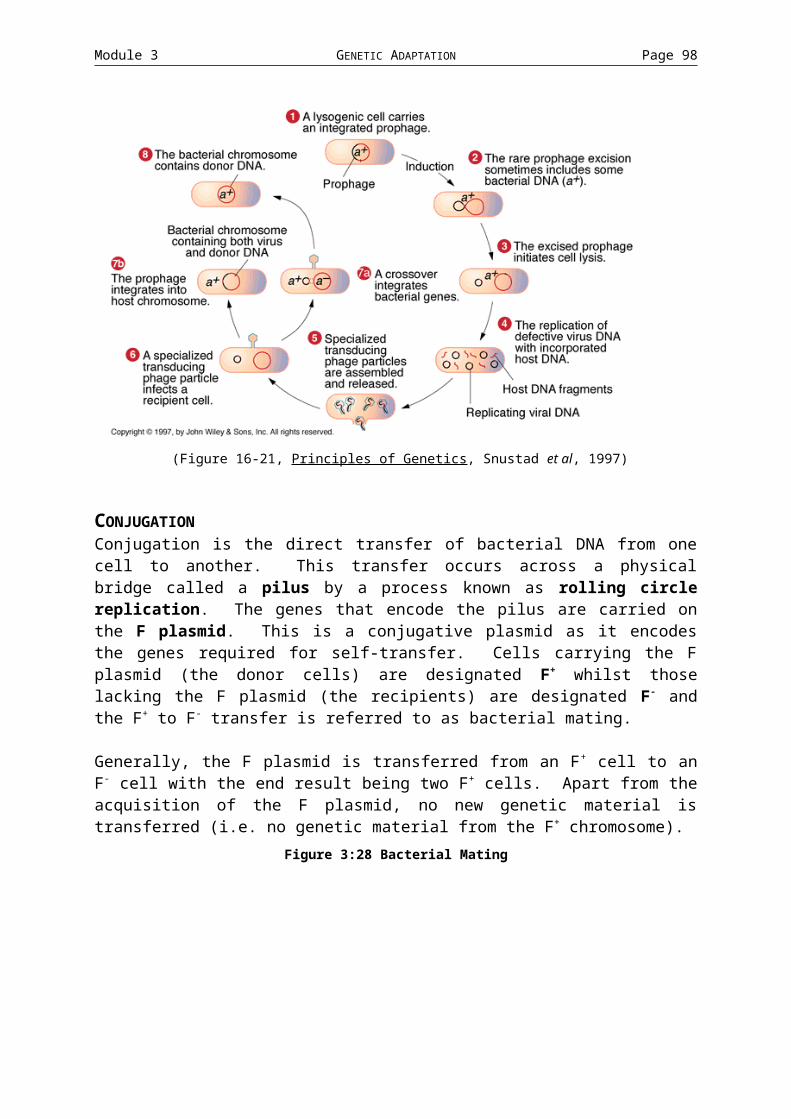
Module 4 PHYSIOLOGICAL ADAPTATION Page 98
Questions
Short answer questions:1. What is an allosteric enzyme?2. What is an operon?3. What is a promoter?4. How do positive and negative allosteric effectors differ?5. What elements make up the lac operon?6. What is global control and give two examples?7. Transcription termination generally involves one of two mechanisms. What are
they and which one is used in the regulation of gene expression?8. What is posttranscriptional regulation and how does it usually work?9. What is autogenous regulation and with what genes is it usually associated with?10.Compare allosteric and competitive inhibition
Medium answer questions:1. Provide an overview of the relationship between genotype and phenotype.2. With the use of a diagram, provide an overview of the general regulation
strategies available to a bacterial cell.3. The lac repressor completely stops transcription of the lac operon structural
genes. True or false and explain your answer.4. Regulation of aspartakinase can control the production of all of the aspartate
family of amino acids. Explain.5. Wghy is catabolite repression significant with respect to lac operon gene
expression?
Large answer questions:1. Explain the process of attenuation in the trp operon under conditions of low and
high tryptophan levels.2. Explain the basic mechanism of signal transduction and why such a process
exists.3. Compare and contrast inducible and repressible operons.4. Feedback inhibition is a dynamic and reversible process. Explain.

MODULE 5
ENERGY
AND
METABOLISM
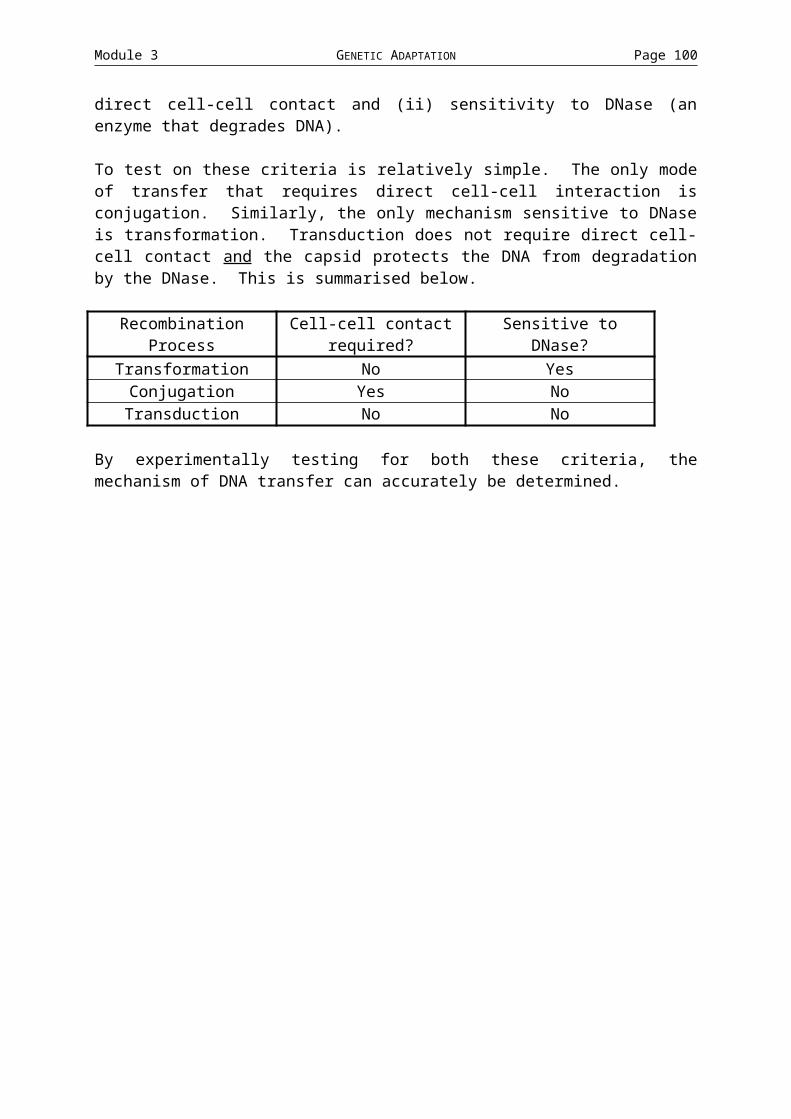
Module 5 ENERGY AND METABOLISM Page 100
MODULE 5: ENERGY AND METABOLISMCellular metabolism consists of a complex network of chemical reactions that capture energy and raw materials and process them into forms that can be utilised by the cell. The generation of ATP and the proton-motive force are key elements in energy transformation. In this module we will examine the general mechanisms at work in generating ATP as well as the diverse collection of variations used by microbes.
Topics:1. Energy Production: an overview2. Glycolysis and Aerobic Respiration3. Alternative approaches to Respiration4. Fermentation5. Photosynthesis6. Summary of Energy Producing Mechanisms
AIMS AND OBJECTIVES* To examine the diversity of microbial metabolisms
* To understand the variety of strategies used to produce energy to drive cellular reactions.
YOU SHOULD…* have an understanding of how energy is produced through respiration
* be able to describe alternative approaches used by some organisms as an alternative to respiration
* know the basic classifications of metabolisms and give examples for each
READING: To be advised
LEARNING EXERCISES find two examples for organisms that belong to each of the types of
microbial metabolisms find examples of biotechnologically important fermentation processes.

Module 5 ENERGY AND METABOLISM Page 101
5.1 ENERGY PRODUCTION: AN OVERVIEWEnergy for cellular processes is stored in the form of ATP (adenosine triphosphate). ATP is used as it has high-energy bonds (although unstable is probably a better description). The amount of energy in these bonds is, in actuality, not particularly large but can be released quickly and easily.
Figure 5:48 Simple overview of microbial metabolism(Figure 5.1. Tortora et al, Microbiology: An Introduction, 1998)
Figure 5:49 Generation of cellular energy(Figure 5.25. Tortora et al, Microbiology: An Introduction, 1998)
Oxidation and Reduction reactions In many of the pathways that will be discussed in this module, the chemical processes of oxidation and reduction play important roles. Oxidation is the removal of electrons from an atom (or molecule). These reactions result in a release of energy and the molecules that lose the electron are termed oxidised. Conversely, reduction is the addition of electrons to an atom or molecule. The molecule that gains the electrons is reduced. Oxidation and reduction reactions are always coupled and are called REDOX reactions.
In cellular processes electrons and protons are generally removed at the same time (which is, of course, the equivalent of removing a hydrogen [1H1] atom). Most biological oxidations actually involve the loss of hydrogen atoms (dehydrogenation).
Figure 5:50 REDOX reactions(Figure 5.9. Tortora et al, Microbiology: An Introduction, 1998)
NAD (nicatinomide adenine dinucleotide) and NADP are Redox co-enzymes. They assists in enzymatic reactions by accepting hydrogen atoms released from substrates during catalysis. NAD is capable of accepting 2 electrons and 1 proton with the remaining proton being released to the surrounding medium.

Module 5 ENERGY AND METABOLISM Page 102
The reduced NADH contains more energy than NAD+ and this energy can be used in later reactions to generate ATP.
Biological Redox reactions are used to extract energy from nutrient molecules. In this process, cells take nutrients from the environment, some of which serve as energy sources, and degrade them from highly reduced compounds (with many H atoms) to highly oxidised compounds. The oxidation of glucose (which will be central to the discussions in the following topics) involves the stepwise release of energy as the parent molecule (C6H12O6) is oxidised to CO2 and H2O. The energy that is released during these reactions is ultimately stored in the form of ATP which, in turn, is used to drive energy dependent reactions.
Generation of ATP ATP is formed by adding an inorganic phosphate group (Pi) to ADP (adenosine diphosphate).
Adenosine-P~P + Pi + energy Adenosine-P~P~P ADP ATP
["~" indicates high energy bonds]
It is the removal of the phosphate group that releases the energy.
There are three general mechanisms that are used for generating ATP from ADP: substrate-level phosphorylation, oxidative phosphorylation and photophosphorylation.
Substrate-level Phosphorylation Substrate-level phosphorylation is the direct transfer of high-energy phosphate groups from a substrate to ADP. The phosphate group has generally acquired its energy during a previous oxidation step.
Oxidative Phosphorylation In oxidative phosphorylation, electrons are transferred from an organic compound to an electron carrier (usually NAD+). These electrons are then passed through a series of different electron carriers to molecules of oxygen or other inorganic molecules. This series of carriers is called an electron transport chain. It is the energy released during the transfer of electrons from one carrier to the next that is used to generate ATP from ADP and inorganic phosphate. This is done by a process of chemiosmosis coupled to an enzyme capable of generating ATP (see Topic 2). The process occurs in the plasma membrane (and in the mitochondria of eukaryotic cells).
Photophosphorylation As the name suggests, photophosphorylation only occurs in photosynthetic cells. In these cells, light trapping pigments such as chlorophyll convert light energy into chemical energy stored as ATP and NADPH. As in oxidative phosphorylation, photophosphorylation requires an electron transport chain.

Module 5 ENERGY AND METABOLISM Page 103
5.2 GLYCOLYSIS AND AEROBIC RESPIRATIONMost microorganisms oxidise carbohydrates as their primary sources of cellular energy through the pathways of carbohydrate catabolism. The most common source is glucose, though other sources such as lipids and proteins can also be used. Two general processes are used to produce energy from glucose - respiration (this topic) and fermentation (Topic 4). Both of these mechanisms generally start with the glycolysis pathway (however alternatives will be mentioned in Topic 3).
Figure 5:51 Overview of respiration and fermentation(Figure 5.10. Tortora et al, Microbiology: An Introduction, 1998)

Module 5 ENERGY AND METABOLISM Page 104
RESPIRATIONRespiration of glucose typically occurs in three stages - glycolysis, the TCA cycle and electron transport chain / chemiosmosis. In gylcolysis, glucose is oxidised to two molecules of pyruvate and some ATP and NADH is produced. During the TCA cycle acteyl-CoA (a byproduct of pyruvate) is oxidised to CO2. Again, some ATP is produced along with NADH and FADH2 (another co-enzyme). It is the final stage, involving the electron transport chain / chemiosmosis, that most of the ATP is generated. NADH and FADH2 are oxidised and contribute their electrons from substrates to a cascade of oxidation-reduction reactions involving a series of additional electron carriers. The energy released from these reactions is used to generate a considerable amount of ATP. Even though glycolysis and the TCA cycle generate little ATP, they provide the electrons that generate a lot of ATP at the electron transport chain stage. These processes are the main focus of this topic.
Fermentation (which will be discussed in Topic 4) still uses glycolysis as the initial stage. Pyruvate, the end-produce of glycolysis, is then converted into a variety of different products (ethanol, lactic acid) depending on the cell. Unlike repiration, however, there is no TCA cycle and an electron transport chain is not required. Another major difference is that ATP is only generated through glycolysis so the ATP yield is much lower.
Gylcolysis Glycolysis is, simply, the oxidation of glucose to pyruvate via the Embden-Meyerhof pathway. The enzymes of glycolysis split glucose (a 6-carbon molecule) into two 3-carbon molecules. These 3-carbon sugars are then oxidised, releasing energy, and rearranged to form two pyruvate molecules.
During the glycolytic pathway, NAD+ is reduced to NADH and two ATP are generated by substrate-level phosphorylation. Two points regarding glycolysis should be noted: (i) it does not require oxygen so glycolysis can occur under both aerobic and anaerobic conditions and (ii) each of the 10 steps in the process is catalysed by a different enzyme.
Glycolysis can be divided into two basic stages. A preparatory stage in which two ATP is used in the phosphorylation of glucose and its restructuring and final separation into two 3-carbon compounds; glyceraldehyde-3-phosphate and dihydroxyacetone phosphate (which is subsequently converted to glyceraldehyde-3-phosphate). The two molecules of glyceraldehyde-3-phosphate are fed into the second stage of the glycolytic pathway, the energy conserving stage. During this multistep stage, the two molecules of glyceraldehyde-3-phosphate are oxidised to pyruvate. This results in the reduction of two molecules of NAD+ to NADH and generated 4 ATP by substrate-level phosphorylation.
Figure 5:52 Glycolysis(Figure 5.11. Tortora et al, Microbiology: An Introduction, 1998)
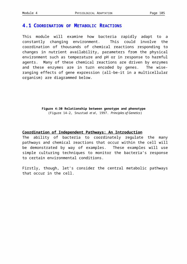
Module 5 ENERGY AND METABOLISM Page 105
CATABOLISM OF 1 GLUCOSE MOLECULE GLYCOLYSIS
NAD FAD ATP Generated by…2 NAD+ Made Used Substrate-level2 NADH 4 ATP 2 ATP phosphorylation

Module 5 ENERGY AND METABOLISM Page 106
Aerobic Respiration The pyruvate that is generated by glycolysis can have one of two fates; cellular respiration or fermentation. Aerobic respiration is an ATP generating process in which molecules are oxidised and the terminal electron acceptor from the electron transport chain is oxygen (an electron transport chain is an essential feature of respiration).
The process of aerobic respiration consists of three components - the TCA cycle, an electron transport chain and the chemiosmotic generation of ATP.
The TCA Cycle The TCA cycle (also known as the citric acid or Krebs cycle) consists of a series of enzyme driven reactions in which the large amount of energy stored in Acetyl-CoA is released in a stepwise manner (Acetyl-CoA is derived directly from pyruvate). During this cycle, a series of oxidation and reduction reactions transfer the energy (electrons) from the substrates to electron carrier coenzymes, typically NAD+ (i.e. the pyruvate derivatives are oxidised and the coenzymes are reduced).
As mentioned above, pyruvate does not directly enter the TCA cycle. Rather, there is a preparatory stage during which the pyruvate molecules lose a CO2
molecule (i.e. the 3-C pyruvate is oxidised to the 2-C Acetyl-CoA). This reaction in coupled to the reduction of NAD+ to NADH. The resulting Acetyl-CoA then loses the CoA and the acetyl group combines with oxaloacetate (4C) to produce citric acid. This reaction requires energy that is obtained from the cleavage of the high-energy bond between the acetyl and CoA groups. This is the first step of the TCA cycle.
Figure 5:53 The TCA cycle(Figure 5.12. Tortora et al, Microbiology: An Introduction, 1998)
The chemical reactions occurring throughout the TCA cycle can be categorised as being either oxidation-reduction reactions or decarboxylation reaction. Decarboxylations are the removal of carbon from a molecule via the release of CO2. The conversions of pyruvateacetyl-CoA, isocitrate-ketoglutaratesuccinyl-CoA are the decarboxylation reactions occurring within this cycle. It is these reactions that represent the complete breakdown of pyruvate (and, therefore, utlimately glucose) into CO2.
The importance of oxidation-reduction reactions have been well established. In this cycle, during each of the four steps where hydrogens are released, they are picked up by the coenzymes NAD and FAD. For example, during the conversion of isocitrate to -ketoglutarate, two hydrogens are released (i.e. the isocitrate is oxidised). NAD+ picks up two electrons and 1 proton, generating NADH. FAD, however, picks up two complete hydrogen atoms, generating FADH2.

Module 5 ENERGY AND METABOLISM Page 107
The fates of the various compounds produced through the TCA cycle vary greatly. The CO2 is released into the atmosphere as a gaseous byproduct of aerobic respiration. Many of the intermediates in the cycle play important roles in other pathways. However, it is the reduced coenzymes NADH amd FADH2 that are the most important products with respect to energy generation as they contain most of the energy originally stored in the glocuse molecule. Furthermore, it is the energy stored in these coenzymes that will be used in the generation of ATP via an electron transport chain.
So, for every 2 molecules of Acetyl-CoA (coming from 2 pyruvates which, in turn, came from 1 glucose) that enter the TCA cycle:
(1) 4xCO2 molecules are liberated by carboxlyation (with the other two CO2
molecules being removed prior to the TCA cycle)(2) 6x NADH are produced through oxidation-reduction(3) 2x FADH2 are produced through oxidation-reduction(4) 2x ATP are generated through substrate-level phosphorylation
CATABOLISM OF 1 GLUCOSE MOLECULE GLYCOLYSIS
NAD FAD ATP Generated by…2 NAD+ Made Used Substrate-level2 NADH 4 ATP 2 ATP phosphorylation
PREPARATORY STAGE (PRE TCA)2 NAD+ Made Used2 NADH
TCA CYCLE6 NAD+ 6 FAD Made Used Substrate-level6 NADH 6 FADH2 2 ATP phosphorylation

Module 5 ENERGY AND METABOLISM Page 108
Electron Transport Chain The electron transport chain is a sequence of reaction whereby electrons are passed through a sequence of carrier molecules that are capable of oxidation and reduction. As the electrons are passed through the chain, there is a stepwise release of energy which drives ATP synthesis through chemiosmosis. The final oxidation (i.e. that of the terminal electron acceptor) is irreversible and in aerobic respiration this molecule is oxygen. The components of the electron transport chain are located in the plasma membrane of the bacterial cell (or in the mitochondria in eukaryotes) and the carrier molecules are classified into three classes.
The flavoproteins are proteins that contain flavin, which is a coenzyme, derived from riboflavin (vitamin B2). These proteins are capable of performing alternating oxidations and reductions. FMN (flavin mononucleotide) is a example of a flavoprotein.
The second group is the cytochromes. These proteins have an iron-containing group (heme) and are capable of existing alternatively as a reduced Fe2+ form and the oxidised Fe3+ form. The examples of cytochromes in the electron transport chain are cytochrome a, a3, b, c, and c1.
The final type of molecule involved with the electron transport chain is the ubiquinones (or coenzymeQ). These are small, non-protein carriers.
Figure 5:54 Electron transport chain(Figure 5.13. Tortora et al, Microbiology: An Introduction, 1998)
The actual components in the electron transport chains in different bacteria differ both in the carriers used and the order they are used in. Having said that, they all achieve the same goal, that of releasing energy (as electrons) from higher energy compounds to lower energy compounds. The basic steps involved in an electron transport chain are detailed below.
Firstly, high-energy electrons are transferred from NADH to FMN. The NADH actually loses 2 electrons and a single proton. FMN picks up the additional proton from the medium resulting in the reduced form, FMNH2. Of the hydrogen picked up by FMNH2, the two protons are passed across the membrane whilst the two electrons are passes to ubiquinone (Q).
Q picks up two protons from the medium and releases them across the membrane. The electrons are passes to cytochrome (cyt) b and subsequently through the remaining cytochromes, bc1caa3. In each of these steps, the cytochrome is reduced as it picks up electrons and oxidised at it gives them up. Cyt a3 passes its electrons to molecular oxygen, which becomes negatively charged. This transfer is irreversible and the negatively charged oxygen picks up protons from the surrounding medium to form H2O.

Module 5 ENERGY AND METABOLISM Page 109
At several points through the electron transport chain, the flow of electrons is accompanied by the active transport of protons across the membrane. This results in the accumulation of protons on the outside of the membrane and it is this buildup of protons that provides the energy for ATP generation by chemiosmosis.
Generation of ATP by Chemiosmosis
Remember: Substances can pass across membranes by…(a) passive diffusion down a concentration gradient (this is an energy yielding
process)or(b) being pumped against a concentration gradient (this requied energy,
usually in the form of ATP)
Figure 5:55 Chemiosmotic generation of ATP(Figure 5.14. Tortora et al, Microbiology: An Introduction, 1998)
In the process of chemiosmosis, the energy produced when protons (H+) move along a concentration gradient is used to synthesis ATP. In respiration, chemiosmosis generated most of the ATP and can be divided into three steps.
(1) As the energetic electrons from the NADH pass down the electron transport chain, some of the carriers also pump (via active transport) protons across the membrane (these are called proton pumps)
(2) The membrane is usually impermeable to protons, so the unidirectional pumping of protons establishes a proton gradient with excess H+ on the outside. This creates a net positive charge on the outside compared to the inside. The potential energy created by this electrochemical gradient is called the proton motive force.
(3) Protons on the outside (where the concentration is greatest) can only pass across the membrane through specific protein channels. Part of these channels in an enzyme, ATPase. The energy released by the flow of H+
through the channel (and the ATPase) is used to drive the formation of ATP from ADP and Pi. The electrons from a single NADH provide the energy (and H+) for the generation of 3 ATP molecules. FADH2, which enters on point lower on the electron transport chain, generates less energy (and H+) and can generate 2 ATP per molecule of FADH2 oxidised.
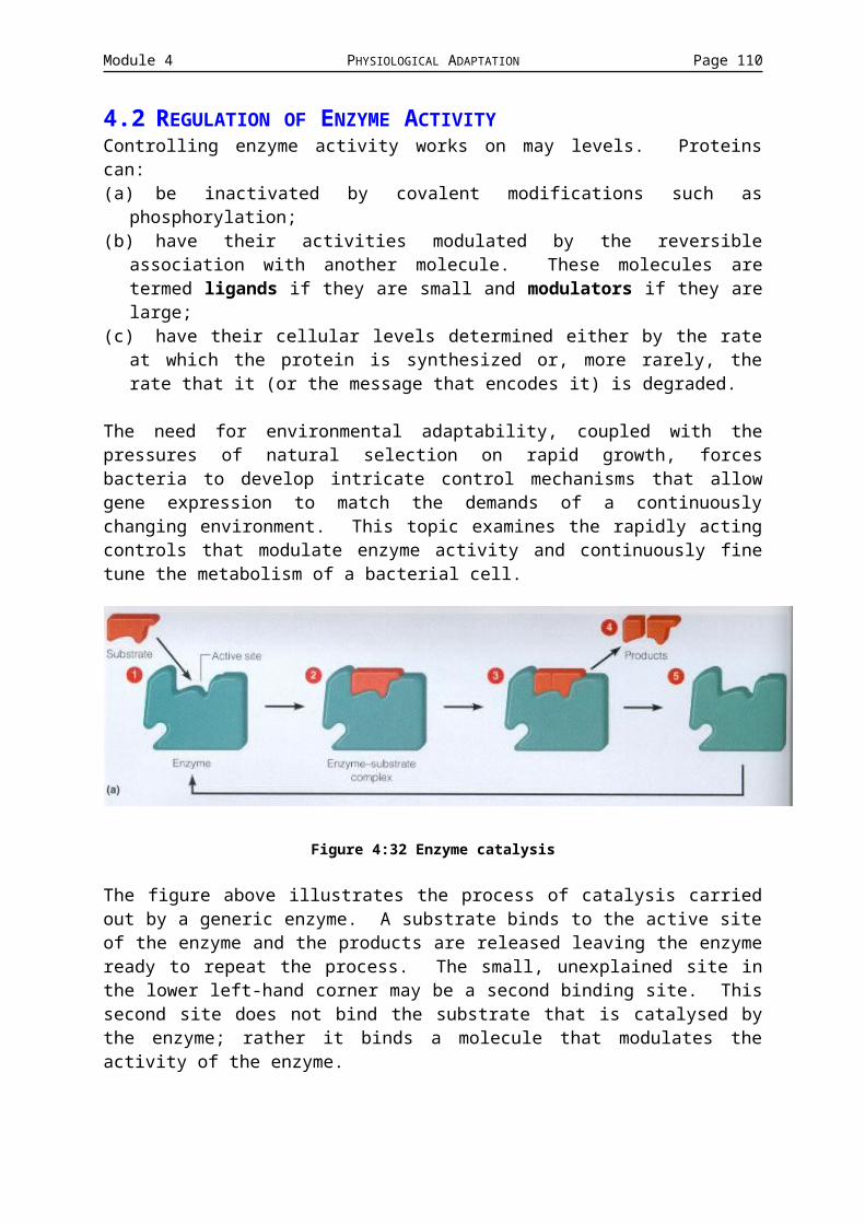
Module 5 ENERGY AND METABOLISM Page 110
Figure 5:56 Electron transport chain(Figure 5.15. Tortora et al, Microbiology: An Introduction, 1998)

Module 5 ENERGY AND METABOLISM Page 111
CATABOLISM OF 1 GLUCOSE MOLECULE GLYCOLYSIS
NAD FAD ATP Generated by…2 NAD+ Made Used Substrate-level
2 NADH 4 ATP 2 ATP phosphorylationPREPARATORY STAGE (PRE TCA)
2 NAD+ Made Used2 NADH
TCA CYCLE6 NAD+ 2 FAD Made Used Substrate-level6 NADH 2 FADH2 2 ATP phosphorylation
ELECTRON TRANSPORT CHAIN / CHEMIOSMOSIS10 NADH 2 FADH2 Made Used 30 from NADH
10 NAD+ 2 FAD 34 ATP 4 from FADH2
TOTAL ATP PRODUCED: 38 ATP 40 made - 2 usedThe overall reaction is…C6H12O6 + 6O2 + 38 ADP + 38 Pi 6CO2 + 6H2O + 38 ATP
Figure 5:57 Summary of respiration(Figure 5.16. Tortora et al, Microbiology: An Introduction, 1998)

Module 5 ENERGY AND METABOLISM Page 112
5.3 ALTERNATIVE APPROACHES TO RESPIRATIONIn this topic, we will look at two alternatives to the Embden-Meyerhof pathway (for glycolysis) and anaerobic respiration.
Pentose-phosphate pathway The pentose-phosphate pathway (also called the hexose monophosphate shunt) operates simultaneously with glycolysis and provides a means of breaking down 5-carbon sugars. Additionally, this pathway also provides key intermediates for nucleic acid synthesis, synthesis of glucose from CO2 in photosynthesis and synthesis of some amino acids. In terms of energy yielding components, it produces the reduced coenzyme NADP (from NADPH) and yields 1 ATP.
Entner-Doudoroff Pathway Organisms can catabolise glucose without either the Embden-Meyerhof pathway or the pentose-phosohate pathway vai the Entner-Doudoroff pathway. This pathway is found in some Gram-negative bacteria and is generaly not found in Gram-positive organisms. The net yield from this pathway is 2 NADPH and 1 ATP per molecule of glucose catabolised.
Anaerobic Respiration Aerobic respiration uses oxygen as the terminal electron acceptor. Anaerobic respiration has a terminal electron acceptor that is an inorganic molecule (or more rarely an organic molecule) other than molecular oxygen. The amount of ATP generated through anaerobic respiration varies depending on the organism and the pathway used. Pseudomonas and Bacillus use NO3
- as the terminal electron acceptor reducing it to NO2
-, N2O or N2. Desulfovibrio uses sulphate (SO4
2-) reducing it to H2S whilst methanogens convert CO32- to CH4 at
the end of the chain.
Only part of the TCA cycle operates under anaerobic conditions and not all the electron carriers are involved in the electron transport chain. ATP generated is never as high as in aerobic respiration so anaerobes tend to grow more slowly than aerobes.

Module 5 ENERGY AND METABOLISM Page 113
5.4 FERMENTATIONFermentation is defined (for our purposes) as a process that…i. releases energy from sugars or other organic molecules such as amino
acids, organic acids, purines and pyrimidines,
ii. does not require oxygen,
iii. does not require the TCA cycle or an electron transport chain
iv. uses an organic molecule as the terminal electron acceptor
v. produces only a small amount of ATP (1 or 2 ATP per molecule of starting material). Much of the energy stored in the original glucose molecule is retained in the chemical bonds of the organic end product.
During fermentation, electrons (and protons) are transferred from reduced coenzymes (NADH and NADPH) to pyruvate or a derivative. The terminal electron acceptor is reduced to the end products of fermentation. The NAD+
and NADP+ are passed back into glycolysis. This is essential because during fermentation, as ATP is only generated via glycolysis.
Figure 5:58 Overview of fermentation(Figure 5.18. Tortora et al, Microbiology: An Introduction, 1998)
Lactic Acid fermentation Lactic acid fermentation is found in Lactobacillus. This organism only produces lactic acid as part of its fermentation process (i.e. it is homolactic). This fermentation results in the spoilage of food but is also used in the production of yogurt (from milk), sauerkraut (from cabbage) and pickles (from cucumbers). Lactic acid fermentation starts with glycolysis generating two pyruvates from a single glucose and generating two ATP. The two molecules of pyruvate are reduced using the electrons from two molecules of NADH to form two molecules of lactic acid (the end product). No further oxidation is possible following the formation of lactic acid. This fermentation yields only a small amount of energy as most of the energy from the glucose remains in the lactic acid.
Figure 5:59 Lactic acid fermentation

Module 5 ENERGY AND METABOLISM Page 114
Alcohol Fermentation This type of fermentation used by organisms such as Saccharomyces and has several commercially useful end products. Alcohol fermentation, as the name suggests, produces ethanol as the end product (= beer). The process also produces CO2 (= nice fluffy bread). Both CO2 and ethanol are waste products of yeast fermentation. As with lactic acid fermentation, glucose is oxidised to two molecules of pyruvate, generating two ATP in the process. The pyruvate is then converted to acetaldehyde (releasing CO2) before being converted to ethanol (accompanied by the oxidation of NADH). Again, this generates a low yield of energy as most of the energy is retained in the ethanol end product.
Figure 5:60 Alcohol fermentation

Module 5 ENERGY AND METABOLISM Page 115
5.5 PHOTOSYNTHESISIn all of the above, organisms acquired energy by oxidising organic compounds. Other organisms synthesise complex organic compounds from simple inorganic substances. The energy required to drive these formations is generally derived from photosynthesis or the conversion of light energy into chemical energy. The chemical energy produced, plus atmospheric CO2, produce (through reduction) reduced carbon compounds, primarily sugars. The process whereby sugars are synthesised from atmospheric CO2 is also called carbon fixation.
The photosynthesis reaction can, generally, be summarised as follows:6CO2 + 12H2O + light energy C6H12O6 + 6O2 + 6H2OElectrons are taken from the H atoms in water (an energy poor molecule) and incorporated into a sugar (an energy rich molecule) with the energy required for this formation being indirectly supplied by light energy. The processes of photosynthesis take place in two stages - a light reaction where light energy is converted to chemical energy and a dark reaction where carbon fixation, using the energy generated by the light reaction, forms the complex sugars from CO2.
The Light Reaction During the light reaction, light energy is used to convert ADP + P i into ATP. This reaction also results in the reduction of NADP to NADPH (an energy rich carrier). The ATP is formed by photophosphorylation which, obviously, only occurs in photosynthetic cells. The general process has light absorbed by a photoreactive centre in the chlorophyll, which results in the excitement of some of its electron. These electrons are then passed down an electron transport chain and ATP is generated by chemiosmosis.
Oxygenic photosynthesis (so named because it produced oxygen) involves two photosystems, photosystems I and II, for the production of ATP and NADPH (which is important for the dark reaction). Each photosystem has its own photoreaction centre (P700 in PSI and P680 in PSII) and these two systems are linked into a unified pathway called the Z pathway of oxidative phosphorylation. The flow of electrons through the Z pathway is unidirectional and, as such, is termed noncyclic photophosphorylation as the electrons are not returned to the original photocentre but are instead replaced by electrons from water or other oxidisable compounds such as H2S.
Figure 5:61 Oxygenic photosynthesis
In contrast, anoxygenic photosynthesis does not produce oxygen. Unlike oxygenic photosynthesis there is only a single photosystem (PSI, P870) and the electrons are returned to the original donor bacteriochlorophyll molecule in a process known as cyclic photophosphorylation.

Module 5 ENERGY AND METABOLISM Page 116
Figure 5:62 Anoxygenic photosynthesis
The Dark Reaction As the name suggests, the dark reaction (also known as the Calvin cycle), occurs independent of light and uses ATP and NADPH to synthesis sugars from atmospheric CO2.
Figure 5:63 The dark reaction(Figure 5.24. Tortora et al, Microbiology: An Introduction, 1998)

Module 5 ENERGY AND METABOLISM Page 117
5.6 SUMMARY OF ENERGY PRODUCING MECHANIMS Having discussed the pathways used to generate energy, we will look at the broad classifications that can be used to group bacteria on the basis of their nutritional requirements for metabolism.
Figure 5:64 Summary of energy producing pathways(Figure 5.22. Tortora et al, Microbiology: An Introduction, 1998)
When considering energy sources, organisms can generally be classified as being either phototrophs, using light as their primary energy source or chemotrophs that use the oxidation-reduction of molecules to generate energy.
The other major classification is based on carbon source. Autotrophs (also called lithotrophs) use carbon dioxide as their primary carbon source whereas heterotrophs (also called organotrophs) require an organic carbon source.
The general metabolic classifications for organisms are derived from the combination of energy and carbon sources.
Photoautotrophs These organisms obtain their energy from light sources and use CO2 as a carbon source. Microorganisms of this metabolic grouping include photosynthetic bacteria (green sulfur, purple sulfur and cyanobacteria) and algae. Cyanobacteria and algae generally use oxygenic photosynthesis whereas the green and purple sulfur bacteria use anoxygenic photosynthesis.
Photoheterotrophs Photoheterotrophs use light as an energy source but are unable to convert CO2 to sugars and, as such, require organic molecules as a source of carbon. These organisms use anoxygenic photosynthesis and include organisms such as Chloroflexus (green nonsulfur bacteria) and Rhodopseudomonas (a purple nonsulfur bacteria).
Chemoautotrophs Chemoautotrophs use electrons from reduced inorganic compounds as an energy source and CO2 as their primary carbon source. Inorganic energy sources include H2S (Beggiatoa), elemental sulfur S (Thiobacillus thiooxidans), ammonia NH3 (Nitrosomonas), nitrite ions NO2
- (Nitrobacter) and ferrous Fe2+ (Thiobacillus ferrooxidans). The energy is derived from these molecules and eventually stored as ATP via oxidative phosphorylation.

Module 5 ENERGY AND METABOLISM Page 118
Chemoheterotrophs Chemoheterotrophs use reduced chemical compounds as an energy source and organic molecules as a carbon source. It organisms with this type of metabolism to which all medically significant microorganisms belong.
Figure 5:65 Summary of microbial metabolisms
(Figure 5.26. Tortora et al, Microbiology: An Introduction, 1998)

Module 5 ENERGY AND METABOLISM Page 119
Questions
Short answer questions:1. Write the overall reaction for the catabolism of glucose.2. What are proton pumps?3. Explain what is meant by dehydrogenation.4. What is a chemoautotroph and give an example.5. What are the two criteria on which organisms can be nutritionally
classified?
Medium answer questions:1. What is the proton motive force and how is it generated?2. Outline the TCA cycle noting the steps that are important for energy
production.3. Explain the process of biological redox reactions and give an example.4. Distinguish between oxygenic and anoxygenic photosynthesis.5. Define fermentation.
Large answer question:Compare the energy produced through respiration and fermentation and explain which is more efficient.