bd mouse hematopoietic stem and progenitor cell … · · 2016-09-12hematopoietic stem and...
TRANSCRIPT

Catalog No. 560492
BD Mouse Hematopoietic Stem and Progenitor Cell Isolation KitInstruction Manual

© 2010, Becton, Dickinson and Company. All rights reserved. No part of this publication may be reproduced, transmitted, transcribed, stored in retrieval systems, or translated into any language or computer language, in any form or by any means: electronic, mechanical, magnetic, optical, chemical, manual, or otherwise, without prior written permission from BD Biosciences.
The information in this guide is subject to change without notice. BD Biosciences reserves the right to change its products and services at any time to incorporate the latest technological developments. Although this guide has been prepared with every precaution to ensure accuracy, BD Biosciences assumes no liability for any errors or omissions, nor for any damages resulting from the application or use of this information. BD Biosciences welcomes customer input on corrections and suggestions for improvement.
BD, BD Logo and all other trademarks are the property of Becton, Dickinson and Company. © 2010 BD
Cy™ is a trademark of Amersham Biosciences Corp. This product is subject to proprietary rights of Amersham Biosciences Corp. and Carnegie Mellon University and made and sold under license from Amersham Biosciences Corp. This product is licensed for sale only for research. It is not licensed for any other use. If you require a commercial license to use this product and do not have one return this material, unopened to BD Biosciences, 2350 Qume Drive, San Jose, CA, 95131-1807 and any money paid for the material will be refunded.
MyeloCult and MethoCult are registered trademarks of StemCell Technologies.
History
BD flow cytometers are class I (1) laser products.
For Research Use Only. Not for use in diagnostic or therapeutic procedures.
Revision Date Change made
23-12114-00 Rev. A 5/2010 Updated BD CompBead quantities in Kit contents.

For Research Use Only. Not for use in diagnostic or therapeutic procedures.
Contents
Chapter 1: About this kit . . . . . . . . . . . . . . . . . . . . . . . . . . . . . . . . . . . . 5Purpose of the kit . . . . . . . . . . . . . . . . . . . . . . . . . . . . . . . . . . . . . . . 6Kit contents . . . . . . . . . . . . . . . . . . . . . . . . . . . . . . . . . . . . . . . . . . . . 9Storage and safe handling . . . . . . . . . . . . . . . . . . . . . . . . . . . . . . . . 12
Chapter 2: Before you begin . . . . . . . . . . . . . . . . . . . . . . . . . . . . . . . . 13Workflow overview . . . . . . . . . . . . . . . . . . . . . . . . . . . . . . . . . . . . . 14Required materials . . . . . . . . . . . . . . . . . . . . . . . . . . . . . . . . . . . . . . 16
Chapter 3: Preparation of cells and beads . . . . . . . . . . . . . . . . . . . . . 19Preparing cells for staining . . . . . . . . . . . . . . . . . . . . . . . . . . . . . . . . 20Staining cells and beads . . . . . . . . . . . . . . . . . . . . . . . . . . . . . . . . . . 22
Chapter 4: Cytometer procedures . . . . . . . . . . . . . . . . . . . . . . . . . . . . 27Running the beads . . . . . . . . . . . . . . . . . . . . . . . . . . . . . . . . . . . . . . 28Setting up the workspace for running cells . . . . . . . . . . . . . . . . . . . . 29Running the cells . . . . . . . . . . . . . . . . . . . . . . . . . . . . . . . . . . . . . . . 31Sorting cells . . . . . . . . . . . . . . . . . . . . . . . . . . . . . . . . . . . . . . . . . . . 34
Chapter 5: Reference . . . . . . . . . . . . . . . . . . . . . . . . . . . . . . . . . . . . . . 37Examples of post-sort applications . . . . . . . . . . . . . . . . . . . . . . . . . 38Troubleshooting . . . . . . . . . . . . . . . . . . . . . . . . . . . . . . . . . . . . . . . 42References . . . . . . . . . . . . . . . . . . . . . . . . . . . . . . . . . . . . . . . . . . . . 43

For Research Use Only. Not for use in diagnostic or therapeutic procedures.
1About this kit
This section covers the following topics:
Purpose of the kit (page 6)
Kit contents (page 9)
Storage and safe handling (page 12)

BD Mouse Hematopoietic Stem and Progenitor Cell Isolation Kit6
For Research Use Only. Not for use in diagnostic or therapeutic procedures.
Purpose of the kit
About this topic This topic explains the purpose of the BD™ Mouse Hematopoietic Stem and Progenitor Cell Isolation Kit (Catalog No. 560492), and provides background for understanding the kit’s components and how they work.
Uses of the kit This kit provides researchers with the reagents necessary to perform multicolor flow cytometric cell sorting of hematopoietic progenitor cells (HPCs) and hematopoietic stem cells (HSCs) from mouse bone marrow samples.
Specific antibodies
Mouse HPCs and HSCs are characterized by a set of cell surface markers,1,2 as well as by their high level of dye efflux activity.3 The BD Mouse Hematopoietic Stem and Progenitor Cell Isolation Kit contains fluorescent antibodies to lineage-specific markers and to markers that help identify HPCs and HSCs.
The kit contains a lineage cocktail that includes specific antibodies for CD3 (a marker for T cells), CD45R (B220) (a marker for B cells), Ly6C and Ly6G (Gr1) (a marker for granulocytes), CD11b (Mac1) (a marker for macrophages), and TER-119 (a marker for red blood cells).
The kit also contains three specific antibodies that are used to isolate HPCs and HSCs. The first is CD34, which is expressed by HPCs and found to be dim to negative on stem cells. The others are Sca-1 and c-Kit (CD117), which are expressed by both HPCs and HSCs. Use of these markers to isolate progenitor and stem cells (called CD34-/dim KSL cells, where KSL is for c-Kit+/Sca-1+/Lineage-), has been reported widely.1,2

Chapter 1: About this kit 7
For Research Use Only. Not for use in diagnostic or therapeutic procedures.
Isotype-control antibodies
This kit contains matching isotype controls for each specific antibody provided. Each isotype control is a non-specific antibody of the same isotype and conjugated to the same fluorochrome as one of the specific antibodies, and is bottled at the same concentration as the specific antibody. The isotype controls are used to quantify any non-specific (background) staining of the specific antibodies.
Cell viability dye This kit contains 7-amino-actinomycin D (7-AAD), a DNA dye that is frequently used to discriminate dead cells from live cells.
Control beads This kit also contains BD™ CompBead positive and negative beads to facilitate application setup for sorting of HPCs and HSCs.
The positive beads are coated with antibodies that will bind to one of the specific antibodies in this kit. The negative beads have no binding capacity.
Once the beads have been stained with the specific antibodies, they provide distinct positive and negative populations used to optimize photomultiplier tube (PMT) settings and calculate fluorescence compensation. Use of these beads ensures consistent application setup and conserves cells.

BD Mouse Hematopoietic Stem and Progenitor Cell Isolation Kit8
For Research Use Only. Not for use in diagnostic or therapeutic procedures.
Which mice to use
We optimized the antibody combination in this kit to work with the C57BL/6 mouse strain, the strain most commonly used for hematopoietic stem cell research. We have also shown that this combination works on DBA mice, but not on Balb/c mice.
We recommend using mice between the ages of 8 and 10 weeks. Because the staining profile changes as mice age, you must identify the optimal age for your research purposes.
Related topics Kit contents (page 9)

Chapter 1: About this kit 9
For Research Use Only. Not for use in diagnostic or therapeutic procedures.
Kit contents
Reagent information
The BD Mouse Hematopoietic Stem and Progenitor Cell Isolation Kit contains the following reagents:
Reagent Details
BD Pharmingen™ FITC Anti-Mouse CD34
Clone. RAM34
Use. Used to stain HPCs in bone marrow
Abbreviation. FITC CD34
Quantity. 1 vial (0.2 mL)
Amount for staining. 20 µL for 1 x 10⁹ cells (per 1 mL, based on cell count before lineage depletion)
BD Pharmingen™ PE Anti-Mouse c-Kit
Clone. 2B8
Use. Used to stain HPCs and HSCs in bone marrow
Abbreviation. PE c-Kit
Quantity. 1 vial (0.2 mL)
Amount for staining. 20 µL for 1 x 10⁹ cells (per 1 mL, based on cell count before lineage depletion)
BD Pharmingen™ PE-Cy™7 Anti-Mouse Sca-1
Clone. D7
Use. Used to stain HPCs and HSCs in bone marrow
Abbreviation. PE-Cy7 Sca-1
Quantity. 1 vial (0.2 mL)
Amount for staining. 20 µL for 1 x 10⁹ cells (per 1 mL, based on cell count before lineage depletion)

BD Mouse Hematopoietic Stem and Progenitor Cell Isolation Kit10
For Research Use Only. Not for use in diagnostic or therapeutic procedures.
BD Pharmingen™ APC Mouse Lineage Antibody Cocktail
Antibodies. CD3, CD45R (B220), Ly6C and Ly6G (Gr1), CD11b (Mac1), and TER-119
Clones. 145-2C11, RA3-6B2, RB6-8C5, M1/70, and TER-119
Use. Used to stain lineage-committed cells in bone marrow
Abbreviation. APC lineage cocktail
Quantity. 1 vial (1 mL)
Amount for staining. 100 µL for 1 x 10⁹ cells (per 1 mL, based on cell count before lineage depletion)
BD Pharmingen™ FITC Rat IgG2a, κ Isotype Control
Clone. R35-95
Use. Used as an isotype control for FITC CD34
Abbreviation. FITC isotype control
Quantity. 1 vial (0.1 mL)
Amount for staining. 1 µL of a 1:10 dilution for 50 µL of cells
BD Pharmingen™ PE Rat IgG2b, κ Isotype Control
Clone. A95-1
Use. Used as an isotype control for PE c-Kit
Abbreviation. PE isotype control
Quantity. 1 vial (0.1 mL)
Amount for staining. 1 µL of a 1:10 dilution for 50 µL of cells
BD Pharmingen™ PE-Cy7 Rat IgG2a, κ Isotype Control
Clone. R35-95
Use. Used as an isotype control for PE-Cy7 Sca-1
Abbreviation. PE-Cy7 isotype control
Quantity. 1 vial (0.1 mL)
Amount for staining. 1 µL of a 1:10 dilution for 50 µL of cells
Reagent Details
Chapter 1: About this kit 11
For Research Use Only. Not for use in diagnostic or therapeutic procedures.
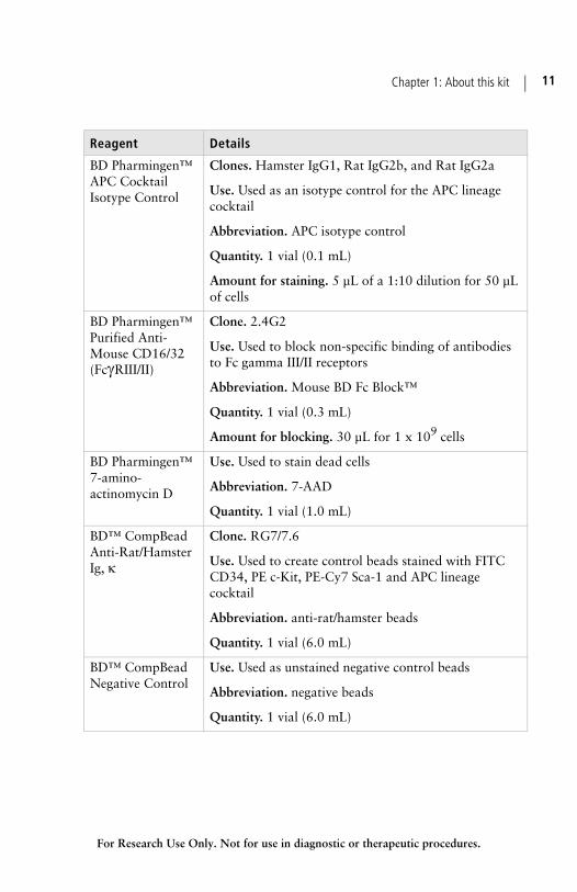
BD Pharmingen™ APC Cocktail Isotype Control
Clones. Hamster IgG1, Rat IgG2b, and Rat IgG2a
Use. Used as an isotype control for the APC lineage cocktail
Abbreviation. APC isotype control
Quantity. 1 vial (0.1 mL)
Amount for staining. 5 µL of a 1:10 dilution for 50 µL of cells
BD Pharmingen™ Purified Anti-Mouse CD16/32 (FcγRIII/II)
Clone. 2.4G2
Use. Used to block non-specific binding of antibodies to Fc gamma III/II receptors
Abbreviation. Mouse BD Fc Block™
Quantity. 1 vial (0.3 mL)
Amount for blocking. 30 µL for 1 x 10⁹ cells
BD Pharmingen™ 7-amino-actinomycin D
Use. Used to stain dead cells
Abbreviation. 7-AAD
Quantity. 1 vial (1.0 mL)
BD™ CompBead Anti-Rat/Hamster Ig, κ
Clone. RG7/7.6
Use. Used to create control beads stained with FITC CD34, PE c-Kit, PE-Cy7 Sca-1 and APC lineage cocktail
Abbreviation. anti-rat/hamster beads
Quantity. 1 vial (6.0 mL)
BD™ CompBead Negative Control
Use. Used as unstained negative control beads
Abbreviation. negative beads
Quantity. 1 vial (6.0 mL)
Reagent Details
BD Mouse Hematopoietic Stem and Progenitor Cell Isolation Kit12
For Research Use Only. Not for use in diagnostic or therapeutic procedures.
Serum proteins Components in this kit contain serum proteins. Source of all serum proteins is from USDA inspected abattoirs located in the United States.
Related topics Purpose of the kit (page 6)
Storage and safe handling (page 12)
Storage and safe handling
About this topic This topic describes the requirements for kit storage and safe handling.
Storage The entire BD Mouse Hematopoietic Stem and Progenitor Cell Isolation Kit must be stored in the dark at 2–8°C. Do not freeze.
Warning The reagents in this kit contain sodium azide. Sodium azide yields highly toxic hydrazoic acid under acidic conditions. Dilute azide compounds in running water before discarding to avoid accumulation of potentially explosive deposits in plumbing.
Related topics Kit contents (page 9)

For Research Use Only. Not for use in diagnostic or therapeutic procedures.
2Before you begin
This section covers the following topics:
Workflow overview (page 14)
Required materials (page 16)

BD Mouse Hematopoietic Stem and Progenitor Cell Isolation Kit14
For Research Use Only. Not for use in diagnostic or therapeutic procedures.
Workflow overview
About this topic This topic provides an overview of the workflow for using the BD Mouse Hematopoietic Stem and Progenitor Cell Isolation Kit to sort HPCs and HSCs.
Workflow overview
The following example shows a typical workflow:
Note: Maintain aseptic conditions throughout the procedures.
Perform flow cytometer QC
Prepare flow cytometer for aseptic sort
Prepare the cells for staining• obtain a single-cell suspension• treat with BD Fc Block• deplete lineage-positive cells
Stain the cellsfor 30 to 60 minutes at 4°C
• unstained cells• cells + all 4 specific antibodies• cells + all 4 isotype controls
then add 7-AAD
Stain the BD CompBeadsfor 15 minutes at RT
Run the beadsto adjust fluorescence settings,
create application settings,and calculate compensation
Set up the workspaceto create tubes, labels, plots, and histograms
Run the cellsto fine-tune scatter settings,
record data from unstained andisotype-control cells,
and set up a gating strategy
Sort the cells
Chapter 2: Before you begin 15
For Research Use Only. Not for use in diagnostic or therapeutic procedures.
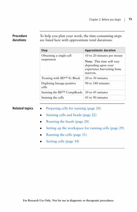
Procedure durations
To help you plan your work, the time-consuming steps are listed here with approximate total durations:
Related topics Preparing cells for staining (page 20)
Staining cells and beads (page 22)
Running the beads (page 28)
Setting up the workspace for running cells (page 29)
Running the cells (page 31)
Sorting cells (page 34)
Step Approximate duration
Obtaining a single-cell suspension
10 to 20 minutes per mouse
Note: This time will vary depending upon your experience harvesting bone marrow.
Treating with BD™ Fc Block 20 to 30 minutes
Depleting lineage-positive cells
90 to 180 minutes
Staining the BD™ CompBeads 30 to 45 minutes
Staining the cells 45 to 90 minutes

BD Mouse Hematopoietic Stem and Progenitor Cell Isolation Kit16
For Research Use Only. Not for use in diagnostic or therapeutic procedures.
Required materials
About this topic This topic describes the reagents, consumables, and equipment that you will need to sort HPCs and HSCs using the BD Mouse Hematopoietic Stem and Progenitor Cell Isolation Kit.
Materials list The following materials are required:
Biological safety cabinet
Note: If you plan to use sorted cells in transplantation or tissue-culture experiments, all sample-preparation steps must be performed in a biological safety cabinet using aseptic technique.
1 M HEPES buffer
Microscope for confirming a single-cell suspension
BD Falcon™ 70-µm cell strainer (Catalog No. 352350), or equivalent (optional, but recommended)
Hemocytometer or other cell counter
Sterile BD Pharmingen Stain buffer (Catalog No. 554656), or equivalent (0.22 µm-filtered 1X DPBS supplemented with 3% heat-inactivated FCS [fetal calf serum])
BD IMag™ Mouse Hematopoietic Progenitor (Stem) Cell Enrichment Set - DM (Catalog No. 558451), or equivalent
BD IMag™ Cell Separation Magnet (Catalog No. 552311)
BD IMag Buffer (10X) [Catalog No. 552362]
BD Falcon round-bottom 12 x 75-mm polystyrene tubes with caps (Catalog No. 352058), or equivalent

Chapter 2: Before you begin 17
For Research Use Only. Not for use in diagnostic or therapeutic procedures.
BD Falcon round-bottom 17 x 100-mm polystyrene tubes (Catalog No. 352057), for sample volumes greater than 3 mL
BD FACSAria™ II flow cytometer, or other flow cytometer equipped for sorting and with a blue (488-nm) laser, a red (633-nm) laser, and detectors for FITC, PE, PE-Cy7, and APC
RPMI 1640 medium, or equivalent (available from Hyclone, Thermo Fisher Scientific, Inc.)
10% FCS (fetal calf serum), heat inactivated (available from Hyclone, Thermo Fisher Scientific, Inc.)
1% penicillin/streptomycin (available from Invitrogen)
Related topics Kit contents (page 9)


For Research Use Only. Not for use in diagnostic or therapeutic procedures.
3Preparation of cells and beads
This section covers the following topics:
Preparing cells for staining (page 20)
Staining cells and beads (page 22)

BD Mouse Hematopoietic Stem and Progenitor Cell Isolation Kit20
For Research Use Only. Not for use in diagnostic or therapeutic procedures.
Preparing cells for staining
About this topic This topic explains how to harvest mouse bone-marrow cells and prepare them for staining with the BD Mouse Hematopoietic Stem and Progenitor Cell Isolation Kit.
Procedural options
This procedure includes a step for treating harvested bone-marrow cells with BD Fc Block to prevent non-specific binding of antibodies to Fc gamma III/II receptors.
This procedure also includes a step for depleting unwanted lineage-positive cells from the cell suspension. We highly recommend performing this step to reduce your sort time by enriching for HPCs and HSCs. However, when planning your experiment and deciding how many mice to use, consider that this step might also result in a loss of HPCs and HSCs.
Aseptic technique
If you plan to use sorted cells in transplantation or tissue-culture experiments, all sample-preparation steps must be performed using aseptic technique.
Before you begin Ensure that you have all of the necessary materials available. See Required materials (page 16) for details.
Collect the hind legs of at least 10 mice.
Decide whether you will complete step 6 for lineage depletion.
Note: The duration of this procedure will vary considerably depending on how many mice you use, your experience harvesting bone marrow, and whether you include the recommended lineage-depletion step. Expect to spend 3 to 5 hours preparing lineage-depleted bone marrow cells from 10 mice.

Chapter 3: Preparation of cells and beads 21
For Research Use Only. Not for use in diagnostic or therapeutic procedures.
Procedure To prepare cells for staining:
1. Isolate bone marrow cells from at least 10 mice, following the procedure in Preparation of bone marrow cells by Mishell et al.4
2. Remove debris from your single-cell suspension by passing it through a 70-µm cell strainer.
3. Determine the cell concentration using the standard method for your hemocytometer or other cell counter.
4. Adjust the cell concentration to between 1 x 10⁸ and 1 x 10⁹ cells per mL in sterile stain buffer at 4°C.
5. Add 30 µL of BD Fc Block per 1 x 10⁹ cells and incubate at 4°C for 10 minutes.
6. Deplete lineage-positive cells using a magnetic separation system, following the manufacturer’s instructions. If you do not perform lineage depletion, proceed to step 9.
7. Wash the cells once with sterile stain buffer (centrifuging at 250g for 10 min).
8. Resuspend the cells in 1,000 µL of sterile stain buffer per 1 x 10⁹ cells (using the cell count before lineage depletion).
9. Transfer the cells to a sterile 12 x 75-mm polystyrene tube and label as the specific-stained cells.
10. Transfer 50 µL of cells to a new 12 x 75-mm tube, label as the unstained control, and store at 4°C.
11. Transfer 50 µL of cells to a new 12 x 75-mm tube and label as the isotype control.

BD Mouse Hematopoietic Stem and Progenitor Cell Isolation Kit22
For Research Use Only. Not for use in diagnostic or therapeutic procedures.
Next step Proceed immediately to Staining cells and beads (page 22).
Related topics Workflow overview (page 14)
Required materials (page 16)
Staining cells and beads
About this topic This topic explains how to stain both your prepared cells and the BD CompBeads with the antibodies provided in the BD Mouse Hematopoietic Stem and Progenitor Cell Isolation Kit.
Before you begin Complete the steps in Preparing cells for staining (page 20).
Prepare sterile stain buffer supplemented with 25 mM HEPES (final concentration). You will use this to adjust the cell concentration.
Aseptic technique
If you plan to use sorted cells in transplantation or tissue-culture experiments, use aseptic technique for all the following steps. You should use aseptic technique when staining the setup beads to minimize potential contamination of the flow cytometer prior to sorting your cells.

Chapter 3: Preparation of cells and beads 23
For Research Use Only. Not for use in diagnostic or therapeutic procedures.
Staining the cells To stain the cells:
1. For each 1 x 10⁹ cells before lineage depletion, add the following to the specific-stained cells tube:
2. For each 50 µL of cells, add the following antibodies to the isotype-control tube:
3. Mix the tubes gently and incubate at 4°C in the dark for 30 to 60 minutes.
Component Volume
FITC CD34 20 µL
PE c-Kit 20 µL
PE-Cy7 Sca-1 20 µL
APC lineage cocktail 100 µL
Component Volume
FITC isotype control 1 µL
PE isotype control 1 µL
PE-Cy7 isotype control 1 µL
APC isotype control 5 µL

BD Mouse Hematopoietic Stem and Progenitor Cell Isolation Kit24
For Research Use Only. Not for use in diagnostic or therapeutic procedures.
Staining the beads
To stain the BD CompBeads:
1. Immediately after starting the cell-stain incubations, add the following to five labeled 12 x 75-mm polystyrene tubes in the order shown (vortex the beads thoroughly immediately before dispensing drops):
2. Vortex the tubes and incubate at room temperature in the dark for 15 minutes.
3. Add 2 mL of sterile stain buffer to the stained beads, vortex, and centrifuge at 200g for 10 minutes.
4. Remove the supernatant and add 1 mL of sterile stain buffer.
Washing the cells
To wash the cells:
1. After the 30 to 60-minute cell-stain incubations are complete, wash each tube twice in 1 to 2 mL of sterile stain buffer (centrifuging at 250g for 10 minutes).
2. Resuspend the isotype control cells in 500 µL of sterile stain buffer.
3. Determine the cell concentration of the specific-stained cells and adjust to between 1 x 10⁷ and 2 x
Volume to add to the tube labeled
Component Negative FITC PE PE-Cy7 APC
sterile stain buffer 100 µL 100 µL 100 µL 100 µL 100 µL
negative beads 1 drop 1 drop 1 drop 1 drop 1 drop
anti-rat/hamster beads — 1 drop 1 drop 1 drop 1 drop
FITC CD34 — 1 µL — — —
PE c-Kit — — 1 µL — —
PE-Cy7 Sca-1 — — — 1 µL —
APC lineage cocktail — — — — 5 µL

Chapter 3: Preparation of cells and beads 25
For Research Use Only. Not for use in diagnostic or therapeutic procedures.
107 cells per mL in sterile stain buffer supplemented with 25 mM HEPES (final concentration).
4. Add 1 drop of 7-AAD for each 1 mL (or less) of cells and swirl to mix.
5. Pass the cells through a 70-µm cell strainer.
6. Place cells on ice and keep on ice throughout the remainder of the experiment.
Next step Proceed to Running the beads (page 28).
Storage is not recommended. Run stained beads and cells within 1 to 2 hours of staining.
Related topics Kit contents (page 9)
Workflow overview (page 14)
Required materials (page 16)


For Research Use Only. Not for use in diagnostic or therapeutic procedures.
4Cytometer procedures
This section provides guidelines for application setup and sorting of cells stained with the BD Mouse Hematopoietic Stem and Progenitor Cell Isolation Kit.
The guidelines and examples in this section use BD FACSDiva™ software and BD FACS™ flow cytometers. However, the fundamental approach to setup and acquisition can be adapted for research labs with other flow cytometers.
This section covers the following topics:
Running the beads (page 28)
Setting up the workspace for running cells (page 29)
Running the cells (page 31)
Sorting cells (page 34)

BD Mouse Hematopoietic Stem and Progenitor Cell Isolation Kit28
For Research Use Only. Not for use in diagnostic or therapeutic procedures.
Running the beads
About this topic This topic describes how to use the prepared BD CompBeads to calculate compensation.
Before you begin Ensure that you run the appropriate instrument setup and QC procedures for your flow cytometer. See your user’s guide for more information.
Perform an aseptic cleaning of your flow cytometer.
Complete the steps in Preparation of cells and beads (page 19).
Procedure To run the prepared beads:
1. Set up the PMTs and compensation using the stained BD CompBeads. Set the P1 gate on the singlet population of beads, and set the P2 gate tightly around the positive peak.

Chapter 4: Cytometer procedures 29
For Research Use Only. Not for use in diagnostic or therapeutic procedures.
2. Repeat this procedure for the PE, PE-Cy7, and APC controls.
Note: You can save these settings and use them as a baseline for future experiments.
Next step Proceed to Setting up the workspace for running cells (page 29).
More information
See Getting Started with BD FACSDiva Software and the BD FACSDiva Software Reference Manual for information about running beads and calculating compensation.
Related topics Workflow overview (page 14)
Setting up the workspace for running cells
About this topic This topic explains how to set up the BD FACSDiva workspace in preparation for running cells.
Before you begin Complete the steps in Running the beads (page 28).
Procedure To set up the workspace for running cells:
1. Create a new specimen using BD FACSDiva software.
2. Create tubes and label them appropriately for your unstained cells, isotype-control cells, and specific-stained cells.

BD Mouse Hematopoietic Stem and Progenitor Cell Isolation Kit30
For Research Use Only. Not for use in diagnostic or therapeutic procedures.
3. If you have previously saved a template for use with this kit, import the template and proceed directly to Running the cells (page 31).
4. Use the Labels tab of the Experiment Layout window to enter parameter labels for each specific antibody.
5. On a global worksheet, create the following plots:
– Plot 1: FSC-A vs 7-AAD-A
– Plot 2: FSC-A vs SSC-A
– Plot 3: FSC-H vs FSC-W
– Plot 4: SSC-H vs SSC-W
– Plot 5: PE-A vs APC-A
– Plot 6: PE-Cy7-A vs PE-A
6. Generate the following histogram:
– FITC-A
7. Select biexponential scaling for all fluorochrome axes.
8. Save this worksheet as a template for use in future experiments.
Next step Proceed to Running the cells (page 31).
More information
See Getting Started with BD FACSDiva Software for information about working in the BD FACSDiva workspace.
See the BD FACSDiva Software Reference Manual for information about how to work with analysis templates.

Chapter 4: Cytometer procedures 31
For Research Use Only. Not for use in diagnostic or therapeutic procedures.
Related topics Workflow overview (page 14)
Running the cells
About this topic This topic explains how to:
Fine-tune scatter settings on your flow cytometer by running your unstained cells
Detect autofluorescence by recording data from your unstained cells
Identify background staining by recording data from your isotype control cells
Set up a gating strategy
Before you begin Complete the steps in Setting up the workspace for running cells (page 29).
Procedure To run the cells:
1. Place the tube of unstained cells on the cytometer and begin acquiring.
2. Adjust the FSC and SSC PMT voltages as needed to ensure that your cell population appears on scale in the scatter plot.
Note: Do not adjust the fluorescence settings at this stage. Adjusting the fluorescence settings now will invalidate your compensation calculations.
3. Record 20,000 events from the unstained cells.
4. Place the tube of isotype-control cells on the cytometer and record 20,000 events.
BD Mouse Hematopoietic Stem and Progenitor Cell Isolation Kit32
For Research Use Only. Not for use in diagnostic or therapeutic procedures.
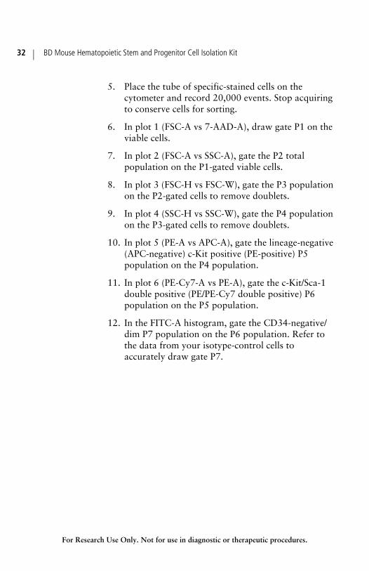
5. Place the tube of specific-stained cells on the cytometer and record 20,000 events. Stop acquiring to conserve cells for sorting.
6. In plot 1 (FSC-A vs 7-AAD-A), draw gate P1 on the viable cells.
7. In plot 2 (FSC-A vs SSC-A), gate the P2 total population on the P1-gated viable cells.
8. In plot 3 (FSC-H vs FSC-W), gate the P3 population on the P2-gated cells to remove doublets.
9. In plot 4 (SSC-H vs SSC-W), gate the P4 population on the P3-gated cells to remove doublets.
10. In plot 5 (PE-A vs APC-A), gate the lineage-negative (APC-negative) c-Kit positive (PE-positive) P5 population on the P4 population.
11. In plot 6 (PE-Cy7-A vs PE-A), gate the c-Kit/Sca-1 double positive (PE/PE-Cy7 double positive) P6 population on the P5 population.
12. In the FITC-A histogram, gate the CD34-negative/dim P7 population on the P6 population. Refer to the data from your isotype-control cells to accurately draw gate P7.

Chapter 4: Cytometer procedures 33
For Research Use Only. Not for use in diagnostic or therapeutic procedures.
Next step Proceed to Sorting cells (page 34).
Related topics Workflow overview (page 14)
Troubleshooting (page 42)

BD Mouse Hematopoietic Stem and Progenitor Cell Isolation Kit34
For Research Use Only. Not for use in diagnostic or therapeutic procedures.
Sorting cells
About this topic This topic provides general guidelines for sorting cells stained with the BD Mouse Hematopoietic Stem and Progenitor Cell Isolation Kit.
For specific procedures, see the user’s guide for your flow cytometer.
Before you begin Complete the steps described in Running the cells (page 31).
Keep samples at 4°C.
Cytometer setup guidelines
We recommend the following cytometer setup for sorting HPCs and HSCs:
A 100-µm nozzle
A drop frequency between 25 and 30 kHz
Pressure of approximately 20 to 25 psi
Follow the instructions in your user’s guide for optimizing the breakoff and drop delay.
Procedure To sort cells:
1. Set up the sort to sort cells based on gate P7.
2. Fill your collection tube with medium containing antibiotics and 10% fetal calf serum.
3. Invert the tubes several times to coat the sides with medium, and install them on the cytometer.
4. Start the sort and monitor the stream during sorting.

Chapter 4: Cytometer procedures 35
For Research Use Only. Not for use in diagnostic or therapeutic procedures.
Related topics Examples of post-sort applications (page 38)
Troubleshooting (page 42)


For Research Use Only. Not for use in diagnostic or therapeutic procedures.
5Reference
This section covers the following topics:
Examples of post-sort applications (page 38)
Troubleshooting (page 42)
References (page 43)
BD Mouse Hematopoietic Stem and Progenitor Cell Isolation Kit38
For Research Use Only. Not for use in diagnostic or therapeutic procedures.
Examples of post-sort applications
About this topic This topic shows an example of a post-sort application for isolated HPCs and HSCs.
Purpose of post-sort applications
Competitive repopulation studies are frequently used to assess the long-term (LT) reconstituting ability of the sorted cells. However, an in vitro assay for LT-HSC function has also been described.5
Example of a Cobblestone-forming cell assay
M2-10B4 cells (ATCCS) were grown to 80–90% confluence in a 6-well tissue culture plate (BD Falcon 35-3046). Cells were treated with 20 µg of mitomycin C for 3 hours at 37°C, washed 3 times with complete tissue culture medium (RPMI-6040, 7.5% FCS, 100 U per mL penicillin, 100 U per mL streptomycin, and 292 µg per mL glutamine), and then cultured overnight.
Lineage-depleted CD34dim/neg KSL bone marrow cells were prepared as described in Preparation of cells and beads (page 19). The cells were plated on top of the M2-10B4 cell monolayer at varying concentrations ranging from 1 x 10⁴ to 2.5 x 10⁵ cells per well in 3 mL of MyeloCult® tissue culture medium (StemCell Technologies).
Cells were cultured at 37°C for 2 to 5 weeks in MyeloCult medium with weekly medium changes. Wells were inspected by phase-contrast microscopy for the appearance of Cobblestone areas. Cobblestone areas were clearly observed at higher frequencies when using the enriched HSC/HPC cells compared to unsorted bone marrow.

Chapter 5: Reference 39
For Research Use Only. Not for use in diagnostic or therapeutic procedures.
The following is a phase-contrast image of Cobblestone cells at 100X magnification:
Example of a colony-forming assay
After three weeks of culture from the Cobblestone-forming cell assay described in the previous example, nonadherent cells were discarded by removing the culture-well medium. Then the wells were washed with 1X Dulbecco’s PBS (DPBS free of Ca2+ and Mg2+), followed by incubation in 2 mL of Trypsin-EDTA solution at 37°C for 2 minutes. With this treatment, the M2-10B4 stromal cells detach from the culture-plate well before the Cobblestone cells.
The M2-10B4 cells and any other floating cells were removed with a pipette and discarded. Then the adherent Cobblestone cells were detached from the plastic wells by incubating in 2 mL of Trypsin-EDTA solution at 37°C for an additional 5 minutes.
The remaining cells were harvested by pipette, then added to an equal volume of medium containing 10% FBS to inactivate the Trypsin-EDTA. The cell suspension was centrifuged at 1200 rpm for 5 minutes, then resuspended in 0.5 mL of IMDM medium containing 2% FBS.
Cobblestone cells
BD Mouse Hematopoietic Stem and Progenitor Cell Isolation Kit40
For Research Use Only. Not for use in diagnostic or therapeutic procedures.
MethoCult® medium (StemCell Technologies) was prepared according to the manufacturer’s instructions. Between 5 x 10² and 5 x 10³ cells were plated in each well of a 12-well plate.
Note: Colonies can be counted 12 to 14 days after initiation of the culture. Setting up replicate wells and plating different cell concentrations per well ensures easy identification and robust quantification of colonies.

Chapter 5: Reference 41
For Research Use Only. Not for use in diagnostic or therapeutic procedures.
The following are representative phase-contrast images of different colony types at 100X magnification:

BD Mouse Hematopoietic Stem and Progenitor Cell Isolation Kit42
For Research Use Only. Not for use in diagnostic or therapeutic procedures.
Troubleshooting
About this topic This topic provides assistance for specific problems that you might encounter while using the BD Mouse Hematopoietic Stem and Progenitor Cell Isolation Kit.
Recommended actions
These are the actions we recommend you take if you encounter the following specific problems. For problems not listed, refer to the BD FACSAria II User’s Guide.
Problem Recommended actions
Sorting pauses automatically on a BD FACSAria II flow cytometer
Try one or more of the following:
Wait for sorting to continue.
If this happens often, stop the sort, turn off the Sweet Spot, and see the BD FACSAria II User’s Guide for instructions on troubleshooting an unstable breakoff.
Sorting stops automatically on a BD FACSAria II flow cytometer
Turn on the stream and see if Drop 1 returns to the original value.
If Drop 1 returns to the original value, set the Sweet Spot and continue sorting.
If Drop 1 returns to a different value but the breakoff still looks acceptable, set the Sweet Spot, optimize the drop delay, and continue sorting.
If the stream is unstable or leaking, follow the directions in the BD FACSAria II User's Guide for unstable stream or leaking around nozzle, as well as to clean a clogged nozzle. When finished, set the Sweet Spot, optimize the drop delay, and continue sorting.
Cells clump during acquisition
Ensure that you stain a single-cell suspension.
Pass the cells through a 70-µm cell strainer.
Treat the cells with DNAse before staining if considerable cell death is observed.
Chapter 5: Reference 43
For Research Use Only. Not for use in diagnostic or therapeutic procedures.
Related topics Preparation of cells and beads (page 19)
Running the cells (page 31)
References
About this topic This topic contains a list of the publications cited in this manual.
References 1. Ikuta K, Weissman IL. Evidence that hematopoietic stem cells express mouse c-kit but do not depend on steel factor for their generation. Proc Natl Acad Sci U S A. 1992;89:1502–1506.
2. Ema H, Morita Y, Yamazaki S, et al. Adult mouse hematopoietic stem cells: purification and single-cell assays. Nat Protoc. 2006;1:2979–2987.
3. Goodell MA, Brose K, Paradis G, Conner AS, Mulligan RC. Isolation and functional properties of murine hematopoietic stem cells that are replicating in vivo. J Exp Med. 1996;183:1797–1806.
4. Mishell BB, Shiigi SM, Henrcy C, et al. Preparation of bone marrow cells. In: Mishell BB, Shiigi SM, eds. Selected Methods in Cellular Immunology. San Francisco: W.H. Freeman and Company. 11–12.
5. Ploemacher RE, van der Sluijs JP, Voerman JS, Brons NH. An in vitro limiting-dilution assay of long-term repopulating hematopoietic stem cells in the mouse. Blood. 1989;74:2755–2763.
Related topics Purpose of the kit (page 6)
Examples of post-sort applications (page 38)

BD Mouse Hematopoietic Stem and Progenitor Cell Isolation Kit44
For Research Use Only. Not for use in diagnostic or therapeutic procedures.
More information
Additional information about the software and cytometers recommended for this application can be found in the Training section of the BD Biosciences website:
bdbiosciences.com/immunocytometry_systems/support/training/

For Research Use Only. Not for use in diagnostic or therapeutic procedures.

23-12114-00 Rev. A
United States877.232.8995
Canada888.259.0187
Europe32.2.400.98.95
Japan0120.8555.90
Asia/Pacific65.6861.0633
Latin America/Caribbean55.11.5185.9995
Becton, Dickinson and CompanyBD BiosciencesSan Jose, CA 95131Toll free: 877.232.8995 (US)Tel: 408.432.9475Fax: 408.954.2347bdbiosciences.com