benign tumours of the ovary - جامعة بابل · is struma ovarii which contains hormonally...
TRANSCRIPT
Benign tumours of the Benign tumours of the OvaryOvary

Development of the ovary:Development of the ovary:It is of triple origin:It is of triple origin:Coelomic epithelium of the Coelomic epithelium of the genital ridge.genital ridge.the underlying mesodermthe underlying mesodermPrimitive germ cellsPrimitive germ cells


Structure of ovaries:Structure of ovaries:
covered by a layer of simple cuboidal covered by a layer of simple cuboidal epitheliumepithelium called germinal (ovarian) called germinal (ovarian) epithelium epithelium Underneath is a dense connective tissue Underneath is a dense connective tissue capsule, the tunica albuginea capsule, the tunica albuginea an outer cortex contain folliclesan outer cortex contain folliclesMedulla:Medulla: loose connective tissue contain blood loose connective tissue contain blood vessels and nerve fibers. vessels and nerve fibers.


Physiological cyst:Physiological cyst:Follicular cyst: Follicular cyst: may persist for may persist for several menstrual cycles & may several menstrual cycles & may achieve a diameter of up to 10 cm.achieve a diameter of up to 10 cm.
may produce estrogen causing may produce estrogen causing menstrual disturbance & endometrial menstrual disturbance & endometrial hyperplasia hyperplasia
Luteal cyst: Luteal cyst: Corpora lutea are not Corpora lutea are not called luteal cyst unless they are more called luteal cyst unless they are more than 3 cm.than 3 cm.

Ovarian tumours are a group of Ovarian tumours are a group of neoplasms affecting the ovary and neoplasms affecting the ovary and have a diverse spectrum of features have a diverse spectrum of features according to the particular tumour according to the particular tumour entity. They include benign, lowentity. They include benign, low--malignant potential/borderline and malignant potential/borderline and malignant subtypes. malignant subtypes.

Histological Classification of benign ovarian tumours :
I- Benign germ cell tumours:Dermoid cyst (mature cystic teratoma)Mature solid teratoma
II- Benign epithelial tumours:Serous cystadenomaMucinous cystadenomaEndometrioid cystadenomaBrenner tumoursClear cell (mesonephroid) tumours

III- Benign sex cord stromal tumours:Granulosa cell tumoursTheca cell tumoursFibromaSertoli-Leydig cell tumours

Benign germ cell tumours:Benign germ cell tumours:
The commonest ovarian tumours seen in The commonest ovarian tumours seen in women less than 30 years old.women less than 30 years old.arise from totipotential germ cells & may arise from totipotential germ cells & may contain elements of all three germ layers contain elements of all three germ layers (embryonic differentiation). (embryonic differentiation).

Dermoid cyst (mature cystic teratoma):Dermoid cyst (mature cystic teratoma):
usually unilocularusually unilocular< 15 cm in diameter< 15 cm in diameterectodermal structures are predominant. lined ectodermal structures are predominant. lined with epithelium like the epidermis & contains with epithelium like the epidermis & contains skin appendages, teeth, sebaceous material, skin appendages, teeth, sebaceous material, hair & nervous tissue. hair & nervous tissue. Endodermal derivatives include thyroid, Endodermal derivatives include thyroid, bronchus & intestine,bronchus & intestine,the mesoderm may be represented by bone, the mesoderm may be represented by bone, cartilage & smooth muscle cartilage & smooth muscle

monodermal teratoma: The classic example monodermal teratoma: The classic example is struma ovarii which contains hormonally is struma ovarii which contains hormonally active thyroid tissue. active thyroid tissue. majority of dermoid cysts are asymptomatic. majority of dermoid cysts are asymptomatic. may undergo torsion or rupture may undergo torsion or rupture spontaneously, either suddenly, causing an spontaneously, either suddenly, causing an acute abdomen & chemical peritonitis; or acute abdomen & chemical peritonitis; or slowly causing chronic granulomatous slowly causing chronic granulomatous peritonitis. peritonitis. 2% contain malignant component2% contain malignant component


Mature cystic teratomaMature cystic teratoma

Benign epithelial tumourBenign epithelial tumour
derived from the coelomic epithelium derived from the coelomic epithelium from which develop Mfrom which develop Müüllerian & llerian & Wolffian structures. Therefore this Wolffian structures. Therefore this may result in development along may result in development along endocervical (mucinous cystadenoma), endocervical (mucinous cystadenoma), endometrial (endomerioid) or tubal endometrial (endomerioid) or tubal (serous) pathways or uroepithelial (serous) pathways or uroepithelial (Brenner) lines respectively.(Brenner) lines respectively.


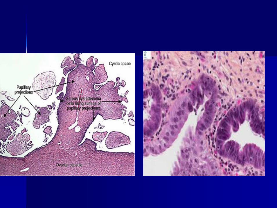
Serous Serous cystadenomacystadenomaThe most common benign epithelial tumour The most common benign epithelial tumour usually unilocular cyst with papilliferous usually unilocular cyst with papilliferous processes on the inner surface. processes on the inner surface. Psammoma bodies are concentric calcified Psammoma bodies are concentric calcified bodies which are more frequent in the bodies which are more frequent in the malignant counterpart. malignant counterpart. The cyst fluid is thin & serous. They are The cyst fluid is thin & serous. They are seldom as large as mucinous tumours.seldom as large as mucinous tumours.

Mucinous cystadenomaMucinous cystadenomaLargeLargeUnilateralUnilateralmultilocular cystsmultilocular cystssmooth inner surface.smooth inner surface.lining epithelium consists of columnar lining epithelium consists of columnar mucusmucus--secreting cells.secreting cells.The cyst fluid is thick & gelatinous.The cyst fluid is thick & gelatinous.
pseudomyxoma peritoneipseudomyxoma peritonei


EndometrioidEndometrioid tumours of tumours of the ovarythe ovary

Clear cell tumourClear cell tumour
arise from serosal cells arise from serosal cells showing little showing little differentiation. The differentiation. The typical histological typical histological appearance is of clear appearance is of clear (hobnail) cells arranged (hobnail) cells arranged in mixed pattern.in mixed pattern.

Brenner tumourBrenner tumour
arise from Wolffian arise from Wolffian metaplasia of the surface metaplasia of the surface epithelium.epithelium.
consists of islands of consists of islands of transitional epithelium in a transitional epithelium in a dense fibrotic stroma giving dense fibrotic stroma giving a solid appearance.a solid appearance.
The vast majority are The vast majority are benign. < 2 cm in benign. < 2 cm in diameter.diameter.
Some secrete oestrogenSome secrete oestrogen

Benign sex cord stromal tumours:Benign sex cord stromal tumours:Constitute a small percentage of Constitute a small percentage of benign ovarian tumours. benign ovarian tumours. They occur at any age from They occur at any age from prepubertal children to elderly, prepubertal children to elderly, postmenopausal women. postmenopausal women. Many secrete hormones & present Many secrete hormones & present with symptoms of inappropriate with symptoms of inappropriate hormone effects hormone effects

Granulosa cell tumorGranulosa cell tumorThese are malignant tumours but are These are malignant tumours but are mentioned here because they are mentioned here because they are generally confined to the ovary when they generally confined to the ovary when they present & so have a good prognosis present & so have a good prognosis

CallCall--Exner bodies are pathognomonic Exner bodies are pathognomonic but present in less than half of cases.but present in less than half of cases.Some secrete oestrogen or inhibin.Some secrete oestrogen or inhibin.

Theca cell tumourTheca cell tumourbenign, solid & unilateral benign, solid & unilateral Oestrogen secreted, cause systemic effects such Oestrogen secreted, cause systemic effects such as precocious puberty, postmenopausal bleeding, as precocious puberty, postmenopausal bleeding, endometrial hyperplasia & endometrial cancer endometrial hyperplasia & endometrial cancer rarely cause ascites or pleural effusion.rarely cause ascites or pleural effusion.

Fibroma: Fibroma: these are hard, mobile & lobulated with a glistening these are hard, mobile & lobulated with a glistening white surface. white surface. While ascites occur with many of the larger While ascites occur with many of the larger fibromas, Meig's syndrome fibromas, Meig's syndrome –– ascites & pleural ascites & pleural effusion in association with fibroma of the ovaryeffusion in association with fibroma of the ovary-- is is seen in only 1% of cases.seen in only 1% of cases.

SertoliSertoli--LeydigLeydig cell tumorcell tumor
usually of lowusually of low--grade malignancy, they grade malignancy, they are rare. are rare. Many produce androgens, & signs of Many produce androgens, & signs of virilization are seen in three quarters virilization are seen in three quarters of patients. Some secrete oestrogen of patients. Some secrete oestrogen

Presentation: Presentation:
Asymptomatic Asymptomatic painpainAbdominal swelling: noticed only when the Abdominal swelling: noticed only when the tumour tumour is veryis very large.large.Pressure effectsPressure effectsMenstrual disturbanceMenstrual disturbanceHormonal effectsHormonal effectsAbnormal cervical smearAbnormal cervical smear

Differential diagnosis of benign ovarian tumours: Differential diagnosis of benign ovarian tumours:
PainPainEctopic pregnancyEctopic pregnancySpontaneous abortionSpontaneous abortionPelvic inflammatory diseaePelvic inflammatory diseaeAppendicitisAppendicitisMeckel's diverticulumMeckel's diverticulumDiverticulitisDiverticulitis

Abdominal swellingAbdominal swellingPregnant uterus Pregnant uterus FibroidFibroidFull bladderFull bladderOvarian malignancyOvarian malignancyColorectal carcinomaColorectal carcinoma
Pressure effectsPressure effectsUrinary tract infectionUrinary tract infection
All other causes of menstrual irregularities, All other causes of menstrual irregularities, precocious puberty & postmenopausal precocious puberty & postmenopausal bleeding.bleeding.

Diagnosis:Diagnosis:HistoryHistory::ExaminationExamination::peritonismperitonism is an ominous sign.is an ominous sign.BimanualBimanual examination is essential for examination is essential for palpating the mass between the vaginal & palpating the mass between the vaginal & abdominal hands, its mobility, texture & abdominal hands, its mobility, texture & consistency, presence of palpable lymph nodes consistency, presence of palpable lymph nodes in the pouch of Douglas. Hard, irregular, fixed in the pouch of Douglas. Hard, irregular, fixed mass is likely to be invasive. mass is likely to be invasive.

Investigations:Investigations:
UltrasoundUltrasound: : mass size, consistency, mass size, consistency, and internal architecture. Bilatrality, and internal architecture. Bilatrality, ascitesascitesDoppler ultrasonographies to evaluate Doppler ultrasonographies to evaluate the resistive index of the mass vessels, the resistive index of the mass vessels, which, when low, indicate a malignancy. which, when low, indicate a malignancy. Radiological investigationsRadiological investigations

Blood test & serum markersBlood test & serum markers: : 1.1. serum CA 125 serum CA 125 2.2. betabeta--human chorionic gonadotrophin human chorionic gonadotrophin
((ββ--hCG) hCG) 3.3. Oestradiol Oestradiol 4.4. Androgen Androgen 5.5. alphaalpha--fetoprotein levels fetoprotein levels

problemproblem
The following masses pose the greatest concern: The following masses pose the greatest concern: Those that have a complex internal structure Those that have a complex internal structure Those that have solid components Those that have solid components associated with pain associated with pain Masses in prepubescent or postmenopausal Masses in prepubescent or postmenopausal women women Large cysts (cysts up to 10 cm have been Large cysts (cysts up to 10 cm have been followed conservatively) followed conservatively)

ManagementManagement::Criteria for observation of asymptomatic ovarian Criteria for observation of asymptomatic ovarian
tumour:tumour:UnilateralUnilateralUnilocularUnilocular cyst without solid componentscyst without solid componentsPremenopausalPremenopausal womenwomen tumour 3tumour 3--10 cm in 10 cm in diameterdiameterPostmenopausal Postmenopausal women tumourwomen tumour 22--6 cm in 6 cm in diameterdiameterNormal CA 125 ( <35mU/mL)Normal CA 125 ( <35mU/mL)No free fluid or masses suggesting No free fluid or masses suggesting omentalomental cake cake or matted bowel loops.or matted bowel loops.

Observation include follow up with US Observation include follow up with US after 3 months, if the cyst is the same after 3 months, if the cyst is the same follow up with US & CA 125 level follow up with US & CA 125 level will be safe.will be safe.
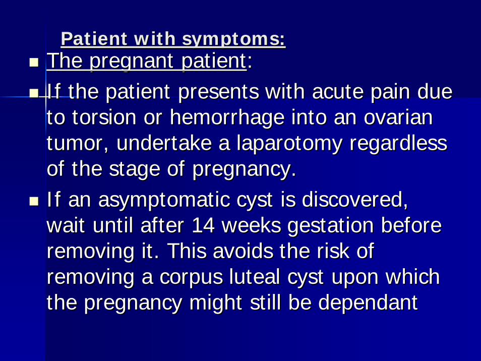
Patient with symptoms:Patient with symptoms:The pregnant patientThe pregnant patient::If the patient presents with acute pain due If the patient presents with acute pain due to torsion or hemorrhage into an ovarian to torsion or hemorrhage into an ovarian tumor, tumor, undertakeundertake a laparotomy regardless a laparotomy regardless of the stage of pregnancy. of the stage of pregnancy. If an asymptomatic cyst is discovered, If an asymptomatic cyst is discovered, waitwait until after 14 weeks gestation before until after 14 weeks gestation before removing it. This avoids the risk of removing it. This avoids the risk of removing a corpus luteal cyst upon which removing a corpus luteal cyst upon which the pregnancy might still be dependant the pregnancy might still be dependant

In the second & third trimesters. Cysts less than In the second & third trimesters. Cysts less than 10 cm in diameter that have a simple 10 cm in diameter that have a simple appearance on ultrasound appearance on ultrasound maymay bebe followed followed ultrasonografically.ultrasonografically. If the cyst is unresolved 6 If the cyst is unresolved 6 weeks postpartum, surgery weeks postpartum, surgery undertakenundertaken..a cyst with features suggestive of a cyst with features suggestive of malignancy on ultrasound or one that is malignancy on ultrasound or one that is growing should be removed growing should be removed surgically. surgically. ManagementManagement may include a Caesarian may include a Caesarian hysterectomy, bilateral salpingohysterectomy, bilateral salpingo--oophorectomy & omentectomy. oophorectomy & omentectomy.

Treatment:Treatment:Laparoscopic procedures: Laparoscopic procedures:
Indications of laparoscopy:Indications of laparoscopy:Uncertainty about the nature of the mass.Uncertainty about the nature of the mass.Tumour suitable for laparoscopic surgery:Tumour suitable for laparoscopic surgery:–– age <35 years.age <35 years.–– ultrasound show no solid component.ultrasound show no solid component.–– simple ovarian cyst.simple ovarian cyst.–– endometriomaendometrioma..

Laparotomy: Laparotomy: IfIf there is any possibility of invasive there is any possibility of invasive disease, a longitudinal skin incision. disease, a longitudinal skin incision. AA sample of sample of asciticascitic fluid or peritoneal fluid or peritoneal washings should be sent for cytological washings should be sent for cytological examination at the beginning of the examination at the beginning of the operation. operation. exploreexplore the whole abdomen thoroughly & the whole abdomen thoroughly & inspectinspect both ovaries.both ovaries.

Age < 35 years old ovarian cystectomy Age < 35 years old ovarian cystectomy Age > 44 years with a unilateral ovarian mass, Age > 44 years with a unilateral ovarian mass, total abdominal hysterectomy, bilateral total abdominal hysterectomy, bilateral salpingosalpingo--oopherectomy & infracolic oopherectomy & infracolic omentectomy. omentectomy. Age 35Age 35--44 years treatment should be 44 years treatment should be individualized. If conservative surgery is individualized. If conservative surgery is planned, preliminary hysteroscopy & curettage planned, preliminary hysteroscopy & curettage of the uterus are essential to exclude a of the uterus are essential to exclude a concomitant endometrial tumour concomitant endometrial tumour