bilateral weight bearing ct imaging for foot · pdf filebilateral weight bearing ct imaging...
TRANSCRIPT
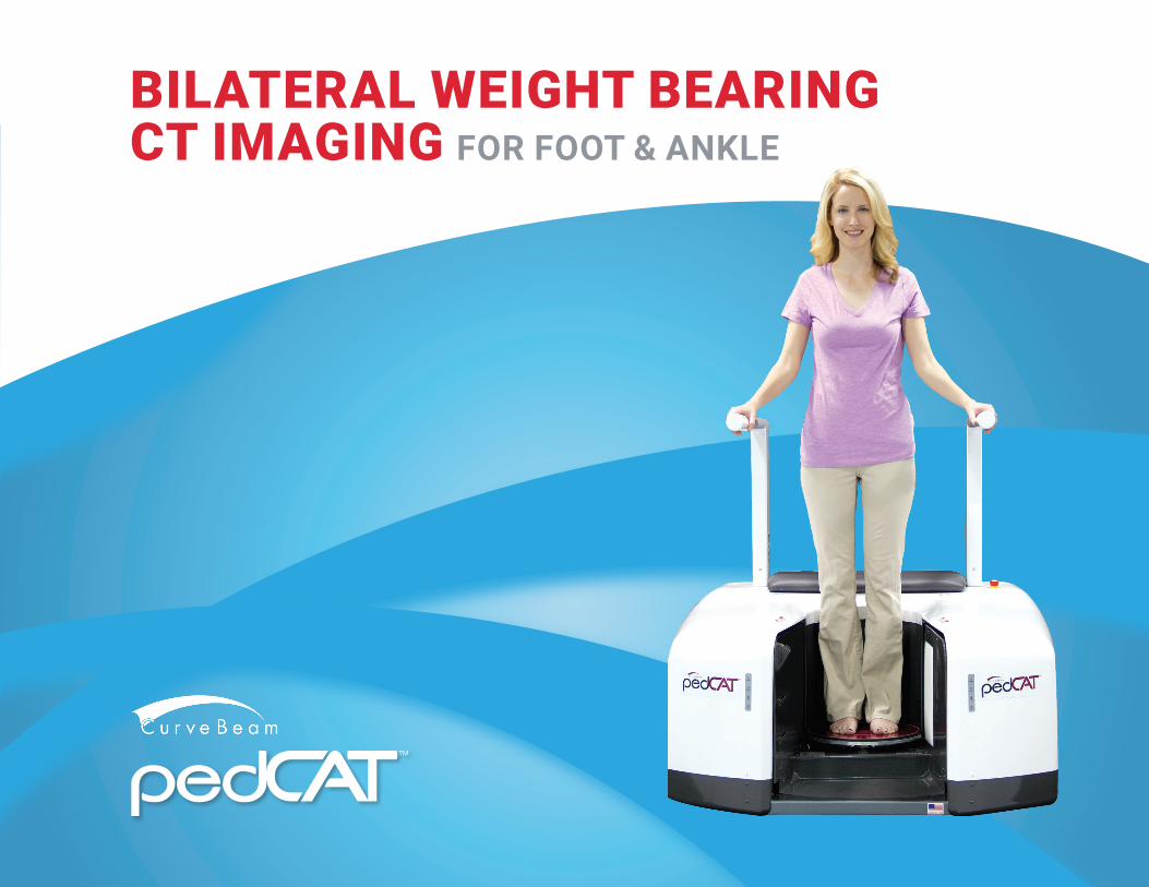
BILATERAL WEIGHT BEARING CT IMAGING FOR FOOT & ANKLE
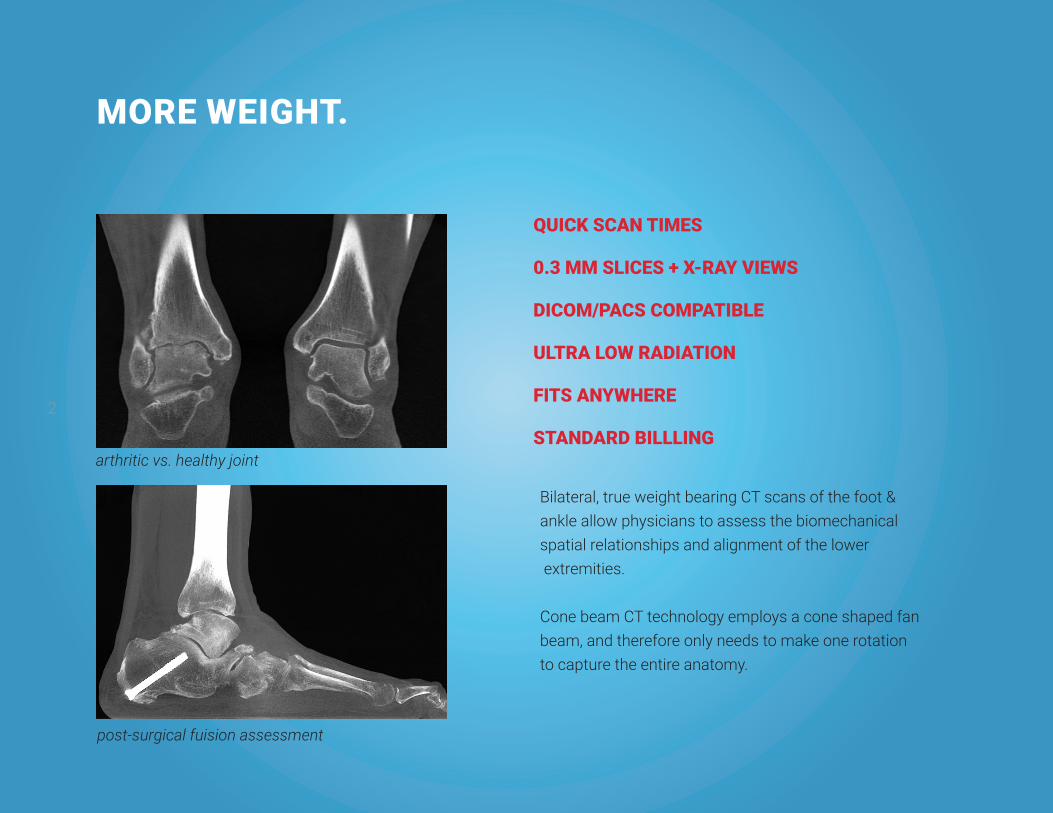
MORE WEIGHT.
Bilateral, true weight bearing CT scans of the foot & ankle allow physicians to assess the biomechanical spatial relationships and alignment of the lower extremities.
Cone beam CT technology employs a cone shaped fan beam, and therefore only needs to make one rotation to capture the entire anatomy.
2
QUICK SCAN TIMES
0.3 MM SLICES + X-RAY VIEWS
DICOM/PACS COMPATIBLE
ULTRA LOW RADIATION
FITS ANYWHERE
STANDARD BILLLINGarthritic vs. healthy joint
post-surgical fuision assessment

3
LESS WAIT.20 seconds for a partial foot scan 48 seconds for a bilateral scan
Scan in weight bearing or seated position.
patient in high heels
fracture

4
PRECISE IMAGINGUltra thin slices - 0.3 mm
3D reconstructions, Multi-Planar slices, and 2D X-Ray views
ADVANCED TOOLSCubeVue, CurveBeam’s custom visualization software, provides multiple alignment measurement tools.
CubeVue Insta-X feature automatically generates standard X-Ray views from the 3D data
CubeVue - 3D & MPR windows
CubeVue - Insta-X windows

5
ULTRA LOW DOSE
Lisfranc injury - elevated metatarsals
rotated sesamoidssyndesmosis - coronal
Technique Micro Sieverts Daily Background Exposure 8
pedCAT Cone Beam CT, medium FOV scan (partial single foot)
2 (2)
pedCAT Cone Beam CT, large FOV scan (both feet in entirety)
5 (2)
Extremity Film X-ray 1 (1)
Extremity Medical CT 25 – 1000 (2), (3), (4)
(1)Radiologyinfo.org developed jointly by American College of Radiology and Radiological Society of North America. www.radiologyinfo.org. (2) John B. Ludlow, Marija Ivanovic, Weightbearing CBCT, MDCT, and 2D Imaging Dosimetry of the Foot & Ankle, International Journal of Diagnostic Imaging, 2014, Vol. I, No. 2(3) Nagel HD. Dose values from CT examinations. In: Nagel HD, ed. Radiation exposure in comput-ed tomography. Hamburg, Germany: CTB Publications, 2002:15 -24(4) Debdut Biswas, BA, Jesse E. Bible, BS, Michael Bohan, BS, Andrew K. Simpson, MD, Peter G.Whang, MD, and Jonathan N. Grauer, MD, Department of Orthopaedics and Rehabilitation, Yale University School of Medicine, New Haven, and Yale-New Haven Hospital, New Haven, ConnecticutRadiation Exposure from Musculoskeletal Computerized Tomographic Scans, J Bone Joint Surg Am. 2009;91:1882-9
syndesmosis - axial

6
FITS ANYWHERE
• Small footprint - 48” x 58”
• Minimal shielding
• Standard 115VAC (220 VAC international) outlet
• No extra heating or cooling required
Podiatric OfficeOrthopedic Office Imaging CenterAmbulatory Surgery Center Hospital/ ED
CubeVue enabled PC & medical grade
monitor
Acquisition console, system server & UPS
Operator console

7
forefoot dislocation
FEATURES AND SPECIFICATIONS
Technical Specifications3D Imaging Volume 20cm (height) x 35cm (diameter) and smaller
Resolution 0.3mm, 0.37mm voxel sizesProcedure Time 20-48 seconds
Max Exposure Time 9 secondsTube Voltage 100-120 kVpTube Current 5 mAImage Detector Amorphous silicon flat panelGray Scale 16 bit
Dimensions 4ft (h) x 4ft (w) x 5ft (d)
Weight 400lbsPower Requirements 1500VA
coalition
post-surgical assessment (non-union) midfoot dislocation
ApprovalsUS FDA 510(k)Health CanadaCE Marking China FDAAustralia TGA Saudi FDA Taiwan FDAHong Kong FDA
• Small footprint - 48” x 58”
• Minimal shielding
• Standard 115VAC (220 VAC international) outlet
• No extra heating or cooling required
US ReimbursementCPT Code 73700 - CT lower extremity without contrast
CurveBeam.com | [email protected] | 866.400.0035
About CurveBeam
CurveBeam designs and manufactures Cone Beam CT imaging equipment
for the orthopedic and podiatric specialties. CurveBeam was founded in
2009 and is privately owned and operated.
CurveBeam’s corporate office is located in Warrington, Pennsylvania, USA.
CurveBeam’s Europe office is located in London, United Kingdom.
The core team behind CurveBeam developed and pioneered the first
commercially viable Cone Beam CT imaging systems for the dental/maxilo-
facial specialties and has been expert in the field since the 1990s.
Rev F72 - 4.3