biochemical characterization of the phosphatase...
TRANSCRIPT
Biochemical Characterization of the Phosphatase Domain of theTumor Suppressor PH Domain Leucine-Rich Repeat ProteinPhosphataseEmma Sierecki† and Alexandra C. Newton*
Department of Pharmacology, University of California San Diego, La Jolla, California 92093, United States
*S Supporting Information
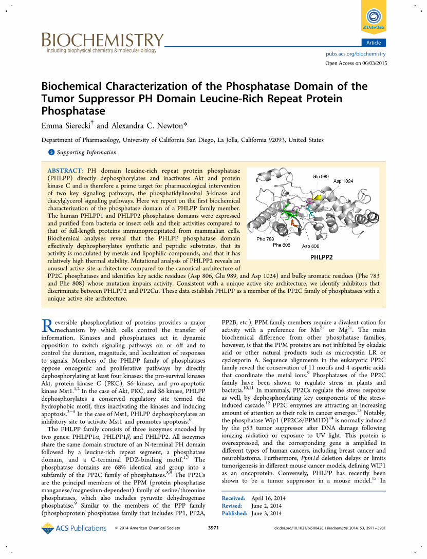
ABSTRACT: PH domain leucine-rich repeat protein phosphatase(PHLPP) directly dephosphorylates and inactivates Akt and proteinkinase C and is therefore a prime target for pharmacological interventionof two key signaling pathways, the phosphatidylinositol 3-kinase anddiacylglycerol signaling pathways. Here we report on the first biochemicalcharacterization of the phosphatase domain of a PHLPP family member.The human PHLPP1 and PHLPP2 phosphatase domains were expressedand purified from bacteria or insect cells and their activities compared tothat of full-length proteins immunoprecipitated from mammalian cells.Biochemical analyses reveal that the PHLPP phosphatase domaineffectively dephosphorylates synthetic and peptidic substrates, that itsactivity is modulated by metals and lipophilic compounds, and that it hasrelatively high thermal stability. Mutational analysis of PHLPP2 reveals anunusual active site architecture compared to the canonical architecture ofPP2C phosphatases and identifies key acidic residues (Asp 806, Glu 989, and Asp 1024) and bulky aromatic residues (Phe 783and Phe 808) whose mutation impairs activity. Consistent with a unique active site architecture, we identify inhibitors thatdiscriminate between PHLPP2 and PP2Cα. These data establish PHLPP as a member of the PP2C family of phosphatases with aunique active site architecture.
Reversible phosphorylation of proteins provides a majormechanism by which cells control the transfer of
information. Kinases and phosphatases act in dynamicopposition to switch signaling pathways on or off and tocontrol the duration, magnitude, and localization of responsesto signals. Members of the PHLPP family of phosphatasesoppose oncogenic and proliferative pathways by directlydephosphorylating at least four kinases: the pro-survival kinasesAkt, protein kinase C (PKC), S6 kinase, and pro-apoptotickinase Mst1.1,2 In the case of Akt, PKC, and S6 kinase, PHLPPdephosphorylates a conserved regulatory site termed thehydrophobic motif, thus inactivating the kinases and inducingapoptosis.3−5 In the case of Mst1, PHLPP dephosphorylates aninhibitory site to activate Mst1 and promotes apoptosis.6
The PHLPP family consists of three isozymes encoded bytwo genes: PHLPP1α, PHLPP1β, and PHLPP2. All isozymesshare the same domain structure of an N-terminal PH domainfollowed by a leucine-rich repeat segment, a phosphatasedomain, and a C-terminal PDZ-binding motif.1,7 Thephosphatase domains are 68% identical and group into asubfamily of the PP2C family of phosphatases.8,9 The PP2Csare the principal members of the PPM (protein phosphatasemanganese/magnesium-dependent) family of serine/threoninephosphatases, which also includes pyruvate dehydrogenasephosphatase.9 Similar to the members of the PPP family(phosphoprotein phosphatase family that includes PP1, PP2A,
PP2B, etc.), PPM family members require a divalent cation foractivity with a preference for Mn2+ or Mg2+. The mainbiochemical difference from other phosphatase families,however, is that the PPM proteins are not inhibited by okadaicacid or other natural products such as microcystin LR orcyclosporin A. Sequence alignments in the eukaryotic PP2Cfamily reveal the conservation of 11 motifs and 4 aspartic acidsthat coordinate the metal ions.9 Phosphatases of the PP2Cfamily have been shown to regulate stress in plants andbacteria.10,11 In mammals, PP2Cs regulate the stress responseas well, by dephosphorylating key components of the stress-induced cascade.12 PP2C enzymes are attracting an increasingamount of attention as their role in cancer emerges.13 Notably,the phosphatase Wip1 (PP2Cδ/PPM1D)14 is normally inducedby the p53 tumor suppressor after DNA damage followingionizing radiation or exposure to UV light. This protein isoverexpressed, and the corresponding gene is amplified indifferent types of human cancers, including breast cancer andneuroblastoma. Furthermore, Ppm1d deletion delays or limitstumorigenesis in different mouse cancer models, defining WIP1as an oncoprotein. Conversely, PHLPP has recently beenshown to be a tumor suppressor in a mouse model.15 In
Received: April 16, 2014Revised: June 2, 2014Published: June 3, 2014
Article
pubs.acs.org/biochemistry
© 2014 American Chemical Society 3971 dx.doi.org/10.1021/bi500428j | Biochemistry 2014, 53, 3971−3981
Open Access on 06/03/2015
humans, PHLPP is frequently lost or reduced, at the gene orprotein level, in diverse cancers, including colon16−18 andbreast19,20 cancers and glioblastoma.21,22 Dysregulation ofPHLPP correlates not only with cancer but also with diseasessuch as obesity, where PHLPP1 is upregulated.23 It also plays aregulatory role in the heart via its negative regulation of Akt.24
Given the broad role of PHLPP in a wide spectrum of diseases,understanding its catalytic mechanisms is essential to itsdevelopment as a therapeutic target.This contribution provides a biochemical and structural
characterization of the intrinsic catalytic activity of PHLPP.Using isolated PP2C domains purified from bacteria, insectcells, or mammalian cells, as well as full-length proteins frommammalian expression, we determine the metal-dependent,pH-dependent activation. We examine the effects of variousfactors on activity, such as reducing potential or lipophiliccompounds. We also describe inactivating mutations in thecatalytic domain.
■ EXPERIMENTAL PROCEDURESBL21(DE3)pLysS competent cells were purchased fromEMD4Biosciences. EGF was purchased from Upstate. Themonoclonal antibody against HA (Covance) and the polyclonalantibody against PHLPP2 (Bethyl) were used to assessexpression. A monoclonal anti-HA antibody (Roche) andprotein A/G Ultralink resin (Pierce) were used forimmunoprecipitation. Palmitoleic and linoleic acids wereobtained from Acros, and stearic acid was from AlfaAesar,oleic acid from Sigma, arachidonic acid from Biomol, andelaidic acid from MP. Ammonium molybdate (Fisher) andmalachite green oxalate (Acros) were obtained at the highestgrade possible. Protein phosphatase peptide substrate(RRAPTVA) and protein tyrosine phosphatase peptidesubstrate (ENDPYINASL) were obtained from Enzo, andHFPQFPSYSAS was ordered from Anaspec. COS7 cells aremaintained in DMEM (Cellgro) supplemented with 5% FBS(Hydroclone) and 1% penicillin/streptomycin at 37 °C in 5%CO2.Purification of the GST-Tagged Phosphatase Domain
of PHLPP1 and PHLPP2. The GST-tagged fusion protein ofthe phosphatase domain of human PHLPP1 (Pro 627−Pro990, PHLPP1α numbering) or PHLPP2 (Gly 745−Asn 1102)was expressed in competent BL21(DE3)pLysS cells andpurified according to the method previously described.3
Purification yielded 15 ± 1 or 14 ± 1 mg/(L of culture),respectively. Purity was assessed on a sodium dodecyl sulfate(SDS) gel and was >90%. The protein was then either kept in adesalting buffer [20 mM Tris and 100 mM NaCl (pH 7.0)] ordialyzed against 1 L of 20 mM Tris-HCl, 2 mM MnCl2, orEDTA (pH 7.0). The enzymes were stored at 4 °C.Purification of the His-Tagged Phosphatase Domain
of PHLPP2. His-tagged PHLPP2 PP2C was expressed andpurified from baculovirus-infected Sf21 cells. Sf21 cells weremaintained in Hink’s TNM-FH medium (Cellgro), supple-mented with 10% FBS and 1% penicillin/streptomycin, andincubated for 4 days with a baculovirus encoding His-PHLPP2PP2C. Purification was conducted using the IMAC purificationkit on Profinia (Bio-Rad), yielding 87 ± 5 μg of purifiedprotein/L of culture medium. Purity was assessed byCoomassie staining of SDS gels and was >90%.Purification of the HA-Tagged Phosphatase Domain
and the Full-Length Protein of PHLPP. The isolatedphosphatase domains of human PHLPP1 and full-length
PHLPP1α, PHLPP1β, and PHLPP2 were overexpressed inCOS7 cells as HA-tagged fusion proteins using FuGENE 6transfection reagent (Promega). Cells were treated with 10 ngof EGF/mL of DMEM for 15 min prior to lysis andimmunoprecipitated as previously described.3
Plasmid Constructs. Mutant constructs were generated bysite-directed mutagenesis of pcDNA3-HA-PHLPP24 using theQuikChange protocol (Agilent Technologies).
Enzymatic Activity Assay. The enzymatic reaction wasconducted at room temperature in a reaction mixturecontaining the appropriate pH buffer at 100 mM (see Table1 of the Supporting Information), 0.02 mg/mL BSA, 4 mMDTT, and 100 mM NaCl, at the pH values noted in thelegends. Divalent cations or inhibitors were added as noted. Inthe case of the isolated PP2C domain purified from bacteria orinsect cells, an exact volume of a solution of a knownconcentration of protein was added to the reaction mixture.When enzymes purified from mammalian cells were used, theA/G beads supporting the proteins were added to the reactionmixture. The relative amount of protein in the assay wasdetermined by Western blot. To determine the kineticparameters kcat and KM, all data were fit to a Michaelis−Menten model, using nonlinear regression.
p-Nitrophenyl Phosphate (pNPP) Assay. This assay waspreviously described.25 Briefly, each well contained 125 μL ofreaction mix (see above), 1 μM enzyme, and variousconcentrations of PNPP. Optical density was recorded overtime at 405 nm. Absorbance was then plotted over time, andthe slope was calculated.
Malachite Green Assay. The malachite green assay used inthis paper was a slightly modified version of the protocoldescribed previously.26 A fresh solution of reagent was preparedfrom 3 volumes of a 34 mM solution of ammonium molybdatein 4 M hydrochloric acid mixed with 1 volume of 518 μMmalachite green in water. Tween 20 was added to a finalconcentration of 1% (v/v). The reaction was stopped byaddition of 65 μL of the reagent to 65 μL of reaction mixture.The reagent was added to the reaction mixture at different timepoints, and the color was allowed to develop for 15 min.Absorbance was recorded at 650 nm. A calibration curve wasobtained each time. The measured OD was converted to theamount of inorganic phosphate released, and data were plottedversus time. The initial slope (picomoles per second), measuredbetween 0 and 25% conversion, was divided by the amount ofprotein (picomoles) to determine the velocity (inverseseconds).
Inhibitors. The following compounds from NCI DiversitySet I were tested: NSC 170008, 7-acetyl-6-ethyl-3,5,8-trihydroxy-9,10-dioxoanthracene-1,2-dicarboxylic acid; NSC73101, 1,2,3,4,7,7-hexachloro-6-(9H-fluoren-2-ylcarbamoyl)-bicyclo[2.2.1]hept-2-ene-5-carboxylic acid; NSC 11241, C.I.Basic Red 6; NSC 270127, N-(2-benzoyl-4-chlorophenyl)-2-chloroacetamide
■ RESULTSKinetic Data. The isolated phosphatase domains of
PHLPP1 and PHLPP2 were expressed in bacteria as N-terminally tagged GST fusion proteins, purified by GST affinitychromatography, and their kinetic parameters were examined.The GST tag had no effect on the enzymatic activity of eitherconstruct (data not shown) and thus was not removed for theanalyses presented. Using pNPP as a substrate, we determinedthe KM and kcat for purified PHLPP1 and PHLPP2 phosphatase
Biochemistry Article
dx.doi.org/10.1021/bi500428j | Biochemistry 2014, 53, 3971−39813972

domains (Figure 1a). The kinetic profiles of both isozymeswere nearly identical: maximal turnover rates, reflected by thekcat values, were (1.8 ± 0.3) × 10−3 s−1 for PHLPP1 and (1.8 ±0.1) × 10−3 s−1 for PHLPP2. Substrate binding energetics forpNPP were also very similar as reflected by KM values of 2.1 ±0.2 and 1.80 ± 0.08 mM for PHLPP1 and PHLPP2,respectively.We next compared the kinetic parameters of isolated
phosphatase domains expressed in bacteria, baculovirus, andmammalian cells (Table 1). The GST-tagged PP2C domain ofPHLPP2 was purified from Sf21 cells after infection bybaculovirus; the HA-tagged PP2C domain of PHLPP1 wasexpressed in COS7 cells and immunoprecipitated using a HAantibody. The KM for pNPP for the PHLPP2 phosphatasedomain purified from insect cells (1.3 ± 0.2 mM) orimmunoprecipitated from mammalian cells (1.5 ± 0.5 mM)was comparable to that for bacterially expressed PHLPP2. Incontrast, the kcat was 200-fold higher for the protein isolatedfrom insect cells (0.37 ± 0.08 s−1) than for the protein purifiedfrom bacteria [(1.8 ± 0.1) × 10−3 s−1]. Because theconcentration of phosphatase was not measured for mammalianphosphatase, kcat was not determined. These data reveal that
the affinity of PHLPP for pNPP is similar regardless of thesource of the protein; however, the catalytic activity is 2 ordersof magnitude higher for phosphatase purified from insect cells(which better mimic the milieu of mammalian cells) thanbacteria.Phosphopeptides were also subjected to PHLPP dephos-
phorylation (Figure 1b). Two sequences were used: athreonine-phosphorylated peptide (RRAPTVA) that is astandard substrate for PP2C27 and a serine-phosphorylatedpeptide (HFPQFpSYSAS), corresponding to the sequence ofthe hydrophobic motif of Akt, a physiologically relevantsubstrate of PHLPP. Both peptides were significantly bettersubstrates for PHLPP than the synthetic substrate pNPP. First,they were dephosphorylated more rapidly than pNPP asreflected by considerably higher kcat values, a difference mostnotable for the bacterially expressed enzyme where kcat wasmore than 600-fold higher for the hydrophobic motif peptide(1.1 ± 0.2 s−1) than for pNPP [(1.8 ± 0.1) × 10−3 s−1].Second, the KM values were >1 order of magnitude lower.Taken together, the efficiency of the reaction, evaluated by thekcat/KM ratio, was enhanced by 2 to ∼4 orders of magnitudedepending on the enzyme source [Table 1; kcat/KM ratio of (8
Figure 1. PHLPP activity is highly dependent on the substrate. (a) PHLPP1 and PHLPP2 have similar phosphatase activities in vitro. The saturatingkinetics of the phosphatase domains of PHLPP1 (empty diamonds) and PHLPP2 (gray squares) with increasing concentrations of the substratepNPP is shown. The rates were determined by measuring the production of pNP at 405 nm in Tricine buffer (pH 7.5). The data are fit to theMichaelis−Menten equation. The graph shows mean values ± SEM for three separate experiments. (b) Activities of the phosphatase domain ofPHLPP2 isolated from bacteria or insect cells are comparable on peptidic substrates. The saturating kinetics of the phosphatase domains of PHLPP2isolated from Sf21 cells (black circles) or Escherichia coli BL21(DE3)pLysS (gray squares) with increasing concentrations of the substrate peptide isshown. The rates were determined by measuring the liberation of free phosphate by the malachite green assay in a Tricine buffer (pH 7.5). The dataare fit to the Michaelis−Menten equation. The graph shows mean values ± SEM for three separate experiments.
Table 1. Kinetic Parametersa
substrate expression system isoform domain kcat (s−1) KM (M) kcat/KM (s−1 M−1)
pNPP bacteria PHLPP1 PP2C (1.8 ± 0.3) × 10−3 (2.1 ± 0.2) × 10−3 0.9 ± 0.2bacteria PHLPP2 PP2C (1.8 ± 0.1) × 10−3 (1.80 ± 0.08) × 10−3 1.0 ± 0.1insect PHLPP2 PP2C (370 ± 80) × 10−3 (1.3 ± 0.2) × 10−3 285 ± 9mammal PHLPP1 PP2C not determined (1.5 ± 0.5) × 10−3 −mammal PHLPP1 full-length not determined (1.5 ± 0.5) × 10−3 −
RRAPTVA bacteria PHLPP2 PP2C 0.15 ± 0.04 (64 ± 3) × 10−6 (2.3 ± 0.7) × 103
insect PHLPP2 PP2C 1.1 ± 0.3 (63 ± 3) × 10−6 (18 ± 5) × 103
HFPQFPSYSAS bacteria PHLPP2 PP2C 1.1 ± 0.2 (139 ± 1) × 10−6 (8 ± 2) × 103
insect PHLPP2 PP2C 0.9 ± 0.2 (40.5 ± 0.3) × 10−6 (22 ± 5) × 103
aPHLPP1 and PHLPP2 phosphatase domains and full-length proteins were obtained as fusion proteins using different expression systems: bacteria[BL21(DE3)pLysS cells], insect cells (Sf21), or mammalian cells (COS7). kcat and KM were determined using either pNPP or peptides as a substrate.Dephosphorylation of pNPP was measured by recording the OD at 405 nm, and dephosphorylation of peptides was assessed using the malachitegreen assay. Reactions occurred in Tricine buffer (pH 7.5). All data were fit to the Michaelis−Menten equation. Data are means ± SEM.
Biochemistry Article
dx.doi.org/10.1021/bi500428j | Biochemistry 2014, 53, 3971−39813973

± 2) × 103 for the hydrophobic motif peptide compared to avalue of 1.0 ± 0.1 using bacterially expressed PHLPP2]. Themost notable feature was that the catalytic rate of the bacteriallyexpressed phosphatase domain was comparable to that of insectcell-expressed phosphatase when peptides were used assubstrates. Thus, the phosphatase domain of PHLPP effectivelydephosphorylates phosphopeptides.pH Profiles. The pH dependence for the dephosphorylation
of pNPP by the bacterially expressed phosphatase domains ofPHLPP1 and PHLPP2 was examined in the absence of addedMn2+. Both enzymes had detectable catalytic activity betweenpH 5 and 9 (Figure 2a). kcat and kcat/KM were then determinedfor various pH values between 5.5 and 8.5. The profiles ofactivity of the two isozymes were very similar. The plot of kcatversus pH reveals two peaks of activity at pH 6.0 and 7.8(Figure 2b). As a general trend, PHLPP1 activity decreased upto 5-fold under basic conditions (pH >8). The data could be fitto a model28 in which one residue is deprotonated. With thismodel, the ionization pH for PHLPP1 is 7.1 ± 0.2 and its pH-independent kcat value is (2.2 ± 0.5) × 10−3 s−1. With the samemodel, the kcat for PHLPP2 does not seem to depend on thepH, at least in this pH range.The kcat/KM versus pH plot (Figure 2c), however, confirms
the existence of two important ionizations for catalysis set atpH 7.84 ± 0.05 and 8.28 ± 0.05 for PHLPP1 and pH 7.84 ±0.07 and 8.2 ± 0.1 for PHLPP2. The pH-independent kcat/KM
for PHLPP1 [(2.940 ± 0.001) × 10−2 M−1 s−1] is comparableto the value for PHLPP2 [(2.017 ± 0.008) × 10−2 M−1 s−1],
stressing once again the strong similarity between the twoisoforms.
Metal Requirement. Next we examined the effect ofdifferent metallic divalent cations on the enzymatic activity ofPHLPP. EDTA, Mg2+, Ca2+, Cu2+, or Ni2+ was added atincreasing concentrations to the reaction mixture, and the initialrate of dephosphorylation was determined at a saturatingsubstrate concentration, using the phosphatase domain ofPHLPP2 purified from bacteria (Figure 3a−c). In every case,the enzyme was active without addition of a metallic ion. Thisactivity was insensitive to EDTA, even at a concentration of 100mM. Mg2+ did not enhance activity; rather, modest inhibitionwas observed at concentrations of >10 mM. Ca2+, Cu2+, andNi2+ inhibited the phosphatase, with half-maximal inhibitionobserved in the presence of approximately 40, 10, and 8 mMdivalent cation, respectively. Zn2+ was the strongest inhibitorwith complete inhibition observed in the presence of 1 mMZnCl2 (data not shown).The influence of Mn2+ on activity was more complex than
expected (Figure 3d). According to the literature, Mn2+ is anactivator of the PP2C family,29 to various degrees depending onthe substrate.30,31 We indeed found that addition of MnCl2 tothe reaction mixture at pH 7.5 increased PHLPP activity whenthe proteins were purified from insect or mammalian cells. A 7-fold increase was observed with the phosphatase domain ofPHLPP2 purified from Sf21 cells, and a more modest 2-foldincrease occurred in the case of PHLPP1 overexpressed inCOS7 cells. However, when bacterial preparations were used asthe enzyme, addition of MnCl2 to the reaction mixture at pH
Figure 2. Effect of pH on the kinetic parameters. (a) Activity of PHLPP1 (empty diamonds) and PHLPP2 (gray squares) phosphatase domains wasevaluated at different pH values. Reactions occurred at 10 mM pNPP and 100 mM pH-appropriate buffer (see Table 1 of the SupportingInformation). Graphs show means ± SEM for at least three separate experiments. (b) kcat and (c) kcat/KM for PHLPP1 and PHLPP2 phosphatasedomains evaluated at different pH values. Saturating kinetics were determined with increasing concentrations of the substrate pNPP (typically 1−15mM) in 100 mM buffer, and data were fit to a Michaelis−Menten equation. pH data in panel c are fit to the equation v = C/(1+ H/Ka + H/Kb).Graphs show means ± SEM.
Biochemistry Article
dx.doi.org/10.1021/bi500428j | Biochemistry 2014, 53, 3971−39813974

7.5 resulted in a decrease in activity, by 70% in the case ofPHLPP1 and 50% for PHLPP2. To understand this apparentlyconflicting result, we examined the dependence of this effect onthe pH of the reaction mixture. MnCl2 was added to a finalconcentration of 1−20 mM to a reaction mixture buffered atvarious pH values, between pH 6 and 8.5 (Figure 3e,f). Thedata show that Mn2+ acts as an inhibitor in both cases, belowpH 8.0. Addition of 10 mM MnCl2 to a reaction mixtureresulted in an average 60% inhibition at these pH values.However, at pH 8.2 and 8.5, PHLPP1 showed a maximal 8-foldboost in activity (Figure 3e), and a 4-fold increase was observedfor PHLPP2 (Figure 3f). Thus, Mn2+ is inhibitory below pH 8and activating above pH 8.Influence of Other Parameters. Varying the ionic
strength of the reaction mixture (set by varying theconcentration of sodium chloride in the reaction mixturefrom 1 to 500 mM) had no effect on the dephosphorylationrate of either PHLPP1 or PHLPP2 (data not shown). Thechemical nature of the buffer also did not have any significantimpact on the activity (Table 1 of the Supporting Information).
However, the concentration of DTT in the solution plays a rolein the enzymatic reaction (Figure 4a). Addition of 0.4−8 mMDTT to the reaction mixture significantly enhanced PHLPP1activity with a maximal 3-fold increase at low DTTconcentrations (<3 mM). On the other hand, DTT had anegligible effect on PHLPP2 activity at concentrations of <10mM. More interestingly, DTT variation revealed the first smallbut significant difference between PHLPP2 and PHLPP1activities, PHLPP2 being ∼2-fold more active than PHLPP1 inthe absence of DTT.It has been reported that PP2Cα and PP2Cβ are activated by
fatty acids.31−33 Specifically, fatty acids enhance the affinity forMn2+, such that they increase the activity recorded atsubsaturating concentrations of Mn2+. We thus tested theeffects of various fatty acids on the kinetic parameters ofPHLPP, using the phosphatase domain of PHLPP2 purifiedfrom insect cells as an example. Fatty acids (100 μM) of variouslengths (16−20 carbons), saturated state (zero to four doublebonds), and configuration (cis vs trans double bond) weretested in the presence of 1 mM MnCl2 (Figure 4b). We found
Figure 3. Influence of the cation on PHLPP activity. Relative activity of PHLPP2 phosphatase domains at 10 mM pNPP with increasingconcentrations of (a) EDTA (red) or MgCl2 (yellow), (b) CaCl2, or (c) CuSO4 (blue) or NiSO4 (orange). The activity is given as a percentage ofthe activity of the protein in the absence of additive. (d) Relative activity of PHLPP2 phosphatase domains isolated from insect cells (black circles)or bacteria (gray squares) and full-length PHLPP1 immunoprecipitated from mammalian cells (white triangles) or the PHLPP1 phosphatase domainfrom bacteria (white diamonds) at 10 mM pNPP with increasing concentrations of MnCl2. k values for PHLPP1 (e) and PHLPP2 (f) phosphatasedomains purified from BL21(DE3)pLysS cells were evaluated at 10 mM pNPP, at different pH values in the absence (empty symbols) or presence(black symbols) of 10 mM MnCl2. Graphs represent means ± SEM for three separate experiments.
Biochemistry Article
dx.doi.org/10.1021/bi500428j | Biochemistry 2014, 53, 3971−39813975

that only oleic acid (18:1 cis) and arachidonic acid (20:4) wereable to increase PHLPP2 activity, by 30 and 50%, respectively.Palmitoleic acid was slightly inhibitory under these conditions.The effects of oleic and arachidonic acids were studied in moredetail and revealed that activation of PHLPP started between30 and 100 μM and increased with the concentration of thecompound (Figure 4c). It should be noted that elaidic acid(18:1 trans), the isomer of oleic acid, did not change thePHLPP activity at any concentration tested (data not shown).These results are in good agreement with previous studiesshowing the importance of the configuration of the doublebond.33 We also verified that the effects of fatty acids werelinked to the presence of Mn2+. Indeed, addition of any fattyacid in solutions containing no supplementary Mn2+ had noeffect on the activity of the protein (data not shown).Thermal Stability of the Domain. The thermal stability of
the isolated phosphatase domain, purified from Escherichia coli,was evaluated. First, we used circular dichroism (CD) tocompare global structural states of PHLPP1 and PHLPP2(Figure 5a). Little difference was observed in the CD spectra ofthe two proteins. The phosphatase domain of PHLPP2 wasthen submitted to increasing temperatures, and effects on
protein structure were assessed using CD (Figure 5b). Changesin the slope of residue ellipticity versus temperature wereinterpreted as structural variations, in this case, unfolding of theprotein. The PP2C domain of PHLPP2 had a temperature ofdenaturation of 50 °C in the reaction buffer. The sametemperature of denaturation could be obtained usingfluorescence polarization as a readout. PHLPP1 and PHLPP2could then be compared, with PHLPP1 being unfolded at aslightly higher temperature (58 °C). We then measured theenzymatic activity of the PHLPP1 or PHLPP2 phosphatasedomain using a fluorescent substrate, difluoromethyl umbeli-ferryl phosphate (DiFMUP). The dephosphorylation rateincreased with an increase in temperature, up to a plateau,and then the enzyme slowly lost activity as heat denatured it(Figure 5c). The optimal temperature for activity was between40 and 50 °C for PHLPP2, with PHLPP1 being more stable(Figure 5c). We then evaluated the importance of the metalliccations on thermal stability. To this end, we assessed thetemperature of denaturation of the enzyme after dialysis against5 mM EDTA. These conditions have been shown to efficientlyremove the Mn2+ ions from the catalytic pocket of PP2Cα.34
We observed a transition in fluorescence at 37 °C, around 10
Figure 4. Activation of PHLPP by DTT and selected lipophilic compounds. (a) Effect of DTT on PHLPP1 and PHLPP2 activity. k values forPHLPP1 (white bars) and PHLPP2 (gray bars) were determined at 10 mM pNPP, in the absence of Mn2+ for different concentrations of DTT. (b)Effect of different lipophilic compounds on PHLPP2 phosphatase activity. Reactions were conducted in Tricine (pH 7.5) at 10 mM pNPP and 1 mMMnCl2 in the presence of 100 μM lipophilic compounds. (c) Relative activity of the PHLPP2 phosphatase domain with increasing concentrations ofoleic (circles) and arachidonic (triangles) acid. Reactions were conducted in Tricine (pH 7.5) at 10 mM pNPP and 5 mM MnCl2. Graphs showmeans ± SEM for three separate experiments. *P < 0.01, and **P < 0.001 (Student’s t test).
Figure 5. Structural stability of the isolated phosphatase domain of PHLPP. (a) Circular dichroism (CD) spectra for PHLPP1 (■) and PHLPP2(△) phosphatase domains, isolated from bacteria. (b) The phosphatase domain of PHLPP2 was subjected to increasing temperatures, and variationsof the mean residue ellipticity at 222 nm (by CD) were recorded. (c) Activity of the phosphatase domain of PHLPP1 (empty diamonds) or PHLPP2(gray squares) was measured by fluorescence at different temperatures using DiFMUP as the substrate. (d) Phosphatase domain PHLPP2 treatedwith EDTA (black squares) and subsequently incubated with MnCl2 (orange squares), MgCl2 (yellow squares), or CaCl2 (pink squares) wassubjected to increasing temperatures, and fluorescence emission at 330 nm was recorded. (e) Activity of PHLPP2 incubated with MnCl2 (orangesquares), MgCl2 (yellow squares), or CaCl2 (pink squares) was measured by fluorescence at different temperatures using DiFMUP as the substrate.
Biochemistry Article
dx.doi.org/10.1021/bi500428j | Biochemistry 2014, 53, 3971−39813976
°C lower than that for the untreated protein. We thenincubated the apoprotein in buffer containing 5 mM Mn2+,Mg2+, or Ca2+ for 48 h and determined the temperature ofdenaturation (Figure 5d). We first verified the Tm we found forMn2+ was identical to that determined for proteins that hadalways been stored in the presence of Mn2+. All three metalions increased thermal stability relative to that of theapoenzyme, in the following order: Mg2+ > Ca2+ > Mn2+.This increase was also observed when using the activity assay asa measure of thermal sensitivity (Figure 5e). These dataindicate that both phosphatase domains are relatively stableproteins, with the phosphatase domain of PHLPP1 beingslightly more stable than that of PHLPP2.Distinct Inhibitor Sensitivity of the PHLPP Phospha-
tase Domain. We next compared the inhibitor sensitivity ofthe PHLPP2 PP2C domain to that of PP2Cα using compoundswe previously identified in a biochemical and virtual screen forPHLPP inhibitors.25 Compounds NSC 27157 and NSC170008 were selective for the PHLPP2 PP2C domain, whereascompounds NSC 73101 and NSC 11241 were selective forPP2Cα (Figure 6). This selectivity underscores the uniquearchitecture of the PHLPP PP2C domain compared to that ofPP2Cα.Mutational Analysis of PHLPP2. We have previously
reported the construction of a homology model for PHLPP225
based on the crystal structure of PP2Cα34 (Figure 7b). We usethis model to predict residues essential for catalysis. As notedabove, PP2Cs share 11 conserved sequence motifs, with keyconserved aspartic acids in motifs 1, 2, 8, and 11 (Figure 7c).9
We identified Asp 806 and Asp 1024 as corresponding to thecatalytic Asp in the conserved 55DGxxG (PP2Cα numbering)of motif 2 (Figure 7c, green) and conserved Gxx282DN (PP2Cαnumbering) of motif 11 (Figure 7c, cyan); these were mutatedto Asn (D806N and D1024N). No Asp corresponding to thatin the consensus Rxxx38D of motif 1 (Figure 7c, orange) waspresent; rather, the corresponding position was occupied byTrp 784. This and the preceding Phe 783 were mutated to Alaand Val, respectively, to test their role in maintaining thepacking of the domain (F783V and W784A). The aspartic acidin the consensus 239DG in motif 8 (Figure 7c, yellow) was alsonot found in PHLPP2; rather, a lysine (Lys 985) aligned withthis residue. This was mutated to leucine (K985L). In thehomology model, we noticed that a glutamate (Glu 989) ispositioned one helix turn from Lys 985, suggesting it could bethe relevant acidic residue in motif 8; this was mutated to Gln(E989Q). Lastly, a previous sequence alignment identified Asp
820 as a conserved aspartic acid as the surrounding motif wasconserved (motif 2, F/YDG conserved in most mammalianPP2Cs) . However, our homology model locates Asp 820 onthe surface of the domain, away from the catalytic site. TheD820N mutation was therefore introduced to assess if thisresidue indeed played a role in catalysis. We also mutated athird bulky aromatic, Phe 808 (F808V), as it could play a rolein a π-stacking with Phe 782 and Tyr 784. The mutations wereintroduced into HA-tagged, full-length PHLPP2. The mutantswere expressed in COS7 cells and partially purified byimmunoprecipitation. Activity was assessed by both pNPPand malachite green assays (Figure 7a). Mutation of Asp 806 orAsp 1024 significantly impaired activity, confirming theirequivalence to the conserved Asp in motifs 2 and 11.Interestingly, mutation of Glu 989 in motif 8 also impairedactivity, suggesting its functional equivalence to the Asp inmotif 8. Mutation of either Phe 783 or Phe 808, but not Trp784, inhibited the enzyme, consistent with important structuralroles for the two Phe residues. Mutation of Asp 820 did notalter activity, validating its positioning away from the catalyticpocket. Mutation of Lys 985 also had no effect on activity.
■ DISCUSSIONPHLPP Phosphatase Activity. PHLPP has the hallmarks
of a PP2C. The phosphatase domain of PHLPP can be purifiedfrom various cells and demonstrates in every case the capacityto dephosphorylate synthetic and natural substrates. Consistentwith a known property of PP2C enzymes, activity is insensitiveto okadaic acid. Sequence alignment predicts that thephosphatase domains of PHLPP1 and PHLPP2 constitute adiscrete subfamily of PP2C. Some distinctions indeed appearupon comparison to other members of the family. When thePP2C domains of PHLPP1 and PHLPP2 are expressed inbacteria, the enzyme is active and dephosphorylates pNPP witha kcat of 1.8 × 10⁻³ s−1 and a kcat/Km of ∼1 M−1 s−1. PP2Cα,which is a standard in the PP2C family, can also be purifiedfrom E. coli and hydrolyzes pNPP with a kcat of 1.02 s−1 and akcat/Km of 859 M−1 s−1.28 PP2Cα is therefore almost 3 orders ofmagnitude more active and efficient at dephosphorylating thissubstrate. Purification from Sf21 cells increases the activity ofthe PHLPP phosphatase domain, but this enzyme is still 3-foldless active and less efficient than PP2Cα. However, when aphosphopeptide is used as the substrate, PHLPP as aphosphatase is almost as good as PP2Cα, even when purifiedfrom bacteria. PP2Cα dephosphorylates the PP2C preferredsubstrate peptide (RRAPTVA) with a kcat of 5.2 s−1 and a kcat/
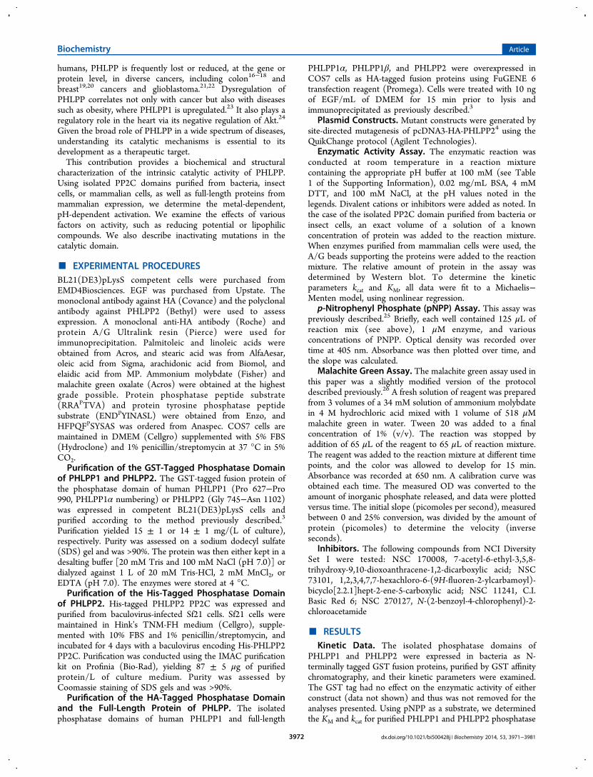
Figure 6. Unique inhibitor sensitivity of the PHLPP PP2C domain. Relative activity of the PHLPP2 PP2C domain (gray squares) and PP2Cα (whitecircles) in the presence of different molecules (NCS 270156, NCS 170008, NCS 73101, and NCS 11241). Compounds NCS 270156 and NCS170008 were selective for PHLPP2 compared to PP2Cα, whereas NCS 73101 inhibits PP2Cα preferentially. Reactions were conducted in Tricine(pH 7.5) at 10 mM pNPP at various inhibitor concentrations. Graphs show means ± SEM for three separate experiments.
Biochemistry Article
dx.doi.org/10.1021/bi500428j | Biochemistry 2014, 53, 3971−39813977

KM of 48 × 103 M−1 s−1 according to one study28 and 16.5 s−1
and 109 × 103 M−1 s−1 according to another.30 PHLPP frombacteria is therefore only 2−4-fold less active than PP2Cα whenpeptides are used as substrates. The proteins purified frominsect cells are slightly more active than the bacteriallyexpressed ones. The Km values are lower for all peptides thanfor pNPP, indicating a better recognition of peptides assubstrates, as expected. The change in kcat, however, was morepronounced than anticipated. Indeed, in the case of PP2Cα,dephosphorylation occurred with an only 5−15-fold increasewhen using RRAPTVA as a substrate compared to pNPP, and
kcat values were even decreased when other peptide sequenceswere used.35 The same reduction in kcat between pNPP andpeptides was observed with PrpZ, a Salmonella enterica PP2C,for example.36 pNPP is supposed to mimic phosphorylatedtyrosine residues, but the data obtained with PHLPP indicate itis a weak mimetic as a phosphotyrosine peptide wasdephosphorylated as effectively as the Ser/Thr phosphopep-tides (data not shown).The major difference between PHLPP and other members of
the PP2C family lies in their metal requirement. PP2C familymembers require either Mn2+ or Mg2+ for their activity. Mn2+ is
Figure 7. Mutational analysis of PHLPP2. (a) Relative activity of point mutants of PHLPP2 using a peptide as the substrate. Point mutants of full-length PHLPP2 were overexpressed as HA-tagged fusion proteins in COS7 cells and immunoprecipitated. They were used to dephosphorylate thethreonine-phosphorylated peptide RRAPTVA in Tricine (pH 7.5) in the presence of 5 mM MnCl2. Release of inorganic phosphate was monitored bythe malachite green assay, and the speed of dephosphorylation was divided by the relative amount of protein, as determined by Western blotting.Activity is given relative to that of wild-type PHLPP2. The graph shows means ± SEM of three separate experiments. (b) Position of the mutatedresidues in the homology model of PHLPP2.25 (c) Sequence alignment of subdomains 1, 2, 8, and 11 of the PP2C family showing conservedRXXXD, DGXXG, DG, and GXXDN (colored orange, green, yellow, and cyan, respectively). Mn2+-coordinating acidic residues are colored red.Below is a schematic of PP2Cα (left) showing four asparates that coordinate the two bound Mn2+ ions (magenta spheres)34 and a homology modelof PHLPP225 showing equivalent acidic residues and structurally important Phe residues.
Biochemistry Article
dx.doi.org/10.1021/bi500428j | Biochemistry 2014, 53, 3971−39813978

most commonly preferred, but Mg2+ is often able to activate theenzymes and, in some instances, is the most efficient cofactor.We showed that PHLPP was active even in the absence of acofactor and that we observed a residual activity in the presenceof EDTA. In a majority of cases, PP2C family members had noin vitro activity in the absence of Mn2+ or Mg2+, on anysubstrate. A few exceptions exist. PP1MH is inactive towardpNPP and phosphopeptides without Mn2+ but was able todephosphorylate casein in the absence of an additional metalliccation.30 Conversely, PP2Cβ has a small basal activity towardphosphopeptides but none toward casein.31 Nevertheless, thisfeature is an oddity in the family. In the case of PHLPP, themetal ions appear to play both a structural role and a catalyticrole. On one hand, EDTA treatment causes a structuralrearrangement, as observed by fluorescence (Figure 5d and SI),resulting in a conformation of the enzyme with a reducedthermal stability but unchanged catalytic activity. On the otherhand, mutation of either key aspartic acid in the catalytic site(D806 or D1024) reduces PHLPP activity by 50%, suggestingthat metal ions are involved in the catalytic process. Thesemetals may be incorporated during protein expression (in aMn2+-rich environment) and are likely to be tightly bound.Additional ions may be more loosely associated with theprotein and play a structural role. Varying the nature of theseions affects the thermal stability of the recombinant enzyme(Figures 5d and 5e) but not its activity. PHLPP activity can beonly enhanced by the addition of Mn2+. This is always the casefor proteins expressed in insect cells or mammalian cells. In thecase of bacterially expressed proteins, the influence of Mn2+ ismore complex, being mainly inhibitory. However, this trend isoverturned above pH 8.0 when these enzymes start to act liketheir counterparts purified in other systems. Activation by Mn2+
is enhanced by fatty acids as described previously,31−33
although the amplitude of the effect was attenuated.Proteins expressed in E. coli showed some differences
compared to proteins expressed in other systems. The mostdramatic feature was their weak activity, as determined by thekcat, using pNPP as a substrate. However, this did not hold truefor other substrates as shown in Table 1. We also found thatthose recombinant proteins were a good model for the metalrequirement for PHLPP activity. Bacteria may lack the properenzymes for post-translational modification or the properchaperones for correct folding to ensure that the enzymesadopt conformations for maximal catalytic activity. We knowthat bacterially expressed protein displayed the odd character-istic of gaining activity upon being stored at 4 °C, consistentwith slow structural rearrangements. Nonetheless, bacteria arestill the preferred source of recombinant proteins as this systemgives the highest yield of PHLPP. Care should be taken whenanalyzing results using bacterially expressed proteins, andcomparison with proteins from mammalian cells should beundertaken to assess the physiological relevance of the results.Mutational Analysis of PHLPP2. The archetypal
mammalian PP2C is characterized by a central, buried β-sandwich composed by two antiparallel β-sheets, flanked by twoantiparallel α-helices. Its catalytic core is composed of twoMn2+ ions coordinated by four invariant Asp residues and anonconserved Glu.37 Mutational analysis of PP2Cα38 orPP2Cβ39 also identifies two important positively chargedresidues, an Arg and a His, the first one positioning thephosphate group correctly and the second one acting as ageneral acid for catalysis (Figure 7c). Using point mutations, wewere able to identify three residues that are important for
catalysis that are likely to coordinate metals: Asp 806(corresponding to Asp 60 in PP2Cα), Asp 1024 (Asp 282 inPP2Cα), and Glu 989 (Asp 239 in PP2Cα). The exactalignment for residue 238 in PP2Cα is actually Lys 985.Mutation of Lys 985 to a neutral residue such as Leu has noeffect, as shown, nor do mutations of this residue to an acidicresidue such as Asp or Glu (data not shown). Glu 989 ispredicted to be located one helix turn away compared toPP2Cα; therefore, we predict the distance between the twometallic centers to be significantly increased. We could notidentify a homologue for Asp 38 (in motif 1) in PHLPP2, butthis residue is not essential for catalysis.38 Mutation of Glu 37or Asp 38 in PP2Cα does not result in a dramatic decrease inactivity; indeed, the D38Q mutant is more active than wild-typePP2Cα. Asp 38 in PP2Cα is located at the beginning of the β3sheet and coordinates the same Mn2+ as Asp 60, which is at theend of the β4 sheet34(Figure 7c). It is possible that this metalliccomplex adds stability to the structure. If so, this feature couldbe recapitulated in PHLPP2 by a putative π-stacking betweenPhe 783 (at the beginning of the β3 sheet) and Phe 808 (at theend of the β4 sheet) (Figure 7b,c). Supporting this hypothesisis the fact that mutation of either residue leads to almostcomplete inhibition of the PHLPP2 activity, even though themutants are normally expressed.The catalytic site we describe is missing elements compared
to the canonical PP2C catalytic site9,34(Figure 7c). Onepossibility is that our homology model is inaccurate and theactual folding of the protein is providing additionalcoordination points for the metal that we missed. Still, thishomology model performed well for determination ofinhibitors and provided us with mechanistic details aboutinhibition.25 Another possibility is that not all residues arerequired for catalytic activity, contrary to previous reports.40
Different approaches can be developed by proteins to stabilizetheir structure or position or activate substrates. The lowactivity of the PP2C domain of PHLPP isolated from bacteria,when compared to other PP2Cs, could indicate that the activesite of PHLPP is indeed different from the rest of the PPMfamily, making it less efficient or more specific towardsubstrates. Only resolution of the crystal structure of thephosphatase domain of PHLPP can definitively reveal the exactarchitecture of the active site.In conclusion, we purified the catalytic domains of PHLPP1
and PHLPP2 from three different cell expression systems:bacteria, insect cells, and mammalian cells. These proteinsdephosphorylate synthetic and peptidic substrates with kineticsthat depend on both the substrate and the expression systems.We determined that PHLPP1 and PHLPP2 have almost similarin vitro activities and respond comparably to the presence ofmetallic ions. We also showed that metallic ions affect thestructural stability of the domain. Finally, we probed forresidues predicted to be important for catalysis in PHLPP2 bysite-directed mutagenesis. We identified three residues that arelikely involved in metal coordination and two that could beimportant for structural integrity. This work is only thebeginning of the biochemical characterization of PHLPP, andmany questions remain. One step is the identification of theresidues involved in the active site of PHLPP1. Even thoughPHLPP1 and PHLPP2 are 58% identical in their PP2C domainamino acid sequences and present comparable kinetic profilesin vitro when using isolated domains purified from bacteria,differences are expected as, for example, the two critical Phe weidentified in PHLPP2 have no equivalent in the PHLPP1
Biochemistry Article
dx.doi.org/10.1021/bi500428j | Biochemistry 2014, 53, 3971−39813979
sequence. Another essential step is to understand theinteractions between the domains and how this affects theactivity of the phosphatase, the final goal being to ascertain howPHLPP phosphatase activity is regulated in the cell.
■ ASSOCIATED CONTENT*S Supporting InformationBuffers used at various pH values (Table 1) and fluorescence ofthe tryptophan for the phosphatase domain of PHLPP2recorded as a function of time following treatment with 100mM EDTA. This material is available free of charge via theInternet at http://pubs.acs.org.
■ AUTHOR INFORMATIONCorresponding Author*Address: 9500 Gilman Drive 0721, La Jolla, CA 92093-0721.E-mail: [email protected]. Phone: (858) 534-4527. Fax:(858) 822-5888.Present Address†E.S.: The University of Queensland, Institute for MolecularBioscience, St. Lucia, Queensland 4072, Australia.FundingThis work was supported by National Institutes of HealthGrant GM067946 (A.C.N.).NotesThe authors declare no competing financial interest.
■ ACKNOWLEDGMENTSWe thank Yann Gambin for help with the structural datacollection and analyses, Bill Sinko and Corina Antal for helpwith sequence alignment and modeling, and David Brautiganand Jack Dixon for helpful advice on phosphatase assays.
■ ABBREVIATIONSPHLPP, PH domain leucine-rich repeat protein phosphatase;pNPP, p-nitrophenyl phosphate; PKC, protein kinase C; SEM,standard error of the mean.
■ REFERENCES(1) Warfel, N. A., and Newton, A. C. (2012) Pleckstrin HomologyDomain Leucine-rich Repeat Protein Phosphatase (PHLPP): A NewPlayer in Cell Signaling. J. Biol. Chem. 287, 3610−3616.(2) Newton, A. C., and Trotman, L. C. (2014) Turning off AKT:PHLPP as a drug target. Annu. Rev. Pharmacol. Toxicol. 54, 537−558.(3) Gao, T., Furnari, F., and Newton, A. C. (2005) PHLPP: APhosphatase that Directly Dephosphorylates Akt, Promotes Apoptosis,and Suppresses Tumor Growth. Mol. Cell 18, 13−24.(4) Brognard, J., Sierecki, E., Gao, T., and Newton, A. C. (2007)PHLPP and a Second Isoform, PHLPP2, Differentially Attenuate theAmplitude of Akt Signaling by Regulating Distinct Akt Isoforms. Mol.Cell 25, 917−931.(5) Gao, T., Brognard, J., and Newton, A. C. (2008) ThePhosphatase PHLPP Controls the Cellular Levels of Protein KinaseC. J. Biol. Chem. 283, 6300−6311.(6) Qiao, M., Wang, Y., Xu, X., Lu, J., Dong, Y., Tao, W., Stein, J.,Stein, G. S., Iglehart, J. D., Shi, Q., and Pardee, A. B. (2010) Mst1 is aninteracting protein that mediates PHLPPs’ induced apoptosis. Mol.Cell 38, 512−523.(7) O’Neill, A. K., Niederst, M. J., and Newton, A. C. (2013)Suppression of survival signalling pathways by the phosphatasePHLPP. FEBS J. 280, 572−583.(8) Brognard, J., and Newton, A. C. (2008) PHLiPPing the switch onAkt and protein kinase C signaling. Trends Endocrinol. Metab. 19, 223−230.
(9) Shi, Y. (2009) Serine/threonine phosphatases: Mechanismthrough structure. Cell 139, 468−484.(10) Rodriguez, P. L. (1998) Protein phosphatase 2C (PP2C)function in higher plants. Plant Mol. Biol. 38, 919−927.(11) Schweighofer, A., Hirt, H., and Meskiene, I. (2004) Plant PP2Cphosphatases: Emerging functions in stress signaling. Trends Plant Sci.9, 236−243.(12) Lammers, T., and Lavi, S. (2007) Role of type 2C proteinphosphatases in growth regulation and in cellular stress signaling. Crit.Rev. Biochem. Mol. Biol. 42, 437−461.(13) Tamura, S., Toriumi, S., Saito, J., Awano, K., Kudo, T. A., andKobayashi, T. (2006) PP2C family members play key roles inregulation of cell survival and apoptosis. Cancer Sci. 97, 563−567.(14) Le Guezennec, X., and Bulavin, D. V. (2010) WIP1 phosphataseat the crossroads of cancer and aging. Trends Biochem. Sci. 35, 109−114.(15) Chen, M., Pratt, C. P., Zeeman, M. E., Schultz, N., Taylor, B. S.,O’Neill, A., Castillo-Martin, M., Nowak, D. G., Naguib, A., Grace, D.M., Murn, J., Navin, N., Atwal, G. S., Sander, C., Gerald, W. L.,Cordon-Cardo, C., Newton, A. C., Carver, B. S., and Trotman, L. C.(2011) Identification of PHLPP1 as a tumor suppressor reveals therole of feedback activation in PTEN-mutant prostate cancerprogression. Cancer Cell 20, 173−186.(16) Liu, J., Weiss, H. L., Rychahou, P., Jackson, L. N., Evers, B. M.,and Gao, T. (2008) Loss of PHLPP expression in colon cancer: Rolein proliferation and tumorigenesis. Oncogene 28, 994−1004.(17) Kaiser, S., Park, Y.-K., Franklin, J., Halberg, R., Yu, M., Jessen,W., Freudenberg, J., Chen, X., Haigis, K., Jegga, A., Kong, S., Sakthivel,B., Xu, H., Reichling, T., Azhar, M., Boivin, G., Roberts, R., Bissahoyo,A., Gonzales, F., Bloom, G., Eschrich, S., Carter, S., Aronow, J.,Kleimeyer, J., Kleimeyer, M., Ramaswamy, V., Settle, S., Boone, B.,Levy, S., and Graff, J. (2007) Transcriptional recapitulation andsubversion of embryonic colon development by mouse colon tumormodels and human colon cancer. Genome Biol. 8, R131.(18) Sabates-Bellver, J., Van der Flier, L. G., de Palo, M., Cattaneo, E.,Maake, C., Rehrauer, H., Laczko, E., Kurowski, M. A., Bujnicki, J. M.,Menigatti, M., Luz, J., Ranalli, T. V., Gomes, V., Pastorelli, A., Faggiani,R., Anti, M., Jiricny, J., Clevers, H., and Marra, G. (2007)Transcriptome Profile of Human Colorectal Adenomas. Mol. CancerRes. 5, 1263−1275.(19) Karnoub, A. E., Dash, A. B., Vo, A. P., Sullivan, A., Brooks, M.W., Bell, G. W., Richardson, A. L., Polyak, K., Tubo, R., and Weinberg,R. A. (2007) Mesenchymal stem cells within tumour stroma promotebreast cancer metastasis. Nature 449, 557−563.(20) Richardson, A. L., Wang, Z. C., De Nicolo, A., Lu, X., Brown,M., Miron, A., Liao, X., Iglehart, J. D., Livingston, D. M., and Ganesan,S. (2006) X chromosomal abnormalities in basal-like human breastcancer. Cancer Cell 9, 121−132.(21) Bredel, M., Bredel, C., Juric, D., Harsh, G. R., Vogel, H., Recht,L. D., and Sikic, B. I. (2005) High-Resolution Genome-Wide Mappingof Genetic Alterations in Human Glial Brain Tumors. Cancer Res. 65,4088−4096.(22) Parsons, D. W., Jones, S., Zhang, X., Lin, J. C.-H., Leary, R. J.,Angenendt, P., Mankoo, P., Carter, H., Siu, I. M., Gallia, G. L., Olivi,A., McLendon, R., Rasheed, B. A., Keir, S., Nikolskaya, T., Nikolsky, Y.,Busam, D. A., Tekleab, H., Diaz, L. A., Jr., Hartigan, J., Smith, D. R.,Strausberg, R. L., Marie, S. K. N., Shinjo, S. M. O., Yan, H., Riggins, G.J., Bigner, D. D., Karchin, R., Papadopoulos, N., Parmigiani, G.,Vogelstein, B., Velculescu, V. E., and Kinzler, K. W. (2008) AnIntegrated Genomic Analysis of Human Glioblastoma Multiforme.Science 321, 1807−1812.(23) Andreozzi, F., Procopio, C., Greco, A., Mannino, G. C., Miele,C., Raciti, G. A., Iadicicco, C., Beguinot, F., Pontiroli, A. E., Hribal, M.L., Folli, F., and Sesti, G. (2011) Increased levels of the Akt-specificphosphatase PH domain leucine-rich repeat protein phosphatase(PHLPP)-1 in obese participants are associated with insulin resistance.Diabetologia 54, 1879−1887.(24) Miyamoto, S., Purcell, N. H., Smith, J. M., Gao, T., Whittaker,R., Huang, K., Castillo, R., Glembotski, C. C., Sussman, M. A.,
Biochemistry Article
dx.doi.org/10.1021/bi500428j | Biochemistry 2014, 53, 3971−39813980

Newton, A. C., and Brown, J. H. (2010) PHLPP-1 NegativelyRegulates Akt Activity and Survival in the Heart. Circ. Res. 107, 476−484.(25) Sierecki, E., Sinko, W., McCammon, J. A., and Newton, A. C.(2010) Discovery of Small Molecule Inhibitors of the PH DomainLeucine-Rich Repeat Protein Phosphatase (PHLPP) by Chemical andVirtual Screening. J. Med. Chem. 53, 6899−6911.(26) Souza, R. C., Junqueira, J. C., Rossoni, R. D., Pereira, C. A.,Munin, E., and Jorge, A. O. (2010) Comparison of the photodynamicfungicidal efficacy of methylene blue, toluidine blue, malachite greenand low-power laser irradiation alone against Candida albicans. Lasersin Medical Science 25, 385−389.(27) Donella Deana, A., Mac Gowan, C. H., Cohen, P., Marchiori, F.,Meyer, H. E., and Pinna, L. A. (1990) An investigation of the substratespecificity of protein phosphatase 2C using synthetic peptidesubstrates; comparison with protein phosphatase 2A. Biochim. Biophys.Acta 2, 199−202.(28) Fjeld, C. C., and Denu, J. M. (1999) Kinetic Analysis of HumanSerine/Threonine Protein Phosphatase 2Cα. J. Biol. Chem. 274,20336−20343.(29) Barford, D., Das, A. K., and Egloff, M.-P. (1998) The Structureand Mechanism of Protein Phosphatases: Insights into Catalysis andRegulation. Annu. Rev. Biophys. Biomol. Struct. 27, 133−164.(30) Sugiura, T., and Noguchi, Y. (2009) Substrate-dependent metalpreference of PPM1H, a cancer-associated protein phosphatase 2C:Comparison with other family members. BioMetals 22, 469−477.(31) Krieglstein, J., Selke, D., Maassen, A., and Klumpp, S. (2003)Activity of PP2Cb is increased by divalent cations and lipophiliccompounds depending on the substrate. Methods Enzymol. 366, 282−289.(32) Krieglstein, J., Hufnagel, B., Dworak, M., Schwarz, S., Kewitz, T.,Reinbold, M., and Klumpp, S. (2008) Influence of various fatty acidson the activity of protein phosphatase type 2C and apoptosis ofendothelial cells and macrophages. Eur. J. Pharm. Sci. 35, 397−403.(33) Klumpp, S., Selke, D., and Hermesmeier, J. (1998) Proteinphosphatase type 2C active at physiological Mg2+: Stimulation byunsaturated fatty acids. FEBS Lett. 437, 229−232.(34) Das, A. K., Helps, N. R., Cohen, P. T., and Bradford, D. (1996)Crystal structure of the protein serine/threonine phosphatase 2C at2.0 Å resolution. EMBO J. 15, 6798−6809.(35) Yamaguchi, H., Durell, S. R., Chatterjee, D. K., Anderson, C. W.,and Appella, E. (2007) The Wip1 phosphatase PPM1D dephosphor-ylates SQ/TQ motifs in checkpoint substrates phosphorylated byPI3K-like kinases. Biochemistry 46, 12594−12603.(36) Lai, S. M., and Le Moual, H. (2005) PrpZ, a Salmonella entericaserovar Typhi serine/threonine protein phosphatase 2C with dualsubstrate specificity. Microbiology 151, 1159−1167.(37) Barford, D. (1996) Molecular mechanisms of the proteinserine/threonine phosphatases. Trends Biochem. Sci. 21, 407−412.(38) Jackson, M. D., Fjeld, C. C., and Denu, J. M. (2003) Probing theFunction of Conserved Residues in the Serine/Threonine PhosphatasePP2Cα. Biochemistry 42, 8513−8521.(39) Kusuda, K., Kobayashi, T., Ikeda, S., Ohnishi, M., Chida, N.,Yanagawa, Y., Shineha, R., Nishihira, T., Satomi, S., Hiraga, A., andTamura, S. (1998) Mutational analysis of the domain structure ofmouse protein phosphatase 2Cβ. Biochem. J. 332, 243−250.(40) Conner, S. H., Kular, G., Peggie, M., Sheperd, S., Schuttelkopf,A. W., Cohen, P., and Van Aalten, D. M. F. (2006) TAK1-bindingprotein 1 is a pseudophosphatase. Biochem. J. 399, 427−434.
Biochemistry Article
dx.doi.org/10.1021/bi500428j | Biochemistry 2014, 53, 3971−39813981