biological bases of behavior presentation...
TRANSCRIPT

UNIT 2 BIOLOGICAL BASES OF BEHAVIOR
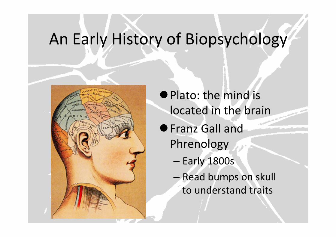
An Early History of Biopsychology
l Plato: the mind is located in the brain
l Franz Gall and Phrenology – Early 1800s – Read bumps on skull to understand traits
If I was to take your brain out of your body, place it into patient needing brain surgery, where would the new ‘self’ call home? Are you simply the end product of a biological and chemical reaction?
Biopsychology Today l Everything we do is ulFmately controlled by our body and brain – Body/brain composed of cells – Brain cells called neurons communicate electrically and chemically
– Different parts of the brain have specific funcFons – Our brains create meaningful experiences from sensory informaFon
– Brain structure and funcFon is influenced by experience
AGENDA
1) Review Text Book Reading 2) Quiz 3) Todays Theme: Hemisphere
Dominance 4) VIDEO: Split Brain Patients 5) Hand back & Discuss Test
(I WILL NEED 5-7 minutes)
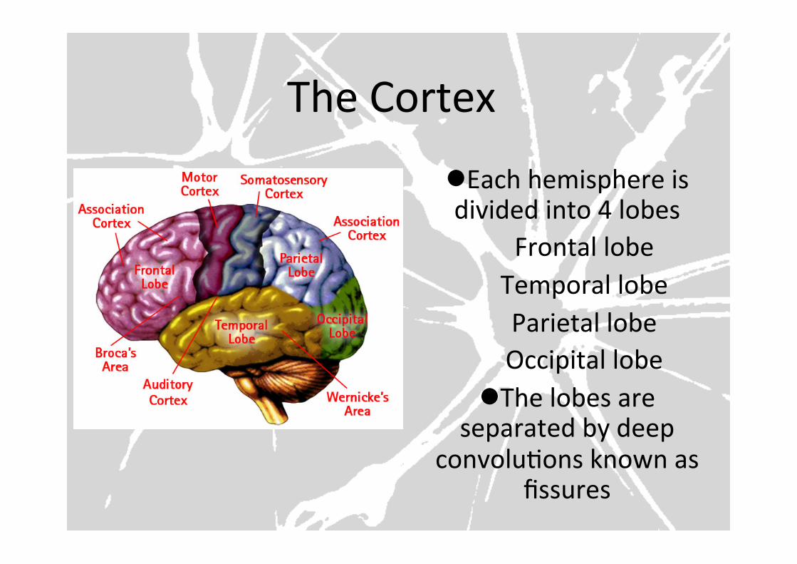
The Cortex
l Each hemisphere is divided into 4 lobes
Frontal lobe Temporal lobe Parietal lobe Occipital lobe
l The lobes are separated by deep
convoluFons known as fissures
Cortex Breakdown…
l Occipital Lobes – Visual cortex – Damage?
l Temporal Lobes – Auditory cortex – Auditory hallucinaFons?
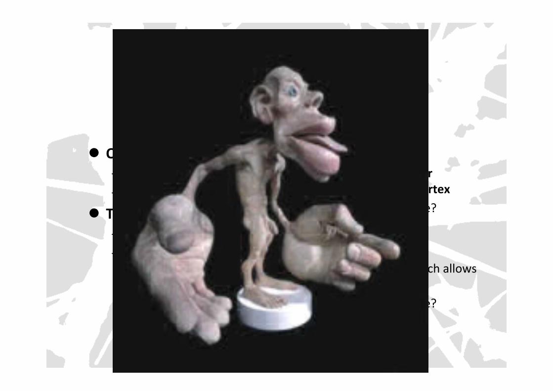
l Parietal Lobes – Primary sensory or
somatosensory cortex – AllocaFon of space?
l Frontal Lobes – Most evolved – Motor cortex, which allows
us to move – AllocaFon of space?
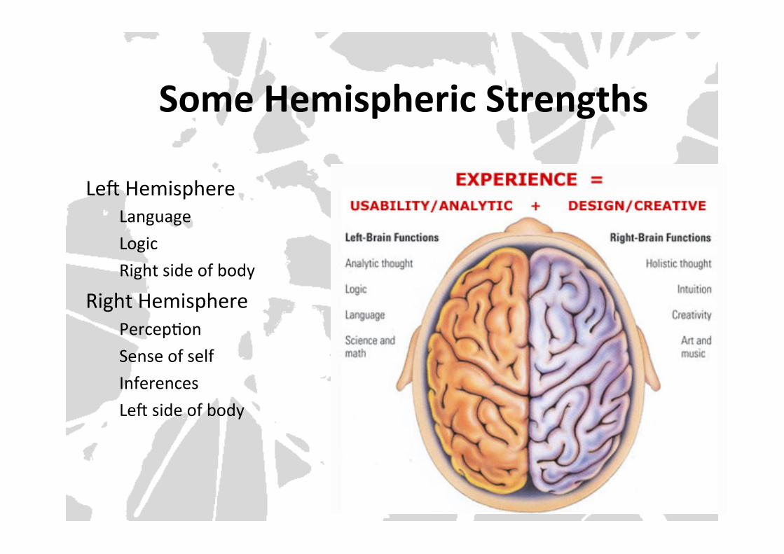
Some Hemispheric Strengths
LeU Hemisphere Language Logic Right side of body
Right Hemisphere PercepFon Sense of self Inferences LeU side of body
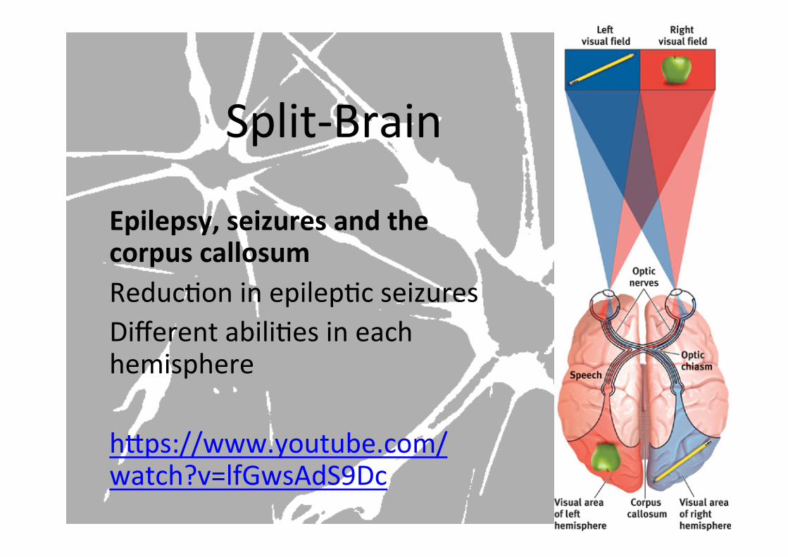
Split-‐Brain
Epilepsy, seizures and the corpus callosum ReducFon in epilepFc seizures Different abiliFes in each hemisphere hYps://www.youtube.com/watch?v=lfGwsAdS9Dc
AGENDA:
1. Map the Brain with the Truine Model
2. COLLABORATION: Map brains parts together
3. VIDEO: Secrets of the Mind
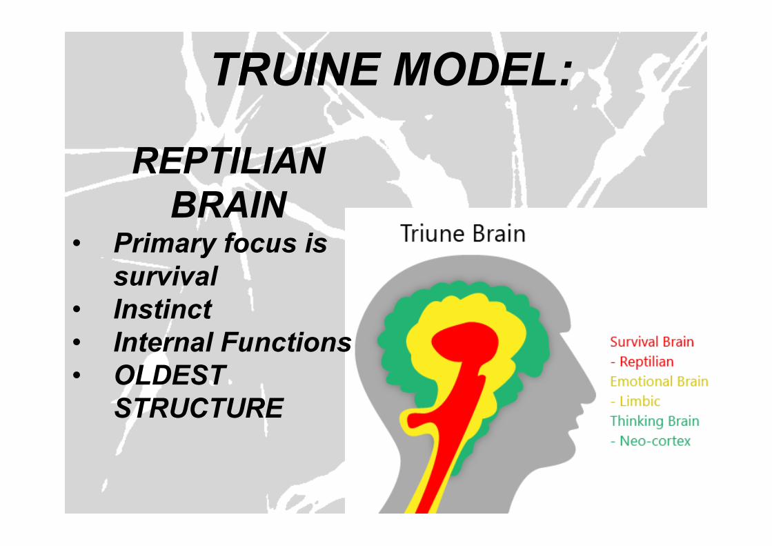
TRUINE MODEL:
REPTILIAN BRAIN
• Primary focus is survival
• Instinct • Internal Functions • OLDEST
STRUCTURE

TRUE STORY:
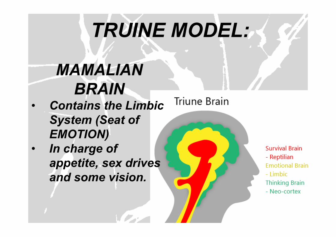
TRUINE MODEL:
MAMALIAN BRAIN
• Contains the Limbic System (Seat of EMOTION)
• In charge of appetite, sex drives and some vision.
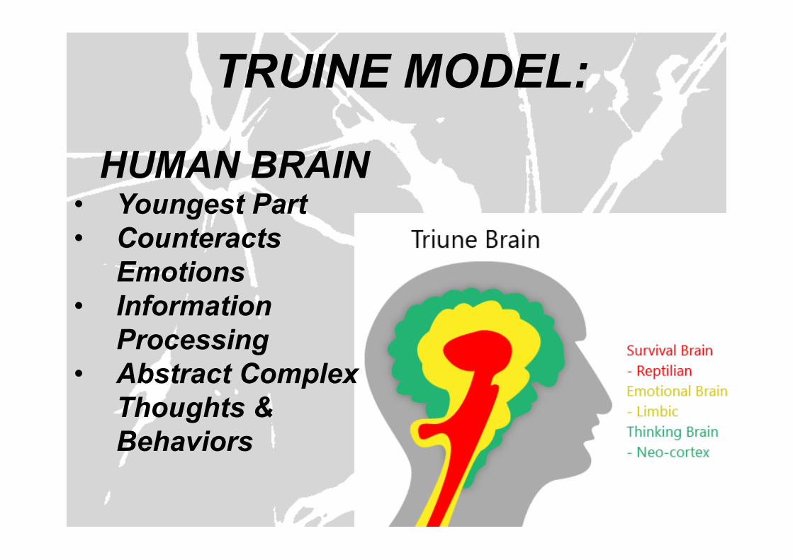
TRUINE MODEL:
HUMAN BRAIN • Youngest Part • Counteracts
Emotions • Information
Processing • Abstract Complex
Thoughts & Behaviors
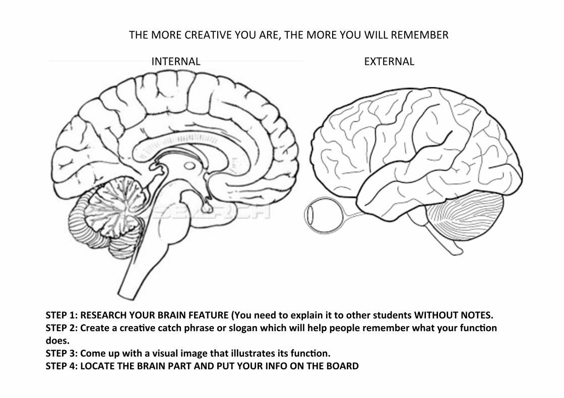
STEP 1: RESEARCH YOUR BRAIN FEATURE (You need to explain it to other students WITHOUT NOTES. STEP 2: Create a creaOve catch phrase or slogan which will help people remember what your funcOon does. STEP 3: Come up with a visual image that illustrates its funcOon. STEP 4: LOCATE THE BRAIN PART AND PUT YOUR INFO ON THE BOARD
THE MORE CREATIVE YOU ARE, THE MORE YOU WILL REMEMBER
INTERNAL EXTERNAL
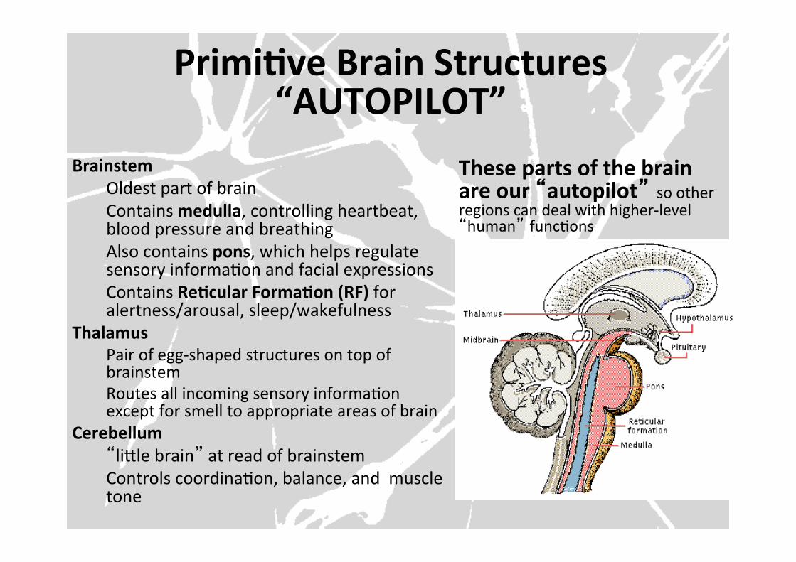
PrimiOve Brain Structures “AUTOPILOT”
Brainstem Oldest part of brain Contains medulla, controlling heartbeat, blood pressure and breathing Also contains pons, which helps regulate sensory informaFon and facial expressions Contains ReOcular FormaOon (RF) for alertness/arousal, sleep/wakefulness
Thalamus Pair of egg-‐shaped structures on top of brainstem Routes all incoming sensory informaFon except for smell to appropriate areas of brain
Cerebellum “liYle brain” at read of brainstem Controls coordinaFon, balance, and muscle tone
These parts of the brain are our “autopilot” so other regions can deal with higher-‐level “human” funcFons
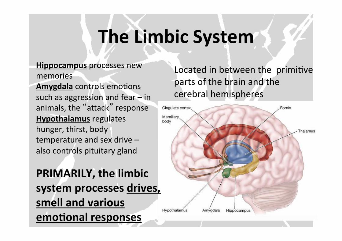
The Limbic System Located in between the primiFve parts of the brain and the cerebral hemispheres
Hippocampus processes new memories Amygdala controls emoFons such as aggression and fear – in animals, the “aYack” response Hypothalamus regulates hunger, thirst, body temperature and sex drive – also controls pituitary gland
PRIMARILY, the limbic system processes drives, smell and various emoOonal responses
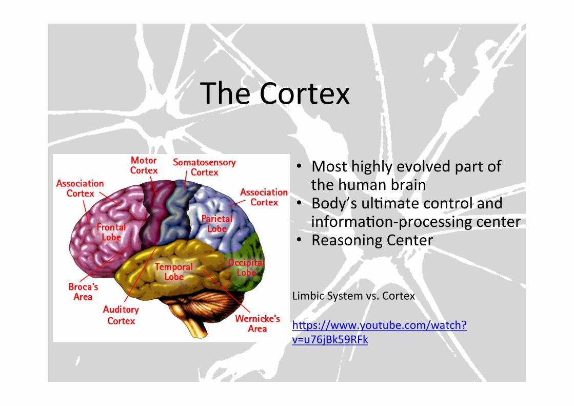
The Cortex
• Most highly evolved part of the human brain
• Body’s ulFmate control and informaFon-‐processing center
• Reasoning Center
Limbic System vs. Cortex hYps://www.youtube.com/watch?v=u76jBk59RFk

AGENDA:
1. QUIZ (Be ready to go at the bell) 2. GET FIRED UP ABOUT NEUROSCIENCE! 3. How does our body communicate with itself? 4. Labeling the Neuron 5. ACTING OUT NEURAL COMMUNICATION!
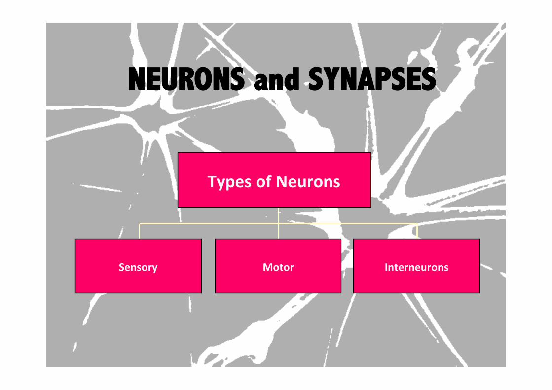
NEURONS and SYNAPSES
Types of Neurons
Sensory Motor Interneurons
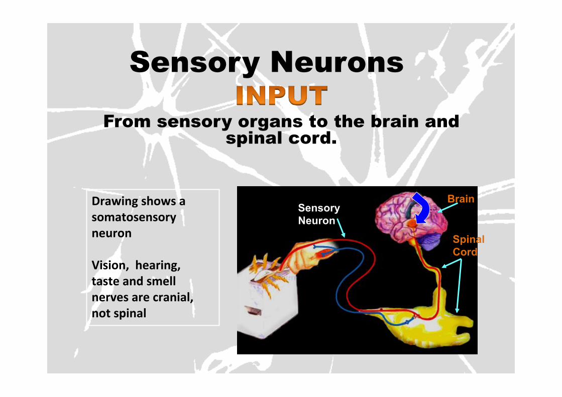
Sensory Neurons
From sensory organs to the brain and spinal cord.
Drawing shows a somatosensory neuron Vision, hearing, taste and smell nerves are cranial, not spinal
Spinal Cord
Brain Sensory Neuron
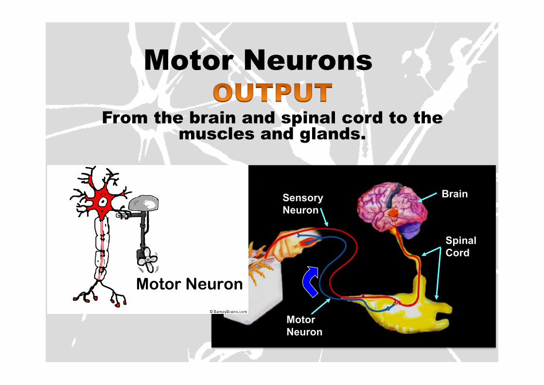
Motor Neurons
From the brain and spinal cord to the muscles and glands.
Spinal Cord
Brain Sensory Neuron
Motor Neuron
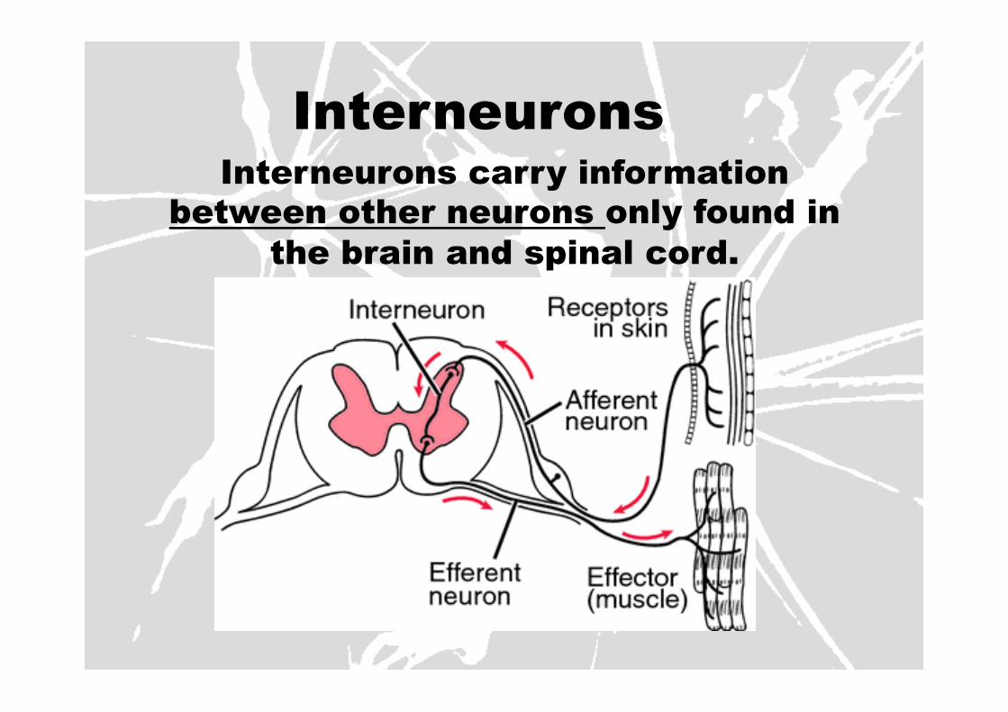
Interneurons Interneurons carry information
between other neurons only found in the brain and spinal cord.
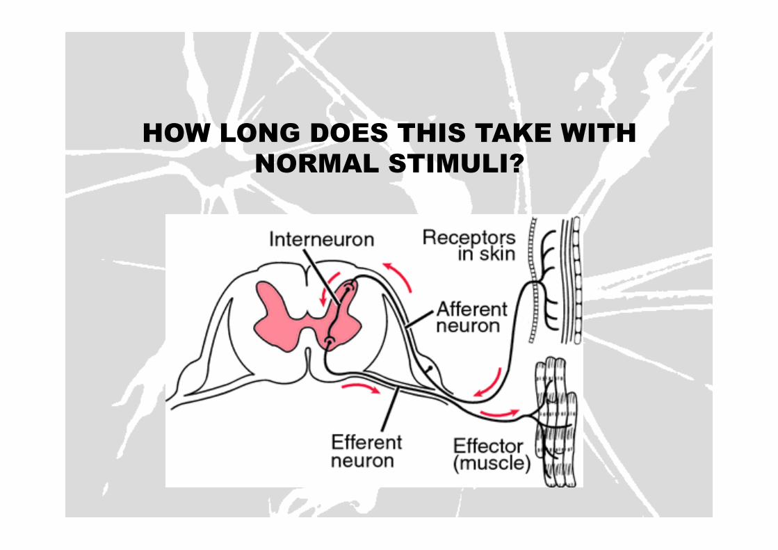
HOW LONG DOES THIS TAKE WITH NORMAL STIMULI?
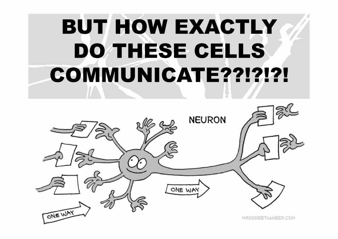
BUT HOW EXACTLY DO THESE CELLS
COMMUNICATE??!?!?!
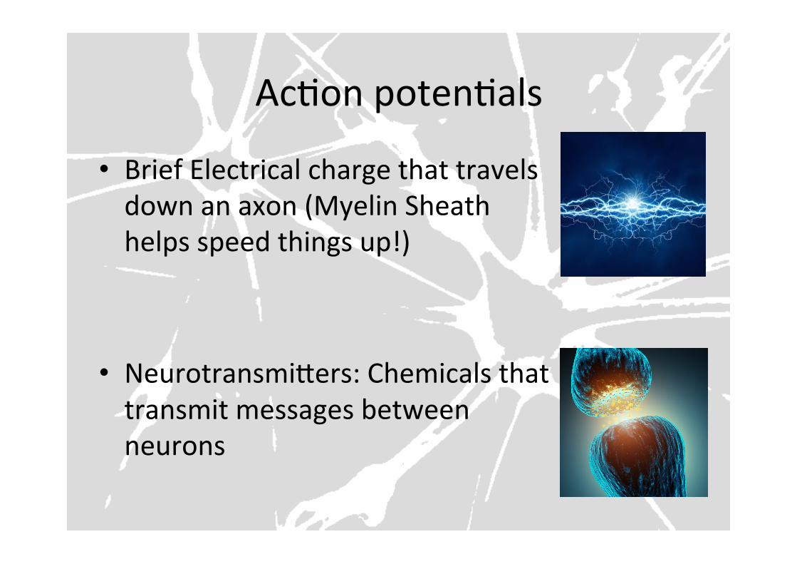
AcFon potenFals
• Brief Electrical charge that travels down an axon (Myelin Sheath helps speed things up!)
• NeurotransmiYers: Chemicals that transmit messages between neurons
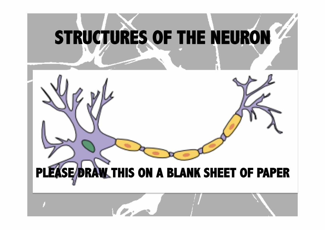
STRUCTURES OF THE NEURON
PLEASE DRAW THIS ON A BLANK SHEET OF PAPER

THE CELL BODY
• CONTAINS THE CELL’S NUCLEUS
• Round, centrally located structure
• Contains DNA • Controls protein manufacturing
• Directs metabolism
• No role in neural signaling
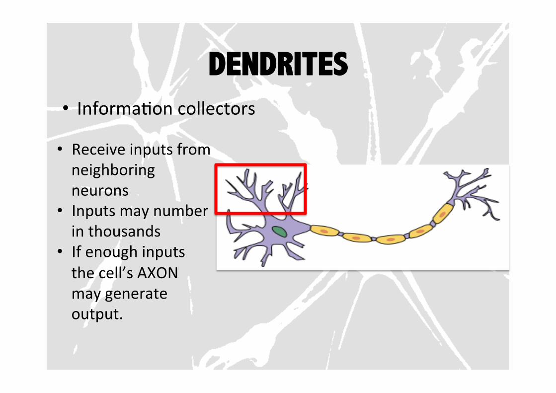
DENDRITES • InformaFon collectors
• Receive inputs from neighboring neurons
• Inputs may number in thousands
• If enough inputs the cell’s AXON may generate output.

DENDRITIC GROWH • Mature neurons generally can’t divide….
• BUT new dendrites can grow.
• Provides room for more connecFons to other neurons.
• NEW CONNECTIONS ARE THE BASIS FOR LEARNING
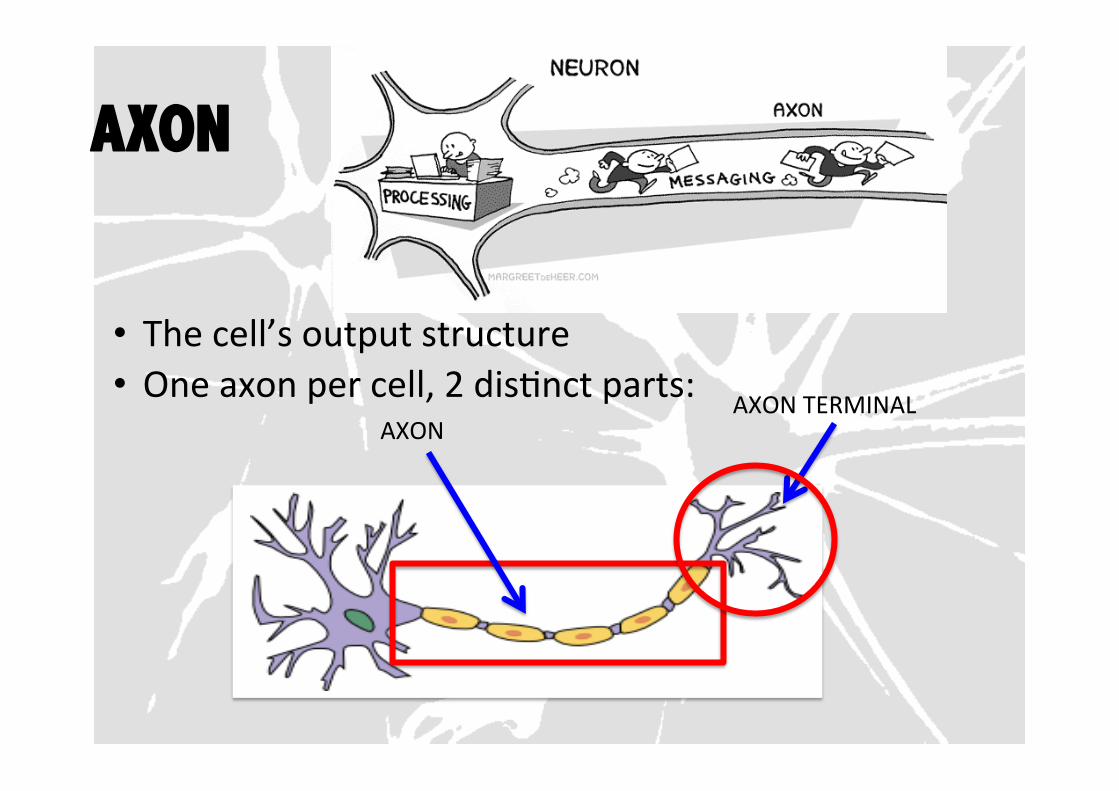
AXON
• The cell’s output structure • One axon per cell, 2 disFnct parts:
AXON AXON TERMINAL
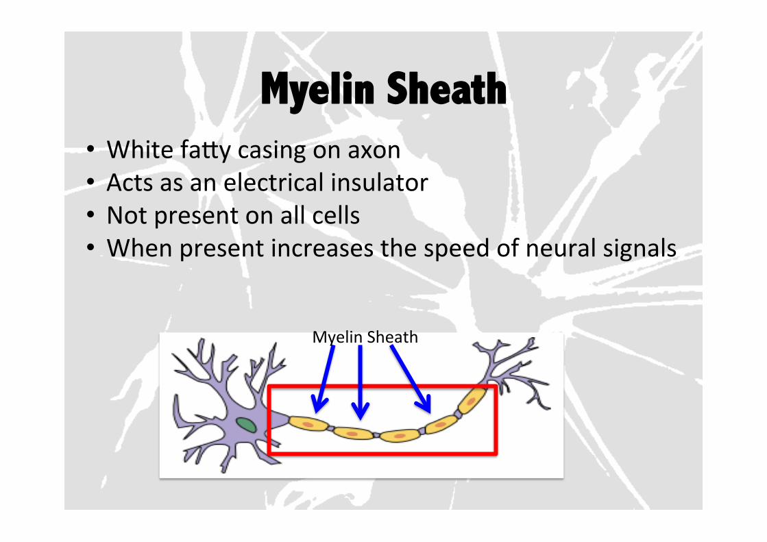
Myelin Sheath • White faYy casing on axon • Acts as an electrical insulator • Not present on all cells • When present increases the speed of neural signals
Myelin Sheath
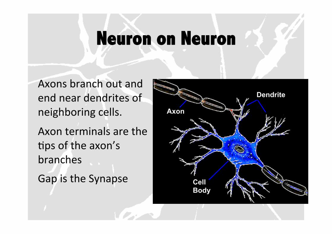
Neuron on Neuron
Axons branch out and end near dendrites of neighboring cells.
Axon terminals are the Fps of the axon’s branches
Gap is the Synapse Cell Body
Dendrite
Axon

Synapse Axon terminals contain small storage sacks called synapFc vesicles
Vesicles contain neurotransmiYer molecules
Sending Neuron
Synapse Axon Terminal
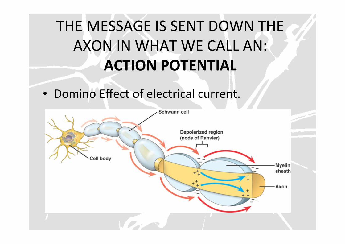
THE MESSAGE IS SENT DOWN THE AXON IN WHAT WE CALL AN:
ACTION POTENTIAL
• Domino Effect of electrical current.
TIME TO APPLY!
LAB TIME!
LETS MAKE IT VISUAL!
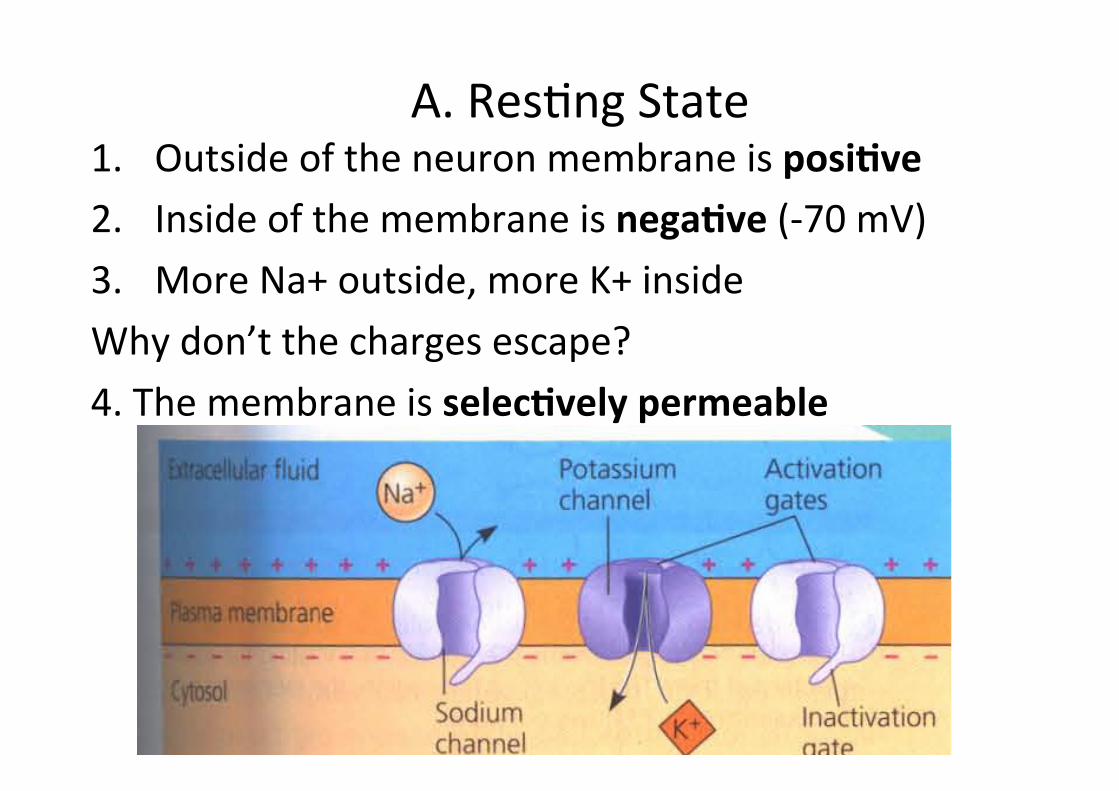
A. ResFng State 1. Outside of the neuron membrane is posiOve 2. Inside of the membrane is negaOve (-‐70 mV) 3. More Na+ outside, more K+ inside Why don’t the charges escape? 4. The membrane is selecOvely permeable
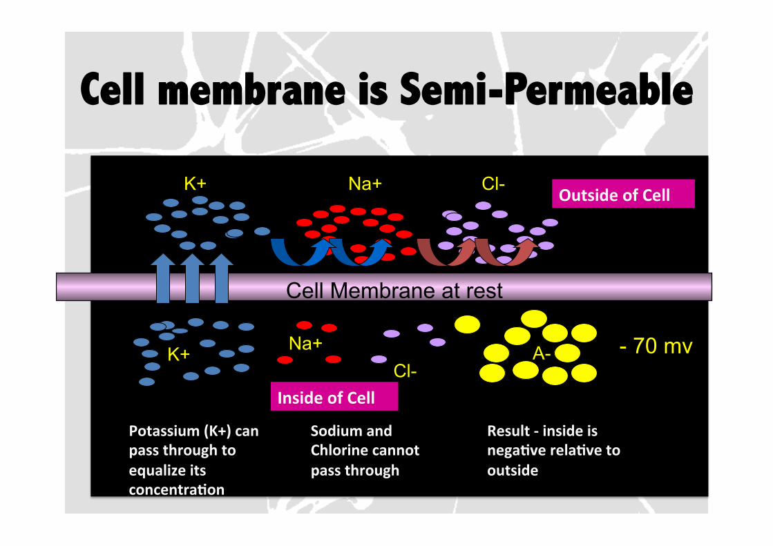
Cell membrane is Semi-Permeable
Cell Membrane at rest
Na+ Cl- K+
Na+ Cl- K+ A-
Outside of Cell
Inside of Cell Potassium (K+) can pass through to equalize its concentraOon
Sodium and Chlorine cannot pass through
Result -‐ inside is negaOve relaOve to outside
- 70 mv
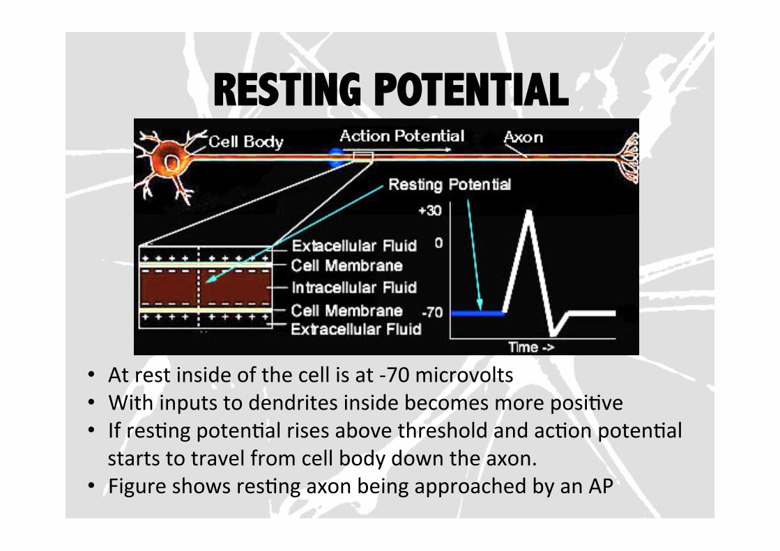
RESTING POTENTIAL
• At rest inside of the cell is at -‐70 microvolts • With inputs to dendrites inside becomes more posiFve • If resFng potenFal rises above threshold and acFon potenFal starts to travel from cell body down the axon.
• Figure shows resFng axon being approached by an AP
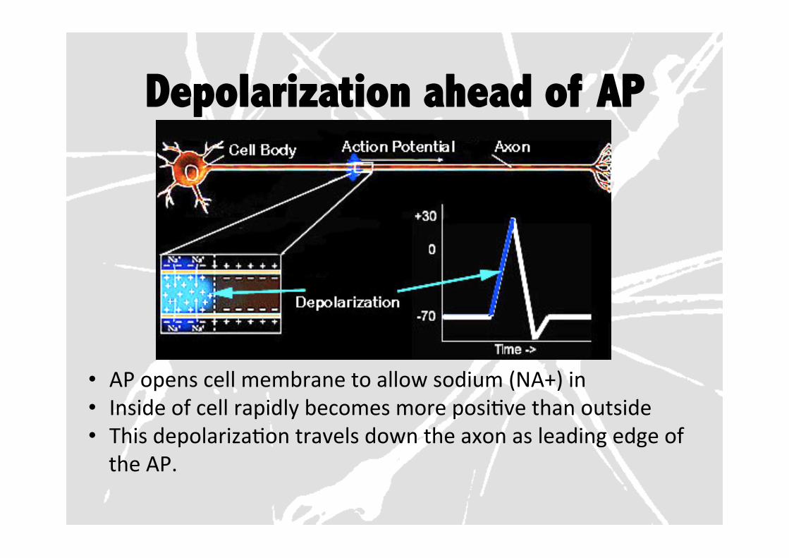
Depolarization ahead of AP
• AP opens cell membrane to allow sodium (NA+) in • Inside of cell rapidly becomes more posiFve than outside • This depolarizaFon travels down the axon as leading edge of the AP.
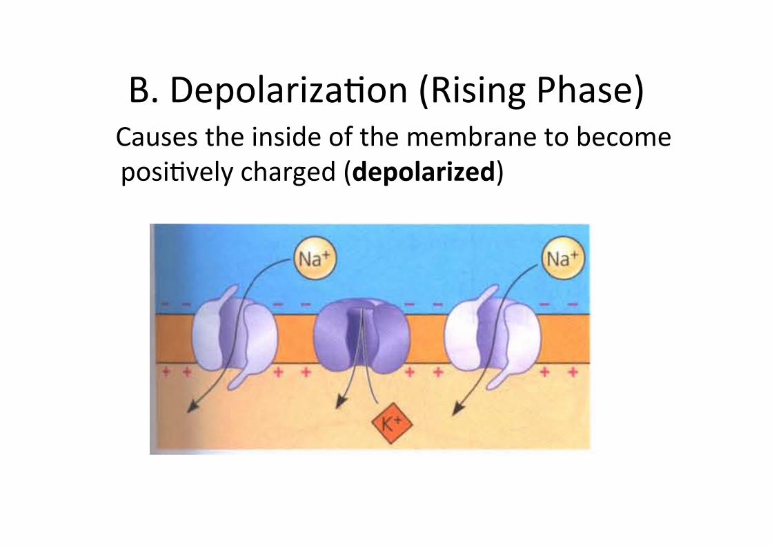
B. DepolarizaFon (Rising Phase) Causes the inside of the membrane to become posiFvely charged (depolarized)
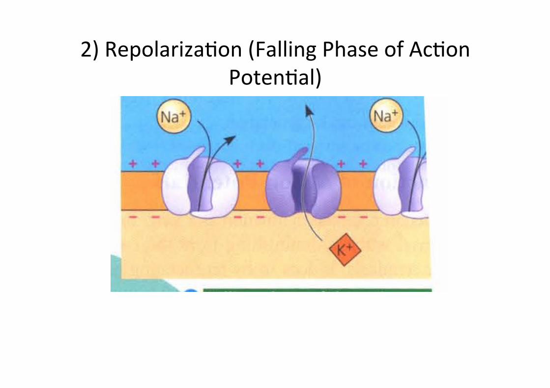
2) RepolarizaFon (Falling Phase of AcFon PotenFal)
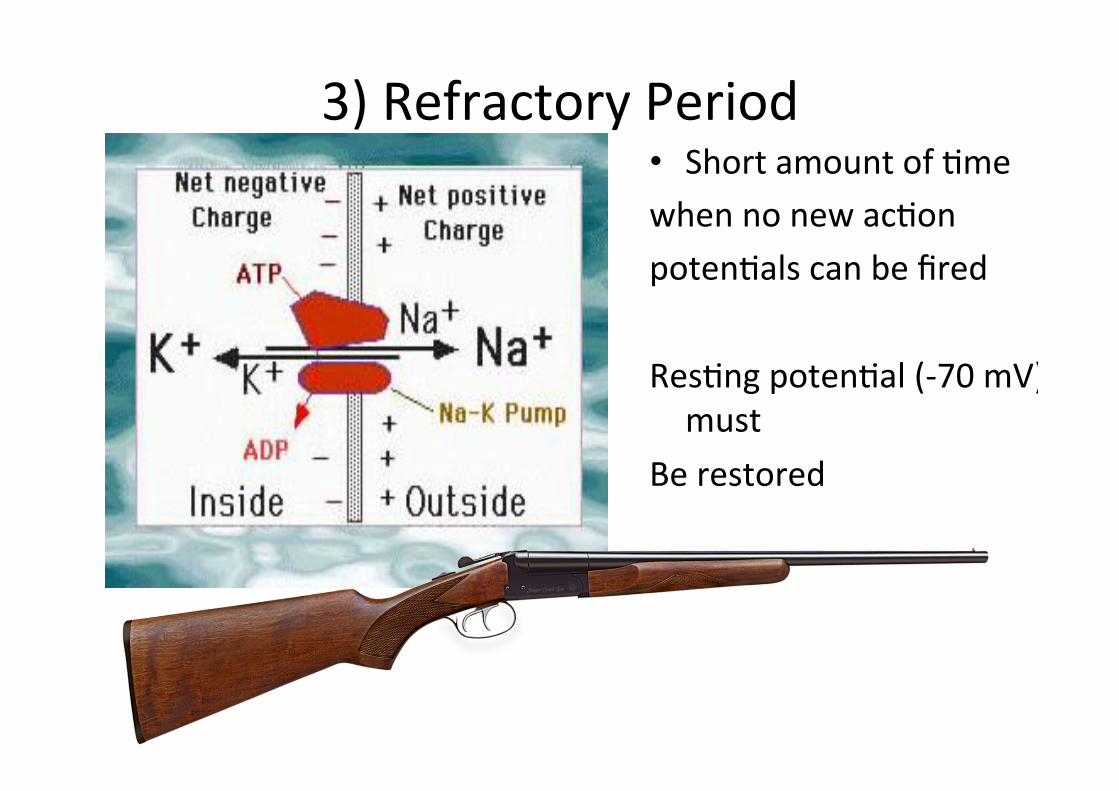
3) Refractory Period • Short amount of Fme when no new acFon potenFals can be fired ResFng potenFal (-‐70 mV) must
Be restored
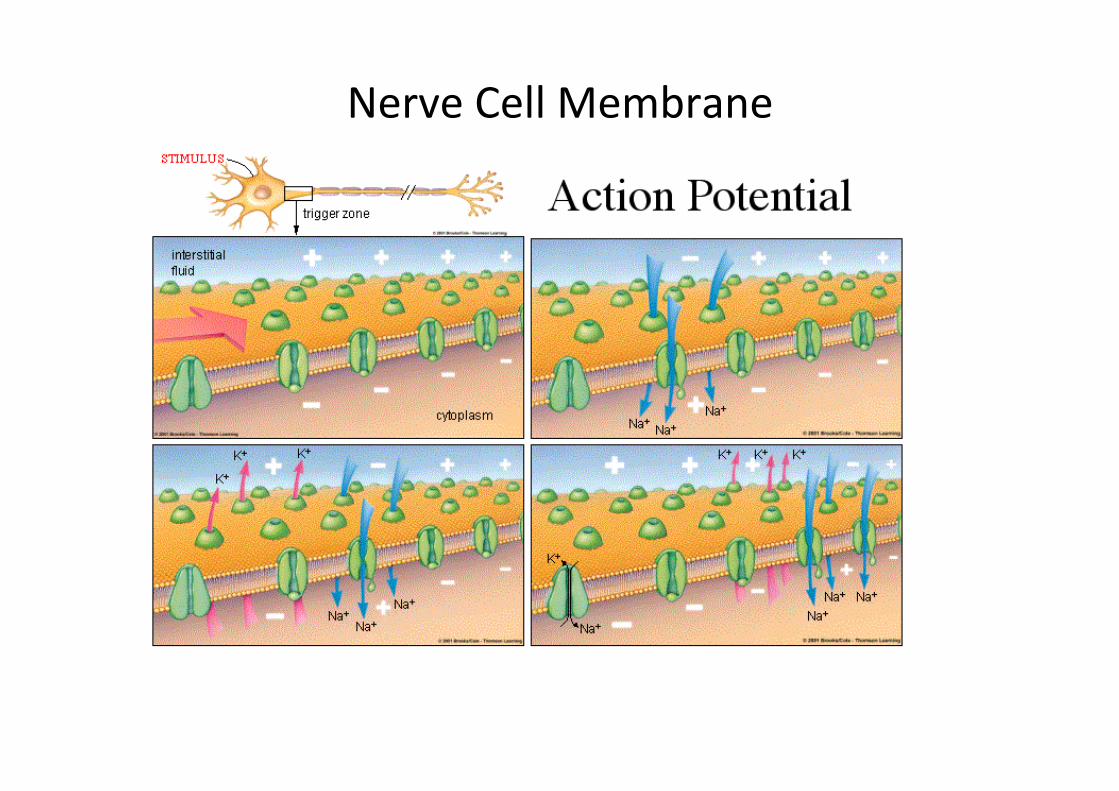
Nerve Cell Membrane
• Each level contains about 10% o the energy in the previous level.

ResFng State
hYp://www.youtube.com/watch?v=90cj4NX87Yk&feature=related
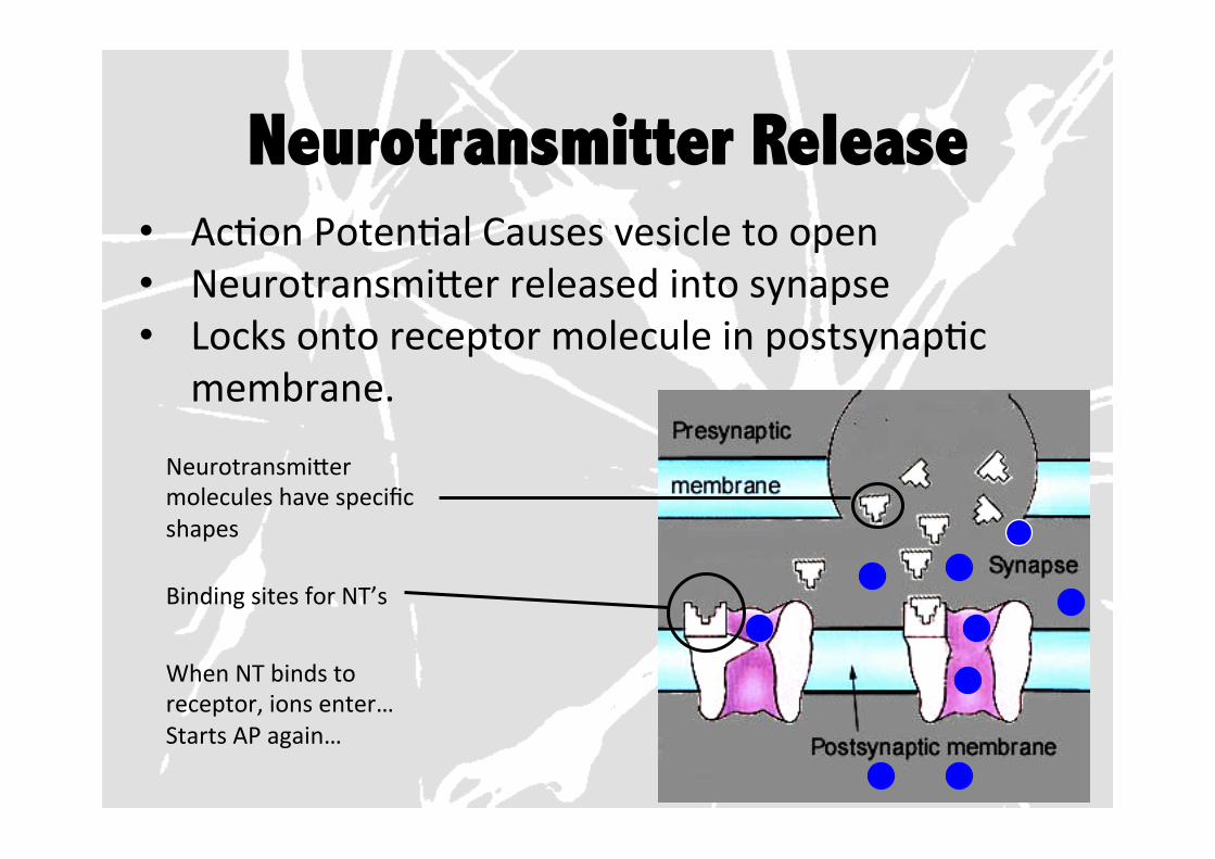
Neurotransmitter Release • AcFon PotenFal Causes vesicle to open • NeurotransmiYer released into synapse • Locks onto receptor molecule in postsynapFc
membrane.
NeurotransmiYer molecules have specific shapes
Binding sites for NT’s
When NT binds to receptor, ions enter… Starts AP again…
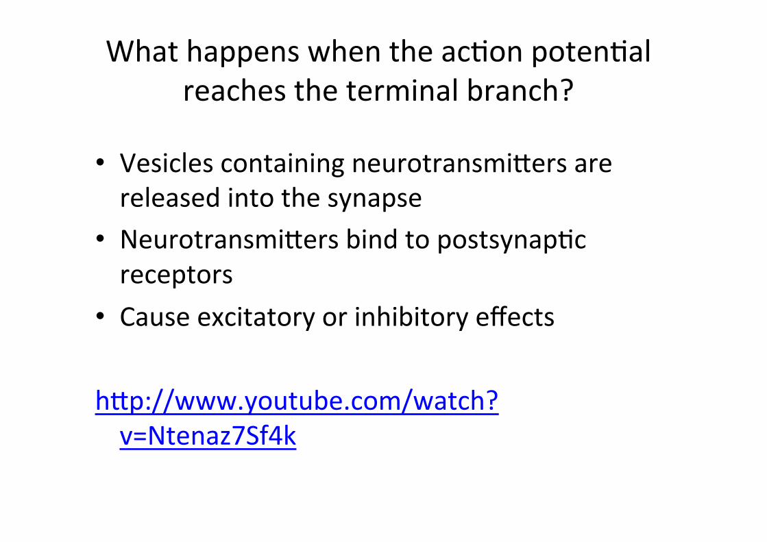
What happens when the acFon potenFal reaches the terminal branch?
• Vesicles containing neurotransmiYers are released into the synapse
• NeurotransmiYers bind to postsynapFc receptors
• Cause excitatory or inhibitory effects hYp://www.youtube.com/watch?v=Ntenaz7Sf4k
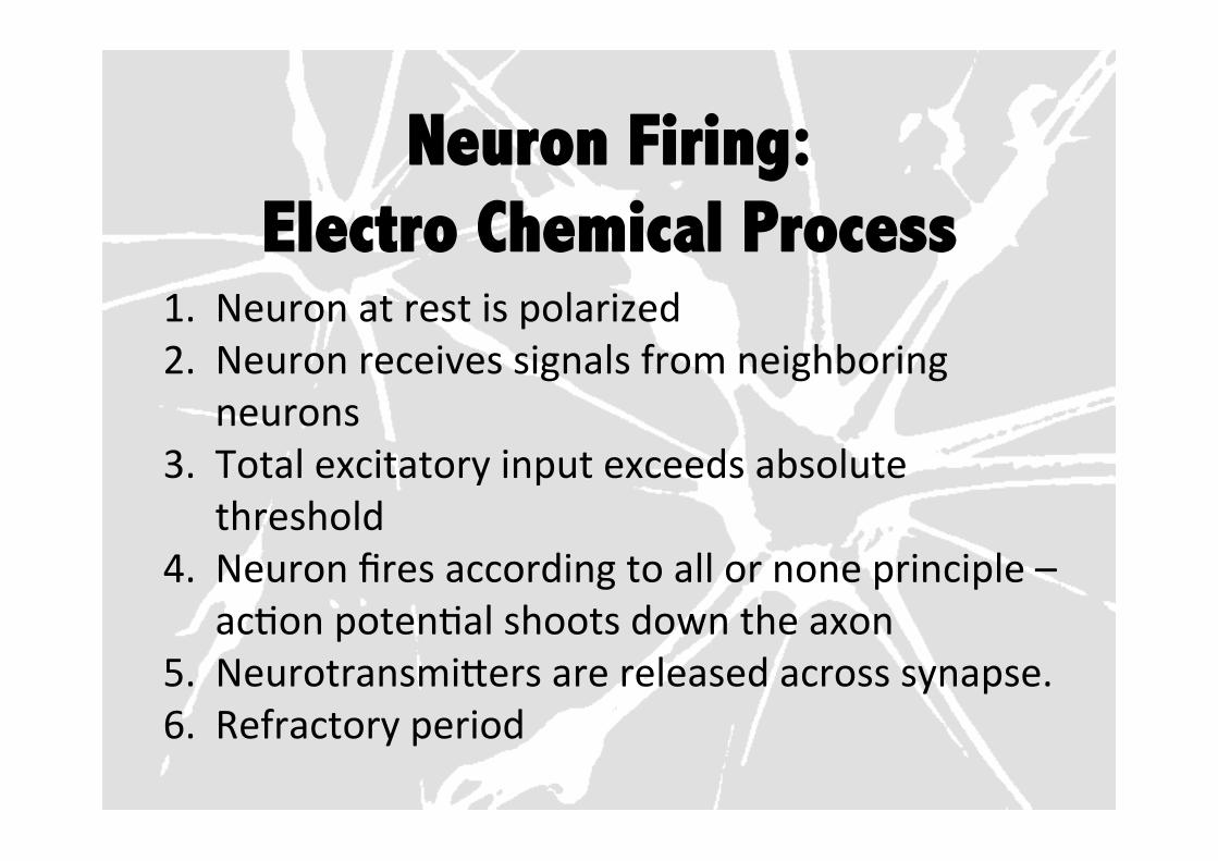
Neuron Firing: Electro Chemical Process
1. Neuron at rest is polarized 2. Neuron receives signals from neighboring
neurons 3. Total excitatory input exceeds absolute
threshold 4. Neuron fires according to all or none principle –
acFon potenFal shoots down the axon 5. NeurotransmiYers are released across synapse. 6. Refractory period
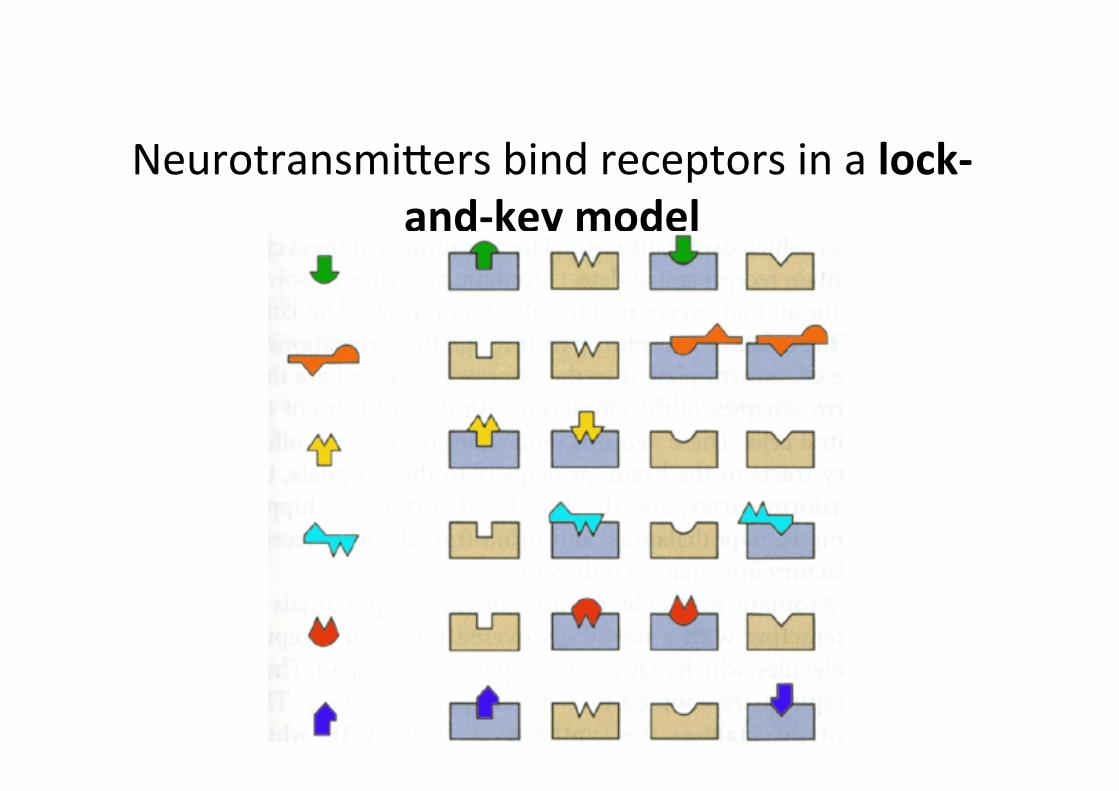
NeurotransmiYers bind receptors in a lock-‐and-‐key model