biological composites complex structures for functional
TRANSCRIPT
Published as:
Eder, M., Shahrouz, A., & Fratzl, P. (2018). Biological composites—
complex structures for functional diversity. Science, 362(6414), 543-547.
doi:10.1126/science.aat8297.
Biological composites – complex structures for
functional diversity
Michaela Eder, Shahrouz Amini, Peter Fratzl
MPIKG Public Access
Max
Pla
nck
Inst
itute
of C
ollo
ids a
nd In
terfa
ces ·
Aut
hor M
anus
crip
t Biological composites – complex structures for functional diversity
Michaela Eder, Shahrouz Amini, Peter Fratzl*
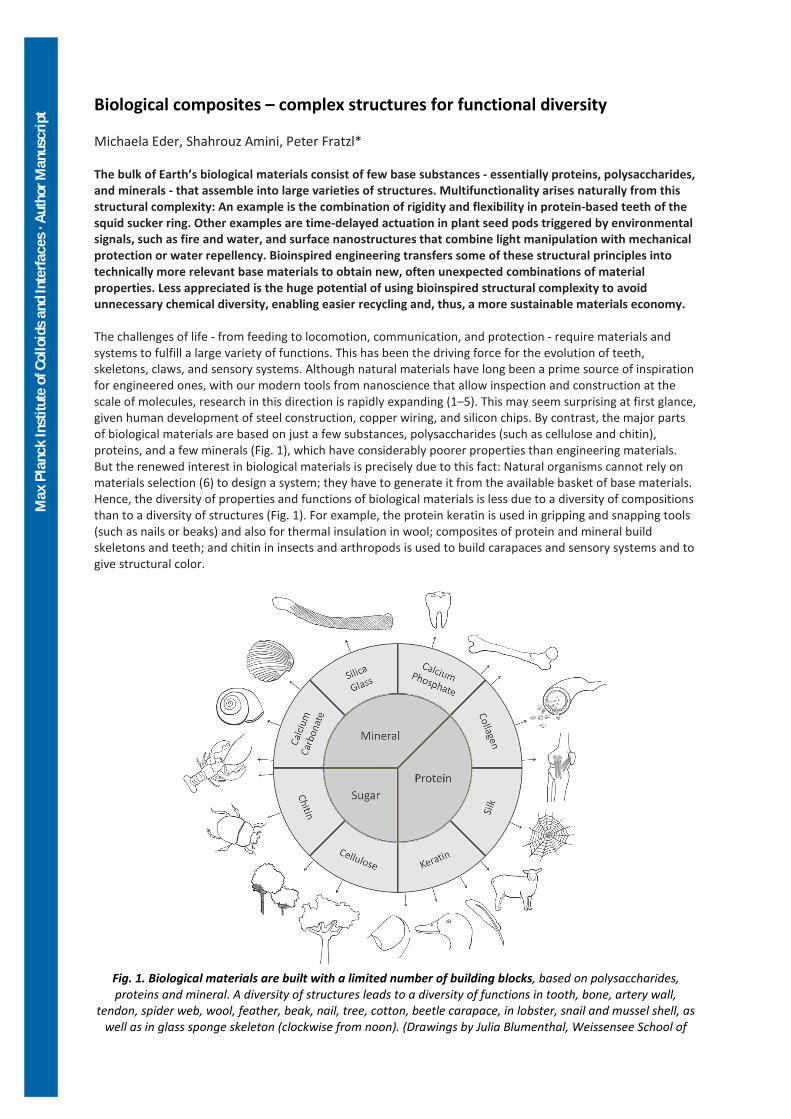
The bulk of Earth’s biological materials consist of few base substances - essentially proteins, polysaccharides, and minerals - that assemble into large varieties of structures. Multifunctionality arises naturally from this structural complexity: An example is the combination of rigidity and flexibility in protein-based teeth of the squid sucker ring. Other examples are time-delayed actuation in plant seed pods triggered by environmental signals, such as fire and water, and surface nanostructures that combine light manipulation with mechanical protection or water repellency. Bioinspired engineering transfers some of these structural principles into technically more relevant base materials to obtain new, often unexpected combinations of material properties. Less appreciated is the huge potential of using bioinspired structural complexity to avoid unnecessary chemical diversity, enabling easier recycling and, thus, a more sustainable materials economy. The challenges of life - from feeding to locomotion, communication, and protection - require materials and systems to fulfill a large variety of functions. This has been the driving force for the evolution of teeth, skeletons, claws, and sensory systems. Although natural materials have long been a prime source of inspiration for engineered ones, with our modern tools from nanoscience that allow inspection and construction at the scale of molecules, research in this direction is rapidly expanding (1–5). This may seem surprising at first glance, given human development of steel construction, copper wiring, and silicon chips. By contrast, the major parts of biological materials are based on just a few substances, polysaccharides (such as cellulose and chitin), proteins, and a few minerals (Fig. 1), which have considerably poorer properties than engineering materials. But the renewed interest in biological materials is precisely due to this fact: Natural organisms cannot rely on materials selection (6) to design a system; they have to generate it from the available basket of base materials. Hence, the diversity of properties and functions of biological materials is less due to a diversity of compositions than to a diversity of structures (Fig. 1). For example, the protein keratin is used in gripping and snapping tools (such as nails or beaks) and also for thermal insulation in wool; composites of protein and mineral build skeletons and teeth; and chitin in insects and arthropods is used to build carapaces and sensory systems and to give structural color.
Fig. 1. Biological materials are built with a limited number of building blocks, based on polysaccharides, proteins and mineral. A diversity of structures leads to a diversity of functions in tooth, bone, artery wall,
tendon, spider web, wool, feather, beak, nail, tree, cotton, beetle carapace, in lobster, snail and mussel shell, as well as in glass sponge skeleton (clockwise from noon). (Drawings by Julia Blumenthal, Weissensee School of

Max
Pla
nck
Inst
itute
of C
ollo
ids a
nd In
terfa
ces ·
Aut
hor M
anus
crip
t
Arts and Design, and Excellence Cluster Image-Knowledge-Gestaltung, Humboldt Universität zu Berlin, Germany).
A study of these materials leads to a number of questions: How does one get valuable functionality based on cheap base materials? How does one build a complex system based on little variation in constituents so as to facilitate recycling? Can we use structure to make materials active and adaptive? How does nature combine several functions in one material? Answers to such questions require considering the function of biological materials in the natural environment to which they adapt. An evolutionary perspective can be particularly useful because it may reveal how evolutionary pressures led to the adaptation of material structure. As a consequence, bioinspired materials research is a relatively new field at the interface between materials science and various branches of biology, including functional morphology, evolutionary and developmental biology, and sensory physiology. Multifunctionality of biological materials Biological materials may need to simultaneously fulfill different functions to serve not only the needs of the (living) organism but also the needs of populations, such as growth, locomotion (7), signaling (8), repair, mechanical stability (9), resistance against light irradiation (10) or against low temperatures (11), and the possibility for functional adaptation (12). Because of this, Torquato et al. stated in 2003 that “the ultimate multifunctional materials are provided by nature” (13). The potential for multifunctionality is inherently contained in the fact that properties are tuned through the internal structure. In particular, hierarchical structuring that is omnipresent in biological materials opens the possibility of adopting different physical properties in different size ranges (14). A typical example would be structural colors that require features at the submicron range corresponding to the wavelengths of visible light, whereas mechanical properties may be controlled at different scales. Most biological materials are based on fibers as primary motif. Bone is an excellent example in which collagen fibers and mineral are assembled into a great variety of structures that have different mechanical performances (15). Bone is not only the mechanical support for our body but also the reservoir for some of the most important ions for the functioning of cells: calcium and phosphate. More generally, hard-soft hybrid materials in the form of multilayers or tessellations allow the tuning of a wide range of properties without a change in composition (16). The combination of toughness and strength is one aspect of mechanical functionality that has been studied extensively in biomaterials such as nacre and bone (2, 17, 18). However, biodiversity is huge, and many more examples of multifunctional biological materials have been and will be studied. Here we focus on three examples. The first is a thermoplastic protein that is manufactured into a graded porous material in the squid sucker ring. The second describes an example of serotiny in plants, in which a seed capsule stays closed for many years only to open and shed seeds during the first rain after a bush fire—all this by a cellulose-based composite without the help of living cells once the material is synthesized. The third discusses a range of organisms for which light-manipulation is performed by the same material needed for mechanical protection or for water repellency. Thermoplastic material with graded properties and strong grip – squid succer ring teeth Despite a fully proteinaceous structure, the toothed sucker rings of squid tentacles present remarkable physicochemical and thermomechanical characteristics, which, by far, transcend the properties of their building blocks (19, 20). These gripping tools, which are used for piercing and anchoring of prey, attain their multifunctionality through one type of protein building block called “suckerins” and through their interactions into complex structural arrangements and architectures (19). The macroscopic geometrical arrangement of the sucker ring provides excellent stability during the lateral bending of these formidable teeth to facilitate an effective puncturing action actuated by their surrounding tentacle musculature (21). As shown in Fig. 2, sucker ring teeth (SRT) consist of an oriented tubular structure with graded pore size and pore volume fraction. Given the homogeneous molecular structure and in the absence of cross-linking or metal ions coordination (22), it is this graded distribution of porosity that provides the graded mechanical properties of SRT, which is a hallmark of many biological materials such as crustacean exoskeleton (5). In peripheral regions, a smaller diameter and
Max
Pla
nck
Inst
itute
of C
ollo
ids a
nd In
terfa
ces ·
Aut
hor M
anus
crip
t
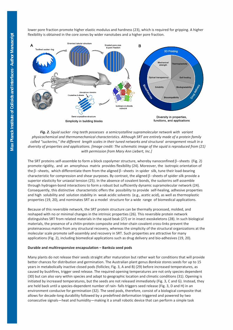
lower pore fraction promote higher elastic modulus and hardness (23), which is required for gripping. A higher flexibility is obtained in the core zones by wider nanotubes and a higher pore fraction.
Fig. 2. Squid sucker ring teeth possesses a semicrystalline supramolecular network with variant physicochemical and thermomechanical characteristics. Although SRT are entirely made of a protein family
called “suckerins,” the different length scales in their tuned networks and structural arrangement result in a diversity of properties and applications. [Image credit: The schematic image of the squid is reproduced from (21)
with permission from Mary Ann Liebert, Inc.] The SRT proteins self-assemble to form a block copolymer structure, whereby nanoconfined β−sheets (Fig. 2) promote rigidity, and an amorphous matrix provides flexibility (24). Moreover, the isotropic orientation of the β−sheets, which differentiate them from the aligned β−sheets in spider silk, tune their load-bearing characteristic for compression and shear purposes. By contrast, the aligned β−sheets of spider silk provide a superior elasticity for uniaxial tension (25). In the absence of covalent bonds, the suckerins self-assemble through hydrogen-bond interactions to form a robust but sufficiently dynamic supramolecular network (24). Consequently, this distinctive characteristic offers the possibility to provide self-healing, adhesive properties and high solubility and solution stability in weak acidic solvents (e.g., acetic acid), as well as thermoplastic properties (19, 20), and nominates SRT as a model structure for a wide range of biomedical applications. Because of this reversible network, the SRT protein structure can be thermally processed, molded, and reshaped with no or minimal changes in the intrinsic properties (26). This reversible protein network distinguishes SRT from related materials in the squid beak (27) or in insect exoskeletons (28). In such biological materials, the presence of a chitin-protein composite and inter-chain covalent cross-links prevent the proteinaceous matrix from any structural recovery, whereas the simplicity of the structural organizations at the molecular scale promote self-assembly and recovery in SRT. Such properties are attractive for many applications (Fig. 2), including biomedical applications such as drug delivery and bio-adhesives (19, 20). Durable and multiresponsive encapsulation – Banksia seed pods Many plants do not release their seeds straight after maturation but rather wait for conditions that will provide better chances for distribution and germination. The Australian plant genus Banksia stores seeds for up to 15 years in metabolically inactive closed pods (follicles; Fig. 3, A and B) (29) before increased temperatures, as caused by bushfires, trigger seed release. The required opening temperatures are not only species dependent (30) but can also vary within species and adapt to geographic location and climatic conditions (31). Opening is initiated by increased temperatures, but the seeds are not released immediately (Fig. 3, C and G). Instead, they are held back until a species-dependent number of rain- falls triggers seed release (Fig. 3, D and H) in an environment conducive for germination (32). The seed pods, therefore, consist of a biological composite that allows for decade-long durability followed by a predefined deformation triggered and powered by two consecutive signals—heat and humidity—making it a small robotic device that can perform a simple task

Max
Pla
nck
Inst
itute
of C
ollo
ids a
nd In
terfa
ces ·
Aut
hor M
anus
crip
t
without an external energy source, triggered by a rare event. Thus, these seed pods may provide additional inspiration in the current quest for actuating soft robotic devices (33–36).
Fig. 3. Opening of B. attenuata infructescences (cones) collected in Western Australia. (A to D) Cones from the North [(A), left side] contain mainly closed follicles (B). Half-open (C) and open follicles (D) are frequently found on cones in the South, where opening temperatures are lower [right infructescence in (A)]. (E) Light micrograph
of the junction zone (JZ) sealed with wax (scale bar 100 mm). (F to H) Virtual cuts through micro-tomographic reconstructions of closed (F), half-open (G), and open follicles (H) showing the seeds with the separator in between [(F) and (G)] and the endocarp-mesocarp bilayer (colored in green and yellow in one of the two
pericarp valves). The white line in (F) indicates the internal valve curvature, which changes with geographic location and climate.
Durability for long-term seed storage and multi-responsiveness for opening and seed release are achieved by a structured functional composite made of cellulose, hemicelluloses, lignin, wax, and tannins. Each follicle consists of two pericarp valves in the centimeter range. The valves are multilayers with endo-, meso-, and exocarp, which have different cellulose orientations (37) that shrink by different amounts upon ripening and, thus, store internal stresses. They are connected by the junction zone (Fig. 3, B, C, and E)—a tissue with a high surface area of interdigitating cells—that is sealed with wax that melts at about 45 to 50°C (31, 38), temperatures that can be reached on summer days in the field. These waxes are not related to seed pod opening but instead contribute to follicle integrity in creating a barrier against water loss and an antiadhesive film against wetting, insects, and microorganisms. The low melting temperatures may furthermore seal microcracks, which probably arise during long-term exposure to environmental challenges such as microbes, strong bird beaks, or weathering. In addition to the self-sealing waxes, condensed tannins in the mechanically weak parenchymatic tissue contribute to integrity by a high antioxidative capacity and increased water retention ability (39, 40). In Banksia attenuata, the initial opening temperatures and the dimensional stability depend on internal follicle geometry, in particular, on the radii of the biaxially curved follicle interiors (31), indicated by the white line in Fig. 3F. Opening at increased temperatures occurs by the softening of the endocarp (green layer in Fig. 3, F and G), which changes the internal force balance and allows the stored pre-stresses to be released (31) by the formation of a crack and an initial opening of the junction zone (Fig. 3C). Further opening for seed release (Fig. 3D) requires wetting and drying cycles, which activate bending of the endo-mesocarp bilayer (32) (Fig. 3H). The B. attenuata example illustrates how adaptation strategies and (multi)functionalities in biological materials can be better understood by including ecological aspects in biomaterials research. Furthermore, it highlights the importance of sampling biological tissues in their natural environments and of relating their properties to the environmental challenges in those habitats. Comparative studies between species also increase our knowledge of biological material functionalities and possibly evolutionary processes. For example, a recent
Max
Pla
nck
Inst
itute
of C
ollo
ids a
nd In
terfa
ces ·
Aut
hor M
anus
crip
t
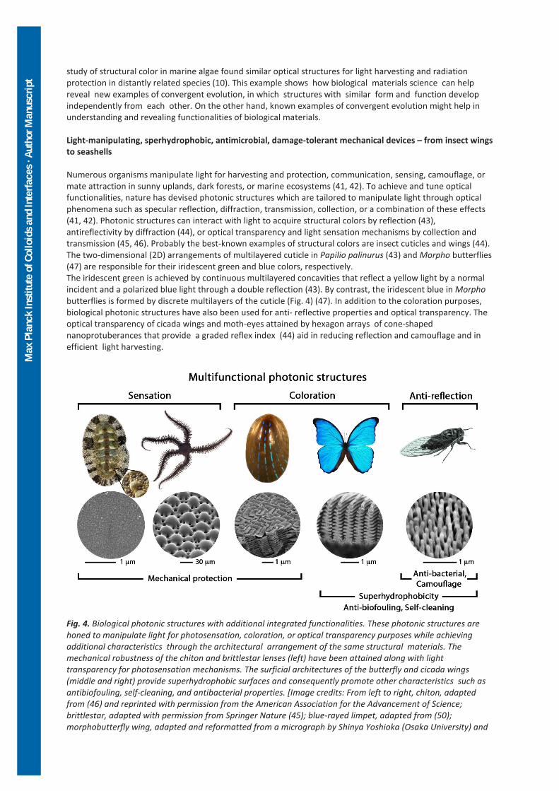
study of structural color in marine algae found similar optical structures for light harvesting and radiation protection in distantly related species (10). This example shows how biological materials science can help reveal new examples of convergent evolution, in which structures with similar form and function develop independently from each other. On the other hand, known examples of convergent evolution might help in understanding and revealing functionalities of biological materials. Light-manipulating, sperhydrophobic, antimicrobial, damage-tolerant mechanical devices – from insect wings to seashells Numerous organisms manipulate light for harvesting and protection, communication, sensing, camouflage, or mate attraction in sunny uplands, dark forests, or marine ecosystems (41, 42). To achieve and tune optical functionalities, nature has devised photonic structures which are tailored to manipulate light through optical phenomena such as specular reflection, diffraction, transmission, collection, or a combination of these effects (41, 42). Photonic structures can interact with light to acquire structural colors by reflection (43), antireflectivity by diffraction (44), or optical transparency and light sensation mechanisms by collection and transmission (45, 46). Probably the best-known examples of structural colors are insect cuticles and wings (44). The two-dimensional (2D) arrangements of multilayered cuticle in Papilio palinurus (43) and Morpho butterflies (47) are responsible for their iridescent green and blue colors, respectively. The iridescent green is achieved by continuous multilayered concavities that reflect a yellow light by a normal incident and a polarized blue light through a double reflection (43). By contrast, the iridescent blue in Morpho butterflies is formed by discrete multilayers of the cuticle (Fig. 4) (47). In addition to the coloration purposes, biological photonic structures have also been used for anti- reflective properties and optical transparency. The optical transparency of cicada wings and moth-eyes attained by hexagon arrays of cone-shaped nanoprotuberances that provide a graded reflex index (44) aid in reducing reflection and camouflage and in efficient light harvesting.
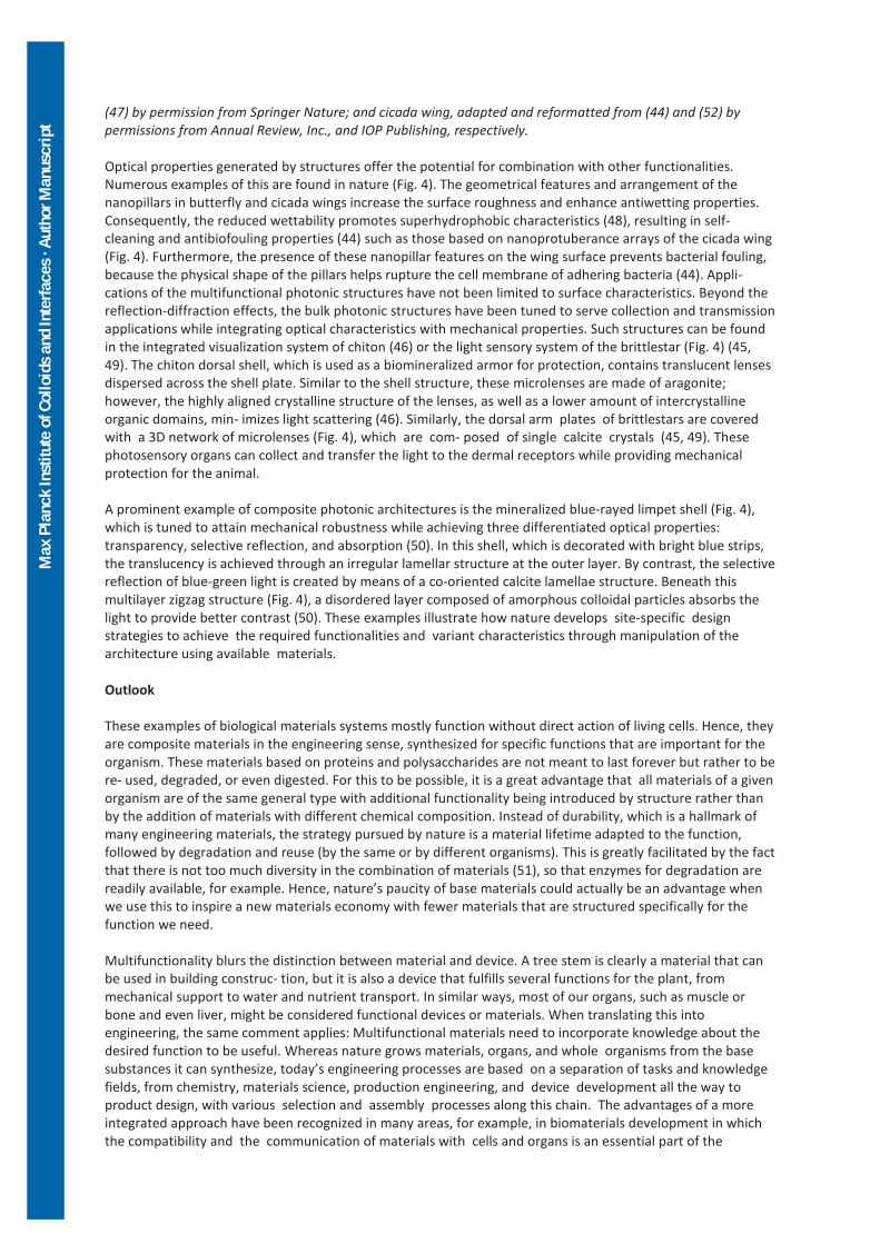
Fig. 4. Biological photonic structures with additional integrated functionalities. These photonic structures are honed to manipulate light for photosensation, coloration, or optical transparency purposes while achieving additional characteristics through the architectural arrangement of the same structural materials. The mechanical robustness of the chiton and brittlestar lenses (left) have been attained along with light transparency for photosensation mechanisms. The surficial architectures of the butterfly and cicada wings (middle and right) provide superhydrophobic surfaces and consequently promote other characteristics such as antibiofouling, self-cleaning, and antibacterial properties. [Image credits: From left to right, chiton, adapted from (46) and reprinted with permission from the American Association for the Advancement of Science; brittlestar, adapted with permission from Springer Nature (45); blue-rayed limpet, adapted from (50); morphobutterfly wing, adapted and reformatted from a micrograph by Shinya Yoshioka (Osaka University) and
Max
Pla
nck
Inst
itute
of C
ollo
ids a
nd In
terfa
ces ·
Aut
hor M
anus
crip
t
(47) by permission from Springer Nature; and cicada wing, adapted and reformatted from (44) and (52) by permissions from Annual Review, Inc., and IOP Publishing, respectively. Optical properties generated by structures offer the potential for combination with other functionalities. Numerous examples of this are found in nature (Fig. 4). The geometrical features and arrangement of the nanopillars in butterfly and cicada wings increase the surface roughness and enhance antiwetting properties. Consequently, the reduced wettability promotes superhydrophobic characteristics (48), resulting in self-cleaning and antibiofouling properties (44) such as those based on nanoprotuberance arrays of the cicada wing (Fig. 4). Furthermore, the presence of these nanopillar features on the wing surface prevents bacterial fouling, because the physical shape of the pillars helps rupture the cell membrane of adhering bacteria (44). Appli- cations of the multifunctional photonic structures have not been limited to surface characteristics. Beyond the reflection-diffraction effects, the bulk photonic structures have been tuned to serve collection and transmission applications while integrating optical characteristics with mechanical properties. Such structures can be found in the integrated visualization system of chiton (46) or the light sensory system of the brittlestar (Fig. 4) (45, 49). The chiton dorsal shell, which is used as a biomineralized armor for protection, contains translucent lenses dispersed across the shell plate. Similar to the shell structure, these microlenses are made of aragonite; however, the highly aligned crystalline structure of the lenses, as well as a lower amount of intercrystalline organic domains, min- imizes light scattering (46). Similarly, the dorsal arm plates of brittlestars are covered with a 3D network of microlenses (Fig. 4), which are com- posed of single calcite crystals (45, 49). These photosensory organs can collect and transfer the light to the dermal receptors while providing mechanical protection for the animal. A prominent example of composite photonic architectures is the mineralized blue-rayed limpet shell (Fig. 4), which is tuned to attain mechanical robustness while achieving three differentiated optical properties: transparency, selective reflection, and absorption (50). In this shell, which is decorated with bright blue strips, the translucency is achieved through an irregular lamellar structure at the outer layer. By contrast, the selective reflection of blue-green light is created by means of a co-oriented calcite lamellae structure. Beneath this multilayer zigzag structure (Fig. 4), a disordered layer composed of amorphous colloidal particles absorbs the light to provide better contrast (50). These examples illustrate how nature develops site-specific design strategies to achieve the required functionalities and variant characteristics through manipulation of the architecture using available materials. Outlook These examples of biological materials systems mostly function without direct action of living cells. Hence, they are composite materials in the engineering sense, synthesized for specific functions that are important for the organism. These materials based on proteins and polysaccharides are not meant to last forever but rather to be re- used, degraded, or even digested. For this to be possible, it is a great advantage that all materials of a given organism are of the same general type with additional functionality being introduced by structure rather than by the addition of materials with different chemical composition. Instead of durability, which is a hallmark of many engineering materials, the strategy pursued by nature is a material lifetime adapted to the function, followed by degradation and reuse (by the same or by different organisms). This is greatly facilitated by the fact that there is not too much diversity in the combination of materials (51), so that enzymes for degradation are readily available, for example. Hence, nature’s paucity of base materials could actually be an advantage when we use this to inspire a new materials economy with fewer materials that are structured specifically for the function we need. Multifunctionality blurs the distinction between material and device. A tree stem is clearly a material that can be used in building construc- tion, but it is also a device that fulfills several functions for the plant, from mechanical support to water and nutrient transport. In similar ways, most of our organs, such as muscle or bone and even liver, might be considered functional devices or materials. When translating this into engineering, the same comment applies: Multifunctional materials need to incorporate knowledge about the desired function to be useful. Whereas nature grows materials, organs, and whole organisms from the base substances it can synthesize, today’s engineering processes are based on a separation of tasks and knowledge fields, from chemistry, materials science, production engineering, and device development all the way to product design, with various selection and assembly processes along this chain. The advantages of a more integrated approach have been recognized in many areas, for example, in biomaterials development in which the compatibility and the communication of materials with cells and organs is an essential part of the

Max
Pla
nck
Inst
itute
of C
ollo
ids a
nd In
terfa
ces ·
Aut
hor M
anus
crip
t
materials research itself. Research on materials and on functional devices are traditionally close in the textile industry or architecture. In general, an increased interaction of materials scientists with product designers could reduce the distinction between material and functional device in all areas of engineering and, thus, provide a growing need for the development of multifunctional materials. References and Notes 1. J. Aizenberg, P. Fratzl, Adv. Funct. Mater. 23, 4398–4399 (2013). 2. U. G. K. Wegst, H. Bai, E. Saiz, A. P. Tomsia, R. O. Ritchie, Nat. Mater. 14, 23–36 (2015). 3. A. R. Studart, Chem. Soc. Rev. 45, 359–376 (2016). 4. F. Barthelat, Z. Yin, M. J. Buehler, Nat. Rev. Mater. 1, 16007 (2016). 5. Z. Liu, M. A. Meyers, Z. Zhang, R. O. Ritchie, Prog. Mater. Sci. 88, 467–498 (2017). 6. M. F. Ashby, Y. J. M. Bréchet, D. Cebon, L. Salvo, Mater. Des. 25, 51–67 (2004). 7. T. N. Sullivan, B. Wang, H. D. Espinosa, M. A. Meyers, Mater. Today 20, 377–391 (2017). 8. E. Moyroud et al., Nature 550, 469–474 (2017). 9. M. F. Ashby, L. J. Gibson, U. Wegst, R. Olive, Proc. R. Soc. London, Math. Phys. Sci. 450, 123–140 (1995). 10. C. J. Chandler, B. D. Wilts, J. Brodie, S. Vignolini, Adv. Opt. Mater. 5, 1600646 (2017). 11. E. Kuprian et al., Plant Cell Environ. 40, 3101–3112 (2017). 12. R. Weinkamer, P. Fratzl, Mater. Sci. Eng. C Mater. Biol. Appl. 31, 1164–1173 (2011). 13. S. Torquato, S. Hyun, A. Donev, J. Appl. Phys. 94, 5748–5755 (2003). 14. R. Weinkamer, P. Fratzl, MRS Bull. 41, 667–671 (2016). 15. N. Reznikov, J. A. M. Steele, P. Fratzl, M. M. Stevens, Nat. Rev. Mater. 1, 16041 (2016). 16. P. Fratzl, O. Kolednik, F. D. Fischer, M. N. Dean, Chem. Soc. Rev. 45, 252–267 (2016). 17. A. R. Studart, Nat. Mater. 13, 433–435 (2014). 18. H.-L. Gao et al., Nat. Commun. 8, 287 (2017). 19. S. H. Hiew, A. Miserez, ACS Biomater. Sci. Eng. 3, 680–693 (2017). 20. A. Pena-Francesch et al., APL Mater. 6, 010701 (2018). 21. K. Kumar et al., Soft Robot. 4, 317–323 (2017). 22. E. Degtyar, M. J. Harrington, Y. Politi, P. Fratzl, Angew. Chem. Int. Ed. 53, 12026–12044 (2014). 23. A. Miserez et al., Adv. Mater. 21, 401–406 (2009). 24. P. A. Guerette et al., ACS Nano 8, 7170–7179 (2014). 25. S. Keten, Z. Xu, B. Ihle, M. J. Buehler, Nat. Mater. 9, 359–367 (2010). 26. V. Latza et al., Nat. Commun. 6, 8313 (2015). 27. A. Miserez, T. Schneberk, C. Sun, F. W. Zok, J. H. Waite, Science 319, 1816–1819 (2008). 28. S. O. Andersen, Insect Biochem. Mol. Biol. 40, 166–178 (2010). 29. B. B. Lamont, D. C. Lemaitre, R. M. Cowling, N. J. Enright, Bot. Rev. 57, 277–317 (1991). 30. A. S. George, Nuytsia 3, 239–473 (1981). 31. J. C. Huss et al., Adv. Sci. 5, 1700572 (2017). 32. R. M. Cowling, B. B. Lamont, Aust. J. Ecol. 10, 169–171 (1985). 33. B. Mazzolai, L. Beccai, V. Mattoli, Front. Bioeng. Biotechnol. 2, 2 (2014). 34. W. Wang et al., Nature 559, 77–82 (2018). 35. W. Hu, G. Z. Lum, M. Mastrangeli, M. Sitti, Nature 554, 81–85 (2018). 36. S. Poppinga et al., Adv. Mater. 30, e1703653 (2018). 37. A. B. Wardrop, Aust. J. Bot. 31, 485–500 (1983). 38. J. C. Huss et al., J. R. Soc. Interface 15, 20180190 (2018). 39. S. Quideau, D. Deffieux, C. Douat-Casassus, L. Pouységu, Angew. Chem. Int. Ed. 50, 586–621 (2011). 40. W. Vermerris, R. M. Nicholson, Phenolic Compound Biochemistry (Springer, 2008). 41. F. P. Barrows, M. H. Bartl, Nanomater. Nanotechnol. 4, 1 (2014). 42. S. Tadepalli, J. M. Slocik, M. K. Gupta, R. R. Naik, S. Singamaneni, Chem. Rev. 117, 12705–12763 (2017). 43. P. Vukusic, J. R. Sambles, C. R. Lawrence, Nature 404, 457 (2000). 44. G. S. Watson, J. A. Watson, B. W. Cribb, Annu. Rev. Entomol.62, 185–205 (2017). 45. J. Aizenberg, A. Tkachenko, S. Weiner, L. Addadi, G. Hendler, Nature 412, 819–822 (2001). 46. L. Li et al., Science 350, 952–956 (2015). 47. P. Vukusic, J. R. Sambles, Nature 424, 852–855 (2003). 48. T. Darmanin, F. Guittard, Mater. Today 18, 273–285 (2015). 49. I. Polishchuk et al., Science 358, 1294–1298 (2017). 50. L. Li et al., Nat. Commun. 6, 6322 (2015). 51. M. F. Ashby, Materials and Sustainable Development (Butterworth-Heinemann, Oxford, 2015). 52. G. Xie et al., Nanotechnology 19, 095605 (2008).

Max
Pla
nck
Inst
itute
of C
ollo
ids a
nd In
terfa
ces ·
Aut
hor M
anus
crip
t
Acknowledgements The authors are grateful for many discussions about active materials, particularly with W. Schäffner, K. Krauthausen, and M. Friedman from Humboldt-Universität zu Berlin and with J. Dunlop from the University of Salzburg, Austria. We thank J. Blumenthal, Weissensee School of Arts and Design, for drawings in Fig. 1. Funding: Partial support was provided by the DFG Cluster of Excellence “Image-Knowledge-Gestaltung” (DFG-EXC 1027). P.F. was also supported by a Leibniz Award of the DFG.