biology stem cells-nih-2001!!
DESCRIPTION
TRANSCRIPT
Stem Cells:Scientific Progress andFuture Research Directions
© 2001 Terese Winslow
STEM CELLS: SCIENTIFIC PROGRESS
AND FUTURERESEARCH DIRECTIONS
June 2001

This page intentionally left blank
Preface . . . . . . . . . . . . . . . . . . . . . . . . . . . . . . . . . . . . . . . . . . . . . . . . . . . . . . . . . . . . i
Executive Summary . . . . . . . . . . . . . . . . . . . . . . . . . . . . . . . . . . . . . . . . . . . . . . . . ES-1
Chapter 1: The Stem Cell . . . . . . . . . . . . . . . . . . . . . . . . . . . . . . . . . . . . . . . . . . . . . 1
Chapter 2: The Embryonic Stem Cell . . . . . . . . . . . . . . . . . . . . . . . . . . . . . . . . . . . . . 5
Chapter 3: The Human Embryonic Stem Celland The Human Embryonic Germ Cell . . . . . . . . . . . . . . . . . . . . . . . . . 11
Chapter 4: The Adult Stem Cell . . . . . . . . . . . . . . . . . . . . . . . . . . . . . . . . . . . . . . . . 23
Chapter 5: Hematopoietic Stem Cells . . . . . . . . . . . . . . . . . . . . . . . . . . . . . . . . . . . 43
Chapter 6: Autoimmune Diseases and the Promiseof Stem Cell-Based Therapies . . . . . . . . . . . . . . . . . . . . . . . . . . . . . . . . 59
Chapter 7: Stem Cells and Diabetes . . . . . . . . . . . . . . . . . . . . . . . . . . . . . . . . . . . . 67
Chapter 8: Rebuilding the Nervous System with Stem Cells . . . . . . . . . . . . . . . . . . . 77
Chapter 9: Can Stem Cells Repair a Damaged Heart? . . . . . . . . . . . . . . . . . . . . . . 87
Chapter 10: Assessing Human Stem Cell Safety . . . . . . . . . . . . . . . . . . . . . . . . . . . . . 93
Chapter 11: Use of Genetically Modified Stem Cells inExperimental Gene Therapies . . . . . . . . . . . . . . . . . . . . . . . . . . . . . . . . 99
Appendix A: Early Development . . . . . . . . . . . . . . . . . . . . . . . . . . . . . . . . . . . . . . . . A-1
Appendix B: Mouse Embryonic Stem Cells . . . . . . . . . . . . . . . . . . . . . . . . . . . . . . . . B-1
Appendix C: Human Embryonic Stem Cells and Embryonic Germ Cells . . . . . . . . . . C-1
Appendix D: Stem Cell Tables
i. Published Reports on Isolation and Differentiation of Mouse Stem Cells . . . . . . . . . . . . . . . . . . . . . . . . . . . . . . . . . . . . . . . . . . . . D-2
ii. Published Reports on Isolation and Differentiation of HumanFetal Tissue Germ Cells . . . . . . . . . . . . . . . . . . . . . . . . . . . . . . . . . D-13
iii. Published Reports on Isolation and Differentiation of HumanEmbryonic Stem Cells . . . . . . . . . . . . . . . . . . . . . . . . . . . . . . . . . . D-14
iv. Published Reports on Isolation and Differentiation of HumanEmbryonic Carcinoma Stem Cells . . . . . . . . . . . . . . . . . . . . . . . . . D-16
v. Published Reports on Isolation and Differentiation of HumanAdult Stem Cells . . . . . . . . . . . . . . . . . . . . . . . . . . . . . . . . . . . . . . D-18
vi. References . . . . . . . . . . . . . . . . . . . . . . . . . . . . . . . . . . . . . . . . . . D-22
TABLE OF CONTENTSTABLE OF CONTENTS

Appendix E: Stem Cell Markers
i. Markers: How Do Researchers Use Them to Identify Stem Cells? . . . . . E-1
ii. Commonly Used Markers to Identify Stem Cells andCharacterize Differentiated Cell Types . . . . . . . . . . . . . . . . . . . . . . . E-5
Appendix F: Glossary and Terms
i. Glossary . . . . . . . . . . . . . . . . . . . . . . . . . . . . . . . . . . . . . . . . . . . . . . F-1
ii. Terms . . . . . . . . . . . . . . . . . . . . . . . . . . . . . . . . . . . . . . . . . . . . . . . F-11
Appendix G: Informational Resources
i. Persons Interviewed . . . . . . . . . . . . . . . . . . . . . . . . . . . . . . . . . . . . . G-1
ii. Special Contributions . . . . . . . . . . . . . . . . . . . . . . . . . . . . . . . . . . . G-4
iii. Acknowledgments . . . . . . . . . . . . . . . . . . . . . . . . . . . . . . . . . . . . . G-5
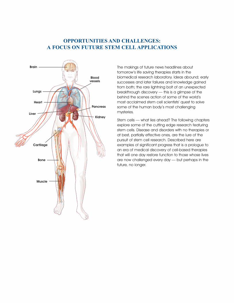
Brain
Lungs
Heart
Liver
Bloodvessels
Pancreas
Kidney
Cartilage
Bone
Muscle
The makings of future news headlines abouttomorrow’s life saving therapies starts in thebiomedical research laboratory. Ideas abound; earlysuccesses and later failures and knowledge gainedfrom both; the rare lightning bolt of an unexpectedbreakthrough discovery — this is a glimpse of thebehind the scenes action of some of the world’smost acclaimed stem cell scientists’ quest to solvesome of the human body’s most challengingmysteries.
Stem cells — what lies ahead? The following chaptersexplore some of the cutting edge research featuringstem cells. Disease and disorders with no therapies orat best, partially effective ones, are the lure of thepursuit of stem cell research. Described here areexamples of significant progress that is a prologue toan era of medical discovery of cell-based therapiesthat will one day restore function to those whose livesare now challenged every day — but perhaps in thefuture, no longer.
OPPORTUNITIES AND CHALLENGES:A FOCUS ON FUTURE STEM CELL APPLICATIONS

REPORT PREPARED BY THE NATIONAL INSTITUTES OF HEALTH
Ruth Kirschstein, M.D.Acting Director
Office of Science PolicyLana R. Skirboll, Ph.D.
Director
i
On February 28, 2001, Tommy G. Thompson,Secretary of Health and Human Services, requestedthat the National Institutes of Health prepare asummary report on the state of the science on stemcells. This report was developed in response to hisrequest. It provides the current information about thebiology of stem cells derived from all sources—embryo, fetal tissue, and adult.
Since 1998, when human pluripotent stem cells werefirst isolated, research on stem cells has receivedmuch public attention, both because of its extraordi-nary promise and because of relevant legal andethical issues. Underlying this recent public scrutiny isdecades of painstaking work by scientists in manyfields, who have been deciphering some of the mostfundamental questions about life with the goal ofimproving health.
In the last several decades, investments in basicresearch have yielded extensive knowledge aboutthe many and complex processes involved in thedevelopment of an organism, including the controlof cellular development. But many questions remain.How does a single cell—the fertilized egg—give riseto a complex, multi-cellular organism? The questionrepresents a fundamental challenge in develop-mental biology. Researchers are now seeking tounderstand in greater detail the genetic factors thatregulate cell differentiation in early development.
Put simply, stem cells are self-renewing, unspecial-ized cells that can give rise to multiple types all ofspecialized cells of the body. The process by whichdividing, unspecialized cells are equipped to per-form specific functions—muscle contraction or nervecell communication, for example—is called differen-tiation, and is fundamental to the development ofthe mature organism. It is now known that stem cells,in various forms, can be obtained from the embryo,the fetus, and the adult.
How and whether stem cells derived from any ofthese sources can be manipulated to replace cellsin diseased tissues, used to screen drugs and toxins,or studied to better understand normal developmentdepends on knowing more about their basic proper-ties. In this respect, stem cell research is in manyways no different than many other areas of modernbiology; it is advancing because new tools and newknowledge are providing the opportunities for newinsights. Like all fields of scientific inquiry, research onstem cells raises as many questions as it answers. Thisreport describes the state of the science of stem cellbiology and gives some clues as to the many andvaried questions that remain to be answered.
WHAT IS THE SCOPE OF THEREPORT?The report is a review of the state of the science ofstem cell research as of June 17, 2001. Included inthis report is subject matter addressing stem cellsfrom adult, fetal tissue, and embryonic sources.Because so much of the progress made to date wasdependent on animal models, a significant emphasisis placed on understandings gained from mousemodels of development and mouse stem cellresearch. The report also devotes substantial attentionto scientific publications on the characterization ofspecialized cells developed from embryonic stemcells and the plasticity of adult stem cells. A generaloverview of early development is provided in theAppendix to assist the reader in understanding thekey events in formation of cells, tissues, and the wholeorganism.
Both scientific and lay publications use a variety ofterms to describe stem cells and their properties. Forthis reason, this report adopts a lexicon of terms andit is used consistently throughout. To aid the reader, aglossary and terms section is provided. In several
PREFACEPREFACE
places in the report, discovery timelines are provided.The various sources of stem cells are described, asare the techniques used to isolate and developthem. A comprehensive listing of various stem cellisolation and characterizations is also included.
In order to ensure the reader is provided informationboth about the basic biology of stem cells, and theirtherapeutic potential, the report contains severalchapters focused on particular diseases which mightbenefit from stem cell research. These chapters onthe use of hematopoietic stem cells, followed byfocus features on specific nervous system diseases,diabetes, heart disease, and autoimmune diseasesserve merely as examples of the many applicationsof stem cells that are being pursued. Also includedare features that review aspects of stem cells astherapeutic delivery tools for gene therapy and,importantly, the safety considerations for developingstem cell-based therapies.
WHAT IS NOT IN THE SCOPE OF THE REPORT?NIH recognizes the compelling ethical and legalissues surrounding human pluripotent stem cellresearch. Because extensive discussions regardingthese issues have been presented in various forumselsewhere, they are not part of this review of the stateof the science. Also, the report does not makerecommendations pertaining to the policies govern-ing Federal funding of such research.
HOW WAS THE REPORTDEVELOPED?The report was prepared under the auspices of theOffice of Science Policy, Office of the Director, NIH.Several approaches were taken to obtain relevantscientific information for the report. A thorough reviewof the extant literature, including more than 1200scientific publications was conducted. Scientificexperts (both domestic and international) from allareas of relevant biomedical research in stem cellswere interviewed in depth. While the majority of thework presented in this report emanates frominvestigators in academic laboratories, extensivediscussions were held with scientists in the privatepharmaceutical and biotechnology sectors. Thus, thereport makes every effort to encompass what isknown and not known about stem cell biology and is,therefore, not limited to research that is or has beenfunded by the NIH.
In recent months, there have been many reports inthe lay press regarding scientific discoveries onvarious types of stem cells. The science representedin this report focuses exclusively on scientificpublications or public presentations. In cases wheretechnical or logistical information key to the under-standing of the details of science was needed,personal communications with the informationsources were cited.
ii
Preface
ES-1
INTRODUCTIONA stem cell is a special kind of cell that has a uniquecapacity to renew itself and to give rise to specializedcell types. Although most cells of the body, such asheart cells or skin cells, are committed to conduct aspecific function, a stem cell is uncommitted andremains uncommitted, until it receives a signal todevelop into a specialized cell. Their proliferativecapacity combined with the ability to become spe-cialized makes stem cells unique. Researchers havefor years looked for ways to use stem cells to replacecells and tissues that are damaged or diseased.Recently, stem cells have received much attention.What is “new” and what has brought stem cell bio-logy to the forefront of science and public policy?
Scientists interested in human development havebeen studying animal development for many years.This research yielded our first glimpse at a class ofstem cells that can develop into any cell type in thebody. This class of stem cells is called pluripotent,meaning the cells have the potential to developalmost all of the more than 200 different known celltypes. Stem cells with this unique property come fromembryos and fetal tissue.
In 1998, for the first time, investigators were able toisolate this class of pluripotent stem cell from earlyhuman embryos and grow them in culture. In the fewyears since this discovery, evidence has emerged thatthese stem cells are, indeed, capable of becomingalmost all of the specialized cells of the body and,thus, may have the potential to generate replace-ment cells for a broad array of tissues and organs,such as the heart, the pancreas, and the nervoussystem. Thus, this class of human stem cell holds thepromise of being able to repair or replace cells ortissues that are damaged or destroyed by many ofour most devastating diseases and disabilities.
At about the same time as scientists were beginningto explore human pluripotent stem cells fromembryos and fetal tissue, a flurry of new informationwas emerging about a class of stem cells that havebeen in clinical use for years—so-called adult stemcells. An adult stem cell is an undifferentiated cellthat is found in a differentiated (specialized) tissue inthe adult, such as blood. It can yield the specializedcell types of the tissue from which it originated. In thebody, it too, can renew itself. During the past decade,scientists discovered adult stem cells in tissues thatwere previously not thought to contain them, such asthe brain. More recently, they reported that adultstem cells from one tissue appear to be capable ofdeveloping into cell types that are characteristic ofother tissues. For example, although adulthematopoietic stem cells from bone marrow havelong been recognized as capable of developing intoblood and immune cells, recently scientists reportedthat, under certain conditions, the same stem cellscould also develop into cells that have many of thecharacteristics of neurons. So, a new concept and anew term emerged-adult stem cell plasticity.
Are human adult and embryonic stem cells equiva-lent in their potential for generating replacement cellsand tissues? Current science indicates that, althoughboth of these cell types hold enormous promise,adult and embryonic stem cells differ in importantways. What is not known is the extent to which thesedifferent cell types will be useful for the developmentof cell-based therapies to treat disease.
Some considerations are noteworthy regarding thisreport. First, in recent months, there have been manydiscussions in the lay press about the anticipatedabilities of stem cells from various sources and pro-jected benefits to be realized from them in replacingcells and tissues in patients with various diseases. Theterminology used to describe stem cells in the lay
EXECUTIVE SUMMARYEXECUTIVE SUMMARY
literature is often confusing or misapplied. Second,even among biomedical researchers, there is a lackof consistency in common terms to describe whatstem cells are and how they behave in the researchlaboratory. Third, the field of stem cell biology isadvancing at an incredible pace with new discover-ies being reported in the scientific literature on aweekly basis.
This summary begins with common definitions andexplanations of key concepts about stem cells. Itends with an assessment of how adult, embryonicand fetal stem cells are similar and how they are dif-ferent. In between lie important details that describewhat researchers have discovered about stem cellsand how they are being used in the laboratory.
DEFINITIONS AND GENERALCONCEPTS ABOUT STEM CELLSIn developing this report, some conventions wereestablished to describe consistently what stem cellsare, what characteristics they have, and how they areused in biomedical research. Here are some of thekey definitions that are used throughout this report.
Stem cell. A stem cell is a cell from the embryo,fetus, or adult that has, under certain conditions, theability to reproduce itself for long periods or, in thecase of adult stem cells, throughout the life of theorganism. It also can give rise to specialized cells thatmake up the tissues and organs of the body. Muchbasic understanding about embryonic stem cells hascome from animal research. In the laboratory, thistype of stem cell can proliferate indefinitely, a proper-ty that is not shared by adult stem cells.
Pluripotent stem cell. A single pluripotent stem cellhas the ability to give rise to types of cells that devel-op from the three germ layers (mesoderm, endo-derm, and ectoderm) from which all the cells of thebody arise. The only known sources of human pluripo-tent stem cells are those isolated and cultured fromearly human embryos and from fetal tissue that wasdestined to be part of the gonads.
Embryonic stem cell. An embryonic stem cell isderived from a group of cells called the inner cellmass, which is part of the early (4- to 5-day) embryocalled the blastocyst. Once removed from the blasto-cyst, the cells of the inner cell mass can be culturedinto embryonic stem cells. These embryonic stem
cells are not themselves embryos. In fact, evidence isemerging that these cells do not behave in the labo-ratory as they would in the developing embryo—thatis, the conditions in which these cells develop in cul-ture are likely to differ from those in the developingembryo.
Embryonic germ cell. An embryonic germ cell isderived from fetal tissue. Specifically, they are isolatedfrom the primordial germ cells of the gonadal ridgeof the 5- to 10-week fetus. Later in development, thegonadal ridge develops into the testes or ovaries andthe primordial germ cells give rise to eggs or sperm.Embryonic stem cells and embryonic germ cells arepluripotent, but they are not identical in their proper-ties and characteristics.
Differentiation. Differentiation is the process by whichan unspecialized cell (such as a stem cell) becomesspecialized into one of the many cells that make upthe body. During differentiation, certain genesbecome activated and other genes become inacti-vated in an intricately regulated fashion. As a result, adifferentiated cell develops specific structures andperforms certain functions. For example, a mature,differentiated nerve cell has thin, fiber-like projectionsthat send and receive the electrochemical signalsthat permit the nerve cell to communicate with othernerve cells. In the laboratory, a stem cell can bemanipulated to become specialized or partiallyspecialized cell types (e.g., heart muscle, nerve, orpancreatic cells) and this is known as directeddifferentiation.
Adult stem cell. An adult stem cell is an undifferenti-ated (unspecialized) cell that occurs in a differentiat-ed (specialized) tissue, renews itself, and becomesspecialized to yield all of the specialized cell types ofthe tissue from which it originated. Adult stem cellsare capable of making identical copies of them-selves for the lifetime of the organism. This property isreferred to as “self-renewal.” Adult stem cells usuallydivide to generate progenitor or precursor cells,which then differentiate or develop into “mature” celltypes that have characteristic shapes and special-ized functions, e.g., muscle cell contraction or nervecell signaling. Sources of adult stem cells includebone marrow, blood, the cornea and the retina ofthe eye, brain, skeletal muscle, dental pulp, liver, skin,the lining of the gastrointestinal tract, and pancreas.The most abundant information about adult human
ES-2
Executive Summary
stem cells comes from studies of hematopoietic(blood-forming) stem cells isolated from the bonemarrow and blood. These adult stem cells have beenextensively studied and applied therapeutically forvarious diseases. At this point, there is no isolatedpopulation of adult stem cells that is capable offorming all the kinds of cells of the body. Adult stemcells are rare. Often they are difficult to identify, iso-late, and purify. There are insufficient numbers of cellsavailable for transplantation and adult stem cells donot replicate indefinitely in culture.
Plasticity. Plasticity is the ability of an adult stem cellfrom one tissue to generate the specialized celltype(s) of another tissue. A recently reported exampleof plasticity is that, under specific experimental condi-tions, adult stem cells from bone marrow generatedcells that resemble neurons and other cell types thatare commonly found in the brain. The concept ofadult stem cell plasticity is new, and the phenome-non is not thoroughly understood. Evidence suggeststhat, given the right environment, some adult stemcells are capable of being “genetically repro-grammed” to generate specialized cells that arecharacteristic of different tissues.
Clonality or clonally derived stem cell. A cell is saidto be clonally derived or to exhibit clonality if it wasgenerated by the division of a single cell and isgenetically identical to that cell. In stem cellresearch, the concept of clonality is important forseveral reasons. For researchers to fully understandand harness the ability of stem cells to generatereplacement cells and tissues, the exact identity ofthose cells’ genetic capabilities and functional quali-ties must be known. Human pluripotent stem cellsfrom embryos and fetal tissue are by their natureclonally derived. However, very few studies haveshown clonal properties of the cells that are devel-oped from adult stem cells. It is crucial to knowwhether a single cell is capable of developing anarray of cell types, or whether multiple stem celltypes, that when grown together, are capable offorming multiple cell types. For instance, recentresearch has shown that a mixture of cells removedfrom fat tissue or umbilical cord blood are capableof developing into blood cells, bone cells, andperhaps others. Researchers have not shown that asingle cell is responsible for giving rise to other celltypes or, if so, what kind of cell it is. These results maywell be attributable to multiple types of precursor cells
in the starting tissue; such results from fat cells may, infact, be due to the presence of hematopoietic stemcells in the fat tissue. The importance of showing thatone cell type can reproducibly become another andself-replicate cannot be overemphasized.
Progenitor or precursor cell. A progenitor or pre-cursor cell occurs in fetal or adult tissues and is par-tially specialized; it divides and gives rise to differenti-ated cells. Researchers often distinguish precursor/progenitor cells from adult stem cells in the followingway: when a stem cell divides, one of the two newcells is often a stem cell capable of replicating itselfagain. In contrast, when a progenitor/precursor celldivides, it can form more progenitor/precursor cells orit can form two specialized cells, neither of which iscapable of replicating itself. Progenitor/precursor cellscan replace cells that are damaged or dead, thusmaintaining the integrity and functions of a tissuesuch as liver or brain. Progenitor/precursor cells giverise to related types of cells-lymphocytes such as Tcells, B cells, and natural killer cells, for example—butin their normal state do not generate a wide varietyof cell types.
CHALLENGES IN STEM CELLRESEARCH It is important to understand some of the difficultiesthat researchers have had in isolating various types ofstem cells, working with the cells in the laboratory,and proving experimentally that the cells are truestem cells. Most of the basic research discoveries onembryonic and adult stem cells come from researchusing animal models, particularly mice.
In 1981, researchers reported methods for growingmouse embryonic stem cells in the laboratory, and ittook nearly 20 years before similar achievementscould be made with human embryonic stem cells.Much of the knowledge about embryonic stem cellshas emerged from two fields of research: appliedreproductive biology, i.e., in vitro fertilization technolo-gies, and basic research on mouse embryology.
There have been many technical challenges thathave been overcome in adult stem cell research aswell. Some of the barriers include: the rare occur-rence of adult stem cells among other, differentiatedcells, difficulties in isolating and identifying the cells(researchers often use molecular “markers” to identify
Executive Summary
ES-3

adult stem cells), and in many cases, difficulties ingrowing adult stem cells in tissue culture. Much of theresearch demonstrating the plasticity of adult stemcells comes from studies of animal models in which amixture of adult stem cells from a donor animal isinjected into another animal, and the developmentof new, specialized cells is traced.
In 1998, James Thomson at the University ofWisconsin-Madison isolated cells from the inner cellmass of the early embryo, called the blastocyst, anddeveloped the first human embryonic stem cell lines.At the same time, John Gearhart at Johns HopkinsUniversity reported the first derivation of humanembryonic germ cells from an isolated population ofcells in fetal gonadal tissue, known as the primordialgerm cells, which are destined to become the eggsand sperm. From both of these sources, theresearchers developed pluripotent stem cell “lines,”which are capable of renewing themselves for longperiods and giving rise to many types of human cellsor tissues. Human embryonic stem cells and embry-onic germ cells differ in some characteristics,however, and do not appear to be equivalent.
Why are the long-term proliferation ability andpluripotency of embryonic stem cells and embryonicgerm cells so important? First, for basic researchpurposes, it is important to understand the geneticand molecular basis by which these cells continue tomake many copies of themselves overlong periodsof time. Second, if the cells are to be manipulatedand used for transplantation, it is important to havesufficient quantities of cells that can be directed todifferentiate into the desired cell type(s) and used totreat the many patients that may be suffering from aparticular disease.
In recent months, other investigators have beensuccessful in using somewhat different approaches toderiving human pluripotent stem cells. At least 5 otherlaboratories have been successful in deriving pluri-potent stem cells from human embryos and oneadditional laboratory has created cell lines from fetaltissue. In each case, the methods for deriving pluri-potent stem cells from human embryos and embry-onic germ cells from fetal tissue are similar, yet theydiffer in the isolation and culture conditions as initiallydescribed by Thomson and Gearhart, respectively. Itis not known to what extent U.S.-based researchersare using these additional sources of embryonic stemand germ cells.
ES-4
Executive Summary
At present, there have been multiple human adultstem cell lines that have been created through acombination of public and private resources (e.g.,hematopoietic stem cells). Substantial adult stem cellresearch has been underway for many years, and inrecent years this has included basic studies on the“plasticity” of such cells.
WHAT KINDS OF RESEARCH MIGHTBE CONDUCTED WITH STEM CELLS?There has been much written about the newdiscoveries of various stem cell types and theirproperties. Importantly, these cells are research toolsand they open many doors of opportunity for bio-medical research.
Transplantation Research—Restoring Vital Body Functions
Stem cells may hold the key to replacing cells lost inmany devastating diseases. There is little doubt thatthis potential benefit underpins the vast interest aboutstem cell research. What are some of thesediseases? Parkinson’s disease, diabetes, chronic heartdisease, end-stage kidney disease, liver failure, andcancer are just a few for which stem cells have thera-peutic potential. For many diseases that shorten lives,there are no effective treatments but the goal is tofind a way to replace what natural processes havetaken away. For example, today, science has broughtus to a point where the immune response can besubdued, so that organs from one person can beused to replace the diseased organs and tissues ofanother. But, despite recent advances in transplanta-tion sciences, there is a shortage of donor organsthat makes it unlikely that the growing demand forlifesaving organ replacements will be fully metthrough organ donation strategies.
The use of stem cells to generate replacement tissuesfor treating neurological diseases is a major focus ofresearch. Spinal cord injury, multiple sclerosis, Parkin-son’s disease, and Alzheimer ’s disease are amongthose diseases for which the concept of replacingdestroyed or dysfunctional cells in the brain or spinalcord is a practical goal. This report features severalrecent advances that demonstrate the regenerativeproperties of adult and embryonic stem cells.
Another major discovery frontier for research on adultand embryonic stem cells is the development of
transplantable pancreatic tissues that can be used totreat diabetes. Scientists in academic and industrialresearch are vigorously pursuing all possible avenuesof research, including ways to direct the specializa-tion of adult and embryonic stem cells to becomepancreatic islet-like cells that produce insulin and canbe used to control blood glucose levels. Researchershave recently shown that human embryonic stemcells to be directly differentiated into cells that pro-duce insulin.
There are common misconceptions about both adultand human embryonic stem cells. First, the lines ofunaltered human embryonic stem cells that exist willnot be suitable for direct use in patients. These cellswill need to be differentiated or otherwise modifiedbefore they can be used clinically. Current chal-lenges are to direct the differentiation of embryonicstem cells into specialized cell populations, and alsoto devise ways to control their development or prolif-eration once placed in patients.
A second misconception is that adult stem cells areready to use as therapies. With the exception of theclinical application of hematopoietic stem cells torestore the blood and immune system, this is not thecase. The therapeutic use of this mixture of cells hasproven safe because the mixture is place back intothe environment from which it was taken, e.g., thebone marrow. In fact, many of the adult stem cellpreparations currently being developed in the labora-tory represent multiple cell types that are not fullycharacterized. In order to safely use stem cells or cellsdifferentiated from them in tissues other than the tissuefrom which they were isolated, researchers will needpurified populations (clonal lines) of adult stem cells.
In addition, the potential for the recipient of a stemcell transplant to reject these tissues as foreign is veryhigh. Modifications to the cells, to the immune sys-tem, or both will be a major requirement for their use.In sum, with the exception of the current practice ofhematopoietic stem cell transplantation, much basicresearch lies ahead before direct patient applicationof stem cell therapies is realized.
Basic Research Applications
Embryonic stem cells will undoubtedly be keyresearch tools for understanding fundamental eventsin embryonic development that one day mayexplain the causes of birth defects and approachesto correct or prevent them. Another important area of
research that links developmental biology and stemcell biology is understanding the genes and mole-cules, such as growth factors and nutrients, thatfunction during the development of the embryo sothat they can be used to grow stem cells in thelaboratory and direct their development intospecialized cell types.
Therapeutic Delivery Systems
Stem cells are already being explored as a vehiclefor delivering genes to specific tissues in the body.Stem cell-based therapies are a major area of inves-tigation in cancer research. For many years, restora-tion of blood and immune system function has beenused as a component in the care of cancer patientswho have been treated with chemotherapeuticagents. Now, researchers are trying to devise moreways to use specialized cells derived from stem cellsto target specific cancerous cells and directly delivertreatments that will destroy or modify them.
Other Applications of Stem Cells
Future uses of human pluripotent cell lines mightinclude the exploration of the effects of chromoso-mal abnormalities in early development. This mightinclude the ability to monitor the development ofearly childhood tumors, many of which are embryon-ic in origin. Another future use of human stem cellsand their derivatives include the testing of candidatetherapeutic drugs. Although animal model testing is amainstay of pharmaceutical research, it cannotalways predict the effects that a developmental drugmay have on human cells. Stem cells will likely beused to develop specialized liver cells to evaluatedrug detoxifying capabilities and represents a newtype of early warning system to prevent adverse reac-tions in patients. The coupling of stem cells with theinformation learned from the human genome projectwill also likely have many unanticipated benefits inthe future.
Critical Evidence and Questions about StemCell Research
What is the evidence that specialized cells generatedfrom human stem cells can replace damaged ordiseased cells and tissues? Currently, there are morequestions than answers.
Most of the evidence that stem cells can be directedto differentiate into specific types of cells suitable for
Executive Summary
ES-5
transplantation—for example, neurons, heart musclecells, or pancreatic islet cells—comes from experi-ments with stem cells from mice. And although moreis known about mouse stem cells, not all of that infor-mation can be translated to the understanding ofhuman stem cells. Mouse and human cells differ insignificant ways, such as the laboratory conditionsthat favor the growth and specialization of specificcell types.
Another important aspect of developing therapiesbased on stem cells will be devising ways to preventthe immune system of recipients from rejecting thedonated cells and tissues that are derived fromhuman pluripotent stem cells. Modifying or evadingthe immune rejection of cells or tissues developedfrom embryonic stem cells will not be able to bedone exclusively using mouse models and humanadult stem cells.
As with any new research tool, it will also be importantto compare the techniques and approaches thatvarious laboratories are using to differentiate and usehuman embryonic stem cells. Such research will pro-vide a more complete understanding of the cells’characteristics. One key finding about the directeddifferentiation of pluripotent stem cells learned thusfar is that relatively subtle changes in culture condi-tions can have dramatic influences on the types ofcells that develop.
What Is Known About Adult Stem Cells?
• To date, published scientific papers indicate thatadult stem cells have been identified in brain,bone marrow, peripheral blood, blood vessels,skeletal muscle, epithelia of the skin and diges-tive system, cornea, dental pulp of the tooth,retina, liver, and pancreas. Thus, adult stem cellshave been found in tissues that develop from allthree embryonic germ layers.
• There is no evidence of an adult stem cell that ispluripotent. It has not been demonstrated thatone adult stem cell can be directed to developinto any cell type of the body. That is, no adultstem cell has been shown to be capable ofdeveloping into cells from all three embryonicgerm layers.
• In the body, adult stem cells can proliferatewithout differentiating for a long period (the char-acteristic referred to as long-term self-renewal),
and they can give rise to mature cell types thathave characteristic shapes and specializedfunctions of a particular tissue.
• Adult stem cells are rare. Often they are difficultto identify, isolate, and purify.
• One important, limiting factor for the use of adultstem cells in future cell-replacement strategies isthat there are insufficient numbers of cellsavailable for transplantation. This is becausemost adult stem cell lines when grown in aculture dish are unable to proliferate in anunspecialized state for long periods of time. Incases where they can be grown under theseconditions, researchers have not been able todirect them to become specialized as function-ally useful cells.
• Stem cells from the bone marrow are the most-studied type of adult stem cells. Currently, theyare used clinically to restore various blood andimmune components to the bone marrow viatransplantation. There are two major types ofstem cells found in bone: hematopoietic stemcells which form blood and immune cells, andstromal (mesenchymal) stem cells that normallyform bone, cartilage, and fat. The restrictedcapacity of hematopoietic stem cells to grow inlarge numbers and remain undifferentiated inthe culture dish is a major limitation to theirbroader use for research and transplantationstudies. Researchers have reported that at leasttwo other populations of adult stem cells occurin bone marrow and blood, but these cells arenot well characterized.
• Evidence to date indicates that umbilical cordblood is an abundant source of hematopoieticstem cells. There do not appear to be anyqualitative differences between the stem cellsobtained from umbilical cord blood and thoseobtained from bone marrow or peripheral blood.
• Several populations of adult stem cells havebeen identified in the brain, particularly in aregion important in memory, known as the hip-pocampus. Their function in the brain is unknown.When the cells are removed from the brain ofmice and grown in tissue culture, their prolifera-tion and differentiation can be influenced byvarious growth factors.
ES-6
Executive Summary
• Current methods for characterizing adult stemcells depend on determining cell-surfacemarkers and making observations about theirdifferentiation patterns in culture dishes.
• Some adult stem cells appear to have thecapability to differentiate into tissues other thanthe ones from which they originated; this isreferred to as plasticity. Reports of human ormouse adult stem cells that demonstrate plastic-ity and the cells they differentiate or specializeinto include: 1) blood and bone marrow(unpurified hematopoietic) stem cells differenti-ate into the 3 major types of brain cells (neurons,oligodendrocytes, and astrocytes), skeletalmuscle cells, cardiac muscle cells, and livercells; 2) bone marrow (stromal) cells differentiatesinto cardiac muscle cells, skeletal muscle cells,fat, bone, and cartilage; and 3) brain stem cellsdifferentiate into blood cells and skeletal muscle cells.
• Very few published research reports on theplasticity of adult stem cells shown that a single,identified adult stem cell can give rise to a differ-entiated cell type of another tissue. That is, thereis limited evidence that a single adult stem cellor genetically identical line of adult stem cellsdemonstrates plasticity. Researchers believe thatit is most likely that a variety of populations ofstem cells may be responsible for the phenom-ena of developing multiple cell types.
• A few experiments have shown plasticity of adultstem cells by demonstrating the development ofmature, fully functional cells in tissues other thanwhich they were derived and the restoration oflost or diminished function in an animal model.
What is Known About Human Pluripotent Stem Cells?
• Since 1998, research teams have refined thetechniques for growing human pluripotent cells inculture systems. Collectively, the studies indicatethat it is now possible to grow these cells for upto two years in a chemically defined medium.
• The cell lines have been shown to have anormal number of chromosomes and theygenerate cell types that originate from all threeprimary germ layers.
• Cultures of human pluripotent stem cells haveactive telomerase, which is an enzyme thatmaintains the length of telomeres and is
important for cells to maintain their capacity toreplicate. Human pluripotent stem cells appearto maintain relatively long telomeres, indicatingthat they have the ability to replicate for many,many generations.
• Evidence of structural, genetic, and functionalcells characteristic of specialized cellsdeveloped from cultured human and mouseembryonic stem cells has been shown for: 1) Pancreatic islet-cell like cells that secreteinsulin (mouse and human); 2) cardiac musclecells with contractile activity (mouse andhuman); 3) blood cells (human and mouse); 4) nerve cells that produce certain brain chemi-cals (mouse).
• At the time of this report, there are approximately30 cell lines of human pluripotent stem cells thathave been derived from human blastocysts orfetal tissue.
• Overall, it appears human embryonic cells andembryonic germ cells are not equivalent in theirpotential to proliferate or differentiate.
What are Some of the Questions that Need tobe Answered about Stem Cells?
• What are the mechanisms that allow humanembryonic stem cells and embryonic germ cellsto proliferate in vitro without differentiating?
• What are the intrinsic controls that keep stemcells from differentiating?
• Is there a universal stem cell? That is, could akind of stem cell exist (possibly circulating in theblood) that can generate the cells of any organor tissue?
• Do adult stem cells exhibit plasticity as a normalevent in the body or is it an artifact of the cultureconditions? If plasticity occurs normally, is it acharacteristic of all adult stem cells? What arethe signals that regulate the proliferation anddifferentiation of stem cells that demonstrateplasticity?
• What are the factors responsible for stem cells to“home” to sites of injury or damage?
• What are the intrinsic controls that direct stemcells along a particular differentiation pathway toform one specialized cell over another? How aresuch intrinsic regulators, in turn, influenced by the
Executive Summary
ES-7
microenvironment, or niche, where stem cellsnormally reside?
• Will the knowledge about the genetic mecha-nisms regulating the specialization of embryoniccells into cells from all embryonic germ layersduring development enable the scientists toengineer adult stem cells to do the same?
• What are the sources of adult stem cells in thebody? Are they “leftover” embryonic stem cells,or do they arise in some other way? And if thelatter is true—which seems to be the case—exactly how do adult stem cells arise, and whydo they remain in an undifferentiated state whenall the cells around them have differentiated?
• How many kinds of adult stem cells exist, and inwhich tissues do they exist?
• Is it possible to manipulate adult stem cells toincrease their ability to proliferate in a culturedish so that adult stem cells can be used as asufficient source of tissue for transplants?
• Does the genetic programming status of stemcells play a significant role in maintaining thecells, directing their differentiation, or determiningtheir suitability for transplant?
• Are the human embryonic stem and germ cellsthat appear to be homogeneous and undiffer-entiated in culture, in fact, homogeneous andundifferentiated? Or are they heterogeneousand/or “partially” differentiated?
• What are the cellular and molecular signals thatare important in activating a human pluripotentstem cell to begin differentiating into a special-ized cell type?
• Will analysis of genes from human pluripotentstem cells reveal a common mechanism thatmaintains cells in an undifferentiated state?
• Do all pluripotent stem cells pass through aprogenitor/precursor cell stage while becomingspecialized? If so, can a precursor or progenitorcell stage be maintained as optimal cells fortherapeutic transplantation?
• What stage of differentiation of stem cells will bebest for transplantation? Would the same stagebe optimal for all transplantation applications, orwill it differ on a case-by-case basis?
• What differentiation stages of stem cells wouldbe best for screening drugs or toxins, or fordelivering potentially therapeutic drugs?
COMPARISONS OF ADULT STEMCELLS AND EMBRYONIC STEM CELLSBiomedical research on stem cells is at an earlystage, but is advancing rapidly. After many years ofisolating and characterizing these cells, researchersare just now beginning to employ stem cells asdiscovery tools and a basis for potential therapies.This new era of research affords an opportunity to usewhat has already been learned to explore the similar-ities and differences of adult and embryonic stemcells. (In this discussion, comments about embryonicstem cells derived from human embryos, andembryonic germ cells derived from fetal tissue, will bereferred to equally as embryonic stem cells, unlessotherwise distinguished.)
How are Adult and Embryonic Stem CellsSimilar?
By definition, stem cells have in common the abilityto self-replicate and to give rise to specialized cellsand tissues (such as cells of the heart, brain, bone,etc.) that have specific functions. In most cases, stemcells can be isolated and maintained in an unspe-cialized state. Scientists use similar techniques (i.e.,cell-surface markers and monitoring the expression ofcertain genes) to identify or characterize stem cellsas being unspecialized. Scientists then use differentgenetic or molecular markers to determine that thecells have differentiated—a process that might becompared to distinguishing a particular cell type byreading its cellular barcode.
Stem cells from both adult and embryonic sourcescan proliferate and specialize when transplanted intoan animal with a compromised immune system.(Immune-deficient animals are less likely to reject thetransplanted tissue). Scientists also have evidencethat differentiated cells generated from either stemcell type, when injected or transplanted into an ani-mal model of disease or injury, undergo “homing,” aprocess whereby the transplanted cells are attractedby and travel to the injured site. Similarly, researchersare finding that the cellular and non-cellular “environ-ment” into which stem cell-derived tissues are placed(e.g., whether they are grown in a culture dish ortransplanted into an animal) prominently influenceshow the cells differentiate.
Another important area that requires substantially moreresearch concerns the immunologic characteristics of
ES-8
Executive Summary
human adult and embryonic stem cells. If any of thesestem cells are to be used as the basis for therapy, it iscritical understand how the body ’s immune system willrespond to the transplantation of tissue derived fromthese cells. At this time, there is no clear advantage ofone stem cell source over the other in this regard.
How are Adult and Embryonic Stem CellsDifferent?
Perhaps the most distinguishing feature of embryonicstem cells and adult stem cells is their source. Mostscientists now agree that adult stem cells exist inmany tissues of the human body (in vivo), althoughthe cells are quite rare. In contrast, it is less certainthat embryonic stem cells exist as such in theembryo. Instead, embryonic stem cells and embry-onic germ cells develop in tissue culture after theyare derived from the inner cell mass of the earlyembryo or from the gonadal ridge tissue of the fetus,respectively.
Depending on the culture conditions, embryonicstem cells may form clumps of cells that can differ-entiate spontaneously to generate many cell types.This property has not been observed in cultures ofadult stem cells. Also, if undifferentiated embryonicstem cells are removed from the culture dish andinjected into a mouse with a compromised immunesystem, a benign tumor called a teratoma candevelop. A teratoma typically contains a mixture ofpartially differentiated cell types. For this reason,scientists do not anticipate that undifferentiatedembryonic stem cells will be used for transplants orother therapeutic applications. It is not known whethersimilar results are observed with adult stem cells.
Stem cells in adult tissues do not appear to have thesame capacity to differentiate as do embryonicstem cells or embryonic germ cells. Embryonic stemand germ cells are clearly pluripotent; they candifferentiate into any tissues derived from all threegerm layers of the embryo (ectoderm, mesoderm,and endoderm). But are adult stem cells alsopluripotent? When they reside in their normal tissuecompartments—the brain, the bone marrow, theepithelial lining of the gut, etc.—they produce thecells that are specific to that kind of tissue and theyhave been found in tissues derived from all threeembryonic layers. But can adult stem cells be takenout of their normal environment and be manipulatedor otherwise induced to have the same differentiation
potential as embryonic stem and germ cells? Todate, there are no definitive answers to these ques-tions, and the answers that do exist are sometimesconflicting.
These sources of stem cells do not seem to have thesame ability to proliferate in culture and at the sametime retain the capacity to differentiate into function-ally useful cells. Human embryonic stem cells can begenerated in abundant quantities in the laboratoryand can be grown (allowed to proliferate) in theirundifferentiated (or unspecialized) state for many,many generations. From a practical perspective inbasic research or eventual clinical application, it issignificant that millions of cells can be generatedfrom one embryonic stem cell in the laboratory. Inmany cases, however, researchers have had difficultyfinding laboratory conditions under which some adultstem cells can proliferate without becomingspecialized. This problem is most pronounced withhematopoietic stem cells isolated from blood orbone marrow. These cells when cultured in thelaboratory either fail to proliferate or do so to alimited extent, although they do proliferate if trans-planted into an animal or human. This technicalbarrier to proliferation has limited the ability ofresearchers to explore the capacity of certain typesof adult stem cells to generate sufficient numbers ofspecialized cells for transplantation purposes.
These differences in culturing conditions contribute tothe contrasts in the experimental systems used toevaluate the ability to become specialized underparticular laboratory conditions. Much of the infor-mation on the directed differentiation of embryonicstem cells into cells with specialized function comesfrom studying mouse or human embryonic cell linesgrown in laboratory culture dishes. In contrast, mostknowledge about the differentiation of adult stemcells differentiation are from observations of cells andtissues in animal models in which mixtures of cellshave been implanted.
Stem cells also differ in their capacity to specializeinto various cell and tissue types. Current evidenceindicates that the capability of adult stem cells togive rise to many different specialized cell types ismore limited than that of embryonic stem cells. Asingle embryonic stem cell has been shown to giverise to specialized cells from all three embryoniclayers. However, it has not yet been shown that a
Executive Summary
ES-9
single adult stem cell can give rise to specializedcells derived from all three embryonic germ celllayers. Therefore, a single adult stem cell has notbeen shown to have the same degree of pluri-potency as embryonic stem cells.
CONCLUSIONSTwo important points about embryonic and adultstem cells have emerged so far: the cells are differ-ent and present immense research opportunities forpotential therapy. As research goes forward, scientistswill undoubtedly find other similarities and differencesbetween adult and embryonic stem cells. During thenext several years, it will be important to compareembryonic stem cells and adult stem cells in terms oftheir ability to proliferate, differentiate, survive andfunction after transplant, and avoid immune rejec-tion. Investigators have shown that differentiated cellsgenerated from both adult and embryonic stem cellscan repair or replace damaged cells and tissues inanimal studies.
Scientists upon making new discoveries often verifyreported results in different laboratories and underdifferent conditions. Similarly, they will often conduct
experiments with different animal models or, in thiscase, different cell lines. However, there have beenvery few studies that compare various stem cell lineswith each other. It may be that one source provesbetter for certain applications, and a different cellsource proves better for others.
For researchers and patients, there are many practi-cal questions about stem cells that cannot yet beanswered. How long will it take to develop therapiesfor Parkinson’s Disease and diabetes with and withouthuman pluripotent stem cells? Can the full range ofnew therapeutic approaches be developed usingonly adult stem cells? How many different sources ofstem cells will be needed to generate the best treat-ments in the shortest period of time?
Predicting the future of stem cell applications isimpossible, particularly given the very early stage ofthe science of stem cell biology. To date, it isimpossible to predict which stem cells—those derivedfrom the embryo, the fetus, or the adult—or whichmethods for manipulating the cells, will best meet theneeds of basic research and clinical applications.The answers clearly lie in conducting more research.
ES-10
Executive Summary
1
WHAT IS A STEM CELL?A stem cell is a cell that has the ability to divide (selfreplicate) for indefinite periods—often throughout thelife of the organism. Under the right conditions, orgiven the right signals, stem cells can give rise (differ-entiate) to the many different cell types that make upthe organism. That is, stem cells have the potential todevelop into mature cells that have characteristicshapes and specialized functions, such as heart cells,skin cells, or nerve cells.
THE DIFFERENTIATION POTENTIALOF STEM CELLS: BASIC CONCEPTSAND DEFINITIONSMany of the terms used to define stem cells dependon the behavior of the cells in the intact organism(in vivo), under specific laboratory conditions (in vitro),or after transplantation in vivo, often to a tissue that isdifferent from the one from which the stem cellswere derived.
For example, the fertilized egg is said to betotipotent—from the Latin totus, meaning entire—because it has the potential to generate all the cellsand tissues that make up an embryo and that sup-port its development in utero. The fertilized eggdivides and differentiates until it produces a matureorganism. Adult mammals, including humans, consistof more than 200 kinds of cells. These include nervecells (neurons), muscle cells (myocytes), skin (epithe-lial) cells, blood cells (erythrocytes, monocytes, lym-phocytes, etc.), bone cells (osteocytes), and cartilagecells (chondrocytes). Other cells, which are essentialfor embryonic development but are not incorporatedinto the body of the embryo, include the extra-embryonic tissues, placenta, and umbilical cord. Allof these cells are generated from a single, totipotentcell—the zygote, or fertilized egg.
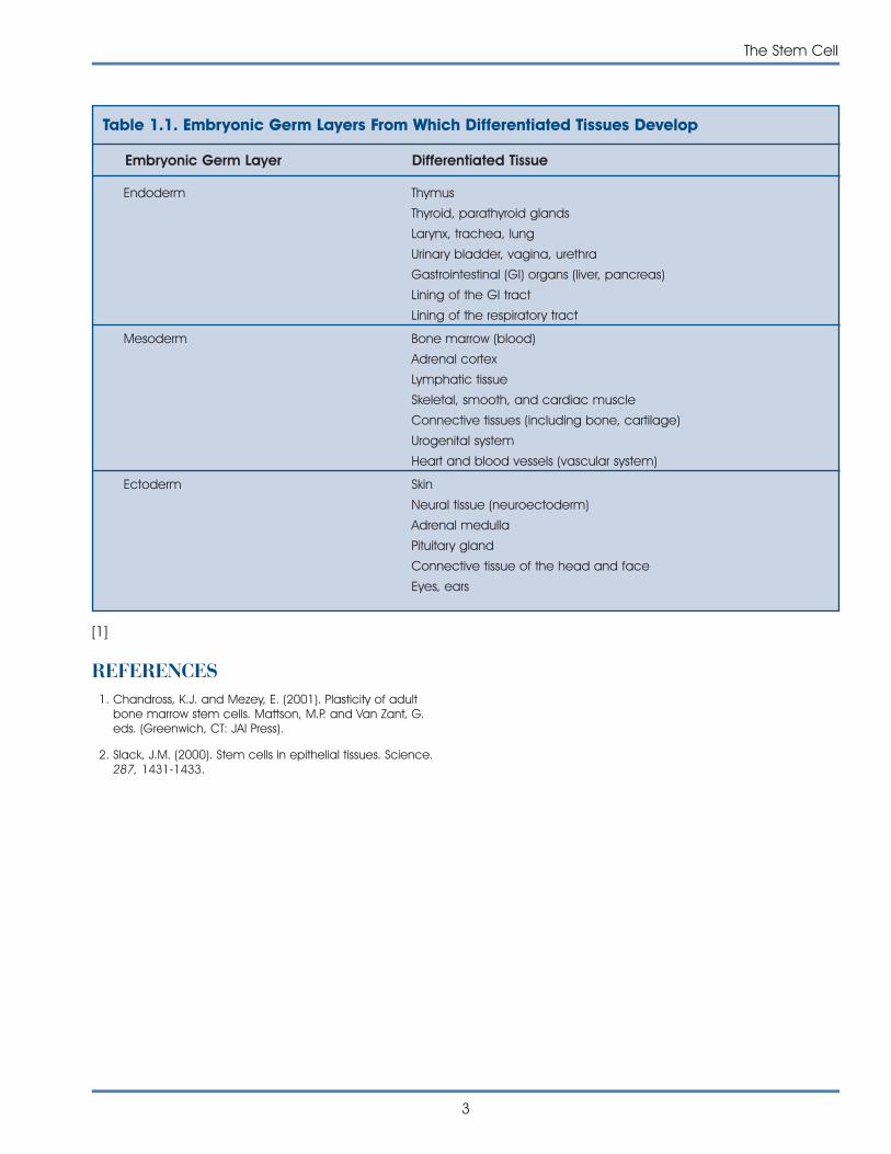
Most scientists use the term pluripotent to describestem cells that can give rise to cells derived from allthree embryonic germ layers—mesoderm, endo-derm, and ectoderm. These three germ layers arethe embryonic source of all cells of the body (seeFigure 1.1. Differentiation of Human Tissues). All of themany different kinds of specialized cells that make upthe body are derived from one of these germ layers(see Table 1.1. Embryonic Germ Layers From WhichDifferentiated Tissues Develop). “Pluri”—derived fromthe Latin plures—means several or many. Thus,pluripotent cells have the potential to give rise toany type of cell, a property observed in the naturalcourse of embryonic development and under certainlaboratory conditions.
Unipotent stem cell, a term that is usually applied toa cell in adult organisms, means that the cells inquestion are capable of differentiating along onlyone lineage. “Uni” is derived from the Latin word unus,which means one. Also, it may be that the adult stemcells in many differentiated, undamaged tissues aretypically unipotent and give rise to just one cell typeunder normal conditions. This process would allow fora steady state of self-renewal for the tissue. However,if the tissue becomes damaged and the replacementof multiple cell types is required, pluripotent stem cellsmay become activated to repair the damage [2].
The embryonic stem cell is defined by its origin—thatis from one of the earliest stages of the developmentof the embryo, called the blastocyst. Specifically,embryonic stem cells are derived from the inner cellmass of the blastocyst at a stage before it wouldimplant in the uterine wall. The embryonic stem cellcan self-replicate and is pluripotent—it can give riseto cells derived from all three germ layers.
The adult stem cell is an undifferentiated (unspecial-ized) cell that is found in a differentiated (specialized)
THE STEM CELLTHE STEM CELL1.1.
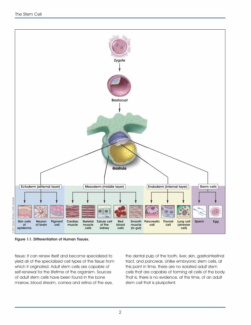
the dental pulp of the tooth, liver, skin, gastrointestinaltract, and pancreas. Unlike embryonic stem cells, atthis point in time, there are no isolated adult stemcells that are capable of forming all cells of the body.That is, there is no evidence, at this time, of an adultstem cell that is pluripotent.
2
The Stem Cell
tissue; it can renew itself and become specialized toyield all of the specialized cell types of the tissue fromwhich it originated. Adult stem cells are capable ofself-renewal for the lifetime of the organism. Sourcesof adult stem cells have been found in the bonemarrow, blood stream, cornea and retina of the eye,
Zygote
Blastocyst
Ectoderm (external layer) Mesoderm (middle layer) Germ cellsEndoderm (internal layer)
Figure 1.1. Differentiation of Human Tissues.
© 2
001
Tere
se W
inslo
w, C
aitl
in D
uckw
all
Skin cellsof
epidermis
Neuronof brain
Pigmentcell
Cardiacmuscle
Skeletalmuscle
cells
Tubule cellof thekidney
Redbloodcells
Smoothmuscle(in gut)
Pancreaticcell
Thyroidcell
Lung cell(alveolar
cell)
Sperm Egg
The Stem Cell
3
[1]
REFERENCES1. Chandross, K.J. and Mezey, E. (2001). Plasticity of adult
bone marrow stem cells. Mattson, M.P. and Van Zant, G.eds. (Greenwich, CT: JAI Press).
2. Slack, J.M. (2000). Stem cells in epithelial tissues. Science.287, 1431-1433.
Table 1.1. Embryonic Germ Layers From Which Differentiated Tissues Develop
Embryonic Germ Layer Differentiated Tissue
Endoderm Thymus
Thyroid, parathyroid glands
Larynx, trachea, lung
Urinary bladder, vagina, urethra
Gastrointestinal (GI) organs (liver, pancreas)
Lining of the GI tract
Lining of the respiratory tract
Mesoderm Bone marrow (blood)
Adrenal cortex
Lymphatic tissue
Skeletal, smooth, and cardiac muscle
Connective tissues (including bone, cartilage)
Urogenital system
Heart and blood vessels (vascular system)
Ectoderm Skin
Neural tissue (neuroectoderm)
Adrenal medulla
Pituitary gland
Connective tissue of the head and face
Eyes, ears

4
The Stem Cell
This page intentionally left blank
5
As stated in the first chapter, an embryonic stem cell(ES cell) is defined by its origin. It is derived from theblastocyst stage of the embryo. The blastocyst is thestage of embryonic development prior to implanta-tion in the uterine wall. At this stage, the preimplan-tation embryo of the mouse is made up of 150 cellsand consists of a sphere made up of an outer layerof cells (the trophectoderm), a fluid-filled cavity(the blastocoel), and a cluster of cells on the interior(the inner cell mass).
Studies of ES cells derived from mouse blastocystsbecame possible 20 years ago with the discovery oftechniques that allowed the cells to be grown in thelaboratory. Embryonic–like stem cells, called embry-onic germ (EG) cells, can also be derived fromprimordial germ (PG) cells (the cells of the developingfetus from which eggs and sperm are formed) of themouse [20] and human fetus [30].
In this chapter the discussion will be limited to mouseembryonic stem cells. Chapter 3 describes thehuman embryonic stem cell.
DO EMBRYONIC STEM CELLSACTUALLY OCCUR IN THE EMBRYO?Some scientists argue that ES cells do not occur inthe embryo as such. ES cells closely resemble thecells of the preimplantation embryo [3], but are not infact the same [32]. An alternative perspective is thatthe embryos of many animal species contain stemcells. These cells proliferate extensively in the embryo,are capable of differentiating into all the types ofcells that occur in the adult, and can be isolated andgrown ex vivo (outside the organism), where theycontinue to replicate and show the potential todifferentiate [18].
For research purposes, the definition of an ES cell ismore than a self-replicating stem cell derived fromthe embryo that can differentiate into almost all ofthe cells of the body. Scientists have found it neces-sary to develop specific criteria that help them betterdefine the ES cell. Austin Smith, whose studies ofmouse ES cells have contributed significantly to thefield, has offered a list of essential characteristics thatdefine ES cells [18, 32].
DEFINING PROPERTIES OF ANEMBRYONIC STEM CELL
• aDerived from the inner cell mass/epiblast of theblastocyst.
• aCapable of undergoing an unlimited numberof symmetrical divisions without differentiating(long-term self-renewal).
• Exhibit and maintain a stable, full (diploid), nor-mal complement of chromosomes (karyotype).
• Pluripotent ES cells can give rise to differentiatedcell types that are derived from all three primarygerm layers of the embryo (endoderm, meso-derm, and ectoderm).
• a,bCapable of integrating into all fetal tissuesduring development. (Mouse ES cells maintainedin culture for long periods can still generate anytissue when they are reintroduced into anembryo to generate a chimeric animal.)
• a,bCapable of colonizing the germ line andgiving rise to egg or sperm cells.
• aClonogenic, that is a single ES cell can giverise to a colony of genetically identical cells, orclones, which have the same properties as theoriginal cell.
THE EMBRYONICSTEM CELLTHE EMBRYONICSTEM CELL
2.2.
• Expresses the transcription factor Oct-4, whichthen activates or inhibits a host of target genesand maintains ES cells in a proliferative, non-differentiating state.
• Can be induced to continue proliferating or todifferentiate.
• Lacks the G1 checkpoint in the cell cycle. EScells spend most of their time in the S phase ofthe cell cycle, during which they synthesize DNA.Unlike differentiated somatic cells, ES cells do notrequire any external stimulus to initiate DNAreplication.
• Do not show X inactivation. In every somatic cellof a female mammal, one of the two X chromo-somes becomes permanently inactivated. Xinactivation does not occur in undifferentiatedES cells.
[a Not shown in human EG cells. b Not shown in human EScells. All of the criteria have been met by mouse ES cells.]
ARE EMBRYONIC STEM CELLSTRULY PLURIPOTENT?Pluripotency—that is the ability to give rise to differen-tiated cell types that are derived from all threeprimary germ layers of the embryo, endoderm,mesoderm, and ectoderm—is what makes ES cellsunique. How do we know that these cells are, indeed,pluripotent? Laboratory-based criteria for testing thepluripotent nature of ES cells derived from miceinclude three kinds of experiments [19]. One test isconducted by injecting ES cells derived from theinner cell mass of one blastocyst into the cavity ofanother blastocyst. The “combination” embryos arethen transferred to the uterus of a pseudopregnantfemale mouse, and the progeny that result arechimeras. Chimeras are a mixture of tissues andorgans of cells derived from both donor ES cells andthe recipient blastocyst.
This test has been extended in studies designed totest whether cultured ES cells can be used to replacethe inner cell mass of a mouse blastocyst and pro-duce a normal embryo. They can, but the process isfar less efficient than that of using cells taken directlyfrom the inner cell mass. Apparently, the ability of EScells to generate a complete embryo depends onthe number of times they have been passagedin vitro [21, 22]. A passage is the process of removing
cells from one culture dish and replating them intofresh culture dishes. Whether the number of passagesaffects the differentiation potential of human ES cellsremains to be determined. (For a detailed discussionof the techniques for maintaining mouse ES cells inculture, see Appendix B. Mouse Embryonic Stem Cells.)
A second method for determining the pluripotency ofmouse ES cells is to inject the cells into adult mice(under the skin or the kidney capsule) that are eithergenetically identical or are immune-deficient, so thetissue will not be rejected. In the host animal, theinjected ES cells develop into benign tumors calledteratomas. When examined under a microscope, itwas noted that these tumors contain cell typesderived from all three primary germ layers of theembryo—endoderm, mesoderm, and ectoderm.Teratomas typically contain gut-like structures such aslayers of epithelial cells and smooth muscle; skeletalor cardiac muscle (which may contract sponta-neously); neural tissue; cartilage or bone; and some-times hair. Thus, ES cells that have been maintainedfor a long period in vitro can behave as pluripotentcells in vivo. They can participate in normal embryo-genesis by differentiating into any cell type in thebody, and they can also differentiate into a widerange of cell types in an adult animal. However,normal mouse ES cells do not generate trophoblasttissues in vivo [32].
A third technique for demonstrating pluripotency isto allow mouse ES cells in vitro to differentiate sponta-neously or to direct their differentiation along specificpathways. The former is usually accomplished byremoving feeder layers and adding leukemia inhibi-tory factor (LIF) to the growth medium. Within a fewdays after changing the culture conditions, ES cellsaggregate and may form embryoid bodies (EBs).In many ways, EBs in the culture dish resembleteratomas that are observed in the animal. EBs con-sist of a disorganized array of differentiated or partiallydifferentiated cell types that are derived from thethree primary germ layers of the embryo—the endo-derm, mesoderm, and ectoderm [32].
The techniques for culturing mouse ES cells from theinner cell mass of the preimplantation blastocyst werefirst reported 20 years ago [9, 19], and versions ofthese standard procedures are used today inlaboratories throughout the world. It is striking that, todate, only three species of mammals have yielded
6
The Embryonic Stem Cell

long-term cultures of self-renewing ES cells: mice,monkeys, and humans [27, 34, 35, 36] (see AppendixB. Mouse Embryonic Stem Cells).
HOW DOES A MOUSE EMBRYONICSTEM CELL STAY UNDIFFERENTIATED?As stated earlier, a true stem cell is capable of main-taining itself in a self-renewing, undifferentiated stateindefinitely. The undifferentiated state of the embry-onic stem cell is characterized by specific cell mark-ers that have helped scientists better understand howembryonic stem cells—under the right culture condi-tions—replicate for hundreds of population doublingsand do not differentiate. To date, two major areasof investigation have provided some clues. Oneincludes attempts to understand the effects ofsecreted factors such as the cytokine leukemiainhibitory factor on mouse ES cells in vitro. The sec-ond area of study involves transcription factors suchas Oct-4. Oct-4 is a protein expressed by mouse andhuman ES cells in vitro, and also by mouse inner cellmass cells in vivo. The cell cycle of the ES also seemsto play a role in preventing differentiation. From stud-ies of these various signaling pathways, it is clear thatmany factors must be balanced in a particular wayfor ES cells to remain in a self-renewing state. If thebalance shifts, ES cells begin to differentiate [18, 31].(For a detailed discussion of how embryonic stemcells maintain their pluripotency, see Appendix B.Mouse Embryonic Stem Cells.)
CAN A MOUSE EMBRYONIC STEM CELLBE DIRECTED TO DIFFERENTIATEINTO A PARTICULAR CELL TYPEIN VITRO? One goal for embryonic stem cell research is thedevelopment of specialized cells such as neurons,heart muscle cells, endothelial cells of blood vessels,and insulin secreting cells similar to those found in thepancreas. The directed derivation of embryonic stemcells is then vital to the ultimate use of such cells inthe development of new therapies.
By far the most common approach to directingdifferentiation is to change the growth conditions ofthe ES cells in specific ways, such as by addinggrowth factors to the culture medium or changingthe chemical composition of the surface on which
the ES cells are growing. For example, the plasticculture dishes used to grow both mouse and humanES cells can be treated with a variety of substancesthat allow the cells either to adhere to the surface ofthe dish or to avoid adhering and instead float in theculture medium. In general, an adherent substratehelps prevent them from interacting and differentiat-ing. In contrast, a nonadherent substrate allows the EScells to aggregate and thereby interact with eachother. Cell-cell interactions are critical to normalembryonic development, so allowing some of these“natural” in vivo interactions to occur in the culturedish is a fundamental strategy for inducing mouse orhuman ES cell differentiation in vitro. In addition,adding specific growth factors to the culture mediumtriggers the activation (or inactivation) of specificgenes in ES cells. This initiates a series of molecularevents that induces the cells to differentiate along aparticular pathway.
Another way to direct differentiation of ES cells is tointroduce foreign genes into the cells via transfectionor other methods [6, 39]. The result of these strategiesis to add an active gene to the ES cell genome,which then triggers the cells to differentiate along aparticular pathway. The approach appears to be aprecise way of regulating ES cell differentiation, but itwill work only if it is possible to identify which genemust be active at which particular stage of differen-tiation. Then, the gene must be activated at theright time—meaning during the correct stage ofdifferentiation—and it must be inserted into thegenome at the proper location.
Another approach to generate mouse ES cells usescloning technology. In theory, the nucleus of adifferentiated mouse somatic cell might be repro-grammed by injecting it into an oocyte. The resultantpluripotent cell would be immunologically com-patible because it would be genetically identical tothe donor cell [25].
All of the techniques just described are still highlyexperimental. Nevertheless, within the past severalyears, it has become possible to generate specific,differentiated, functional cell types by manipulatingthe growth conditions of mouse ES cells in vitro. It isnot possible to explain how the directed differentia-tion occurs, however. No one knows how or whengene expression is changed, what signal-transductionsystems are triggered, or what cell-cell interactions
The Embryonic Stem Cell
7
must occur to convert undifferentiated ES cells intoprecursor cells and, finally, into differentiated cellsthat look and function like their in vivo counterparts.
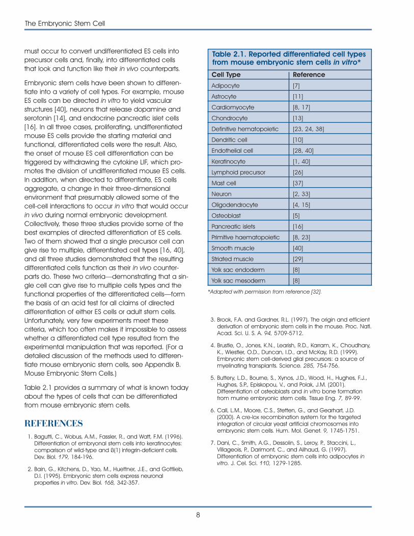
Embryonic stem cells have been shown to differen-tiate into a variety of cell types. For example, mouseES cells can be directed in vitro to yield vascularstructures [40], neurons that release dopamine andserotonin [14], and endocrine pancreatic islet cells[16]. In all three cases, proliferating, undifferentiatedmouse ES cells provide the starting material andfunctional, differentiated cells were the result. Also,the onset of mouse ES cell differentiation can betriggered by withdrawing the cytokine LIF, which pro-motes the division of undifferentiated mouse ES cells.In addition, when directed to differentiate, ES cellsaggregate, a change in their three-dimensionalenvironment that presumably allowed some of thecell-cell interactions to occur in vitro that would occurin vivo during normal embryonic development.Collectively, these three studies provide some of thebest examples of directed differentiation of ES cells.Two of them showed that a single precursor cell cangive rise to multiple, differentiated cell types [16, 40],and all three studies demonstrated that the resultingdifferentiated cells function as their in vivo counter-parts do. These two criteria—demonstrating that a sin-gle cell can give rise to multiple cells types and thefunctional properties of the differentiated cells—formthe basis of an acid test for all claims of directeddifferentiation of either ES cells or adult stem cells.Unfortunately, very few experiments meet thesecriteria, which too often makes it impossible to assesswhether a differentiated cell type resulted from theexperimental manipulation that was reported. (For adetailed discussion of the methods used to differen-tiate mouse embryonic stem cells, see Appendix B.Mouse Embryonic Stem Cells.)
Table 2.1 provides a summary of what is known todayabout the types of cells that can be differentiatedfrom mouse embryonic stem cells.
REFERENCES1. Bagutti, C., Wobus, A.M., Fassler, R., and Watt, F.M. (1996).
Differentiation of embryonal stem cells into keratinocytes:comparison of wild-type and ß(1) integrin-deficient cells.Dev. Biol. 179, 184-196.
2. Bain, G., Kitchens, D., Yao, M., Huettner, J.E., and Gottlieb,D.I. (1995). Embryonic stem cells express neuronalproperties in vitro. Dev. Biol. 168, 342-357.
8
The Embryonic Stem Cell
Table 2.1. Reported differentiated cell typesfrom mouse embryonic stem cells in vitro*
Cell Type Reference
Adipocyte [7]
Astrocyte [11]
Cardiomyocyte [8, 17]
Chondrocyte [13]
Definitive hematopoietic [23, 24, 38]
Dendritic cell [10]
Endothelial cell [28, 40]
Keratinocyte [1, 40]
Lymphoid precursor [26]
Mast cell [37]
Neuron [2, 33]
Oligodendrocyte [4, 15]
Osteoblast [5]
Pancreatic islets [16]
Primitive haematopoietic [8, 23]
Smooth muscle [40]
Striated muscle [29]
Yolk sac endoderm [8]
Yolk sac mesoderm [8]
*Adapted with permission from reference [32].
3. Brook, F.A. and Gardner, R.L. (1997). The origin and efficientderivation of embryonic stem cells in the mouse. Proc. Natl.Acad. Sci. U. S. A. 94, 5709-5712.
4. Brustle, O., Jones, K.N., Learish, R.D., Karram, K., Choudhary,K., Wiestler, O.D., Duncan, I.D., and McKay, R.D. (1999).Embryonic stem cell-derived glial precursors: a source ofmyelinating transplants. Science. 285, 754-756.
5. Buttery, L.D., Bourne, S., Xynos, J.D., Wood, H., Hughes, F.J.,Hughes, S.P., Episkopou, V., and Polak, J.M. (2001).Differentiation of osteoblasts and in vitro bone formationfrom murine embryonic stem cells. Tissue Eng. 7, 89-99.
6. Call, L.M., Moore, C.S., Stetten, G., and Gearhart, J.D.(2000). A cre-lox recombination system for the targetedintegration of circular yeast artificial chromosomes intoembryonic stem cells. Hum. Mol. Genet. 9, 1745-1751.
7. Dani, C., Smith, A.G., Dessolin, S., Leroy, P., Staccini, L.,Villageois, P., Darimont, C., and Ailhaud, G. (1997).Differentiation of embryonic stem cells into adipocytes invitro. J. Cel. Sci. 110, 1279-1285.
8. Doetschman, T., Eistetter, H., Katz, M., Schmit, W., andKemler, R. (1985). The in vitro development of blastocyst-derived embryonic stem cell lines: formation of visceralyolk sac, blood islands and myocardium. J. Embryol. Exp.Morph. 87, 27-45.
9. Evans, M.J. and Kaufman, M.H. (1981). Establishment inculture of pluripotential cells from mouse embryos. Nature.292, 154-156.
10. Fairchild, P.J., Brook, F.A., Gardner, R.L., Graca, L., Strong, V.,Tone, Y., Tone, M., Nolan, K.F., and Waldmann, H. (2000).Directed differentiation of dendritic cells from mouseembryonic stem cells. Curr. Biol. 10, 1515-1518.
11. Fraichard, A., Chassande, O., Bilbaut, G., Dehay, C.,Savatier, P., and Samarut, J. (1995). In vitro differentiationof embryonic stem cells into glial cells and functionalneurons. J. Cell Sci. 108, 3181-3188.
12. Itskovitz-Eldor, J., Schuldiner, M., Karsenti, D., Eden, A.,Yanuka, O., Amit, M., Soreq, H., and Benvenisty, N. (2000).Differentiation of human embryonic stem cells into embry-oid bodies comprising the three embryonic germ layers.Mol. Med. 6, 88-95.
13. Kramer, J., Hegert, C., Guan, K., Wobus, A.M., Muller, P.K.,and Rohwedel, J. (2000). Embryonic stem cell-derivedchondrogenic differentiation in vitro: activation by BMP-2and BMP-4. Mech. Dev. 92, 193-205.
14. Lee, S.H., Lumelsky, N., Studer, L., Auerbach, J.M., andMcKay, R.D. (2000). Efficient generation of midbrain andhindbrain neurons from mouse embryonic stem cells. Nat.Biotechnol. 18, 675-679.
15. Liu, S., Qu, Y., Stewart, T.J., Howard, M.J., Chakrabortty, S.,Holekamp, T.F., and McDonald, J.W. (2000). Embryonicstem cells differentiate into oligodendrocytes and myeli-nate in culture and after spinal cord transplantation. Proc.Natl. Acad. Sci. U. S. A. 97, 6126-6131.
16. Lumelsky, N., Blondel, O., Laeng, P., Velasco, I., Ravin, R.,and McKay, R. (2001). Differentiation of Embryonic StemCells to Insulin-Secreting Structures Similiar to PancreaticIslets. Science. 292, 1389-1394.
17. Maltsev, V.A., Rohwedel, J., Hescheler, J., and Wobus, A.M.(1993). Embryonic stem cells differentiate in vitro into car-diomyocytes representing sinusnodal, atrial and ventricularcell types. Mech. Dev. 44, 41-50.
18. Marshak, D.R., Gottlieb, D., Kiger, A.A., Fuller, M.T., Kunath, T.,Hogan, B., Gardner, R.L., Smith, A., Klar, A.J.S., Henrique, D.,D’Urso, G., Datta, S., Holliday, R., Astle, C.M., Chen, J.,Harrison, D.E., Xie, T., Spradling, A., Andrews, P.W., Przyborski,S.A., Thomson, J.A., Kunath, T., Strumpf, D., Rossant, J.,Tanaka, S., Orkin, S.H., Melchers, F., Rolink, A., Keller, G.,Pittenger, M.F., Marshak, D.R., Flake, A.W., Panicker, M.M.,Rao, M., Watt, F.M., Grompe, M., Finegold, M.J., Kritzik, M.R.,Sarvetnick, N., and Winton, D.J. (2001). Stem cell biology,Marshak, D.R., Gardner, R.L., and Gottlieb, D. eds. (ColdSpring Harbor, New York: Cold Spring Harbor LaboratoryPress).
19. Martin, G.R. (1981). Isolation of a pluripotent cell line fromearly mouse embryos cultured in medium conditioned byteratocarcinoma stem cells. Proc. Natl. Acad. Sci. U. S. A.78, 7634-7638.
20. Matsui, Y., Zsebo, K., and Hogan, B.L. (1992). Derivation ofpluripotential embryonic stem cells from murine primordialgerm cells in culture. Cell. 70, 841-847.
21. Nagy, A., Gocza, E., Diaz, E.M., Prideaux, V.R., Ivanyi, E.,Markkula, M., and Rossant, J. (1990). Embryonic stem cellsalone are able to support fetal development in the mouse.Development. 110, 815-821.
22. Nagy, A., Rossant, J., Nagy, R., Abramow-Newerly, W., andRoder, J.C. (1993). Derivation of completely cell culture-derived mice from early-passage embryonic stem cells.Proc. Natl. Acad. Sci. U. S. A. 90, 8424-8428.
23. Nakano, T., Kodama, H., and Honjo, T. (1996). In vitrodevelopment of primitive and definitive erythrocytes fromdifferent precursors. Science. 272, 722-724.
24. Nishikawa, S.I., Nishikawa, S., Hirashima, M., Matsuyoshi, N.,and Kodama, H. (1998). Progressive lineage analysis by cellsorting and culture identifies FLK1(+)VE-cadherin(+) cells ata diverging point of endothelial and hemopoietic lineages.Development. 125, 1747-1757.
25. Odorico, J.S., Kaufman, D.S., and Thomson, J.A. (2001).Multilineage differentiation from human embryonic stemcell lines. Stem Cells. 19, 193-204.
26. Potocnik, A.J., Nielsen, P.J., and Eichmann, K. (1994). In vitrogeneration of lymphoid precursors from embryonic stemcells. EMBO. J. 13, 5274-5283.
27. Reubinoff, B.E., Pera, M.F., Fong, C.Y., Trounson, A., andBongso, A. (2000). Embryonic stem cell lines from humanblastocysts: somatic differentiation in vitro. Nat. Biotechnol.18, 399-404.
28. Risau, W., Sariola, H., Zerwes, H.G., Sasse, J., Ekblom, P.,Kemler, R., and Doetschman, T. (1988). Vasculogenesis andangiogenesis in embryonic-stem-cell-derived embryoidbodies. Development. 102, 471-478.
29. Rohwedel, J., Maltsev, V., Bober, E., Arnold, H.H., Hescheler,J., and Wobus, A.M. (1994). Muscle cell differentiation ofembryonic stem cells reflects myogenesis in vivo: develop-mentally regulated expression of myogenic determinationgenes and functional expression of ionic currents. Dev. Biol.164, 87-101.
30. Shamblott, M.J., Axelman, J., Wang, S., Bugg, E.M.,Littlefield, J.W., Donovan, P.J., Blumenthal, P.D., Huggins,G.R., and Gearhart, J.D. (1998). Derivation of pluripotentstem cells from cultured human primordial germ cells.Proc. Natl. Acad. Sci. U. S. A. 95, 13726-13731.
31. Smith, A., personal communication.
32. Smith, A.G. (2001). Origins and properties of mouseembryonic stem cells. Annu. Rev. Cell. Dev. Biol.
The Embryonic Stem Cell
9
33. Strubing, C., Ahnert-Hilger, G., Shan, J., Wiedenmann, B.,Hescheler, J., and Wobus, A.M. (1995). Differentiation ofpluripotent embryonic stem cells into the neuronal lineagein vitro gives rise to mature inhibitory and excitatoryneurons. Mech. Dev. 53, 275-287.
34. Thomson, J.A., Kalishman, J., Golos, T.G., Durning, M., Harris,C.P., Becker, R.A., and Hearn, J.P. (1995). Isolation of a pri-mate embryonic stem cell line. Proc. Natl. Acad. Sci. U. S.A. 92, 7844-7848.
35. Thomson, J.A. and Marshall, V.S. (1998). Primate embryonicstem cells. Curr. Top. Dev. Biol. 38, 133-165.
36. Thomson, J.A., Itskovitz-Eldor, J., Shapiro, S.S., Waknitz, M.A.,Swiergiel, J.J., Marshall, V.S., and Jones, J.M. (1998).Embryonic stem cell lines derived from human blastocysts.Science. 282, 1145-1147.
37. Tsai, M., Wedemeyer, J., Ganiatsas, S., Tam, S.Y., Zon, L.I.,and Galli, S.J. (2000). In vivo immunological function ofmast cells derived from embryonic stem cells: anapproach for the rapid analysis of even embryonic lethalmutations in adult mice in vivo. Proc. Natl. Acad. Sci. U. S. A.97, 9186-9190.
38. Wiles, M.V. and Keller, G. (1991). Multiple hematopoieticlineages develop from embryonic stem (ES) cells in culture.Development. 111, 259-267.
39. Wiles, M.V., Vauti, F., Otte, J., Fuchtbauer, E.M., Ruiz, P.,Fuchtbauer, A., Arnold, H.H., Lehrach, H., Metz, T., vonMelchner, H., and Wurst, W. (2000). Establishment of agene-trap sequence tag library to generate mutant micefrom embryonic stem cells. Nat. Genet. 24, 13-14.
40. Yamashita, J., Itoh, H., Hirashima, M., Ogawa, M.,Nishikawa, S., Yurugi, T., Naito, M., Nakao, K., and Nishikawa,S. (2000). Flk1-positive cells derived from embryonic stemcells serve as vascular progenitors. Nature. 408, 92-96.
10
The Embryonic Stem Cell
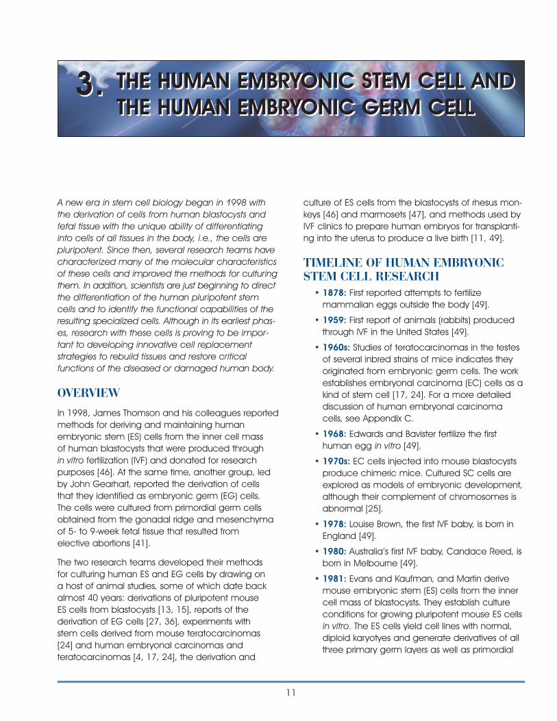
11
A new era in stem cell biology began in 1998 withthe derivation of cells from human blastocysts andfetal tissue with the unique ability of differentiatinginto cells of all tissues in the body, i.e., the cells arepluripotent. Since then, several research teams havecharacterized many of the molecular characteristicsof these cells and improved the methods for culturingthem. In addition, scientists are just beginning to directthe differentiation of the human pluripotent stemcells and to identify the functional capabilities of theresulting specialized cells. Although in its earliest phas-es, research with these cells is proving to be impor-tant to developing innovative cell replacementstrategies to rebuild tissues and restore critical functions of the diseased or damaged human body.
OVERVIEWIn 1998, James Thomson and his colleagues reportedmethods for deriving and maintaining humanembryonic stem (ES) cells from the inner cell mass of human blastocysts that were produced through in vitro fertilization (IVF) and donated for research purposes [46]. At the same time, another group, ledby John Gearhart, reported the derivation of cells that they identified as embryonic germ (EG) cells. The cells were cultured from primordial germ cellsobtained from the gonadal ridge and mesenchymaof 5- to 9-week fetal tissue that resulted from elective abortions [41].
The two research teams developed their methods for culturing human ES and EG cells by drawing on a host of animal studies, some of which date backalmost 40 years: derivations of pluripotent mouse ES cells from blastocysts [13, 15], reports of the derivation of EG cells [27, 36], experiments with stem cells derived from mouse teratocarcinomas [24] and human embryonal carcinomas and teratocarcinomas [4, 17, 24], the derivation and
culture of ES cells from the blastocysts of rhesus mon-keys [46] and marmosets [47], and methods used byIVF clinics to prepare human embryos for transplanti-ng into the uterus to produce a live birth [11, 49].
TIMELINE OF HUMAN EMBRYONICSTEM CELL RESEARCH
• 1878: First reported attempts to fertilize mammalian eggs outside the body [49].
• 1959: First report of animals (rabbits) producedthrough IVF in the United States [49].
• 1960s: Studies of teratocarcinomas in the testesof several inbred strains of mice indicates theyoriginated from embryonic germ cells. The workestablishes embryonal carcinoma (EC) cells as akind of stem cell [17, 24]. For a more detaileddiscussion of human embryonal carcinomacells, see Appendix C.
• 1968: Edwards and Bavister fertilize the firsthuman egg in vitro [49].
• 1970s: EC cells injected into mouse blastocystsproduce chimeric mice. Cultured SC cells areexplored as models of embryonic development,although their complement of chromosomes isabnormal [25].
• 1978: Louise Brown, the first IVF baby, is born inEngland [49].
• 1980: Australia’s first IVF baby, Candace Reed, isborn in Melbourne [49].
• 1981: Evans and Kaufman, and Martin derivemouse embryonic stem (ES) cells from the innercell mass of blastocysts. They establish cultureconditions for growing pluripotent mouse ES cellsin vitro. The ES cells yield cell lines with normal,diploid karyotyes and generate derivatives of allthree primary germ layers as well as primordial
THE HUMAN EMBRYONIC STEM CELL ANDTHE HUMAN EMBRYONIC GERM CELL
3.3. THE HUMAN EMBRYONIC STEM CELL ANDTHE HUMAN EMBRYONIC GERM CELL
germ cells. Injecting the ES cells into miceinduces the formation of teratomas [15, 26]. The first IVF baby, Elizabeth Carr, is born in theUnited States [49].
• 1984-88: Andrews et al., develop pluripotent,genetically identical (clonal) cells called embry-onal carcinoma (EC) cells from Tera-2, a cell lineof human testicular teratocarcinoma [5]. Clonedhuman teratoma cells exposed to retinoic aciddifferentiate into neuron-like cells and other celltypes [3, 44].
• 1989: Pera et al., derive a clonal line of humanembryonal carcinoma cells, which yields tissuesfrom all three primary germ layers. The cells areaneuploid (fewer or greater than the normalnumber of chromosomes in the cell) and theirpotential to differentiate spontaneously in vitrois typically limited. The behavior of human ECcell clones differs from that of mouse ES or EC cells [33].
• 1994: Human blastocysts created for reproductive purposes using IVF and donated by patients for research, are generated from the2-pronuclear stage. The inner cell mass of theblastocyst is maintained in culture and generatesaggregates with trophoblast-like cells at theperiphery and ES-like cells in the center. The cellsretain a complete set of chromosomes (normalkaryotype); most cultures retain a stem cell-likemorphology, although some inner cell massclumps differentiate into fibroblasts. The culturesare maintained for two passages [6, 7].
• 1995-96: Non-human primate ES cells arederived and maintained in vitro, first from theinner cell mass of rhesus monkeys [46], and thenfrom marmosets [47]. The primate ES cells arediploid and have normal karyotypes. They arepluripotent and differentiate into cells typesderived from all three primary germ layers. Theprimate ES cells resemble human EC cells andindicate that it should be possible to derive andmaintain human ES cells in vitro.
• 1998: Thomson et al., derive human ES cellsfrom the inner cell mass of normal human blastocysts donated by couples undergoingtreatment for infertility. The cells are culturedthrough many passages, retain their normal karyotypes, maintain high levels of telomeraseactivity, and express a panel of markers typical
of human EC cells non-human primate ES cells.Several (non-clonal) cell lines are established that form teratomas when injected into immune-deficient mice. The teratomas includecell types derived from all three primary germlayers, demonstrating the pluripotency of humanES cells [48]. Gearhart and colleagues derivehuman embryonic germ (EG) cells from thegonadal ridge and mesenchyma of 5- to 9-week fetal tissue that resulted from electiveabortions. They grow EG cells in vitro for approxi-mately 20 passages, and the cells maintain normal karyotypes. The cells spontaneously formaggregates that differentiate spontaneously, andultimately contain derivatives of all three primarygerm layers. Other indications of their pluripotency include the expression of a panel ofmarkers typical of mouse ES and EG cells. TheEG cells do not form teratomas when injectedinto immune-deficient mice [41].
• 2000: Scientists in Singapore and Australia led byPera, Trounson, and Bongso derive human EScells from the inner cell mass of blastocystsdonated by couples undergoing treatment forinfertility. The ES cells proliferate for extended periods in vitro, maintain normal karyotypes, differentiate spontaneously into somatic cell lineages derived from all three primary germ layers, and form teratomas when injected intoimmune-deficient mice.
• 2001: As human ES cell lines are shared andnew lines are derived, more research groupsreport methods to direct the differentiation of the cells in vitro. Many of the methods areaimed at generating human tissues for transplantation purposes, including pancreaticislet cells, neurons that release dopamine, andcardiac muscle cells.
DERIVATION OF HUMAN EMBRYONICSTEM CELLSThe first documentation of the isolation of embryonicstem cells from human blastocysts was in 1994 [7].Since then, techniques for deriving and culturinghuman ES cells have been refined [38, 48]. The abilityto isolate human ES cells from blastocysts and growthem in culture seems to depend in large part on theintegrity and condition of the blastocyst from whichthe cells are derived. In general, blastocysts with a
12
The Human Embryonic Stem Cell and The Human Embryonic Germ Cell
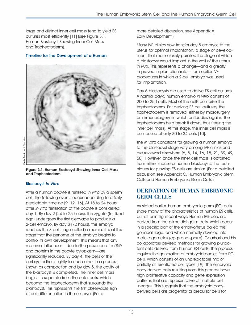
large and distinct inner cell mass tend to yield ES cultures most efficiently [11] (see Figure 3.1. Human Blastocyst Showing Inner Cell Mass and Trophectoderm).
Timeline for the Development of a Human
Blastocyst In Vitro
After a human oocyte is fertilized in vitro by a spermcell, the following events occur according to a fairlypredictable timeline [9, 12, 16]. At 18 to 24 hoursafter in vitro fertilization of the oocyte is consideredday 1. By day 2 (24 to 25 hours), the zygote (fertilizedegg) undergoes the first cleavage to produce a 2-cell embryo. By day 3 (72 hours), the embryoreaches the 8-cell stage called a morula. It is at thisstage that the genome of the embryo begins to control its own development. This means that anymaternal influences—due to the presence of mRNAand proteins in the oocyte cytoplasm—are significantly reduced. By day 4, the cells of theembryo adhere tightly to each other in a processknown as compaction and by day 5, the cavity ofthe blastocyst is completed. The inner cell massbegins to separate from the outer cells, whichbecome the trophectoderm that surrounds the blastocyst. This represents the first observable sign of cell differentiation in the embryo. (For a
more detailed discussion, see Appendix A. Early Development.)
Many IVF clinics now transfer day-5 embryos to theuterus for optimal implantation, a stage of develop-ment that more closely parallels the stage at which a blastocyst would implant in the wall of the uterus in vivo. This represents a change—and a greatlyimproved implantation rate—from earlier IVF procedures in which a 2-cell embryo was used for implantation.
Day-5 blastocysts are used to derive ES cell cultures.A normal day-5 human embryo in vitro consists of200 to 250 cells. Most of the cells comprise the trophectoderm. For deriving ES cell cultures, the trophectoderm is removed, either by microsurgery or immunosurgery (in which antibodies against thetrophectoderm help break it down, thus freeing theinner cell mass). At this stage, the inner cell mass iscomposed of only 30 to 34 cells [10].
The in vitro conditions for growing a human embryoto the blastocyst stage vary among IVF clinics andare reviewed elsewhere [6, 8, 14, 16, 18, 21, 39, 49,50]. However, once the inner cell mass is obtainedfrom either mouse or human blastocysts, the tech-niques for growing ES cells are similar. (For a detaileddiscussion see Appendix C. Human Embryonic StemCells and Human Embryonic Germ Cells.)
DERIVATION OF HUMAN EMBRYONICGERM CELLS As stated earlier, human embryonic germ (EG) cellsshare many of the characteristics of human ES cells,but differ in significant ways. Human EG cells arederived from the primordial germ cells, which occurin a specific part of the embryo/fetus called thegonadal ridge, and which normally develop intomature gametes (eggs and sperm). Gearhart and hiscollaborators devised methods for growing pluripo-tent cells derived from human EG cells. The processrequires the generation of embryoid bodies from EGcells, which consists of an unpredictable mix of partially differentiated cell types [19]. The embryoidbody-derived cells resulting from this process havehigh proliferative capacity and gene expression patterns that are representative of multiple cell lineages. This suggests that the embryoid body-derived cells are progenitor or precursor cells for
The Human Embryonic Stem Cell and The Human Embryonic Germ Cell
13
Figure 3.1. Human Blastocyst Showing Inner Cell Massand Trophectoderm.
Pho
to C
red
it: M
r. J.
Co
nag
han
a variety of differentiated cell types [19]. (For a moredetailed description of the derivation of EG cells, seeAppendix C. Human Embryonic Stem Cells andHuman Embryonic Germ Cells.)
PLURIPOTENCY OF HUMANEMBRYONIC STEM CELLS ANDEMBRYONIC GERM CELLSAs stated earlier, a truly pluripotent stem cell is a cellthat is capable of self-renewal and of differentiatinginto most all of the cells of the body, including cells ofall three germ layers. Human ES and EG cells in vitroare capable of long-term self-renewal, while retaininga normal karyotype [1, 38, 41, 42, 48]. Human EScells can proliferate for two years through 300 population doublings [29] or even 450 populationdoublings [30]. Cultures derived from embryoid bodies generated by human embryonic germ cells have less capacity for proliferation. Most will proliferate for 40 population doublings; the maximumreported is 70 to 80 population doublings [42].
To date, several laboratories have demonstrated thathuman ES cells in vitro are pluripotent; they can produce cell types derived from all three embryonicgerm layers [1, 20, 38, 40].
Currently, the only test of the in vivo pluripotency ofhuman ES cells is to inject them into immune-deficient mice where they generate differentiatedcells that are derived from all three germ layers.These include gut epithelium (which, in the embryo, is derived from endoderm); smooth and striated muscle (derived from mesoderm); and neural epithelium, and stratified squamous epithelium(derived from ectoderm) [20, 38, 48].
However, two aspects of in vivo pluripotency typicallyused in animals have not been met by human EScells: evidence that cells have the capacity to beinjected into a human embryo and form an organ-ism made up of cells from two genetic lineages; and evidence that they have the ability to generategerm cells, the precursors to eggs and sperm in adeveloping organism. These are theoretical considerations, however, because such tests usinghuman ES cells have not been conducted. In anycase, these two demonstrations of human ES cellpluripotency are not likely to be critical for potentialtherapeutic uses of the cells—in transplants or drugdevelopment, for example [43].
14
The Human Embryonic Stem Cell and The Human Embryonic Germ Cell
COMPARISONS BETWEEN HUMANEMBRYONIC STEM CELLS ANDEMBRYONIC GERM CELLSThe ES cells derived from human blastocysts byThomson and his colleagues, and from human EGcells derived by Gearhart and his collaborators, aresimilar in many respects. In both cases, the cells replicate for an extended period of time, show nochromosomal abnormalities, generate both XX(female) and XY (male) cultures, and express a set of markers regarded as characteristic of pluripotentcells. When the culture conditions are adjusted topermit differentiation (see below for details), both ES and EG cells spontaneously differentiate into derivatives of all three primary germ layers—endoderm, mesoderm, and ectoderm (see Table3.1. Comparison of Mouse, Monkey, and HumanPluripotent Stem Cells).
However, the ES cells derived from human blastocystsand EG cells differ not only in the tissue sources fromwhich they are derived, they also vary with respect totheir growth characteristics in vitro, and their behaviorin vivo [34]. In addition, human ES cells have beenpropagated for approximately two years in vitro, forseveral hundred population doublings [1], whereashuman embryoid body-derived cells from cultures ofembryonic germ cells have been maintained for only70 to 80 population doublings [42]. Also, human EScells will generate teratomas containing differentiatedcell types, if injected into immunocompromised mice colonies, while human EG cells will not [20, 37, 38, 41, 48].
Several research groups are trying to grow human EScells without feeder layers of mouse embryo fibro-blasts (MEF), which are labor-intensive to generate. At a recent meeting, scientists from the GeronCorporation reported that they have grown human EScell without feeder layers, in medium conditioned byMEFs and supplemented with basic FGF [51].
DIRECTED DIFFERENTIATION OFHUMAN EMBRYONIC STEM CELLSAND EMBRYONIC GERM CELLSIN VITROCurrently, a major goal for embryonic stem cellresearch is to control the differentiation of human ESand EG cell lines into specific kinds of cells—an
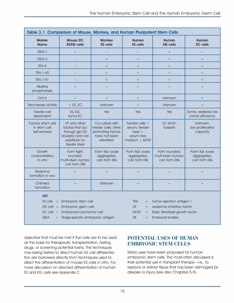
objective that must be met if the cells are to be usedas the basis for therapeutic transplantation, testingdrugs, or screening potential toxins. The techniquesnow being tested to direct human ES cell differentia-tion are borrowed directly from techniques used todirect the differentiation of mouse ES cells in vitro. Formore discussion on directed differentiation of humanES and EG cells see Appendix C.
POTENTIAL USES OF HUMANEMBRYONIC STEM CELLSMany uses have been proposed for human embryonic stem cells. The most-often discussed istheir potential use in transplant therapy—i.e., toreplace or restore tissue that has been damaged bydisease or injury (see also Chapters 5-9).
The Human Embryonic Stem Cell and The Human Embryonic Germ Cell
15
MarkerName
Mouse EC/ES/EG cells
MonkeyES cells
HumanES cells
HumanEG cells
HumanEC cells
SSEA-1 + – – + –
Table 3.1. Comparison of Mouse, Monkey, and Human Pluripotent Stem Cells
SSEA-3 – + + + +
SEA-4 – + + + +
TRA-1-60 – + + + +
TRA-1-81 – + + + +
Alkalinephosphatase
+ + + + +
Oct-4 + + + Unknown +
Telomerase activity + ES, EC Unknown + Unknown +
Feeder-celldependent
ES, EG,some EC
Yes Yes Yes Some; relatively lowclonal efficiency
Factors which aidin stem cellself-renewal
LIF and otherfactors that actthrough gp130
receptor and cansubstitute forfeeder layer
Co-culture withfeeder cells; otherpromoting factors
have not beenidentified
Feeder cells +serum; feeder
layer +serum-free
medium + bFGF
LIF, bFGF,forskolin
Unknown;low proliferative
capacity
Growthcharacteristics
in vitro
Form tight,rounded,
multi-layer clumps;can form EBs
Form flat, looseaggregates;can form EBs
Form flat, looseaggregates;can form EBs
Form rounded,multi-layer clumps;
can form EBs
Form flat, looseaggregates;can form EBs
Teratomaformation in vivo
+ + + – +
Chimeraformation
+ Unknown + – +
KEY
ES cell =
EG cell =
EC cell =
SSEA =
Embryonic stem cell
Embryonic germ cell
Embryonal carcinoma cell
Stage-specific embryonic antigen
TRA =
LIF =
bFGF =
EB =
Tumor rejection antigen-1
Leukemia inhibitory factor
Basic fibroblast growth factor
Embryoid bodies
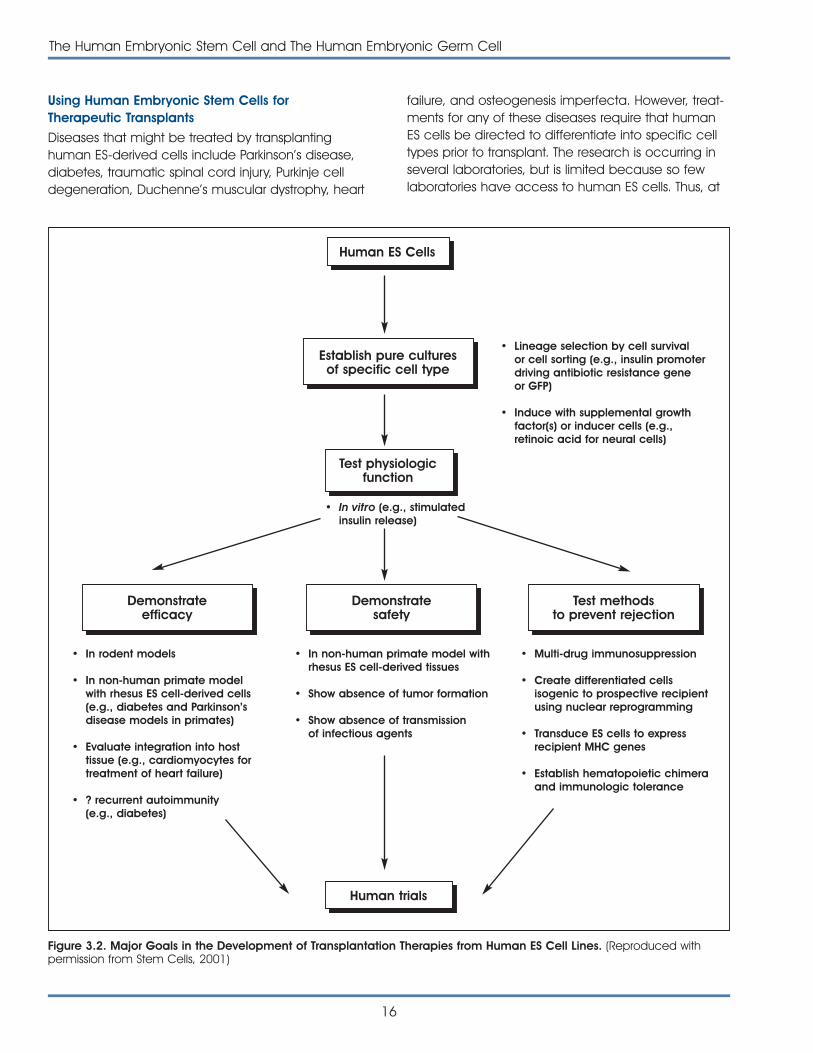
Using Human Embryonic Stem Cells forTherapeutic Transplants
Diseases that might be treated by transplantinghuman ES-derived cells include Parkinson’s disease,diabetes, traumatic spinal cord injury, Purkinje celldegeneration, Duchenne’s muscular dystrophy, heart
failure, and osteogenesis imperfecta. However, treat-ments for any of these diseases require that humanES cells be directed to differentiate into specific celltypes prior to transplant. The research is occurring inseveral laboratories, but is limited because so fewlaboratories have access to human ES cells. Thus, at
16
The Human Embryonic Stem Cell and The Human Embryonic Germ Cell
Figure 3.2. Major Goals in the Development of Transplantation Therapies from Human ES Cell Lines. (Reproduced withpermission from Stem Cells, 2001)
Human ES Cells
Establish pure culturesof specific cell type
Test physiologicfunction
Demonstrateefficacy
Demonstratesafety
Human trials
Test methodsto prevent rejection
• Lineage selection by cell survivalor cell sorting (e.g., insulin promoterdriving antibiotic resistance geneor GFP)
• Induce with supplemental growthfactor(s) or inducer cells (e.g.,retinoic acid for neural cells)
• In vitro (e.g., stimulatedinsulin release)
• In rodent models
• In non-human primate modelwith rhesus ES cell-derived cells(e.g., diabetes and Parkinson’sdisease models in primates)
• Evaluate integration into hosttissue (e.g., cardiomyocytes fortreatment of heart failure)
• ? recurrent autoimmunity(e.g., diabetes)
• In non-human primate model withrhesus ES cell-derived tissues
• Show absence of tumor formation
• Show absence of transmissionof infectious agents
• Multi-drug immunosuppression
• Create differentiated cellsisogenic to prospective recipientusing nuclear reprogramming
• Transduce ES cells to expressrecipient MHC genes
• Establish hematopoietic chimeraand immunologic tolerance
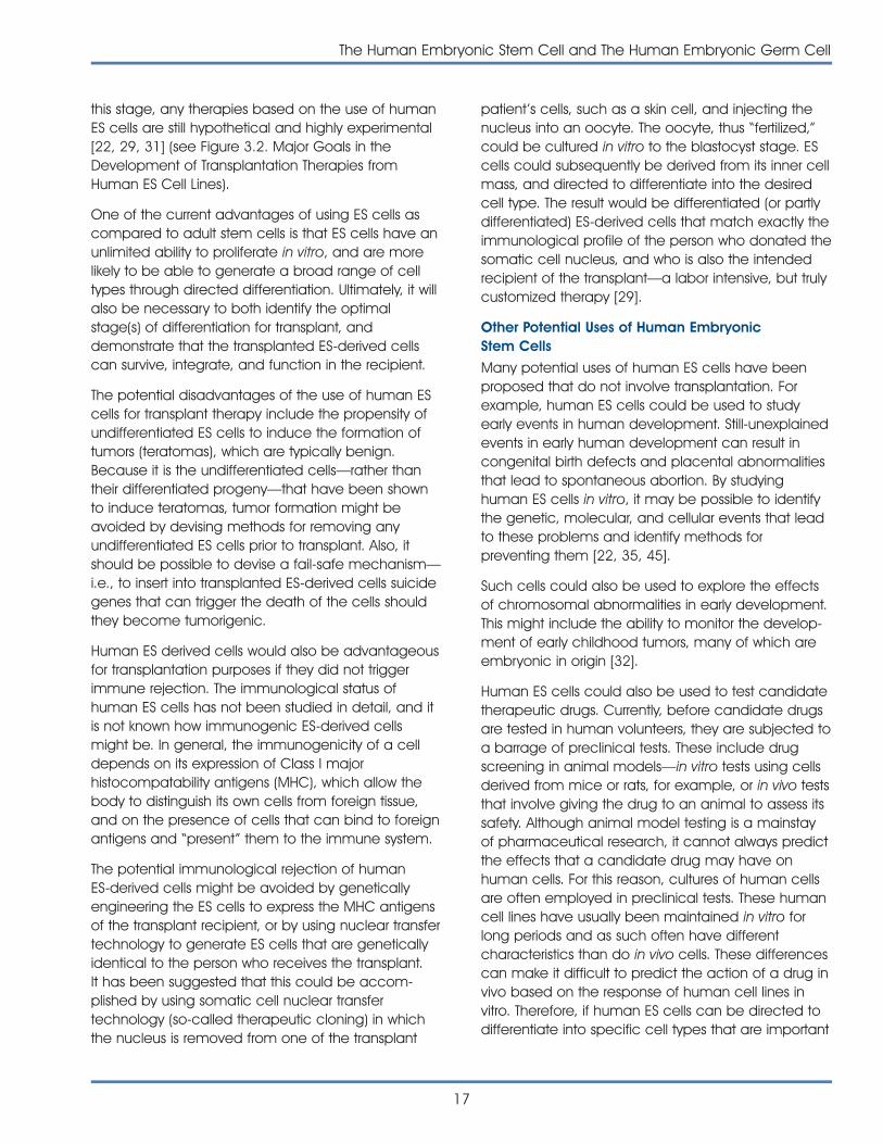
this stage, any therapies based on the use of humanES cells are still hypothetical and highly experimental[22, 29, 31] (see Figure 3.2. Major Goals in theDevelopment of Transplantation Therapies fromHuman ES Cell Lines).
One of the current advantages of using ES cells ascompared to adult stem cells is that ES cells have anunlimited ability to proliferate in vitro, and are morelikely to be able to generate a broad range of celltypes through directed differentiation. Ultimately, it willalso be necessary to both identify the optimalstage(s) of differentiation for transplant, and demonstrate that the transplanted ES-derived cellscan survive, integrate, and function in the recipient.
The potential disadvantages of the use of human EScells for transplant therapy include the propensity ofundifferentiated ES cells to induce the formation oftumors (teratomas), which are typically benign.Because it is the undifferentiated cells—rather thantheir differentiated progeny—that have been shownto induce teratomas, tumor formation might beavoided by devising methods for removing any undifferentiated ES cells prior to transplant. Also, itshould be possible to devise a fail-safe mechanism—i.e., to insert into transplanted ES-derived cells suicidegenes that can trigger the death of the cells shouldthey become tumorigenic.
Human ES derived cells would also be advantageousfor transplantation purposes if they did not triggerimmune rejection. The immunological status ofhuman ES cells has not been studied in detail, and itis not known how immunogenic ES-derived cellsmight be. In general, the immunogenicity of a celldepends on its expression of Class I major histocompatability antigens (MHC), which allow thebody to distinguish its own cells from foreign tissue,and on the presence of cells that can bind to foreignantigens and “present” them to the immune system.
The potential immunological rejection of human ES-derived cells might be avoided by geneticallyengineering the ES cells to express the MHC antigensof the transplant recipient, or by using nuclear transfertechnology to generate ES cells that are geneticallyidentical to the person who receives the transplant. It has been suggested that this could be accom-plished by using somatic cell nuclear transfer technology (so-called therapeutic cloning) in whichthe nucleus is removed from one of the transplant
patient’s cells, such as a skin cell, and injecting thenucleus into an oocyte. The oocyte, thus “fertilized,”could be cultured in vitro to the blastocyst stage. EScells could subsequently be derived from its inner cellmass, and directed to differentiate into the desiredcell type. The result would be differentiated (or partlydifferentiated) ES-derived cells that match exactly theimmunological profile of the person who donated thesomatic cell nucleus, and who is also the intendedrecipient of the transplant—a labor intensive, but trulycustomized therapy [29].
Other Potential Uses of Human EmbryonicStem Cells
Many potential uses of human ES cells have beenproposed that do not involve transplantation. Forexample, human ES cells could be used to studyearly events in human development. Still-unexplainedevents in early human development can result incongenital birth defects and placental abnormalitiesthat lead to spontaneous abortion. By studyinghuman ES cells in vitro, it may be possible to identifythe genetic, molecular, and cellular events that leadto these problems and identify methods for preventing them [22, 35, 45].
Such cells could also be used to explore the effectsof chromosomal abnormalities in early development.This might include the ability to monitor the develop-ment of early childhood tumors, many of which areembryonic in origin [32].
Human ES cells could also be used to test candidatetherapeutic drugs. Currently, before candidate drugsare tested in human volunteers, they are subjected toa barrage of preclinical tests. These include drugscreening in animal models—in vitro tests using cellsderived from mice or rats, for example, or in vivo teststhat involve giving the drug to an animal to assess itssafety. Although animal model testing is a mainstayof pharmaceutical research, it cannot always predictthe effects that a candidate drug may have onhuman cells. For this reason, cultures of human cellsare often employed in preclinical tests. These humancell lines have usually been maintained in vitro forlong periods and as such often have different characteristics than do in vivo cells. These differencescan make it difficult to predict the action of a drug invivo based on the response of human cell lines invitro. Therefore, if human ES cells can be directed todifferentiate into specific cell types that are important
The Human Embryonic Stem Cell and The Human Embryonic Germ Cell
17
for drug screening, the ES-derived cells may be morelikely to mimic the in vivo response of the cells/tissuesto the drug(s) being tested and so offer safer, andpotentially cheaper, models for drug screening.
Human ES cells could be employed to screen potential toxins. The reasons for using human ES cellsto screen potential toxins closely resemble those forusing human ES-derived cells to test drugs (above).Toxins often have different effects on different animalspecies, which makes it critical to have the best possible in vitro models for evaluating their effects on human cells.
Finally, human ES cells could be used to develop newmethods for genetic engineering (see Figure 3.3.Genetic Manipulation of Human Embryonic StemCells). Currently, the genetic complement of mouseES cells in vitro can be modified easily by techniquessuch as homologous recombination. This is a methodfor replacing or adding genes, which requires that aDNA molecule be artificially introduced into thegenome and then expressed. Using this method,genes to direct differentiation to a specific cell typeor genes that express a desired protein product mightbe introduced into the ES cell line. Ultimately, if suchtechniques could be developed using human ES
cells, it may be possible to devise better methods forgene therapy [35] (see Chapter 10. Assessing HumanStem Cell Safety).
SUMMARYWhat Do We Know About Human EmbryonicStem Cells?
Since 1998, research teams have refined the techniques for growing human ES cells in vitro [1, 20,38]. Collectively, the studies indicate that it is nowpossible to grow human ES cells for more than a yearin serum-free medium on feeder layers. The cellshave normal karyotype and are pluripotent; theygenerate teratomas that contain differentiated celltypes derived from all three primary germ layers. Thelong-term cultures of human ES cells have activetelomerase and maintain relatively long telomeres,another marker of proliferating cells.
Overall, the pluripotent cells that can be generatedin vitro from human ES cells and human EG cells areapparently not equivalent in their potential to proliferate or differentiate. (ES cells are derived fromthe inner cell mass of the preimplantation blastocyst,approximately 5 days post-fertilization, whereashuman EG cells are derived from fetal primordial
18
The Human Embryonic Stem Cell and The Human Embryonic Germ Cell
Deletion ofMHC genes
Substitution ofMHC genes
orX X
Tissue-specificdifferentiation
MHC-deficienttissue
Self-MHCexpressing
tissue
A. Genetic manipulationof MHC genes
ES cells
XX
XX
Neuron
Muscle Pancreaticislet cells
Hematopoieticdifferentiation
Cotransplantation
ES cells
C. Hematopoietic chimera: complete, mixed, micro
Enucleatedoocyte
Transfer of donated nucleus
Tissue-specificdifferentiation
ES-cellsderived from
nuclear transferproduct
Differentiatedtissue expressingdonor nucleus
MHC genes
Micropipet
Holding pipet
B. Nuclear reprogramming
Figure 3.3. Genetic Manipulation of Human Embryonic Stem Cells. (Reproduced with permission from Stem Cells, 2001)
germ cells, 5 to 10 weeks post-fertilization.) ES cellscan proliferate for up to 300 population doublings,while cells derived from embryoid bodies that aregenerated from embryonic germ cells (fetal tissue)double a maximum of 70 to 80 times in vitro.
ES cells appear to have a broader ability to differenti-ate. Both kinds of cells spontaneously generate neuralprecursor-type cells (widely regarded as a defaultpathway for differentiation), and both generate cellsthat resemble cardiac myocytes [19, 45]. However,human ES and EG cells in vitro will spontaneouslygenerate embryoid bodies that consist of cell typesfrom all three primary germ layers [1, 20, 38, 42].
What Do We Need To Know About HumanEmbryonic Stem Cells?
Scientists are just beginning to understand the biologyof human embryonic stem cells, and many key questions remain unanswered or only partlyanswered. For example, in order to refine andimprove ES cell culture systems, it is important thatscientists identify the mechanisms that allow humanES cells in vitro to proliferate without differentiating[29]. Once the mechanisms that regulate human ESproliferation are known, it will likely be possible toapply this knowledge to the long-standing challengeof improving the in vitro self-renewal capabilities ofadult stem cells.
It will also be important to determine whether thegenetic imprinting status of human ES cells plays anysignificant role in maintaining the cells, directing theirdifferentiation, or determining their suitability for transplant. One of the effects of growing mouse blastocysts in culture is a change in the methylationof specific genes that control embryonic growth and development [23]. Do similar changes in geneimprinting patterns occur in human ES cells (or blastocysts)? If so, what is their effect on in vitrodevelopment and on any differentiated cell typesthat may be derived from cultured ES cells?
Efforts will need to be made to determine whethercultures of human ES cells that appear to be homogeneous and undifferentiated are, in fact,homogeneous and undifferentiated. Is it possible that human ES cells in vitro cycle in and out of partially differentiated states? And if that occurs, howwill it affect attempts to direct their differentiation ormaintain the cells in a proliferating state [28]?
Scientists will need to identify which signal transductionpathways must be activated to induce human ES celldifferentiation along a particular pathway. This includesunderstanding ligand-receptor interaction and theintracellular components of the signaling system, aswell as identifying the genes that are activated or inac-tivated during differentiation of specific cell types [29].
Identifying intermediate stages of human ES cell differentiation will also be important. As human EScells differentiate in vitro, do they form distinct precursor or progenitor cells that can be identifiedand isolated? If ES cells do form such intermediatecell types, can the latter be maintained and expanded? Would such precursor or progenitor cellsbe useful for therapeutic transplantation [19]?
Finally, scientists will need to determine what differentiation stages of human ES-derived cells areoptimal for other practical applications. For example,what differentiation stages of ES-derived cells wouldbe best for screening drugs or toxins, or for deliveringpotentially therapeutic drugs?
REFERENCES1. Amit, M., Carpenter, M.K., Inokuma, M.S., Chiu, C.P., Harris,
C.P., Waknitz, M.A., Itskovitz-Eldor, J., and Thomson, J.A.(2000). Clonally derived human embryonic stem cell linesmaintain pluripotency and proliferative potential forprolonged periods of culture. Dev. Biol. 227, 271-278.
2. Andrews, P.W., Damjanov, I., Simon, D., Banting, G.S.,Carlin, C., Dracopoli, N.C., and Fogh, J. (1984). Pluripotentembryonal carcinoma clones derived from the humanteratocarcinoma cell line Tera-2. Differentiation in vivoand in vitro. Lab. Invest. 50, 147-162.
3. Andrews, P.W. (1988). Human teratocarcinomas. Biochim.Biophys. Acta. 948, 17-36.
4. Andrews, P.W. (1998). Teratocarcinomas and humanembryology: pluripotent human EC cell lines. Reviewarticle. APMIS 106, 158-167.
5. Andrews, P. W., personal communication.
6. Bongso, A., Fong, C.Y., Ng, S.C., and Ratnam, S.S. (1994).Blastocyst transfer in human in vitro: fertilization; the use ofembryo co-culture. Cell Biol. Int. 18, 1181-1189.
7. Bongso, A., Fong, C.Y., Ng, S.C., and Ratnam, S. (1994).Isolation and culture of inner cell mass cells from humanblastocysts. Hum. Reprod. 9, 2110-2117.
8. Bongso, A., Fong, C.Y., Ng, S.C., and Ratnam, S.S. (1995).Co-culture techniques for blastocyst transfer and embry-onic stem cell production. Asst. Reprod. Rev. 5, 106-114.
The Human Embryonic Stem Cell and The Human Embryonic Germ Cell
19
9. Bongso, A. (1996). Behaviour of human embryos in vitro inthe first 14 days: blastocyst transfer and embryonic stemcell production. Clin. Sci. (Colch.) 91, 248-249.
10. Bongso, A., Fong, C.Y., Mathew, J., Ng, L.C., Kumar, J., andNg, S.C. (1999). The benefits to human IVF by transferringembryos after the in vitro embryonic block: alternatives today 2 transfers. Asst. Reprod. Rev.
11. Bongso, A. (1999). Handbook on blastocyst culture,(Singapore: Sydney Press Indusprint).
12. Bongso, A., personal communication.
13. Bradley, A., Evans, M., Kaufman, M.H., and Robertson, E.(1984). Formation of germ-line chimaeras from embryo-derived teratocarcinoma cell lines. Nature. 309, 255-256.
14. De Vos, A. and Van Steirteghem, A. (2000). Zona hardening,zona drilling and assisted hatching: new achievements inassisted reproduction. Cells Tissues Organs. 166, 220-227.
15. Evans, M.J. and Kaufman, M.H. (1981). Establishment inculture of pluripotential cells from mouse embryos.Nature. 292, 154-156.
16. Fong, C.Y., Bongso, A., Ng, S.C., Kumar, J., Trounson, A.,and Ratnam, S. (1998). Blastocyst transfer after enzymatictreatment of the zona pellucida: improving in-vitro fertiliza-tion and understanding implantation. Hum. Reprod. 13, 2926-2932.
17. Friedrich, T.D., Regenass, U., and Stevens, L.C. (1983).Mouse genital ridges in organ culture: the effects oftemperature on maturation and experimental induction ofteratocarcinogenesis. Differentiation. 24, 60-64.
18. Gardner, D.K. and Schoolcraft, W.B. (1999). Culture andtransfer of human blastocysts. Curr. Opin. Obstet. Gynecol.11, 307-311.
19. Gearhart, J., personal communication.
20. Itskovitz-Eldor, J., Schuldiner, M., Karsenti, D., Eden, A.,Yanuka, O., Amit, M., Soreq, H., and Benvenisty, N. (2000).Differentiation of human embryonic stem cells into embry-oid bodies comprising the three embryonic germ layers.Mol. Med. 6, 88-95.
21. Jones, G.M., Trounson, A.O., Lolatgis, N., and Wood, C.(1998). Factors affecting the success of human blastocystdevelopment and pregnancy following in vitro fertilizationand embryo transfer. Fertil. Steril. 70, 1022-1029.
22. Jones, J.M. and Thomson, J.A. (2000). Human embryonicstem cell technology. Semin. Reprod. Med. 18, 219-223.
23. Khosla, S., Dean, W., Brown, D., Reik, W., and Feil, R. (2001). Culture of preimplantation mouse embryosaffects fetal development and the expression of imprintedgenes. Biol. Reprod. 64, 918-926.
24. Kleinsmith, L.J. and Pierce Jr, G.B. (1964). Multipotentialityof single embryonal carcinoma cells. Cancer Res. 24,1544-1551.
25. Martin, G.R. (1980). Teratocarcinomas and mammalianembryogenesis. Science. 209, 768-776.
26. Martin, G.R. (1981). Isolation of a pluripotent cell line fromearly mouse embryos cultured in medium conditioned byteratocarcinoma stem cells. Proc. Natl. Acad. Sci. U. S. A.78, 7634-7638.
27. Matsui, Y., Toksoz, D., Nishikawa, S., Nishikawa, S., Williams,D., Zsebo, K., and Hogan, B.L. (1991). Effect of steel factorand leukaemia inhibitory factor on murine primordial germcells in culture. Nature. 353, 750-752.
28. McKay, R., personal communication.
29. Odorico, J.S., Kaufman, D.S., and Thomson, J.A. (2001).Multilineage Differentiation from Human Embryonic StemCell Lines. Stem Cells. 19, 193-204.
30. Okarma, T., personal communication.
31. Pedersen, R.A. (1999). Embryonic stem cells for medicine.Sci. Am. 280, 68-73.
32. Pera, M., personal communication.
33. Pera, M.F., Cooper, S., Mills, J., and Parrington, J.M. (1989). Isolation and characterization of a multipotentclone of human embryonal carcinoma cells.Differentiation. 42, 10-23.
34. Pera, M.F., Reubinoff, B., and Trounson, A. (2000). Humanembryonic stem cells. J. Cell Sci. 113 (Pt 1), 5-10.
35. Rathjen, P.D., Lake, J., Whyatt, L.M., Bettess, M.D., andRathjen, J. (1998). Properties and uses of embryonic stemcells: prospects for application to human biology andgene therapy. Reprod. Fertil. Dev. 10, 31-47.
36. Resnick, J.L., Bixler, L.S., Cheng, L., and Donovan, P.J. (1992).Long-term proliferation of mouse primordial germ cells inculture. Nature. 359, 550-551.
37. Reubinoff BE, Pera, M., Fong, C.Y., and Trounson, A. andBongso, A. (2000). Research Errata. Nat. Biotechnol. 18,559.
38. Reubinoff, B.E., Pera, M.F., Fong, C.Y., Trounson, A., andBongso, A. (2000). Embryonic stem cell lines from humanblastocysts: somatic differentiation in vitro. Nat. Biotechnol.18, 399-404.
39. Sathananthan, A.H. (1997). Ultrastructure of the humanegg. Hum. Cell. 10, 21-38.
40. Schuldiner, M., Yanuka, O., Itskovitz-Eldor, J., Melton, D., andBenvenisty, N. (2000). Effects of eight growth factors on thedifferentiation of cells derived from human embryonic stemcells. Proc. Natl. Acad. Sci. U. S. A. 97, 11307-11312.
41. Shamblott, M.J., Axelman, J., Wang, S., Bugg, E.M.,Littlefield, J.W., Donovan, P.J., Blumenthal, P.D., Huggins,G.R., and Gearhart, J.D. (1998). Derivation of pluripotentstem cells from cultured human primordial germ cells.Proc. Natl. Acad. Sci. U. S. A. 95, 13726-13731.
20
The Human Embryonic Stem Cell and The Human Embryonic Germ Cell
42. Shamblott, M.J., Axelman, J., Littlefield, J.W., Blumenthal,P.D., Huggins, G.R., Cui, Y., Cheng, L., and Gearhart, J.D.(2001). Human embryonic germ cell derivatives express abroad range of develpmentally distinct markers andproliferate extensively in vitro. Proc. Natl. Acad. Sci. U. S. A.98, 113-118.
43. Smith, A.G. (2001). Origins and properties of mouseembryonic stem cells. Annu. Rev. Cell. Dev. Biol.
44. Thompson, S., Stern, P.L., Webb, M., Walsh, F.S., Engstrom,W., Evans, E.P., Shi, W.K., Hopkins, B., and Graham, C.F.(1984). Cloned human teratoma cells differentiate intoneuron-like cells and other cell types in retinoic acid. J. CellSci. 72, 37-64.
45. Thomson, J., personal communication.
46. Thomson, J.A., Kalishman, J., Golos, T.G., Durning, M., Harris,C.P., Becker, R.A., and Hearn, J.P. (1995). Isolation of aprimate embryonic stem cell line. Proc. Natl. Acad. Sci. U. S. A. 92, 7844-7848.
47. Thomson, J.A., Kalishman, J., Golos, T.G., Durning, M., Harris,C.P., and Hearn, J.P. (1996). Pluripotent cell lines derivedfrom common marmoset (Callithrix jacchus) blastocysts.Biol. Reprod. 55, 254-259.
48. Thomson, J.A., Itskovitz-Eldor, J., Shapiro, S.S., Waknitz, M.A.,Swiergiel, J.J., Marshall, V.S., and Jones, J.M. (1998).Embryonic stem cell lines derived from human blastocysts.Science. 282, 1145-1147.
49. Trounson, A.O., Gardner, D.K., Baker, G., Barnes, F.L.,Bongso, A., Bourne, H., Calderon, I., Cohen, J., Dawson, K.,Eldar-Geve, T., Gardner, D.K., Graves, G., Healy, D., Lane,M., Leese, H.J., Leeton, J., Levron, J., Liu, D.Y., MacLachlan,V., Munné, S., Oranratnachai, A., Rogers, P., Rombauts, L.,Sakkas, D., Sathananthan, A.H., Schimmel, T., Shaw, J.,Trounson, A.O., Van Steirteghem, A., Willadsen, S., andWood, C. (2000b). Handbook of in vitro fertilization, (BocaRaton, London, New York, Washington, D.C.: CRC Press).
50. Trounson, A.O., Anderiesz, C., and Jones, G. (2001).Maturation of human oocytes in vitro and their develop-mental competence. Reproduction. 121, 51-75.
51. Xu, C., Inokuma, M.S., Denham, J., Golds, K., Kundu, P.,Gold, J.D., and Carpenter, M.K. Keystone symposia.Pluripotent stem cells: biology and applications. Growth ofundifferentiated human embryonic stem cells on defined matrices with conditioned medium. Poster abstract. 133.
The Human Embryonic Stem Cell and The Human Embryonic Germ Cell
21

22
The Human Embryonic Stem Cell and The Human Embryonic Germ Cell
This page intentionally left blank.
23
For many years, researchers have been seeking tounderstand the body’s ability to repair and replacethe cells and tissues of some organs, but not others.After years of work pursuing the how and why ofseemingly indiscriminant cell repair mechanisms, scientists have now focused their attention on adultstem cells. It has long been known that stem cellsare capable of renewing themselves and that theycan generate multiple cell types. Today, there is newevidence that stem cells are present in far more tissues and organs than once thought and that thesecells are capable of developing into more kinds ofcells than previously imagined. Efforts are nowunderway to harness stem cells and to takeadvantage of this new found capability, with the goalof devising new and more effective treatments for ahost of diseases and disabilities. What lies ahead forthe use of adult stem cells is unknown, but it iscertain that there are many research questions to beanswered and that these answers hold great promisefor the future.
WHAT IS AN ADULT STEM CELL?Adult stem cells, like all stem cells, share at least twocharacteristics. First, they can make identical copiesof themselves for long periods of time; this ability toproliferate is referred to as long-term self-renewal.Second, they can give rise to mature cell types thathave characteristic morphologies (shapes) andspecialized functions. Typically, stem cells generatean intermediate cell type or types before theyachieve their fully differentiated state. The intermedi-ate cell is called a precursor or progenitor cell.Progenitor or precursor cells in fetal or adult tissues arepartly differentiated cells that divide and give rise todifferentiated cells. Such cells are usually regarded as“committed” to differentiating along a particularcellular development pathway, although this
characteristic may not be as definitive as oncethought [82] (see Figure 4.1. Distinguishing Features ofProgenitor/Precursor Cells and Stem Cells).
Adult stem cells are rare. Their primary functions areto maintain the steady state functioning of a cell—called homeostasis—and, with limitations, to replacecells that die because of injury or disease [44, 58].For example, only an estimated 1 in 10,000 to 15,000cells in the bone marrow is a hematopoietic (blood-forming) stem cell (HSC) [105]. Furthermore, adultstem cells are dispersed in tissues throughout themature animal and behave very differently,depending on their local environment. For example,HSCs are constantly being generated in the bonemarrow where they differentiate into mature types ofblood cells. Indeed, the primary role of HSCs is toreplace blood cells [26] (see Chapter 5. Hemato-poietic Stem Cells). In contrast, stem cells in the smallintestine are stationary, and are physically separatedfrom the mature cell types they generate. Gut epi-thelial stem cells (or precursors) occur at the bases ofcrypts—deep invaginations between the mature,differentiated epithelial cells that line the lumen of theintestine. These epithelial crypt cells divide fairly often,but remain part of the stationary group of cells theygenerate [93].
Unlike embryonic stem cells, which are defined bytheir origin (the inner cell mass of the blastocyst),adult stem cells share no such definitive means ofcharacterization. In fact, no one knows the origin ofadult stem cells in any mature tissue. Some haveproposed that stem cells are somehow set asideduring fetal development and restrained fromdifferentiating. Definitions of adult stem cells vary inthe scientific literature range from a simple descrip-tion of the cells to a rigorous set of experimentalcriteria that must be met before characterizing a
THE ADULT STEM CELLTHE ADULT STEM CELL4.4.
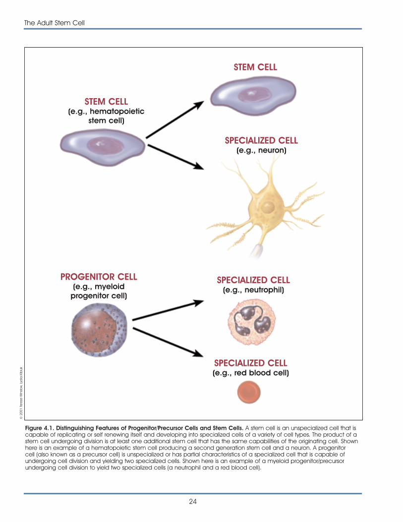
24
The Adult Stem Cell
Figure 4.1. Distinguishing Features of Progenitor/Precursor Cells and Stem Cells. A stem cell is an unspecialized cell that iscapable of replicating or self renewing itself and developing into specialized cells of a variety of cell types. The product of astem cell undergoing division is at least one additional stem cell that has the same capabilities of the originating cell. Shownhere is an example of a hematopoietic stem cell producing a second generation stem cell and a neuron. A progenitorcell (also known as a precursor cell) is unspecialized or has partial characteristics of a specialized cell that is capable ofundergoing cell division and yielding two specialized cells. Shown here is an example of a myeloid progenitor/precursorundergoing cell division to yield two specialized cells (a neutrophil and a red blood cell).
© 2
001
Tere
se W
inslo
w, L
ydia
Kib
iuk
STEM CELL(e.g., hematopoietic
stem cell)
STEM CELL
SPECIALIZED CELL(e.g., red blood cell)
PROGENITOR CELL(e.g., myeloid
progenitor cell)
SPECIALIZED CELL(e.g., neutrophil)
SPECIALIZED CELL(e.g., neuron)
particular cell as an adult stem cell. Most of the infor-mation about adult stem cells comes from studies ofmice. The list of adult tissues reported to contain stemcells is growing and includes bone marrow, peripheralblood, brain, spinal cord, dental pulp, blood vessels,skeletal muscle, epithelia of the skin and digestivesystem, cornea, retina, liver, and pancreas.
In order to be classified as an adult stem cell, the cellshould be capable of self-renewal for the lifetime ofthe organism. This criterion, although fundamental tothe nature of a stem cell, is difficult to prove in vivo. Itis nearly impossible, in an organism as complex as ahuman, to design an experiment that will allow thefate of candidate adult stem cells to be identified invivo and tracked over an individual’s entire lifetime.
Ideally, adult stem cells should also be clonogenic. Inother words, a single adult stem cell should be ableto generate a line of genetically identical cells, whichthen gives rise to all the appropriate, differentiatedcell types of the tissue in which it resides. Again, thisproperty is difficult to demonstrate in vivo; in practice,scientists show either that a stem cell is clonogenic invitro, or that a purified population of candidate stemcells can repopulate the tissue.
An adult stem cell should also be able to give rise tofully differentiated cells that have mature phenotypes,are fully integrated into the tissue, and are capableof specialized functions that are appropriate for thetissue. The term phenotype refers to all the observ-able characteristics of a cell (or organism); its shape(morphology); interactions with other cells and thenon-cellular environment (also called the extracellularmatrix); proteins that appear on the cell surface(surface markers); and the cell’s behavior (e.g.,secretion, contraction, synaptic transmission).
The majority of researchers who lay claim to havingidentified adult stem cells rely on two of these char-acteristics—appropriate cell morphology, and thedemonstration that the resulting, differentiated celltypes display surface markers that identify them asbelonging to the tissue. Some studies demonstratethat the differentiated cells that are derived fromadult stem cells are truly functional, and a few studiesshow that cells are integrated into the differentiatedtissue in vivo and that they interact appropriately withneighboring cells. At present, there is, however, apaucity of research, with a few notable exceptions, inwhich researchers were able to conduct studies of
genetically identical (clonal) stem cells. In order tofully characterize the regenerating and self-renewalcapabilities of the adult stem cell, and therefore totruly harness its potential, it will be important todemonstrate that a single adult stem cell can,indeed, generate a line of genetically identical cells,which then gives rise to all the appropriate, differenti-ated cell types of the tissue in which it resides.
EVIDENCE FOR THE PRESENCE OFADULT STEM CELLSAdult stem cells have been identified in many animaland human tissues. In general, three methods areused to determine whether candidate adult stemcells give rise to specialized cells. Adult stem cellscan be labeled in vivo and then they can betracked. Candidate adult stem cells can also beisolated and labeled and then transplanted back intothe organism to determine what becomes of them.Finally, candidate adult stem cells can be isolated,grown in vitro and manipulated, by adding growthfactors or introducing genes that help determinewhat differentiated cells types they will yield. Forexample, currently, scientists believe that stem cells inthe fetal and adult brain divide and give rise to morestem cells or to several types of precursor cells, whichgive rise to nerve cells (neurons), of which there aremany types.
It is often difficult—if not impossible—to distinguishadult, tissue-specific stem cells from progenitor cells,which are found in fetal or adult tissues and are partlydifferentiated cells that divide and give rise to differ-entiated cells. These are cells found in many organsthat are generally thought to be present to replacecells and maintain the integrity of the tissue.Progenitor cells give rise to certain types of cells—such as the blood cells known as T lymphocytes,B lymphocytes, and natural killer cells—but are notthought to be capable of developing into all the celltypes of a tissue and as such are not truly stem cells.The current wave of excitement over the existence ofstem cells in many adult tissues is perhaps fuelingclaims that progenitor or precursor cells in those tis-sues are instead stem cells. Thus, there are reports ofendothelial progenitor cells, skeletal muscle stemcells, epithelial precursors in the skin and digestivesystem, as well as some reports of progenitors or stemcells in the pancreas and liver. A detailed summaryof some of the evidence for the existence of stem
The Adult Stem Cell
25
cells in various tissues and organs is presented later inthe chapter.
ADULT STEM CELL PLASTICITY It was not until recently that anyone seriously consid-ered the possibility that stem cells in adult tissuescould generate the specialized cell types of anothertype of tissue from which they normally reside—eithera tissue derived from the same embryonic germlayer or from a different germ layer (see Table 1.1.Embryonic Germ Layers From Which DifferentiatedTissues Develop). For example, studies have shownthat blood stem cells (derived from mesoderm) maybe able to generate both skeletal muscle (alsoderived from mesoderm) and neurons (derived fromectoderm). That realization has been triggered by aflurry of papers reporting that stem cells derived fromone adult tissue can change their appearance andassume characteristics that resemble those of differ-entiated cells from other tissues.
The term plasticity, as used in this report, means thata stem cell from one adult tissue can generate thedifferentiated cell types of another tissue. At this time,there is no formally accepted name for this phenom-enon in the scientific literature. It is variously referredto as “plasticity” [15, 52], “unorthodox differentiation”[10] or “transdifferentiation” [7, 54].
Approaches for Demonstrating Adult Stem CellPlasticity
To be able to claim that adult stem cells demon-strate plasticity, it is first important to show that a cellpopulation exists in the starting tissue that has theidentifying features of stem cells. Then, it is necessaryto show that the adult stem cells give rise to cell typesthat normally occur in a different tissue. Neither ofthese criteria is easily met. Simply proving the exis-tence of an adult stem cell population in a differenti-ated tissue is a laborious process. It requires that thecandidate stem cells are shown to be self-renewing,and that they can give rise to the differentiated celltypes that are characteristic of that tissue.
To show that the adult stem cells can generate othercell types requires them to be tracked in their newenvironment, whether it is in vitro or in vivo. In general,this has been accomplished by obtaining the stemcells from a mouse that has been genetically engi-neered to express a molecular tag in all its cells. It isthen necessary to show that the labeled adult stem
26
The Adult Stem Cell
cells have adopted key structural and biochemicalcharacteristics of the new tissue they are claimed tohave generated. Ultimately—and most importantly—itis necessary to demonstrate that the cells can inte-grate into their new tissue environment, survive in thetissue, and function like the mature cells of the tissue.
In the experiments reported to date, adult stem cellsmay assume the characteristics of cells that havedeveloped from the same primary germ layer or adifferent germ layer (see Figure 4.2. PreliminaryEvidence of Plasticity Among Nonhuman Adult StemCells). For example, many plasticity experimentsinvolve stem cells derived from bone marrow, whichis a mesodermal derivative. The bone marrow stemcells may then differentiate into another mesoder-mally derived tissue such as skeletal muscle [28, 43],cardiac muscle [51, 71] or liver [4, 54, 97].
Alternatively, adult stem cells may differentiate into atissue that—during normal embryonic develop-ment—would arise from a different germ layer. Forexample, bone marrow-derived cells may differenti-ate into neural tissue, which is derived from embryon-ic ectoderm [15, 65]. And—reciprocally—neural stemcell lines cultured from adult brain tissue may differ-entiate to form hematopoietic cells [13], or even giverise to many different cell types in a chimeric embryo[17]. In both cases cited above, the cells would bedeemed to show plasticity, but in the case of bonemarrow stem cells generating brain cells, the findingis less predictable.
In order to study plasticity within and across germlayer lines, the researcher must be sure that he/she isusing only one kind of adult stem cell. The vast major-ity of experiments on plasticity have been conductedwith adult stem cells derived either from the bonemarrow or the brain. The bone marrow-derived cellsare sometimes sorted—using a panel of surfacemarkers—into populations of hematopoietic stemcells or bone marrow stromal cells [46, 54, 71]. TheHSCs may be highly purified or partially purified,depending on the conditions used. Another way toseparate population of bone marrow cells is by frac-tionation to yield cells that adhere to a growth sub-strate (stromal cells) or do not adhere (hematopoieticcells) [28].
To study plasticity of stem cells derived from the brain,the researcher must overcome several problems.Stem cells from the central nervous system (CNS),
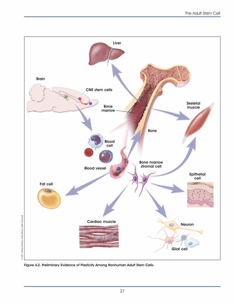
The Adult Stem Cell
27
Liver
Brain
CNS stem cells
Bonemarrow
Bone
Bloodcell
Blood vessel
Neuron
Epithelialcell
Bone marrowstromal cell
Glial cell
Cardiac muscle
Fat cell
Skeletalmuscle
Figure 4.2. Preliminary Evidence of Plasticity Among Nonhuman Adult Stem Cells.
© 2
001
Tere
se W
inslo
w, L
ydia
Kib
iuk,
Ca
itlin
Duc
kwa
ll
unlike bone marrow cells, do not occur in a single,accessible location. Instead, they are scattered inthree places, at least in rodent brain—the tissuearound the lateral ventricles in the forebrain, a migra-tory pathway for the cells that leads from the ventri-cles to the olfactory bulbs, and the hippocampus.Many of the experiments with CNS stem cells involvethe formation of neurospheres, round aggregates ofcells that are sometimes clonally derived. But it is notpossible to observe cells in the center of a neu-rosphere, so to study plasticity in vitro, the cells areusually dissociated and plated in monolayers. Tostudy plasticity in vivo, the cells may be dissociatedbefore injection into the circulatory system of therecipient animal [13], or injected as neurospheres[17].
What is the Evidence for Plasticity?
The differentiated cell types that result from plasticityare usually reported to have the morphological char-acteristics of the differentiated cells and to displaytheir characteristic surface markers. In reports thattransplanted adult stem cells show plasticity in vivo,the stem cells typically are shown to have integratedinto a mature host tissue and assumed at least someof its characteristics [15, 28, 51, 65, 71]. Many plastic-ity experiments involve injury to a particular tissue,which is intended to model a particular humandisease or injury [13, 54, 71]. However, there is limitedevidence to date that such adult stem cells cangenerate mature, fully functional cells or that the cellshave restored lost function in vivo [54]. Most of thestudies that show the plasticity of adult stem cellsinvolve cells that are derived from the bone marrow[15, 28, 54, 65, 77] or brain [13, 17]. To date, adultstem cells are best characterized in these two tissues,which may account for the greater number of plas-ticity studies based on bone marrow and brain.Collectively, studies on plasticity suggest that stemcell populations in adult mammals are not fixedentities, and that after exposure to a new environ-ment, they may be able to populate other tissuesand possibly differentiate into other cell types.
It is not yet possible to say whether plasticity occursnormally in vivo. Some scientists think it may [14, 64],but as yet there is no evidence to prove it. Also, it isnot yet clear to what extent plasticity can occur inexperimental settings, and how—or whether—thephenomenon can be harnessed to generate tissues
that may be useful for therapeutic transplantation. Ifthe phenomenon of plasticity is to be used as a basisfor generating tissue for transplantation, the tech-niques for doing it will need to be reproducible andreliable (see Chapter 10. Assessing Human Stem CellSafety). In some cases, debate continues aboutobservations that adult stem cells yield cells of tissuetypes different than those from which they wereobtained [7, 68].
EXPERIMENTAL EVIDENCE OF ADULTSTEM CELLS AND PLASTICITYAdult Stem Cells of the Nervous System
More than 30 years ago, Altman and Das showedthat two regions of the postnatal rat brain, the hip-pocampus and the olfactory bulb, contain dividingcells that become neurons [5, 6]. Despite thesereports, the prevailing view at the time was that nervecells in the adult brain do not divide. In fact, thenotion that stem cells in the adult brain can generateits three major cell types—astrocytes and oligoden-drocytes, as well as neurons—was not accepted untilfar more recently. Within the past five years, a seriesof studies has shown that stem cells occur in theadult mammalian brain and that these cells cangenerate its three major cell lineages [35, 48, 63, 66,90, 96, 104] (see Chapter 8. Rebuilding the NervousSystem with Stem Cells).
Today, scientists believe that stem cells in the fetaland adult brain divide and give rise to more stemcells or to several types of precursor cells. Neuronalprecursors (also called neuroblasts) divide and giverise to nerve cells (neurons), of which there are manytypes. Glial precursors give rise to astrocytes or oligo-dendrocytes. Astrocytes are a kind of glial cell, whichlend both mechanical and metabolic support forneurons; they make up 70 to 80 percent of the cellsof the adult brain. Oligodendrocytes make myelin,the fatty material that ensheathes nerve cell axonsand speeds nerve transmission. Under normal, in vivoconditions, neuronal precursors do not give rise toglial cells, and glial precursors do not give rise toneurons. In contrast, a fetal or adult CNS (centralnervous system—the brain and spinal cord) stem cellmay give rise to neurons, astrocytes, or oligodendro-cytes, depending on the signals it receives and itsthree-dimensional environment within the brain tissue.
28
The Adult Stem Cell
There is now widespread consensus that the adultmammalian brain does contain stem cells. However,there is no consensus about how many populationsof CNS stem cells exist, how they may be related,and how they function in vivo. Because there are nomarkers currently available to identify the cells in vivo,the only method for testing whether a given popula-tion of CNS cells contains stem cells is to isolate thecells and manipulate them in vitro, a process thatmay change their intrinsic properties [67].
Despite these barriers, three groups of CNS stem cellshave been reported to date. All occur in the adultrodent brain and preliminary evidence indicates theyalso occur in the adult human brain. One groupoccupies the brain tissue next to the ventricles,regions known as the ventricular zone and the sub-ventricular zone (see discussion below). The ventriclesare spaces in the brain filled with cerebrospinal fluid.During fetal development, the tissue adjacent to theventricles is a prominent region of actively dividingcells. By adulthood, however, this tissue is much small-er, although it still appears to contain stem cells [70].
A second group of adult CNS stem cells, described inmice but not in humans, occurs in a streak of tissuethat connects the lateral ventricle and the olfactorybulb, which receives odor signals from the nose. Inrodents, olfactory bulb neurons are constantly beingreplenished via this pathway [59, 61]. A third possiblelocation for stem cells in adult mouse and humanbrain occurs in the hippocampus, a part of the brainthought to play a role in the formation of certain kindsof memory [27, 34].
Central Nervous System Stem Cells in theSubventricular Zone. CNS stem cells found in the fore-brain that surrounds the lateral ventricles are hetero-geneous and can be distinguished morphologically.Ependymal cells, which are ciliated, line the ventri-cles. Adjacent to the ependymal cell layer, in aregion sometimes designated as the subependymalor subventricular zone, is a mixed cell population thatconsists of neuroblasts (immature neurons) thatmigrate to the olfactory bulb, precursor cells, andastrocytes. Some of the cells divide rapidly, whileothers divide slowly. The astrocyte-like cells can beidentified because they contain glial fibrillary acidicprotein (GFAP), whereas the ependymal cells stainpositive for nestin, which is regarded as a marker ofneural stem cells. Which of these cells best qualifiesas a CNS stem cell is a matter of debate [76].
A recent report indicates that the astrocytes thatoccur in the subventricular zone of the rodent brainact as neural stem cells. The cells with astrocytemarkers appear to generate neurons in vivo, asidentified by their expression of specific neuronalmarkers. The in vitro assay to demonstrate that theseastrocytes are, in fact, stem cells involves their abilityto form neurospheres—groupings of undifferentiatedcells that can be dissociated and coaxed to differen-tiate into neurons or glial cells [25]. Traditionally, theseastrocytes have been regarded as differentiatedcells, not as stem cells and so their designation asstem cells is not universally accepted.
A series of similar in vitro studies based on theformation of neurospheres was used to identify thesubependymal zone as a source of adult rodent CNSstem cells. In these experiments, single, candidatestem cells derived from the subependymal zone areinduced to give rise to neurospheres in the presenceof mitogens—either epidermal growth factor (EGF) orfibroblast growth factor-2 (FGF-2). The neurospheresare dissociated and passaged. As long as a mitogenis present in the culture medium, the cells continueforming neurospheres without differentiating. Somepopulations of CNS cells are more responsive to EGF,others to FGF [100]. To induce differentiation intoneurons or glia, cells are dissociated from theneurospheres and grown on an adherent surface in serum-free medium that contains specific growthfactors. Collectively, the studies demonstrate that apopulation of cells derived from the adult rodentbrain can self-renew and differentiate to yield thethree major cell types of the CNS cells [41, 69, 74, 102].
Central Nervous System Stem Cells in the VentricularZone. Another group of potential CNS stem cells inthe adult rodent brain may consist of the ependymalcells themselves [47]. Ependymal cells, which areciliated, line the lateral ventricles. They have beendescribed as non-dividing cells [24] that function aspart of the blood-brain barrier [22]. The suggestionthat ependymal cells from the ventricular zone of theadult rodent CNS may be stem cells is thereforeunexpected. However, in a recent study, in which twomolecular tags—the fluorescent marker Dil, and anadenovirus vector carrying lacZ tags—were used tolabel the ependymal cells that line the entire CNSventricular system of adult rats, it was shown thatthese cells could, indeed, act as stem cells. A few
The Adult Stem Cell
29
days after labeling, fluorescent or lacZ+ cells wereobserved in the rostral migratory stream (which leadsfrom the lateral ventricle to the olfactory bulb), andthen in the olfactory bulb itself. The labeled cells inthe olfactory bulb also stained for the neuronalmarkers �III tubulin and Map2, which indicated thatependymal cells from the ventricular zone of theadult rat brain had migrated along the rostralmigratory stream to generate olfactory bulb neuronsin vivo [47].
To show that Dil+ cells were neural stem cells andcould generate astrocytes and oligodendrocytes aswell as neurons, a neurosphere assay was performedin vitro. Dil-labeled cells were dissociated from theventricular system and cultured in the presence ofmitogen to generate neurospheres. Most of theneurospheres were Dil+; they could self-renew andgenerate neurons, astrocytes, and oligodendrocyteswhen induced to differentiate. Single, Dil+ ependymalcells isolated from the ventricular zone could alsogenerate self-renewing neurospheres and differen-tiate into neurons and glia.
To show that ependymal cells can also divide in vivo,bromodeoxyuridine (BrdU) was administered in thedrinking water to rats for a 2- to 6-week period.Bromodeoxyuridine (BrdU) is a DNA precursor that isonly incorporated into dividing cells. Through a seriesof experiments, it was shown that ependymal cellsdivide slowly in vivo and give rise to a population ofprogenitor cells in the subventricular zone [47]. Adifferent pattern of scattered BrdU-labeled cells wasobserved in the spinal cord, which suggested thatependymal cells along the central canal of the cordoccasionally divide and give rise to nearby ependy-mal cells, but do not migrate away from the canal.
Collectively, the data suggest that CNS ependymalcells in adult rodents can function as stem cells. Thecells can self-renew, and most proliferate via asym-metrical division. Many of the CNS ependymal cellsare not actively dividing (quiescent), but they can bestimulated to do so in vitro (with mitogens) or in vivo(in response to injury). After injury, the ependymal cellsin the spinal cord only give rise to astrocytes, not toneurons. How and whether ependymal cells from theventricular zone are related to other candidate popu-lations of CNS stem cells, such as those identified inthe hippocampus [34], is not known.
Are ventricular and subventricular zone CNS stemcells the same population? These studies and otherleave open the question of whether cells that directlyline the ventricles—those in the ventricular zone—orcells that are at least a layer removed from thiszone—in the subventricular zone are the same popu-lation of CNS stem cells. A new study, based on thefinding that they express different genes, confirmsearlier reports that the ventricular and subventricularzone cell populations are distinct. The new researchutilizes a technique called representational differenceanalysis, together with cDNA microarray analysis, tomonitor the patterns of gene expression in the com-plex tissue of the developing and postnatal mousebrain. The study revealed the expression of a panel ofgenes known to be important in CNS development,such as L3-PSP (which encodes a phosphoserinephosphatase important in cell signaling), cyclin D2 (acell cycle gene), and ERCC-1 (which is important inDNA excision repair). All of these genes in the recentstudy were expressed in cultured neurospheres, aswell as the ventricular zone, the subventricular zone,and a brain area outside those germinal zones. Thisanalysis also revealed the expression of novel genessuch as A16F10, which is similar to a gene in anembryonic cancer cell line. A16F10 was expressed inneurospheres and at high levels in the subventricularzone, but not significantly in the ventricular zone.Interestingly, several of the genes identified in culturedneurospheres were also expressed in hematopoieticcells, suggesting that neural stem cells and blood-forming cells may share aspects of their geneticprograms or signaling systems [38]. This finding mayhelp explain recent reports that CNS stem cellsderived from mouse brain can give rise tohematopoietic cells after injection into irradiatedmice [13].
Central Nervous System Stem Cells in theHippocampus. The hippocampus is one of the oldestparts of the cerebral cortex, in evolutionary terms,and is thought to play an important role in certainforms of memory. The region of the hippocampus inwhich stem cells apparently exist in mouse andhuman brains is the subgranular zone of the dentategyrus. In mice, when BrdU is used to label dividingcells in this region, about 50% of the labeled cellsdifferentiate into cells that appear to be dentate
30
The Adult Stem Cell
gyrus granule neurons, and 15% become glial cells.The rest of the BrdU-labeled cells do not have arecognizable phenotype [90]. Interestingly, many, ifnot all the BrdU-labeled cells in the adult rodenthippocampus occur next to blood vessels [33].
In the human dentate gyrus, some BrdU-labeled cellsexpress NeuN, neuron-specific enolase, or calbindin,all of which are neuronal markers. The labeledneuron-like cells resemble dentate gyrus granulecells, in terms of their morphology (as they did inmice). Other BrdU-labeled cells express glial fibrillaryacidic protein (GFAP) an astrocyte marker. The studyinvolved autopsy material, obtained with familyconsent, from five cancer patients who had beeninjected with BrdU dissolved in saline prior to theirdeath for diagnostic purposes. The patients ranged inage from 57 to 72 years. The greatest number ofBrdU-labeled cells were identified in the oldestpatient, suggesting that new neuron formation in thehippocampus can continue late in life [27].
Fetal Central Nervous System Stem Cells. Notsurprisingly, fetal stem cells are numerous in fetaltissues, where they are assumed to play an importantrole in the expansion and differentiation of all tissuesof the developing organism. Depending on thedevelopmental stage of an animal, fetal stem cellsand precursor cells—which arise from stem cells—may make up the bulk of a tissue. This is certainly truein the brain [48], although it has not been demon-strated experimentally in many tissues.
It may seem obvious that the fetal brain containsstem cells that can generate all the types of neuronsin the brain as well as astrocytes and oligodendro-cytes, but it was not until fairly recently that the con-cept was proven experimentally. There has been along-standing question as to whether or not the samecell type gives rise to both neurons and glia. In stud-ies of the developing rodent brain, it has now beenshown that all the major cell types in the fetal brainarise from a common population of progenitor cells[20, 34, 48, 80, 108].
Neural stem cells in the mammalian fetal brain areconcentrated in seven major areas: olfactory bulb,ependymal (ventricular) zone of the lateral ventricles(which lie in the forebrain), subventricular zone (nextto the ependymal zone), hippocampus, spinal cord,cerebellum (part of the hindbrain), and the cerebralcortex. Their number and pattern of development
vary in different species. These cells appear torepresent different stem cell populations, rather thana single population of stem cells that is dispersed inmultiple sites. The normal development of the braindepends not only on the proliferation and differen-tiation of these fetal stem cells, but also on agenetically programmed process of selective celldeath called apoptosis [76].
Little is known about stem cells in the human fetalbrain. In one study, however, investigators derivedclonal cell lines from CNS stem cells isolated from thediencephalon and cortex of human fetuses, 10.5weeks post-conception [103]. The study is unusual,not only because it involves human CNS stem cellsobtained from fetal tissue, but also because the cellswere used to generate clonal cell lines of CNS stemcells that generated neurons, astrocytes, andoligodendrocytes, as determined on the basis ofexpressed markers. In a few experiments describedas “preliminary,” the human CNS stem cells wereinjected into the brains of immunosuppressed ratswhere they apparently differentiated into neuron-likecells or glial cells.
In a 1999 study, a serum-free growth medium thatincluded EGF and FGF2 was devised to grow thehuman fetal CNS stem cells. Although most of thecells died, occasionally, single CNS stem cells sur-vived, divided, and ultimately formed neurospheresafter one to two weeks in culture. The neurospherescould be dissociated and individual cells replated.The cells resumed proliferation and formed new neu-rospheres, thus establishing an in vitro system that (likethe system established for mouse CNS neurospheres)could be maintained up to 2 years. Depending onthe culture conditions, the cells in the neurospherescould be maintained in an undifferentiated dividingstate (in the presence of mitogen), or dissociatedand induced to differentiate (after the removal ofmitogen and the addition of specific growth factorsto the culture medium). The differentiated cells con-sisted mostly of astrocytes (75%), some neurons (13%)and rare oligodendrocytes (1.2%). The neurons gen-erated under these conditions expressed markersindicating they were GABAergic, [the major type ofinhibitory neuron in the mammalian CNS responsiveto the amino acid neurotransmitter, gamma-aminobutyric acid (GABA)]. However, catecholamine-like cells that express tyrosine hydroxylase (TH, acritical enzyme in the dopamine-synthesis pathway)
The Adult Stem Cell
31
could be generated, if the culture conditions werealtered to include different medium conditioned by arat glioma line (BB49). Thus, the report indicates thathuman CNS stem cells obtained from early fetusescan be maintained in vitro for a long time withoutdifferentiating, induced to differentiate into the threemajor lineages of the CNS (and possibly two kinds ofneurons, GABAergic and TH-positive), and engraft (inrats) in vivo [103].
Central Nervous System Neural Crest Stem Cells.Neural crest cells differ markedly from fetal or adultneural stem cells. During fetal development, neuralcrest cells migrate from the sides of the neural tubeas it closes. The cells differentiate into a range oftissues, not all of which are part of the nervous system[56, 57, 91]. Neural crest cells form the sympatheticand parasympathetic components of the peripheralnervous system (PNS), including the network of nervesthat innervate the heart and the gut, all the sensoryganglia (groups of neurons that occur in pairs alongthe dorsal surface of the spinal cord), and Schwanncells, which (like oligodendrocytes in the CNS) makemyelin in the PNS. The non-neural tissues that arisefrom the neural crest are diverse. They populatecertain hormone-secreting glands—including theadrenal medulla and Type I cells in the carotidbody—pigment cells of the skin (melanocytes),cartilage and bone in the face and skull, andconnective tissue in many parts of the body [76].
Thus, neural crest cells migrate far more extensivelythan other fetal neural stem cells during develop-ment, form mesenchymal tissues, most of whichdevelop from embryonic mesoderm as well as thecomponents of the CNS and PNS which arises fromembryonic ectoderm. This close link, in neural crestdevelopment, between ectodermally derived tissuesand mesodermally derived tissues accounts in partfor the interest in neural crest cells as a kind of stemcell. In fact, neural crest cells meet several criteria ofstem cells. They can self-renew (at least in the fetus)and can differentiate into multiple cells types, whichinclude cells derived from two of the three embryonicgerm layers [76].
Recent studies indicate that neural crest cells persistlate into gestation and can be isolated from E14.5 ratsciatic nerve, a peripheral nerve in the hindlimb. Thecells incorporate BrdU, indicating that they are divid-ing in vivo. When transplanted into chick embryos, the
rat neural crest cells develop into neurons and glia,an indication of their stem cell-like properties [67].However, the ability of rat E14.5 neural crest cellstaken from sciatic nerve to generate nerve and glialcells in chick is more limited than neural crest cellsderived from younger, E10.5 rat embryos. At theearlier stage of development, the neural tube hasformed, but neural crest cells have not yet migratedto their final destinations. Neural crest cells from early developmental stages are more sensitive tobone morphogenetic protein 2 (BMP2) signaling,which may help explain their greater differentiationpotential [106].
Stem Cells in the Bone Marrow and Blood
The notion that the bone marrow contains stem cellsis not new. One population of bone marrow cells, thehematopoietic stem cells (HSCs), is responsible forforming all of the types of blood cells in the body.HSCs were recognized as a stem cells more than 40years ago [9, 99]. Bone marrow stromal cells—amixed cell population that generates bone, cartilage,fat, fibrous connective tissue, and the reticular net-work that supports blood cell formation—weredescribed shortly after the discovery of HSCs [30, 32,73]. The mesenchymal stem cells of the bonemarrow also give rise to these tissues, and mayconstitute the same population of cells as the bonemarrow stromal cells [78]. Recently, a population ofprogenitor cells that differentiates into endothelialcells, a type of cell that lines the blood vessels, wasisolated from circulating blood [8] and identified asoriginating in bone marrow [89]. Whether theseendothelial progenitor cells, which resemble theangioblasts that give rise to blood vessels duringembryonic development, represent a bona fidepopulation of adult bone marrow stem cells remainsuncertain. Thus, the bone marrow appears to containthree stem cell populations—hematopoietic stemcells, stromal cells, and (possibly) endothelialprogenitor cells (see Figure 4.3. Hematopoietic andStromal Stem Cell Differentiation).
Two more apparent stem cell types have beenreported in circulating blood, but have not beenshown to originate from the bone marrow. Onepopulation, called pericytes, may be closely relatedto bone marrow stromal cells, although their originremains elusive [12]. The second population of blood-born stem cells, which occur in four species of
32
The Adult Stem Cell
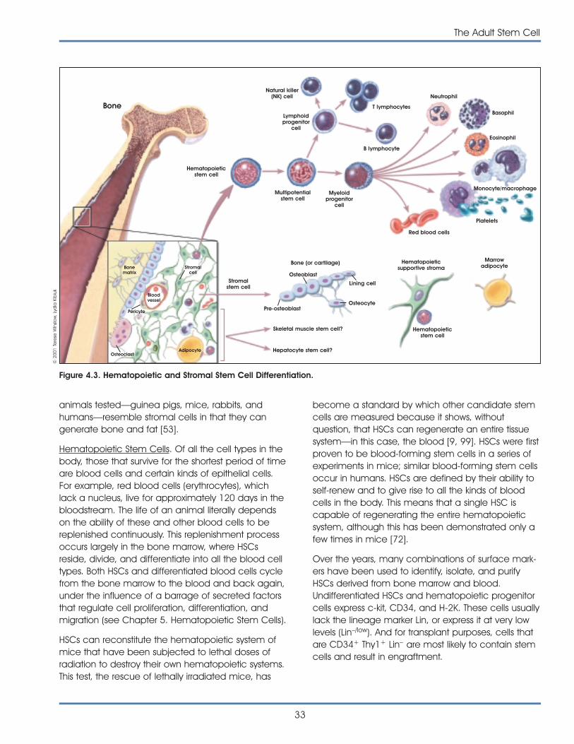
animals tested—guinea pigs, mice, rabbits, andhumans—resemble stromal cells in that they cangenerate bone and fat [53].
Hematopoietic Stem Cells. Of all the cell types in thebody, those that survive for the shortest period of timeare blood cells and certain kinds of epithelial cells.For example, red blood cells (erythrocytes), whichlack a nucleus, live for approximately 120 days in thebloodstream. The life of an animal literally dependson the ability of these and other blood cells to bereplenished continuously. This replenishment processoccurs largely in the bone marrow, where HSCsreside, divide, and differentiate into all the blood celltypes. Both HSCs and differentiated blood cells cyclefrom the bone marrow to the blood and back again,under the influence of a barrage of secreted factorsthat regulate cell proliferation, differentiation, andmigration (see Chapter 5. Hematopoietic Stem Cells).
HSCs can reconstitute the hematopoietic system ofmice that have been subjected to lethal doses ofradiation to destroy their own hematopoietic systems.This test, the rescue of lethally irradiated mice, has
become a standard by which other candidate stemcells are measured because it shows, withoutquestion, that HSCs can regenerate an entire tissuesystem—in this case, the blood [9, 99]. HSCs were firstproven to be blood-forming stem cells in a series ofexperiments in mice; similar blood-forming stem cellsoccur in humans. HSCs are defined by their ability toself-renew and to give rise to all the kinds of bloodcells in the body. This means that a single HSC iscapable of regenerating the entire hematopoieticsystem, although this has been demonstrated only afew times in mice [72].
Over the years, many combinations of surface mark-ers have been used to identify, isolate, and purifyHSCs derived from bone marrow and blood.Undifferentiated HSCs and hematopoietic progenitorcells express c-kit, CD34, and H-2K. These cells usuallylack the lineage marker Lin, or express it at very lowlevels (Lin–/low). And for transplant purposes, cells thatare CD34+ Thy1+ Lin– are most likely to contain stemcells and result in engraftment.
The Adult Stem Cell
33
Bone
Hematopoieticstem cell
Stromalstem cell
Bone (or cartilage) Hematopoieticsupportive stroma
Marrowadipocyte
Hematopoieticstem cell
Osteoblast
Pre-osteoblastOsteocyte
Skeletal muscle stem cell?
Hepatocyte stem cell?AdipocyteOsteoclast
Pericyte
Bloodvessel
Bonematrix
Stromalcell
Lining cell
Multipotentialstem cell
Myeloidprogenitor
cell
Lymphoidprogenitor
cell
Natural killer(NK) cell
T lymphocytes
B lymphocyte
Red blood cells
Neutrophil
Basophil
Eosinophil
Platelets
Monocyte/macrophage
Figure 4.3. Hematopoietic and Stromal Stem Cell Differentiation.
© 2
001
Tere
se W
inslo
w, L
ydia
Kib
iuk
Two kinds of HSCs have been defined. Long-termHSCs proliferate for the lifetime of an animal. Inyoung adult mice, an estimated 8 to 10 % of long-term HSCs enter the cell cycle and divide eachday. Short-term HSCs proliferate for a limited time,possibly a few months. Long-term HSCs have highlevels of telomerase activity. Telomerase is an enzymethat helps maintain the length of the ends of chromo-somes, called telomeres, by adding on nucleotides.Active telomerase is a characteristic of undifferentiat-ed, dividing cells and cancer cells. Differentiated,human somatic cells do not show telomerase activity.In adult humans, HSCs occur in the bone marrow,blood, liver, and spleen, but are extremely rare in anyof these tissues. In mice, only 1 in 10,000 to 15,000bone marrow cells is a long-term HSC [105].
Short-term HSCs differentiate into lymphoid andmyeloid precursors, the two classes of precursors forthe two major lineages of blood cells. Lymphoid pre-cursors differentiate into T cells, B cells, and naturalkiller cells. The mechanisms and pathways that leadto their differentiation are still being investigated [1, 2].Myeloid precursors differentiate into monocytes andmacrophages, neutrophils, eosinophils, basophils,megakaryocytes, and erythrocytes [3]. In vivo, bonemarrow HSCs differentiate into mature, specializedblood cells that cycle constantly from the bonemarrow to the blood, and back to the bone marrow[26]. A recent study showed that short-term HSCs area heterogeneous population that differ significantly interms of their ability to self-renew and repopulate thehematopoietic system [42].
Attempts to induce HSC to proliferate in vitro—onmany substrates, including those intended to mimicconditions in the stroma—have frustrated scientists formany years. Although HSCs proliferate readily in vivo,they usually differentiate or die in vitro [26]. Thus,much of the research on HSCs has been focused onunderstanding the factors, cell-cell interactions, andcell-matrix interactions that control their proliferationand differentiation in vivo, with the hope that similarconditions could be replicated in vitro. Many of thesoluble factors that regulate HSC differentiation in vivoare cytokines, which are made by different cell typesand are then concentrated in the bone marrow bythe extracellular matrix of stromal cells—the sites ofblood formation [45, 107]. Two of the most-studiedcytokines are granulocyte-macrophage colony-stimu-lating factor (GM-CSF) and interleukin-3 (IL-3) [40, 81].
Also important to HSC proliferation and differentiationare interactions of the cells with adhesion moleculesin the extracellular matrix of the bone marrow stroma[83, 101, 110].
Bone Marrow Stromal Cells. Bone marrow (BM) stromalcells have long been recognized for playing animportant role in the differentiation of mature bloodcells from HSCs (see Figure 4.3. Hematopoietic andStromal Stem Cell Differentiation). But stromal cellsalso have other important functions [30, 31]. In addi-tion to providing the physical environment in whichHSCs differentiate, BM stromal cells generate carti-lage, bone, and fat. Whether stromal cells are bestclassified as stem cells or progenitor cells for these tis-sues is still in question. There is also a question as towhether BM stromal cells and so-called mesenchy-mal stem cells are the same population [78].
BM stromal cells have many features that distinguishthem from HSCs. The two cell types are easy to sepa-rate in vitro. When bone marrow is dissociated, andthe mixture of cells it contains is plated at low density,the stromal cells adhere to the surface of the culturedish, and the HSCs do not. Given specific in vitro con-ditions, BM stromal cells form colonies from a singlecell called the colony forming unit-F (CFU-F). Thesecolonies may then differentiate as adipocytes ormyelosupportive stroma, a clonal assay that indicatesthe stem cell-like nature of stromal cells. Unlike HSCs,which do not divide in vitro (or proliferate only to alimited extent), BM stromal cells can proliferate for upto 35 population doublings in vitro [16]. They growrapidly under the influence of such mitogens asplatelet-derived growth factor (PDGF), epidermalgrowth factor (EGF), basic fibroblast growth factor(bFGF), and insulin-like growth factor-1 (IGF-1) [12].
To date, it has not been possible to isolate a popula-tion of pure stromal cells from bone marrow. Panels ofmarkers used to identify the cells include receptors forcertain cytokines (interleukin-1, 3, 4, 6, and 7) recep-tors for proteins in the extracellular matrix, (ICAM-1and 2, VCAM-1, the alpha-1, 2, and 3 integrins, andthe beta-1, 2, 3 and 4 integrins), etc. [64]. Despite theuse of these markers and another stromal cell markercalled Stro-1, the origin and specific identity of stro-mal cells have remained elusive. Like HSCs, BMstromal cells arise from embryonic mesoderm duringdevelopment, although no specific precursor or stemcell for stromal cells has been isolated and identified.
34
The Adult Stem Cell
One theory about their origin is that a common kindof progenitor cell—perhaps a primordial endothelialcell that lines embryonic blood vessels—gives rise toboth HSCs and to mesodermal precursors. The lattermay then differentiate into myogenic precursors (thesatellite cells that are thought to function as stemcells in skeletal muscle), and the BM stromal cells[10].
In vivo, the differentiation of stromal cells into fat andbone is not straightforward. Bone marrow adipocytesand myelosupportive stromal cells—both of whichare derived from BM stromal cells—may be regardedas interchangeable phenotypes [10, 11]. Adipocytesdo not develop until postnatal life, as the bonesenlarge and the marrow space increases to accom-modate enhanced hematopoiesis. When the skele-ton stops growing, and the mass of HSCs decreasesin a normal, age-dependent fashion, BM stromal cellsdifferentiate into adipocytes, which fill the extraspace. New bone formation is obviously greaterduring skeletal growth, although bone “turns over”throughout life. Bone forming cells are osteoblasts,but their relationship to BM stromal cells is not clear.New trabecular bone, which is the inner region ofbone next to the marrow, could logically developfrom the action of BM stromal cells. But the outsidesurface of bone also turns over, as does bone next tothe Haversian system (small canals that form concen-tric rings within bone). And neither of these surfaces isin contact with BM stromal cells [10, 11].
Adult Stem Cells in Other Tissues
It is often difficult—if not impossible—to distinguishadult, tissue-specific stem cells from progenitor cells.With that caveat in mind, the following summaryidentifies reports of stem cells in various adult tissues.
Endothelial Progenitor Cells. Endothelial cells line theinner surfaces of blood vessels throughout the body,and it has been difficult to identify specific endothe-lial stem cells in either the embryonic or the adultmammal. During embryonic development, just aftergastrulation, a kind of cell called the hemangioblast,which is derived from mesoderm, is presumed to bethe precursor of both the hematopoietic andendothelial cell lineages. The embryonic vasculatureformed at this stage is transient and consists of bloodislands in the yolk sac. But hemangioblasts, per se,have not been isolated from the embryo and theirexistence remains in question. The process of forming
new blood vessels in the embryo is called vasculoge-nesis. In the adult, the process of forming blood ves-sels from pre-existing blood vessels is called angio-genesis [50].
Evidence that hemangioblasts do exist comes fromstudies of mouse embryonic stem cells that aredirected to differentiate in vitro. These studies haveshown that a precursor cell derived from mouse EScells that express Flk-1 [the receptor for vascularendothelial growth factor (VEGF) in mice] can giverise to both blood cells and blood vessel cells [88,109]. Both VEGF and fibroblast growth factor-2 (FGF-2)play critical roles in endothelial cell differentiation in vivo [79].
Several recent reports indicate that the bone marrowcontains cells that can give rise to new blood vesselsin tissues that are ischemic (damaged due to thedeprivation of blood and oxygen) [8, 29, 49, 94]. Butit is unclear from these studies what cell type(s) in thebone marrow induced angiogenesis. In a study whichsought to address that question, researchers foundthat adult human bone marrow contains cells thatresemble embryonic hemangioblasts, and maytherefore be called endothelial stem cells.
In more recent experiments, human bone marrow-derived cells were injected into the tail veins of ratswith induced cardiac ischemia. The human cellsmigrated to the rat heart where they generated newblood vessels in the infarcted muscle (a process akinto vasculogenesis), and also induced angiogenesis.The candidate endothelial stem cells are CD34+ (amarker for HSCs), and they express the transcriptionfactor GATA-2 [51]. A similar study using transgenicmice that express the gene for enhanced green fluo-rescent protein (which allows the cells to be tracked),showed that bone-marrow-derived cells could repopu-late an area of infarcted heart muscle in mice, andgenerate not only blood vessels, but also cardiomy-ocytes that integrated into the host tissue [71] (seeChapter 9. Can Stem Cells Repair a Damaged Heart?).
And, in a series of experiments in adult mammals,progenitor endothelial cells were isolated fromperipheral blood (of mice and humans) by using anti-bodies against CD34 and Flk-1, the receptor for VEGF.The cells were mononuclear blood cells (meaningthey have a nucleus) and are referred to as MBCD34+
cells and MBFlk1+ cells. When plated in tissue-culture
The Adult Stem Cell
35
dishes, the cells attached to the substrate, becamespindle-shaped, and formed tube-like structures thatresemble blood vessels. When transplanted into miceof the same species (autologous transplants) withinduced ischemia in one limb, the MBCD34+ cellspromoted the formation of new blood vessels [8].Although the adult MBCD34+ and MBFlk1+ cells functionin some ways like stem cells, they are usuallyregarded as progenitor cells.
Skeletal Muscle Stem Cells. Skeletal muscle, like thecardiac muscle of the heart and the smooth musclein the walls of blood vessels, the digestive system,and the respiratory system, is derived from embryonicmesoderm. To date, at least three populations ofskeletal muscle stem cells have been identified:satellite cells, cells in the wall of the dorsal aorta, andso-called “side population” cells.
Satellite cells in skeletal muscle were identified 40years ago in frogs by electron microscopy [62], andthereafter in mammals [84]. Satellite cells occur onthe surface of the basal lamina of a mature musclecell, or myofiber. In adult mammals, satellite cellsmediate muscle growth [85]. Although satellite cellsare normally non-dividing, they can be triggered toproliferate as a result of injury, or weight-bearingexercise. Under either of these circumstances, musclesatellite cells give rise to myogenic precursor cells,which then differentiate into the myofibrils that typifyskeletal muscle. A group of transcription factorscalled myogenic regulatory factors (MRFs) playimportant roles in these differentiation events. The so-called primary MRFs, MyoD and Myf5, help regulatemyoblast formation during embryogenesis. The sec-ondary MRFs, myogenin and MRF4, regulate the ter-minal differentiation of myofibrils [86].
With regard to satellite cells, scientists have beenaddressing two questions. Are skeletal muscle satellitecells true adult stem cells or are they instead precur-sor cells? Are satellite cells the only cell type that canregenerate skeletal muscle. For example, a recentreport indicates that muscle stem cells may alsooccur in the dorsal aorta of mouse embryos, andconstitute a cell type that gives rise both to musclesatellite cells and endothelial cells. Whether the dorsalaorta cells meet the criteria of a self-renewing musclestem cell is a matter of debate [21].
Another report indicates that a different kind of stemcell, called an SP cell, can also regenerate skeletal
muscle may be present in muscle and bone marrow.SP stands for a side population of cells that can beseparated by fluorescence-activated cell sortinganalysis. Intravenously injecting these muscle-derivedstem cells restored the expression of dystrophin inmdx mice. Dystrophin is the protein that is defectivein people with Duchenne’s muscular dystrophy; mdxmice provide a model for the human disease.Dystrophin expression in the SP cell-treated mice waslower than would be needed for clinical benefit.Injection of bone marrow- or muscle-derived SP cellsinto the dystrophic muscle of the mice yieldedequivocal results that the transplanted cells had inte-grated into the host tissue. The authors conclude thata similar population of SP stem cells can be derivedfrom either adult mouse bone marrow or skeletalmuscle, and suggest “there may be some directrelationship between bone marrow-derived stem cellsand other tissue- or organ-specific cells” [43]. Thus,stem cell or progenitor cell types from variousmesodermally-derived tissues may be able to gen-erate skeletal muscle.
Epithelial Cell Precursors in the Skin and DigestiveSystem. Epithelial cells, which constitute 60 percent ofthe differentiated cells in the body are responsible forcovering the internal and external surfaces of thebody, including the lining of vessels and other cavi-ties. The epithelial cells in skin and the digestive tractare replaced constantly. Other epithelial cell popula-tions—in the ducts of the liver or pancreas, forexample—turn over more slowly. The cell populationthat renews the epithelium of the small intestineoccurs in the intestinal crypts, deep invaginations inthe lining of the gut. The crypt cells are oftenregarded as stem cells; one of them can give rise to an organized cluster of cells called a structural-proliferative unit [93].
The skin of mammals contains at least three popula-tions of epithelial cells: epidermal cells, hair folliclecells, and glandular epithelial cells, such as those thatmake up the sweat glands. The replacement patternsfor epithelial cells in these three compartments differ,and in all the compartments, a stem cell populationhas been postulated. For example, stem cells in thebulge region of the hair follicle appear to give rise tomultiple cell types. Their progeny can migrate downto the base of the follicle where they become matrixcells, which may then give rise to different cell typesin the hair follicle, of which there are seven [39]. The
36
The Adult Stem Cell
bulge stem cells of the follicle may also give rise tothe epidermis of the skin [95].
Another population of stem cells in skin occurs in thebasal layer of the epidermis. These stem cells prolifer-ate in the basal region, and then differentiate as theymove toward the outer surface of the skin. The ker-atinocytes in the outermost layer lack nuclei and actas a protective barrier. A dividing skin stem cell candivide asymmetrically to produce two kinds of daugh-ter cells. One is another self-renewing stem cell. Thesecond kind of daughter cell is an intermediate pre-cursor cell which is then committed to replicate a fewtimes before differentiating into keratinocytes. Self-renewing stem cells can be distinguished from thisintermediate precusor cell by their higher level of �1integrin expression, which signals keratinocytes to pro-liferate via a mitogen-activated protein (MAP) kinase[112]. Other signaling pathways include that triggeredby �-catenin, which helps maintain the stem-cellstate [111], and the pathway regulated by the onco-protein c-Myc, which triggers stem cells to give rise totransit amplifying cells [36].
Stem Cells in the Pancreas and Liver. The status ofstem cells in the adult pancreas and liver is unclear.During embryonic development, both tissues arisefrom endoderm. A recent study indicates that asingle precursor cell derived from embryonic endo-derm may generate both the ventral pancreas andthe liver [23]. In adult mammals, however, both thepancreas and the liver contain multiple kinds ofdifferentiated cells that may be repopulated orregenerated by multiple types of stem cells. In thepancreas, endocrine (hormone-producing) cellsoccur in the islets of Langerhans. They include thebeta cells (which produce insulin), the alpha cells(which secrete glucagon), and cells that release thepeptide hormones somatostatin and pancreaticpolypeptide. Stem cells in the adult pancreas arepostulated to occur in the pancreatic ducts or in theislets themselves. Several recent reports indicate thatstem cells that express nestin—which is usuallyregarded as a marker of neural stem cells—cangenerate all of the cell types in the islets [60, 113](see Chapter 7. Stem Cells and Diabetes).
The identity of stem cells that can repopulate the liverof adult mammals is also in question. Recent studiesin rodents indicate that HSCs (derived frommesoderm) may be able to home to liver after it isdamaged, and demonstrate plasticity in becoming
into hepatocytes (usually derived from endoderm)[54, 77, 97]. But the question remains as to whethercells from the bone marrow normally generate hepa-tocytes in vivo. It is not known whether this kind ofplasticity occurs without severe damage to the liver orwhether HSCs from the bone marrow generate ovalcells of the liver [18]. Although hepatic oval cells existin the liver, it is not clear whether they actually gener-ate new hepatocytes [87, 98]. Oval cells may arisefrom the portal tracts in liver and may give rise toeither hepatocytes [19, 55] and to the epithelium ofthe bile ducts [37, 92]. Indeed, hepatocytes them-selves, may be responsible for the well-know regen-erative capacity of liver.
SUMMARYWhat Do We Know About Adult Stem Cells?
• Adult stem cells can proliferate without differenti-ating for a long period (a characteristic referredto as long-term self-renewal), and they can giverise to mature cell types that have characteristicshapes and specialized functions.
• Some adult stem cells have the capability todifferentiate into tissues other than the ones fromwhich they originated; this is referred to asplasticity.
• Adult stem cells are rare. Often they are difficultto identify and their origins are not known.Current methods for characterizing adult stemcells are dependent on determining cell surfacemarkers and observations about their differentia-tion patterns in test tubes and culture dishes.
• To date, published scientific literature indicatesthat adult stem cells have been derived frombrain, bone marrow, peripheral blood, dentalpulp, spinal cord, blood vessels, skeletal muscle,epithelia of the skin and digestive system,cornea, retina, liver, and pancreas; thus, adultstem cells have been found in tissues thatdevelop from all three embryonic germ layers.
• Hematopoietic stem cells from bone marrow arethe most studied and used for clinical applica-tions in restoring various blood and immunecomponents to the bone marrow via transplan-tation. There are at least two other populations ofadult stem cells that have been identified frombone marrow and blood.
The Adult Stem Cell
37
• Several populations of adult stem cells havebeen identified in the brain, particularly thehippocampus. Their function is unknown.Proliferation and differentiation of brain stem cellsare influenced by various growth factors.
• There are now several reports of adult stem cellsin other tissues (muscle, blood, and fat) thatdemonstrate plasticity. Very few publishedresearch reports on plasticity of adult stem cellshave, however, included clonality studies. That is,there is limited evidence that a single adult stemcell or genetically identical line of adult stemcells demonstrates plasticity.
• Rarely have experiments that claim plasticitydemonstrated that the adult stem cells havegenerated mature, fully functional cells or thatthe cells have restored lost function in vivo.
What Do We Need to Know About Adult Stem Cells?
• What are the sources of adult stem cells in thebody? Are they “leftover” embryonic stem cells,or do they arise in some other way? And if thelatter is true—which seems to be the case—exactly how do adult stem cells arise, and why do they remain in an undifferentiated state, when all the cells around them havedifferentiated?
• Is it possible to manipulate adult stem cells toincrease their ability to proliferate in vitro, so thatadult stem cells can be used as a sufficientsource of tissue for transplants?
• How many kinds of adult stem cells exist, and inwhich tissues do they exist? Evidence is accu-mulating that, although they occur in smallnumbers, adult stem cells are present in manydifferentiated tissues.
• What is the best evidence that adult stem cellsshow plasticity and generate cell types of othertissues?
• Is it possible to manipulate adult stem cells toincrease their ability to proliferate in vitro so thatadult stem cells can be used as a sufficientsource of tissue for transplants?
• Is there a universal stem cell? An emergingconcept is that, in adult mammals, there maybe a population of “universal” stem cells.Although largely theoretical, the concept hassome experimental basis. A candidate, universal
adult stem cell may be one that circulates in the blood stream, can escape from the blood,and populate various adult tissues. In more thanone experimental system, researchers havenoted that dividing cells in adult tissues oftenappear near a blood vessel, such as candidatestem cells in the hippocampus, a region of thebrain [75].
• Do adult stem cells exhibit plasticity as a normalevent in vivo? If so, is this true of all adult stemcells? What are the signals that regulate theproliferation and differentiation of stem cells thatdemonstrate plasticity?
REFERENCES1. Akashi, K., Traver, D., Kondo, M., and Weissman, I.L. (1999).
Lymphoid development from hematopoietic stem cells. Int.J. Hematol. 69, 217-226.
2. Akashi, K., Kondo, M., Cheshier, S., Shizuru, J., Gandy, K.,Domen, J., Mebius, R., Traver, D., and Weissman, I.L. (1999).Lymphoid development from stem cells and the commonlymphocyte progenitors. Cold Spring Harb. Symp. Quant.Biol. 64, 1-12.
3. Akashi, K., Traver, D., Miyamoto, T., and Weissman, I.L.(2000). A clonogenic common myeloid progenitor thatgives rise to all myeloid lineages. Nature. 404, 193-197.
4. Alison, M.R., Poulsom, R., Jeffery, R., Dhillon, A.P., Quaglia,A., Jacob, J., Novelli, M., Prentice, G., Williamson, J., andWright, N.A. (2000). Hepatocytes from non-hepatic adultstem cells. Nature. 406, 257.
5. Altman, J. and Das, G.D. (1965). Autoradiographic and his-tological evidence of postnatal hippocampal neurogene-sis in rats. J. Comp. Neurol. 124, 319-335.
6. Altman, J. (1969). Autoradiographic and histological studiesof postnatal neurogenesis. IV. Cell proliferation and migra-tion in the anterior forebrain, with special reference to per-sisting neurogenesis in the olfactory bulb. J. Comp. Neurol.137, 433-457.
7. Anderson, D.J., Gage, F.H., and Weissman, I.L. (2001). Canstem cells cross lineage boundaries? Nat. Med. 7, 393-395.
8. Asahara, T., Murohara, T., Sullivan, A., Silver, M., van der ZeeR., Li, T., Witzenbichler, B., Schatteman, G., and Isner, J.M.(1997). Isolation of putative progenitor endothelial cells forangiogenesis. Science. 275, 964-967.
9. Becker, A.J., McCullough, E.A., and Till, J.E. (1963).Cytological demonstration of the clonal nature of spleencolonies derived from transplanted mouse marrow cells.Nature. 197, 452-454.
10. Bianco, P. and Cossu, G. (1999). Uno, nessuno e centomila:searching for the identity of mesodermal progenitors. Exp.Cell Res. 251, 257-263.
38
The Adult Stem Cell
11. Bianco, P., Riminucci, M., Kuznetsov, S., and Robey, P.G.(1999). Multipotential cells in the bone marrow stroma:regulation in the context of organ physiology. Crit. Rev.Eukaryotic. Gene Expr. 9, 159-173.
12. Bianco, P., Riminucci, M., Gronthos, S., and Robey, P.G.(2001). Bone marrow stromal stem cells: nature, biology,and potential applications. Stem Cells. 19, 180-192.
13. Bjornson, C.R., Rietze, R.L., Reynolds, B.A., Magli, M.C., andVescovi, A.L. (1999). Turning brain into blood: a hemato-poietic fate adopted by adult neural stem cells in vivo.Science. 283, 534-537.
14. Blau, H., personal communication.
15. Brazelton, T.R., Rossi, F.M., Keshet, G.I., and Blau, H.M.(2000). From marrow to brain: expression of neuronalphenotypes in adult mice. Science. 290, 1775-1779.
16. Bruder, S.P., Jaiswal, N., and Haynesworth, S.E. (1997).Growth kinetics, self-renewal, and the osteogenic potentialof purified human mesenchymal stem cells during exten-sive subcultivation and following cryopreservation. J. Cell.Biochem. 64, 278-294.
17. Clarke, D.L., Johansson, C.B., Wilbertz, J., Veress, B., Nilsson,E., Karlström, H., Lendahl, U., and Frisen, J. (2000).Generalized potential of adult neural stem cells. Science.288, 1660-1663.
18. Crosby, H.A. and Strain, A.J. (2001). Adult liver stem cells:bone marrow, blood, or liver derived? Gut. 48, 153-154.
19. Dabeva, M.D. and Shafritz, D.A. (1993). Activation, prolifera-tion, and differentiation of progenitor cells into hepatocytesin the D-galactosamine model of liver regeneration. Am. J.Pathol. 143, 1606-1620.
20. Davis, A.A. and Temple, S. (1994). A self-renewing multipo-tential stem cell in embryonic rat cerebral cortex. Nature.372, 263-266.
21. De Angelis, L., Berghella, L., Coletta, M., Lattanzi, L., Zanchi,M., Cusella-De Angelis, M.G., Ponzetto, C., and Cossu, G.(1999). Skeletal myogenic progenitors originating fromembryonic dorsal aorta coexpress endothelial and myo-genic markers and contribute to postnatal muscle growthand regeneration. J. Cell Biol. 147, 869-877.
22. Del Bigio, M.R. (1995). The ependyma: a protective barrierbetween brain and cerebrospinal fluid. Glia. 14, 1-13.
23. Deutsch, G., Jung, J., Zheng, M., Lora, J., and Zaret, K.S.(2001). A bipotential precursor population for pancreas andliver within the embryonic endoderm. Development. 128,871-881.
24. Doetsch, F., Garcia-Verdugo, J.M., and Alvarez-Buylla, A.(1997). Cellular composition and three-dimensional organi-zation of the subventricular germinal zone in the adultmammalian brain. J. Neurosci. 17, 5046-5061.
25. Doetsch, F., Caille, I., Lim, D.A., Garcia-Verdugo, J.M., andAlvarez-Buylla, A. (1999). Subventricular zone astrocytes areneural stem cells in the adult mammalian brain. Cell. 97,703-716.
26. Domen, J. and Weissman, I.L. (1999). Self-renewal, differen-tiation or death: regulation and manipulation ofhematopoietic stem cell fate. Mol. Med. Today. 5, 201-208.
27. Eriksson, P.S., Perfilieva, E., Bjork-Eriksson, T., Alborn, A.M.,Nordborg, C., Peterson, D.A., and Gage, F.H. (1998).Neurogenesis in the adult human hippocampus. Nat. Med.4, 1313-1317.
28. Ferrari, G., Cusella-De Angelis, G., Coletta, M., Paolucci, E.,Stornaiuolo, A., Cossu, G., and Mavilio, F. (1998). Muscleregeneration by bone marrow-derived myogenic progeni-tors. Science. 279, 1528-1530.
29. Folkman, J. (1998). Therapeutic angiogenesis in ischemiclimbs. Circulation. 97, 1108-1110.
30. Friedenstein, A.J., Piatetzky-Shapiro, I.I., and Petrakova, K.V.(1966). Osteogenesis in transplants of bone marrow cells. J. Embryol. Exp. Morphol. 16, 381-390.
31. Friedenstein, A.J., Petrakova, K.V., Kurolesova, A.I., andFrolova, G.P. (1968). Heterotopic of bone marrow. Analysisof precursor cells for osteogenic and hematopoietictissues. Transplantation. 6, 230-247.
32. Friedenstein, A.J., Chailakhjan, R.K., and Lalykina, K.S.(1970). The development of fibroblast colonies in mono-layer cultures of guinea-pig bone marrow and spleen cells.Cell Tissue Kinet. 3, 393-403.
33. Gage, F., personal communication.
34. Gage, F.H., Ray, J., and Fisher, L.J. (1995). Isolation, charac-terization, and use of stem cells from the CNS. Annu. Rev.Neurosci. 18, 159-192.
35. Gage, F.H., Coates, P.W., Palmer, T.D., Kuhn, H.G., Fisher, L.J., Suhonen, J.O., Peterson, D.A., Suhr, S.T., and Ray, J.(1995). Survival and differentiation of adult neuronalprogenitor cells transplanted to the adult brain. Proc. Natl.Acad. Sci. U. S. A. 92, 11879-11883.
36. Gandarillas, A. and Watt, F.M. (1997). c-Myc promotesdifferentiation of human epidermal stem cells. Genes Dev.11, 2869-2882.
37. Germain, L., Noel, M., Gourdeau, H., and Marceau, N.(1988). Promotion of growth and differentiation of rat ductu-lar oval cells in primary culture. Cancer Res. 48, 368-378.
38. Geschwind, D.H., Ou, J., Easterday, M.C., Dougherty, J.D.,Jackson, R.L., Chen, Z., Antoine, H., Terskikh, A., Weissman,I.L., Nelson, S.F., and Kornblum, H.I. (2001). A genetic analy-sis of neural progenitor differentiation. Neuron. 29, 325-339.
39. Ghazizadeh, S. and Taichman, L.B. (2001). Multiple classesof stem cells in cutaneous epithelium: a lineage analysis ofadult mouse skin. EMBO J. 20, 1215-1222.
40. Gordon, M.Y., Riley, G.P., Watt, S.M., and Greaves, M.F.(1987). Compartmentalization of a haematopoietic growthfactor (GM-CSF) by glycosaminoglycans in the bonemarrow microenvironment. Nature. 326, 403-405.
The Adult Stem Cell
39
41. Gritti, A., Parati, E.A., Cova, L., Frolichsthal, P., Galli, R.,Wanke, E., Faravelli, L., Morassutti, D.J., Roisen, F., Nickel,D.D., and Vescovi, A.L. (1996). Multipotential stem cells fromthe adult mouse brain proliferate and self-renew inresponse to basic fibroblast growth factor. J. Neurosci. 16,1091-1100.
42. Guenechea, G., Gan, O.I., Dorrell, C., and Dick, J.E. (2001).Distinct classes of human stem cells that differ in prolifera-tive and self-renewal potential. Nat. Immunol. 2, 75-82.
43. Gussoni, E., Soneoka, Y., Strickland, C.D., Buzney, E.A., Khan,M.K., Flint, A.F., Kunkel, L.M., and Mulligan, R.C. (1999).Dystrophin expression in the mdx mouse restored by stemcell transplantation. Nature. 401, 390-394.
44. Holtzer. H. (1978). Cell lineages, stem cells and the‘quantal’ cell cycle concept. In: Stem cells and tissuehomeostasis. Eds: B.I. Lord, C.S. Potten, and R.J. Cole.(Cambridge, New York: Cambridge University Press). 1-28.
45. Hunt, P., Robertson, D., Weiss, D., Rennick, D., Lee, F., andWitte, O.N. (1987). A single bone marrow-derived stromalcell type supports the in vitro growth of early lymphoid andmyeloid cells. Cell. 48, 997-1007.
46. Jackson, K., Majka SM, Wang H, Pocius J, Hartley CJ,Majesky MW, Entman ML, Michael LH, Hirschi KK, andGoodell MA (2001). Regeneration of ischemic cardiacmuscle and vascular endothelium by adult stem cells. J. Clin. Invest. 107, 1-8.
47. Johansson, C.B., Momma, S., Clarke, D.L., Risling, M.,Lendahl, U., and Frisen, J. (1999). Identification of a neuralstem cell in the adult mammalian central nervous system.Cell. 96, 25-34.
48. Johe, K.K., Hazel, T.G., Muller, T., Dugich-Djordjevic, M.M.,and McKay, R.D. (1996). Single factors direct the differentia-tion of stem cells from the fetal and adult central nervoussystem. Genes Dev. 10, 3129-3140.
49. Kalka, C., Masuda, H., Takahashi, T., Kalka-Moll, W.M., Silver,M., Kearney, M., Li, T., Isner, J.M., and Asahara, T. (2000).Transplantation of ex vivo expanded endothelial progenitorcells for therapeutic neovascularization. Proc. Natl. Acad.Sci. U. S. A. 97, 3422-3427.
50. Keller, G. (2001). The hemangioblast. Marshak, D.R.,Gardner, D.K., and Gottlieb, D. eds. (Cold Spring Harbor,New York: Cold Spring Harbor Laboratory Press). 329-348.
51. Kocher, A.A., Schuster, M.D., Szabolcs, M.J., Takuma, S.,Burkhoff, D., Wang, J., Homma, S., Edwards, N.M., andItescu, S. (2001). Neovascularization of ischemic myo-cardium by human bone-marrow-derived angioblastsprevents cardiomyocyte apoptosis, reduces remodelingand improves cardiac function. Nat. Med. 7, 430-436.
52. Krause, D.S., Theise, N.D., Collector, M.I., Henegariu, O.,Hwang, S., Gardner, R., Neutzel, S., and Sharkis, S.J. (2001).Multi-organ, multi-lineage engraftment by a single bonemarrow-derived stem cell. Cell. 105, 369-377.
53. Kuznetsov, S.A., Mankani, M.H., Gronthos, S., Satomura, K.,Bianco, P., and Robey P.G. (2001). Circulating skeletal stemcells. J. Cell Biol. 153, 1133-1140.
54. Lagasse, E., Connors, H., Al Dhalimy, M., Reitsma, M.,Dohse, M., Osborne, L., Wang, X., Finegold, M., Weissman,I.L., and Grompe, M. (2000). Purified hematopoietic stemcells can differentiate into hepatocytes in vivo. Nat. Med. 6,1229-1234.
55. Lazaro, C.A., Rhim, J.A., Yamada, Y., and Fausto, N. (1998).Generation of hepatocytes from oval cell precursors in cul-ture. Cancer Res. 58, 5514-5522.
56. Le Douarin, N.M. (1980). The ontogeny of the neural crest inavian embryo chimaeras. Nature. 286, 663-669.
57. Le Douarin, N.M. and Kalcheim, C. (1999). The migration ofneural crest cells. In: The neural crest. (Cambridge, NewYork: Cambridge University Press). 23-59.
58. Leblond, C.P. (1964). Classification of cell populations onthe basis of their proliferative behavior. National CancerInstitute. 14, 119-150.
59. Lois, C. and Alvarez-Buylla, A. (1994). Long-distanceneuronal migration in the adult mammalian brain. Science. 264, 1145-1148.
60. Lumelsky, N., Blondel, O., Laeng, P., Velasco, I., Ravin, R.,and McKay, R. (2001). Differentiation of Embryonic StemCells to Insulin-Secreting Structures Similiar to PancreaticIslets. Science. 292, 1309-1599.
61. Luskin, M.B. (1993). Restricted proliferation and migration ofpostnatally generated neurons derived from the forebrainsubventricular zone. Neuron. 11, 173-189.
62. Mauro, A. (1961). Satellite cell of skeletal muscle fibers. J. Biophys. Biochem. Cytol. 9, 493-495.
63. McKay, R. (1997). Stem cells in the central nervous system.Science. 276, 66-71.
64. McKay, R., personal communication.
65. Mezey, E., Chandross, K.J., Harta, G., Maki, R.A., andMcKercher, S.R. (2000). Turning blood into brain: cells bear-ing neuronal antigens generated in vivo from bone marrow.Science. 290, 1779-1782.
66. Momma, S., Johansson, C.B., and Frisen, J. (2000). Get toknow your stem cells. Curr. Opin. Neurobiol. 10, 45-49.
67. Morrison, S.J., White, P.M., Zock, C., and Anderson, D.J.(1999). Prospective identification, isolation by flow cyto-metry, and in vivo self-renewal of multipotent mammalianneural crest stem cells. Cell. 96, 737-749.
68. Morrison, S.J. (2001). Neuronal differentiation: Proneuralgenes inhibit gliogenesis. Curr. Biol. 11, R349-R351.
69. Morshead, C.M., Reynolds, B.A., Craig, C.G., McBurney,M.W., Staines, W.A., Morassutti, D., Weiss, S., and van der,K.D. (1994). Neural stem cells in the adult mammalianforebrain: a relatively quiescent subpopulation ofsubependymal cells. Neuron. 13, 1071-1082.
70. Morshead, C.M. and van der Kooy, K.D. (2001). A new ‘spin’on neural stem cells? Curr. Opin. Neurobiol. 11, 59-65.
40
The Adult Stem Cell
71. Orlic, D., Kajstura, J., Chimenti, S., Jakoniuk, I., Anderson,S.M., Li, B., Pickel, J., McKay, R., Nadal-Ginard, B., Bodine,D.M., Leri, A., and Anversa, P. (2001). Bone marrow cellsregenerate infarcted myocardium. Nature. 410, 701-705.
72. Osawa, M., Hanada, K., Hamada, H., and Nakauchi, H.(1996). Long-term lymphohematopoietic reconstitution by a single CD34- low/negative hematopoietic stem cell.Science. 273, 242-245.
73. Owen, M. (1988). Marrow derived stromal stem cells. J. CellScience Supp. 10, 63-76.
74. Palmer, T.D., Takahashi, J., and Gage, F.H. (1997). The adultrat hippocampus contains primordial neural stem cells.Mol. Cell. Neurosci. 8, 389-404.
75. Palmer, T.D., Willhoite, A.R., and Gage, F.H. (2000). Vascularniche for adult hippocampal neurogenesis. J. Comp.Neurol. 425, 479-494.
76. Panicker, M. and Rao, M. (2001). Stem cells and neuro-genesis. Marshak, D.R., Gardner, D.K., and Gottlieb, D. eds.(Cold Spring Harbor, New York: Cold Spring HarborLaboratory Press). 399-438.
77. Petersen, B.E., Bowen, W.C., Patrene, K.D., Mars, W.M.,Sullivan, A.K., Murase, N., Boggs, S.S., Greenberger, J.S., andGoff, J.P. (1999). Bone marrow as a potential source ofhepatic oval cells. Science. 284, 1168-1170.
78. Pittenger, M.F. and Marshak, D.R. (2001). Mesenchymalstem cells of human adult bone marrow. Marshak, D.R.,Gardner, D.K., and Gottlieb, D. eds. (Cold Spring Harbor,New York: Cold Spring Harbor Laboratory Press). 349-374.
79. Poole, T.J., Finkelstein, E.B., and Cox, C.M. (2001). The roleof FGF and VEGF in angioblast induction and migrationduring vascular development. Dev. Dyn. 220, 1-17.
80. Reynolds, B.A. and Weiss, S. (1992). Generation of neuronsand astrocytes from isolated cells of the adult mammaliancentral nervous system. Science. 255, 1707-1710.
81. Roberts, R., Gallagher, J., Spooncer, E., Allen, T.D.,Bloomfield, F., and Dexter, T.M. (1988). Heparan sulphatebound growth factors: a mechanism for stromal cellmediated haemopoiesis. Nature. 332, 376-378.
82. Robey, P.G. (2000). Stem cells near the century mark. J. Clin. Invest. 105, 1489-1491.
83. Roy, V. and Verfaillie, C.M. (1999). Expression and functionof cell adhesion molecules on fetal liver, cord blood andbone marrow hematopoietic progenitors: implications foranatomical localization and developmental stage specificregulation of hematopoiesis. Exp. Hematol. 27, 302-312.
84. Schultz, E. (1976). Fine structure of satellite cells in growingskeletal muscle. Am. J. Anat. 147, 49-70.
85. Schultz, E. (1996). Satellite cell proliferative compartments ingrowing skeletal muscles. Dev. Biol. 175, 84-94.
86. Seale, P. and Rudnicki, M.A. (2000). A new look at the origin,function, and “stem-cell” status of muscle satellite cells.Dev. Biol. 218, 115-124.
87. Sell, S. (1990). Is there a liver stem cell? Cancer Res. 50,3811-3815.
88. Shalaby, F., Rossant, J., Yamaguchi, T.P., Gertsenstein, M.,Wu, X.F., Breitman, M.L., and Schuh, A.C. (1995). Failure of blood-island formation and vasculogenesis in Flk-1-deficient mice. Nature. 376, 62-66.
89. Shi, Q., Rafii, S., Wu, M.H., Wijelath, E.S., Yu, C., Ishida, A.,Fujita, Y., Kothari, S., Mohle, R., Sauvage, L.R., Moore, M.A.,Storb, R.F., and Hammond, W.P. (1998). Evidence for circu-lating bone marrow-derived endothelial cells. Blood. 92,362-367.
90. Shihabuddin, L.S., Palmer, T.D., and Gage, F.H. (1999). Thesearch for neural progenitor cells: prospects for the thera-py of neurodegenerative disease. Mol. Med. Today. 5,474-480.
91. Sieber-Blum, M. (2000). Factors controlling lineage speci-fication in the neural crest. Int. Rev. Cytol. 197, 1-33.
92. Sirica, A.E., Mathis, G.A., Sano, N., and Elmore, L.W. (1990).Isolation, culture, and transplantation of intrahepatic bil-iary epithelial cells and oval cells. Pathobiology. 58, 44-64.
93. Slack, J.M. (2000). Stem Cells in Epithelial Tissues. Science.287, 1431-1433.
94. Takahashi, T., Kalka, C., Masuda, H., Chen, D., Silver, M.,Kearney, M., Magner, M., Isner, J.M., and Asahara, T.(1999). Ischemia- and cytokine-induced mobilization ofbone marrow-derived endothelial progenitor cells forneovascularization. Nat. Med. 5, 434-438.
95. Taylor, G., Lehrer, M.S., Jensen, P.J., Sun, T.T., and Lavker,R.M. (2000). Involvement of follicular stem cells in formingnot only the follicle but also the epidermis. Cell. 102, 451-461.
96. Temple, S. and Alvarez-Buylla, A. (1999). Stem cells in theadult mammalian central nervous system. Curr. Opin.Neurobiol. 9, 135-141.
97. Theise, N.D., Nimmakayalu, M., Gardner, R., Illei, P.B.,Morgan, G., Teperman, L., Henegariu, O., and Krause, D.S.(2000). Liver from bone marrow in humans. Hepatology.32, 11-16.
98. Thorgeirsson, S.S. (1993). Hepatic stem cells. Am. J. Pathol.142, 1331-1333.
99. Till, J.E. and McCullough, E.A. (1961). A direct measure-ment of the radiation sensitivity of normal mouse bonemarrow cells. Radiat. Res. 14, 213-222.
100. Tropepe, V., Sibilia, M., Ciruna, B.G., Rossant, J., Wagner,E.F., and van der Kooy D. (1999). Distinct neural stem cellsproliferate in response to EGF and FGF in the developingmouse telencephalon. Dev. Biol. 208, 166-188.
101. Verfaillie, C.M. (1998). Adhesion receptors as regulators ofthe hematopoietic process. Blood. 92, 2609-2612.
The Adult Stem Cell
41
102. Vescovi, A.L., Reynolds, B.A., Fraser, D.D., and Weiss, S.(1993). bFGF regulates the proliferative fate of unipotent(neuronal) and bipotent (neuronal/astroglial) EGF-generated CNS progenitor cells. Neuron. 11, 951-966.
103. Vescovi, A.L., Gritti, A., Galli, R., and Parati, E.A. (1999).Isolation and intracerebral grafting of nontransformedmultipotential embryonic human CNS stem cells. J.Neurotrauma. 16, 689-693.
104. Weiss, S. and van der Kooy D. (1998). CNS stem cells:where’s the biology (a.k.a. beef)? J. Neurobiol. 36, 307-314.
105. Weissman, I.L. (2000). Stem cells: units of development,units of regeneration, and units in evolution. Cell. 100,157-168.
106. White, P.M., Morrison, S.J., Orimoto, K., Kubu, C.J., Verdi,J.M., and Anderson, D.J. (2001). Neural crest stem cellsundergo cell-intrinsic developmental changes in sensitivityto instructive differentiation signals. Neuron. 29, 57-71.
107. Whitlock, C.A., Tidmarsh, G.F., Muller-Sieburg, C., andWeissman, I.L. (1987). Bone marrow stromal cell lines withlymphopoietic activity express high levels of a pre-B neo-plasia-associated molecule. Cell. 48, 1009-1021.
108. Williams, B.P., Read, J., and Price, J. (1991). The generationof neurons and oligodendrocytes from a common pre-cursor cell. Neuron. 7, 685-693.
109. Yamashita, J., Itoh, H., Hirashima, M., Ogawa, M.,Nishikawa, S., Yurugi, T., Naito, M., Nakao, K., andNishikawa, S. (2000). Flk1-positive cells derived fromembryonic stem cells serve as vascular progenitors.Nature. 408, 92-96.
110. Zandstra, P.W., Lauffenburger, D.A., and Eaves, C.J. (2000).A ligand-receptor signaling threshold model of stem celldifferentiation control: a biologically conserved mecha-nism applicable to hematopoiesis. Blood. 96, 1215-1222.
111. Zhu, A.J. and Watt, F.M. (1999). beta-catenin signallingmodulates proliferative potential of human epidermal ker-atinocytes independently of intercellular adhesion.Development. 126, 2285-2298.
112. Zhu, A.J., Haase, I., and Watt, F.M. (1999). Signaling viabeta1 integrins and mitogen-activated protein kinasedetermines human epidermal stem cell fate in vitro. Proc.Natl. Acad. Sci. U. S. A. 96, 6728-6733.
113. Zulewski, H., Abraham, E.J., Gerlach, M.J., Daniel, P.B.,Moritz, W., Muller, B., Vallejo, M., Thomas, M.K., andHabener, J.F. (2001). Multipotential nestin-positive stemcells isolated from adult pancreatic islets differentiate exvivo into pancreatic endocrine, exocrine, and hepaticphenotypes. Diabetes. 50, 521-533.
42
The Adult Stem Cell
43
With more than 50 years of experience studyingblood-forming stem cells called hematopoietic stemcells, scientists have developed sufficient understand-ing to actually use them as a therapy. Currently, noother type of stem cell, adult, fetal or embryonic,has attained such status. Hematopoietic stem celltransplants are now routinely used to treat patientswith cancers and other disorders of the blood andimmune systems. Recently, researchers haveobserved in animal studies that hematopoietic stemcells appear to be able to form other kinds of cells,such as muscle, blood vessels, and bone. If this canbe applied to human cells, it may eventually bepossible to use hematopoietic stem cells to replacea wider array of cells and tissues than once thought.
Despite the vast experience with hematopoietic stemcells, scientists face major roadblocks in expandingtheir use beyond the replacement of blood andimmune cells. First, hematopoietic stem cells areunable to proliferate (replicate themselves) anddifferentiate (become specialized to other cell types) invitro (in the test tube or culture dish). Second, scientistsdo not yet have an accurate method to distinguishstem cells from other cells recovered from the bloodor bone marrow. Until scientists overcome thesetechnical barriers, they believe it is unlikely thathematopoietic stem cells will be applied as cellreplacement therapy in diseases such as diabetes,Parkinson’s Disease, spinal cord injury, and many others.
INTRODUCTIONBlood cells are responsible for constant maintenanceand immune protection of every cell type of thebody. This relentless and brutal work requires thatblood cells, along with skin cells, have the greatestpowers of self-renewal of any adult tissue.
The stem cells that form blood and immune cells areknown as hematopoietic stem cells (HSCs). They are
ultimately responsible for the constant renewal ofblood—the production of billions of new blood cellseach day. Physicians and basic researchers haveknown and capitalized on this fact for more than 50years in treating many diseases. The first evidenceand definition of blood-forming stem cells camefrom studies of people exposed to lethal doses ofradiation in 1945.
Basic research soon followed. After duplicatingradiation sickness in mice, scientists found they couldrescue the mice from death with bone marrowtransplants from healthy donor animals. In the early1960s, Till and McCulloch began analyzing the bonemarrow to find out which components were respon-sible for regenerating blood [56]. They defined whatremain the two hallmarks of an HSC: it can renewitself and it can produce cells that give rise to all thedifferent types of blood cells (see Chapter 4. TheAdult Stem Cell).
WHAT IS A HEMATOPOIETICSTEM CELL?A hematopoietic stem cell is a cell isolated from theblood or bone marrow that can renew itself, candifferentiate to a variety of specialized cells, canmobilize out of the bone marrow into circulatingblood, and can undergo programmed cell death,called apoptosis—a process by which cells that aredetrimental or unneeded self-destruct.
A major thrust of basic HSC research since the 1960shas been identifying and characterizing these stemcells. Because HSCs look and behave in culture likeordinary white blood cells, this has been a difficultchallenge and this makes them difficult to identify bymorphology (size and shape). Even today, scientistsmust rely on cell surface proteins, which serve, onlyroughly, as markers of white blood cells.
HEMATOPOIETICSTEM CELLSHEMATOPOIETICSTEM CELLS
5.5.
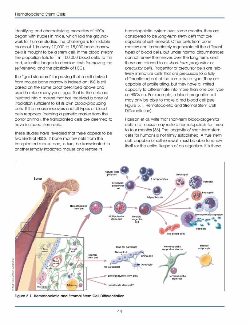
hematopoietic system over some months, they areconsidered to be long-term stem cells that arecapable of self-renewal. Other cells from bonemarrow can immediately regenerate all the differenttypes of blood cells, but under normal circumstancescannot renew themselves over the long term, andthese are referred to as short-term progenitor orprecursor cells. Progenitor or precursor cells are rela-tively immature cells that are precursors to a fullydifferentiated cell of the same tissue type. They arecapable of proliferating, but they have a limitedcapacity to differentiate into more than one cell typeas HSCs do. For example, a blood progenitor cellmay only be able to make a red blood cell (seeFigure 5.1. Hematopoietic and Stromal Stem CellDifferentiation).
Harrison et al. write that short-term blood-progenitorcells in a mouse may restore hematopoiesis for threeto four months [36]. The longevity of short-term stemcells for humans is not firmly established. A true stemcell, capable of self-renewal, must be able to renewitself for the entire lifespan of an organism. It is these
44
Hematopoietic Stem Cells
Identifying and characterizing properties of HSCsbegan with studies in mice, which laid the ground-work for human studies. The challenge is formidableas about 1 in every 10,000 to 15,000 bone marrowcells is thought to be a stem cell. In the blood streamthe proportion falls to 1 in 100,000 blood cells. To thisend, scientists began to develop tests for proving theself-renewal and the plasticity of HSCs.
The “gold standard” for proving that a cell derivedfrom mouse bone marrow is indeed an HSC is stillbased on the same proof described above andused in mice many years ago. That is, the cells areinjected into a mouse that has received a dose ofirradiation sufficient to kill its own blood-producingcells. If the mouse recovers and all types of bloodcells reappear (bearing a genetic marker from thedonor animal), the transplanted cells are deemed tohave included stem cells.
These studies have revealed that there appear to betwo kinds of HSCs. If bone marrow cells from thetransplanted mouse can, in turn, be transplanted toanother lethally irradiated mouse and restore its
Bone
Hematopoieticstem cell
Stromalstem cell
Bone (or cartilage) Hematopoieticsupportive stroma
Marrowadipocyte
Hematopoieticstem cell
Osteoblast
Pre-osteoblastOsteocyte
Skeletal muscle stem cell?
Hepatocyte stem cell?AdipocyteOsteoclast
Pericyte
Bloodvessel
Bonematrix
Stromalcell
Lining cell
Multipotentialstem cell
Myeloidprogenitor
cell
Lymphoidprogenitor
cell
Natural killer(NK) cell
T lymphocytes
B lymphocyte
Red blood cells
Neutrophil
Basophil
Eosinophil
Platelets
Monocyte/macrophage
Figure 5.1. Hematopoietic and Stromal Stem Cell Differentiation.
© 2
001
Tere
se W
inslo
w, L
ydia
Kib
iuk
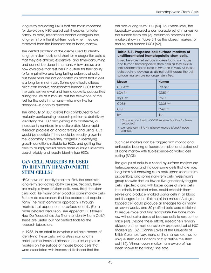
Hematopoietic Stem Cells
45
long-term replicating HSCs that are most importantfor developing HSC-based cell therapies. Unfortu-nately, to date, researchers cannot distinguish thelong-term from the short-term cells when they areremoved from the bloodstream or bone marrow.
The central problem of the assays used to identifylong-term stem cells and short-term progenitor cells isthat they are difficult, expensive, and time-consumingand cannot be done in humans. A few assays arenow available that test cells in culture for their abilityto form primitive and long-lasting colonies of cells,but these tests are not accepted as proof that a cellis a long-term stem cell. Some genetically alteredmice can receive transplanted human HSCs to testthe cells’ self-renewal and hematopoietic capabilitiesduring the life of a mouse, but the relevance of thistest for the cells in humans—who may live fordecades—is open to question.
The difficulty of HSC assays has contributed to twomutually confounding research problems: definitivelyidentifying the HSC and getting it to proliferate, orincrease its numbers, in a culture dish. More rapidresearch progress on characterizing and using HSCswould be possible if they could be readily grown inthe laboratory. Conversely, progress in identifyinggrowth conditions suitable for HSCs and getting thecells to multiply would move more quickly if scientistscould reliably and readily identify true HSCs.
CAN CELL MARKERS BE USEDTO IDENTIFY HEMATOPOIETICSTEM CELLS?HSCs have an identity problem. First, the ones withlong-term replicating ability are rare. Second, thereare multiple types of stem cells. And, third, the stemcells look like many other blood or bone marrow cells.So how do researchers find the desired cell popula-tions? The most common approach is throughmarkers that appear on the surface of cells. (For amore detailed discussion, see Appendix E.i. Markers:How Do Researchers Use Them to Identify Stem Cells?)These are useful, but not perfect tools for theresearch laboratory.
In 1988, in an effort to develop a reliable means ofidentifying these cells, Irving Weissman and hiscollaborators focused attention on a set of proteinmarkers on the surface of mouse blood cells thatwere associated with increased likelihood that the
cell was a long-term HSC [50]. Four years later, thelaboratory proposed a comparable set of markers forthe human stem cell [3]. Weissman proposes themarkers shown in Table 5.1 as the closest markers formouse and human HSCs [62].
Such cell markers can be tagged with monoclonalantibodies bearing a fluorescent label and culled outof bone marrow with fluorescence-activated cellsorting (FACS).
The groups of cells thus sorted by surface markers areheterogeneous and include some cells that are true,long-term self-renewing stem cells, some shorter-termprogenitors, and some non-stem cells. Weissman’sgroup showed that as few as five genetically taggedcells, injected along with larger doses of stem cellsinto lethally irradiated mice, could establish them-selves and produce marked donor cells in all bloodcell lineages for the lifetime of the mouse. A singletagged cell could produce all lineages for as manyas seven weeks, and 30 purified cells were sufficientto rescue mice and fully repopulate the bone mar-row without extra doses of backup cells to rescue themice [49]. Despite these efforts, researchers remaindivided on the most consistently expressed set of HSCmarkers [27, 32]. Connie Eaves of the University ofBritish Columbia says none of the markers are tied tounique stem cell functions or truly define the stemcell [14]. “Almost every marker I am aware of hasbeen shown to be fickle,” she says.
Table 5.1. Proposed cell-surface markers ofundifferentiated hematopoietic stem cells.Listed here are cell surface markers found on mouseand human hematopoietic stem cells as they exist intheir undifferentiated state in vivo and in vitro. As thesecells begin to develop as distinct cell lineages the cellsurface markers are no longer identified.
Mouse Human
CD34 low/- CD 34+
SCA-1+ CD59+*
Thy1+/low Thy1+
CD38+ CD38 low/-
C-kit+ C-kit -/low
lin-* lin -**
* Only one of a family of CD59 markers has thus far beenevaluated.
** Lin- cells lack 13 to 14 different mature blood-lineagemarkers.
46
Hematopoietic Stem Cells
More recently, Diane Krause and her colleagues atYale University, New York University, and Johns HopkinsUniversity, used a new technique to home in on a sin-gle cell capable of reconstituting all blood cell line-ages of an irradiated mouse [27]. After marking bonemarrow cells from donor male mice with a nontoxicdye, they injected the cells into female recipientmice that had been given a lethal dose of radiation.Over the next two days, some of the injected cellsmigrated, or homed, to the bone marrow of therecipients and did not divide; when transplanted intoa second set of irradiated female mice, they eventu-ally proved to be a concentrated pool of self-renew-ing stem cells. The cells also reconstituted blood pro-duction. The scientists estimate that their techniqueconcentrated the long-term stem cells 500 to 1,000-fold compared with bone marrow.
WHAT ARE THE SOURCES OFHEMATOPOIETIC STEM CELLS?Bone Marrow
The classic source of hematopoietic stem cells (HSCs)is bone marrow. For more than 40 years, doctorsperformed bone marrow transplants by anesthetizingthe stem cell donor, puncturing a bone—typically ahipbone—and drawing out the bone marrow cellswith a syringe. About 1 in every 100,000 cells in themarrow is a long-term, blood-forming stem cell; othercells present include stromal cells, stromal stem cells,blood progenitor cells, and mature and maturingwhite and red blood cells.
Peripheral Blood
As a source of HSCs for medical treatments, bonemarrow retrieval directly from bone is quickly fadinginto history. For clinical transplantation of humanHSCs, doctors now prefer to harvest donor cells fromperipheral, circulating blood. It has been known fordecades that a small number of stem and progenitorcells circulate in the bloodstream, but in the past 10years, researchers have found that they can coax thecells to migrate from marrow to blood in greaternumbers by injecting the donor with a cytokine, suchas granulocyte-colony stimulating factor (GCSF). Thedonor is injected with GCSF a few days before thecell harvest. To collect the cells, doctors insert anintravenous tube into the donor ’s vein and pass hisblood through a filtering system that pulls out CD34+
white blood cells and returns the red blood cells tothe donor. Of the cells collected, just 5 to 20 percent
will be true HSCs. Thus, when medical researcherscommonly refer to peripherally harvested “stemcells,” this is something of a misnomer. As is true forbone marrow, the CD34+ cells are a mixture of stemcells, progenitors, and white blood cells of variousdegrees of maturity.
In the past three years, the majority of autologous(where the donor and recipient are the same person)and allogeneic (where the donor and recipient aredifferent individuals) “bone marrow” transplants haveactually been white blood cells drawn from peripheralcirculation, not bone marrow. Richard Childs, an intra-mural investigator at the NIH, says peripheral harvestof cells is easier on the donor—with minimal pain, noanesthesia, and no hospital stay—but also yieldsbetter cells for transplants [6]. Childs points to evi-dence that patients receiving peripherally harvestedcells have higher survival rates than bone marrowrecipients do. The peripherally harvested cells containtwice as many HSCs as stem cells taken from bonemarrow and engraft more quickly. This meanspatients may recover white blood cells, platelets, andtheir immune and clotting protection several daysfaster than they would with a bone marrow graft.Scientists at Stanford report that highly purified,mobilized peripheral cells that have CD34+ andThy-1+ surface markers engraft swiftly and withoutcomplication in breast cancer patients receiving anautologous transplant of the cells after intensivechemotherapy [41].
Umbilical Cord Blood
In the late 1980s and early 1990s, physicians beganto recognize that blood from the human umbilicalcord and placenta was a rich source of HSCs. Thistissue supports the developing fetus during pregnancy,is delivered along with the baby, and, is usually dis-carded. Since the first successful umbilical cordblood transplants in children with Fanconi anemia,the collection and therapeutic use of these cells hasgrown quickly. The New York Blood Center ’s PlacentalBlood Program, supported by NIH, is the largest U.S.public umbilical cord blood bank and now has13,000 donations available for transplantation intosmall patients who need HSCs. Since it began col-lecting umbilical cord blood in 1992, the center hasprovided thousands of cord blood units to patients.Umbilical cord blood recipients—typically children—have now lived in excess of eight years, relying on theHSCs from an umbilical cord blood transplant [31, 57].
Hematopoietic Stem Cells
47
There is a substantial amount of research being con-ducted on umbilical cord blood to search for ways toexpand the number of HSCs and compare and con-trast the biological properties of cord blood with adultbone marrow stem cells. There have been sugges-tions that umbilical cord blood contains stem cellsthat have the capability of developing cells of multi-ple germ layers (multipotent) or even all germ layers,e.g., endoderm, ectoderm, and mesoderm(pluripotent). To date, there is no published scientificevidence to support this claim. While umbilical cordblood represents a valuable resource for HSCs,research data have not conclusively shown qualita-tive differences in the differentiated cells producedbetween this source of HSCs and peripheral bloodand bone marrow.
Fetal Hematopoietic System
An important source of HSCs in research, but not inclinical use, is the developing blood-producing tissuesof fetal animals. Hematopoietic cells appear early inthe development of all vertebrates. Most extensivelystudied in the mouse, HSC production sweepsthrough the developing embryo and fetus in waves.Beginning at about day 7 in the life of the mouseembryo, the earliest hematopoietic activity is indicat-ed by the appearance of blood islands in the yolksac (see Appendix A. Early Development). The point isdisputed, but some scientists contend that yolk sacblood production is transient and will generate someblood cells for the embryo, but probably not the bulkof the HSCs for the adult animal [12, 26, 44].According to this proposed scenario, most stem cellsthat will be found in the adult bone marrow andcirculation are derived from cells that appear slightlylater and in a different location. This other wave ofhematopoietic stem cell production occurs in theAGM—the region where the aorta, gonads, and fetalkidney (mesonephros) begin to develop. The cellsthat give rise to the HSCs in the AGM may also giverise to endothelial cells that line blood vessels. [13].These HSCs arise at around days 10 to 11 in themouse embryo (weeks 4 to 6 in human gestation),divide, and within a couple of days, migrate to theliver [11]. The HSCs in the liver continue to divide andmigrate, spreading to the spleen, thymus, and—nearthe time of birth—to the bone marrow.
Whereas an increasing body of fetal HSC research isemerging from mice and other animals, there ismuch less information about human fetal and
embryonic HSCs. Scientists in Europe, includingCoulombel, Peault, and colleagues, first describedhematopoietic precursors in human embryos only afew years ago [20, 53]. Most recently, Gallacher andothers reported finding HSCs circulating in the bloodof 12- to 18-week aborted human fetuses [16, 28, 54]that was rich in HSCs. These circulating cells haddifferent markers than did cells from fetal liver, fetalbone marrow, or umbilical cord blood.
Embryonic Stem Cells and Embryonic Germ Cells
In 1985, it was shown that it is possible to obtainprecursors to many different blood cells from mouseembryonic stem cells [9]. Perkins was able to obtainall the major lineages of progenitor cells from mouseembryoid bodies, even without adding hemato-poietic growth factors [45].
Mouse embryonic stem cells in culture, given the rightgrowth factors, can generate most, if not all, thedifferent blood cell types [19], but no one has yetachieved the “gold standard” of proof that they canproduce long-term HSCs from these sources—namelyby obtaining cells that can be transplanted intolethally irradiated mice to reconstitute long-termhematopoiesis [32].
The picture for human embryonic stem and germcells is even less clear. Scientists from JamesThomson’s laboratory reported in 1999 that they wereable to direct human embryonic stem cells—whichcan now be cultured in the lab—to produce bloodprogenitor cells [23]. Israeli scientists reported thatthey had induced human ES cells to producehematopoietic cells, as evidenced by their produc-tion of a blood protein, gamma-globin [21]. Cell linesderived from human embryonic germ cells (culturedcells derived originally from cells in the embryo thatwould ultimately give rise to eggs or sperm) that arecultured under certain conditions will produce CD34+
cells [47]. The blood-producing cells derived fromhuman ES and embryonic germ (EG) cells have notbeen rigorously tested for long-term self-renewal orthe ability to give rise to all the different blood cells.
As sketchy as data may be on the hematopoieticpowers of human ES and EG cells, blood experts areintrigued by their clinical potential and their potentialto answer basic questions on renewal and differen-tiation of HSCs [19]. Connie Eaves, who has madecomparisons of HSCs from fetal liver, cord blood,
48
Hematopoietic Stem Cells
and adult bone marrow, expects cells derived fromembryonic tissues to have some interesting traits. Shesays actively dividing blood-producing cells from EScell culture—if they are like other dividing cells—willnot themselves engraft or rescue hematopoiesis in ananimal whose bone marrow has been destroyed.However, they may play a critical role in developingan abundant supply of HSCs grown in the lab.Indications are that the dividing cells will also morereadily lend themselves to gene manipulations thando adult HSCs. Eaves anticipates that HSCs derivedfrom early embryo sources will be developmentallymore “plastic” than later HSCs, and more capable ofself-renewal [14].
HOW DO HSCs FROM VARYINGSOURCES DIFFER?Scientists in the laboratory and clinic are beginning tomeasure the differences among HSCs from differentsources. In general, they find that HSCs taken fromtissues at earlier developmental stages have agreater ability to self-replicate, show different homingand surface characteristics, and are less likely to berejected by the immune system—making thempotentially more useful for therapeutic transplantation.
Stem cell populations of the bone marrow
When do HSCs move from the early locations in thedeveloping fetus to their adult “home” in the bonemarrow? European scientists have found that the rela-tive number of CD34+ cells in the collections of cordblood declined with gestational age, but expressionof cell-adhesion molecules on these cells increased.
The authors believe these changes reflect prepara-tions for the cells to relocate—from homing in fetalliver to homing in bone marrow [52].
The point is controversial, but a paper by Chen et al.provides evidence that at least in some strains ofmice, HSCs from old mice are less able to repopu-late bone marrow after transplantation than are cellsfrom young adult mice [5]. Cells from fetal mice were50 to 100 percent better at repopulating marrowthan were cells from young adult mice were. Thespecific potential for repopulating marrow appears tobe strain-specific, but the scientists found this poten-tial declined with age for both strains. Other scientistsfind no decreases or sometimes increases in num-bers of HSCs with age [51]. Because of the difficulty in identifying a long-term stem cell, it remains difficultto quantify changes in numbers of HSCs as a person ages.
Effectiveness of Transplants of Adult versus UmbilicalCord Blood Stem Cells
A practical and important difference between HSCscollected from adult human donors and from um-bilical cord blood is simply quantitative. Doctors arerarely able to extract more than a few million HSCsfrom a placenta and umbilical cord—too few to usein a transplant for an adult, who would ideally get 7 to10 million CD34+ cells per kilogram body weight, butoften adequate for a transplant for a child [33, 48].
Leonard Zon says that HSCs from cord blood are lesslikely to cause a transplantation complication calledgraft-versus-host disease, in which white blood cells
Ihor Lemischka and colleagues at Princeton Universityand the Computational Biology and InformaticsLaboratory at the University of Pennsylvania are colla-borating to record all the findings about hematopoieticstem cell (HSC) genes and markers in the Stem CellDatabase.
The collaborators started the database five years ago.Its goal is listing and annotating all the genes that aredifferentially expressed in mouse liver HSCs and theircellular progeny. The database is growing to includehuman HSCs from different blood sources, and a
related database, constructed in collaboration withKateri A. Moore, also at Princeton University, will docu-ment all genes active in stromal cells, which provide themicroenvironment in which stem cells are maintained.The combined power of the two databases, along withnew tools and methods for studying molecular biology,will help researchers put together a complete portrait ofthe hematopoietic stem cell and how it works. Thedatabases will continue to grow and take advantage of other efforts, such as those to complete the genesequences of mammals. Data will be publicly availableto researchers around the world.
The Stem Cell Databasehttp://stemcell.princeton.edu
Hematopoietic Stem Cells
49
from a donor attack tissues of the recipient [65]. In arecent review of umbilical cord blood transplantation,Laughlin cites evidence that cord blood causes lessgraft-versus-host disease [31]. Laughlin writes that it isyet to be determined whether umbilical cord bloodHSCs are, in fact, longer lived in a transplant recipient.
In lab and mouse-model tests comparing CD34+
cells from human cord with CD34+ cells derived fromadult bone marrow, researchers found cord bloodhad greater proliferation capacity [24]. White bloodcells from cord blood engrafted better in a mousemodel, which was genetically altered to tolerate thehuman cells, than did their adult counterparts.
Effectiveness in Transplants of Peripheral VersusBone Marrow Stem Cells
In addition to being far easier to collect, peripherallyharvested white blood cells have other advantagesover bone marrow. Cutler and Antin’s review says thatperipherally harvested cells engraft more quickly, butare more likely to cause graft-versus-host disease [8].Prospecting for the most receptive HSCs for genetherapy, Orlic and colleagues found that mouse HSCsmobilized with cytokines were more likely to take upgenes from a viral vector than were non-mobilizedbone marrow HSCs [43].
WHAT DO HEMATOPOIETIC STEMCELLS DO AND WHAT FACTORS AREINVOLVED IN THESE ACTIVITIES?As stated earlier, an HSC in the bone marrow has fouractions in its repertoire: 1) it can renew itself, 2) it candifferentiate, 3) it can mobilize out of the bonemarrow into circulation (or the reverse), or 4) it canundergo programmed cell death, or apoptosis.Understanding the how, when, where, which, and whyof this simple repertoire will allow researchers tomanipulate and use HSCs for tissue and organ repair.
Self-renewal of Hematopoietic Stem Cells
Scientists have had a tough time trying to grow—or even maintain—true stem cells in culture. This is animportant goal because cultures of HSCs that couldmaintain their characteristic properties of self-renewaland lack of differentiation could provide an unlimitedsource of cells for therapeutic transplantation andstudy. When bone marrow or blood cells are observedin culture, one often observes large increases in thenumber of cells. This usually reflects an increase in
differentiation of cells to progenitor cells that can giverise to different lineages of blood cells but cannotrenew themselves. True stem cells divide and replacethemselves slowly in adult bone marrow.
New tools for gene-expression analysis will now allowscientists to study developmental changes in telo-merase activity and telomeres. Telomeres are regionsof DNA found at the end of chromosomes that areextended by the enzyme telomerase. Telomeraseactivity is necessary for cells to proliferate and activitydecreases with age leading to shortened telomeres.Scientists hypothesize that declines in stem cellrenewal will be associated with declines in telomerelength and telomerase activity. Telomerase activity inhematopoietic cells is associated with self-renewalpotential [40].
Because self-renewal divisions are rare, hard toinduce in culture, and difficult to prove, scientists donot have a definitive answer to the burning question:what puts—or perhaps keeps—HSCs in a self-renewaldivision mode? HSCs injected into an anemic patientor mouse—or one whose HSCs have otherwise beensuppressed or killed—will home to the bone marrowand undergo active division to both replenish all thedifferent types of blood cells and yield additional self-renewing HSCs. But exactly how this happens remainsa mystery that scientists are struggling to solve bymanipulating cultures of HSCs in the laboratory.
Two recent examples of progress in the culturingstudies of mouse HSCs are by Ema and coworkersand Audet and colleagues [2, 15]. Ema et al. foundthat two cytokines—stem cell factor and thrombo-poietin—efficiently induced an unequal first celldivision in which one daughter cell gave rise torepopulating cells with self-renewal potential. Audet et al. found that activation of the signaling moleculegp130 is critical to survival and proliferation of mouseHSCs in culture.
Work with specific cytokines and signaling moleculesbuilds on several earlier studies demonstrating mod-est increases in the numbers of stem cells that couldbe induced briefly in culture. For example, Van Zantand colleagues used continuous-perfusion cultureand bioreactors in an attempt to boost human HSCnumbers in single cord blood samples incubated forone to two weeks [58]. They obtained a 20-foldincrease in “long-term culture initiating cells.”
50
Hematopoietic Stem Cells
More clues on how to increase numbers of stem cellsmay come from looking at other animals and variousdevelopmental stages. During early developmentalstages—in the fetal liver, for example—HSCs mayundergo more active cell division to increase theirnumbers, but later in life, they divide far less often [30,42]. Culturing HSCs from 10- and 11-day-old mouseembryos, Elaine Dzierzak at Erasmus University in theNetherlands finds she can get a 15-fold increase inHSCs within the first 2 or 3 days after she removes theAGM from the embryos [38]. Dzierzak recognizes thatthis is dramatically different from anything seen withadult stem cells and suggests it is a difference withpractical importance. She suspects that the increaseis not so much a response to what is going on in theculture but rather, it represents the developmentalmomentum of this specific embryonic tissue. That is, itis the inevitable consequence of divisions that werecued by that specific embryonic microenvironment.After five days, the number of HSCs plateaus and canbe maintained for up to a month. Dzierzak says thatthe key to understanding how adult-derived HSCs canbe expanded and manipulated for clinical purposesmay very well be found by defining the cellular com-position and complex molecular signals in the AGMregion during development [13].
In another approach, Lemischka and coworkers havebeen able to maintain mouse HSCs for four to sevenweeks when they are grown on a clonal line of cells(AFT024) derived from the stroma, the other majorcellular constituent of bone marrow [39]. No oneknows which specific factors secreted by the stromalcells maintain the stem cells. He says ongoing genecloning is rapidly zeroing in on novel molecules fromthe stromal cells that may “talk” to the stem cells andpersuade them to remain stem cells—that is, continue to divide and not differentiate.
If stromal factors provide the key to stem cell self-renewal, research on maintaining stromal cells maybe an important prerequisite. In 1999, researchers atOsiris Therapeutics and Johns Hopkins Universityreported culturing and expanding the numbers ofmesenchymal stem cells, which produce the stromalenvironment [46]. Whereas cultured HSCs rush todifferentiate and fail to retain primitive, self-renewingcells, the mesenchymal stem cells could beincreased in numbers and still retained their powers togenerate the full repertoire of descendant lineages.
Differentiation of HSCs into Components of theBlood and Immune System
Producing differentiated white and red blood cells isthe real work of HSCs and progenitor cells. M.C.MacKey calculates that in the course of producinga mature, circulating blood cell, the originalhematopoietic stem cell will undergo between 17and 19.5 divisions, “giving a net amplification ofbetween ~170,000 and ~720,000” [35].
Through a series of careful studies of cultured cells—often cells with mutations found in leukemia patientsor cells that have been genetically altered—investigators have discovered many key growthfactors and cytokines that induce progenitor cells tomake different types of blood cells. These factorsinteract with one another in complex ways to createa system of exquisite genetic control and coordina-tion of blood cell production.
Migration of Hematopoietic Stem Cells Into andOut of Marrow and Tissues
Scientists know that much of the time, HSCs live inintimate connection with the stroma of bone marrowin adults (see Chapter 4. The Adult Stem Cell). ButHSCs may also be found in the spleen, in peripheralblood circulation, and other tissues. Connection tothe interstices of bone marrow is important to boththe engraftment of transplanted cells and to themaintenance of stem cells as a self-renewing popu-lation. Connection to stroma is also important to theorderly proliferation, differentiation, and maturation ofblood cells [63].
Weissman says HSCs appear to make brief forays outof the marrow into tissues, then duck back into mar-row [62]. At this time, scientists do not understand whyor how HSCs leave bone marrow or return to it [59].Scientists find that HSCs that have been mobilizedinto peripheral circulation are mostly non-dividingcells [64]. They report that adhesion molecules on thestroma, play a role in mobilization, in attachment tothe stroma, and in transmitting signals that regulateHSC self-renewal and progenitor differentiation [61].
Apoptosis and Regulation of Hematopoietic StemCell Populations
The number of blood cells in the bone marrow andblood is regulated by genetic and molecularmechanisms. How do hematopoietic stem cells know
Hematopoietic Stem Cells
51
when to stop proliferating? Apoptosis is the processof programmed cell death that leads cells to self-destruct when they are unneeded or detrimental. Ifthere are too few HSCs in the body, more cells divideand boost the numbers. If excess stem cells wereinjected into an animal, they simply wouldn’t divideor would undergo apoptosis and be eliminated [62].Excess numbers of stem cells in an HSC transplantactually seem to improve the likelihood and speed ofengraftment, though there seems to be no rigorousidentification of a mechanism for this empiricalobservation.
The particular signals that trigger apoptosis in HSCsare as yet unknown. One possible signal for apoptosismight be the absence of life-sustaining signals frombone marrow stroma. Michael Wang and othersfound that when they used antibodies to disrupt theadhesion of HSCs to the stroma via VLA-4/VCAM-1,the cells were predisposed to apoptosis [61].
Understanding the forces at play in HSC apoptosis isimportant to maintaining or increasing their numbersin culture. For example, without growth factors, sup-plied in the medium or through serum or other feederlayers of cells, HSCs undergo apoptosis. Domen andWeissman found that stem cells need to get twogrowth factor signals to continue life and avoidapoptosis: one via a protein called BCL-2, the otherfrom steel factor, which, by itself, induces HSCs toproduce progenitor cells but not to self-renew [10].
WHAT ARE THE CLINICAL USES OFHEMATOPOIETIC STEM CELLS?Leukemia and Lymphoma
Among the first clinical uses of HSCs were thetreatment of cancers of the blood—leukemia andlymphoma, which result from the uncontrolledproliferation of white blood cells. In these applica-tions, the patient’s own cancerous hematopoieticcells were destroyed via radiation or chemotherapy,then replaced with a bone marrow transplant, or, as isdone now, with a transplant of HSCs collected fromthe peripheral circulation of a matched donor. Amatched donor is typically a sister or brother of thepatient who has inherited similar human leukocyteantigens (HLAs) on the surface of their cells. Cancersof the blood include acute lymphoblastic leukemia,acute myeloblastic leukemia, chronic myelogenousleukemia (CML), Hodgkin’s disease, multiplemyeloma, and non-Hodgkin’s lymphoma.
Thomas and Clift describe the history of treatment forchronic myeloid leukemia as it moved from largelyineffective chemotherapy to modestly successfuluse of a cytokine, interferon, to bone marrow trans-plants—first in identical twins, then in HLA-matchedsiblings [55]. Although there was significant risk ofpatient death soon after the transplant either frominfection or from graft-versus-host disease, for the firsttime, many patients survived this immediate chal-lenge and had survival times measured in years oreven decades, rather than months. The authors write,“In the space of 20 years, marrow transplantationhas contributed to the transformation of [chronicmyelogenous leukemia] CML from a fatal diseaseto one that is frequently curable. At the same time,experience acquired in this setting has improved ourunderstanding of many transplant-related problems.It is now clear that morbidity and mortality are notinevitable consequences of allogeneic transplanta-tion, [and] that an allogeneic effect can add to theanti-leukemic power of conditioning regimens…”
In a recent development, CML researchers havetaken their knowledge of hematopoietic regulationone step farther. On May 10, 2001, the Food andDrug Administration approved Gleevec™ (imatinibmesylate), a new, rationally designed oral drug fortreatment of CML. The new drug specifically targets amutant protein, produced in CML cancer cells, thatsabotages the cell signals controlling orderly divisionof progenitor cells. By silencing this protein, the newdrug turns off cancerous overproduction of whiteblood cells, so doctors do not have to resort to bonemarrow transplantation. At this time, it is unknownwhether the new drug will provide sustained remissionor will prolong life for CML patients.
Inherited Blood Disorders
Another use of allogeneic bone marrow transplants isin the treatment of hereditary blood disorders, suchas different types of inherited anemia (failure to pro-duce blood cells), and inborn errors of metabolism(genetic disorders characterized by defects in keyenzymes need to produce essential body compo-nents or degrade chemical byproducts). The blooddisorders include aplastic anemia, beta-thalassemia,Blackfan-Diamond syndrome, globoid cell leuko-dystrophy, sickle-cell anemia, severe combinedimmunodeficiency, X-linked lymphoproliferativesyndrome, and Wiskott-Aldrich syndrome. Inborn errorsof metabolism that are treated with bone marrow
transplants include: Hunter ’s syndrome, Hurler ’s syn-drome, Lesch Nyhan syndrome, and osteopetrosis.Because bone marrow transplantation has carried asignificant risk of death, this is usually a treatment oflast resort for otherwise fatal diseases.
Hematopoietic Stem Cell Rescue in CancerChemotherapy
Chemotherapy aimed at rapidly dividing cancer cellsinevitably hits another target—rapidly dividinghematopoietic cells. Doctors may give cancerpatients an autologous stem cell transplant toreplace the cells destroyed by chemotherapy. Theydo this by mobilizing HSCs and collecting them fromperipheral blood. The cells are stored while thepatient undergoes intensive chemotherapy or radio-therapy to destroy the cancer cells. Once the drugshave washed out of a patient’s body, the patientreceives a transfusion of his or her stored HSCs.Because patients get their own cells back, there is nochance of immune mismatch or graft-versus-hostdisease. One problem with the use of autologousHSC transplants in cancer therapy has been thatcancer cells are sometimes inadvertently collectedand reinfused back into the patient along with thestem cells. One team of investigators finds that theycan prevent reintroducing cancer cells by purifyingthe cells and preserving only the cells that areCD34+, Thy-1+ [41].
Graft-Versus-Tumor Treatment of Cancer
One of the most exciting new uses of HSCtransplantation puts the cells to work attacking other-wise untreatable tumors. A group of researchers inNIH’s intramural research program recently describedthis approach to treating metastatic kidney cancer[7]. Just under half of the 38 patients treated so
52
Hematopoietic Stem Cells
far have had their tumors reduced. The researchprotocol is now expanding to treatment of other solidtumors that resist standard therapy, including cancerof the lung, prostate, ovary, colon, esophagus, liver,and pancreas.
This experimental treatment relies on an allogeneicstem cell transplant from an HLA-matched siblingwhose HSCs are collected peripherally. The patient’sown immune system is suppressed, but not totallydestroyed. The donor ’s cells are transfused into thepatient, and for the next three months, doctorsclosely monitor the patient’s immune cells, using DNAfingerprinting to follow the engraftment of the donor ’scells and regrowth of the patient’s own blood cells.They must also judiciously suppress the patient’simmune system as needed to deter his/her T cellsfrom attacking the graft and to reduce graft-versus-host disease.
A study by Joshi et al. shows that umbilical cordblood and peripherally harvested human HSCs showantitumor activity in the test tube against leukemiacells and breast cancer cells [22]. Grafted into amouse model that tolerates human cells, HSCs attackhuman leukemia and breast cancer cells. Althoughuntreated cord blood lacks natural killer (NK) lympho-cytes capable of killing tumor cells, researchers havefound that at least in the test tube and in mice, theycan greatly enhance the activity and numbers ofthese cells with cytokines IL-15 [22, 34].
Other Applications of Hematopoietic Stem Cells
Substantial basic and limited clinical research explor-ing the experimental uses of HSCs for other diseasesis underway. Among the primary applications areautoimmune diseases, such as diabetes, rheumatoid
Launched in 1987, the National Marrow Donor Program(NMDP) was created to connect patients who needblood-forming stem cells or bone marrow with potentialnonrelated donors. About 70 percent of patients whoneed a life-saving HSC transplant cannot find a matchin their own family.
The NMDP is made up of an international network ofcenters and banks that collect cord blood, bonemarrow, and peripherally harvested stem cells and that
recruit potential donors. As of February 28, 2001, theNMDP listed 4,291,434 potential donors. Since its start,the Minneapolis-based group has facilitated almost12,000 transplants—75 percent of them for leukemia.Major recruiting efforts have led to substantial increasesin the number of donations from minorities, but thechance that African Americans, Native Americans,Asian/Pacific Islanders, or Hispanics will find a match isstill lower than it is for Caucasians.
The National Marrow Donor Programhttp://www.marrow.org
Hematopoietic Stem Cells
53
arthritis, and system lupus erythematosis. Here, thebody ’s immune system turns to destroying bodytissues. Experimental approaches similar to thoseapplied above for cancer therapies are being con-ducted to see if the immune system can bereconstituted or reprogrammed. More detailed dis-cussion on this application is provided in Chapter 6.Autoimmune Diseases and the Promise of Stem Cell-Based Therapies. The use of HSCs as a means todeliver genes to repair damaged cells is anotherapplication being explored. The use of HSCs for genetherapies is discussed in detail in Chapter 11. Use ofGenetically Modified Stem Cells in ExperimentalGene Therapies.
PLASTICITY OF HEMATOPOIETICSTEM CELLS A few recent reports indicate that scientists havebeen able to induce bone marrow or HSCs todifferentiate into other types of tissue, such as brain,muscle, and liver cells. These concepts and theexperimental evidence supporting this concept arediscussed in Chapter 4. The Adult Stem Cell.
Research in a mouse model indicates that cells fromgrafts of bone marrow or selected HSCs may hometo damaged skeletal and cardiac muscle or liver andregenerate those tissues [4, 29]. One recent advancehas been in the study of muscular dystrophy, agenetic disease that occurs in young people andleads to progressive weakness of the skeletal muscles.Bittner and colleagues used mdx mice, a geneticallymodified mouse with muscle cell defects similar tothose in human muscular dystrophy. Bone marrowfrom non-mdx male mice was transplanted intofemale mdx mice with chronic muscle damage;after 70 days, researchers found that nuclei from themales had taken up residence in skeletal and cardiacmuscle cells.
Lagasse and colleagues’ demonstration of liver repairby purified HSCs is a similarly encouraging sign thatHSCs may have the potential to integrate into andgrow in some non-blood tissues. These scientistslethally irradiated female mice that had an unusualgenetic liver disease that could be halted with adrug. The mice were given transplants of geneticallymarked, purified HSCs from male mice that did nothave the liver disease. The transplants were given achance to engraft for a couple of months while the
mice were on the liver-protective drug. The drug wasthen removed, launching deterioration of the liver—and a test to see whether cells from the transplantwould be recruited and rescue the liver. The scientistsfound that transplants of as few as 50 cells led toabundant growth of marked, donor-derived liver cellsin the female mice.
Recently, Krause has shown in mice that a singleselected donor hematopoietic stem cell could domore than just repopulate the marrow andhematopoietic system of the recipient [27]. Theseinvestigators also found epithelial cells derived fromthe donors in the lungs, gut, and skin of the recipientmice. This suggests that HSCs may have grown in theother tissues in response to infection or damage fromthe irradiation the mice received.
In humans, observations of male liver cells in femalepatients who have received bone marrow grafts frommales, and in male patients who have received livertransplants from female donors, also suggest thepossibility that some cells in bone marrow have thecapacity to integrate into the liver and formhepatocytes [1].
WHAT ARE THE BARRIERS TO THEDEVELOPMENT OF NEW ANDIMPROVED TREATMENTS USINGHEMATOPOIETIC STEM CELLS? Boosting the Numbers of Hematopoietic Stem Cells
Clinical investigators share the same fundamentalproblem as basic investigators—limited ability to growand expand the numbers of human HSCs. Cliniciansrepeatedly see that larger numbers of cells in stemcell grafts have a better chance of survival in apatient than do smaller numbers of cells. The limitednumber of cells available from a placenta andumbilical cord blood transplant currently means thatcord blood banks are useful to pediatric but not adultpatients. Investigators believe that the main cause offailure of HSCs to engraft is host-versus-graft disease,and larger grafts permit at least some donor cellsto escape initial waves of attack from a patient’sresidual or suppressed immune system [6]. Ability toexpand numbers of human HSCs in vivo or in vitrowould clearly be an enormous boost to all currentand future medical uses of HSC transplantation.
54
Hematopoietic Stem Cells
Once stem cells and their progeny can be multipliedin culture, gene therapists and blood experts couldcombine their talents to grow limitless quantities of“universal donor” stem cells, as well as progenitorsand specific types of red and white blood cells. If thecells were engineered to be free of markers thatprovoke rejection, these could be transfused to anyrecipient to treat any of the diseases that are nowaddressed with marrow, peripheral, cord, or othertransfused blood. If gene therapy and studies of theplasticity of HSCs succeed, the cells could also begrown to repair other tissues and treat non-blood-related disorders [32].
Several research groups in the United States, Canada,and abroad have been striving to find the key factoror factors for boosting HSC production. Typicalapproaches include comparing genes expressed inprimitive HSCs versus progenitor cells; comparinggenes in actively dividing fetal HSCs versus adultHSCs; genetic screening of hematopoieticallymutated zebrafish; studying dysregulated genes incancerous hematopoietic cells; analyzing stromal orfeeder-layer factors that appear to boost HSCdivision; and analyzing factors promoting homingand attachment to the stroma. Promising candidatefactors have been tried singly and in combination,and researchers claim they can now increase thenumber of long-term stem cells 20-fold, albeit briefly,in culture.
The specific assays researchers use to prove that theirexpanded cells are stem cells vary, which makes itdifficult to compare the claims of different researchgroups. To date, there is only a modest ability toexpand true, long-term, self-renewing human HSCs.Numbers of progenitor cells are, however, morereadily increased. Kobari et al., for example, canincrease progenitor cells for granulocytes andmacrophages 278-fold in culture [25].
Some investigators are now evaluating whether thesecomparatively modest increases in HSCs are clinicallyuseful. At this time, the increases in cell numbers arenot sustainable over periods beyond a few months,and the yield is far too low for mass production. Inaddition, the cells produced are often not rigorouslycharacterized. A host of other questions remain—from how well the multiplied cells can be altered forgene therapy to their potential longevity, immuno-genicity, ability to home correctly, and susceptibility to
cancerous transformation. Glimm et al. [17] highlightsome of these problems, for example, with theirconfirmation that human stem cells lose their abilityto repopulate the bone marrow as they enter andprogress through the cell cycleælike mouse stemcells that have been stimulated to divide lose theirtransplantability [18]. Observations on the inverserelationship between progenitor cell division rate andlongevity in strains of mice raise an additional con-cern that culture tricks or selection of cells thatexpand rapidly may doom the cells to a short life.
Pragmatically, some scientists say it may not benecessary to be able to induce the true, long-termHSC to divide in the lab. If they can manipulateprogenitors and coax them into division on com-mand, gene uptake, and differentiation into keyblood cells and other tissues, that may be sufficientto accomplish clinical goals. It might be sufficient toboost HSCs or subpopulations of hematopoietic cellswithin the body by chemically prodding the bonemarrow to supply the as-yet-elusive factors torejuvenate cell division.
Outfoxing the Immune System in Host, Graft, andPathogen Attacks
Currently, the risks of bone marrow transplants—graftrejection, host-versus-graft disease, and infectionduring the period before HSCs have engrafted andresumed full blood cell production—restrict their useto patients with serious or fatal illnesses. Allogeneicgrafts must come from donors with a close HLAmatch to the patient (see Chapter 6. AutoimmuneDiseases and the Promise of Stem Cell-BasedTherapies). If doctors could precisely manipulateimmune reactions and protect patients frompathogens before their transplants begin to function,HSC transplants could be extended to less ill patientsand patients for whom the HLA match was not asclose as it must now be. Physicians might usetransplants with greater impunity in gene therapy,autoimmune disease, HIV/AIDS treatment, and thepreconditioning of patients to accept a major organtransplant.
Scientists are zeroing in on subpopulations of T cellsthat may cause or suppress potentially lethal host-versus-graft rejection and graft-versus-host disease inallogeneic-transplant recipients. T cells in a graft area two-edged sword. They fight infections and help
Hematopoietic Stem Cells
55
the graft become established, but they also cancause graft-versus-host disease. Identifying sub-populations of T cells responsible for deleterious andbeneficial effects—in the graft, but also in residualcells surviving or returning in the host—could allowclinicians to make grafts safer and to ratchet upgraft-versus-tumor effects [48]. Understanding thepresentation of antigens to the immune system andthe immune system’s healthy and unhealthyresponses to these antigens and maturation andprogrammed cell death of T cells is crucial.
The approach taken by investigators at Stanford—purifying peripheral blood—may also help eliminatethe cells causing graft-versus-host disease. Transplantsin mouse models support the idea that purified HSCs,cleansed of mature lymphocytes, engraft readily andavoid graft-versus-host disease [60].
Knowledge of the key cellular actors in autoimmunedisease, immune grafting, and graft rejection couldalso permit scientists to design gentler “minitrans-plants.” Rather than obliterating and replacing thepatient’s entire hematopoietic system, they couldreplace just the faulty components with a selection ofcells custom tailored to the patient’s needs. Cliniciansare currently experimenting with deletion of T cellsfrom transplants in some diseases, for example,thereby reducing graft-versus-host disease.
Researchers are also experimenting with the possibilityof knocking down the patient’s immune system—butnot knocking it out. A blow that is sublethal to thepatient’s hematopoietic cells given before an allo-geneic transplant can be enough to give the graft achance to take up residence in the bone marrow.The cells replace some or all of the patient’s originalstem cells, often making their blood a mix of donorand original cells. For some patients, this mix of cellswill be enough to accomplish treatment objectivesbut without subjecting them to the vicious side effects and infection hazards of the most powerfultreatments used for total destruction of their hemato-poietic systems [37].
Understanding the Differentiating Environment andDevelopmental Plasticity
At some point in embryonic development, all cellsare plastic, or developmentally flexible enough togrow into a variety of different tissues. Exactly what isit about the cell or the embryonic environment thatinstructs cells to grow into one organ and not another?
Could there be embryological underpinnings to theapparent plasticity of adult cells? Researchers havesuggested that a lot of the tissues that are showingplasticity are adjacent to one another after gastru-lation in the sheet of mesodermal tissue that will goon to form blood—muscle, blood vessels, kidney,mesenchyme, and notochord. Plasticity may reflectderivation from the mesoderm, rather than being afixed trait of hematopoietic cells. One lab is nowstudying the adjacency of embryonic cells and howthe developing embryo makes the decision to makeone tissue instead of another—and whether thedecision is reversible [65].
In vivo studies of the plasticity of bone marrow orpurified stem cells injected into mice are in theirinfancy. Even if follow-up studies confirm and moreprecisely characterize and quantify plasticity potentialof HSCs in mice, there is no guarantee that it willoccur or can be induced in humans.
SUMMARYGrounded in half a century of research, the study ofhematopoietic stem cells is one of the most excitingand rapidly advancing disciplines in biomedicinetoday. Breakthrough discoveries in both the laboratoryand clinic have sharply expanded the use andsupply of life-saving stem cells. Yet even morepromising applications are on the horizon andscientists’ current inability to grow HSCs outside thebody could delay or thwart progress with these newtherapies. New treatments include graft-versus-tumortherapy for currently incurable cancers, autologoustransplants for autoimmune diseases, and genetherapy and tissue repair for a host of other problems.The techniques, cells, and knowledge thatresearchers have now are inadequate to realize thefull promise of HSC-based therapy.
Key issues for tapping the potential of hematopoieticstem cells will be finding ways to safely and efficientlyexpand the numbers of transplantable human HSCsin vitro or in vivo. It will also be important to gain abetter understanding of the fundamentals of howimmune cells work—in fighting infections, in causingtransplant rejection, and in graft-versus-host disease aswell as master the basics of HSC differentiation.Concomitant advances in gene therapy techniquesand the understanding of cellular plasticity could makeHSCs one of the most powerful tools for healing.
56
Hematopoietic Stem Cells
REFERENCES1. Alison, M.R., Poulsom, R., Jeffery, R., Dhillon, A.P., Quaglia,
A., Jacob, J., Novelli, M., Prentice, G., Williamson, J., andWright, N.A. (2000). Hepatocytes from non-hepatic adultstem cells. Nature. 406, 257.
2. Audet, J., Miller, C.L., Rose-John, S., Piret, J.M., and Eaves,C.J. (2001). Distinct role of gp130 activation in promotingself-renewal divisions by mitogenically stimulated murinehematopoietic stem cells. Proc. Natl. Acad. Sci. U. S. A. 98,1757-1762.
3. Baum, C.M., Weissman, I.L., Tsukamoto, A.S., Buckle, A.M.,and Peault, B. (1992). Isolation of a candidate humanhematopoietic stem-cell population. Proc. Natl. Acad. Sci.U. S. A. 89, 2804-2808.
4. Bittner, R.E., Schofer, C., Weipoltshammer, K., Ivanova, S.,Streubel, B., Hauser, E., Freilinger, M., Hoger, H., Elbe-Burger,A., and Wachtler, F. (1999). Recruitment of bone-marrow-derived cells by skeletal and cardiac muscle in adultdystrophic mdx mice. Anat. Embryol. (Berl) 199, 391-396.
5. Chen, J., Astle, C.M., and Harrison, D.E. (1999).Development and aging of primitive hematopoietic stemcells in BALB/cBy mice. Exp. Hematol. 27, 928-935.
6. Childs, R., personal communication.
7. Childs, R., Chernoff, A., Contentin, N., Bahceci, E., Schrump,D., Leitman, S., Read, E.J., Tisdale, J., Dunbar, C., Linehan,W.M., Young, N.S., and Barrett, A.J. (2000). Regression ofmetastatic renal-cell carcinoma after nonmyeloablativeallogeneic peripheral-blood stem-cell transplantation. N. Engl. J. Med. 343, 750-758.
8. Cutler, C. and Antin, J.H. (2001). Peripheral blood stem cellsfor allogeneic transplantation: a review. Stem Cells. 19,108-117.
9. Doetschman, T., Eistetter, H., Katz, M., Schmit, W., andKemler, R. (1985). The in vitro development of blastocyst-derived embryonic stem cell lines: formation of visceralyolk sac, blood islands and myocardium. J. Embryol. Exp.Morph. 87, 27-45.
10. Domen, J. and Weissman, I.L. (2000). Hematopoietic stemcells need two signals to prevent apoptosis; BCL-2 canprovide one of these, Kitl/c-Kit signaling the other. J. Exp.Med. 192, 1707-1718.
11. Dzierzak, E., Medvinsky, A., and de Bruijn, M. (1998).Qualitative and quantitative aspects of haematopoieticcell development in the mammalian embryo. Immunol.Today. 19, 228-236.
12. Dzierzak, E. (1999). Embryonic beginnings of definitivehematopoietic stem cells. Ann. N. Y. Acad. Sci. 872, 256-262.
13. Dzierzak, E., personal communication.
14. Eaves, C. J., personal communication.
15. Ema, H., Takano, H., Sudo, K., and Nakauchi, H. (2000). In vitro self-renewal division of hematopoietic stem cells. J. Exp. Med. 192, 1281-1288.
16. Gallacher, L., Murdoch, B., Wu, D., Karanu, F., Fellows, F.,and Bhatia, M. (2000). Identification of novel circulatinghuman embryonic blood stem cells. Blood. 96, 1740-1747.
17. Glimm, H., Oh, I.H., and Eaves, C.J. (2000). Humanhematopoietic stem cells stimulated to proliferate in vitrolose engraftment potential during their S/G(2)/M transit anddo not reenter G(0). Blood. 96, 4185-4193.
18. Gothot, A., van der Loo, J.C., Clapp, D.W., and Srour, E.F.(1998). Cell cycle-related changes in repopulating capaci-ty of human mobilized peripheral blood CD34+ cells innon-obese diabetic/severe combined immune-deficientmice. Blood. 92, 2641-2649.
19. Hole, N. (1999). Embryonic stem cell-derivedhaematopoiesis. Cells Tissues Organs. 165, 181-189.
20. Huyhn, A., Dommergues, M., Izac, B., Croisille, L., Katz, A.,Vainchenker, W., and Coulombel, L. (1995).Characterization of hematopoietic progenitors from humanyolk sacs and embryos. Blood. 86, 4474-4485.
21. Itskovitz-Eldor, J., Schuldiner, M., Karsenti, D., Eden, A.,Yanuka, O., Amit, M., Soreq, H., and Benvenisty, N. (2000).Differentiation of human embryonic stem cells into embry-oid bodies comprising the three embryonic germ layers.Mol. Med. 6, 88-95.
22. Joshi, S.S., Tarantolo, S.R., Kuszynski, C.A., and Kessinger, A.(2000). Antitumor therapeutic potential of activated humanumbilical cord blood cells against leukemia and breastcancer. Clin. Cancer Res. 6, 4351-4358.
23. Kaufman, D.S., Lewis, R.L., Auerbach, R., and Thomson, J.A.(1999). Directed differentiation of human embryonic stemcells into hematopoietic colony forming cells. Blood. 94(Supplement part 1), 34a.
24. Kim, D.K., Fujiki, Y., Fukushima, T., Ema, H., Shibuya, A., andNakauchi, H. (1999). Comparison of hematopoietic activi-ties of human bone marrow and umbilical cord bloodCD34 positive and negative cells. Stem Cells. 17, 286-294.
25. Kobari, L., Pflumio, F., Giarratana, M., Li, X., Titeux, M., Izac,B., Leteurtre, F., Coulombel, L., and Douay, L. (2000). In vitroand in vivo evidence for the long-term multilineage(myeloid, B, NK, and T) reconstitution capacity of ex vivoexpanded human CD34+ cord blood cells. Exp. Hematol.28, 1470-1480.
26. Koichi, T., Akashi, K., and Weissman, I.L. (2001). Stem cellsand hematolymphoic development. Zon, L.I. ed. OxfordPress.
27. Krause, D.S., Theise, N.D., Collector, M.I., Henegariu, O.,Hwang, S., Gardner, R., Neutzel, S., and Sharkis, S.J. (2001).Multi-organ, multi-lineage engraftment by a single bonemarrow-derived stem cell. Cell. 105, 369-377.
28. Labastie, M.C., Cortes, F., Romeo, P.H., Dulac, C., andPeault, B. (1998). Molecular identity of hematopoietic pre-cursor cells emerging in the human embryo. Blood. 92,3624-3635.
Hematopoietic Stem Cells
57
29. Lagasse, E., Connors, H., Al Dhalimy, M., Reitsma, M.,Dohse, M., Osborne, L., Wang, X., Finegold, M., Weissman,I.L., and Grompe, M. (2000). Purified hematopoietic stemcells can differentiate into hepatocytes in vivo. Nat. Med. 6,1229-1234.
30. Lansdorp, P.M., Dragowska, W., and Mayani, H. (1993).Ontogeny-related changes in proliferative potential ofhuman hematopoietic cells. J. Exp. Med. 178, 787-791.
31. Laughlin, M.J. (2001). Umbilical cord blood for allogeneictransplantation in children and adults. Bone MarrowTransplant. 27, 1-6.
32. Lemischka, I. R., personal communication.
33. Lickliter, J.D., McGlave, P.B., DeFor, T.E., Miller, J.S., Ramsay,N.K., Verfaillie, C.M., Burns, L.J., Wagner, J.E., Eastlund, T.,Dusenbery, K., and Weisdorf, D.J. (2000). Matched-pairanalysis of peripheral blood stem cells compared to mar-row for allogeneic transplantation. Bone Marrow Transplant.26, 723-728.
34. Lin, S.J., Yang, M.H., Chao, H.C., Kuo, M.L., and Huang, J.L.(2000). Effect of interleukin-15 and Flt3-ligand on naturalkiller cell expansion and activation: umbilical cord vs. adultperipheral blood mononuclear cells. Pediatr. AllergyImmunol. 11, 168-174.
35. MacKey, M.C. (2001). Cell kinetic status of haematopoieticstem cells. Cell. Prolif. 34, 71-83.
36. Marshak, D.R., Gottlieb, D., Kiger, A.A., Fuller, M.T., Kunath, T.,Hogan, B., Gardner, R.L., Smith, A., Klar, A.J.S., Henrique, D.,D’Urso, G., Datta, S., Holliday, R., Astle, C.M., Chen, J.,Harrison, D.E., Xie, T., Spradling, A., Andrews, P.W., Przyborski,S.A., Thomson, J.A., Kunath, T., Strumpf, D., Rossant, J.,Tanaka, S., Orkin, S.H., Melchers, F., Rolink, A., Keller, G.,Pittenger, M.F., Marshak, D.R., Flake, A.W., Panicker, M.M.,Rao, M., Watt, F.M., Grompe, M., Finegold, M.J., Kritzik, M.R.,Sarvetnick, N., and Winton, D.J. (2001). Stem cell biology,Marshak, D.R., Gardner, R.L., and Gottlieb, D. eds. (ColdSpring Harbor, New York: Cold Spring Harbor LaboratoryPress).
37. McSweeney, P.A. and Storb, R. (1999). Mixed chimerism:preclinical studies and clinical applications. Biol. BloodMarrow Transplant. 5, 192-203.
38. Medvinsky, A. and Dzierzak, E. (1996). Definitivehematopoiesis is autonomously initiated by the AGMregion. Cell. 86, 897-906.
39. Moore, K.A., Ema, H., and Lemischka, I.R. (1997). In vitromaintenance of highly purified, transplantable hematopoi-etic stem cells. Blood. 89, 4337-4347.
40. Morrison, S.J., Prowse, K.R., Ho, P., and Weissman, I.L. (1996).Telomerase activity in hematopoietic cells is associatedwith self- renewal potential. Immunity. 5, 207-216.
41. Negrin, R.S., Atkinson, K., Leemhuis, T., Hanania, E., Juttner,C., Tierney, K., Hu, W.W., Johnston, L.J., Shizurn, J.A., Stockerl-Goldstein, K.E., Blume, K.G., Weissman, I.L., Bower, S.,Baynes, R., Dansey, R., Karanes, C., Peters, W., and Klein, J.(2000). Transplantation of highly purified CD34+ Thy-1+
hematopoietic stem cells in patients with metastatic breastcancer. Biol. Blood Marrow Transplant. 6, 262-271.
42. Oh, I.H., Lau, A., and Eaves, C.J. (2000). During ontogenyprimitive (CD34+ CD38-) hematopoietic cells show alteredexpression of a subset of genes associated with earlycytokine and differentiation responses of their adult coun-terparts. Blood. 96, 4160-4168.
43. Orlic, D., Girard, L.J., Anderson, S.M., Pyle, L.C., Yoder, M.C.,Broxmeyer, H.E., and Bodine, D.M. (1998). Identification ofhuman and mouse hematopoietic stem cell populationsexpressing high levels of mRNA encoding retrovirus recep-tors. Blood. 91, 3247-3254.
44. Orlic, D., Bock, T.A., and Kanz, L. (1999). Hematopoieticstem cells biology and transplantation. Annals of The NewYork Academy of Sciences (New York, NY).
45. Perkins, A.C. (1998). Enrichment of blood from embryonicstem cells in vitro. Reprod. Fertil. Dev. 10, 563-572.
46. Pittenger, M.F., Mackay, A.M., Beck, S.C., Jaiswal, R.K.,Douglas, R., Mosca, J.D., Moorman, M.A., Simonetti, D.W.,Craig, S., and Marshak, D.R. (1999). Multilineage potentialof adult human mesenchymal stem cells. Science. 284,143-147.
47. Shamblott, M.J., Axelman, J., Littlefield, J.W., Blumenthal,P.D., Huggins, G.R., Cui, Y., Cheng, L., and Gearhart, J.D.(2001). Human embryonic germ cell derivatives express abroad range of developmentally distinct markers and prolif-erate extensively in vitro. Proc. Natl. Acad. Sci. U. S. A. 98,113-118.
48. Sharp, J.G., Kessinger, A., Lynch, J.C., Pavletic, Z.S., andJoshi, S.S. (2000). Blood stem cell transplantation: factorsinfluencing cellular immunological reconstitution. J.Hematother. Stem Cell Res. 9, 971-981.
49. Smith, L.G., Weissman, I.L., and Heimfeld, S. (1991). Clonalanalysis of hematopoietic stem-cell differentiation in vivo.Proc. Natl. Acad. Sci. U. S. A. 88, 2788-2792.
50. Spangrude, G.J., Heimfeld, S., and Weissman, I.L. (1988).Purification and characterization of mouse hematopoieticstem cells. Science. 241, 58-62.
51. Sudo, K., Ema, H., Morita, Y., and Nakauchi, H. (2000). Age-associated characteristics of murine hematopoietic stemcells. J. Exp. Med. 192, 1273-1280.
52. Surbek, D.V., Steinmann, C., Burk, M., Hahn, S., Tichelli, A.,and Holzgreve, W. (2000). Developmental changes inadhesion molecule expressions in umbilical cord bloodCD34+ hematopoietic progenitor and stem cells. Am. J.Obstet. Gynecol. 183, 1152-1157.
53. Tavian, M., Coulombel, L., Luton, D., Clemente, H.S.,Dieterlen-Lievre, F., and Peault, B. (1996). Aorta-associatedCD34+ hematopoietic cells in the early human embryo.Blood. 87, 67-72.
54. Tavian, M., Hallais, M.F., and Peault, B. (1999). Emergenceof intraembryonic hematopoietic precursors in the pre-liverhuman embryo. Development. 126, 793-803.
55. Thomas, E.D. and Clift, R.A. (1999). Allogenic transplantationfor chronic myeloid leukemia. Thomas, E.D., Blume, K.G.,and Forman, S.J. eds. Blackwell Sci., 807-815.
58
Hematopoietic Stem Cells
56. Till, J.E. and McCullough, E.A. (1961). A direct measurementof the radiation sensitivity of normal mouse bone marrowcells. Radiat. Res. 14, 213-222.
57. U.S. Department of Health and Human Services. (2000).Report to Congress on the Status of Umbilical Cord BloodTransplantation.
58. Van Zant, G., Rummel, S.A., Koller, M.R., Larson, D.B.,Drubachevsky, I., Palsson, M., and Emerson, S.G. (1994).Expansion in bioreactors of human progenitor populationsfrom cord blood and mobilized peripheral blood. BloodCells. 20, 482-490.
59. Verfaillie, C. M., personal communication.
60. Verlinden, S.F., Mulder, A.H., de Leeuw, J.P., and vanBekkum, D.W. (1998). T lymphocytes determine the devel-opment of xeno GVHD and of human hemopoiesis inNOD/SCID mice following human umbilical cord bloodtransplantation. Stem Cells. 16, Suppl. 1, 205-217.
61. Wang, M.W., Consoli, U., Lane, C.M., Durett, A., Lauppe,M.J., Champlin, R., Andreeff, M., and Deisseroth, A.B.(1998). Rescue from apoptosis in early (CD34-selected)versus late (non-CD34- selected) human hematopoieticcells by very late antigen 4- and vascular cell adhesionmolecule (VCAM) 1-dependent adhesion to bone marrow
stromal cells. Cell Growth Differ. 9, 105-112.
62. Weissman, I. L., personal communication.
63. Whetton, A.D. and Graham, G.J. (1999). Homing andmobilization in the stem cell niche. Trends Cell. Biol. 9,233-238.
64. Wright, D.E., Cheshier, S.H., Wagers, A.J., Randall, T.D.,Christensen, J.L., and Weissman, I.L. (2001).Cyclophosphamide/granulocyte colony-stimulating factorcauses selective mobilization of bone marrow hematopoi-etic stem cells into the blood after M-phase of the cellcycle.
65. Zon, L. I., personal communication.
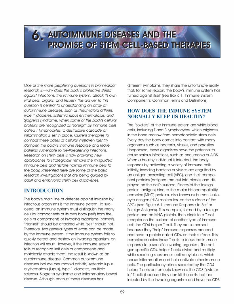
59
One of the more perplexing questions in biomedicalresearch is—why does the body’s protective shieldagainst infections, the immune system, attack its ownvital cells, organs, and tissues? The answer to thisquestion is central to understanding an array ofautoimmune diseases, such as rheumatoid arthritis,type 1 diabetes, systemic lupus erythematosus, andSjogren’s syndrome. When some of the body's cellularproteins are recognized as “foreign” by immune cellscalled T lymphocytes, a destructive cascade ofinflammation is set in place. Current therapies tocombat these cases of cellular mistaken identitydampen the body’s immune response and leavepatients vulnerable to life-threatening infections.Research on stem cells is now providing newapproaches to strategically remove the misguidedimmune cells and restore normal immune cells tothe body. Presented here are some of the basicresearch investigations that are being guided byadult and embryonic stem cell discoveries.
INTRODUCTIONThe body’s main line of defense against invasion byinfectious organisms is the immune system. To suc-ceed, an immune system must distinguish the manycellular components of its own body (self) from thecells or components of invading organisms (nonself).“Nonself” should be attacked while “self” should not.Therefore, two general types of errors can be madeby the immune system. If the immune system fails toquickly detect and destroy an invading organism, aninfection will result. However, if the immune systemfails to recognize self cells or components andmistakenly attacks them, the result is known as anautoimmune disease. Common autoimmunediseases include rheumatoid arthritis, systemic lupuserythematosis (lupus), type 1 diabetes, multiplesclerosis, Sjogren’s syndrome and inflammatory boweldisease. Although each of these diseases has
different symptoms, they share the unfortunate realitythat, for some reason, the body’s immune system hasturned against itself (see Box 6.1. Immune SystemComponents: Common Terms and Definitions).
HOW DOES THE IMMUNE SYSTEMNORMALLY KEEP US HEALTHY? The “soldiers” of the immune system are white bloodcells, including T and B lymphocytes, which originatein the bone marrow from hematopoietic stem cells.Every day the body comes into contact with manyorganisms such as bacteria, viruses, and parasites.Unopposed, these organisms have the potential tocause serious infections, such as pneumonia or AIDS.When a healthy individual is infected, the bodyresponds by activating a variety of immune cells.Initially, invading bacteria or viruses are engulfed byan antigen presenting cell (APC), and their compo-nent proteins (antigens) are cut into pieces and dis-played on the cell’s surface. Pieces of the foreignprotein (antigen) bind to the major histocompatibilitycomplex (MHC) proteins, also known as human leuko-cyte antigen (HLA) molecules, on the surface of theAPCs (see Figure 6.1 Immune Response to Self orForeign Antigens). This complex, formed by a foreignprotein and an MHC protein, then binds to a T cellreceptor on the surface of another type of immunecell, the CD4 helper T cell. They are so namedbecause they “help” immune responses proceedand have a protein called CD4 on their surface. Thiscomplex enables these T cells to focus the immuneresponse to a specific invading organism. The anti-gen-specific CD4 helper T cells divide and multiplywhile secreting substances called cytokines, whichcause inflammation and help activate other immunecells. The particular cytokines secreted by the CD4helper T cells act on cells known as the CD8 “cytotox-ic” T cells (because they can kill the cells that areinfected by the invading organism and have the CD8
AUTOIMMUNE DISEASES AND THE PROMISE OF STEM CELL-BASED THERAPIESAUTOIMMUNE DISEASES AND THEPROMISE OF STEM CELL-BASED THERAPIES
6.6.

marrow. Upon their departure from the bone marrow,immature T cells undergo a final maturation processin the thymus, a small organ located in the upperchest, before being dispersed to the body with therest of the immune cells (e.g., B cells). Within thethymus, T cells undergo an important process that“educates” them to distinguish between self (theproteins of their own body) and nonself (the invadingorganism’s) antigens. Here, the T cells are selected fortheir ability to bind to the particular MHC proteinsexpressed by the individual. The particular array ofMHCs varies slightly between individuals, and thisvariation is the basis of the immune response when atransplanted organ is rejected. MHCs and other lesseasily characterized molecules called minor histo-compatibility antigens are genetically determinedand this is the reason why donor organs from relativesof the recipient are preferred over unrelated donors.
60
Autoimmune Diseases and the Promise of Stem Cell-Based Therapies
protein on their surface). The helper T cells can alsoactivate antigen-specific B cells to produce antibod-ies, which can neutralize and help eliminate bacteriaand viruses from the body. Some of the antigen-specif-ic T and B cells that are activated to rid the body ofinfectious organisms become long-lived “memory”cells. Memory cells have the capacity to act quicklywhen confronted with the same infectious organism atlater times. It is the memory cells that cause us tobecome “immune” from later reinfections with thesame organism.
HOW DO THE IMMUNE CELLS OFTHE BODY KNOW WHAT TO ATTACKAND WHAT NOT TO? All immune and blood cells develop from multipotenthematopoietic stem cells that originate in the bone
Figure 6.1. Immune Response to Self or Foreign Antigens.
Foreign (or self)antigen
AntigenPresentingCell (APC)
MHC/antigencomplex
Cytokines
Antibodies
B Cell
Production ofpathogenicself-reactiveantibodies
Whole selfantigen
Free pathogenclearance by
specific antibody
CD4helperT cell
CD8cytotoxic
T cell
Cell death
Infected cells (displaysforeign T cell epitopeon its surface) or self
(i.e., loss of selftolerance)
CD4helperT cell
Antigen-specificT cell receptor
© 2
001
Tere
se W
inslo
w
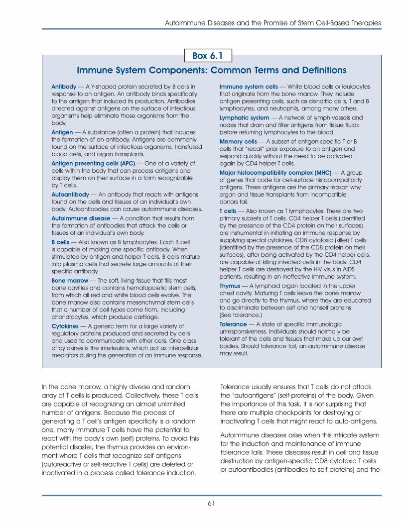
Autoimmune Diseases and the Promise of Stem Cell-Based Therapies
61
In the bone marrow, a highly diverse and randomarray of T cells is produced. Collectively, these T cellsare capable of recognizing an almost unlimitednumber of antigens. Because the process ofgenerating a T cell’s antigen specificity is a randomone, many immature T cells have the potential toreact with the body ’s own (self) proteins. To avoid thispotential disaster, the thymus provides an environ-ment where T cells that recognize self-antigens(autoreactive or self-reactive T cells) are deleted orinactivated in a process called tolerance induction.
Tolerance usually ensures that T cells do not attackthe “autoantigens” (self-proteins) of the body. Giventhe importance of this task, it is not surprising thatthere are multiple checkpoints for destroying orinactivating T cells that might react to auto-antigens.
Autoimmune diseases arise when this intricate systemfor the induction and maintenance of immunetolerance fails. These diseases result in cell and tissuedestruction by antigen-specific CD8 cytotoxic T cellsor autoantibodies (antibodies to self-proteins) and the
Antibody — A Y-shaped protein secreted by B cells inresponse to an antigen. An antibody binds specificallyto the antigen that induced its production. Antibodiesdirected against antigens on the surface of infectiousorganisms help eliminate those organisms from thebody.
Antigen — A substance (often a protein) that inducesthe formation of an antibody. Antigens are commonlyfound on the surface of infectious organisms, transfusedblood cells, and organ transplants.
Antigen presenting cells (APC) — One of a variety ofcells within the body that can process antigens anddisplay them on their surface in a form recognizableby T cells.
Autoantibody — An antibody that reacts with antigensfound on the cells and tissues of an individual’s ownbody. Autoantibodies can cause autoimmune diseases.
Autoimmune disease — A condition that results fromthe formation of antibodies that attack the cells ortissues of an individual’s own body.
B cells — Also known as B lymphocytes. Each B cellis capable of making one specific antibody. Whenstimulated by antigen and helper T cells, B cells matureinto plasma cells that secrete large amounts of theirspecific antibody
Bone marrow — The soft, living tissue that fills mostbone cavities and contains hematopoietic stem cells,from which all red and white blood cells evolve. Thebone marrow also contains mesenchymal stem cellsthat a number of cell types come from, includingchondrocytes, which produce cartilage.
Cytokines — A generic term for a large variety ofregulatory proteins produced and secreted by cellsand used to communicate with other cells. One classof cytokines is the interleukins, which act as intercellularmediators during the generation of an immune response.
Immune system cells — White blood cells or leukocytesthat originate from the bone marrow. They includeantigen presenting cells, such as dendritic cells, T and Blymphocytes, and neutrophils, among many others.
Lymphatic system — A network of lymph vessels andnodes that drain and filter antigens from tissue fluidsbefore returning lymphocytes to the blood.
Memory cells — A subset of antigen-specific T or Bcells that “recall” prior exposure to an antigen andrespond quickly without the need to be activatedagain by CD4 helper T cells.
Major histocompatibility complex (MHC) — A groupof genes that code for cell-surface histocompatibilityantigens. These antigens are the primary reason whyorgan and tissue transplants from incompatibledonors fail.
T cells — Also known as T lymphocytes. There are twoprimary subsets of T cells. CD4 helper T cells (identifiedby the presence of the CD4 protein on their surfaces)are instrumental in initiating an immune response bysupplying special cytokines. CD8 cytotoxic (killer) T cells(identified by the presence of the CD8 protein on theirsurfaces), after being activated by the CD4 helper cells,are capable of killing infected cells in the body. CD4helper T cells are destroyed by the HIV virus in AIDSpatients, resulting in an ineffective immune system.
Thymus — A lymphoid organ located in the upperchest cavity. Maturing T cells leave the bone marrowand go directly to the thymus, where they are educatedto discriminate between self and nonself proteins.(See tolerance.)
Tolerance — A state of specific immunologicunresponsiveness. Individuals should normally betolerant of the cells and tissues that make up our ownbodies. Should tolerance fail, an autoimmune diseasemay result.
Immune System Components: Common Terms and Definitions
Box 6.1
62
Autoimmune Diseases and the Promise of Stem Cell-Based Therapies
accompanying inflammatory process. Thesemechanisms can lead to the destruction of the jointsin rheumatoid arthritis, the destruction of the insulin-producing beta cells of the pancreas in type 1diabetes, or damage to the kidneys in lupus. The rea-sons for the failure to induce or maintain toleranceare enigmatic. However, genetic factors, along withenvironmental and hormonal influences and certaininfections, may contribute to tolerance and thedevelopment of autoimmune disease [4, 7].
HEMATOPOIETIC STEM CELLTHERAPY FOR AUTOIMMUNEDISEASESThe current treatments for many autoimmune diseasesinclude the systemic use of anti-inflammatory drugsand potent immunosuppressive and immunomodu-latory agents (i.e., steroids and inhibitor proteins thatblock the action of inflammatory cytokines). However,despite their profound effect on immune responses,these therapies are unable to induce clinically signifi-cant remissions in certain patients. In recent years,researchers have contemplated the use of stem cellsto treat autoimmune disorders. Discussed here issome of the rationale for this approach, with a focuson experimental stem cell therapies for lupus,rheumatoid arthritis, and type 1 diabetes.
The immune-mediated injury in autoimmune diseasescan be organ-specific, such as type 1 diabeteswhich is the consequence of the destruction of thepancreatic beta islet cells or multiple sclerosis whichresults from the breakdown of the myelin covering ofnerves. These autoimmune diseases are amenableto treatments involving the repair or replacement ofdamaged or destroyed cells or tissue (see Chapter 7.Stem Cells and Diabetes and Chapter 11. Use ofGenetically Modified Stem Cells in ExperimentalGene Therapies). In contrast, non-organ-specificautoimmune diseases, such as lupus, are character-ized by widespread injury due to immune reactionsagainst many different organs and tissues.
One approach is being evaluated in early clinicaltrials of patients with poorly responsive, life-threateninglupus. This is a severe disease affecting multipleorgans in the body including muscles, skin, joints, andkidneys as well as the brain and nerves. Over 239,000Americans, of which more than 90 percent arewomen, suffer from lupus. In addition, lupus
disproportionately afflicts African-American andHispanic women [11]. A major obstacle in the treat-ment of non-organ-specific autoimmune diseasessuch as lupus is the lack of a single specific target forthe application of therapy.
The objective of hematopoietic stem cell therapy forlupus is to destroy the mature, long-lived, and auto-reactive immune cells and to generate a new,properly functioning immune system. In most of thesetrials, the patient’s own stem cells have been used ina procedure known as autologous (from “one’s self”)hematopoietic stem cell transplantation. First, patientsreceive injections of a growth factor, which coaxeslarge numbers of hematopoietic stem cells to bereleased from the bone marrow into the blood stream.These cells are harvested from the blood, purifiedaway from mature immune cells, and stored. Aftersufficient quantities of these cells are obtained, thepatient undergoes a regimen of cytotoxic (cell-killing)drug and/or radiation therapy, which eliminates themature immune cells. Then, the hematopoietic stemcells are returned to the patient via a blood transfusioninto the circulation where they migrate to the bonemarrow and begin to differentiate to become matureimmune cells. The body ’s immune system is thenrestored. Nonetheless, the recovery phase, until theimmune system is reconstituted represents a period ofdramatically increased susceptibility to bacterial, fun-gal, and viral infection, making this a high-risk therapy.
Recent reports suggest that this replacement therapymay fundamentally alter the patient’s immune sys-tem. Richard Burt and his colleagues [18] conducteda long-term follow-up (one to three years) of sevenlupus patients who underwent this procedure andfound that they remained free from active lupus andimproved continuously after transplantation, withoutthe need for immunosuppressive medications. Oneof the hallmarks of lupus is that during the natural pro-gression of disease, the normally diverse repertoire ofT cells become limited in the number of different anti-gens they recognize, suggesting that an increasingproportion of the patient’s T cells are autoreactive.Burt and colleagues found that following hemato-poietic stem cell transplantation, levels of T celldiversity were restored to those of healthy individuals.This finding provides evidence that stem cell replace-ment may be beneficial in reestablishing tolerance inT cells, thereby decreasing the likelihood of diseasereoccurrence.
Autoimmune Diseases and the Promise of Stem Cell-Based Therapies
63
DEVELOPMENT OF HEMATOPOIETICSTEM CELL LINES FORTRANSPLANTATIONThe ability to generate and propagate unlimitednumbers of hematopoietic stem cells outside thebody—whether from adult, umbilical cord blood,fetal, or embryonic sources—would have a majorimpact on the safety, cost, and availability of stemcells for transplantation. The current approach ofisolating hematopoietic stem cells from a patient’sown peripheral blood places the patient at risk for aflare-up of their autoimmune disease. This is a poten-tial consequence of repeated administration of thestem cell growth factors needed to mobilizehematopoietic stem cells from the bone marrow tothe blood stream in numbers sufficient for trans-plantation. In addition, contamination of the purifiedhematopoietic stem cells with the patient’s matureautoreactive T and B cells could affect the success ofthe treatment in some patients. Propagation of purecell lines in the laboratory would avoid these potentialdrawbacks and increase the numbers of stem cellsavailable to each patient, thus shortening the at-riskinterval before full immune reconstitution.
Whether embryonic stem cells will provide advan-tages over stem cells derived from cord blood oradult bone marrow hematopoietic stem cells remainsto be determined. However, hematopoietic stemcells, whether from umbilical cord blood or bonemarrow, have a more limited potential for self-renewal than do pluripotent embryonic stem cells.Although new information will be needed to directthe differentiation of embryonic stem cells intohematopoietic stem cells, hematopoietic cells arepresent in differentiated cultures from human embry-onic stem cells [9] and from human fetal-derivedembryonic germ stem cells [17].
One potential advantage of using hematopoieticstem cell lines for transplantation in patients withautoimmune diseases is that these cells could begenerated from unaffected individuals or, as pre-disposing genetic factors are defined, from embryonicstem cells lacking these genetic influences. In addition,use of genetically selected or genetically engineeredcell types may further limit the possibility of diseaseprogression or reemergence.
One risk of using nonself hematopoietic stem cells isof immune rejection of the transplanted cells.Immune rejection is caused by MHC protein differ-ences between the donor and the patient (recipient).In this scenario, the transplanted hematopoietic stemcells and their progeny are rejected by the patient’sown T cells, which are originating from the patient’ssurviving bone marrow hematopoietic stem cells. Inthis regard, embryonic stem cell-derived hemato-poietic stem cells may offer distinct advantages overcord blood and bone marrow hematopoietic stemcell lines in avoiding rejection of the transplant.Theoretically, banks of embryonic stem cells express-ing various combinations of the three most criticalMHC proteins could be generated to allow closematching to the recipient’s MHC composition.
Additionally, there is evidence that embryonic stemcells are considerably more receptive to geneticmanipulation than are hematopoietic stem cells (seeChapter 11. Use of Genetically Modified Stem Cells inExperimental Gene Therapies).
This characteristic means that embryonic stem cellscould be useful in strategies that could prevent theirrecognition by the patient’s surviving immune cells.For example, it may be possible to introduce therecipient’s MHC proteins into embryonic stem cellsthrough targeted gene transfer. Alternatively, it istheoretically possible to generate a universal donorembryonic stem cell line by genetic alteration orremoval of the MHC proteins. Researchers haveaccomplished this by genetically altering a mouseso that it has little or no surface expression of MHCmolecules on any of the cells or tissues. There is norejection of pancreatic beta islet cells from thesegenetically altered mice when the cells are trans-planted into completely MHC-mismatched mice [13].Additional research will be needed to determine thefeasibility of these alternative strategies for preventionof graft rejection in humans [6].
Jon Odorico and colleagues have shown that expres-sion of MHC proteins on mouse embryonic stem cellsand differentiated embryonic stem cell progeny iseither absent or greatly decreased compared withMHC expression on adult cells [8]. These preliminaryfindings raise the intriguing possibility that lines derivedfrom embryonic stem cells may be inherently lesssusceptible to rejection by the recipient’s immune
64
Autoimmune Diseases and the Promise of Stem Cell-Based Therapies
system than lines derived from adult cells. This couldhave important implications for the transplantation ofcells other than hematopoietic stem cells.
Another potential advantage of using pure popula-tions of donor hematopoietic stem cells achievedthrough stem cell technologies would be a lowerincidence and severity of graft-versus-host disease, apotentially fatal complication of bone marrow trans-plantation. Graft-versus-host disease results from theimmune-mediated injury to recipient tissues thatoccurs when mature organ-donor T cells remain with-in the organ at the time of transplant. Such maturedonor alloreactive T cells would be absent from purepopulations of multipotent hematopoietic stem cells,and under ideal conditions of immune toleranceinduction in the recipient’s thymus, the donor-derivedmature T cell population would be tolerant to the host.
GENE THERAPY AND STEM CELLAPPROACHES FOR THE TREATMENTOF AUTOIMMUNE DISEASESGene therapy is the genetic modification of cells toproduce a therapeutic effect (see Chapter 11. Use ofGenetically Modified Stem Cells in ExperimentalGene Therapies). In most investigational protocols,DNA containing the therapeutic gene is transferredinto cultured cells, and these cells are subsequentlyadministered to the animal or patient. DNA can alsobe injected directly, entering cells at the site of theinjection or in the circulation. Under ideal conditions,cells take up the DNA and produce the therapeuticprotein encoded by the gene.
Currently, there is an extensive amount of genetherapy research being conducted in animal modelsof autoimmune disease. The goal is to modify theaberrant, inflammatory immune response that ischaracteristic of autoimmune diseases [15, 19].Researchers most often use one of two generalstrategies to modulate the immune system. The firststrategy is to block the actions of an inflammatorycytokine (secreted by certain activated immune cellsand inflamed tissues) by transferring a gene into cellsthat encodes a “decoy” receptor for that cytokine.Alternatively, a gene is transferred that encodes ananti-inflammatory cytokine, redirecting the auto-inflammatory immune response to a more “tolerant”state. In many animal studies, promising results havebeen achieved by using these approaches, and the
studies have advanced understanding of the diseaseprocesses and the particular inflammatory cytokinesinvolved in disease progression [15, 19].
Serious obstacles to the development of effectivegene therapies for humans remain, however.Foremost among these are the difficulty of reliablytransferring genetic material into adult and slowlydividing cells (including hematopoietic stem cells)and of producing long-lasting expression of theintended protein at levels that can be tightly con-trolled in response to disease activity. Importantly,embryonic stem cells are substantially more permis-sive to gene transfer compared with adult cells, andembryonic cells sustain protein expression duringextensive self-renewal. Whether adult-derived stemcells, other than hematopoietic stem cells, aresimilarly amenable to gene transfer has not yetbeen determined.
Ultimately, stem cell gene therapy should allow thedevelopment of novel methods for immune modula-tion in autoimmune diseases. One example is thegenetic modification of hematopoietic stem cells ordifferentiated tissue cells with a “decoy” receptor forthe inflammatory cytokine interferon gamma to treatlupus. For example, in a lupus mouse model, genetransfer of the decoy receptor, via DNA injection,arrested disease progression [12]. Other investigatorshave used a related but distinct approach in amouse model of type 1 diabetes. Interleukin-12 (IL-12), an inflammatory cytokine, plays a prominentrole in the development of diabetes in these mice.The investigators transferred the gene for a modifiedform of IL-12, which blocks the activity of the naturalIL-12, into pancreatic beta islet cells (the target ofautoimmune injury in type 1 diabetes). The islet cellgene therapy prevented the onset of diabetes inthese mice [20]. Theoretically, embryonic stem cellsor adult stem cells could be genetically modifiedbefore or during differentiation into pancreatic betaislet cells to be used for transplantation. The resultingimmune-modulating islet cells might diminish theoccurrence of ongoing autoimmunity, increase thelikelihood of long-term function of the transplantedcells, and eliminate the need for immunosuppressivetherapy following transplantation.
Researchers are exploring similar genetic approachesto prevent progressive joint destruction and loss ofcartilage and to repair damaged joints in animal
Autoimmune Diseases and the Promise of Stem Cell-Based Therapies
65
models of rheumatoid arthritis. Rheumatoid arthritis isa debilitating autoimmune disease characterized byacute and chronic inflammation, in which theimmune system primarily attacks the joints of thebody. In a recent study, investigators geneticallytransferred an anti-inflammatory cytokine, interleukin-4(IL-4), into a specialized, highly efficient antigen-presenting cell called a dendritic cell, and theninjected these IL-4-secreting cells into mice that canbe induced to develop a form of arthritis similar torheumatoid arthritis in humans. These IL-4-secretingdendritic cells are presumed to act on the CD4helper T cells to reintroduce tolerance to self-proteins.Treated mice showed complete suppression of theirdisease and, in addition to its immune-modulatoryproperties, IL-4 blocked bone resorption (a seriouscomplication of rheumatoid arthritis), making it aparticularly attractive cytokine for this therapy [10].However, one obstacle to this approach is that humandendritic cells are difficult to isolate in large numbers.
Investigators have also directed the differentiation ofdendritic cells from mouse embryonic stem cells,indicating that a stem cell-based approach mightwork in patients with rheumatoid arthritis [5]. Longer-term follow-up and further characterization will beneeded in animal models before researchersproceed with the development of such an approachin humans. In similar studies, using other inhibitors ofinflammatory cytokines such as a decoy receptor fortumor necrosis factor–� (a prominent inflammatorycytokine in inflamed joints), an inhibitor of nuclearfactor–�� (a protein within cells that turns on theproduction of many inflammatory cytokines), andinterleukin-13 (an anti-inflammatory cytokine),researchers have shown promising results in animalmodels of rheumatoid arthritis [19]. Because of thecomplexity and redundancy of immune systemsignaling networks, it is likely that a multifacetedapproach involving inhibitors of several differentinflammatory cytokines will be successful, whereasapproaches targeting single cytokines might fail orproduce only short-lived responses. In addition, othercell types may prove to be even better vehicles forthe delivery of gene therapy in this disease.
Chondrocytes, cells that build cartilage in joints, mayprovide another avenue for stem cell-based treat-ment of rheumatoid arthritis. These cells have beenderived from human bone marrow stromal stem cells
derived from human bone marrow [14]. Little is knownabout the intermediate cells that ultimately differ-entiate into chondrocytes. In addition to adult bonemarrow as a source for stromal stem cells, humanembryonic stem cells can differentiate into precursorcells believed to lead ultimately to the stromal stemcells [16]. However, extensive research is needed toreliably achieve the directed derivation of the stromalstem cells from embryonic stem cells and, subse-quently, the differentiation of chondrocytes fromthese stromal stem cells.
The ideal cell for optimum cartilage repair may be amore primitive cell than the chondrocyte, such as thestromal cell, or an intermediate cell in the pathway(e.g., a connective tissue precursor) leading to thechondrocyte. Stromal stem cells can generate newchondrocytes and facilitate cartilage repair in a rab-bit model [3]. Such cells may also prove to be idealtargets for the delivery of immune-modulatory genetherapy. Like hematopoietic stem cells, stromal stemcells have been used in animal models for delivery ofgene therapy [1]. For example, a recent studydemonstrated that genetically engineered chondro-cytes, expressing a growth factor, can enhance thefunction of transplanted chondrocytes [2].
Two obstacles to the use of adult stromal stem cellsor chondrocytes are the limited numbers of thesecells that can be harvested and the difficulties inpropagating them in the laboratory. Embryonic stemcells, genetically modified and expanded beforedirected differentiation to a connective tissue stemcell, may be an attractive alternative.
Collectively, these results illustrate the tremendouspotential these cells may offer for the treatment ofrheumatoid arthritis and other autoimmune diseases.
CONCLUSIONStem cell-based therapies offer many excitingpossibilities for the development of novel treatments,and perhaps even cures, for autoimmune diseases. A challenging research effort remains to fully realizethis potential and to address the many remainingquestions, which include how best to direct the differ-entiation of specific cell types and determine whichparticular type of stem cell will be optimum for eachtherapeutic approach. Gene therapy with cytokinesor their inhibitors is still in its infancy, but stem cells or
66
Autoimmune Diseases and the Promise of Stem Cell-Based Therapies
their progeny may provide one of the better avenuesfor future delivery of immune-based therapies.Ultimately, the potential to alleviate these devastatingchronic diseases with the use of stem cell-basedtechnologies is enormous.
REFERENCES1. Allay, J.A., Dennis, J.E., Haynesworth, S.E., Majumdar, M.K.,
Clapp, D.W., Shultz, L.D., Caplan, A.I., and Gerson, S.L.(1997). LacZ and interleukin-3 expression in vivo after retro-viral transduction of marrow-derived human osteogenicmesenchymal progenitors. Hum. Gene Ther. 8, 1417-1427.
2. Brower-Toland, B.D., Saxer, R.A., Goodrich, L.R., Mi, Z.,Robbins, P.D., Evans, C.H., and Nixon, A.J. (2001). Directadenovirus-mediated insulin-like growth factor I genetransfer enhances transplant chondrocyte function. Hum.Gene Ther. 12, 117-129.
3. Caplan, A.I., Elyaderani, M., Mochizuki, Y., Wakitani, S., andGoldberg, V.M. (1997). Principles of cartilage repair andregeneration. Clin. Orthop. 342, 254-269.
4. Cooper, G.S., Dooley, M.A., Treadwell, E.L., St Clair, E.W.,Parks, C.G., and Gilkeson, G.S. (1998). Hormonal, environ-mental, and infectious risk factors for developing systemiclupus erythematosus. Arthritis Rheum. 41, 1714-1724.
5. Fairchild, P.J., Brook, F.A., Gardner, R.L., Graca, L., Strong, V.,Tone, Y., Tone, M., Nolan, K.F., and Waldmann, H. (2000).Directed differentiation of dendritic cells from mouseembryonic stem cells. Curr. Biol. 10, 1515-1518.
6. Gearhart, J. (1998). New potential for human embryonicstem cells. Science. 282, 1061-1062.
7. Grossman, J.M. and Tsao, B.P. (2000). Genetics and sys-temic lupus erythematosus. Curr. Rheumatol. Rep. 2, 13-18.
8. Harley, C.B., Gearhart, J., Jaenisch, R., Rossant, J., andThomson, J. (2001). Keystone Symposia. Pluripotent stemcells: biology and applications. Durango, CO.
9. Itskovitz-Eldor, J., Schuldiner, M., Karsenti, D., Eden, A.,Yanuka, O., Amit, M., Soreq, H., and Benvenisty, N. (2000).Differentiation of human embryonic stem cells into embry-oid bodies comprising the three embryonic germ layers.Mol. Med. 6, 88-95.
10. Kim, S.H., Kim, S., Evans, C.H., Ghivizzani, S.C., Oligino, T.,and Robbins, P.D. (2001). Effective treatment of establishedmurine collagen-induced arthritis by systemic administra-tion of dendritic cells genetically modified to express IL-4.J. Immunol. 166, 3499-3505.
11. Lawrence, R.C., Helmick, C.G., Arnett, F.C., Deyo, R.A.,Felson, D.T., Giannini, E.H., Heyse, S.P., Hirsch, R., Hochberg,M.C., Hunder, G.G., Liang, M.H., Pillemer, S.R., Steen, V.D.,and Wolfe, F. (1998). Estimates of the prevalence of arthritisand selected musculoskeletal disorders in the United States.Arthritis Rheum. 41, 778-799.
12. Lawson, B.R., Prud’homme, G.J., Chang, Y., Gardner, H.A.,Kuan, J., Kono, D.H., and Theofilopoulos, A.N. (2000).Treatment of murine lupus with cDNA encoding IFN-gammaR/Fc. J. Clin. Invest. 106, 207-215.
13. Osorio, R.W., Ascher, N.L., Jaenisch, R., Freise, C.E., Roberts,J.P., and Stock, P.G. (1993). Major histocompatibility com-plex class I deficiency prolongs islet allograft survival.Diabetes. 42, 1520-1527.
14. Pittenger, M.F., Mackay, A.M., Beck, S.C., Jaiswal, R.K.,Douglas, R., Mosca, J.D., Moorman, M.A., Simonetti, D.W.,Craig, S., and Marshak, D.R. (1999). Multilineage potentialof adult human mesenchymal stem cells. Science. 284,143-147.
15. Prud’homme, G.J. (2000). Gene therapy of autoimmunediseases with vectors encoding regulatory cytokines orinflammatory cytokine inhibitors. J. Gene. Med. 2, 222-232.
16. Schuldiner, M., Yanuka, O., Itskovitz-Eldor, J., Melton, D., andBenvenisty, N. (2000). Effects of eight growth factors on thedifferentiation of cells derived from human embryonic stemcells. Proc. Natl. Acad. Sci. U. S. A. 97, 11307-11312.
17. Shamblott, M.J., Axelman, J., Littlefield, J.W., Blumenthal,P.D., Huggins, G.R., Cui, Y., Cheng, L., and Gearhart, J.D.(2000). Human embryonic germ cell derivatives express abroad range of develpmentally distinct markers and prolif-erate extensively in vitro. Proc. Natl. Acad. Sci. U. S. A. 98,113-118.
18. Traynor, A.E., Schroeder, J., Rosa, R.M., Cheng, D., Stefka, J.,Mujais, S., Baker, S., and Burt, R.K. (2000). Treatment ofsevere systemic lupus erythematosus with high-dosechemotherapy and haemopoietic stem-cell transplanta-tion: a phase I study. Lancet. 356, 701-707.
19. Tsokos, G.C. and Nepom, G.T. (2000). Gene therapy in thetreatment of autoimmune diseases. J. Clin. Invest. 106,181-183.
20. Yasuda, H., Nagata, M., Arisawa, K., Yoshida, R., Fujihira, K.,Okamoto, N., Moriyama, H., Miki, M., Saito, I., Hamada, H.,Yokono, K., and Kasuga, M. (1998). Local expression ofimmunoregulatory IL-12p40 gene prolonged syngeneic isletgraft survival in diabetic NOD mice. J. Clin. Invest. 102,1807-1814.
67
Diabetes exacts its toll on many Americans, youngand old. For years, researchers have painstakinglydissected this complicated disease caused by thedestruction of insulin producing islet cells of thepancreas. Despite progress in understanding theunderlying disease mechanisms for diabetes, there isstill a paucity of effective therapies. For years investi-gators have been making slow, but steady, progresson experimental strategies for pancreatic transplan-tation and islet cell replacement. Now, researchershave turned their attention to adult stem cells thatappear to be precursors to islet cells and embryonicstem cells that produce insulin.
INTRODUCTIONFor decades, diabetes researchers have beensearching for ways to replace the insulin-producingcells of the pancreas that are destroyed by apatient’s own immune system. Now it appears thatthis may be possible. Each year, diabetes affectsmore people and causes more deaths than breastcancer and AIDS combined. Diabetes is the seventhleading cause of death in the United States today,with nearly 200,000 deaths reported each year. TheAmerican Diabetes Association estimates that nearly16 million people, or 5.9 percent of the United Statespopulation, currently have diabetes.
Diabetes is actually a group of diseases character-ized by abnormally high levels of the sugar glucose inthe bloodstream. This excess glucose is responsiblefor most of the complications of diabetes, whichinclude blindness, kidney failure, heart disease, stroke,neuropathy, and amputations. Type 1 diabetes, alsoknown as juvenile-onset diabetes, typically affectschildren and young adults. Diabetes develops whenthe body’s immune system sees its own cells as foreign and attacks and destroys them. As a result,
the islet cells of the pancreas, which normally produce insulin, are destroyed. In the absence ofinsulin, glucose cannot enter the cell and glucoseaccumulates in the blood. Type 2 diabetes, alsocalled adult-onset diabetes, tends to affect older,sedentary, and overweight individuals with a familyhistory of diabetes. Type 2 diabetes occurs when thebody cannot use insulin effectively. This is calledinsulin resistance and the result is the same as withtype 1 diabetes—a build up of glucose in the blood.
There is currently no cure for diabetes. People withtype 1 diabetes must take insulin several times a dayand test their blood glucose concentration three tofour times a day throughout their entire lives. Frequentmonitoring is important because patients who keeptheir blood glucose concentrations as close to normal as possible can significantly reduce many ofthe complications of diabetes, such as retinopathy (a disease of the small blood vessels of the eyewhich can lead to blindness) and heart disease, that tend to develop over time. People with type 2diabetes can often control their blood glucose concentrations through a combination of diet, exercise, and oral medication. Type 2 diabetes often progresses to the point where only insulin therapy will control blood glucose concentrations.
Each year, approximately 1,300 people with type 1diabetes receive whole-organ pancreas transplants.After a year, 83 percent of these patients, on average, have no symptoms of diabetes and do not have to take insulin to maintain normal glucoseconcentrations in the blood. However, the demandfor transplantable pancreases outweighs their availability. To prevent the body from rejecting thetransplanted pancreas, patients must take powerfuldrugs that suppress the immune system for their entirelives, a regimen that makes them susceptible to a
STEM CELLSAND DIABETESSTEM CELLSAND DIABETES
7.7.
DEVELOPMENT OF THE PANCREASBefore discussing cell-based therapies for diabetes, it is important to understand how the pancreas develops. In mammals, the pancreas contains threeclasses of cell types: the ductal cells, the acinar cells,and the endocrine cells. The endocrine cells producethe hormones glucagon, somatostatin, pancreaticpolypeptide (PP), and insulin, which are secreted intothe blood stream and help the body regulate sugarmetabolism. The acinar cells are part of the exocrinesystem, which manufactures digestive enzymes, andductal cells from the pancreatic ducts, which connect the acinar cells to digestive organs.
In humans, the pancreas develops as an outgrowthof the duodenum, a part of the small intestine. Thecells of both the exocrine system—the acinar cells—and of the endocrine system—the islet cells—seemto originate from the ductal cells during develop-ment. During development these endocrine cellsemerge from the pancreatic ducts and form aggregates that eventually form what is known asIslets of Langerhans. In humans, there are four typesof islet cells: the insulin-producing beta cells; thealpha cells, which produce glucagon; the delta cells,which secrete somatostatin; and the PP-cells, whichproduce pancreatic polypeptide. The hormonesreleased from each type of islet cell have a role inregulating hormones released from other islet cells. Inthe human pancreas, 65 to 90 percent of islet cellsare beta cells, 15 to 20 percent are alpha-cells, 3 to10 percent are delta cells, and one percent is PPcells. Acinar cells form small lobules contiguous withthe ducts (see Figure 7.1. Insulin Production in theHuman Pancreas). The resulting pancreas is a combination of a lobulated, branched acinar glandthat forms the exocrine pancreas, and, embedded in the acinar gland, the Islets of Langerhans, whichconstitute the endocrine pancreas.
During fetal development, new endocrine cellsappear to arise from progenitor cells in the pancre-atic ducts. Many researchers maintain that some sort of islet stem cell can be found intermingled withductal cells during fetal development and that thesestem cells give rise to new endocrine cells as thefetus develops. Ductal cells can be distinguishedfrom endocrine cells by their structure and by thegenes they express. For example, ductal cells typically express a gene known as cytokeratin-9
68
Stem Cells and Diabetes
host of other diseases. Many hospitals will not performa pancreas transplant unless the patient also needs akidney transplant. That is because the risk of infectiondue to immunosuppressant therapy can be a greaterhealth threat than the diabetes itself. But if a patient isalso receiving a new kidney and will require immuno-suppressant drugs anyway, many hospitals will perform the pancreas transplant.
Over the past several years, doctors have attemptedto cure diabetes by injecting patients with pancreaticislet cells—the cells of the pancreas that secreteinsulin and other hormones. However, the requirementfor steroid immunosuppressant therapy to preventrejection of the cells increases the metabolicdemand on insulin-producing cells and eventuallythey may exhaust their capacity to produce insulin.The deleterious effect of steroids is greater for islet cell transplants than for whole-organ transplants. As a result, less than 8 percent of islet cell transplantsperformed before last year had been successful.
More recently, James Shapiro and his colleagues inEdmonton, Alberta, Canada, have developed anexperimental protocol for transplanting islet cells thatinvolves using a much larger amount of islet cells anda different type of immunosuppressant therapy. In arecent study, they report that [17], seven of sevenpatients who received islet cell transplants no longerneeded to take insulin, and their blood glucose concentrations were normal a year after surgery. The success of the Edmonton protocol is now beingtested at 10 centers around the world.
If the success of the Edmonton protocol can beduplicated, many hurdles still remain in using thisapproach on a wide scale to treat diabetes. First,donor tissue is not readily available. Islet cells used intransplants are obtained from cadavers, and the pro-cedure requires at least two cadavers per transplant.The islet cells must be immunologically compatible,and the tissue must be freshly obtained—within eighthours of death. Because of the shortage of organdonors, these requirements are difficult to meet andthe waiting list is expected to far exceed available tissue, especially if the procedure becomes widelyaccepted and available. Further, islet cell transplantrecipients face a lifetime of immunosuppressant therapy, which makes them susceptible to other serious infections and diseases.
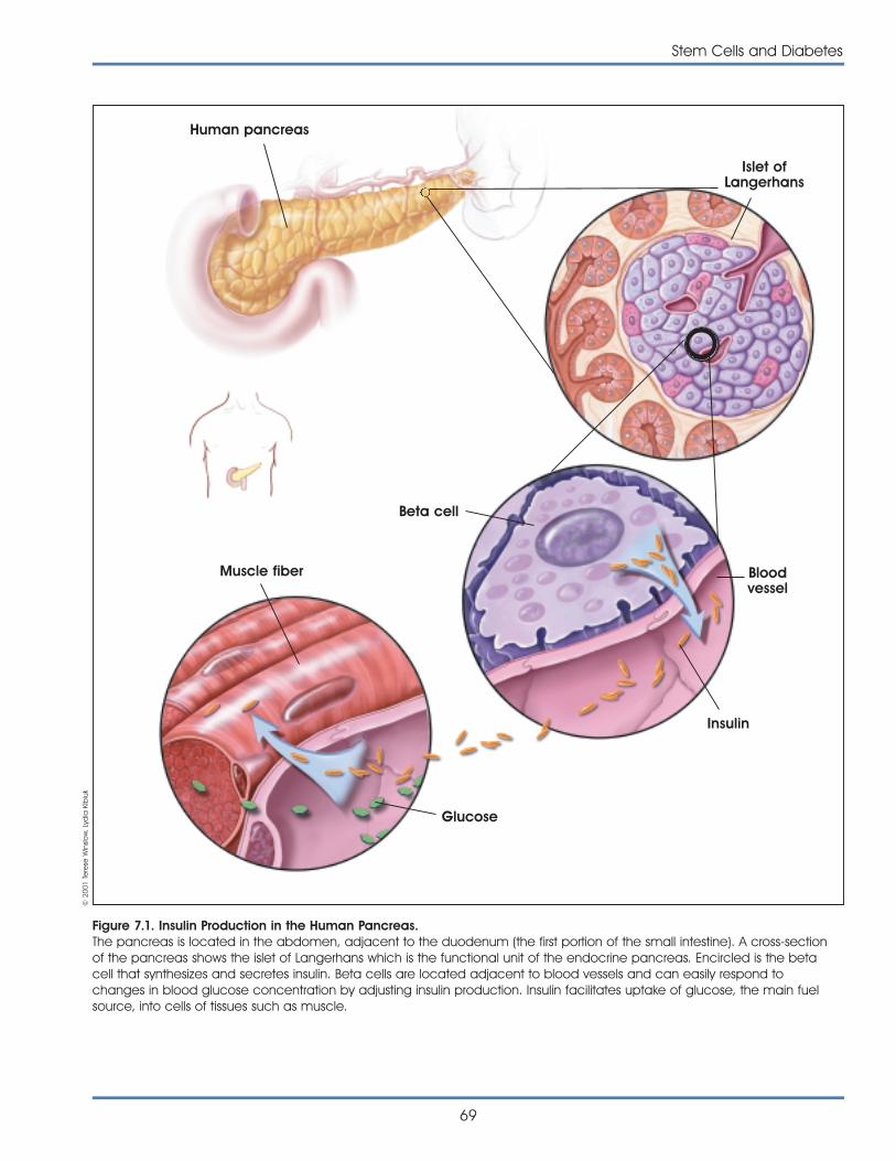
Stem Cells and Diabetes
69
Figure 7.1. Insulin Production in the Human Pancreas.The pancreas is located in the abdomen, adjacent to the duodenum (the first portion of the small intestine). A cross-sectionof the pancreas shows the islet of Langerhans which is the functional unit of the endocrine pancreas. Encircled is the betacell that synthesizes and secretes insulin. Beta cells are located adjacent to blood vessels and can easily respond tochanges in blood glucose concentration by adjusting insulin production. Insulin facilitates uptake of glucose, the main fuelsource, into cells of tissues such as muscle.
Human pancreas
Islet ofLangerhans
Beta cell
Muscle fiber
Glucose
Bloodvessel
Insulin
© 2
001
Tere
se W
inslo
w, L
ydia
Kib
iuk
70
Stem Cells and Diabetes
(CK-9), which encodes a structural protein. Beta isletcells, on the other hand, express a gene called PDX-1, which encodes a protein that initiates transcription from the insulin gene. These genes,called cell markers, are useful in identifying particularcell types.
Following birth and into adulthood, the source of newislet cells is not clear, and some controversy existsover whether adult stem cells exist in the pancreas.Some researchers believe that islet stem cell-like cellscan be found in the pancreatic ducts and even inthe islets themselves. Others maintain that the ductalcells can differentiate into islet precursor cells, whileothers hold that new islet cells arise from stem cells inthe blood. Researchers are using several approachesfor isolating and cultivating stem cells or islet pre-cursor cells from fetal and adult pancreatic tissue. Inaddition, several new promising studies indicate thatinsulin-producing cells can be cultivated from embry-onic stem cell lines.
DEVELOPMENT OF CELL-BASEDTHERAPIES FOR DIABETESIn developing a potential therapy for patients withdiabetes, researchers hope to develop a system thatmeets several criteria. Ideally, stem cells should beable to multiply in culture and reproduce themselvesexactly. That is, the cells should be self-renewing.Stem cells should also be able to differentiate in vivo to produce the desired kind of cell. For diabetestherapy, it is not clear whether it will be desirable toproduce only beta cells—the islet cells that manufac-ture insulin—or whether other types of pancreatic isletcells are also necessary. Studies by Bernat Soria andcolleagues, for example, indicate that isolated betacells—those cultured in the absence of the othertypes of islet cells—are less responsive to changes inglucose concentration than intact islet clusters madeup of all islet cell types. Islet cell clusters typicallyrespond to higher-than-normal concentrations of glucose by releasing insulin in two phases: a quickrelease of high concentrations of insulin and a slowerrelease of lower concentrations of insulin. In this manner the beta cells can fine-tune their response to glucose. Extremely high concentrations of glucosemay require that more insulin be released quickly,while intermediate concentrations of glucose can behandled by a balance of quickly and slowly released insulin.
Isolated beta cells, as well as islet clusters with lower-than-normal amounts of non-beta cells, do notrelease insulin in this biphasic manner. Instead insulinis released in an all-or-nothing manner, with no fine-tuning for intermediate concentrations of glucosein the blood [5, 18]. Therefore, many researchersbelieve that it will be preferable to develop a systemin which stem or precursor cell types can be cultured to produce all the cells of the islet cluster inorder to generate a population of cells that will beable to coordinate the release of the appropriateamount of insulin to the physiologically relevant concentrations of glucose in the blood.
FETAL TISSUE AS SOURCE FOR ISLET CELLS Several groups of researchers are investigating theuse of fetal tissue as a potential source of islet pro-genitor cells. For example, using mice, researchershave compared the insulin content of implants fromseveral sources of stem cells—fresh human fetal pancreatic tissue, purified human islets, and culturedislet tissue [2]. They found that insulin content was initially higher in the fresh tissue and purified islets.However, with time, insulin concentration decreasedin the whole tissue grafts, while it remained the samein the purified islet grafts. When cultured islets wereimplanted, however, their insulin content increasedover the course of three months. The researchersconcluded that precursor cells within the culturedislets were able to proliferate (continue to replicate)and differentiate (specialize) into functioning islet tissue, but that the purified islet cells (already differentiated) could not further proliferate when grafted. Importantly, the researchers found, however,that it was also difficult to expand cultures of fetal isletprogenitor cells in culture [7].
ADULT TISSUE AS SOURCE FORISLET CELLSMany researchers have focused on culturing islet cellsfrom human adult cadavers for use in developingtransplantable material. Although differentiated betacells are difficult to proliferate and culture, someresearchers have had success in engineering suchcells to do this. For example, Fred Levine and his colleagues at the University of California, San Diego,have engineered islet cells isolated from humancadavers by adding to the cells’ DNA special genes
Stem Cells and Diabetes
71
that stimulate cell proliferation. However, becauseonce such cell lines that can proliferate in culture areestablished, they no longer produce insulin. The celllines are further engineered to express the beta isletcell gene, PDX-1, which stimulates the expression ofthe insulin gene. Such cell lines have been shown topropagate in culture and can be induced to differentiate to cells, which produce insulin. Whentransplanted into immune-deficient mice, the cellssecrete insulin in response to glucose. The researchersare currently investigating whether these cells willreverse diabetes in an experimental diabetes model in mice [6, 8].
These investigators report that these cells do not produce as much insulin as normal islets, but it is within an order of magnitude. The major problem indealing with these cells is maintaining the delicatebalance between growth and differentiation. Cellsthat proliferate well do not produce insulin efficiently,and those that do produce insulin do not proliferatewell. According to the researchers, the major issue isdeveloping the technology to be able to grow largenumbers of these cells that will reproducibly producenormal amounts of insulin [9].
Another promising source of islet progenitor cells liesin the cells that line the pancreatic ducts. Someresearchers believe that multipotent (capable offorming cells from more than one germ layer) stemcells are intermingled with mature, differentiated ductcells, while others believe that the duct cells themselves can undergo a differentiation, or areversal to a less mature type of cell, which can thendifferentiate into an insulin-producing islet cell.
Susan Bonner-Weir and her colleagues reported lastyear that when ductal cells isolated from adulthuman pancreatic tissue were cultured, they couldbe induced to differentiate into clusters that contained both ductal and endocrine cells. Over the course of three to four weeks in culture, the cellssecreted low amounts of insulin when exposed to low concentrations of glucose, and higher amountsof insulin when exposed to higher glucose concentrations. The researchers have determined by immunochemistry and ultrastructural analysis thatthese clusters contain all of the endocrine cells of the islet [4].
Bonner-Weir and her colleagues are working with primary cell cultures from duct cells and have not
established cells lines that can grow indefinitely.However the cells can be expanded. According tothe researchers, it might be possible in principle to doa biopsy and remove duct cells from a patient andthen proliferate the cells in culture and give thepatient back his or her own islets. This would work withpatients who have type 1 diabetes and who lackfunctioning beta cells, but their duct cells remainintact. However, the autoimmune destruction wouldstill be a problem and potentially lead to destructionof these transplanted cells [3]. Type 2 diabetespatients might benefit from the transplantation ofcells expanded from their own duct cells since theywould not need any immunosuppression. However,many researchers believe that if there is a geneticcomponent to the death of beta cells, then betacells derived from ductal cells of the same individualwould also be susceptible to autoimmune attack.
Some researchers question whether the ductal cellsare indeed undergoing a dedifferentiation or whethera subset of stem-like or islet progenitors populate thepancreatic ducts and may be co-cultured along with the ductal cells. If ductal cells die off but isletprecursors proliferate, it is possible that the islet precursor cells may overtake the ductal cells in culture and make it appear that the ductal cells are dedifferentiating into stem cells. According toBonner-Weir, both dedifferentiated ductal cells andislet progenitor cells may occur in pancreatic ducts.
Ammon Peck of the University of Florida, VijayakumarRamiya of Ixion Biotechnology in Alachua, FL, andtheir colleagues [13, 14] have also cultured cells fromthe pancreatic ducts from both humans and mice.Last year, they reported that pancreatic ductalepithelial cells from adult mice could be cultured toyield islet-like structures similar to the cluster of cellsfound by Bonner-Weir. Using a host of islet-cell markersthey identified cells that produced insulin, glucagon,somatostatin, and pancreatic polypeptide. When thecells were implanted into diabetic mice, the diabeteswas reversed.
Joel Habener has also looked for islet-like stem cellsfrom adult pancreatic tissue. He and his colleagueshave discovered a population of stem-like cells withinboth the adult pancreas islets and pancreatic ducts.These cells do not express the marker typical of ductal cells, so they are unlikely to be ductal cells,according to Habener. Instead, they express a markercalled nestin, which is typically found in developing
72
Stem Cells and Diabetes
neural cells. The nestin-positive cells do not expressmarkers typically found in mature islet cells. However,depending upon the growth factors added, the cellscan differentiate into different types of cells, includingliver, neural, exocrine pancreas, and endocrine pan-creas, judged by the markers they express, and canbe maintained in culture for up to eight months [20].
EMBRYONIC STEM CELLSThe discovery of methods to isolate and grow humanembryonic stem cells in 1998 renewed the hopes ofdoctors, researchers, and diabetes patients and theirfamilies that a cure for type 1 diabetes, and perhapstype 2 diabetes as well, may be within striking distance. In theory, embryonic stem cells could becultivated and coaxed into developing into theinsulin-producing islet cells of the pancreas. With aready supply of cultured stem cells at hand, the theory is that a line of embryonic stem cells could be grown up as needed for anyone requiring a transplant. The cells could be engineered to avoidimmune rejection. Before transplantation, they couldbe placed into nonimmunogenic material so thatthey would not be rejected and the patient wouldavoid the devastating effects of immunosuppressantdrugs. There is also some evidence that differentiatedcells derived from embryonic stem cells might be less likely to cause immune rejection (see Chapter10. Assessing Human Stem Cell Safety). Although having a replenishable supply of insulin-producingcells for transplant into humans may be a long wayoff, researchers have been making remarkableprogress in their quest for it. While some researchershave pursued the research on embryonic stem cells, other researchers have focused on insulin-producingprecursor cells that occur naturally in adult and fetal tissues.
Since their discovery three years ago, several teamsof researchers have been investigating the possibilitythat human embryonic stem cells could be developed as a therapy for treating diabetes. Recent studies in mice show that embryonic stemcells can be coaxed into differentiating into insulin-producing beta cells, and new reports indicatethat this strategy may be possible using humanembryonic cells as well.
Last year, researchers in Spain reported using mouseembryonic stem cells that were engineered to allowresearchers to select for cells that were differentiating
into insulin-producing cells [19]. Bernat Soria and hiscolleagues at the Universidad Miguel Hernandez inSan Juan, Alicante, Spain, added DNA containingpart of the insulin gene to embryonic cells from mice.The insulin gene was linked to another gene that rendered the mice resistant to an antibiotic drug. Bygrowing the cells in the presence of an antibiotic,only those cells that were activating the insulin promoter were able to survive. The cells were clonedand then cultured under varying conditions. Cells cultured in the presence of low concentrations of glucose differentiated and were able to respond tochanges in glucose concentration by increasinginsulin secretion nearly sevenfold. The researchersthen implanted the cells into the spleens of diabetic mice and found that symptoms of diabetes were reversed.
Manfred Ruediger of Cardion, Inc., in Erkrath,Germany, is using the approach developed by Soriaand his colleagues to develop insulin-producinghuman cells derived from embryonic stem cells. Byusing this method, the non-insulin-producing cells willbe killed off and only insulin-producing cells shouldsurvive. This is important in ensuring that undifferenti-ated cells are not implanted that could give rise totumors [15]. However, some researchers believe thatit will be important to engineer systems in which allthe components of a functioning pancreatic islet areallowed to develop.
Recently Ron McKay and his colleagues described aseries of experiments in which they induced mouseembryonic cells to differentiate into insulin-secretingstructures that resembled pancreatic islets [10].McKay and his colleagues started with embryonicstem cells and let them form embryoid bodies—anaggregate of cells containing all three embryonicgerm layers. They then selected a population of cellsfrom the embryoid bodies that expressed the neuralmarker nestin (see Appendix B. Mouse EmbryonicStem Cells). Using a sophisticated five-stage culturingtechnique, the researchers were able to induce thecells to form islet-like clusters that resembled thosefound in native pancreatic islets. The cells respondedto normal glucose concentrations by secreting insulin,although insulin amounts were lower than thosesecreted by normal islet cells (see Figure 7.2.Development of Insulin-Secreting Pancreatic-Like Cells From Mouse Embryonic Stem Cells). When the cells were injected into diabetic mice, they
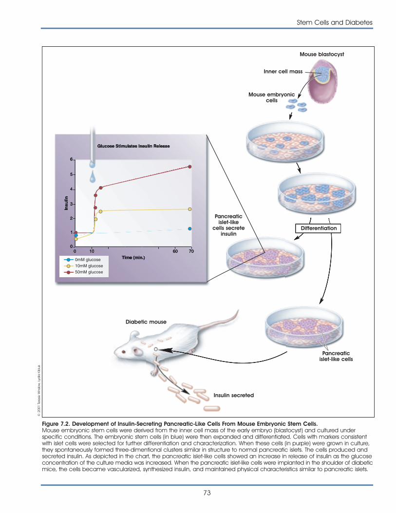
Stem Cells and Diabetes
73
Figure 7.2. Development of Insulin-Secreting Pancreatic-Like Cells From Mouse Embryonic Stem Cells.Mouse embryonic stem cells were derived from the inner cell mass of the early embryo (blastocyst) and cultured underspecific conditions. The embryonic stem cells (in blue) were then expanded and differentiated. Cells with markers consistentwith islet cells were selected for further differentiation and characterization. When these cells (in purple) were grown in culture,they spontaneously formed three-dimentional clusters similar in structure to normal pancreatic islets. The cells produced andsecreted insulin. As depicted in the chart, the pancreatic islet-like cells showed an increase in release of insulin as the glucoseconcentration of the culture media was increased. When the pancreatic islet-like cells were implanted in the shoulder of diabeticmice, the cells became vascularized, synthesized insulin, and maintained physical characteristics similar to pancreatic islets.
Mouse blastocyst
Inner cell mass
Mouse embryoniccells
Pancreaticislet-like
cells secreteinsulin
Differentiation
Pancreaticislet-like cells
Insulin secreted
Diabetic mouse
© 2
001
Tere
se W
inslo
w, L
ydia
Kib
iuk
74
Stem Cells and Diabetes
survived, although they did not reverse the symptomsof diabetes.
According to McKay, this system is unique in that theembryonic cells form a functioning pancreatic islet,complete with all the major cell types. The cellsassemble into islet-like structures that contain anotherlayer, which contains neurons and is similar to intactislets from the pancreas [11]. Several research groupsare trying to apply McKay ’s results with mice toinduce human embryonic stem cells to differentiateinto insulin-producing islets.
Recent research has also provided more evidencethat human embryonic cells can develop into cellsthat can and do produce insulin. Last year, Melton,Nissim Benvinisty of the Hebrew University inJerusalem, and Josef Itskovitz-Eldor of the Technion inHaifa, Israel, reported that human embryonic stemcells could be manipulated in culture to express thePDX-1 gene, a gene that controls insulin transcription[16]. In these experiments, researchers culturedhuman embryonic stem cells and allowed them tospontaneously form embryoid bodies (clumps ofembryonic stem cells composed of many types ofcells from all three germ layers). The embryoid bodieswere then treated with various growth factors, includ-ing nerve growth factor. The researchers found thatboth untreated embryoid bodies and those treatedwith nerve growth factor expressed PDX-1. Embryonicstem cells prior to formation of the aggregatedembryoid bodies did not express PDX-1. Becauseexpression of the PDX-1 gene is associated with theformation of beta islet cells, these results suggest thatbeta islet cells may be one of the cell types thatspontaneously differentiate in the embryoid bodies.The researchers now think that nerve growthfactor may be one of the key signals for inducing the differentiation of beta islet cells and can beexploited to direct differentiation in the laboratory.Complementing these findings is work done by JonOdorico of the University of Wisconsin in Madisonusing human embryonic cells of the same source. In preliminary findings, he has shown that humanembryonic stem cells can differentiate and expressthe insulin gene [12].
More recently, Itskovitz-Eldor and his Technion colleagues further characterized insulin-producingcells in embryoid bodies [1]. The researchers foundthat embryonic stem cells that were allowed to
spontaneously form embryoid bodies contained asignificant percentage of cells that express insulin.Based on the binding of antibodies to the insulin protein, Itskovitz-Eldor estimates that 1 to 3 percent of the cells in embryoid bodies are insulin-producingbeta-islet cells. The researchers also found that cellsin the embryoid bodies express glut-2 and islet-specif-ic glucokinase, genes important for beta cell functionand insulin secretion. Although the researchers didnot measure a time-dependent response to glucose,they did find that cells cultured in the presence ofglucose secrete insulin into the culture medium. Theresearchers concluded that embryoid bodies containa subset of cells that appear to function as beta cellsand that the refining of culture conditions may soonyield a viable method for inducing the differentiationof beta cells and, possibly, pancreatic islets.
Taken together, these results indicate that the devel-opment of a human embryonic stem cell system thatcan be coaxed into differentiating into functioninginsulin-producing islets may soon be possible.
FUTURE DIRECTIONSUltimately, type 1 diabetes may prove to be especially difficult to cure, because the cells aredestroyed when the body ’s own immune systemattacks and destroys them. This autoimmunity mustbe overcome if researchers hope to use transplantedcells to replace the damaged ones. Manyresearchers believe that at least initially, immunosup-pressive therapy similar to that used in the Edmontonprotocol will be beneficial. A potential advantage ofembryonic cells is that, in theory, they could be engineered to express the appropriate genes thatwould allow them to escape or reduce detection bythe immune system. Others have suggested that atechnology should be developed to encapsulate orembed islet cells derived from islet stem or progenitorcells in a material that would allow small moleculessuch as insulin to pass through freely, but would notallow interactions between the islet cells and cells ofthe immune system. Such encapsulated cells couldsecrete insulin into the blood stream, but remaininaccessible to the immune system.
Before any cell-based therapy to treat diabetesmakes it to the clinic, many safety issues must beaddressed (see Chapter 10. Assessing Human StemCell Safety). A major consideration is whether any
Stem Cells and Diabetes
75
precursor or stem-like cells transplanted into the bodymight revert to a more pluripotent state and inducethe formation of tumors. These risks would seeminglybe lessened if fully differentiated cells are used intransplantation.
But before any kind of human islet-precursor cells can be used therapeutically, a renewable source ofhuman stem cells must be developed. Althoughmany progenitor cells have been identified in adulttissue, few of these cells can be cultured for multiplegenerations. Embryonic stem cells show the greatestpromise for generating cell lines that will be free ofcontaminants and that can self renew. However,most researchers agree that until a therapeuticallyuseful source of human islet cells is developed, allavenues of research should be exhaustively investigated, including both adult and embryonicsources of tissue.
REFERENCES1. Assady, S., Maor, G., Amit, M., Itskovitz-Eldor, J., Skorecki,
K.L., and Tzukerman, M. (2001). Insulin production byhuman embryonic stem cells. Diabetes. 50. http://www.diabetes.org/Diabetes_Rapids/Suheir_Assady_06282001.pdf
2. Beattie, G.M., Otonkoski, T., Lopez, A.D., and Hayek, A.(1997). Functional beta-cell mass after transplantation ofhuman fetal pancreatic cells: differentiation or prolifera-tion? Diabetes. 46, 244-248.
3. Bonner-Weir, S., personal communication.
4. Bonner-Weir, S., Taneja, M., Weir, G.C., Tatarkiewicz, K., Song,K.H., Sharma, A., and O'Neil, J.J. (2000). In vitro cultivationof human islets from expanded ductal tissue. Proc. Natl.Acad. Sci. U. S. A. 97, 7999-8004.
5. Bosco, D. and Meda, P. (1997). Reconstructing islet functionin vitro. Adv. Exp. Med. Biol. 426, 285-298.
6. Dufayet de la Tour, D., Halvorsen, T., Demeterco, C.,Tyrberg, B., Itkin-Ansari, P., Loy, M., Yoo, S.J., Hao, S., Bossie,S., and Levine, F. (2001). b-cell differentiation from ahuman pancreatic cell line in vitro and in vivo. Mol.Endocrinol. 15, 476-483.
7. Hayek, A., personal communication.
8. Itkin-Ansari, P., Demeterco, C., Bossie, S., Dufayet de la Tour,D., Beattie, G.M., Movassat, J., Mally, M.I., Hayek, A., andLevine, F. (2001). PDX-1 and cell-cell contact act in synergyto promote d-cell development in a human pancreaticendocrine precursor cell line. Mol. Endocrinol. 14, 814-822.
9. Levine, F., personal communication.
10. Lumelsky, N., Blondel, O., Laeng, P., Velasco, I., Ravin, R.,and McKay, R. (2001). Differentiation of Embryonic StemCells to Insulin-Secreting Structures Similiar to PancreaticIslets. Science. 292, 1389-1394.
11. McKay, R., personal communication
12. Odorico, J. S., personal communication.
13. Peck, A., personal communication.
14. Ramiya, V. K., personal communication.
15. Ruediger, M., personal communication.
16. Schuldiner, M., Yanuka, O., Itskovitz-Eldor, J., Melton, D., andBenvenisty, N. (2000). Effects of eight growth factors on thedifferentiation of cells derived from human embryonic stemcells. Proc. Natl. Acad. Sci. U. S. A. 97, 11307-11312.
17. Shapiro, J., Lakey, J.R.T., Ryan, E.A., Korbutt, G.S., Toth, E.,Warnock, G.L., Kneteman, N.M., and Rajotte, R.V. (2000).Islet transplantation in seven patients with type 1 diabetesmellitus using a glucocorticoid-free immunosuppressiveregimen. N. Engl. J. Med. 343, 230-238.
18. Soria, B., Martin, F., Andreu, E., Sanchez-Andrés, J.V.,Nacher, V., and Montana, E. (1996). Diminished fraction ofblockable ATP-sensitive K+ channels in islets transplantedinto diabetic mice. Diabetes. 45, 1755-1760.
19. Soria, B., Roche, E., Berná, G., Leon-Quinto, T., Reig, J.A.,and Martin, F. (2000). Insulin-secreting cells derived fromembryonic stem cells normalize glycemia in streptozotocin-induced diabetic mice. Diabetes. 49, 157-162.
20. Zulewski, H., Abraham, E.J., Gerlach, M.J., Daniel, P.B.,Moritz, W., Muller, B., Vallejo, M., Thomas, M.K., andHabener, J.F. (2001). Multipotential nestin-positive stem cellsisolated from adult pancreatic islets differentiate ex vivointo pancreatic endocrine, exocrine, and hepatic pheno-types. Diabetes. 50, 521-533.

76
Stem Cells and Diabetes
This page intentionally left blank
77
Today, most treatments for damage to the brain orspinal cord aim to relieve symptoms and limit furtherdamage. But recent research into the regenerationmechanisms of the central nervous system, includingthe discovery of stem cells in the adult brain that cangive rise to new neurons and neural support cells,has raised hopes that researchers can find ways toactually repair central nervous system damage.Research on stem cells in nervous system disorders isone of the few areas in which there is evidence thatcell-replacement therapy can restore lost function.
STEM CELLS BRING NEWSTRATEGIES FOR DEVELOPINGREPLACEMENT NEURONSJust a decade ago, neuroscience textbooks heldthat neurons in the adult human brain and spinalcord could not regenerate. Once dead, it wasthought, central nervous system neurons were gonefor good. Because rebuilding nervous tissue seemedout of the question, research focused almost entirelyon therapeutic approaches to limiting further damage.
That dogma that brain tissue could not be regeneratedis history. In the mid-1990s, neuroscientists learned thatsome parts of the adult human brain do, in fact, gener-ate new neurons, at least under certain circumstances.Moreover, they found that the new neurons arise from"neural stem cells” in the fetal as well as the adult brain(see Chapter 4. The Adult Stem Cell). These undifferenti-ated cells resemble cells in a developing fetus that giverise to the brain and spinal cord. The researchers alsofound that these neural stem cells could generatemany, if not all, types of cells found in the brain. Thisincludes neurons—the main message carriers in thenervous system, which use long, thin projections calledaxons to transmit signals over long distances—as wellas crucial neural-support cells called oligodendrocytesand astrocytes (see Figure 8.1. The Neuron).
The discovery of a regenerative capacity in the adultcentral nervous system holds out the promise that itmay eventually be possible to repair damage fromterrible degenerative diseases such as Parkinson’sDisease and amyotrophic lateral sclerosis (ALS, alsoknown as Lou Gehrig’s disease), as well as from brainand spinal cord injuries resulting from stroke or trauma(see Box 8.1. Early Research Shows Stem Cells CanImprove Movement in Paralyzed Mice).
Researchers are pursuing two fundamental strategiesto exploit this discovery. One is to grow differentiatedcells in a laboratory dish that are suitable for implan-tation into a patient by starting with undifferentiatedneural cells. The idea is either to treat the cells in cul-ture to nudge them toward the desired differentiatedneuronal cell type before implantation, or to implantthem directly and rely on signals inside the body todirect their maturation into the right kind of brain cell.A variety of stem cells might be used for this task,including so-called “neural precursor cells” that areinwardly committed to differentiating into a particularcell type but are outwardly not yet changed orpluripotent embryonic stem cells—cells derived froma very early stage human embryo that retain thecapacity to become any cell type in the body andthat can be maintained in culture for a very longtime without differentiating.
The other repair strategy relies on finding growthhormones and other “trophic factors”—growth fac-tors, hormones, and other signaling molecules thathelp cells survive and grow—that can fire up apatient’s own stem cells and endogenous repairmechanisms, to allow the body to cope with dam-age from disease or injury. Researchers are vigorouslypursuing both strategies to find therapies for centralnervous system disorders that involve cell death, buta great deal more basic research must be carriedout before effective new therapies emerge.
REBUILDING THE NERVOUS SYSTEM WITH STEM CELLSREBUILDING THE NERVOUS SYSTEM WITH STEM CELLS
8.8.
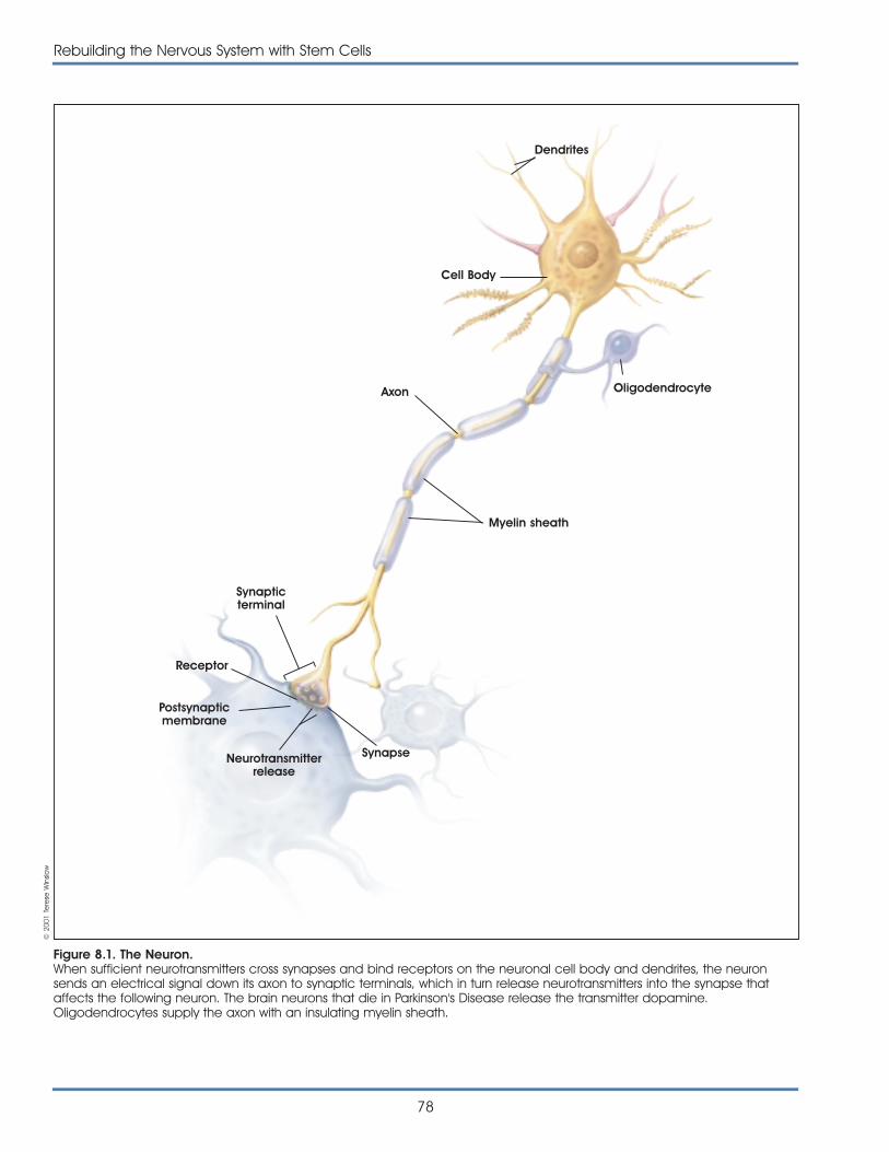
78
Rebuilding the Nervous System with Stem Cells
Figure 8.1. The Neuron.When sufficient neurotransmitters cross synapses and bind receptors on the neuronal cell body and dendrites, the neuronsends an electrical signal down its axon to synaptic terminals, which in turn release neurotransmitters into the synapse thataffects the following neuron. The brain neurons that die in Parkinson's Disease release the transmitter dopamine.Oligodendrocytes supply the axon with an insulating myelin sheath.
Dendrites
Cell Body
Oligodendrocyte
Myelin sheath
Synapticterminal
Receptor
Postsynapticmembrane
Neurotransmitterrelease
Synapse
Axon
© 2
001
Tere
se W
inslo
w
Rebuilding the Nervous System with Stem Cells
79
Researchers at Johns Hopkins University recently reportedpreliminary evidence that cells derived from embryonicstem cells can restore movement in an animal modelof amyotrophic lateral sclerosis (ALS) [1]. This degenera-tive disorder, also called as Lou Gehrig’s disease, pro-gressively destroys special nerves found in the spinalcord, known as motor neurons, that control movement.Patients with ALS develop increasing muscle weaknessover months to years, which ultimately leads to paralysisand death. The cause is largely unknown, and there areno effective treatments.
In this new study, the researchers used a rat model ofALS to test for possible nerve cell- restoring properties ofstem cells. The rats were exposed to Sindbis virus, whichinfects the central nervous system and destroys themotor neurons in the spinal cord. Rats that survive areleft with paralyzed muscles in their hindquarters andweakened back limbs. Scientists assess the degree ofimpairment by measuring the rats’ movement, quantify-ing electrical activity in the nerves serving the backlimbs, and visually judging the extent of nerve damagethrough a microscope.
The researchers wanted to see whether stem cells couldrestore nerves and improve mobility in rats. Because sci-entists have had difficulty sustaining stem cell linesderived from rat embryos, the investigators conductedtheir experiments with embryonic germ cells that JohnGearhart and colleagues isolated from human fetal tis-sue in 1998. These cells can produce unchangedcopies of themselves when maintained in culture, andthey form into clumps called embryoid bodies. Undercertain conditions, research has shown that the cells inthe embryoid bodies begin to look and function likeneurons when subjected to specific laboratory condi-tions [2]. The researchers had an idea that these embry-oid body cells in their nonspecialized state mightbecome specialized as replacement neurons if placedinto the area of the damaged spinal cord. So theycarefully prepared cells from the embryoid bodies andinjected them into the fluid surrounding the spinal cordof the paralyzed rats that had their motor neuronsdestroyed by the Sindbis virus.
To test this idea, the researchers selected from laborato-ry culture dishes barely differentiated embryonic germscells that displayed the molecular markers of neuralstem cells, including the proteins nestin and neuron spe-cific enolase. They grew these cells in large quantitiesand injected them into the fluid surrounding the spinalcords of partially paralyzed, Sindbis-virus-treated rats.
The response was impressive. Three months after theinjections, many of the treated rats were able to movetheir hind limbs and walk, albeit clumsily, while the ratsthat did not receive cell injections remained paralyzed.Moreover, at autopsy the researchers found that cellsderived from human embryonic germ cells had migrat-ed throughout the spinal fluid and continued to devel-op, displaying both the shape and molecular markerscharacteristic of mature motor neurons. The researchersare quick to caution that their results are preliminary,and that they do not know for certain whether the treat-ment helped the paralyzed rats because new neuronstook the place of the old, or because trophic factorsfrom the injected cells facilitated the recovery of therats’ remaining nerve cells and helped the rats improvein their ability to use their hind limbs. Nor do they knowhow well this strategy will translate into a therapy forhuman neurodegenerative diseases like ALS. And theyemphasize that there are many hurdles to cross beforethe use of stem cells to repair damaged motor neuronsin patients can be considered. Nevertheless, researchersare excited about these results, which, if confirmed,would represent a major step toward using specializedstem cells from embryonic and fetal tissue sources torestore nervous system function.
REFERENCES1. Kerr, D.A., Llado, J., Shamblott, M., Maragakis, N., Irani, D.N.,
Dike, S., Sappington, A., Gearhart, J., and Rothstein, J. (2001).Human embryonic germ cell derivatives facillitate motorrecovery of rats with diffuse motor neuron injury.
2. Shamblott, M.J., Axelman, J., Wang, S., Bugg, E.M., Littlefield,J.W., Donovan, P.J., Blumenthal, P.D., Huggins, G.R., andGearhart, J.D. (1998). Derivation of pluripotent stem cellsfrom cultured human primordial germ cells. Proc. Natl. Acad.Sci. U. S. A. 95, 13726-13731.
Early Research Shows Stem CellsCan Improve Movement in Paralyzed Mice
Box 8.1
MULTIPLE APPROACHES FOR USINGSTEM CELLS IN PARKINSON’SDISEASE RESEARCHEfforts to develop stem cell based therapies forParkinson’s Disease provide a good example ofresearch aimed at rebuilding the central nervous sys-
tem. As is the case with other disorders, both the cell-implantation and the trophic-factor strategies areunder active development. Both approaches arepromising. This is especially true of cell implantation,which involves using primary tissue transplanteddirectly form developing fetal brain tissue. Parkinson’sis a progressive movement disorder that usually strikes
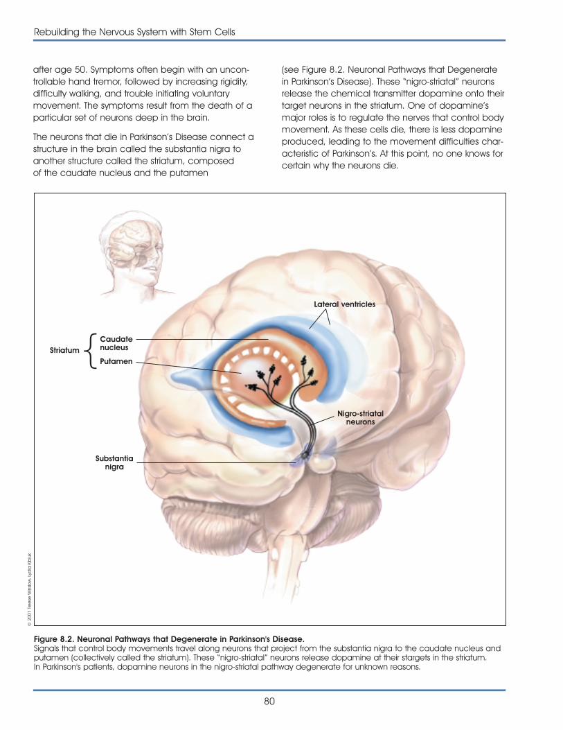
after age 50. Symptoms often begin with an uncon-trollable hand tremor, followed by increasing rigidity,difficulty walking, and trouble initiating voluntarymovement. The symptoms result from the death of aparticular set of neurons deep in the brain.
The neurons that die in Parkinson’s Disease connect astructure in the brain called the substantia nigra toanother structure called the striatum, composedof the caudate nucleus and the putamen
80
Rebuilding the Nervous System with Stem Cells
(see Figure 8.2. Neuronal Pathways that Degeneratein Parkinson’s Disease). These “nigro-striatal” neuronsrelease the chemical transmitter dopamine onto theirtarget neurons in the striatum. One of dopamine’smajor roles is to regulate the nerves that control bodymovement. As these cells die, there is less dopamineproduced, leading to the movement difficulties char-acteristic of Parkinson’s. At this point, no one knows forcertain why the neurons die.
Striatum
}
Lateral ventricles
Nigro-striatalneurons
Substantianigra
Figure 8.2. Neuronal Pathways that Degenerate in Parkinson's Disease.Signals that control body movements travel along neurons that project from the substantia nigra to the caudate nucleus andputamen (collectively called the striatum). These “nigro-striatal” neurons release dopamine at their stargets in the striatum.In Parkinson's patients, dopamine neurons in the nigro-striatal pathway degenerate for unknown reasons.
© 2
001
Tere
se W
inslo
w, L
ydia
Kib
iuk
Caudatenucleus
Putamen
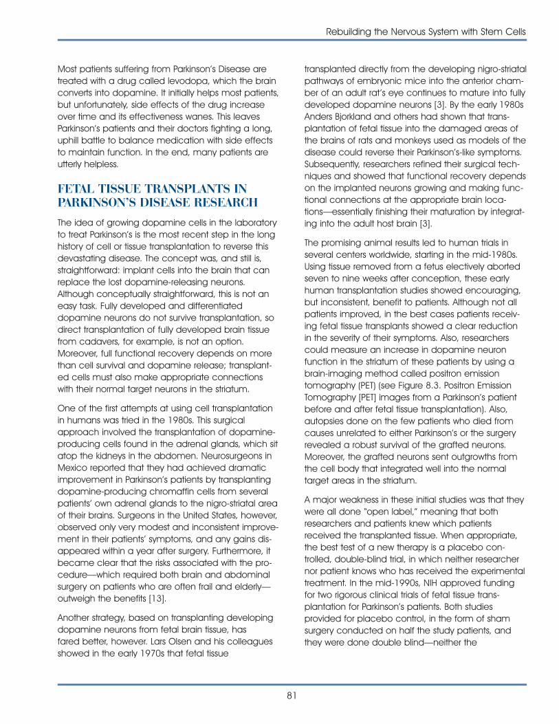
Most patients suffering from Parkinson’s Disease aretreated with a drug called levodopa, which the brainconverts into dopamine. It initially helps most patients,but unfortunately, side effects of the drug increaseover time and its effectiveness wanes. This leavesParkinson’s patients and their doctors fighting a long,uphill battle to balance medication with side effectsto maintain function. In the end, many patients areutterly helpless.
FETAL TISSUE TRANSPLANTS INPARKINSON’S DISEASE RESEARCHThe idea of growing dopamine cells in the laboratoryto treat Parkinson’s is the most recent step in the longhistory of cell or tissue transplantation to reverse thisdevastating disease. The concept was, and still is,straightforward: implant cells into the brain that canreplace the lost dopamine-releasing neurons.Although conceptually straightforward, this is not aneasy task. Fully developed and differentiateddopamine neurons do not survive transplantation, sodirect transplantation of fully developed brain tissuefrom cadavers, for example, is not an option.Moreover, full functional recovery depends on morethan cell survival and dopamine release; transplant-ed cells must also make appropriate connectionswith their normal target neurons in the striatum.
One of the first attempts at using cell transplantationin humans was tried in the 1980s. This surgicalapproach involved the transplantation of dopamine-producing cells found in the adrenal glands, which sitatop the kidneys in the abdomen. Neurosurgeons inMexico reported that they had achieved dramaticimprovement in Parkinson’s patients by transplantingdopamine-producing chromaffin cells from severalpatients’ own adrenal glands to the nigro-striatal areaof their brains. Surgeons in the United States, however,observed only very modest and inconsistent improve-ment in their patients’ symptoms, and any gains dis-appeared within a year after surgery. Furthermore, itbecame clear that the risks associated with the pro-cedure—which required both brain and abdominalsurgery on patients who are often frail and elderly—outweigh the benefits [13].
Another strategy, based on transplanting developingdopamine neurons from fetal brain tissue, has fared better, however. Lars Olsen and his colleaguesshowed in the early 1970s that fetal tissue
transplanted directly from the developing nigro-striatalpathways of embryonic mice into the anterior cham-ber of an adult rat’s eye continues to mature into fullydeveloped dopamine neurons [3]. By the early 1980sAnders Bjorkland and others had shown that trans-plantation of fetal tissue into the damaged areas ofthe brains of rats and monkeys used as models of thedisease could reverse their Parkinson’s-like symptoms.Subsequently, researchers refined their surgical tech-niques and showed that functional recovery dependson the implanted neurons growing and making func-tional connections at the appropriate brain loca-tions—essentially finishing their maturation by integrat-ing into the adult host brain [3].
The promising animal results led to human trials inseveral centers worldwide, starting in the mid-1980s.Using tissue removed from a fetus electively abortedseven to nine weeks after conception, these earlyhuman transplantation studies showed encouraging,but inconsistent, benefit to patients. Although not allpatients improved, in the best cases patients receiv-ing fetal tissue transplants showed a clear reductionin the severity of their symptoms. Also, researcherscould measure an increase in dopamine neuronfunction in the striatum of these patients by using abrain-imaging method called positron emissiontomography (PET) (see Figure 8.3. Positron EmissionTomography [PET] images from a Parkinson’s patientbefore and after fetal tissue transplantation). Also,autopsies done on the few patients who died fromcauses unrelated to either Parkinson’s or the surgeryrevealed a robust survival of the grafted neurons.Moreover, the grafted neurons sent outgrowths fromthe cell body that integrated well into the normaltarget areas in the striatum.
A major weakness in these initial studies was that theywere all done “open label,” meaning that bothresearchers and patients knew which patientsreceived the transplanted tissue. When appropriate,the best test of a new therapy is a placebo con-trolled, double-blind trial, in which neither researchernor patient knows who has received the experimentaltreatment. In the mid-1990s, NIH approved fundingfor two rigorous clinical trials of fetal tissue trans-plantation for Parkinson’s patients. Both studiesprovided for placebo control, in the form of shamsurgery conducted on half the study patients, andthey were done double blind—neither the
Rebuilding the Nervous System with Stem Cells
81
researchers evaluating the effects of the surgery northe patients themselves knew who got tissue implants.
The results of one of these trials, led by Curt Freed,were published recently [5]. Compared with control,patients who received the fetal-tissue transplantshowed no significant benefit in a subjective assess-ment of the patient’s quality of life, which was thestudy ’s primary endpoint. Moreover, two years aftersurgery, 5 out of 33 treated patients developed per-sistent dyskinesia—uncontrolled flailing movements—that had not been observed in the open-label workdescribed above.
The Freed study results, nonetheless, provide impor-tant information about the ability of dopamineneurons to survive in humans. Moreover, PET-scanningdata from the treated patients, as well as autopsiesof two patients who died of unrelated causes severalmonths after the surgery, showed that many of thedopamine neurons survived and grew. Researchersare now awaiting the results of the second NIH-sponsored double-blind trial, led by Warren Olanow[12]. The procedures used in this study differ substan-tially from those of Freed and his colleagues—including the tissue-handling method, the number ofcells implanted, the use of immunosuppressive drugs
to limit rejection of the implanted tissue, and the testsused to assess patient response—and are closer tothose used in the most successful of the early open-label experiments.
Most Parkinson’s researchers are still hopeful that thecell-implantation approach will one day lead to auseful and widely used therapy for Parkinson’sDisease. At the same time, however, mostresearchers are also convinced they must find adifferent source of cells for transplant. The logisticaland technical problems involved in recoveringenough developing dopamine neurons from fetaltissue are very great. Moreover, it is virtually impossibleto standardize the tissue collected from differentfetuses and to fully characterize the cells implanted.This absence of tissue standardization makes it verydifficult to determine the most important factors thatlead to a good patient response and may add risk(see Chapter 10. Assessing Human Stem Cell Safety).
One alternative to cell implantation with human fetaltissue is to use fetal cells and tissues from animals.Researchers at Diacrin and Genzyme, twobiotechnology companies, recently announcedpreliminary results from a clinical trial in which 10Parkinson’s patients received neural cells from the
82
Rebuilding the Nervous System with Stem Cells
Before Surgery After Surgery
Dopamine-Neuron Transplantation
Figure 8.3. Positron Emission Tomography (PET) images from a Parkinson’s patientbefore and after fetal tissue transplantation. The image taken before surgery (left)shows uptake of a radioactive form of dopamine (red) only in the caudate nucleus,indicating that dopamine neurons have degenerated. Twelve months after surgery, animage from the same patient (right) reveals increased dopamine function, especiallyin the putamen. (Reprinted with permission from N Eng J Med 2001;344 (10) p. 710.)
brains of fetal pigs. Eighteen months after the surgery,treated patients did not improve enough to show astatistically significant difference from eight controlpatients who received a sham immunosuppressionregimen and underwent sham surgery. Autopsy ofone treated patient who died of a pulmonaryembolism eight months after surgery revealed that asmall portion of the transplanted pig cells had sur-vived [2], but PET studies looking for improvement indopamine uptake in all treated patients did not showclear improvement. The researchers are still analyzingtheir data [15].
RAISING NEURONS FORREPLACEMENT IN PATIENTS WITHPARKINSON’S DISEASEWhat Parkinson’s researchers ultimately want is arenewable source of cells that can differentiate intofunctional dopamine neurons when placed in thestriatum. Laboratory-grown cells derived from a stemcell may be the best potential alternative source fortransplantable material. One way to get these is tofind the right combination of growth factors and cell-culture conditions to bring undifferentiated cells alongin a culture dish to a point where they are committedto becoming dopamine neurons, then implant themto finish growth and differentiation in the host brain.Another possibility is to put less-committed cells into adamaged brain and rely on “environmental” signalsin the brain to guide them into becoming the rightkind of replacement cell. These developmental sig-nals may be expressed in the brain transiently follow-ing neural degeneration or acute damage.
Whether the cells ultimately implanted are half-differentiated or completely immature, however,researchers need a reliable source. To that end, theyhave identified a whole host of different immaturecells that may have the potential to become,among other things, dopamine neurons, and theyare now in the process of sorting out how best tomake them do so. Neural stem cells isolated fromanimals and humans cannot be grown efficiently inthe lab without changing them in some way, such asby engineering them to express a gene normallyturned on only early in development. Embryonic stemcells—derived from the inner cell mass of an embryoat the blastocyst stage, when only a few hundredcells are present—can be kept in culture in a com-pletely undifferentiated state. They are still capable of
becoming not just nervous system cells but every celltype in the body. If researchers want to be able toimplant cells derived from undifferentiated embryon-ic stem cells, they must take care that no cells in themix give rise to unwanted cell types, such as muscleor bone, within the nervous system. Stem cells fromother tissues—including umbilical cord blood andhuman bone marrow—can also be coaxed todisplay many of the surface-protein “markers” char-acteristic of nervous system cells. It is not yet clear,however, whether these cells are capable of givingrise to fully functional neurons.
A great deal of basic research remains to be done tofind which of these cells provides the best way to geta workable therapy for Parkinson’s Disease. For exam-ple, although researchers have shown for certain thatboth primary human fetal cells and mouse embryon-ic stem cells can become fully functional dopamineneurons, they do not yet know if adult neural stemcells have the same potential. Also, no one has yetpublished evidence that cells from any renewablesource that are laboratory-directed to differentiateinto dopamine neurons can eliminate symptoms inanimal models of Parkinson’s when implanted.
Researchers are making rapid progress, however. Forexample, Ron McKay and his colleagues at NIHreported in 1998 that they were able to expand apopulation of neurons from embryonic mouse brainin culture, and that these cells relieved Parkinson’s-likesymptoms in a rat model [16]. And last year, McKay ’slab also described a procedure for efficiently con-verting mouse pluripotent embryonic stem cells intoneurons that have all the characteristics of dopamineneurons, including the ability to form synapses [17].McKay and other researchers say they have encour-aging unpublished results that dopamine precursorsderived from mouse embryonic stem cells can elimi-nate symptoms in rat models of Parkinson’s Disease[7, 10].
Privately funded researchers are following an analo-gous path using pluripotent human embryonic stemcells. Thomas Okarma of Geron Corporation confirmsthat his company is testing the potential of humanembryonic stem cells in animal models of Parkinson’sDisease, but the results are not yet complete [11]. Inabstracts presented at a recent conference, Geronreports having succeeded in directing humanembryonic stem cells to become mature neural cells
Rebuilding the Nervous System with Stem Cells
83
in laboratory culture, including cells that have thestructural and chemical characteristics of dopamineneurons [6].
TURNING ON THE BRAIN’S OWNSTEM CELLS AS A REPAIRMECHANISMParkinson’s researchers are also looking for ways tospark the repair mechanisms already in a patient’sbrain to fix damage that these mechanisms couldnot otherwise manage. This strategy is less developedthan cell implantation, but it also holds promise [1]. In the future, researchers may use stem cells fromembryonic or adult sources not to replace lost cellsdirectly, but rather to turn on the body ’s own repairmechanisms. Alternatively, researchers may findeffective drug treatments that help a patient’s ownstem cells and repair mechanisms work moreeffectively.
Stem cells in the adult primate brain occur in twolocations. One, the subventricular zone, is an areaunder fluid-filled spaces called ventricles. The other isthe dentate gyrus of the hippocampus. In primates,very few new neurons normally appear in eitherplace, which is why the phenomenon escapednotice until recently. Researchers showed in the mid-1990s that when the brain is injured, stem cells inthese two areas proliferate and migrate toward thesite of the damage. The researchers are now trying todiscover how far this kind of response can go towardameliorating certain kinds of damage.
Recent research shows the direction that this may beheading for Parkinson’s Disease. James Fallon andcolleagues studied the effects on rat brain of a pro-tein called transforming growth factor alpha (TGF�)—a natural peptide found in the body from the veryearliest stages of embryonic development onwardthat is important in activating normal repair processesin several organs, including liver and skin. Fallon'sstudies suggest that the brain’s normal repair processmay never be adequately triggered in a slowly devel-oping degenerative disease like Parkinson's and thatproviding more TGF� can turn it on. Specifically,Fallon found that TGF� injected into healthy rat braincauses stem cells in the subventricular zone to prolif-erate for several days, after which they disappear. Butif the researchers make similar injections into rats inwhich they first damage the nigro-striatal neurons with
a toxin called 6-hydroxydopamine—a frequently usedanimal model for Parkinson’s Disease—two thingshappen. After several days of cell proliferation, Fallonobserves what he calls a “wave of migration” of thestem cells to the damaged areas, where they differ-entiate into dopamine neurons. Most importantly, thetreated rats do not show the behavioral abnormalitiesassociated with the loss of the neurons. Whether thebeneficial effect on symptoms is the result of thenewly formed cells or some other trophic effect is notyet entirely clear [4].
STEM CELLS’ FUTURE ROLE INSPINAL CORD INJURY REPAIR Parkinson’s Disease is only one of many nervoussystem disorders that researchers are trying to solveby regenerating damaged tissue. But Parkinson’s,difficult as it is to reverse, is a relatively easy targetbecause a regenerative therapy need only replaceone particular cell type in one part of the brain.
Therapies for other disorders face much biggerhurdles. Complete restoration after severe spinal cordinjury, for example, is probably far in the future, if itcan ever be done at all. Many cell types aredestroyed in these injuries, including neurons thatcarry messages between the brain and the rest ofthe body. Getting these neurons to grow past aninjury site and connect appropriately with their targetsis extraordinarily difficult. But spinal cord injury patientswould benefit greatly from an even limited restorationof lost functions—gaining partial use of a limb insteadof none, or restoring bladder control, or being freedfrom pain. Such limited restoration of part of apatient’s lost functions is, for some less severe types ofinjury, perhaps a more achievable goal.
In many spinal injuries, the spinal cord is not actuallycut and at least some of the signal-carrying neuronalaxons are intact. But the surviving axons no longercarry messages because cells called oligodendro-cytes, which make the axons’ insulating myelinsheath, are lost. Researchers have recently made thefirst steps in learning to replace these lost myelin-pro-ducing cells [14]. For example, researchers haveshown that stem cells can aid remyelination inrodents [8, 9]. Specifically, they found that injection ofoligodendrocytes derived from mouse embryonicstem cells could remyelinate axons in chemicallydemyelinated rat spinal cord and that the treated
84
Rebuilding the Nervous System with Stem Cells
rats regained limited use of their hind limbs com-pared with the controls. They are not certain, however,whether the limited increase in function theyobserved in rats is actually due to the remyelinationor an unidentified trophic effect of the treatment.
Spinal injury researchers emphasize that much morebasic and preclinical research must be done beforeattempting human trials using stem cell therapies torepair the damaged nervous system. Despite the factthat there is much basic work left to do and manyfundamental questions still to be answered,researchers are hopeful that effective repair for once-hopeless nervous system damage may eventually beachieved. Whether through developing replacementcells or activating the body ’s own stem cells in vivo,research on the use of stem cells for nervous systemdisorders is a rapidly advancing field. This researchpromises to answer key questions about how to repairnervous system damage and how to restore keybody functions damaged by disease or disability.
REFERENCES1. Bjorklund, A. and Lindvall, O. (2000). Self-repair in the brain.
Nature. 405, 892-895.
2. Deacon, T., Schumacher, J., Dinsmore, J., Thomas, C.,Palmer, P., Kott, S., Edge, A., Penney, D., Kassissieh, S.,Dempsey, P., and Isacson, O. (1997). Histological evidenceof fetal pig neural cell survival after transplantation into apatient with Parkinson's disease. Nat. Med. 3, 350-353.
3. Dunnett, S.B., Bjorklund, A., and Lindvall, O. (2001). Cell thera-py in Parkinson's disease—stop or go? Nat. Rev. Neurosci. 2,365-369.
4. Fallon, J., Reid, S., Kinyamu, R., Opole, I., Opole, R., Baratta,J., Korc, M., Endo, T.L., Duong, A., Nguyen, G., Karkehabadhi,M., Twardzik, D., and Loughlin, S. (2000). In vivo induction ofmassive proliferation, directed migration, and differentiationof neural cells in the adult mammalian brain. Proc. Natl.Acad. Sci. U. S. A. 97, 14686-14691.
5. Freed, C.R., Greene, P.E., Breeze, R.E., Tsai, W.Y.,DuMouchel, W., Kao, R., Dillon, S., Winfield, H., Culver, S.,Trojanowski, J.Q., Eidelberg, D., and Fahn, S. (2001).Transplantation of embryonic dopamine neurons for severeParkinson's disease. N. Engl. J. Med. 344, 710-719.
6. Harley, C.B., Gearhart, J., Jaenisch, R., Rossant, J., andThomson, J. (2001). Pluripotent stem cells: biology andapplications. Durango, CO.
7. Isacson, O., personal communication.
8. Liu, S., Qu, Y., Stewart, T.J., Howard, M.J., Chakrabortty, S.,Holekamp, T.F., and McDonald, J.W. (2000). Embryonicstem cells differentiate into oligodendrocytes and myeli-nate in culture and after spinal cord transplantation. Proc.Natl. Acad. Sci. U. S. A. 97, 6126-6131.
9. McDonald, J.W., Liu, X.Z., Qu, Y., Liu, S., Mickey, S.K., Turetsky,D., Gottlieb, D.I., and Choi, D.W. (1999). Transplantedembryonic stem cells survive, differentiate and promoterecovery in injured rat spinal cord. Nat. Med. 5, 1410-1412.
10. McKay, R., personal communication.
11. Okarma, T., personal communication.
12. Olanow, C. W., personal communication.
13. Quinn, N.P. (1990). The clinical application of cell graftingtechniques in patients with Parkinson's disease. Prog. BrainRes. 82, 619-625.
14. Raisman, G. (2001). Olfactory ensheathing cells—anothermiracle cure for spinal cord injury? Nat. Rev. Neurosci. 2,369-374.
15. Schumacher, J.M., Ellias, S.A., Palmer, E.P., Kott, H.S.,Dinsmore, J., Dempsey, P.K., Fischman, A.J., Thomas, C.,Feldman, R.G., Kassissieh, S., Raineri, R., Manhart, C.,Penney, D., Fink, J.S., and Isacson, O. (2000).Transplantation of embryonic porcine mesencephalictissue in patients with PD. Neurology. 54, 1042-1050.
16. Studer, L., Csete, M., Lee, S.H., Kabbani, N., Walikonis, J.,Wold, B., and McKay, R. (2000). Enhanced proliferation,survival, and dopaminergic differentiation of CNS precur-sors in lowered oxygen. J. Neurosci. 20, 7377-7383.
17. Studer, L., Tabar, V., and McKay, R.D. (1998). Transplantationof expanded mesencephalic precursors leads to recoveryin parkinsonian rats. Nat. Neurosci. 1, 290-295.
Rebuilding the Nervous System with Stem Cells
85

86
Rebuilding the Nervous System with Stem Cells
This page intentionally left blank
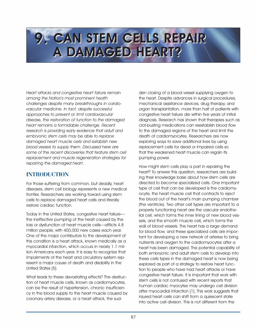
87
Heart attacks and congestive heart failure remainamong the Nation’s most prominent healthchallenges despite many breakthroughs in cardio-vascular medicine. In fact, despite successfulapproaches to prevent or limit cardiovasculardisease, the restoration of function to the damagedheart remains a formidable challenge. Recentresearch is providing early evidence that adult andembryonic stem cells may be able to replacedamaged heart muscle cells and establish newblood vessels to supply them. Discussed here aresome of the recent discoveries that feature stem cellreplacement and muscle regeneration strategies forrepairing the damaged heart.
INTRODUCTIONFor those suffering from common, but deadly, heartdiseases, stem cell biology represents a new medicalfrontier. Researchers are working toward using stemcells to replace damaged heart cells and literallyrestore cardiac function.
Today in the United States, congestive heart failure—the ineffective pumping of the heart caused by theloss or dysfunction of heart muscle cells—afflicts 4.8million people, with 400,000 new cases each year.One of the major contributors to the development ofthis condition is a heart attack, known medically as amyocardial infarction, which occurs in nearly 1.1 mil-lion Americans each year. It is easy to recognize thatimpairments of the heart and circulatory system rep-resent a major cause of death and disability in theUnited States [5].
What leads to these devastating effects? The destruc-tion of heart muscle cells, known as cardiomyocytes,can be the result of hypertension, chronic insufficien-cy in the blood supply to the heart muscle caused bycoronary artery disease, or a heart attack, the sud-
den closing of a blood vessel supplying oxygen tothe heart. Despite advances in surgical procedures,mechanical assistance devices, drug therapy, andorgan transplantation, more than half of patients withcongestive heart failure die within five years of initialdiagnosis. Research has shown that therapies such asclot-busting medications can reestablish blood flowto the damaged regions of the heart and limit thedeath of cardiomyocytes. Researchers are nowexploring ways to save additional lives by usingreplacement cells for dead or impaired cells sothat the weakened heart muscle can regain itspumping power.
How might stem cells play a part in repairing theheart? To answer this question, researchers are build-ing their knowledge base about how stem cells aredirected to become specialized cells. One importanttype of cell that can be developed is the cardiomy-ocyte, the heart muscle cell that contracts to ejectthe blood out of the heart’s main pumping chamber(the ventricle). Two other cell types are important to aproperly functioning heart are the vascular endothe-lial cell, which forms the inner lining of new blood ves-sels, and the smooth muscle cell, which forms thewall of blood vessels. The heart has a large demandfor blood flow, and these specialized cells are impor-tant for developing a new network of arteries to bringnutrients and oxygen to the cardiomyocytes after aheart has been damaged. The potential capability ofboth embryonic and adult stem cells to develop intothese cells types in the damaged heart is now beingexplored as part of a strategy to restore heart func-tion to people who have had heart attacks or havecongestive heart failure. It is important that work withstem cells is not confused with recent reports thathuman cardiac myocytes may undergo cell divisionafter myocardial infarction [1]. This work suggests thatinjured heart cells can shift from a quiescent stateinto active cell division. This is not different from the
CAN STEM CELLS REPAIRA DAMAGED HEART?CAN STEM CELLS REPAIRA DAMAGED HEART?
9.9.
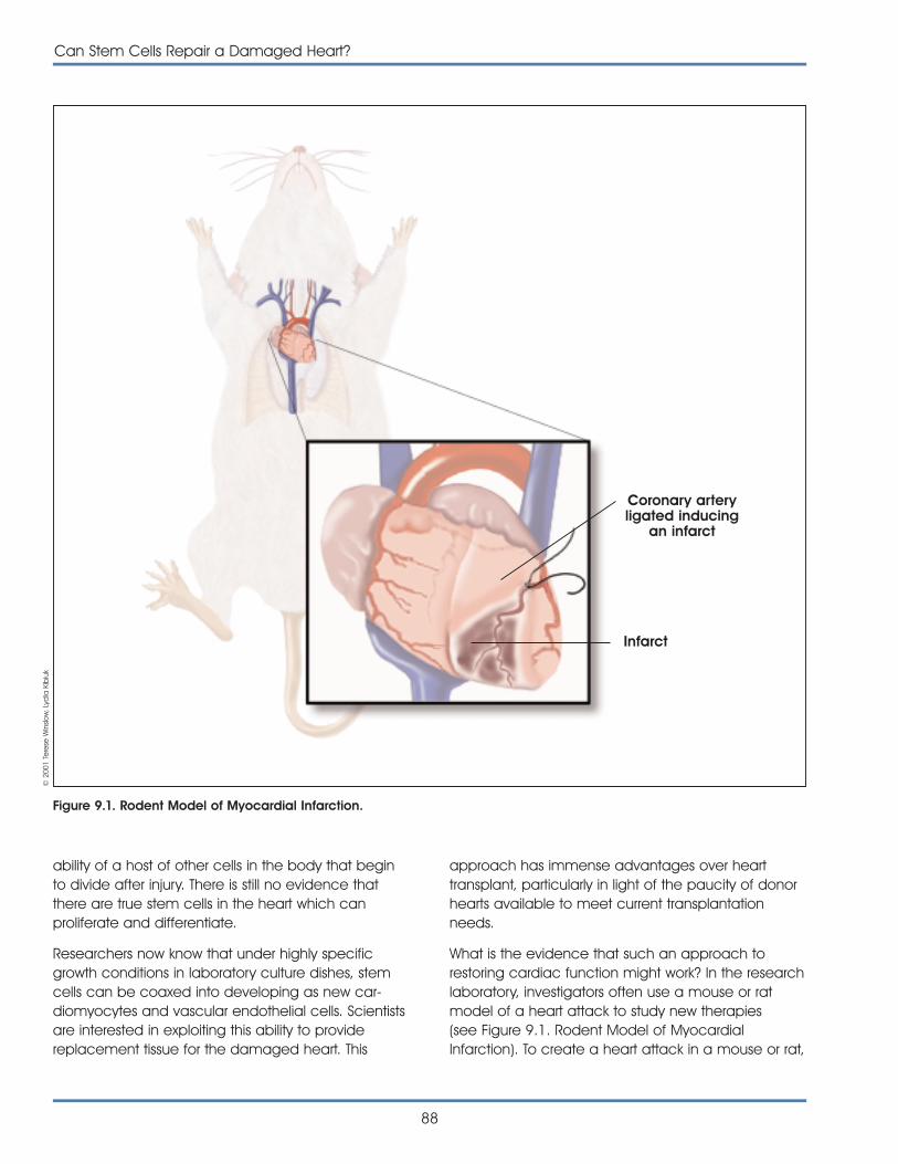
ability of a host of other cells in the body that beginto divide after injury. There is still no evidence thatthere are true stem cells in the heart which canproliferate and differentiate.
Researchers now know that under highly specificgrowth conditions in laboratory culture dishes, stemcells can be coaxed into developing as new car-diomyocytes and vascular endothelial cells. Scientistsare interested in exploiting this ability to providereplacement tissue for the damaged heart. This
approach has immense advantages over hearttransplant, particularly in light of the paucity of donorhearts available to meet current transplantationneeds.
What is the evidence that such an approach torestoring cardiac function might work? In the researchlaboratory, investigators often use a mouse or ratmodel of a heart attack to study new therapies(see Figure 9.1. Rodent Model of MyocardialInfarction). To create a heart attack in a mouse or rat,
88
Can Stem Cells Repair a Damaged Heart?
Figure 9.1. Rodent Model of Myocardial Infarction.
Coronary arteryligated inducing
an infarct
Infarct
© 2
001
Tere
se W
inslo
w, L
ydia
Kib
iuk
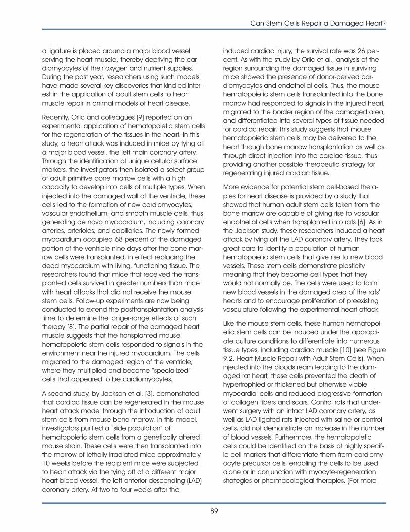
Can Stem Cells Repair a Damaged Heart?
89
a ligature is placed around a major blood vesselserving the heart muscle, thereby depriving the car-diomyocytes of their oxygen and nutrient supplies.During the past year, researchers using such modelshave made several key discoveries that kindled inter-est in the application of adult stem cells to heartmuscle repair in animal models of heart disease.
Recently, Orlic and colleagues [9] reported on anexperimental application of hematopoietic stem cellsfor the regeneration of the tissues in the heart. In thisstudy, a heart attack was induced in mice by tying offa major blood vessel, the left main coronary artery.Through the identification of unique cellular surfacemarkers, the investigators then isolated a select groupof adult primitive bone marrow cells with a highcapacity to develop into cells of multiple types. Wheninjected into the damaged wall of the ventricle, thesecells led to the formation of new cardiomyocytes,vascular endothelium, and smooth muscle cells, thusgenerating de novo myocardium, including coronaryarteries, arterioles, and capillaries. The newly formedmyocardium occupied 68 percent of the damagedportion of the ventricle nine days after the bone mar-row cells were transplanted, in effect replacing thedead myocardium with living, functioning tissue. Theresearchers found that mice that received the trans-planted cells survived in greater numbers than micewith heart attacks that did not receive the mousestem cells. Follow-up experiments are now beingconducted to extend the posttransplantation analysistime to determine the longer-range effects of suchtherapy [8]. The partial repair of the damaged heartmuscle suggests that the transplanted mousehematopoietic stem cells responded to signals in theenvironment near the injured myocardium. The cellsmigrated to the damaged region of the ventricle,where they multiplied and became “specialized”cells that appeared to be cardiomyocytes.
A second study, by Jackson et al. [3], demonstratedthat cardiac tissue can be regenerated in the mouseheart attack model through the introduction of adultstem cells from mouse bone marrow. In this model,investigators purified a “side population” ofhematopoietic stem cells from a genetically alteredmouse strain. These cells were then transplanted intothe marrow of lethally irradiated mice approximately10 weeks before the recipient mice were subjectedto heart attack via the tying off of a different majorheart blood vessel, the left anterior descending (LAD)coronary artery. At two to four weeks after the
induced cardiac injury, the survival rate was 26 per-cent. As with the study by Orlic et al., analysis of theregion surrounding the damaged tissue in survivingmice showed the presence of donor-derived car-diomyocytes and endothelial cells. Thus, the mousehematopoietic stem cells transplanted into the bonemarrow had responded to signals in the injured heart,migrated to the border region of the damaged area,and differentiated into several types of tissue neededfor cardiac repair. This study suggests that mousehematopoietic stem cells may be delivered to theheart through bone marrow transplantation as well asthrough direct injection into the cardiac tissue, thusproviding another possible therapeutic strategy forregenerating injured cardiac tissue.
More evidence for potential stem cell-based thera-pies for heart disease is provided by a study thatshowed that human adult stem cells taken from thebone marrow are capable of giving rise to vascularendothelial cells when transplanted into rats [6]. As inthe Jackson study, these researchers induced a heartattack by tying off the LAD coronary artery. They tookgreat care to identify a population of humanhematopoietic stem cells that give rise to new bloodvessels. These stem cells demonstrate plasticitymeaning that they become cell types that theywould not normally be. The cells were used to formnew blood vessels in the damaged area of the rats’hearts and to encourage proliferation of preexistingvasculature following the experimental heart attack.
Like the mouse stem cells, these human hematopoi-etic stem cells can be induced under the appropri-ate culture conditions to differentiate into numeroustissue types, including cardiac muscle [10] (see Figure9.2. Heart Muscle Repair with Adult Stem Cells). Wheninjected into the bloodstream leading to the dam-aged rat heart, these cells prevented the death ofhypertrophied or thickened but otherwise viablemyocardial cells and reduced progressive formationof collagen fibers and scars. Control rats that under-went surgery with an intact LAD coronary artery, aswell as LAD-ligated rats injected with saline or controlcells, did not demonstrate an increase in the numberof blood vessels. Furthermore, the hematopoieticcells could be identified on the basis of highly specif-ic cell markers that differentiate them from cardiomy-ocyte precursor cells, enabling the cells to be usedalone or in conjunction with myocyte-regenerationstrategies or pharmacological therapies. (For more
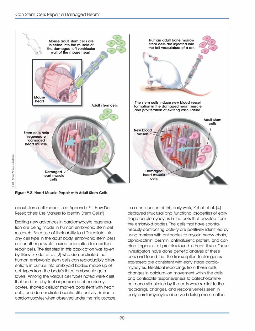
about stem cell markers see Appendix E.i. How DoResearchers Use Markers to Identify Stem Cells?)
Exciting new advances in cardiomyocyte regenera-tion are being made in human embryonic stem cellresearch. Because of their ability to differentiate intoany cell type in the adult body, embryonic stem cellsare another possible source population for cardiac-repair cells. The first step in this application was takenby Itskovitz-Eldor et al. [2] who demonstrated thathuman embryonic stem cells can reproducibly differ-entiate in culture into embryoid bodies made up ofcell types from the body ’s three embryonic germlayers. Among the various cell types noted were cellsthat had the physical appearance of cardiomy-ocytes, showed cellular markers consistent with heartcells, and demonstrated contractile activity similar tocardiomyocytes when observed under the microscope.
90
Can Stem Cells Repair a Damaged Heart?
In a continuation of this early work, Kehat et al. [4]displayed structural and functional properties of earlystage cardiomyocytes in the cells that develop fromthe embryoid bodies. The cells that have sponta-neously contracting activity are positively identified byusing markers with antibodies to myosin heavy chain,alpha-actinin, desmin, antinaturietic protein, and car-diac troponin—all proteins found in heart tissue. Theseinvestigators have done genetic analysis of thesecells and found that the transcription-factor genesexpressed are consistent with early stage cardio-myocytes. Electrical recordings from these cells,changes in calcium-ion movement within the cells,and contractile responsiveness to catecholaminehormone stimulation by the cells were similar to therecordings, changes, and responsiveness seen inearly cardiomyocytes observed during mammalian
Figure 9.2. Heart Muscle Repair with Adult Stem Cells.
Mouse adult stem cells areinjected into the muscle of
the damaged left ventricularwall of the mouse heart.
Human adult bone marrowstem cells are injected intothe tail vasculature of a rat.
The stem cells induce new blood vesselformation in the damaged heart muscleand proliferation of existing vasculature.
Mouseheart
Stem cells helpregeneratedamaged
heart muscle.
Damagedheart muscle
cells
Adult stem cells
Adult stemcells
New bloodvessels
Damagedheart muscle
cells
© 2
001
Tere
se W
inslo
w, L
ydia
Kib
iuk
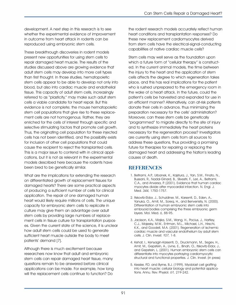
development. A next step in this research is to seewhether the experimental evidence of improvementin outcome from heart attack in rodents can bereproduced using embryonic stem cells.
These breakthrough discoveries in rodent modelspresent new opportunities for using stem cells torepair damaged heart muscle. The results of thestudies discussed above are growing evidence thatadult stem cells may develop into more cell typesthan first thought. In those studies, hematopoieticstem cells appear to be able to develop not only intoblood, but also into cardiac muscle and endothelialtissue. This capacity of adult stem cells, increasinglyreferred to as “plasticity,” may make such adult stemcells a viable candidate for heart repair. But thisevidence is not complete; the mouse hematopoieticstem cell populations that give rise to these replace-ment cells are not homogenous. Rather, they areenriched for the cells of interest through specific andselective stimulating factors that promote cell growth.Thus, the originating cell population for these injectedcells has not been identified, and the possibility existsfor inclusion of other cell populations that couldcause the recipient to reject the transplanted cells.This is a major issue to contend with in clinical appli-cations, but it is not as relevant in the experimentalmodels described here because the rodents havebeen bred to be genetically similar.
What are the implications for extending the researchon differentiated growth of replacement tissues fordamaged hearts? There are some practical aspectsof producing a sufficient number of cells for clinicalapplication. The repair of one damaged humanheart would likely require millions of cells. The uniquecapacity for embryonic stem cells to replicate inculture may give them an advantage over adultstem cells by providing large numbers of replace-ment cells in tissue culture for transplantation purpos-es. Given the current state of the science, it is unclearhow adult stem cells could be used to generatesufficient heart muscle outside the body to meetpatients’ demand [7].
Although there is much excitement becauseresearchers now know that adult and embryonicstem cells can repair damaged heart tissue, manyquestions remain to be answered before clinicalapplications can be made. For example, how longwill the replacement cells continue to function? Do
the rodent research models accurately reflect humanheart conditions and transplantation responses? Dothese new replacement cardiomyocytes derivedfrom stem cells have the electrical-signal-conductingcapabilities of native cardiac muscle cells?
Stem cells may well serve as the foundation uponwhich a future form of “cellular therapy” is construct-ed. In the current animal models, the time betweenthe injury to the heart and the application of stemcells affects the degree to which regeneration takesplace, and this has real implications for the patientwho is rushed unprepared to the emergency room inthe wake of a heart attack. In the future, could thepatient’s cells be harvested and expanded for use inan efficient manner? Alternatively, can at-risk patientsdonate their cells in advance, thus minimizing thepreparation necessary for the cells’ administration?Moreover, can these stem cells be genetically“programmed” to migrate directly to the site of injuryand to synthesize immediately the heart proteinsnecessary for the regeneration process? Investigatorsare currently using stem cells from all sources toaddress these questions, thus providing a promisingfuture for therapies for repairing or replacing thedamaged heart and addressing the Nation’s leadingcauses of death.
REFERENCES1. Beltrami, A.P., Urbanek, K., Kajstura, J., Yan, S.M., Finato, N.,
Bussani, R., Nadal-Ginard, B., Silvestri, F., Leri, A., Beltrami,C.A., and Anversa, P. (2001). Evidence that human cardiacmyocytes divide after myocardial infarction. N. Engl. J.Med. 344, 1750-1757.
2. Itskovitz-Eldor, J., Schuldiner, M., Karsenti, D., Eden, A.,Yanuka, O., Amit, M., Soreq, H., and Benvenisty, N. (2000).Differentiation of human embryonic stem cells intoembryoid bodies comprising the three embryonic germlayers. Mol. Med. 6, 88-95.
3. Jackson, K.A., Majka, S.M., Wang, H., Pocius, J., Hartley,C.J., Majesky, M.W., Entman, M.L., Michael, L.H., Hirschi,K.K., and Goodell, M.A. (2001). Regeneration of ischemiccardiac muscle and vascular endothelium by adult stemcells. J. Clin. Invest. 107, 1-8.
4. Kehat, I., Kenyagin-Karsenti, D., Druckmann, M., Segev, H.,Amit, M., Gepstein, A., Livne, E., Binah, O., Itskovitz-Eldor, J.,and Gepstein, L. (2001). Human embryonic stem cells candifferentiate into myocytes portraying cardiomyocyticstructural and functional properties. J. Clin. Invest. (in press)
5. Kessler, P.D. and Byrne, B.J. (1999). Myoblast cell graftinginto heart muscle: cellular biology and potential applica-tions. Annu. Rev. Physiol. 61, 219-242.
Can Stem Cells Repair a Damaged Heart?
91

6. Kocher, A.A., Schuster, M.D., Szabolcs, M.J., Takuma, S.,Burkhoff, D., Wang, J., Homma, S., Edwards, N.M., andItescu, S. (2001). Neovascularization of ischemic myocardi-um by human bone-marrow-derived angioblasts preventscardiomyocyte apoptosis, reduces remodeling andimproves cardiac function. Nat. Med. 7, 430-436.
7. Lanza, R., personal communication.
8. Orlic, D., personal communication.
9. Orlic, D., Kajstura, J., Chimenti, S., Jakoniuk, I., Anderson,S.M., Li, B., Pickel, J., McKay, R., Nadal-Ginard, B., Bodine,D.M., Leri, A., and Anversa, P. (2001). Bone marrow cellsregenerate infarcted myocardium. Nature. 410, 701-705.
10. Pittenger, M.F., Mackay, A.M., Beck, S.C., Jaiswal, R.K.,Douglas, R., Mosca, J.D., Moorman, M.A., Simonetti, D.W.,Craig, S., and Marshak, D.R. (1999). Multilineage potentialof adult human mesenchymal stem cells. Science. 284,143-147.
92
Can Stem Cells Repair a Damaged Heart?
93
The isolation of human stem cells offers the promiseof a remarkable array of novel therapeutics. Biologictherapies derived from such cells—through tissueregeneration and repair as well as through thetargeted delivery of genetic material—are expectedto be effective in the treatment of a wide range ofmedical conditions. Efforts to analyze and assess thesafety of using human stem cells in the clinical settingare vitally important to this endeavor.
Transplanted human stem cells are dynamic bio-logical entities that interact intimately with—and areinfluenced by—the physiology of the recipient. Beforethey are transplanted, cultured human stem cells aremaintained under conditions that promote either theself-renewing expansion of undifferentiated progeni-tors or the acquisition of differentiated propertiesindicative of the phenotype the cells will assume.After incompletely differentiated human stem cellsare transplanted, additional fine-tuning occurs as aconsequence of instructions received from the cells’physiologic microenvironments within the recipient.The capabilities to self-renew and differentiate thatare inherent to human stem cells point simultaneouslyto their perceived therapeutic potential and to thechallenge of assessing their safety.
Assessing human stem cell safety requires the imple-mentation of a comprehensive strategy. Each step inthe human stem cell development process—begin-ning with identifying and evaluating suitable humanstem cell sources—must be carefully scrutinized.Included in this global assessment are the derivation,expansion, manipulation, and characterization ofhuman stem cell lines, as well as preclinical efficacyand toxicity testing in appropriate animal models.Being able to trace back from the cell populationprepared for transplantation to the source of thefounder human stem cells also allows each safetycheckpoint to be connected, one to the other.
WEAVING A STEM CELL SAFETY NETA diversity of opinion exists among researchers aboutthe feasibility of initiating pilot clinical studies usinghuman stem cells. Some are of the view that it isreasonable to expect within the next five years thathuman stem cells will be used in transplantationsettings to replace dead or dying cells within organssuch as the failing heart or that genetically modifiedhuman stem cells will be created for delivery oftherapeutic genes. Others argue that a good dealmore information about the basic biology of humanstem cells needs to be accumulated before theirtherapeutic potential in humans can be assessed.
Clinical studies involving the transplantation of blood-restoring, or hematopoietic, stem cells have beenunder way for a number of years. Reconstituting theblood and immune systems through stem cell trans-plantation is an established practice for treatinghematological malignancies such as leukemia andlymphoma. Transplanting hematopoietic stem cellsresident in the bone marrow or isolated from cordblood or circulating peripheral blood is used tocounter the destruction of certain bone marrow cellscaused by high-intensity chemotherapeutic regimensused to battle various solid tumors. Moreover, clinicaltrials are being conducted to assess the safety andefficacy of using hematopoietic stem cell transplan-tation to treat various autoimmune conditions includ-ing multiple sclerosis, lupus, and rheumatoid arthritis.
Although precedents exist for the clinical use ofhuman stem cells, there is considerable reluctanceto proceed with clinical trials involving human stemcells derived from embryonic and fetal sources. Thishesitancy extends to adult human stem cells of non-hematopoietic origin, even though, by contrast, theirplasticity is generally considered to be lower than thatof their embryo- and fetus-derived counterparts. For
ASSESSING HUMANSTEM CELL SAFETYASSESSING HUMANSTEM CELL SAFETY
10.10.
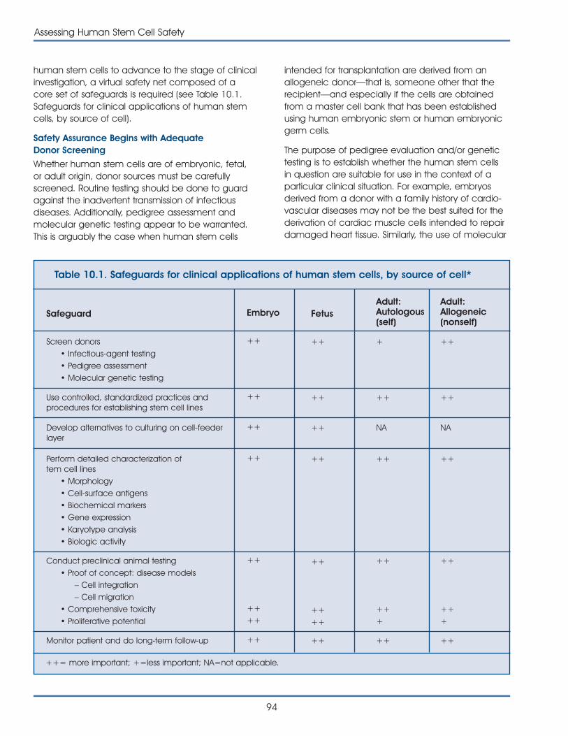
human stem cells to advance to the stage of clinicalinvestigation, a virtual safety net composed of acore set of safeguards is required (see Table 10.1.Safeguards for clinical applications of human stemcells, by source of cell).
Safety Assurance Begins with AdequateDonor Screening
Whether human stem cells are of embryonic, fetal,or adult origin, donor sources must be carefullyscreened. Routine testing should be done to guardagainst the inadvertent transmission of infectiousdiseases. Additionally, pedigree assessment andmolecular genetic testing appear to be warranted.This is arguably the case when human stem cells
intended for transplantation are derived from anallogeneic donor—that is, someone other that therecipient—and especially if the cells are obtainedfrom a master cell bank that has been establishedusing human embryonic stem or human embryonicgerm cells.
The purpose of pedigree evaluation and/or genetictesting is to establish whether the human stem cellsin question are suitable for use in the context of aparticular clinical situation. For example, embryosderived from a donor with a family history of cardio-vascular diseases may not be the best suited for thederivation of cardiac muscle cells intended to repairdamaged heart tissue. Similarly, the use of molecular
94
Assessing Human Stem Cell Safety
Table 10.1. Safeguards for clinical applications of human stem cells, by source of cell*
SafeguardAdult:Allogeneic(nonself)
Adult:Autologous(self)
Fetus Embryo
Screen donors• Infectious-agent testing• Pedigree assessment• Molecular genetic testing
+++++++
Use controlled, standardized practices andprocedures for establishing stem cell lines
++++++++
Develop alternatives to culturing on cell-feederlayer
NANA++++
Perform detailed characterization of tem cell lines
• Morphology• Cell-surface antigens• Biochemical markers• Gene expression• Karyotype analysis• Biologic activity
++++++++
Conduct preclinical animal testing• Proof of concept: disease models
– Cell integration– Cell migration
• Comprehensive toxicity• Proliferative potential
++
+++
++
++ ++
++
++ ++
Monitor patient and do long-term follow-up ++
++
+++
++++++
++= more important; +=less important; NA=not applicable.
Assessing Human Stem Cell Safety
95
genetic analysis could detect a mutation in the genefor alpha-synuclein. This gene is known to be respon-sible for the rare occurrence of early onset Parkinson’sDisease. Detecting such a genetic abnormality inneuronal progenitor cells derived from an establishedembryonic germ cell line could block the use ofthose cells as a treatment for a number of neuro-degenerative conditions, including Parkinson’s Disease.
The number of genes known to be directly respon-sible for causing disease or anomalous physiologicfunction is relatively small. Advances in techniques foridentifying, isolating, and analyzing genes, coupledwith the wealth of information destined to becomeavailable as one outcome of the human genomesequencing projects, will raise this number.Considerably more will also be learned about howmultiple gene products, each contributing an incre-mental quantity to the overall sum, predispose anindividual to develop particular diseases. Clearly, itwill eventually not be possible, or even necessary, toscreen every source of human stem cells for theentire panoply of disease-associated genes. Thescreening of targeted genes will be conducted withinthe context of the relevant clinical population.
Using Controlled, Standardized Practices andProcedures for Establishing Cultured Human StemCell Lines Enhances Safety
To ensure the integrity, uniformity, and reliability ofhuman stem cell preparations intended for clinicaluse, it is essential to demonstrate that rigorouslycontrolled, standardized practices and proceduresare being followed in establishing and maintaininghuman stem cell lines in culture.
Human stem cells from virtually every source otherthan blood-derived hematopoietic stem cells aremaintained in tissue culture for some defined periodof time. This is necessary to obtain a sufficient num-ber of cells for use in clinical studies involving trans-plantation. Culturing human stem cells requires theuse of formulated liquid media supplemented withgrowth factors and other chemical substances thatpromote cellular replication and govern the differenti-ation of the cultured human stem cells. Since humanstem cells are a dynamic, biological entity, failure tostandardize procedures for maintaining and expand-ing cells in culture could result in unintended alter-ations in the intrinsic properties of the cells. The initialseeding density of the cells, the frequency with whichthe culture medium is replenished, and the density
cells are permitted to achieve before subdividing willall affect the characteristics of human stem cellsmaintained in culture. Altering the concentrations ofsupplemental growth factors and chemical sub-stances, even switching from one supplier to another,may lead to changes in cell growth rate, expressionof defining cell markers, and differentiation potential.Alterations in stem cell properties caused by the useof nonstandardized culture practices are likely toaffect the behavior and effectiveness of the cellsonce transplanted.
One particular concern is how safe it is to use serumderived from cows as a supplement to culturemedia. Due to the outbreak of bovine spongiformencephalopathy (BSE) in cattle herds, primarily thoseraised in the United Kingdom, only serum producedfrom cows reared in countries certified to be free ofBSE should be used. Consumption of beef contami-nated with the agent responsible for causing BSE haslead to the limited emergence of new variantCreutzfeldt-Jakob disease (nvCJD) in humans. This dis-ease results in the relentless destruction of brain tissueand is invariably fatal. Placing neural stem cells con-taminated with the BSE infectious agent in a patient’snervous system to investigate cellular-replacementtherapies for neurological disorders would be bothirresponsible and devastating. Researchers areengaged in a vigorous effort to develop serum-free,chemically defined media that obviate risks asso-ciated with the use of bovine serum.
Alternatives to Culturing on a Feeder Layer ofAnimal Cells Improve Safety
An issue unique to the culturing of human embryonicstem and embryonic germ cells involves the use ofmouse embryonic fibroblast feeder cells to keep theembryonic cells in a proliferating, undifferentiatedcondition. Human embryonic stem and embryonicgerm cells are seeded directly onto a bed of irradiatedmouse feeder cells. Transplanting into humans stemcell preparations derived from founder cells that havebeen in direct, intimate contact with nonhumananimal cells constitutes xenotransplantation—the useof organs, tissues, and cells derived from animals totreat human disease. The principal concern of xeno-transplantation is the unintended transfer of animalviruses into humans.
Researchers are devoting considerable attention todeveloping culture conditions that do not use mousefeeder cells. In February of this year, scientists from
Geron Corporation, a biotech company focusing onthe development of embryonic stem cell technologyfor treating disease, presented findings at a scientificconference demonstrating that human embryonicstem cells can be maintained without mouse feedercells. Human embryonic stem cells seeded on acommercially available basement membrane matrixin media conditioned by feeder cells retain theirproliferative potential and capacity to form all threeembryonic germ layers (mesoderm, endoderm, andectoderm). This suggests that human embryonicstem cells maintained in the absence of directculture on a mouse feeder cell layer are comparableto human embryonic stem cells co-cultured withmouse feeder cells.
Detailed Characterization of Human Stem CellPopulations Reinforces the Safety Net
Detailed characterization of cell preparations intend-ed for transplantation is critical to the development ofhuman stem cells for clinical use. Identifying the cellsthat make up an human stem cell population intend-ed for clinical study requires identifying cells exhibitingthe desired phenotype within the preparation, as wellas those that do not. This poses considerable chal-lenges because human embryonic stem and embry-onic germ cells have the capacity to give rise to alldifferentiated cell types, while adult human stemcells, though generally more restricted in their plas-ticity, are capable of generating all cell types thatmake up the tissue from which they were derived.
On the basis of the complex biological properties ofhuman stem cells, including their potential to differen-tiate along multiple lineages and give rise to a varietyof cell types, it is expected that the characterizationof stem cell preparations will require a panel oforthogonal assessments. Parameters that will proveuseful in establishing identity include 1) cell morphology(visual microscopic inspection of cells to assess theirappearance), 2) expression of unique cell-surfaceantigens (as is the case for CD34+ hematopoieticstem cells), 3) characterization of biochemicalmarkers such as a tissue-specific enzymatic activity(e.g., enzymes that produce neurotransmitters fornerve cells), and 4) expression of genes that areunique to a particular cell type. Further, analysis of thenuclear chromosomal karyotype may be used toassess genetic stability of established human embry-onic stem and embryonic germ cell lines maintained
96
Assessing Human Stem Cell Safety
in culture for extended periods of time. Continueddevelopment and standardization of DNA microarrayanalysis (simultaneous screening for many genes)and proteomics (protein profiling) technologies willsignificantly enhance stem cell characterization.
Rigorous and quantitative identification of cell typeswithin a heterogeneous population of differentiatinghuman stem cells provides the means to gaugepurity of a cellular preparation. In turn, this permitsevaluation of the extent to which purity of a humanstem cell preparation predicts efficacy after trans-plantation. It is not necessarily the case that homo-genous populations composed of a single cell typewill be more effective as a cell-replacement therapythan mixed populations of cells. It is conceivable thatthe reason differentiation of cultured stem cellsobtained from the brain leads to formation of all thecell types found within the nervous system (namely,neurons, astrocytes, and oligodendrocytes) is thattheir coincidental presence is required to ensuremaximum survival and functional capability. Theinteraction of various phenotypic cell types within apreparation of progenitor cells obtained after thecontrolled differentiation of cultured human embry-onic stem cells is being actively investigated.
Once the purity profile has been established for apopulation of human stem cells generated usingstandardized procedures, deviations that occur out-side what is expected due to normal biologic varia-tion serve as a harbinger that significant, and possiblydeleterious, changes may have occurred. Such alter-ations could reflect the introduction of genetic muta-tions as a consequence of culture conditions used topromote expansion and to induce differentiation ofthe progenitor cell population.
Before clinical studies involving human stem celltransplantation can be done, it is essential to demon-strate that human stem cell preparations possessrelevant biological activity. The bioassay provides aquantitative measure of the potency of a cell prep-aration and ensures that cells destined for trans-plantation are not inert. Assays may be based on abiologic activity such as insulin release from pan-creatic islet-like cells, glycogen storage by cellsintended for regeneration of liver tissue, or synchro-nous contraction in the case of stem cell-derivedcardiomyocytes to be used for repairing damagedheart muscle. When cells that have not acquired fully
differentiated functionality are to be transplanted, itmay be appropriate to use surrogate markers thatpredict the acquisition of the intended biologicactivity upon further differentiation. (For example,counting tyrosine hydroxylase-expressing neuralprogenitor cells in a mixed population of cells intend-ed to provide dopaminergic neurons for treatingParkinson’s Disease could predict the acquisition ofrelevant biologic activity after transplantation.)
Proof of Concept, Toxicity Testing, and Evaluationof Proliferative Potential in Animal Models AreImportant to the Assessment of Human StemCell Safety
A critical element of the safety net is the transplan-tation of human stem cells into animals to demon-strate that the therapy does what it is supposed to do(“proof of concept”) and to assess toxicity. Admittedly,animal models of human disease are imperfectbecause most human maladies do not spontan-eously occur in animals. Chemical, surgical, andimmunologic methods are used to damage neurons;induce diabetes; simulate heart attacks, stroke, andhypertension; or compromise organ function. In situa-tions when focal genetic lesions are known to causedisease, the creation of transgenic mouse colonies inwhich the culpable gene is either eliminated or over-expressed results in disease models that are capableof faithfully reproducing human-disease-specificpathologies.
Human stem cells must be transplanted into animalmodels of human disease. Transplantation of neuralstem cells should demonstrate measurable evidenceof efficacy in models of neurodegenerative disease,such as Parkinson’s Disease, Huntington’s disease,and amyotrophic lateral sclerosis (ALS), Alzheimer ’sdisease, as well as spinal cord injury and stroke.Improved liver function after transplantation ofhepatocyte precursors should be observed in ananimal model of hepatic failure. Normalization ofblood insulin concentrations and amelioration ofdiabetic disease symptoms should result from thetransplantation of pancreatic islet progenitors in amouse model of diabetes. It is likely that in all cases,immunosuppression will be required due to immu-nologic incompatibility between humans and theanimal model species (usually mouse or rat).
In addition to efficacy, evidence for anatomic andfunctional integration of transplanted human stemcells should be assessed. human stem cells destinedfor transplantation may be tagged with a marker,such as green fluorescent protein, that allows trans-planted cells to be readily identified upon histologicalexamination. A similar approach should be used toevaluate the migration of transplanted human stemcells from the site of injection into adjacent and moredistant tissues. The migration of transplanted humanstem cells to a nontarget site and subsequent differ-entiation into a tissue type that is inappropriate forthat anatomic location could be problematic.
Questions about the use of embryonic comparedwith adult stem cells with respect to robustness anddurability should be addressed in animal-transplanta-tion models. Similarly, the issue of whether less-differ-entiated cells will be more effective than more-differ-entiated cells following transplantation should beinvestigated. Continued advancements in noninva-sive imaging technologies, such as magnetic reso-nance imaging (MRI) and positron emission tomogra-phy (PET scanning), will allow these events to beobserved in real time with reasonable resolution andwithout having to use large numbers of animals.
From the perspective of toxicology, the proliferativepotential of undifferentiated human embryonic andembryonic germ cells evokes the greatest level ofconcern. A characteristic of human embryonic stemcells is their capacity to generate teratomas whentransplanted into immunologically incompetentstrains of mice. Undifferentiated embryonic stem cellsare not considered as suitable for transplantation dueto the risk of unregulated growth. The question thatremains is, at what point during differentiation doesthis risk become insignificant, if ever? Identifying thestage at which the risk for tumor formation is mini-mized will depend on whether the process of stemcell differentiation occurs only in a forward directionor is reversible. Before clinical trials are begun inhumans, the issue of unregulated growth potentialand its relationship to stem cell differentiation mustbe evaluated. It is essential that careful toxicologystudies are performed that are of the appropriateduration and that involve transplantation intoimmunocompromised animals of undifferentiated orpartially differentiated embryonic stem cells, as wellas adult stem cells.
Assessing Human Stem Cell Safety
97

98
Assessing Human Stem Cell Safety
This page intentionally left blank
99
To date, only nonembryonic human stem cells havebeen used in cell-based gene therapy studies. Theinherent limitations of these stem cells, as discussedbelow, have prompted scientists to ponder andexplore whether human embryonic stem cells mightovercome the current barriers to the clinical successof cell-based gene therapies.
PRINCIPLES AND PROMISE OF GENETHERAPYGene therapy is a relatively recent, and still highlyexperimental, approach to treating human disease.While traditional drug therapies involve the adminis-tration of chemicals that have been manufacturedoutside the body, gene therapy takes a very differentapproach: directing a patient’s own cells to produceand deliver a therapeutic agent. The instructions forthis are contained in the therapeutic transgene (thenew genetic material introduced into the patient).Gene therapy uses genetic engineering—the intro-duction or elimination of specific genes by usingmolecular biology techniques to physically mani-pulate genetic material—to alter or supplement thefunction of an abnormal gene by providing a copyof a normal gene, to directly repair such a gene, orto provide a gene that adds new functions orregulates the activity of other genes.
Clinical efforts to apply genetic engineering tech-nology to the treatment of human diseases date to1989. Initially, gene therapy clinical trials focused oncancer, infectious diseases, or disorders in which onlya single gene is abnormal, such as cystic fibrosis.Increasingly however, efforts are being directedtoward complex, chronic diseases that involve morethan one gene. Prominent examples include heartdisease, inadequate blood flow to the limbs, arthritis,and Alzheimer’s disease.
The potential success of gene therapy technologydepends not only on the delivery of the therapeutictransgene into the appropriate human target cells,but also on the ability of the gene to functionproperly in the cell. Both requirements poseconsiderable technical challenges.
Gene therapy researchers have employed two majorstrategies for delivering therapeutic transgenes intohuman recipients (see Figure 11.1. Strategies forDelivering Therapeutic Transgenes into Patients). Thefirst is to “directly” infuse the gene into a person.Viruses that have been altered to prevent them fromcausing disease are often used as the vehicle fordelivering the gene into certain human cell types, inmuch the same way as ordinary viruses infect cells.This delivery method is fairly imprecise and limited tothe specific types of human cells that the viral vehiclecan infect. For example, some viruses commonlyused as gene-delivery vehicles can only infect cellsthat are actively dividing. This limits their usefulness intreating diseases of the heart or brain, because theseorgans are largely composed of nondividing cells.Nonviral vehicles for directly delivering genes intocells are also being explored, including the use ofplain DNA and DNA wrapped in a coat of fattymolecules known as liposomes.
The second strategy involves the use of living cells todeliver therapeutic transgenes into the body. In thismethod, the delivery cells—often a type of stem cell,a lymphocyte, or a fibroblast—are removed from thebody, and the therapeutic transgene is introducedinto them via the same vehicles used in the pre-viously described direct-gene-transfer method. Whilestill in the laboratory, the genetically modified cellsare tested and then allowed to grow and multiplyand, finally, are infused back into the patient.
USE OF GENETICALLY MODIFIEDSTEM CELLS IN EXPERIMENTALGENE THERAPIES
USE OF GENETICALLY MODIFIEDSTEM CELLS IN EXPERIMENTALGENE THERAPIES
11.11.
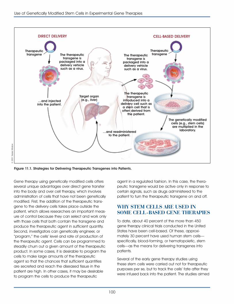
Gene therapy using genetically modified cells offersseveral unique advantages over direct gene transferinto the body and over cell therapy, which involvesadministration of cells that have not been geneticallymodified. First, the addition of the therapeutic trans-gene to the delivery cells takes place outside thepatient, which allows researchers an important meas-ure of control because they can select and work onlywith those cells that both contain the transgene andproduce the therapeutic agent in sufficient quantity.Second, investigators can genetically engineer, or“program,” the cells’ level and rate of production ofthe therapeutic agent. Cells can be programmed tosteadily churn out a given amount of the therapeuticproduct. In some cases, it is desirable to program thecells to make large amounts of the therapeuticagent so that the chances that sufficient quantitiesare secreted and reach the diseased tissue in thepatient are high. In other cases, it may be desirableto program the cells to produce the therapeutic
agent in a regulated fashion. In this case, the thera-peutic transgene would be active only in response tocertain signals, such as drugs administered to thepatient to turn the therapeutic transgene on and off.
WHY STEM CELLS ARE USED INSOME CELL-BASED GENE THERAPIESTo date, about 40 percent of the more than 450gene therapy clinical trials conducted in the UnitedStates have been cell-based. Of these, approxi-mately 30 percent have used human stem cells—specifically, blood-forming, or hematopoietic, stemcells—as the means for delivering transgenes intopatients.
Several of the early gene therapy studies usingthese stem cells were carried out not for therapeuticpurposes per se, but to track the cells’ fate after theywere infused back into the patient. The studies aimed
100
Use of Genetically Modified Stem Cells in Experimental Gene Therapies
Figure 11.1. Strategies for Delivering Therapeutic Transgenes into Patients.
DIRECT DELIVERY CELL-BASED DELIVERY
Therapeutictransgene
…and injectedinto the patient.
Target organ(e.g., liver)
…and readministeredto the patient.
TherapeutictransgeneThe therapeutic
transgene ispackaged into adelivery vehiclesuch as a virus.
The therapeutictransgene is
packaged into adelivery vehiclesuch as a virus.
The therapeutictransgene is
introduced into adelivery cell such as
a stem cell that isoften derived from
the patient.
The genetically modifiedcells (e.g., stem cells)are multiplied in the
laboratory.
© 2
001
Tere
se W
inslo
w
Use of Genetically Modified Stem Cells in Experimental Gene Therapies
101
to determine where the stem cells ended up andwhether they were indeed producing the desiredgene product, and if so, in what quantities and forwhat length of time. Of the stem cell-based genetherapy trials that have had a therapeutic goal,approximately one-third have focused on cancers(e.g., ovarian, brain, breast, myeloma, leukemia, andlymphoma), one-third on human immunodeficiencyvirus disease (HIV-1), and one-third on so-calledsingle-gene diseases (e.g., Gaucher ’s disease, severecombined immune deficiency (SCID), Fanconianemia, Fabry disease, and leukocyte adherencedeficiency).
But why use stem cells for this method of gene thera-py, and why hematopoietic stem cells in particular?The major reason for using stem cells in cell-basedgene therapies is that they are a self-renewingpopulation of cells and thus may reduce oreliminate the need for repeated administrations ofthe gene therapy.
Since the advent of gene therapy research,hematopoietic stem cells have been a delivery cellof choice for several reasons. First, although small innumber, they are readily removed from the body viathe circulating blood or bone marrow of adults or theumbilical cord blood of newborn infants. In addition,they are easily identified and manipulated in thelaboratory and can be returned to patients relativelyeasily by injection.
The ability of hematopoietic stem cells to give rise tomany different types of blood cells means that oncethe engineered stem cells differentiate, the thera-peutic transgene will reside in cells such as T and Blymphocytes, natural killer cells, monocytes,macrophages, granulocytes, eosinophils, basophils,and megakaryocytes. The clinical applications ofhematopoietic stem cell-based gene therapies arethus also diverse, extending to organ transplantation,blood and bone marrow disorders, and immunesystem disorders.
In addition, hematopoietic stem cells “home,” ormigrate, to a number of different spots in the body—primarily the bone marrow, but also the liver, spleen,and lymph nodes. These may be strategic locationsfor localized delivery of therapeutic agents for disor-ders unrelated to the blood system, such as liverdiseases and metabolic disorders such asGaucher ’s disease.
The only type of human stem cell used in genetherapy trials so far is the hematopoietic stem cell.However, several other types of stem cells are beingstudied as gene-delivery-vehicle candidates. Theyinclude muscle-forming stem cells known asmyoblasts, bone-forming stem cells calledosteoblasts, and neural stem cells.
Myoblasts appear to be good candidates for use ingene therapy because of an unusual and advanta-geous biological property: when injected into muscle,they fuse with nearby muscle fibers and become anintegral part of the muscle tissue. Moreover, sincemuscle tissue is generally well supplied with nervesand blood, the therapeutic agents produced by thetransgene are also accessible to nerves and thecirculatory system. Thus, myoblasts may not only beuseful for treating muscle disorders such as musculardystrophy, but also possibly nonmuscle disorders suchas neurodegenerative diseases, inherited hormonedeficiencies, hemophilia, and cancers.
Several promising animal studies of myoblast-mediated gene therapy have been reported [17]. Forinstance, this approach was successful in correctingliver and spleen abnormalities associated with alysosomal storage disease in mice. Investigators havealso achieved stable production of the human clot-ting factor IX deficient in hemophilia at therapeuticconcentrations in mice for at least eight months.Myoblasts engineered to secrete erythropoietin (ahormone that stimulates red blood cell production)were successful in reversing a type of anemiaassociated with end-stage renal disease in a mousemodel of renal failure.
Another animal study of myoblast-mediated genetransfer involved a mouse model of familial amy-otrophic lateral sclerosis (ALS, also known as LouGehrig’s disease), a fatal disorder characterized byprogressive degeneration of the brain and spinalcord nerves that control muscle activity. Investigatorsinjected myoblasts containing the transgene for ahuman nerve growth factor into the muscles of theALS mice before the onset of disease symptoms andmotor neuron degeneration. The transgene remainedactive in the muscle for up to 12 weeks, and, mostimportantly, the gene therapy successfully delayedthe onset of disease symptoms, slowed muscle atro-phy, and delayed the deterioration of motor skills [16].
In a series of experiments in rodents, a team of inves-tigators has been testing neural stem cells as vehiclesfor cell-based gene therapy for brain tumors knownas gliomas. Gliomas are virtually impossible to treatbecause the tumor cells readily invade the surround-ing tissue and migrate extensively into the normalbrain. The researchers genetically modified humanneural stem cells to produce a protein—cytosinedeaminase—that converts a nontoxic precursor druginto an active form that kills cancer cells. The engi-neered neural stem cells were then injected into thebrains of mice with human-derived gliomas. Withintwo weeks of the gene therapy and systemic treat-ment with the precursor drug, the tumors had shrunkby 80 percent. The animal studies also revealed thatneural stem cells were able to quickly and accurately“find” glioma cells, regardless of whether the stemcells were implanted directly into the tumors, implant-ed far from the tumors (but still within the brain), orinjected into circulating blood outside the brain [1].
Another cell-based gene therapy system underinvestigation involves the use of osteoblasts, or bone-forming stem cells. In a recent preliminary studyexamining a gene therapy approach to bone repairand regeneration, researchers genetically engineeredosteoblasts to produce a bone growth factor. Theosteoblasts were added to a biodegradable matrixthat could act as a “scaffold” for new bone forma-tion. Within a month after the cell-impregnatedscaffold was implanted into mice, new bone forma-tion was detectable. Although this work is in the veryearly stages, it offers hope of an effective alternativeto conventional bone-grafting techniques [14].
HOW EMBRYONIC STEM CELLSMIGHT PLAY A ROLE IN GENETHERAPY RESEARCHWith one notable exception, no therapeutic effectshave been achieved in gene therapy trials to date.The first successful gene therapy occurred in a recentFrench study in which a therapeutic transgene forcorrecting X-linked severe combined immunedeficiency was introduced into the bone marrowcells of children, resulting in improved function of theirimmune systems and correction of the disease [5].This encouraging success aside, the generally disap-pointing results are due, in part, to the inherent limita-tions of adult and cord blood stem cells. In principleat least, the use of human embryonic stem cells
102
Use of Genetically Modified Stem Cells in Experimental Gene Therapies
might overcome some of these limitations, but furtherresearch will be needed to determine whetherembryonic stem cells are better suited to meet theneeds of gene therapy applications than are adultstem cells.
One important feature of the optimal cell for deliver-ing a therapeutic transgene would be its ability toretain the therapeutic transgene even as it prolifer-ates or differentiates into specialized cells. Most of thecell-based gene therapies attempted so far haveused viral vehicles to introduce the transgene into thehematopoietic stem cell. One way to accomplish thisis to insert the therapeutic transgene into the one ofthe chromosomes of the stem cell. Retroviruses areable to do this, and for this reason, they are oftenused as the vehicle for infecting the stem cell andintroducing the therapeutic transgene into thechromosomal DNA. However, mouse retroviruses areonly efficient at infecting cells that are actively divid-ing. Unfortunately, hematopoietic stem cells are qui-escent and seldom divide. The percentage of stemcells that actually receive the therapeutic transgenehas usually been too low to attain a therapeuticeffect. Because of this problem, investigators havebeen exploring the use of viral vehicles that caninfect nondividing cells, such as lentiviruses (e.g., HIV)or adeno-associated viruses. This approach has notbeen entirely successful, however, because ofproblems relating to the fact that the cells themselvesare not in an active state [13, 19].
One approach to improving the introduction of trans-genes into hematopoietic stem cells has been tostimulate the cells to divide so that the viral vehiclescan infect them and insert the therapeutic transgene.Inder Verma of the Salk Institute has noted, however,that this manipulation can change other importantproperties of the hematopoietic stem cells, such asplasticity, self-renewal, and the ability to survive andgrow when introduced into the patient [23]. This possi-bility might be overcome with the use of embryonicstem cells if they require less manipulation. And infact, some preliminary data suggest that retroviralvectors may work more efficiently with embryonicstem cells than with the more mature adult stemcells. For example, researchers have noted that retro-viral vectors introduce transgenes into human fetalcord blood stem cells more efficiently than into cordblood stem cells from newborns, and that the fetalcord blood stem cells also had a higher proliferative
capacity (i.e., they underwent more subsequent celldivisions). This suggests that fetal cord blood stem cellsmight be useful in cell-based in utero gene therapyto correct hematopoietic disorders before birth [15, 21].
In some cases—such as a treatment of a chronicdisease—achieving continued production of the ther-apeutic transgene over the life of the patient will bevery important. Generally, however, gene therapiesusing hematopoietic stem cells have encountered aphenomenon known as “gene silencing,” where, overtime, the therapeutic transgene gets “turned off” dueto cellular mechanisms that alter the structure of thearea of the chromosome where the therapeuticgene has been inserted [6, 7, 11, 22, 24]. Whetherthe use of embryonic stem cells in gene therapycould overcome this problem is unknown, althoughpreliminary evidence suggests that this phenomenonmay occur in these cells as well [8,18].
Persistence of the cell containing the therapeutictransgene is equally important for ensuring continuedavailability of the therapeutic agent. Verma notedthat the optimal cells for cell-mediated gene transferwould be cells that will persist for “the rest of thepatient’s life; they can proliferate and they wouldmake the missing protein constantly and forever” [23].Persistence, or longevity, of the cells can come aboutin two ways: a long life span for an individual cell, ora self-renewal process whereby a short-lived cellundergoes successive cell divisions while maintainingthe therapeutic transgene. Ideally, then, the geneti-cally modified cell for use in cell-based gene therapyshould be able to self-renew (in a controlled mannerso tumors are not formed) so that the therapeuticagent is available on a long-term basis. This is one ofthe reasons why stem cells are used, but adult stemcells seem to be much more limited in the numberof times they can divide compared with embryonicstem cells. The difference between the ability of adultand embryonic stem cells to self-renew has beendocumented in the mouse, where embryonic stemscells were shown to have a much higher proliferativecapacity than do adult hematopoietic stem cells [25].
Researchers are beginning to understand the biolo-gical basis of the difference in proliferative capacitybetween adult and embryonic stem cells. Persistenceof cells and the ability to undergo successive celldivisions are in part, at least, a function of the lengthof structures at the tips of chromosomes calledtelomeres. Telomere length is, in turn, maintained by
an enzyme known as telomerase. Low levels oftelomerase activity result in short telomeres and, thus,fewer rounds of cell division—in other words, shorterlongevity. Higher levels of telomerase activity result inlonger telomeres, more possible cell divisions, andoverall longer persistence. Mouse embryonic stemcells have been found to have longer telomeres andhigher levels of telomerase activity compared withadult stem cells and other more specialized cells inthe body. As mouse embryonic stem cells give rise tohematopoietic stem cells, telomerase activity levelsdrop, suggesting a decrease in the self-renewingpotential of the hematopoietic stem cells [3,4]. (Formore detailed information regarding telomeres andtelomerase, see Figure C.2. Telomeres andTelomerase.)
Human embryonic stem cells have also been shownto maintain pluripotency (the ability to give rise toother, more specialized cell types) and the ability toproliferate for long periods in cell culture in the labo-ratory [2]. Adult stem cells appear capable of only alimited number of cell divisions, which would preventlong-term expression of the therapeutic gene need-ed to correct chronic diseases. “Embryonic stem cellscan be maintained in culture, whereas that is nearlyimpossible with cord blood stem cells,” says RobertHawley of the American Red Cross Jerome H. HollandLaboratory for Biomedical Sciences, who is develop-ing gene therapy vectors for insertion into humanhematopoietic cells [12]. “So with embryonic stemcells, you have the possibility of long-term mainte-nance and expansion of cell lines, which has notbeen possible with hematopoietic stem cells.”
The patient’s immune system response can beanother significant challenge in gene therapy. Mostcells have specific proteins on their surface that allowthe immune system to recognize them as either “self”or “nonself.” These proteins are known as major histo-compatibility proteins, or MHC proteins. If adult stemcells for use in gene therapy cannot be isolated fromthe patient, donor cells can be used. But because ofthe differences in MHC proteins among individuals,the donor stem cells may be recognized as nonselfby the patient’s immune system and be rejected.
John Gearhart of Johns Hopkins University and PeterRathjen at the University of Adelaide speculate thatembryonic stem cells may be useful for avoidingsuch immune reactions [10, 20]. For instance, it maybe possible to establish an extensive “bank” of
Use of Genetically Modified Stem Cells in Experimental Gene Therapies
103
embryonic stem cell lines, each with a different setof MHC genes. Then, an embryonic stem cell that isimmunologically compatible for a patient could beselected, genetically modified, and triggered todevelop into the appropriate type of adult stem cellthat could be administered to the patient. By gene-tically modifying the MHC genes of an embryonicstem cell, it may also be possible to create a “univer-sal” cell that would be compatible with all patients.Another approach might be to “customize” embry-onic stem cells such that cells derived from themhave a patient’s specific MHC proteins on theirsurface and then to genetically modify them for usein gene therapy. Such approaches are hypotheticalat this point, however, and research is needed toassess their feasibility.
Ironically, the very qualities that make embryonicstem cells potential candidates for gene therapy (i.e.,pluripotency and unlimited proliferative capacity) alsoraise safety concerns. In particular, undifferentiatedembryonic stem cells can give rise to teratomas,tumors composed of a number of different tissuetypes (see Chapter 10. Assessing Human Stem CellSafety). It may thus be preferable to use a differen-tiated derivative of genetically modified embryonicstem cells that can still give rise to a limited numberof cell types (akin to an adult stem cell). CautionsEsmail Zanjani of the University of Nevada, “We coulddifferentiate embryonic stem cells into, say, liver cells,and then use them, but I don’t see how we can takeembryonic stem cells per se and put genes intothem to use therapeutically” [26].
Further research is needed to determine whether thedifferentiated stem cells retain the advantages, suchas longer life span, of the embryonic stem cells fromwhich they were derived. Because of the difficulty inisolating and purifying many of the types of adultstem cells, embryonic stem cells may still be bettertargets for gene transfer. The versatile embryonicstem cell could be genetically modified, and then, intheory, it could be induced to give rise to all varietiesof adult stem cells. Also, since the genetically modi-fied stem cells can be easily expanded, large, purepopulations of the differentiated cells could be pro-duced and saved. Even if the differentiated cellswere not as long-lived as the embryonic stem cells,there would still be sufficient genetically modifiedcells to give to the patient whenever the needarises again.
Achieving clinical success with cell-based genetherapy will require new knowledge and advances inseveral key areas, including the design of viral andnonviral vehicles for introducing transgenes into cells,the ability to direct where in a cell the transgene isintroduced, the ability to direct the genetically modi-fied stem cells or the secreted therapeutic agent todiseased tissues, optimization and regulation of theproduction of the therapeutic agent within the stemcell, and management of immune reactions to thegene therapy process. The ability of embryonic stemcells to generate a wide variety of specialized celltypes and being able to maintain them in the labor-atory would make embryonic stem cells a promisingmodel for exploring critical questions in many ofthese areas.
“There are possibilities of long-term maintenance andexpansion of embryonic stem cells and of differen-tiation along specific lineages that have not beenpossible with hematopoietic stem cells,” Zanjani says.“And if they [embryonic stem cells] could be used [inthe laboratory] as a model for differentiation, youcould evaluate … vectors for gene delivery and getan idea of how genes are translated in patients.”Cynthia Dunbar, a gene therapy researcher at theNational Institutes of Health, similarly notes thatembryonic stem cells could be useful not only inscreening new viral and nonviral vectors designed tointroduce therapeutic transgenes into cells, but espe-cially for testing levels of production of the therapeu-tic agent after the embryonic stem cells differentiatein culture [9]. Explains Dunbar, “These behaviors arehard to predict for human cells based on animalstudies … so this would be a very useful laboratorytool.” Indeed, the major contribution of embryonicstem cells to gene therapy may be to advance thegeneral scientific knowledge needed to overcomemany of the current technical hurdles to successfultherapeutic gene transfer.
REFERENCES1. Aboody, K.S., Brown, A., Rainov, N.G., Bower, K.A., Liu, S.,
Yang, W., Small, J.E., Herrlinger, U., Ourednik, V., Black, P.M.,Breakefield, X.O., and Snyder, E.Y. (2000). Neural stem cellsdisplay extensive tropism for pathology in adult brain:evidence from intracranial gliomas. Proc. Natl. Acad. Sci. U. S. A. 97, 12846-12851.
104
Use of Genetically Modified Stem Cells in Experimental Gene Therapies
2. Amit, M., Carpenter, M.K., Inokuma, M.S., Chiu, C.P., Harris,C.P., Waknitz, M.A., Itskovitz-Eldor, J., and Thomson, J.A.(2000). Clonally derived human embryonic stem cell linesmaintain pluripotency and proliferative potential forprolonged periods of culture. Dev. Biol. 227, 271-278.
3. Armstrong, L., Lako, M., Lincoln, J., Cairns, P.M., and Hole, N.(2000). mTert expression correlates with telomerase activityduring the differentiation of murine embryonic stem cells.Mech. Dev. 97, 109-116.
4. Betts, D.H., Bordignon, V., Hill, J.R., Winger, Q., Westhusin,M.E., Smith, L.C., and King, W.A. (2001). Reprogramming oftelomerase activity and rebuilding of telomere length incloned cattle. Proc. Natl. Acad. Sci. U. S. A. 98, 1077-1082.
5. Cavazzana-Calvo, M., Hacein-Bey, S., de Saint, B.G., Gross,F., Yvon, E., Nusbaum, P., Selz, F., Hue, C., Certain, S.,Casanova, J.L., Bousso, P., Deist, F.L., and Fischer, A. (2000).Gene therapy of human severe combined immuno-deficiency (SCID)-X1 disease. Science. 288, 669-672.
6. Challita, P.M. and Kohn, D.B. (1994). Lack of expression froma retroviral vector after transduction of murine hemato-poietic stem cells is associated with methylation in vivo.Proc. Natl. Acad. Sci. U. S. A. 91, 2567-2571.
7. Chen, W.Y. and Townes, T.M. (2000). Molecular mechanismfor silencing virally transduced genes involves histonedeacetylation and chromatin condensation. Proc. Natl.Acad. Sci. U. S. A. 97, 377-382.
8. Cherry, S.R., Biniszkiewicz, D., van Parijs, L., Baltimore, D., andJaenisch, R. (2000). Retroviral expression in embryonic stemcells and hematopoietic stem cells. Mol. Cell. Biol. 20,7419-7426.
9. Dunbar, C., personal communication.
10. Gearhart, J. (1998). New potential for human embryonicstem cells. Science. 282, 1061-1062.
11. Halene, S. and Kohn, D.B. (2000). Gene therapy usinghematopoietic stem cells: Sisyphus approaches the crest.Hum. Gene Ther. 11, 1259-1267.
12. Hawley, R., personal communication.
13. Korin, Y.D. and Zack, J.A. (1998). Progression to the G(1)bphase of the cell cycle is required for completion ofhuman immunodeficiency virus type 1 reverse transcriptionin T cells. J. Virol. 72, 3161-3168.
14. Laurencin, C.T., Attawia, M.A., Lu, L.Q., Borden, M.D., Lu,H.H., Gorum, W.J., and Lieberman, J.R. (2001). Poly(lactide-co-glycolide)/hydroxyapatite delivery of BMP-2-producing cells:a regional gene therapy approach to bone regeneration.Biomaterials. 22, 1271-1277.
15. Luther-Wyrsch, A., Costello, E., Thali, M., Buetti, E., Nissen, C.,Surbek, D., Holzgreve, W., Gratwohl, A., Tichelli, A., andWodnar-Filipowicz, A. (2001). Stable transduction with lenti-viral vectors and amplification of immature hematopoieticprogenitors from cord blood of preterm human fetuses.Hum. Gene. Ther. 12, 377-389.
16. Mohajeri, M.H., Figlewicz, D.A., and Bohn, M.C. (1999).Intramuscular grafts of myoblasts genetically modified tosecrete glial cell line-derived neurotrophic factor preventmotoneuron loss and disease progression in a mousemodel of familial amyotrophic lateral sclerosis. Hum. GeneTher. 10, 1853-1866.
17. Ozawa, C.R., Springer, M.L., and Blau, H.M. (2000). A novelmeans of drug delivery: myoblast-mediated gene therapyand regulatable retroviral vectors. Annu. Rev. Pharmacol.Toxicol. 40, 295-317.
18. Pannell, D., Osborne, C.S., Yao, S., Sukonnik, T., Pasceri, P.,Karaiskakis, A., Okano, M., Li, E., Lipshitz, H.D., and Ellis, J.(2000). Retrovirus vector silencing is de novo methylaseindependent and marked by a repressive histone code.EMBO J. 19, 5884-5894.
19. Park, F., Ohashi, K., Chiu, W., Naldini, L., and Kay, M.A.(2000). Efficient lentiviral transduction of liver requires cellcycling in vivo. Nat. Genet. 24, 49-52.
20. Rathjen, P.D., Lake, J., Whyatt, L.M., Bettess, M.D., andRathjen, J. (1998). Properties and uses of embryonic stemcells: prospects for application to human biology andgene therapy. Reprod. Fertil. Dev. 10, 31-47.
21. Shields, L.E., Kiem, H.P., and Andrews, R.G. (2000). Highlyefficient gene transfer into preterm CD34+ hematopoieticprogenitor cells. Am. J. Obstet. Gynecol. 183, 732-737.
22. Struhl, K. (1998). Histone acetylation and transcriptionalregulatory mechanisms. Genes. Dev. 12, 599-606.
23. Verma, I., personal communication.
24. Wade, P.A., Pruss, D., and Wolffe, A.P. (1997). Histoneacetylation: chromatin in action. Trends Biochem. Sci. 22,128-132.
25. Yoder, M.C. and Hiatt, K. (1999). Murine yolk sac and bonemarrow hematopoietic cells with high proliferative potentialdisplay different capacities for producing colony-formingcells ex vivo. J. Hemato. Stem Cell Res. 8, 421-430.
26. Zanjani, E., personal communication.
Use of Genetically Modified Stem Cells in Experimental Gene Therapies
105

106
Use of Genetically Modified Stem Cells in Experimental Gene Therapies
This page intentionally left blank
A-1
How does a single cell—the fertilized egg—give riseto a complex, multicellular organism? The questionreflects one of the greatest mysteries of life, and rep-resents a fundamental challenge in developmentalbiology. As yet, knowledge about the processes bywhich a fertilized egg divides (cleavage), forms a ballof cells (morula), develops a cavity (blastocyst stage),forms the three primary germ layers of cells that willultimately give rise to all the cell types of the body(gastrula stage), and ultimately generates all thespecialized tissues and organs of a mature organismis far from complete. Little is known about the specificgenes that regulate these early events or how inter-actions among cells or how cellular interactions withother factors in the three-dimensional environment ofthe early embryo affect development. The processesby which a fertilized egg becomes an embryo,called embryogenesis, include coordinated celldivision, cell specialization, cell migration, andgenetically programmed cell death [24, 35].
A description of the stages of early embryogenesis inhumans and mice follows. It includes an explanationof some of the more technical terms and conceptsthat are used throughout the document. It alsoincludes a selective discussion of some of the genes,molecules, signaling pathways, and other influenceson early embryonic development in the living organ-ism (in vivo) that are used in experiments with stemcells maintained in the laboratory (in vitro).
EXPERIMENTAL SYSTEMS USED TOUNDERSTAND EMBRYOGENESISMany kinds of experimental systems have been usedto understand how a fertilized egg produces a blas-tocyst, the first structure in which any cell specializa-tion occurs, and a gastrula, in which the three embry-onic germ layers—endoderm, mesoderm, andectoderm—first appear. They include experimentswith yeast cells; invertebrates such as tiny, jellyfish-like
hydra, the microscopic roundworm Caenorhabditiselegans, and the fruit fly Drosophila melanogaster;and vertebrates such as amphibians, chick embryosand more recently, zebrafish Danio rerio, which aretransparent as embryos and allow the detailedmonitoring of cell differentiation and migration duringdevelopment. The vast majority of studies on embryo-genesis in mammals has been conducted in mice.For obvious ethical reasons, detailed research onhuman embryos has been limited. But the study ofembryogenesis in all of these systems yields as manyquestions as answers.
For example, what signals the earliest cell differen-tiation events in the embryo? What regulates theactivity of genes that are important for embryonicdevelopment? How and when are the axes of theembryo’s body—anterior-posterior (head-tail), dorsal-ventral (back-belly), left-right—determined? What roledoes genetically controlled cell death, also known asapoptosis, play in embryogenesis? What influencesthe cell cycle, the controlled series of molecularevents that leads to cell division or the cessation ofcell division?
Much of the information about human embryonicdevelopment comes from studies of embryogenesisin the mouse. Like mammalian embryonic develop-ment in general, many aspects of embryogenesis inmice resembles that of humans, but development inmice also differs in several important respects fromhuman development. For example, embryonic andfetal development in mice takes 18 to 20 days; inhumans, the process takes nine months. The placentaforms and functions differently in the two species. Inhumans, an embryonic disk develops after theembryo implants in the uterine wall, whereas in micean egg cylinder forms. The yolk sac of a mouseembryo persists and functions throughout gestation;in humans, the yolk sac functions only in earlyembryogenesis [28]. The primary roles of the human
EARLY DEVELOPMENT
APPENDIX A:
EARLY DEVELOPMENT
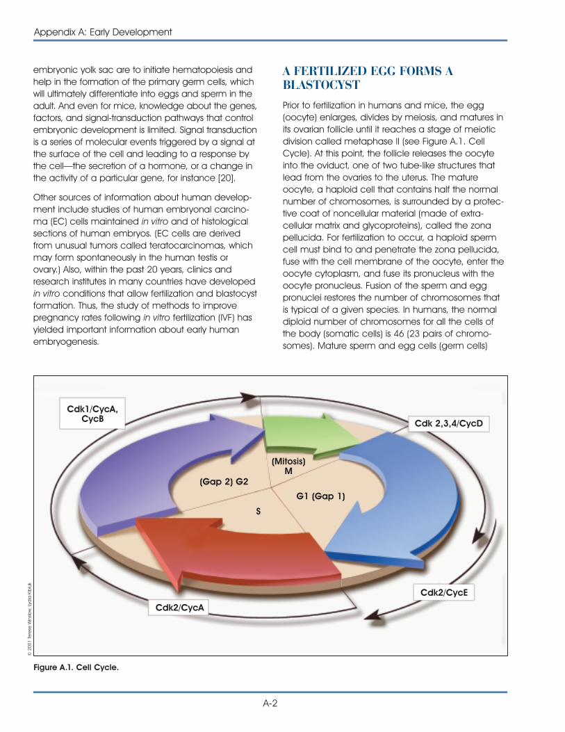
embryonic yolk sac are to initiate hematopoiesis andhelp in the formation of the primary germ cells, whichwill ultimately differentiate into eggs and sperm in theadult. And even for mice, knowledge about the genes,factors, and signal-transduction pathways that controlembryonic development is limited. Signal transductionis a series of molecular events triggered by a signal atthe surface of the cell and leading to a response bythe cell—the secretion of a hormone, or a change inthe activity of a particular gene, for instance [20].
Other sources of information about human develop-ment include studies of human embryonal carcino-ma (EC) cells maintained in vitro and of histologicalsections of human embryos. (EC cells are derivedfrom unusual tumors called teratocarcinomas, whichmay form spontaneously in the human testis orovary.) Also, within the past 20 years, clinics andresearch institutes in many countries have developedin vitro conditions that allow fertilization and blastocystformation. Thus, the study of methods to improvepregnancy rates following in vitro fertilization (IVF) hasyielded important information about early humanembryogenesis.
A-2
Appendix A: Early Development
A FERTILIZED EGG FORMS ABLASTOCYSTPrior to fertilization in humans and mice, the egg(oocyte) enlarges, divides by meiosis, and matures inits ovarian follicle until it reaches a stage of meioticdivision called metaphase II (see Figure A.1. CellCycle). At this point, the follicle releases the oocyteinto the oviduct, one of two tube-like structures thatlead from the ovaries to the uterus. The matureoocyte, a haploid cell that contains half the normalnumber of chromosomes, is surrounded by a protec-tive coat of noncellular material (made of extra-cellular matrix and glycoproteins), called the zonapellucida. For fertilization to occur, a haploid spermcell must bind to and penetrate the zona pellucida,fuse with the cell membrane of the oocyte, enter theoocyte cytoplasm, and fuse its pronucleus with theoocyte pronucleus. Fusion of the sperm and eggpronuclei restores the number of chromosomes thatis typical of a given species. In humans, the normaldiploid number of chromosomes for all the cells ofthe body (somatic cells) is 46 (23 pairs of chromo-somes). Mature sperm and egg cells (germ cells)
Cdk1/CycA,CycB Cdk 2,3,4/CycD
Cdk2/CycA
Cdk2/CycE
(Gap 2) G2
G1 (Gap 1)
(Mitosis)M
S
Figure A.1. Cell Cycle.
© 2
001
Tere
se W
inslo
w, L
ydia
Kib
iuk
in contrast, carry only 23 chromosomes, the haploidnumber [1].
Under normal conditions, fertilization of the humanoocyte occurs in the oviduct, near the ovary. Ahuman egg is many times larger than a sperm cell,which means the oocyte contributes most of thecytoplasm of the zygote, another name for the fertil-ized egg. As a result, any maternal gene products inthe zygote cytoplasm influence its first few divisions,called cleavages. Within several days, and afterseveral cleavages, the genome (all the DNA orhereditary information in the cell’s chromosomes) ofthe zygote becomes activated and controls subse-quent embryonic development [19, 28]. Also, duringthese initial cleavages, the resulting daughter cells donot increase in size. Rather, as early cell division pro-ceeds, the amount of cytoplasm of each daughtercell is reduced by half, and the total volume of theearly embryo remains unchanged from that of thefertilized egg [30, 35].
After fertilization, the zygote makes its way to theuterus, a journey that takes three to four days in miceand five to seven days in humans. As it travels, thezygote divides. The first cleavage produces twoidentical cells and then divides again to produce fourcells. If these cells separate, genetically identicalembryos result, the basis of identical twinning. Usually,however, the cells remain together, dividing asynchro-nously to produce 8 cells, 16 cells, and so on [19].Each early round of cell division takes approximately36 hours, according to information gleaned from thestudy of human embryos in vitro [34]. In humans andmice, at about the eight-cell stage, the embryocompacts, meaning that the formerly “loose” ball ofcells comes together in a tight array that is inter-connected by gap junctions. These specializedmembrane structures consist of an array of six proteinmolecules called connexins, which form a pore thatallows the exchange of ions and small moleculesbetween cells [27].
Recent information from studies of mouse embryosindicates that even at this early stage of embryo-genesis, the anterior-posterior axis of the embryo hasbeen established, a point of some concern for invitro fertilization techniques, which disrupt earlypatterning events. The establishment of the anterior-posterior axis is critical to normal fetal development,because it helps determine the overall body plan ofthe embryo [3, 15, 18].
By the 16-cell stage, the compacted embryo istermed a morula. In mice, the first evidence that cellshave become specialized occurs when the outercells of the 16-cell morula divide to produce an outerrim of cells—the trophectoderm—and an inner coreof cells, the inner cell mass [19]. Although the signalswithin the 16-cell morula that regulate the differentia-tion of the trophectoderm are largely unknown, it isclear that the outer cells of the morula are polarized.That is, one side of the cell differs from the other side.Thus, in the first differentiation event of embryogene-sis, the outer, polar cells give rise to trophectodermand the inner, apolar cells become the inner cellmass. This suggests that individual cells of the earlyembryo exhibit more intrinsic polarity than hadbeen thought [27].
Ultimately, the cells of the inner cell mass will give riseto all the tissues of the embryo’s body, as well as tothe nontrophoblast tissues that support the develop-ing embryo. The latter are referred to as extra-embryonic tissues and include the yolk sac, allantois,and amnion. The trophectoderm, in turn, will gener-ate the trophoblast cells of the chorion, the embryo’scontribution to the extraembryonic tissue known asthe placenta [19, 28].
The cells of the inner cell mass and trophectodermcontinue to divide. Information gained from the studyof mouse embryos suggests that the two tissues needto interact; the inner cell mass helps maintain theability of trophectoderm cells to divide, and thetrophectoderm appears to support the continueddevelopment of the inner cell mass [32]. Secretedparacrine factors (molecular signals that affect othercell types), including fibroblast growth factor-4(FGF-4), which is released from inner cell mass cells[46], help direct embryogenesis at this stage. FGF-4signaling also helps regulate the division and differ-entiation of trophectoderm cells [29].
By embryonic day 3 (E3.0) in the mouse and days 5to 6 in human development [14], the embryo devel-ops a cavity called the blastocoel. It fills with a wateryfluid secreted by trophectodermal cells and trans-ported in from the exterior. As a result of cavitationand the physical separation and differentiation of thetrophectoderm from the inner cell mass, the morulabecomes a blastocyst. Its chief structural features arethe outer sphere of flattened trophectoderm cells(which become the trophoblast), the small, round
Appendix A: Early Development
A-3
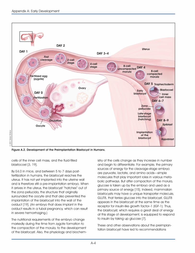
cells of the inner cell mass, and the fluid-filledblastocoel [3, 19].
By E4.0 in mice, and between 5 to 7 days post-fertilization in humans, the blastocyst reaches theuterus. It has not yet implanted into the uterine walland is therefore still a pre-implantation embryo. Whenit arrives in the uterus, the blastocyst “hatches” out ofthe zona pellucida, the structure that originallysurrounded the oocyte and that also prevented theimplantation of the blastocyst into the wall of theoviduct [19]. (An embryo that does implant in theoviduct results in a tubal pregnancy, which can resultin severe hemorrhaging.)
The nutritional requirements of the embryo changemarkedly during the time from zygote formation tothe compaction of the morula, to the developmentof the blastocyst. Also, the physiology and biochem-
istry of the cells change as they increase in numberand begin to differentiate. For example, the primarysources of energy for the cleavage-stage embryoare pyruvate, lactate, and amino acids—simplemolecules that play important roles in various meta-bolic pathways. But after compaction of the morula,glucose is taken up by the embryo and used as aprimary source of energy [15]. Indeed, mammalianblastocysts may have a unique transporter molecule,GLUT8, that ferries glucose into the blastocyst. GLUT8appears in the blastocyst at the same time as thereceptor for insulin-like growth factor–1 (IGF-1). Thus,the blastocyst, which requires a great deal of energyat this stage of development, is equipped to respondto insulin by taking up glucose [7].
These and other observations about the preimplan-tation blastocyst have led to recommendations
A-4
Appendix A: Early Development
Figure A.2. Development of the Preimplantation Blastocyst in Humans.
DAY 0
DAY 1
DAY 2
DAY 4
DAY 5
DAY 6–7
DAY 8–9
DAY 3–4
Ovulation
Oocyte
Ovary
Uterus
Fertilized egg(zygote)
Firstcleavage 2-cell
stage 4-cellstage 8-cell
uncompactedmorula 8-cell
compactedmorula
Trophectoderm
Blastocoel
Epiblast
Hypoblast
Implantationof the
blastocyst
Innercell mass
Earlyblastocyst
Late-stageblastocyst(hatching)
Zonapellucida
Fertilization
© 2
001
Tere
se W
inslo
w
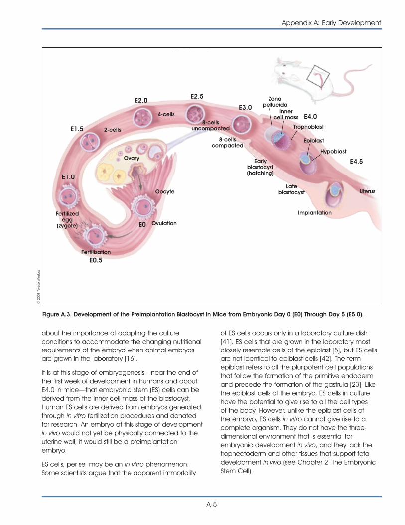
about the importance of adapting the cultureconditions to accommodate the changing nutritionalrequirements of the embryo when animal embryosare grown in the laboratory [16].
It is at this stage of embryogenesis—near the end ofthe first week of development in humans and aboutE4.0 in mice—that embryonic stem (ES) cells can bederived from the inner cell mass of the blastocyst.Human ES cells are derived from embryos generatedthrough in vitro fertilization procedures and donatedfor research. An embryo at this stage of developmentin vivo would not yet be physically connected to theuterine wall; it would still be a preimplantationembryo.
ES cells, per se, may be an in vitro phenomenon.Some scientists argue that the apparent immortality
of ES cells occurs only in a laboratory culture dish[41]. ES cells that are grown in the laboratory mostclosely resemble cells of the epiblast [5], but ES cellsare not identical to epiblast cells [42]. The termepiblast refers to all the pluripotent cell populationsthat follow the formation of the primitive endodermand precede the formation of the gastrula [23]. Likethe epiblast cells of the embryo, ES cells in culturehave the potential to give rise to all the cell typesof the body. However, unlike the epiblast cells ofthe embryo, ES cells in vitro cannot give rise to acomplete organism. They do not have the three-dimensional environment that is essential forembryonic development in vivo, and they lack thetrophectoderm and other tissues that support fetaldevelopment in vivo (see Chapter 2. The EmbryonicStem Cell).
Figure A.3. Development of the Preimplantation Blastocyst in Mice from Embryonic Day 0 (E0) Through Day 5 (E5.0).
Appendix A: Early Development
A-5
E1.0
Fertilizedegg
(zygote)
Fertilization
OvulationE0
Oocyte
Ovary
E1.5
E2.0E2.5
E4.5
E3.0
2-cells
4-cells8-cells
uncompacted
8-cellscompacted
Earlyblastocyst(hatching)
Lateblastocyst
Implantation
Uterus
Trophoblast
Epiblast
Hypoblast
Zonapellucida
Innercell mass E4.0
© 2
001
Tere
se W
inslo
w
E0.5
THE BLASTOCYST IMPLANTS IN THEUTERINE WALLMany of the molecular and cellular events that occurduring the second week of human embryonic devel-opment, and at the end of the first week of embryo-genesis in the mouse, help establish the placenta.The placenta connects the fetal and maternal blood-streams and provides nutrients to the embryothroughout the remainder of gestation.
On or about postfertilization days 8 to 9 in humans(and E4.5 in the mouse), the ball-shaped embryoimplants into in the uterine wall (see Figure A.2.Development of the Preimplantation Blastocyst inHumans). The inner cell mass of the human embryoat this stage has split into layers. One is the hypoblast,which lies next to the blastocoel and gives rise to theprimitive endoderm. (Later, the primitive endodermwill give rise to the outer layer of the yolk sac, acurious reminder of reptilian ancestry in mammalianembryos.) The other cell layer that develops from theinner cell mass is the epiblast. It will give rise to all thecells of the embryo’s body [19, 23].
The epiblast can be thought of as the group of cellsthat succeeds the inner cell mass. Pluripotent cellsare defined differently in scientific articles and textbooks. In general, however, pluripotent cells arecapable of giving rise to all the kinds of cells thatoccur in the mature organism. So at this stage ofembryogenesis, the only pluripotent cells are theundifferentiated cells of the epiblast.
By E6.0 in the mouse, three differentiated cell typesexist: the trophoblast, the epiblast (also called theembryonic ectoderm or primitive ectoderm at thisstage), and the primitive endoderm (see Figure A.3.Development of the Preimplantation Blastocyst inMice). During the next major phase of development,termed gastrulation, the embryonic ectoderm willdifferentiate into the three primary germ layers—endoderm, mesoderm, and ectoderm. Thus, theembryonic ectoderm has succeeded the epiblast asthe tissue that will generate the body of the embryo.The primitive endoderm differentiates into parietaland visceral endoderm, the anterior region of whichwill help regulate the development of the body planduring gastrulation [23].
Prior to gastrulation, the majority of cells (approxi-mately 75 percent) in the preimplantation blastocyst
comprise the trophectoderm and the primitiveendoderm. The preimplantation mouse embryoconsists of approximately 200 cells, approximately 20to 25 of which are inner cell mass or epiblast cells[20, 23]. The day 5, preimplantation human embryocontains 200 to 250 cells, only 30 to 34 of which areinner cell mass cells [4].
The extraembryonic cells of both species differentiateinto the tissues that will convey nutrients to theembryo and remove its waste products. For example,some of the trophoblast cells invade the epitheliallining of the uterus (also known as the decidua), andform a multinucleated tissue called a syncitium. Thissyncytiotrophoblast, as it is called, then developslacunae (cavities). By postfertilization day 10 to11 inhumans, the syncytiotrophoblast becomes suppliedwith maternal blood vessels. The fusion of the embry-onic chorion and the maternal decidua and vasculartissue generates the placenta [19, 29].
The formation of the placenta is a critical process inhuman embryogenesis. Without a healthy placenta,the embryo does not survive; its malformation cantrigger a spontaneous abortion [49]. The placentaanchors the developing embryo to the uterine walland connects it to the maternal bloodstream, thussupplying the embryo with ions and metabolites andproviding a waste-removal mechanism for theembryo [10, 26]. Later, the umbilical cord connectsthe embryo to the chorion portion of the placenta.The cord contains the fetal arteries and veins. Usually,the maternal and fetal blood do not mix directly.Instead, soluble substances pass through fingerlikeprojections called villi that have embedded in theuterine wall, and that have also developed from thetrophoblast of the embryo [19].
In mice, some of the genes that regulate the devel-opment of the placenta have been identified. Oneis the Mash2 gene, which is expressed in the tropho-blast cells of the embryo after it implants into theuterine wall. If Mash2 is inactivated, the placentadoes not form and the embryo dies (at E10.5 in themouse) [21, 45]. However, it is not known whether thesame genes that regulate placenta formation in themouse act in humans.
Meanwhile, during postfertilization days 7 to 14 ofhuman development, the epiblast splits to form theamnionic cavity. The cavity fills with fluid and cushionsthe embryo throughout gestation.
A-6
Appendix A: Early Development
THE BLASTOCYST BECOMESA GASTRULAAt the start of the third week of human development,and about E6.0 in the mouse (the egg-cylinderstage), the cells of the epiblast begin to differentiate.By the end of the third week, they will have generatedthe three primary germ layers of the embryo—endo-derm, mesoderm, and ectoderm. A detailed descrip-tion of all the events of this critical stage of differen-tiation—known as gastrulation—is beyond the scopeof this report. However, the onset of gastrulation istriggered at the posterior end of the embryo with theformation of a structure called the node (fromHensen’s node in chick embryogenesis). The node,together with another important signaling center, theanterior visceral endoderm (AVE), helps regulate theformation of the pattern of the embryo’s body at thisstage of development [19].
The process of gastrulation begins between days 14and 16 of human development and at about E6.5 inthe mouse. At that time, a primitive streak forms in aspecific region of the epiblast along the posterior axisof the embryo. Little is known about the signals thatregulate the generation of the primitive streak,although the genes goosecoid, T, Evx-1, and follistatinare expressed [23]. Nevertheless, the forward migra-tion of the posterior epiblast cells occurs as theircell-cell contacts break down, and they releaseenzymes that digest the basement membrane thatlies underneath. This allows the epiblast cells tomigrate into the space between the epiblast andthe visceral endoderm [6].
The forward-moving epiblast cells also spread later-ally, a migration that induces the formation of themesoderm and the notochord. The notochord is atemporary, rod-like structure that develops along thedorsal surface of the embryo and will ultimatelyconnect the anterior visceral endoderm (AVE) andthe node. Cells at the anterior end of the notochordwill eventually underlie the forebrain [19].
At the anterior end of the primitive streak is the node,a two-layered structure and important signalingcenter in the embryo. The ventral layer of cells in thenode comes from the epiblast and generates thenotochordal plate, which then forms the notochord.Endoderm, which will give rise to the gut, also devel-ops near the node, along the sides of the notochord.
Meanwhile, the anterior region of the mesoderm thatdevelops from the primitive streak is preparing to giverise to the heart. The anterior epiblast is generatingthe neuroectoderm and the ectoderm that coversthe surface of the embryo. The ectodermal tissuethat lies dorsal to the notochord will generate theneural plate, which will round up to form the neuraltube, the precursor to the central nervous system(brain and spinal cord) [23].
Thus, by the end of the third week of embryonicdevelopment in humans, and by E8.0 in the mouse,the primitive ectoderm of the postimplantationblastocyst has generated the ectoderm, mesoderm,and endoderm of the gastrula (see Figure A.4.Development of Human Embryonic Tissues). Theseand other complex processes result in the formationof the tissues and organs that occur in an adultmammal (see Figure 1.1. Differentiation of HumanTissues). They require the activation and inactivationof specific genes at specific times, highly integratedcell-cell interactions, and interactions between cellsand their noncellular environment, the extracellularmatrix [3, 19].
In general, the embryonic “outer” layer, or ectoderm,gives rise to the following tissues: central nervoussystem (brain and spinal cord) and peripheral nervoussystem; outer surface or skin of the organism; corneaand lens of the eye; epithelium that lines the mouthand nasal cavities and the anal canal; epitheliumof the pineal gland, pituitary gland, and adrenalmedulla; and cells of the neural crest (which givesrise to various facial structures, pigmented skin cellscalled melanocytes, and dorsal root ganglia, clustersof nerve cells along the spinal cord). The embryonic“middle” layer, or mesoderm, gives rise to skeletal,smooth, and cardiac muscle; structures of theurogenital system (kidneys, ureters, gonads, andreproductive ducts); bone marrow and blood; fat;bone, and cartilage; other connective tissues; andthe lining of the body cavity. The embryonic “inner”layer, or endoderm, gives rise to the epithelium of theentire digestive tract (excluding the mouth and analcanal); epithelium of the respiratory tract; structuresassociated with the digestive tract (liver and pan-creas); thyroid, parathyroid, and thymus glands;epithelium of the reproductive ducts and glands;epithelium of the urethra and bladder [19].
Appendix A: Early Development
A-7
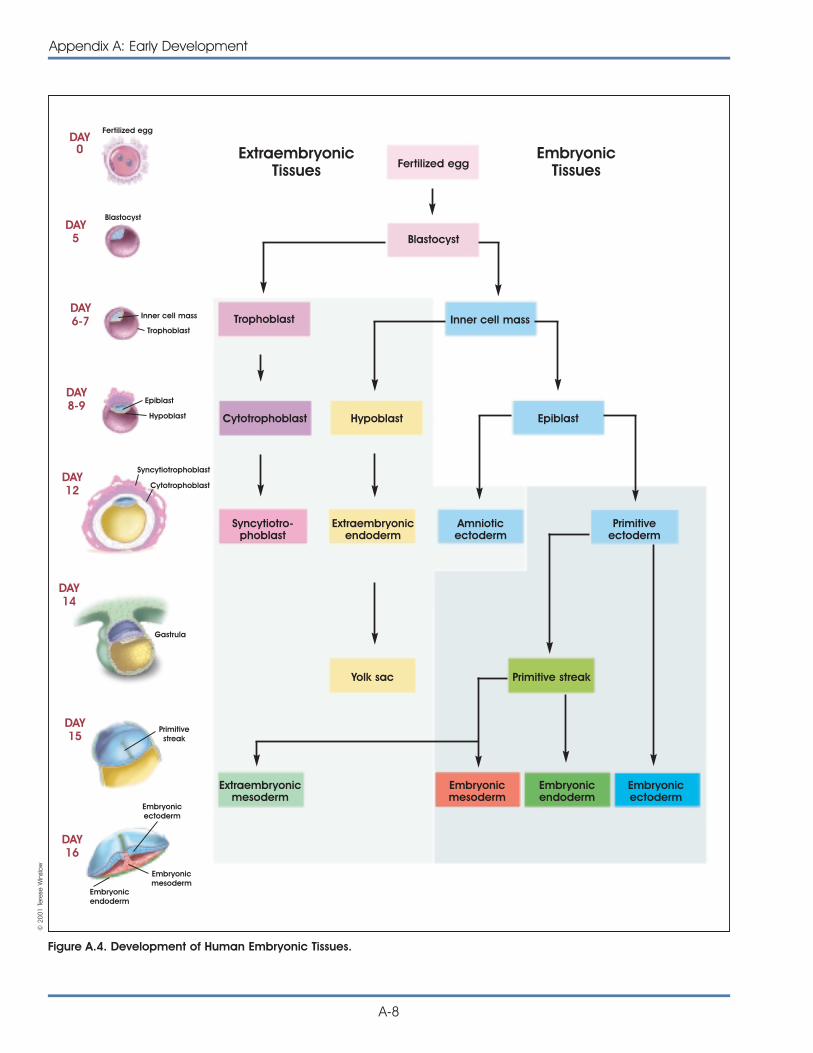
A-8
Appendix A: Early Development
Figure A.4. Development of Human Embryonic Tissues.
Fertilized eggEmbryonic
TissuesExtraembryonic
Tissues
Fertilized egg
Blastocyst
Inner cell mass
Trophoblast
Epiblast
Hypoblast
Syncytiotrophoblast
Cytotrophoblast
Gastrula
Primitivestreak
Embryonicendoderm
Embryonicmesoderm
Embryonicectoderm
Blastocyst
Trophoblast Inner cell mass
Cytotrophoblast Hypoblast Epiblast
Syncytiotro-phoblast
Yolk sac Primitive streak
Extraembryonicmesoderm
Embryonicmesoderm
Embryonicendoderm
Embryonicectoderm
Extraembryonicendoderm
Amnioticectoderm
Primitiveectoderm
DAY0
DAY5
DAY6-7
DAY8-9
DAY12
DAY14
DAY15
DAY16
© 2
001
Tere
se W
inslo
w
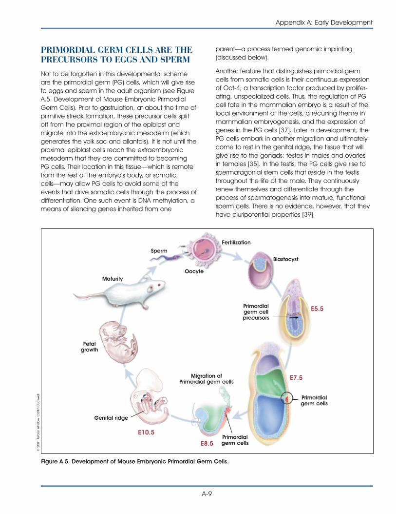
PRIMORDIAL GERM CELLS ARE THEPRECURSORS TO EGGS AND SPERM Not to be forgotten in this developmental schemeare the primordial germ (PG) cells, which will give riseto eggs and sperm in the adult organism (see FigureA.5. Development of Mouse Embryonic PrimordialGerm Cells). Prior to gastrulation, at about the time ofprimitive streak formation, these precursor cells splitoff from the proximal region of the epiblast andmigrate into the extraembryonic mesoderm (whichgenerates the yolk sac and allantois). It is not until theproximal epiblast cells reach the extraembryonicmesoderm that they are committed to becomingPG cells. Their location in this tissue—which is remotefrom the rest of the embryo’s body, or somatic,cells—may allow PG cells to avoid some of theevents that drive somatic cells through the process ofdifferentiation. One such event is DNA methylation, ameans of silencing genes inherited from one
parent—a process termed genomic imprinting(discussed below).
Another feature that distinguishes primordial germcells from somatic cells is their continuous expressionof Oct-4, a transcription factor produced by prolifer-ating, unspecialized cells. Thus, the regulation of PGcell fate in the mammalian embryo is a result of thelocal environment of the cells, a recurring theme inmammalian embryogenesis, and the expression ofgenes in the PG cells [37]. Later in development, thePG cells embark in another migration and ultimatelycome to rest in the genital ridge, the tissue that willgive rise to the gonads: testes in males and ovariesin females [35]. In the testis, the PG cells give rise tospermatagonial stem cells that reside in the testisthroughout the life of the male. They continuouslyrenew themselves and differentiate through theprocess of spermatogenesis into mature, functionalsperm cells. There is no evidence, however, that theyhave pluripotential properties [39].
Appendix A: Early Development
A-9
Figure A.5. Development of Mouse Embryonic Primordial Germ Cells.
Fertilization
Sperm
Oocyte
Blastocyst
Maturity
Fetalgrowth
Primordialgerm cellprecursors
Primordialgerm cells
Primordialgerm cells
Genital ridge
Migration ofPrimordial germ cells
E5.5
E7.5
E8.5
E10.5
© 2
001
Tere
se W
inslo
w, C
aitl
in D
uckw
all
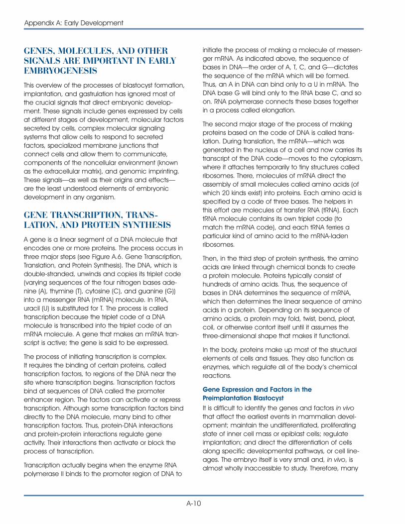
GENES, MOLECULES, AND OTHERSIGNALS ARE IMPORTANT IN EARLYEMBRYOGENESIS This overview of the processes of blastocyst formation,implantation, and gastrulation has ignored most ofthe crucial signals that direct embryonic develop-ment. These signals include genes expressed by cellsat different stages of development, molecular factorssecreted by cells, complex molecular signalingsystems that allow cells to respond to secretedfactors, specialized membrane junctions thatconnect cells and allow them to communicate,components of the noncellular environment (knownas the extracellular matrix), and genomic imprinting.These signals—as well as their origins and effects—are the least understood elements of embryonicdevelopment in any organism.
GENE TRANSCRIPTION, TRANS-LATION, AND PROTEIN SYNTHESISA gene is a linear segment of a DNA molecule thatencodes one or more proteins. The process occurs inthree major steps (see Figure A.6. Gene Transcription,Translation, and Protein Synthesis). The DNA, which isdouble-stranded, unwinds and copies its triplet code(varying sequences of the four nitrogen bases ade-nine (A), thymine (T), cytosine (C), and guanine (G))into a messenger RNA (mRNA) molecule. In RNA,uracil (U) is substituted for T. The process is calledtranscription because the triplet code of a DNAmolecule is transcribed into the triplet code of anmRNA molecule. A gene that makes an mRNA tran-script is active; the gene is said to be expressed.
The process of initiating transcription is complex.It requires the binding of certain proteins, calledtranscription factors, to regions of the DNA near thesite where transcription begins. Transcription factorsbind at sequences of DNA called the promoterenhancer region. The factors can activate or represstranscription. Although some transcription factors binddirectly to the DNA molecule, many bind to othertranscription factors. Thus, protein-DNA interactionsand protein-protein interactions regulate geneactivity. Their interactions then activate or block theprocess of transcription.
Transcription actually begins when the enzyme RNApolymerase II binds to the promoter region of DNA to
initiate the process of making a molecule of messen-ger mRNA. As indicated above, the sequence ofbases in DNA—the order of A, T, C, and G—dictatesthe sequence of the mRNA which will be formed.Thus, an A in DNA can bind only to a U in mRNA. TheDNA base G will bind only to the RNA base C, and soon. RNA polymerase connects these bases togetherin a process called elongation.
The second major stage of the process of makingproteins based on the code of DNA is called trans-lation. During translation, the mRNA—which wasgenerated in the nucleus of a cell and now carries itstranscript of the DNA code—moves to the cytoplasm,where it attaches temporarily to tiny structures calledribosomes. There, molecules of mRNA direct theassembly of small molecules called amino acids (ofwhich 20 kinds exist) into proteins. Each amino acid isspecified by a code of three bases. The helpers inthis effort are molecules of transfer RNA (tRNA). EachtRNA molecule contains its own triplet code (tomatch the mRNA code), and each tRNA ferries aparticular kind of amino acid to the mRNA-ladenribosomes.
Then, in the third step of protein synthesis, the aminoacids are linked through chemical bonds to createa protein molecule. Proteins typically consist ofhundreds of amino acids. Thus, the sequence ofbases in DNA determines the sequence of mRNA,which then determines the linear sequence of aminoacids in a protein. Depending on its sequence ofamino acids, a protein may fold, twist, bend, pleat,coil, or otherwise contort itself until it assumes thethree-dimensional shape that makes it functional.
In the body, proteins make up most of the structuralelements of cells and tissues. They also function asenzymes, which regulate all of the body ’s chemicalreactions.
Gene Expression and Factors in thePreimplantation Blastocyst
It is difficult to identify the genes and factors in vivothat affect the earliest events in mammalian devel-opment; maintain the undifferentiated, proliferatingstate of inner cell mass or epiblast cells; regulateimplantation; and direct the differentiation of cellsalong specific developmental pathways, or cell line-ages. The embryo itself is very small and, in vivo, isalmost wholly inaccessible to study. Therefore, many
A-10
Appendix A: Early Development
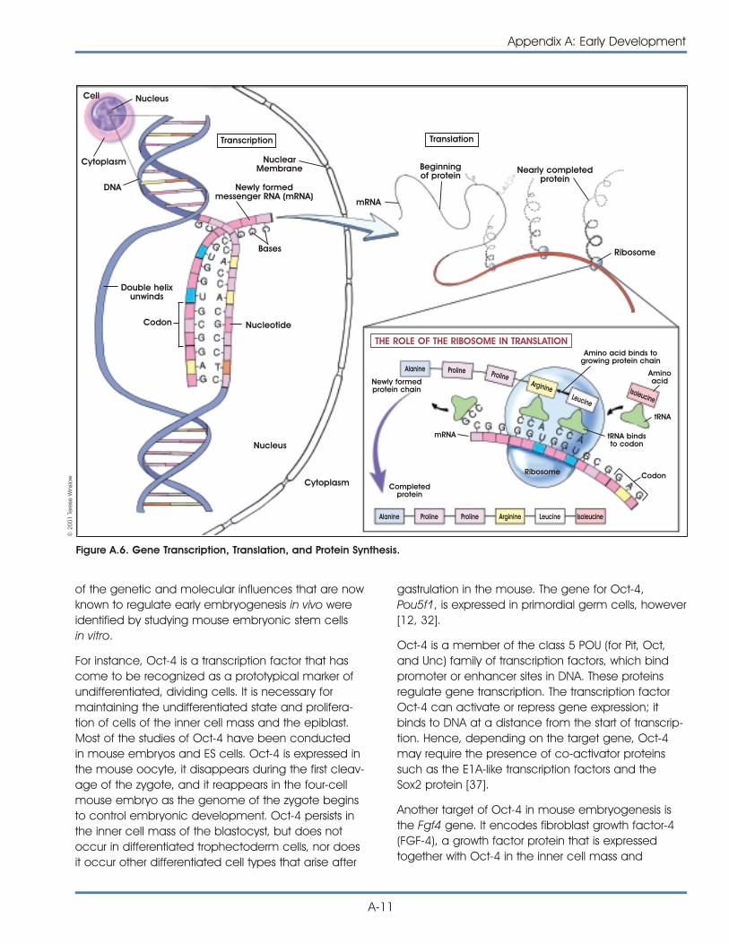
of the genetic and molecular influences that are nowknown to regulate early embryogenesis in vivo wereidentified by studying mouse embryonic stem cellsin vitro.
For instance, Oct-4 is a transcription factor that hascome to be recognized as a prototypical marker ofundifferentiated, dividing cells. It is necessary formaintaining the undifferentiated state and prolifera-tion of cells of the inner cell mass and the epiblast.Most of the studies of Oct-4 have been conductedin mouse embryos and ES cells. Oct-4 is expressed inthe mouse oocyte, it disappears during the first cleav-age of the zygote, and it reappears in the four-cellmouse embryo as the genome of the zygote beginsto control embryonic development. Oct-4 persists inthe inner cell mass of the blastocyst, but does notoccur in differentiated trophectoderm cells, nor doesit occur other differentiated cell types that arise after
gastrulation in the mouse. The gene for Oct-4,Pou5f1, is expressed in primordial germ cells, however[12, 32].
Oct-4 is a member of the class 5 POU (for Pit, Oct,and Unc) family of transcription factors, which bindpromoter or enhancer sites in DNA. These proteinsregulate gene transcription. The transcription factorOct-4 can activate or repress gene expression; itbinds to DNA at a distance from the start of transcrip-tion. Hence, depending on the target gene, Oct-4may require the presence of co-activator proteinssuch as the E1A-like transcription factors and theSox2 protein [37].
Another target of Oct-4 in mouse embryogenesis isthe Fgf4 gene. It encodes fibroblast growth factor-4(FGF-4), a growth factor protein that is expressedtogether with Oct-4 in the inner cell mass and
Appendix A: Early Development
A-11
Cell Nucleus
Transcription Translation
Cytoplasm
DNA Newly formedmessenger RNA (mRNA)
Bases
Double helixunwinds
Codon Nucleotide
Nucleus
Cytoplasm
NuclearMembrane Beginning
of protein
mRNA
Nearly completedprotein
Ribosome
Aminoacid
tRNA bindsto codon
Amino acid binds togrowing protein chain
tRNA
CodonRibosome
mRNA
Alanine Proline Proline Arginine Leucine Isoleucine
Alanine Proline ProlineArginine
Leucine
Newly formedprotein chain
Completedprotein
THE ROLE OF THE RIBOSOME IN TRANSLATION
Figure A.6. Gene Transcription, Translation, and Protein Synthesis.
Isoleucine
© 2
001
Tere
se W
inslo
w
epiblast [32]. FGF-4 is a paracrine signal, meaningthat it is released from one cell type and it acts onanother. In this case, FGF-4 is released from proliferat-ing inner cell mass cells and it affects the surroundingtrophectoderm. FGF-4 may also act as an autocrinesignal, meaning that it may affect the same inner cellmass cells that released it [13].
A series of recent experiments indicates that the levelof Oct-4 expression—not simply its presence orabsence in a cell—determines how mouse embry-onic stem cells differentiate and whether theycontinue to proliferate [33] (see Appendix B. MouseEmbryonic Stem Cells).
Two proteins, leptin and STAT3, which are producedby maternal granulosa cells that surround the oocyte,are apparently secreted into the oocyte as it maturesin its ovarian follicle. By the time the mouse or humanzygote reaches the four-cell stage, leptin and STAT3are concentrated in what may be the founder cell ofthe embryonic trophectoderm. Later, when the tro-phectoderm differentiates and separates from theinner cells mass of the blastocyst, leptin and STAT3 areexpressed only in the trophectoderm, where they playa critical role during implantation [12].
Leukemia inhibitory factor (LIF), a cytokine, also playsan important role during implantation. The LIF gene isexpressed in cultured mouse [31] and bovine blasto-cysts, as is the gene for its receptor. The mRNA for thereceptor for LIF is expressed in human blastocysts [12,47]. LIF, therefore, seems to be important for earlymammalian blastocyst development, as well asimplantation. It is also essential for the survival of theprimordial germ cells, which will become eggs andsperm in the mature organism [22]. And if mouseembryonic stem (ES) cells are cultured from the innercell mass of a blastocyst without the presence of“feeder” layers of cells, they require the addition of LIFto the culture medium in order to survive and pro-liferate [40]. Curiously, cultures of human ES cells donot respond to LIF [36, 48].
Regulation of Body Patterning in the Embryo
As the embryo forms, its overall body pattern is deter-mined by the establishment of three clear axes—theanterior-posterior axis (head-tail), the dorsal-ventral(back-belly) axis, and left-right asymmetry. The estab-lishment of these body axes at the correct time isfundamental to normal embryonic development. Forinstance, the central nervous system develops along
the dorsal surface, with the largest concentration ofneuronal tissue—the brain—at the anterior end ofthe embryo. The limbs develop symmetrically andbilaterally, whereas the heart—although it begins asa symmetrical structure—ultimately comes to pointtoward the left side of the trunk. Some internal struc-tures are paired (the kidneys, lungs, adrenal glands,testes, and ovaries), whereas many are not (the heart,gut, pancreas, spleen, liver, and uterus) [19].
Information about the establishment of these bodyaxes and their role in development is far from com-plete. For example, the anterior-posterior axis of themouse blastocyst may be determined before itimplants and is certainly established before gastru-lation [15, 17]. An unanswered question, however, iswhether this early embryonic axis helps determine thelater development of the embryo. The early axis mayplay a role in primitive streak formation, and requiresthe expression of Wnt, which helps regulate theformation of one of embryo’s chief signaling centers:the node [38].
As indicated above, an important group of cells thatproduces molecular signals that help determine theanterior-posterior axis of the mouse embryo is theanterior visceral endoderm (AVE). The AVE expressesdifferent genes along its length. At E5.0 in the mouse,for example, the Hex gene—a member of the familyof homeobox genes that help regulate body pattern-ing of the mouse embryo—is expressed in the distalvisceral endoderm. These cells migrate to becomethe AVE, which forms on the opposite side of theembryo from the primitive streak, thus establishing theanterior-posterior axis of the fetus [17].
Then, between E6.0 and E7.0 in the mouse, theanterior region of the AVE, where the heart will form,expresses Mrg1. The medial region expresses thetranscription factor genes Otx2 and Lim1, as well asother genes. The region of the AVE that lies next to thepart of the epiblast that will give rise to oral ectodermand the forebrain expresses Hesx1, another home-obox gene. Collectively, the AVE and the genes itexpresses help regulate the development of theanterior end of the embryo [3].
Other genes, notably Bmp4, also help shape themouse embryo prior to gastrulation. BMP stands forbone morphogenetic protein, a family of proteinsthat help regulate the differentiation of mesenchymalcells, which are derived from mesoderm, including
A-12
Appendix A: Early Development
bone-forming osteoblasts, and adipocytes, which arefat cells. They also play a role in CNS development.Bmp4, which is expressed in the extraembryonicectoderm next to the epiblast and also in the innercell mass of the E3.5 and E4.5 mouse blastocyst,may activate genes in epiblast cells that thenmigrate to form the primitive streak. Wnt3 apparentlyhelps induce the formation of both the primitivestreak and the node in mammals, although there isno evidence indicating that Wnt3 expression isrequired for mesoderm induction. However, formationof the embryo’s head region, obviously a key anteriorstructure, seems to require inhibition of the activities ofWnt and Bmp4—a potential role of the AVE [3, 18].
Therefore, coordinating the embryo’s “decisions”about its body pattern is a hierarchy of genes.Overall, the Hox genes specify anterior-posterior polar-ity. Their normal function can be subverted by retinoicacid, which can activate Hox genes in inappropriateplaces. Less is known about the establishment of thedorsal-ventral axis. It may be determined in the blas-tocyst, or even in the oocyte [16]; it is clearly estab-lished when the notochord develops. Genes such asNodal and Lefty help determine left-right asymmetry.Genes that regulate body patterning in embryonicdevelopment are well conserved throughout evolu-tion among both vertebrates and invertebrates [19].
Regulation of Cell Differentiation in EarlyEmbryogenesis
Myriad other genetic and molecular signals conspireto regulate cell differentiation in the embryo. Factorsin a cell’s environment bind to receptor molecules inits membrane and activate a series of intracellularresponses that may result in gene activation or inacti-vation. The process by which a cell responds to anexternal signal is called signal transduction, and isitself the subject of many articles and books.
One of the earliest genes to be involved in cell differ-entiation in the preimplantation blastocyst are thosethat encode the GATA class of transcription factors.GATA-6 is expressed in some inner cell mass cells ofthe E3.5 mouse blastocyst; GATA-4 is expressed in theE5.5 parietal and visceral endoderm. GATA-6 expres-sion is required for the formation of the visceralendoderm; the role of GATA-4 is less clear. Other genessuch as HNF-4, which encodes a transcription factor,and STAT3, which encodes a protein important in acytokine signaling pathway, are expressed later, during
the early differentiation of the visceral endoderm [18].
Other genes are expressed in the pregastrulationepiblast; examples are Brachyury and Cripto, whichencode secreted growth factors. Still others, includingNodal and Otx2, are expressed in both the epiblastand the visceral endoderm [18].
A host of genes is expressed along the primitivestreak. These include HNF-3ß in the notochord, node,and floor plate (which will underlie the forebrain);nodal, goosecoid, T, and Lim-1, in the node;Follistatin and T for the remainder of the streak; andFGF-4 just caudal to the node.
It is far easier to monitor the expression of particulargenes than it is to identify their function(s) duringdevelopment. One of the most useful kinds of experi-ments for determining the function of a gene involvesits permanent inactivation—to create a knockoutmouse, for example—followed by studies of impairedfunctions in the gene-deficient animal. Similarresearch strategies obviously cannot be used todetermine the functions of specific genes in humanembryogenesis. However, it is possible to identifyhuman genes that are important for development bystudying heritable abnormalities or congenital defectsthat have a genetic basis. Then, the function of thehuman genes—which almost certainly will have simi-lar effects in mice—can be assessed in more detailby generating knockout mice that lack the gene.
THE CELL CYCLEMany cells of the early embryo are in a constantstate of dividing or of preparing to divide. The seriesof molecular events that regulate these processes iscalled the cell cycle (see Figure A.1. Cell Cycle).
The cell cycle includes four main phases: DNA syn-thesis (S phase), G2 (a gap phase during which thecell increases in size and prepares to divide), celldivision (also called mitosis, M phase), and G1 (a gapphase of cell growth and replication of the centrioles).When a cell exits the cell cycle, to differentiate, forexample, it is said to be in G0. Progression throughthe cell cycle is regulated by the activation of cyclin-dependent kinases (Cdks), enzymes that attachphosphate groups onto other proteins. Particular Cdksand their associated cyclins regulate the transitionfrom one phase of the cell cycle to the next. Forexample, in mammalian cells, Cdk2 and cyclin E
Appendix A: Early Development
A-13
regulate the transition from G1 to S, whereas Cdk1and cyclins A and B regulate the transition from S toG2. And recently, it has become clear that the cellcycle has several checkpoint mechanisms, duringwhich the cell stops its progression through the cyclewhile it repairs damaged DNA [11].
The activity of the cell cycle varies, depending on thestatus of the cell and the cues—such as cytokinestimulation—the cell receives from its environment.Some cells “cycle” quickly, dividing in a matter ofhours. Others cycle slowly, and some do not cycle atall. The epiblast cells of the postimplantation E5.5 toE6.0 mouse blastocyst, for example, have a meancycle time of 11.5 hours. But a day later, at E6.5 toE7.0, epiblast cells have a mean cell cycle time ofonly 4.4 hours [23]. In contrast, the cycle time for cellsin the cleavage-stage human embryo—a muchearlier developmental stage—is approximately 36hours [34]. And cells that are terminally differentiat-ed—mature nerve cells in the brain, for example—have stopped dividing altogether. What factors regu-late the cell cycle during development, or how thecell cycle alters gene expression or any other eventin embryogenesis, remains largely unknown.
CELL DEATH IS A NORMAL PROCESSDURING EMBRYOGENESISIt is a general characteristic of undifferentiated cells—including embryonic cells in vivo or in vitro— thatwhen they stop dividing, they differentiate, becomequiescent or senescent (stop their progress throughthe cell cycle and enter a period of temporary orpermanent “rest”), or die. In vivo or in vitro, theprocess of cell death can occur by necrosis or apop-tosis. The latter is a form of genetically controlled celldeath that, in itself, is an important aspect of normalembryonic development in vivo. When the geneticprogram for apoptosis becomes activated, the cellcommits a form of molecular suicide. Its DNA dis-integrates in a characteristic manner, blebs (smallpouches) form in the cell membrane, and the celldies. The genetic controls for apoptosis differ,depending on the cell type, but all involve activatingproteases called caspases, enzymes that destroy theprotein components of cells.
As the body of an embryo develops, apoptosis helpsshape it. For example, apoptosis helps control thespacing of nerve cells in the brain and spinal cord; ithelps generate the space in the middle ear, and it
causes the death of skin cells between fingers andtoes—the typical “webbing” of fetal digits [19].
Many of the genes that regulate apoptosis werediscovered in studies of the microscopic roundworm,C. elegans. The mammalian counterparts of thesegenes are very similar in terms of their DNAsequences, and are called homologues. For exam-ple, in C. elegans, the ced-4 and ced-3 genes areactivated (in that order) prior to apoptosis. They, inturn, activate enzymes called caspases, whichactually trigger apoptosis. But the regulatory pathwaythat leads to cell death is complex. Anotherapoptosis-control gene called ced-9 can blockactivation of ced-4 and ced-3, and thereby “rescue”a cell from apoptosis. The mammalian homologuesof ced-9 are members of the BCL-2 gene family,which prevent apoptosis in mammalian cells—and in C. elegans, if they are introduced into cellsfrom the worm [19].
Another mammalian apoptosis gene, Apaf-1 workswith caspase-9 to bring about cell death. It is interest-ing to note that the silencing of the Apaf-1 gene—rather than a mutation in its DNA sequence—wasrecently linked to cancer metastasis [43]. In fact,several of the genes that normally regulate apoptosisinhibit the formation of tumors because they triggerthe death of cells with damaged DNA that mightotherwise replicate to produce a tumor. Because oftheir normal, protective function against the develop-ment of cancer, such genes are termed tumor-suppressor genes. Many tumor-suppressor genes,including Apaf-1, are associated with the so-calledp53 tumor-suppressor pathway. If even one of theapoptosis-regulating genes becomes mutated, thetumor-suppressor pathway can fail, a step toward thedevelopment of cancer.
SOME COMPARISONS BETWEENEMBRYOGENESIS AND ONCOGENESISThere are many molecular links between the regula-tion of normal embryogenesis and the induction ofcancer, which is called oncogenesis. A compre-hensive review of the similarities between the twoexceeds the scope of this report. However, it is usefulto point out that at least some of the genes, factors,and cell-cell interactions critical for normal embry-onic development also play a role in—or are alteredin—tumor development. The example cited above
A-14
Appendix A: Early Development
indicates that some of the genes that function duringapoptosis in the embryo also protect the matureorganism from developing tumors.
A different, and obvious, parallel between embryo-genesis and oncogenesis can be observed in thespontaneous formation of tumors in the gonads ofmammals, including humans. These unusual tumors,which include teratomas, embryonal carcinomas,and teratocarcinomas, develop from the germ cellsin the testes or ovaries. The tumors have provoked agreat deal of interest because they often containhighly differentiated cells and tissues such as teeth,hair, neural cells, and epithelial cells. The structuresare disorganized, but often recognizable [2].
Although teratomas are benign, embryonal carcino-mas and teratocarcinomas are highly malignant. Thelatter contain a kind of stem cell, called an embry-onal carcinoma (EC) cell, which in mice and humansresembles embryonic stem (ES) cells. Human EC cells,unlike ES cells, typically have abnormal chromo-somes. The chromosomes in mouse EC cells mayappear to be normal, although they carry geneticdefects. Nevertheless, mouse EC cells can contributeto normal embryonic development if they are intro-duced into a mouse blastocyst, which is thenimplanted in the uterus of a pseudopregnant female[2] (see Table A.1. Comparison of Mouse, Monkey,and Human ES, EG, and EC Cells).
Other genes recently identified as important in thedevelopment of human cancers are also activeduring embryonic development. For instance, thehuman breast cancer genes, BRCA1 and BRCA2,and their counterparts in mice are expressed in thethree primary germ layers during embryogenesis,particularly in cell types undergoing the most rapidproliferation. The expression of these genes isdependent on the stage of the cell cycle, with peakexpression during the G1/S transition and lowestexpression in cells in the G1 or G0 phase. In mouseand nonhuman primate (cynomolgus monkey)embryos, the temporal and spatial patterns of Brca1and Brca2 expression are virtually identical, despitethe fact that the coding sequences for the genesand their promoters differ between the species. Inhumans, BRCA1 and BRCA2 probably function duringthe development of mammary epithelium, althoughlittle is known about their role in this process. Mutantforms of the genes appear to cause breast cancer
only if the mutations occur in the germ line. Somaticmutations of BRCA1 and BRCA2 are not linked tobreast cancer [8].
DNA METHYLATION AND GENOMICIMPRINTING AFFECT EMBRYONICDEVELOPMENTDNA methylation is the process of adding methylgroups to specific cytosine residues in the promoterregions of DNA. DNA methylation is a genome-widephenomenon; it occurs in many genes dependingon the stage of development and the differentiationstatus of a cell. When the methyl groups are boundat their designated sites in DNA, transcription factorscannot bind to the DNA and gene transcription isturned off. Also, DNA methylation causes a rearrange-ment of the structure of chromatin, the combinationof DNA and protein that forms the chromosomes.DNA methylation patterns change during develop-ment, and their rearrangement in different tissues atdifferent times is an important method for controllinggene expression [19].
Also important to embryonic development is theprocess of genomic imprinting, which causes certainto be genes turned on or off, depending on whetherthey are inherited from the mother or the father.Several mechanisms of genomic imprinting exist inmammals. A common method of imprinting is DNAmethylation. Once methylated, or “marked,” a genemay be activated or inactivated. Thus, the process ofmarking a gene as being inherited from either thefather or the mother is genomic imprinting [19].
For most of the genes known to undergo imprinting,specific regulatory regions have been identifiedwhere methylation takes place. The methylationmarks are acquired during gametogenesis, theprocess of sperm and egg formation, and theypersist during the development of the pre- and post-implantation embryo [9, 44]. In contrast, the genesof nonimprinted embryos acquire their methylationpatterns after blastocyst implantation, as do EScells in vitro [50].
The genomes of germ cells and the zygote are large-ly demethylated, although the sites associated withparental-specific imprints remain methylated. In thepreimplantation blastocyst, the nonimprinted genesof undifferentiated cells remain demethylated, which
Appendix A: Early Development
A-15
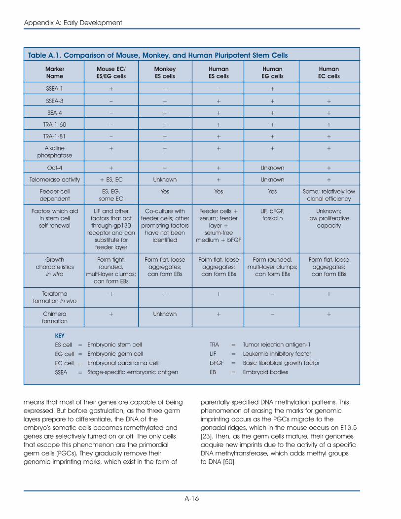
means that most of their genes are capable of beingexpressed. But before gastrulation, as the three germlayers prepare to differentiate, the DNA of theembryo’s somatic cells becomes remethylated andgenes are selectively turned on or off. The only cellsthat escape this phenomenon are the primordialgerm cells (PGCs). They gradually remove theirgenomic imprinting marks, which exist in the form of
parentally specified DNA methylation patterns. Thisphenomenon of erasing the marks for genomicimprinting occurs as the PGCs migrate to thegonadal ridges, which in the mouse occurs on E13.5[23]. Then, as the germ cells mature, their genomesacquire new imprints due to the activity of a specificDNA methyltransferase, which adds methyl groupsto DNA [50].
A-16
Appendix A: Early Development
MarkerName
Mouse EC/ES/EG cells
MonkeyES cells
HumanES cells
HumanEG cells
HumanEC cells
SSEA-1 + – – + –
Table A.1. Comparison of Mouse, Monkey, and Human Pluripotent Stem Cells
SSEA-3 – + + + +
SEA-4 – + + + +
TRA-1-60 – + + + +
TRA-1-81 – + + + +
Alkalinephosphatase
+ + + + +
Oct-4 + + + Unknown +
Telomerase activity + ES, EC Unknown + Unknown +
Feeder-celldependent
ES, EG,some EC
Yes Yes Yes Some; relatively lowclonal efficiency
Factors which aidin stem cellself-renewal
LIF and otherfactors that actthrough gp130
receptor and cansubstitute forfeeder layer
Co-culture withfeeder cells; otherpromoting factors
have not beenidentified
Feeder cells +serum; feeder
layer +serum-free
medium + bFGF
LIF, bFGF,forskolin
Unknown;low proliferative
capacity
Growthcharacteristics
in vitro
Form tight,rounded,
multi-layer clumps;can form EBs
Form flat, looseaggregates;can form EBs
Form flat, looseaggregates;can form EBs
Form rounded,multi-layer clumps;
can form EBs
Form flat, looseaggregates;can form EBs
Teratomaformation in vivo
+ + + – +
Chimeraformation
+ Unknown + – +
KEY
ES cell =
EG cell =
EC cell =
SSEA =
Embryonic stem cell
Embryonic germ cell
Embryonal carcinoma cell
Stage-specific embryonic antigen
TRA =
LIF =
bFGF =
EB =
Tumor rejection antigen-1
Leukemia inhibitory factor
Basic fibroblast growth factor
Embryoid bodies
REFERENCES1. Alberts, B., Bray, D., Lewis, J., Raff, M., Roberts, K., and
Watson, J.D. (1994). Molecular biology of the cell,(New York: Garland Publishing, Inc.).
2. Andrews, P.W., Przyborski, S.A., and Thomson, J. (2001).Embryonal carcinoma cells as embryonic stem cells.Marshak, D.R., Gardner, D.K., and Gottlieb, D. eds. ColdSpring Harbor Laboratory Press, 231-266.
3. Beddington, R.S. and Robertson, E.J. (1999). Axis develop-ment and early asymmetry in mammals. Cell. 96, 195-209.
4. Bongso, A., personal communication.
5. Brook, F.A. and Gardner, R.L. (1997). The origin and efficientderivation of embryonic stem cells in the mouse. Proc. Natl.Acad. Sci. U. S. A. 94, 5709-5712.
6. Burdsal, C.A., Flannery, M.L., and Pedersen, R.A. (1998).FGF-2 alters the fate of mouse epiblast from ectoderm tomesoderm in vitro. Dev. Biol. 198, 231-244.
7. Carayannopoulos, M.O., Chi, M.M., Cui, Y., Pingsterhaus,J.M., McKnight, R.A., Mueckler, M., Devaskar, S.U., andMoley, K.H. (2000). GLUT8 is a glucose transporter respon-sible for insulin-stimulated glucose uptake in the blastocyst.Proc. Natl. Acad. Sci. U. S. A. 97, 7313-7318.
8. Chodosh, L.A. (1998). Expression of BRCA1 and BRCA2 innormal and neoplastic cells. J. Mammary Gland Biol.Neoplasia. 3, 389-402.
9. Constância, M., Pickard, B., Kelsey, G., and Reik, W. (1998).Imprinting mechanisms. Genome Res. 8, 881-900.
10. Cronier, L., Bastide, B., Defamie, N., Niger, C., Pointis, G.,Gasc, J.M., and Malassine, A. (2001). Involvement of gapjunctional communication and connexin expression introphoblast differentiation of the human placenta. Histol.Histopathol. 16, 285-295.
11. D’Urso, G. and Datta, S. (2001). Cell cycle control,checkpoints, and stem cell biology. Marshak, D.R., Gardner,D.K., and Gottlieb, D. eds. Cold Spring Harbor LaboratoryPress, 61-94.
12. Edwards, R.G. (2000). The role of embryonic polarities inpreimplantation growth and implantation of mammalianembryos. Hum. Reprod. 15 Suppl 6, 1-8.
13. Feldman, B., Poueymirou, W., Papaioannou, V.E., DeChiara,T.M., and Goldfarb, M. (1995). Requirement of FGF-4 forpostimplantation mouse development. Science. 267,246-249.
14. Fong, C.Y., Bongso, A., Ng, S.C., Kumar, J., Trounson, A.,and Ratnam, S. (1998). Blastocyst transfer after enzymatictreatment of the zona pellucida: improving in-vitro fertiliza-tion and understanding implantation. Hum. Reprod. 13,2926-2932.
15. Gardner, R.L. (1997). The early blastocyst is bilaterallysymmetrical and its axis of symmetry is aligned with theanimal-vegetal axis of the zygote in the mouse.Development. 124, 289-301.
16. Gardner, D.K. (1998). Changes in requirements andutilization of nutrients during mammalian preimplantationembryo development and their significance in embryoculture. Theriogenology. 49, 83-102.
17. Gardner, R.L. (1999). Polarity in early mammalian develop-ment. Curr. Opin. Genet. Dev. 9, 417-421.
18. Gardner, R.L. (2001). The initial phase of embryonicpatterning in mammals. Int. Rev. Cytol. 203, 233-290.
19. Gilbert, S.F. (2000). Developmental biology. (Sunderland,MA: Sinauer Associates).
20. Gossler, A. (1992). Early embryonic development ofanimals, Hennig, W., Nover, L., and Scheer, U. eds.(Berlin, New York: Springer-Verlag).
21. Guillemot, F., Nagy, A., Auerbach, A., Rossant, J., andJoyner, A.L. (1994). Essential role of Mash-2 in extra-embryonic development. Nature. 371, 333-336.
22. Hara, T., Tamura, K., de Miguel, M.P., Mukouyama, Y., Kim,H., Kogo, H., Donovan, P.J., and Miyajima, A. (1998). Distinctroles of oncostatin M and leukemia inhibitory factor in thedevelopment of primordial germ cells and sertoli cells inmice. Dev. Biol. 201, 144-153.
23. Hogan, B., Beddington, R., Constantini, F., and Lacy, E.(1994). Manipulating the mouse embryo a laboratorymanual, (Cold Spring Harbor, New York: Cold Spring HarborLaboratory Press).
24. Hogan, B.L. (1999). Morphogenesis. Cell. 96, 225-233.
25. Hogan, B. (2001). Primordial germ cells as stem cells.Marshak, D.R., Gardner, D.K., and Gottlieb, D. eds. ColdSpring Harbor Laboratory Press, 189-204.
26. Janatpour, M.J., Utset, M.F., Cross, J.C., Rossant, J., Dong,J., Israel, M.A., and Fisher, S.J. (1999). A repertoire ofdifferentially expressed transcription factors that offersinsight into mechanisms of human cytotrophoblastdifferentiation. Dev. Genet. 25, 146-157.
27. Johnson, M.H., Maro, B., and Takeichi, M. (1986). The roleof cell adhesion in the synchronization and orientation ofpolarization in 8-cell mouse blastomeres. J. Embryol. Exp.Morphol. 93, 239-255.
28. Jones, J.M. and Thomson, J.A. (2000). Human embryonicstem cell technology. Semin. Reprod. Med. 18, 219-223.
29. Kunath, T., Strumpf, D., Rossant, J., and Tanaka, S. (2001).Trophoblast stem cells. Marshak, D.R., Gardner, D.K., andGottlieb, D. eds. Cold Spring Harbor Laboratory Press, 267-288.
30. Marshak, D.R., Gottlieb, D., Kiger, A.A., Fuller, M.T., Kunath, T.,Hogan, B., Gardner, R.L., Smith, A., Klar, A.J.S., Henrique, D.,D’Urso, G., Datta, S., Holliday, R., Astle, C.M., Chen, J., Harrison,D.E., Xie, T., Spradling, A., Andrews, P.W., Przyborski, S.A.,Thomson, J.A., Kunath, T., Strumpf, D., Rossant, J., Tanaka, S.,Orkin, S.H., Melchers, F., Rolink, A., Keller, G., Pittenger, M.F.,Marshak, D.R., Flake, A.W., Panicker, M.M., Rao, M., Watt, F.M.,Grompe, M., Finegold, M.J., Kritzik, M.R., Sarvetnick, N., andWinton, D.J. (2001e). Stem cell biology, Marshak, D.R.,Gardner, R.L., and Gottlieb, D. eds. (Cold Spring Harbor,New York: Cold Spring Harbor Laboratory Press).
Appendix A: Early Development
A-17
31. Nichols, J., Davidson, D., Taga, T., Yoshida, K., Chambers,I., and Smith, A. (1996). Complementary tissue-specificexpression of LIF and LIF-receptor mRNAs in early mouseembryogenesis. Mech. Dev. 57, 123-131.
32. Nichols, J., Zevnik, B., Anastassiadis, K., Niwa, H., Klewe-Nebenius, D., Chambers, I., Scholer, H., and Smith, A.(1998). Formation of pluripotent stem cells in themammalian embryo depends on the POU transcriptionfactor Oct4. Cell. 95, 379-391.
33. Niwa, H., Miyazaki, J., and Smith, A.G. (2000). Quantitativeexpression of Oct-3/4 defines differentiation, dedifferen-tiation or self-renewal of ES cells. Nat. Genet. 24, 372-376.
34. Odorico, J.S., Kaufman, D.S., and Thomson, J.A. (2001).Multilineage Differentiation from Human Embryonic StemCell Lines. Stem Cells. 19, 193-204.
35. Pelton, T.A., Bettess, M.D., Lake, J., Rathjen, J., and Rathjen,P.D. (1998). Developmental complexity of early mammalianpluripotent cell populations in vivo and in vitro. Reprod.Fertil. Dev. 10, 535-549.
36. Pera, M.F., Reubinoff, B., and Trounson, A. (2000). Humanembryonic stem cells. J. Cell Sci. 113 ( Pt 1), 5-10.
37. Pesce, M., Anastassiadis, K., and Scholer, H.R. (1999). Oct-4:lessons of totipotency from embryonic stem cells. CellsTissues Organs. 165, 144-152.
38. Rossant, J., personal communication.
39. Shinohara, T., Orwig, K.E., Avarbock, M.R., and Brinster, R.L.(2000). Spermatogonial stem cell enrichment by multi-parameter selection of mouse testis cells. Proc. Natl. Acad.Sci. U. S. A. 97, 8346-8351.
40. Smith, A.G., Heath, J.K., Donaldson, D.D., Wong, G.G.,Moreau, J., Stahl, M., and Rogers, D. (1988). Inhibition ofpluripotential embryonic stem cell differentiation by purifiedpolypeptides. Nature. 336, 688-690.
41. Smith, A. (2001). Embryonic stem cells. Marshak, D.R.,Gardner, D.K., and Gottlieb, D. eds. Cold Spring HarborLaboratory Press, 205-230.
42. Smith, A.G. (2001). Origins and properties of mouseembryonic stem cells. Annu. Rev. Cell. Dev. Biol. 1-22.
43. Soengas, M.S., Capodieci, P., Polsky, D., Mora, J., Esteller,M., Opitz-Araya, X., McCombie, R., Herman, J.G., Gerald,W.L., Lazebnik, Y.A., Cordon-Cardo, C., and Lowe, S.W.(2001). Inactivation of the apoptosis effector Apaf-1 inmalignant melanoma. Nature. 409, 207-211.
44. Surani, M.A. (1998). Imprinting and the initiation of genesilencing in the germ line. Cell. 93, 309-312.
45. Tanaka, M., Gertsenstein, M., Rossant, J., and Nagy, A.(1997). Mash2 acts cell autonomously in mouse spon-giotrophoblast development. Dev. Biol. 190, 55-65.
46. Tanaka, S., Kunath, T., Hadjantonakis, A.K., Nagy, A., andRossant, J. (1998). Promotion of trophoblast stem cellproliferation by FGF4. Science. 282, 2072-2075.
47. Teruel, M., Smith, R., and Catalano, R. (2000). Growthfactors and embryo development. Biocell. 24, 107-122.
48. Thomson, J.A., Itskovitz-Eldor, J., Shapiro, S.S., Waknitz, M.A.,Swiergiel, J.J., Marshall, V.S., and Jones, J.M. (1998).Embryonic stem cell lines derived from human blastocysts.Science. 282, 1145-1147.
49. Trounson, A.O., Gardner, D.K., Baker, G., Barnes, F.L.,Bongso, A., Bourne, H., Calderon, I., Cohen, J., Dawson, K.,Eldar-Geve, T., Gardner, D.K., Graves, G., Healy, D., Lane,M., Leese, H.J., Leeton, J., Levron, J., Liu, D.Y., MacLachlan,V., Munné, S., Oranratnachai, A., Rogers, P., Rombauts, L.,Sakkas, D., Sathananthan, A.H., Schimmel, T., Shaw, J.,Trounson, A.O., Van Steirteghem, A., Willadsen, S., andWood, C. (2000). Handbook of in vitro fertilization, (BocaRaton, London, New York, Washington, D.C.: CRC Press).
50. Tucker, K.L., Beard, C., Dausmann, J., Jackson-Grusby, L.,Laird, P.W., Lei, H., Li, E., and Jaenisch, R. (1996). Germ-linepassage is required for establishment of methylation andexpression patterns of imprinted but not of nonimprintedgenes. Genes Dev. 10, 1008-1020.
A-18
Appendix A: Early Development
B-1
MOUSE EMBRYONIC STEMCELL CULTURESThe techniques for culturing mouse embryonic stem(ES) cells from the inner cell mass of the preimplan-tation blastocyst were first reported 20 years ago [6,11], and versions of these standard procedures areused today in laboratories throughout the world.Additionally, studies of embryonal carcinoma (EC)cells from mice and humans [2, 30] have helpedestablish parameters for growing and assessing EScells. It is striking that, to date, only three species ofmammals have yielded long-term cultures of self-renewing ES cells: mice, monkeys, and humans [21,34, 35, 36] (see Figure B.1. Origins of MousePluripotent Stem Cells).
In mice, the efficiency of generating ES cells is influ-enced by the genetic strain of laboratory mice andindividual factors that affect pregnant females. Onlya few strains of laboratory mice—notably 129,C57BL/6, and a hybrid strain—yield cultures of EScells. Even then, ES cells derived from C57BL/6 blasto-cysts do not behave as reliably as do ES cells fromthe 129 strain of mice. The former are more difficultto propagate in vitro, generate chimeras less effi-ciently than do ES cells from the 129 strain of mice,and infrequently contribute to the germ line [4].
Another influence on the efficiency with which ES cellscan be cultured from mouse blastocysts is the preg-nancy status of the female. Pregnant mice that are indiapause tend to yield ES cells with greater success.Diapause occurs in female mice that have producedone litter and are still nursing when they becomepregnant again. Diapause is a naturally occurringdelay in the process of blastocyst implantation, whichcauses an arrest in embryonic development and asmall increase in the number of epiblast cells [28].These findings have led to the notion that geneticfactors that are peculiar to specific strains of inbredmice, and other in vivo influences such as diapause,
determine, to a great extent, whether mouse ES cellscan be derived from a given blastocyst.
Generating cultures of mouse or human ES cells thatremain in a proliferating, undifferentiated state is amultistep process that typically includes the following.First, the inner cell mass of a preimplantation blasto-cyst is removed from the trophectoderm that sur-rounds it. (For cultures of human ES cells, blastocystsare generated by in vitro fertilization and donated forresearch.) The small plastic culture dishes used togrow the cells contain growth medium supplementedwith fetal calf serum, and are sometimes coated witha “feeder” layer of nondividing cells. The feeder cellsare often mouse embryonic fibroblast (MEF) cells thathave been chemically inactivated so they will notdivide. Mouse ES cells can be grown in vitro withoutfeeder layers if the cytokine leukemia inhibitory factor(LIF) is added to the culture medium (see below).Human ES cells, however do not respond to LIF.
Second, after several days to a week, proliferatingcolonies of cells are removed and dispersed into newculture dishes, each of which also contains an MEFfeeder layer. Under these in vitro conditions, the EScells aggregate to form colonies. Some coloniesconsist of dividing, nondifferentiated cells; in othercolonies, cells may be differentiating. It is difficult tomaintain human ES cells in dispersed cultures wherecells do not aggregate, although mouse ES cells canbe cultured this way. Depending on the culture con-ditions, it may also be difficult to prevent the spon-taneous differentiation of mouse or human ES cells.
In the third major step required to generate ES celllines, the individual, nondifferentiating colonies aredissociated and replated into new dishes, a stepcalled passage. This replating process establishes a“line” of ES cells. The line of cells is “clonal” if a singleES cell generates it. Following some version of thisfundamental procedure, human and mouse ES cellscan be grown and passaged for two or more years,
MOUSE EMBRYONICSTEM CELLS
APPENDIX B:
MOUSE EMBRYONICSTEM CELLS
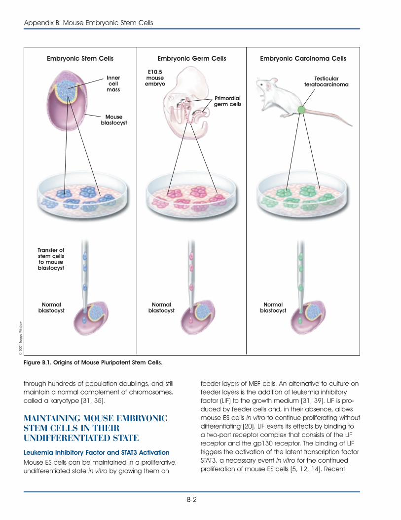
through hundreds of population doublings, and stillmaintain a normal complement of chromosomes,called a karyotype [31, 35].
MAINTAINING MOUSE EMBRYONICSTEM CELLS IN THEIRUNDIFFERENTIATED STATELeukemia Inhibitory Factor and STAT3 Activation
Mouse ES cells can be maintained in a proliferative,undifferentiated state in vitro by growing them on
B-2
Appendix B: Mouse Embryonic Stem Cells
feeder layers of MEF cells. An alternative to culture onfeeder layers is the addition of leukemia inhibitoryfactor (LIF) to the growth medium [31, 39]. LIF is pro-duced by feeder cells and, in their absence, allowsmouse ES cells in vitro to continue proliferating withoutdifferentiating [20]. LIF exerts its effects by binding toa two-part receptor complex that consists of the LIFreceptor and the gp130 receptor. The binding of LIFtriggers the activation of the latent transcription factorSTAT3, a necessary event in vitro for the continuedproliferation of mouse ES cells [5, 12, 14]. Recent
Figure B.1. Origins of Mouse Pluripotent Stem Cells.
Embryonic Stem Cells Embryonic Germ Cells Embryonic Carcinoma Cells
Innercell
mass
Mouseblastocyst
Transfer ofstem cellsto mouseblastocyst
Normalblastocyst
Normalblastocyst
Normalblastocyst
E10.5mouseembryo
Primordialgerm cells
Testicularteratocarcinoma
© 2
001
Tere
se W
inslo
w
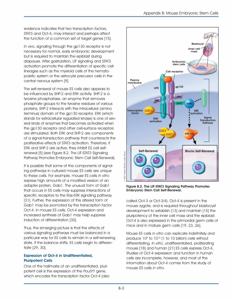
evidence indicates that two transcription factors,STAT3 and Oct-4, may interact and perhaps affectthe function of a common set of target genes [15].
In vivo, signaling through the gp130 receptor is notnecessary for normal, early embryonic developmentbut is required to maintain the epiblast duringdiapause. After gastrulation, LIF signaling and STAT3activation promote the differentiation of specific celllineages such as the myeloid cells of the hemato-poietic system or the astrocyte precursor cells in thecentral nervous system [9].
The self-renewal of mouse ES cells also appears tobe influenced by SHP-2 and ERK activity. SHP-2 is atyrosine phosphatase, an enzyme that removesphosphate groups to the tyrosine residues of variousproteins. SHP-2 interacts with the intracellular (aminoterminus) domain of the gp130 receptor. ERK (whichstands for extracellular regulated kinase) is one of sev-eral kinds of enzymes that becomes activated whenthe gp130 receptor and other cell-surface receptorsare stimulated. Both ERK and SHP-2 are componentsof a signal-transduction pathway that counteracts theproliferative effects of STAT3 activation. Therefore, ifERK and SHP-2 are active, they inhibit ES cell self-renewal [5] (see Figure B.2. The LIF-STAT3 SignalingPathway Promotes Embryonic Stem Cell Self-Renewal).
It is possible that some of the components of signal-ing pathways in cultured mouse ES cells are uniqueto these cells. For example, mouse ES cells in vitroexpress high amounts of a modified version of anadapter protein, Gab1. The unusual form of Gab1that occurs in ES cells may suppress interactions ofspecific receptors to the Ras-ERK signaling pathway[31]. Further, the expression of this altered form ofGab1 may be promoted by the transcription factorOct-4. In mouse ES cells, Oct-4 expression andincreased synthesis of Gab1 may help suppressinduction of differentiation [30].
Thus, the emerging picture is that the effects ofvarious signaling pathways must be balanced in aparticular way for ES cells to remain in a self-renewingstate. If the balance shifts, ES cells begin to differen-tiate [29, 30].
Expression of Oct-4 in Undifferentiated,Pluripotent Cells
One of the hallmarks of an undifferentiated, pluri-potent cell is the expression of the Pou5f1 gene,which encodes the transcription factor Oct-4 (also
called Oct-3 or Oct-3/4). Oct-4 is present in themouse zygote, and is required throughout blastocystdevelopment to establish [13] and maintain [15] thepluripotency of the inner cell mass and the epiblast.Oct-4 is also expressed in the primordial germ cells ofmice and in mature germ cells [19, 23, 26].
Mouse ES cells in vitro can replicate indefinitely andproduce 109 to 1010 (1 to 10 billion) cells withoutdifferentiating. In vitro, undifferentiated, proliferatingmouse [18] and human [21] ES cells express Oct-4.Studies of Oct-4 expression and function in humancells are incomplete, however, and most of theinformation about Oct-4 comes from the study ofmouse ES cells in vitro.
Appendix B: Mouse Embryonic Stem Cells
B-3
Figure B.2. The LIF-STAT3 Signaling Pathway PromotesEmbryonic Stem Cell Self-Renewal.
Blastocyst
Inner cellmass
Embryonicstem cells
Cell receptors
Plasmamembrane
LIFR
JAKJAK
SHP2
Transcriptionfactor
STATactivation
ERKactivation
SignalTransduction
Pathway
Gab
ERK 1/2
Blocks Self-RenewalSelf-Renewal
STAT3
STAT3STAT3
gp130
© 2
001
Tere
se W
inslo
w, L
ydia
Kib
iuk
As is the case with inner cell mass and epiblast cellsin vivo, Oct-4 expression in vitro is required to main-tain the pluripotent, undifferentiated state of ES cells.If Oct-4 expression is inhibited in cultured mouse EScells, the cells generate trophectoderm. If Oct-4expression is artificially increased, mouse ES cells dif-ferentiate into primitive endoderm and mesoderm.Therefore, the level of Oct-4 expression dictates asignificant aspect of the developmental program ofmouse ES cells, making the protein a candidate“master regulator” of ES cell pluripotency [15].
How and why the Oct-4 transcription factor plays suchan important role in early embryogenesis depend onthe genes it regulates. Seven to eight target genes forOct-4 have been identified to date; it activates someand represses others. In fact, the overall impact ofOct-4 may be to prevent the expression of genes thatare required for differentiation [19].
The Cell Cycle of Mouse Embryonic Stem Cells: ItsRole in Preventing Differentiation
Like the cells of the epiblast in the preimplantationmouse embryo, mouse ES cells in vitro have anunusual cell cycle. Specifically, the G1 checkpointdoes not appear to operate in proliferating epiblastand ES cells [25, 38]. This may explain why it has notbeen possible to induce quiescence—withdrawalfrom the cell cycle to a G1 or G0 state—inundifferentiated ES cells [29].
However, if ES cells begin to differentiate by formingembryoid bodies, cyclin D expression increases, theG1 phase of the cell cycle becomes longer, and therate of cell division slows [25]. This can occur if LIF orfeeder layers are withdrawn from mouse ES cell cul-tures. Then, cell division continues for only a few daysas the process of differentiation begins [29]. Perhapsconstant cell proliferation somehow inhibits celldifferentiation, and once the signals for cell divisionare removed, differentiation can occur [37].
Markers of Undifferentiated Embryonic Stem Cells
ES and EC cells, as well as cells of the inner cell massof mouse blastocysts, express a panel of surfacemarkers that are used to characterize undifferen-tiated, pluripotent embryonic cells. (see Table B.1.Comparison of Mouse, Monkey, and HumanPluripotent Stem Cells). The markers also distinguishmouse ES and EC cells from human ES and EC cells.For example mouse ES and EC cells express thestage-specific embryonic antigen SSEA-1, whereas
human ES and EC cells do not. But human ES and ECcells express SSEA-3 and SSEA-4, whereas mouse ESand EC cells do not [21, 35].
Human EG cells, which are derived from primordialgerm cells, express all three markers: SSEA-1, SSEA-3,and SSEA-4. The biological significance of the expres-sion patterns of these surface antigens is unclear, butSSEA-1 expression may be related to the growth char-acteristic of the cells in vitro. Undifferentiated humanES and EC cells tend to grow in flat, relatively loosecolonies. In contrast, mouse ES and EC colonies tendto be multilayered and compact [27]. Alternatively, thesurface expression of various SSEAs may reflect a differ-ence in the developmental stages of the cells [17].
Other markers used to identify ES cells are the surfaceantigens TRA1-60, TRA1-81, and the enzyme alkalinephosphatase. All occur in human ES [3, 27, 35], asthey do in their mouse counterparts.
Genomic Imprinting in Embryonic Stem Cells
It is known that if genomic imprinting patterns aredisturbed before blastocyst implantation in vivo, fetalabnormalities may result. In genomic imprinting,DNA methylation marks certain genes, depending onwhether they are inherited from the mother or thefather. The marked genes are turned on or off in anon-random pattern that is determined by parentalorigin. Imprinting marks are erased in the primordialgerm cells and then reestablished during theformation of eggs and sperm.
However, when embryonic development occursin vitro or when ES cells are grown in tissue culture,normal patterns of genomic imprinting may bedisturbed. For example, mouse embryos that weregrown in vitro in the presence of fetal calf serum—a very different environment than the oviduct—andthen allowed to develop in vivo, showed abnormalgenomic imprinting patterns and abnormal develop-ment. Apparently, the presence of fetal calf serum,a common ingredient in mouse and human EScultures, decreases the expression of certainimprinted genes [8].
How or whether the use of fetal calf serum forculturing mouse or human ES cells affects genomicimprinting and the behavior of ES cells in vitro is notknown. But for mouse ES cells, the parental imprintingpattern apparently persists in vitro [16, 22]. Theimprinting pattern of human ES cells in vitro has notyet been determined.
B-4
Appendix B: Mouse Embryonic Stem Cells
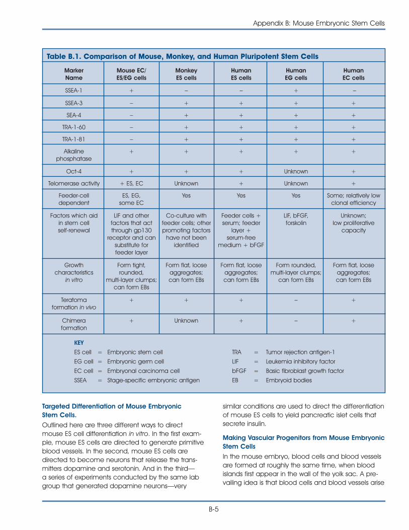
similar conditions are used to direct the differentiationof mouse ES cells to yield pancreatic islet cells thatsecrete insulin.
Making Vascular Progenitors from Mouse EmbryonicStem Cells
In the mouse embryo, blood cells and blood vesselsare formed at roughly the same time, when bloodislands first appear in the wall of the yolk sac. A pre-vailing idea is that blood cells and blood vessels arise
Targeted Differentiation of Mouse EmbryonicStem Cells.
Outlined here are three different ways to directmouse ES cell differentiation in vitro. In the first exam-ple, mouse ES cells are directed to generate primitiveblood vessels. In the second, mouse ES cells aredirected to become neurons that release the trans-mitters dopamine and serotonin. And in the third—a series of experiments conducted by the same labgroup that generated dopamine neurons—very
MarkerName
Mouse EC/ES/EG cells
MonkeyES cells
HumanES cells
HumanEG cells
HumanEC cells
SSEA-1 + – – + –
Table B.1. Comparison of Mouse, Monkey, and Human Pluripotent Stem Cells
SSEA-3 – + + + +
SEA-4 – + + + +
TRA-1-60 – + + + +
TRA-1-81 – + + + +
Alkalinephosphatase
+ + + + +
Oct-4 + + + Unknown +
Telomerase activity + ES, EC Unknown + Unknown +
Feeder-celldependent
ES, EG,some EC
Yes Yes Yes Some; relatively lowclonal efficiency
Factors which aidin stem cellself-renewal
LIF and otherfactors that actthrough gp130
receptor and cansubstitute forfeeder layer
Co-culture withfeeder cells; otherpromoting factors
have not beenidentified
Feeder cells +serum; feeder
layer +serum-free
medium + bFGF
LIF, bFGF,forskolin
Unknown;low proliferative
capacity
Growthcharacteristics
in vitro
Form tight,rounded,
multi-layer clumps;can form EBs
Form flat, looseaggregates;can form EBs
Form flat, looseaggregates;can form EBs
Form rounded,multi-layer clumps;
can form EBs
Form flat, looseaggregates;can form EBs
Teratomaformation in vivo
+ + + – +
Chimeraformation
+ Unknown + – +
KEY
ES cell =
EG cell =
EC cell =
SSEA =
Embryonic stem cell
Embryonic germ cell
Embryonal carcinoma cell
Stage-specific embryonic antigen
TRA =
LIF =
bFGF =
EB =
Tumor rejection antigen-1
Leukemia inhibitory factor
Basic fibroblast growth factor
Embryoid bodies
Appendix B: Mouse Embryonic Stem Cells
B-5
from a common precursor cell derived frommesoderm, the hemangioblast. After hemangioblastsdifferentiate from the mesoderm, they aggregate toform blood islands. The inner cells of the blood islandsbecome hematopoietic stem cells, or blood-formingcells. The outer cells of the blood islands becomeangioblasts, which give rise to the blood vessels.A recent study showed that mouse ES cells in vitrocould be induced to follow this in vivo develop-mental pathway.
In vivo, blood vessel formation occurs in two ways:by vasculogenesis and angiogenesis. Vasculogenesishelps establish the blood islands and the capillarynetwork that connects them. During angiogenesis,new blood vessels form by remodeling or adding toexisting vessels. Both vasculogenesis and angio-genesis are regulated by the actions of a series ofparacrine growth factors, which include fibroblastgrowth factor–2 (FGF-2), vascular endothelial growthfactor (VEGF), and later (in the adult) platelet-derivedgrowth factor (PDGF) and transforming growth factorbeta (TGFß). Each of these growth factors binds tospecific receptors. VEGF, for instance, binds to twodifferent receptors: VEGF-R1, also known as Flt1, andVEGF-R2, also known as Flk1 [7].
To make vascular progenitors from mouse ES cells,Shin-Ichi Nishikawa of Kyoto University GraduateSchool of Medicine in Japan and his colleagues triedto mimic this in vivo pathway for blood vessel forma-tion [40]. They grew undifferentiated ES cells oncollagen-coated dishes in medium containing fetalcalf serum but no leukemia inhibitory factor (LIF). Thisinduced the generation of cells that express Flk1, areceptor for VEGF. Several days later, the cells beganto differentiate. Nearly all the mouse ES cellsexpressed �-smooth muscle actin (SMA), a marker formural cells. (Mural cells, which include pericytes andsmooth muscle cells, normally interact in vivo withendothelial cells to make blood vessels.) When VEGFwas added to the culture medium, sheets ofendothelial cells formed that expressed platelet-endothelial cell adhesion molecule (PECAM1) andother endothelial cell markers. At this point, the culturecontained two differentiating cell types, endothelialcells and mural cells.
Therefore, it appeared that the mouse ES cells haddifferentiated into Flk1+ precursor cells, which thengave rise to both mural cells and endothelial cells
in vitro. To test that hypothesis, single Flk1+ cells werecultured. The individual ES cells generated three kindsof colonies: pure mural cells (SMA+), pure endothelialcells (PECAM1+), and mixed mural and endothelialcells. That result indicated that ES cells can give riseto Flk1+ cells that are precursors for both mural andendothelial cells.
The next test was to see whether the mural cells andendothelial cells generated from Flk1+ precursorscould assemble into primitive blood vessels in vitro.They did. By growing hundreds of Flk1+ cells in colla-gen gel suspensions with fetal calf serum and VEGF,tube-like structures formed within three to five days.This change in the culture conditions allowed the EScells to grow in suspension and interact with eachother. As a result, the cells spontaneously organizedthemselves into tube-like structures that resembleblood vessels in vivo. The tubes were composed ofendothelial (PECAM1+) cells and mural (SMA+) cells.Occasionally, they formed branching structures,which is typical of the organization of blood vesselsin vivo. Also, blood cells (bearing the markers CD45and Ter119) formed inside the tubes, which alsomimicked the organization of blood islands in theearly embryo in vivo.
The final test was to see whether the Flk1+ cells gener-ated from mouse ES cells in vitro would differentiateinto endothelial cells and mural cells in vivo. Again,they did. Flk1+ cells were engineered to express LacZ(which allows the cells to be tracked visually) andinjected into the developing hearts of stage 16-17chick embryos. The donor mouse cells populatedblood vessels in the chicks’ head, yolk sac, heart, andregions between the somites, forming endothelialcells and mural cells in those regions.
Thus, undifferentiated mouse ES cells can bedirected to differentiate into Flk1+ precursors thatgive rise to endothelial cells and mural cells in vitroand in vivo. Further, the differentiated cells can formtube-like vascular structures in vitro. The experimentsnot only demonstrate the power of directed differen-tiation of ES cells into individual cell types, they alsoshow that ES cells can generate multiple cell typesthat then spontaneously organize themselves intotissues that resemble those in vivo. In addition, theexperiments by Nishikawa and his co-workers [40]reveal that Flk1+ cells are important for generatingblood vessels in vivo.
B-6
Appendix B: Mouse Embryonic Stem Cells
Making Dopamine Neurons from MouseEmbryonic Stem Cells
A second example of the directed differentiation ofmouse ES cells in vitro yielded the formation ofparticular kinds of neurons that normally occur in themammalian midbrain and hindbrain. For a long time,the goal of efficiently inducing the formation of theseneurons—which release the neurotransmittersdopamine and serotonin, respectively— was highlydesired, but elusive. In Parkinson’s Disease, a keypopulation of midbrain neurons that releasesdopamine dies. So finding a way to grow largequantities of nerve cells in vitro that might be ableto replace lost dopamine neurons in vivo is a clinicalpriority (see Chapter 8. Rebuilding the Nervous Systemwith Stem Cells).
Last year, Ron McKay and his colleagues reported anefficient technique for inducing mouse ES cells in vitroto differentiate into dopamine neurons of the mid-brain and serotonin neurons, which normally populatethe hindbrain. Like Nishikawa and his colleagues [40],McKay and his collaborators [10] triggered the differ-entiation of mouse ES cells in vitro at various stagesby changing the growth conditions to mimic, in part,those that occur during embryogenesis in vivo. Theresulting differentiated nerve cells look and functionlike their in vivo counterparts.
During embryogenesis, central nervous system (CNS)development is a long, complex process thatdepends on a highly coordinated series of cellularand molecular events. Different signals direct theformation of the neurectoderm from the epiblast, aprocess that ultimately results in the formation of theCNS, the brain and spinal cord. Later, other signalsregulate the development of different parts of thebrain. For example, early in the formation of thebrain, the homeobox genes OTX1 and OTX2 areexpressed [28]. Cells of the epiblast express OTX2before the onset of gastrulation. Then, during gastru-lation, OTX2 is expressed in the anterior neurecto-derm, where it is necessary for the formation of themidbrain and forebrain. Meanwhile, OTX1 expressionoccurs in the region of the neurectoderm that givesrise to the dorsal forebrain. Interactions between OTX1and OTX2 are thought to help shape the midbrainand hindbrain [1].
Once these major brain structures form, variousgenes control the development of individual nervecell types. For example, the genes Pax2, Pax5, Wnt1,
En1, and Nurr1 help control the differentiation ofneurons that release the transmitters dopamine andserotonin [24, 33]. Furthermore, when the proteinssonic hedgehog (SHH) and fibroblast growth factor-8(FGF-8) are added to explant cultures (small chunksof tissue maintained in vitro) of neural plate, thedevelopment of midbrain neurons is enhanced [41].
Taking into account these and other findings, McKayand his coworkers devised an in vitro system forcontrolling the differentiation of mouse ES cells intomidbrain neurons that release dopamine and hind-brain neurons that release serotonin [11]. The cultureconditions they used differ from those devised byNishikawa and his colleagues (described above), butthe starting material—undifferentiated, proliferatingmouse ES cells—was the same in both experiments.McKay and his colleagues cultured mouse ES cells infive distinct stages, each of which they identified bythe changes in culture conditions and the behaviorof the cells (see Figure B.3. Directed Differentiation ofMouse Embryonic Stem Cells Into Neurons orPancreatic Islet-Like Clusters).
In stage 1, undifferentiated mouse ES cells were dis-sociated into single cells and plated at low density.They proliferated in plastic culture dishes coated withgelatin. The growth media contained LIF and fetalcalf serum and was supplemented with amino acids,conditions that promoted the proliferation of undiffer-entiated ES cells. In stage 2, the cells were inducedto form embryoid bodies by dissociating them andreplating at a higher density on a nonadherent sur-face. These conditions allowed the cells to aggre-gate and begin the process of differentiation. Afterfour days, the cells were replated on an adherentsubstrate in the original (stage 1) growth medium.Twenty-four hours later, the growth medium wasreplaced with serum-free insulin/transferrin/selenium/fibronectin (ITSFn) medium. This switch to a serum-freemedium (one lacking fetal calf serum) caused manycells to die but allowed the survival of cells thatexpress nestin. This intermediate filament protein isused as a marker to identify CNS stem cells in vivoand in vitro, although it is also expressed by other celltypes. Stage 1 neurons expressed high levels of OXT2,which decreased in stages 2 and 3. OXT1 was notexpressed until the cells reached stage 3.
Guiding the mouse ES cells through stages 4 and 5of in vitro development yielded fully differentiateddopaminergic and serotoninergic neurons. After 6 to
Appendix B: Mouse Embryonic Stem Cells
B-7
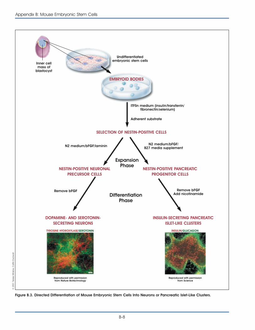
B-8
Appendix B: Mouse Embryonic Stem Cells
Figure B.3. Directed Differentiation of Mouse Embryonic Stem Cells Into Neurons or Pancreatic Islet-Like Clusters.
Inner cellmass of
blastocyst
Undifferentiatedembryonic stem cells
ITFSn medium (insulin/transferrin/fibronectin/selenium)
Adherent substrate
N2 medium/bFGF/laminin N2 medium/bFGF/B27 media supplement
Remove bFGFAdd nicotinamide
Remove bFGF
ExpansionPhase
DifferentiationPhase
EMBRYOID BODIES
SELECTION OF NESTIN-POSITIVE CELLS
NESTIN-POSITIVE NEURONALPRECURSOR CELLS
DOPAMINE- AND SEROTONIN-SECRETING NEURONS
INSULIN-SECRETING PANCREATICISLET-LIKE CLUSTERS
NESTIN-POSITIVE PANCREATICPROGENITOR CELLS
TYROSINE HYDROXYLASE/SEROTONIN
Reproduced with permissionfrom Nature Biotechnology
INSULIN/GLUCAGON
Reproduced with permissionfrom Science
© 2
001
Tere
se W
inslo
w, C
aitl
in D
uckw
all
10 days in the medium that selects for cells thatexpress nestin, the cells were dissociated andinduced to divide in another medium, N2, supple-mented with laminin and basic FGF, a growth factorthat induces proliferation. Other critical additives toyield stage 4 cells were SHH and FGF-8. Cells atstages 3 and 4 express genes that, in vivo, trigger thedevelopment of dopaminergic and serotinergicneurons—namely, Pax2, Pax5, Wnt1, En1, and Nurr1.Stage 5, the final stage of differentiation, wasachieved by removing basic FGF from the growthmedium (which made the cells stop dividing) andgrowing the cells for 6 to 15 days in N2 mediumsupplemented with laminin and ascorbic acid, acombination of additives that induces the differen-tiation of serotonin neurons.
This complex, multistage differentiation process yield-ed a higher percentage (30 percent) of neurons thatexpress tyrosine hydroxylase (TH, the rate-limitingenzyme in the synthesis of dopamine) than any otherreported in vivo or in vitro technique. The cells wereconfirmed to be true dopamine neurons by severalfunctional assays. The neurons secreted dopamineinto the culture medium, showed the electricalactivity typical of neurons, and responded to theaddition of a high concentration of potassium ions(via the addition of potassium chloride) by releasingmore dopamine, much as they would in vivo. A sep-arate population of neurons in the mouse ES cell cul-tures stained positive for serotonin. The differentiationof serotoninergic neurons could be induced by addingonly SHH to the culture medium; addition of FGF-8was not required. Thus, mouse ES cells in vitro can bedirected to differentiate at a high efficiency intoneurons that release either dopamine or serotonin.
Making Pancreatic Islet Cells from MouseEmbryonic Stem Cells
The experimental strategy is similar to that describedabove [10] and is based on a five-stage, in vitrosystem. As before (to differentiate neurons that pro-duce dopamine), undifferentiated mouse ES cells areinduced to proliferate in LIF-supplemented medium(stage 1). Then, the cells are induced to form embry-oid bodies (EBs) in serum-free ITSFn medium withoutLIF (stage 2). ES stage 1 cells expressed Oct-4, a tran-scription factor that characterizes undifferentiated,proliferating, pluripotent cells. Again, cells that expressnestin survive in serum-free medium, whereas othercell types do not, thus creating an environment that
“selects” for nestin-positive cells (stage 3). As before,cells that express nestin are expanded by adding themitogen basic FGF to the serum-free medium (stage4). When basic FGF is withdrawn, the cells stopdividing and begin their final stages of differentiation.It is at this point that the techniques for generatingneurons that release dopamine and pancreatic isletcells that release insulin diverge.
To generate neurons that release dopamine, ES-derived cells were cultured in medium that containedSHH and FGF-8 and later, an N2 medium supple-mented with laminin and ascorbic acid [10]. Togenerate pancreatic islet cells, however, B27 culturemedium was used for stage 4, and nicotinamide wasadded to stage 5 cultures. Another change in thepancreatic islet culture system was to co-cultureindividual stage 4 or 5 ES cells, which were taggedwith the marker green fluorescent protein (GFP), withnontagged ES cells. This meant that an individual,tagged ES cell could be followed so its develop-mental fate could be traced, a technique that madepossible the clonal analysis of the labeled ES cell andits progeny. Tagged, GFP-expressing ES cells gave riseto both pancreatic islet cells and neurons, indicatingthat the same cell acted as the precursor for bothdifferentiated cell types (see Figure B.3. DirectedDifferentiation of Mouse Embryonic Stem Cells IntoNeurons or Pancreatic Islet-Like Clusters).
The tests that identified the differentiated cells typesas pancreatic islet cells and neurons included assaysof various markers. The ES cells at stages 1 and 5expressed GATA-4 and HNFb, markers for embryonicendoderm and extra-embryonic endoderm. This indi-cates that endodermal markers are present in undif-ferentiated ES cells. But stage 5 cells express addition-al markers that are characteristic of endocrine pan-creatic islet cells: mouse insulin I and II, islet amyloidpolypeptide, and the glucose transporter GLUT-2.Other cells stained positive for glucagon, a hormoneproduced by the alpha cells of the pancreas, andsomatostatin, a peptide hormone produced by pan-creatic endocrine cells in vivo. Nerve cells that sur-rounded the clusters of islet cells—a spontaneouslyforming, in vitro arrangement of cell types that mim-icked their arrangement in vivo—stained positive forneuron-specific tubulin. No cells stained positive forboth pancreatic islet markers and neuronal markers,indicating that the two cell types had differentiatedcompletely from a common precursor cell.
Appendix B: Mouse Embryonic Stem Cells
B-9
Other tests demonstrated the functional properties ofthe pancreatic islet cells differentiated from mouseES cells. Adding glucose to the culture mediumtriggered the release of insulin in a dose-dependentmanner. Agonists and antagonists of insulin releasein vivo stimulated or blocked insulin release in vitro,indicating that the pharmacological responses of theES-derived islet cells in vitro mirrored in vivo responses.Finally, when cell clusters of the cultured pancreaticislets were grafted under the skin of diabetic mice(whose diabetes was induced by treatment withstreptozocin), the grafts survived and became infil-trated with blood vessels. The ES-derived pancreaticislets released only one-fiftieth the amount of insulinthey released as islet cells in vivo, however, thediabetic mice experienced no correction of theirhyperglycemia (see Chapter 7. Stem Cells andDiabetes; and Figure 7.2. Development of InsulinSecreting Pancreatic-Like Cells from MouseEmbryonic Stem Cells).
Taken together, the three studies show that the differ-entiation of lines of mouse ES cells can be directedin vitro to yield vascular structures [40], neurons thatrelease dopamine and serotonin [10], and endocrinepancreatic islet cells. In all three cases, proliferating,undifferentiated mouse ES cells provided the startingmaterial and functional, differentiated cells were theresult. Also, in all three experiments, the onset ofmouse ES cell differentiation was triggered by with-drawing the cytokine LIF, which promotes the divisionof undifferentiated mouse ES cells, but — inexplicably— does not have the same effect on human ES cells.In addition, the ES cells in all three examples citedwere induced to aggregate, a change in theirthree-dimensional environment that presumablyallowed some of the cell-cell interactions to occur invitro that would occur in vivo during normal embry-onic development.
Collectively, these three studies provide some of thebest examples of directed differentiation of mouseES cells in vitro. Two of them showed that a single pre-cursor cell can give rise to multiple, differentiated celltypes[10, 40], and all of three studies demonstratedthat the resulting differentiated cells function as theirin vivo counterparts do.
These two criteria – demonstrating that a single cellcan give rise to multiple cells types (clonal analysis),and that the functional properties of the differentiated
cells – form the basis of an acid test for all claims ofdirected differentiation of either ES cells or of adultstem cells. Unfortunately, very few experiments meetthese criteria, which too often makes it impossible toassess whether a differentiated cell type resulted fromthe experimental manipulation that was reported.
REFERENCES1. Acampora, D. and Simeone, A. (1999). The TINS Lecture.
Understanding the roles of Otx1 and Otx2 in the control ofbrain morphogenesis. Trends. Neurosci. 22, 116-122.
2. Andrews, P.W., Casper, J., Damjanov, I., Duggan-Keen, M.,Giwercman, A., Hata, J., von Keitz, A., Looijenga, L.H.,Millan, J.L., Oosterhuis, J.W., Pera, M., Sawada, M., Schmoll,H.J., Skakkebaek, N.E., van Putten, W., and Stern, P. (1996).Comparative analysis of cell surface antigens expressed bycell lines derived from human germ cell tumours. Int. J.Cancer. 66, 806-816.
3. Andrews, P.W., Damjanov, I., Simon, D., Banting, G.S., Carlin,C., Dracopoli, N.C., and Fogh, J. (1984). Pluripotentembryonal carcinoma clones derived from the humanteratocarcinoma cell line Tera-2. Differentiation in vivo andin vitro. Lab. Invest. 50, 147-162.
4. Brook, F.A. and Gardner, R.L. (1997). The origin and efficientderivation of embryonic stem cells in the mouse. Proc. Natl.Acad. Sci. U. S. A. 94, 5709-5712.
5. Burdon, T., Chambers, I., Stracey, C., Niwa, H., and Smith,A. (1999). Signaling mechanisms regulating self-renewaland differentiation of pluripotent embryonic stem cells.Cells Tissues Organs. 165, 131-143.
6. Evans, M.J. and Kaufman, M.H. (1981). Establishment inculture of pluripotential cells from mouse embryos. Nature.292, 154-156.
7. Gilbert, S.F. (2000). Developmental biology. (Sunderland,MA: Sinauer Associates).
8. Khosla, S., Dean, W., Brown, D., Reik, W., and Feil, R. (2001).Culture of preimplantation mouse embryos affects fetaldevelopment and the expression of imprinted genes. Biol.Reprod. 64, 918-926.
9. Kishimoto, T., Taga, T., and Akira, S. (1994). Cytokine signaltransduction. Cell. 76, 253-262.
10. Lee, S.H., Lumelsky, N., Studer, L., Auerbach, J.M., andMcKay, R.D. (2000). Efficient generation of midbrain andhindbrain neurons from mouse embryonic stem cells. Nat.Biotechnol. 18, 675-679.
11. Martin, G.R. (1981). Isolation of a pluripotent cell line fromearly mouse embryos cultured in medium conditioned byteratocarcinoma stem cells. Proc. Natl. Acad. Sci. U. S. A.78, 7634-7638.
12. Matsuda, T., Nakamura, T., Nakao, K., Arai, T., Katsuki, M.,Heike, T., and Yokota, T. (1999). STAT3 activation is sufficientto maintain an undifferentiated state of mouse embryonicstem cells. EMBO J. 18, 4261-4269.
B-10
Appendix B: Mouse Embryonic Stem Cells
13. Nichols, J., Zevnik, B., Anastassiadis, K., Niwa, H., Klewe-Nebenius, D., Chambers, I., Scholer, H., and Smith, A.(1998). Formation of pluripotent stem cells in themammalian embryo depends on the POU transcriptionfactor Oct4. Cell. 95, 379-391.
14. Niwa, H., Burdon, T., Chambers, I., and Smith, A. (1998).Self-renewal of pluripotent embryonic stem cells is mediat-ed via activation of STAT3. Genes Dev. 12, 2048-2060.
15. Niwa, H., Miyazaki, J., and Smith, A.G. (2000). Quantitativeexpression of Oct-3/4 defines differentiation, dedifferen-tiation or self-renewal of ES cells. Nat. Genet. 24, 372-376.
16. O’Shea, K.S. (1999). Embryonic stem cell models ofdevelopment. Anat. Rec. 257, 32-41.
17. Pera, M., personal communication.
18. Pesce, M., Gross, M.K., and Scholer, H.R. (1998). In line withour ancestors: Oct-4 and the mammalian germ. Bioessays.20, 722-732.
19. Pesce, M., Wang, X., Wolgemuth, D.J., and Scholer, H.(1998). Differential expression of the Oct-4 transcriptionfactor during mouse germ cell differentiation. Mech. Dev.71, 89-98.
20. Rathjen, P.D., Toth, S., Willis, A., Heath, J.K., and Smith, A.G.(1990). Differentiation inhibiting activity is produced inmatrix-associated and diffusible forms that are generatedby alternate promoter usage. Cell. 62, 1105-1114.
21. Reubinoff, B.E., Pera, M.F., Fong, C.Y., Trounson, A., andBongso, A. (2000). Embryonic stem cell lines from humanblastocysts: somatic differentiation in vitro. Nat. Biotechnol.18, 399-404.
22. Rohwedel, j., Sehlmeyer, U., Shan, J., Meister, A., andWobus, A.M. (1996). Primordial germ cell-derived mouseembryonic germ (EG) cells in vitro resemble undifferen-tiated stem cells with respect to differentiation capacityand cell cycle distribution. Cell. Biol. Int. 20, 279-587.
23. Rosner, J.L. (1990). Reflections of science as a product.Nature. 345, 108.
24. Rowitch, D.H. and McMahon, A.P. (1995). Pax-2 expressionin the murine neural plate precedes and encompasses theexpression domains of Wnt-1 and En-1. Mech. Dev. 52, 3-8.
25. Savatier, P., Lapillonne, H., van Grunsven, L.A., Rudkin, B.B.,and Samarut, J. (1996). Withdrawal of differentiationinhibitory activity/leukemia inhibitory factor up-regulatesD-type cyclins and cyclin-dependent kinase inhibitors inmouse embryonic stem cells. Oncogene. 12, 309-322.
26. Schöler, H.R., Ruppert, S., Suzuki, N., Chowdhury, K., andGruss, P. (1990). New type of POU domain in germ line-specific protein Oct-4. Nature. 344, 435-439.
27. Shamblott, M.J., Axelman, J., Wang, S., Bugg, E.M.,Littlefield, J.W., Donovan, P.J., Blumenthal, P.D., Huggins,G.R., and Gearhart, J.D. (1998). Derivation of pluripotentstem cells from cultured human primordial germ cells.Proc. Natl. Acad. Sci. U. S. A. 95, 13726-13731.
28. Simeone, A. (1998). Otx1 and Otx2 in the developmentand evolution of the mammalian brain. EMBO J. 17,6790-6798.
29. Smith, A.G. (2001). Embryonic stem cells. Marshak, D.R.,Gardner, D.K., and Gottlieb, D. eds. (Cold Spring Harbor,New York: Cold Spring Harbor Laboratory Press). 205-230.
30. Smith, A., personal communication.
31. Smith, A.G. (2001). Origins and properties of mouseembryonic stem cells. Annu. Rev. Cell. Dev. Biol.
32. Stevens, L.C. (1970). The development of transplantableteratocarcinomas from intratesticular grafts of pre-andpostimplantation mouse embryos. Dev. Biol. 21, 364-382.
33. Stoykova, A. and Gruss, P. (1994). Roles of Pax-genes indeveloping and adult brain as suggested by expressionpatterns. J. Neurosci. 14, 1395-1412.
34. Thomson, J.A., Kalishman, J., Golos, T.G., Durning, M., Harris,C.P., Becker, R.A., and Hearn, J.P. (1995). Isolation of aprimate embryonic stem cell line. Proc. Natl. Acad. Sci.U. S. A. 92, 7844-7848.
35. Thomson, J.A., Itskovitz-Eldor, J., Shapiro, S.S., Waknitz, M.A.,Swiergiel, J.J., Marshall, V.S., and Jones, J.M. (1998).Embryonic stem cell lines derived from human blastocysts.Science. 282, 1145-1147.
36. Thomson, J.A. and Marshall, V.S. (1998). Primate embryonicstem cells. Curr. Top. Dev. Biol. 38, 133-165.
37. Weissman, I.L. (2000). Stem cells: units of development,units of regeneration, and units in evolution. Cell. 100,157-168.
38. Wianny, F., Real, F.X., Mummery, C.L., van Rooijen, M., Lahti,J., Samarut, J., and Savatier, P. (1998). G1-phase regulators,Cyclin D1, Cyclin D2, and Cyclin D3: up-regulation at gas-trulation and dynamic expression during neurolation. Dev.Dyn. 212, 49-62.
39. Williams, R.L., Hilton, D.J., Pease, S., Willson, T.A., Stewart,C.L., Gearing, D.P., Wagner, E.F., Metcalf, D., Nicola, N.A.,and Gough, N.M. (1988). Myeloid leukaemia inhibitoryfactor maintains the developmental potential ofembryonic stem cells. Nature. 336, 684-687.
40. Yamashita, J., Itoh, H., Hirashima, M., Ogawa, M.,Nishikawa, S., Yurugi, T., Naito, M., Nakao, K., and Nishikawa,S. (2000). Flk1-positive cells derived from embryonic stemcells serve as vascular progenitors. Nature. 408, 92-96.
41. Ye, W., Shimamura, K., Rubenstein, J.L., Hynes, M.A.,and Rosenthal, A. (1998). FGF and Shh signals controldopaminergic and serotonergic cell fate in the anteriorneural plate. Cell. 93, 755-766.
Appendix B: Mouse Embryonic Stem Cells
B-11

B-12
Appendix B: Mouse Embryonic Stem Cells
This page intentionally left blank
C-1
METHODS FOR GROWING HUMANEMBRYONIC STEM CELLS IN VITROTo grow cultures of human ES cells, Thomson and hiscollaborators used 36 fresh or frozen embryos gener-ated in IVF laboratories from couples undergoingtreatment for infertility. From the 14 embryos thatdeveloped to the blastocyst stage, they established5 human ES cell lines—H1, H7, H9, H13 and H14 [35].Four of the 5 lines were derived from frozen embryosprovided to Thomson’s laboratory by Josef Itskovitz-Eldor, of the Rambam Medical Center in Haifa, Israel.The ES cell line from the fifth, fresh embryo wasderived from an embryo donated in Wisconsin.
To generate human ES cell cultures, cells from theinner cell mass of a human blastocyst were culturedin a multi-step process. The pluripotent cells of theinner cell mass were separated from the surroundingtrophectoderm by immunosurgery, the antibody-mediated dissolution of the trophectoderm. The innercell masses were plated in culture dishes containinggrowth medium supplemented with fetal bovineserum on feeder layers of mouse embryonic fibro-blasts that had been gamma-irradiated to preventtheir replication. After 9 to 15 days, when inner cellmasses had divided and formed clumps of cells,cells from the periphery of the clumps were chemi-cally or mechanically dissociated and replated inthe same culture conditions. Colonies of apparentlyhomogeneous cells were selectively removed,mechanically dissociated, and replated. These wereexpanded and passaged, thus creating a cell line.None of the initial 5 human ES cell lines generated inthis manner was derived clonally (cloned from a
single cell and are, therefore, genetically identical)[35] (see Figure C.1. Techniques for GeneratingEmbryonic Stem Cell Cultures).
The five original human ES cell lines continued todivide without differentiating for 5 to 6 months [35].Since then, the H9 line has divided for nearly twoyears in vitro, for more than 300 population doublingsand has yielded two subclones, H9.1 and H9.2 [1]1.All the ES cell lines express high levels of telomerase[1, 36], the enzyme that helps maintain telomereswhich protect the ends of chromosomes. Telomeraseactivity and long telomeres are characteristic ofproliferating cells in embryonic tissues and of germcells. Human somatic cells, however, do not showtelomerase activity and their telomeres are consider-ably shorter. Unlike ES cells, differentiated somaticcells also stop dividing in culture—a phenomenoncalled replicative senescence (see Figure C.2.Telomeres and Telomerase).
Three of the human ES cell lines generated byThomson were XY (male) and two were XX (female);all maintained a normal karyotype. Like monkey EScells [34], human ES cells express a panel of surfacemakers that include the stage-specific embryonicantigens SSEA-3 and SSEA-4, as well as TRA-1-60,TRA-1-81, and alkaline phosphatase [14, 25, 26, 35].Mouse ES cells do not express SSEA-3 or SSEA-4; theyexpress SSEA-1, which human and monkey ES cellsdo not. Human ES cells also express the transcriptionfactor Oct-4 [26], as mouse ES cells do.
A somewhat different technique for deriving andculturing human ES cells has now been reported by
HUMAN EMBRYONIC STEM CELLSAND HUMAN EMBRYONIC GERM CELLS
APPENDIX C:
HUMAN EMBRYONIC STEM CELLSAND HUMAN EMBRYONIC GERM CELLS
1The H9.1 and H9.2 clonal cell lines were produced by first plating 105 of the parent H9 cells per well in tissue-culture plates. The culture mediumcontained KnockOut Dulbecco’s modified minimal essential medium (a serum-free substitute for the 20% fetal bovine serum used in the 1998experiments), and basic FGF, which is necessary to maintain cell proliferation and prevent differentiation. To generate clonal cell lines from individualH9 ES cells, 384 single cells were removed from these cultures and transferred individually to the wells of larger plates that contained non-dividingmouse embryonic fibroblasts (MEF) as feeder layers. The single ES cells proliferated and, every 7 days, were dissociated and replated, a process thatgenerate two clonal cell lines, H9.1 and H9.2.
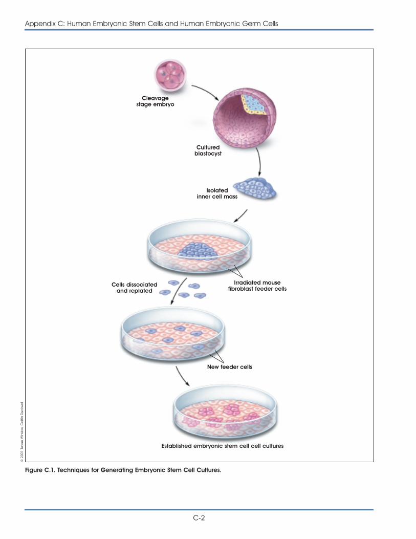
C-2
Appendix C: Human Embryonic Stem Cells and Human Embryonic Germ Cells
Figure C.1. Techniques for Generating Embryonic Stem Cell Cultures.
Cleavagestage embryo
Culturedblastocyst
Isolatedinner cell mass
Cells dissociatedand replated
Irradiated mousefibroblast feeder cells
New feeder cells
Established embryonic stem cell cell cultures
© 2
001
Tere
se W
inslo
w, C
aitl
in D
uckw
all
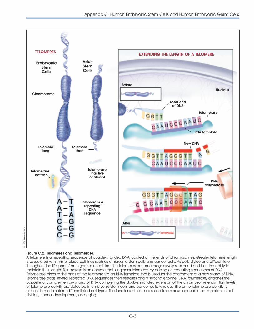
Appendix C: Human Embryonic Stem Cells and Human Embryonic Germ Cells
C-3
Figure C.2. Telomeres and Telomerase.A telomere is a repeating sequence of double-stranded DNA located at the ends of chromosomes. Greater telomere lengthis associated with immortalized cell lines such as embryonic stem cells and cancer cells. As cells divide and differentiatethroughout the lifespan of an organism or cell line, the telomeres become progressively shortened and lose the ability tomaintain their length. Telomerase is an enzyme that lengthens telomeres by adding on repeating sequences of DNA.Telomerase binds to the ends of the telomere via an RNA template that is used for the attachment of a new strand of DNA.Telomerase adds several repeated DNA sequences then releases and a second enzyme, DNA Polymerase, attaches theopposite or complementary strand of DNA completing the double stranded extension of the chromosome ends. High levelsof telomerase activity are detected in embryonic stem cells and cancer cells, whereas little or no telomerase activity ispresent in most mature, differentiated cell types. The functions of telomeres and telomerase appear to be important in celldivision, normal development, and aging.
EXTENDING THE LENGTH OF A TELOMERETELOMERES
Chromosome
Telomerelong
Telomeraseactive
Telomeraseinactive
or absent
BeforeNucleus
Short endof DNA
Telomerase
RNA template
New DNA
DNApolymerase
After
Telomere is arepeating
DNAsequence
Telomereshort
EmbryonicStemCells
AdultStemCells
© 2
001
Tere
se W
inslo
w
investigators in Singapore and Australia. The humanblastocysts are cultured in Singapore, where mono-layer cultures of human ES cells growing on feederlayers are prepared. The primary cultures are shippedto Australia, where the colonies of growing cells aredissociated mechanically and replated. LIF and fetalbovine serum are added to the growth medium. Thecells do not grow well without serum, although it is notclear that LIF has any effect. Under these in vitro con-ditions, the ES cells tend to clump and differentiatespontaneously as they are passaged. In vivo, afterinjection into the testes of immunocompromised mice,the ES cells differentiate into bone, cartilage, squa-mous and cuboidal epithelium, neural cells, glandularepithelium, and striated muscle [25, 26]. Six human EScell lines have been generated from 12 blastocysts, ahigh yield by any standard. The original two cell lineswere generated from fresh embryos; the other fourcells lines were generated from frozen embryos [23].
Recent reports have identified additional human EScell lines that have been developed. New derivationshave been conducted from the blastocyst of frozenembryos at two centers in India (National Centre forBiological Sciences, University of Agriculture Sciences,Bangalore; Harkishondas Hospital in collaboration withReliance Biotechnology, Bombay). They used deriva-tion techniques that differ from those of the Thomsonlaboratory including the use of laser ablation for theremoval of the inner cell mass [22, 24, 33, 36]. Anadditional preparation of human ES cell lines hasbeen conducted in San Francisco [10]. There are nopublications to date on these cell lines and theextent of the research being conducted is not known.
By several criteria, all of the human ES cell lines gen-erated to date are pluripotent. When injected underthe skin or into the testes of immunocompromisedmice—an in vivo method of determining pluripotency—the human ES cells form teratomas that containderivatives of all three primary germ layers. Whenallowed to differentiate in vitro (by culturing the cellsin the absence of MEF feeder layers), the human EScells differentiate spontaneously. Subsequent studiesindicate that in vitro differentiation of these human EScell lines is extensive; the cells can generate manycell types that are derived from all three primarygerm layers [1, 14, 25, 26]. However, the extent towhich these human ES cell lines will differentiate invitro does not match their more extensive differen-tiation capability in vivo (in teratomas) [19].
C-4
Appendix C: Human Embryonic Stem Cells and Human Embryonic Germ Cells
METHODS FOR THE DERIVATION ANDCULTURE OF HUMAN EMBRYONICGERM CELLSTo derive cultures of human embryonic germ (EG)-likecells, Gearhart and his colleagues grew cells from 38initial cultures of primordial germ cells (PGC), whichwere obtained from the gonadal ridge and mesen-tery of 5 to 9-week gestation fetal tissue. (PGCs giverise to the germ cells, eggs and sperm, in the adult.)The PGCs were mechanically and chemically disso-ciated, then plated on a feeder layer of non-dividing,mouse STO fibroblasts in growth medium supple-mented with fetal bovine serum [31]. Unlike thegrowth conditions initially reported for human ES cells[35], the medium for human PGCs cells also con-tained the cytokine, leukemia inhibitory factor (LIF),a mitogen (basic fibroblast growth factor, bFGF),and forskolin.
After one to three weeks in vitro, the human PGCshad formed dense, multilayered colonies of cellsthat resembled mouse ES or EG cells. Cells in thesecolonies expressed SSEA-1, SSEA-3, SSEA-4, TRA1-60,TRA-1-81, and alkaline phosphatase. A small, variablepercentage (1 to 20 %) of the PGC-derived cellcolonies spontaneously formed embryoid bodies. Thegrowth medium for embryoid body cultures lackedLIF, bFGF, and forskolin. The embryoid bodies werecollected from the cultures and either examined forthe cell types they contained, or replated into singlewells of a tissue culture plate for 14 days. The rangeof cell types in the human PGC-derived embryoidbodies included derivatives of all three embryonicgerm layers—endoderm, mesoderm, and ecto-derm—based on the appearance of the cells andthe surface markers they expressed. This result wasinterpreted to mean that the PGC-derived cells werepluripotent, however, it was not possible to demon-strate pluripotency in vivo by generating the forma-tion of teratomas in mice [31].
In their next series of experiments, Gearhart and hiscollaborators devised methods for growing stem cellsderived from human EG cells. The process requiresthe generation of embryoid bodies, which form spon-taneously from EG cells that remain attached to thesubstrate. The embryoid bodies then float freely in theculture medium. Each embryoid body consists of anunpredictable mix of partially differentiated cell types,but allowing the embryoid bodies to form is the most
consistent way of allowing EG-derived cells to differ-entiate [11]. The process involves several stages ofcell derivation in a different kinds of growth media.Cells from low-serum cultures were passaged, chemi-cally dissociated, and resuspended in a culturemedia that contains 50% fetal bovine serum, andfrozen in this state. To measure proliferation, culturesare derived from the frozen embryoid bodies andgrown in the same media used to grow the disso-ciated cells. Clonal cell lines are then derived fromthe embryoid body-derived cultures [32].
The embryoid body-derived cells resulting from thisprocess have high proliferative capacity and geneexpression patterns that are representative of multiplecell lineages. This suggests that the embryoid body-derived cells are progenitor or precursor cells for avariety of differentiated cell types [11].
Recently, Neil Hanley and David Wilson from theUniversity of Southampton, United Kingdom, havederived EG cells from the primordial germ cells of thefetal gonadal ridge. Using material at 8 -10 weeksgestation, cells were derived slightly differently formthe methods of Shamblott et al using a combinationof irradiated fibroblast feeder layers and gelatincoated tissue culture dishes [12]. This method andthe further characterization of the alkaline phosphatase/SSEA1-positive EG cells currently remains unpublished.
DIRECTED DIFFERENTIATION OFHUMAN EMBRYONIC STEM CELLS ANDEMBRYONIC GERM CELLS IN VITROAs with cultures of mouse ES cells, human ES cellsbegin to differentiate if they are removed from feederlayers and grown in suspension culture on a non-adherent surface. The human ES cells form embryoidbodies which, in the early stages, may be simple orcystic and filled with fluid. Although human embryoidbodies vary in their cellular content, many includecells that look like neurons and heart muscle cells[14, 25, 26].
After the human embryoid bodies form, they can bedissociated and replated in monolayer cultures whichare then exposed to specific growth factors thatinfluence further cell differentiation. Some growthfactors induce cell types that would normally bederived from ectoderm in the embryo; these includeretinoic acid, epidermal growth factor (EGF), bonemorphogenic protein 4 (BMP4), and basic fibroblast
growth factor (bFGF). Other growth factors, such asactivin-A and transforming growth factor–beta 1(TGF-ß1) trigger the differentiation of mesodermally-derived cells. Two other factors, hepatocyte growthfactor (HGF) and nerve growth factor (NGF), promotedifferentiation into all three germ layers, includingendoderm. When these eight growth factors wereadded individually to cell cultures derived fromembryoid bodies (generated from the H9 line fromThomson’s laboratory), the cells differentiated into 11cell types that represented all three germ layers. Theidentify of the differentiated human embryoid body-derived cells was determined by their morphology,growth characteristics and expression of messengerRNA (mRNA) for specific markers [30] (see Figure A.6Gene Transcription, Translation, and Protein Synthesis).
Human embryoid body-derived cells will differentiatespontaneously into many kinds of cells, without theaddition of growth factors. However, the addition ofone of a number of growth factors resulted in culturesthat were more likely to be populated by only one ortwo types of differentiated cells, as measured bymRNA transcripts expressed by the cells. Humanembryoid body-derived cultures treated with bFGFdifferentiated largely into epidermal epithelial cellsthat express keratin, a protein in skin. Cells in activin-A–treated cultures formed muscle cell-like syncytium—fused, multinucleated populations of similar cells—that express the enzyme muscle-specific enolase.And cultures treated with retinoic acid differentiatedinto cells that resemble neurons and express neurofil-ament H. However, the same growth factor typicallyinduced the expression of multiple markers; none ofthe resulting cell populations was homogeneous [30].
Spontaneous differentiation of human ES cells intohematopoietic cells, which form all the lineages ofblood cells, is rare in vitro. However, by co-culturinghuman ES cells with mouse bone marrow stromalcells (irradiated to prevent their replication) in growthmedium that contains fetal bovine serum, but noadded growth factors, the cells differentiate to formwhat appear to be hematopoietic precursor cells.The partly differentiated cells express CD34, a markerfor blood cell precursors. If these partly differentiatedhuman ES cells are replated under conditions thatallow them to form colonies of hematopoietic cells,they differentiate into erythroid cells, macrophages,granulocytes, and megakaryocytes [19] (see Chapter5. Hematopoietic Stem Cells).
Appendix C: Human Embryonic Stem Cells and Human Embryonic Germ Cells
C-5
As indicated, human ES cells maintained in vitro havetendency to differentiate spontaneously, a charac-teristic that may not always be desirable. Thus, it maybe necessary to devise methods that allow undiffer-entiated ES cells be selected from a culture that con-tains a mixture of differentiated, partially differentiat-ed, and undifferentiated cells types. The undifferen-tiated ES cells could then be used for the purposesof directed differentiation, or they could be removedfrom cultures in which the differentiated cell types arethe desired product. In either case, a suggestedmethod for identifying undifferentiated ES cells is tointroduce a marker gene—such as that encodinggreen fluorescence protein (GFP)—whose expressionis driven by a gene that is specifically expressed inproliferating, undifferentiated cells, such as Rex1.Then, undifferentiated cells that express GFP can beselectively removed from human ES cultures by usinga fluorescence activated cells sorter (FACS) [9] (seeAppendix E.i. Markers: How Do Researchers Use Themto Identify Stem Cells?).
Joseph Itskovitz-Eldor and his colleagues are trying todirect the differentiation of human ES cells into car-diac myocytes. They use several of the human ESlines generated in James Thomson’s laboratory. Theyreport a number of cells in embryoid bodies thathave contractile activity and express genetic markersconsistent commonly found in cardiac myocytes [16](see Chapter 9. Can Stem Cells Repair a DamagedHeart?). Karl Skorecki and his collaborators have hadsuccess in directing the differentiation of human EScell lines (originating from the Thomson laboratoryderivation) into pancreatic islet-like cells that secreteinsulin. They have also reported the expression ofinsulin genes found in islet-like cells of the pancreas[6] (see Chapter 7. Stem Cells and Diabetes).
A new report indicates that it may be possible todirect the differentiation of human EG cells intoneuronal cells that may play a role in restoring somefunction to paralyzed animals. The SDEC human cellline in this study was generated from embryoidbodies that formed in culture by the aggregation ofhuman EG cells, and is referred to as an embryoid-derived cell line. SDEC cells express a panel ofneuronal markers that include nestin, neurofilament,tau protein, neuron-specific enolase; the cells alsoexpress the glial cells markers glial fibrillary acidicprotein, galactocerebroside, and CNPase. No in vitroassays that indicate cell function have been reported
for SDEC cell assays. However, when SDEC cells wereinjected into the central canal of the spinal cord ofrats—whose hind limbs were paralyzed by aninduced form of amytrophic lateral sclerosis (ALS,also known as Lou Gehrig’s disease)—the majority ofanimals showed some functional recovery. It is notclear whether the human embryoid body-derivedcells replaced some of the spinal motor neuronsdamaged by the experimental ALS, or whether theinjected cells triggered neurons in the recipientanimals to recover lost function [17] (see Chapter 8.Rebuilding the Nervous System with Stem Cells).
Several groups of investigators are trying to direct thedifferentiation of human ES cell lines, but their work isnot yet published. They reported their findings ininterviews with the NIH or during presentations atscientific meetings. They include, but are not limitedto, the following:
• Martin Pera, Alan Trounson, and their coworkersare trying to direct the differentiation of humanES cells along a neural lineage using the BMPantagonist noggin. They generate an apparentlyhomogenous population of cells, but have notyet characterized it [23].
• Brenda Kahan, Jon Odorico, and their coworkersare trying to direct the differentiation of thehuman ES lines H1 and H9 into pancreatic isletcells, which are endodermal derivatives. Theyinduce the formation of embryoid bodies inmedium lacking bFGF and assay cultures for theexpression of transcripts for the endodermalmarkers hnf3, lfabp, ifabp, and villin 1. Differen-tiated progeny from these cells express thegenes for insulin, glucagon, and somatostatin,which are normally expressed in pancreaticislet cells [15].
• Micha Drukker, Nissim Benvenisty, and theircolleagues are trying to direct the differentiationof human ES cells into neurons by addingretinoic acid or ß-NGF to the growth medium.They report that about 80% of embryoid bodiesexposed to these factors contain differentiatedneuronal cells, as determined by morphologyand the expression of receptors for dopamine orserotonin [8].
• Su-Chang Zhang, James Thomson, and theircollaborators are trying to direct the differentiationof human ES cells into neural epithelial cells, by
C-6
Appendix C: Human Embryonic Stem Cells and Human Embryonic Germ Cells
selecting cells from embryoid bodies thatexpress nestin, glial fibrillary acidic protein (GFAP,an astrocyte marker), neural cell adhesion mole-cule (NCAM), and Musashi-1. The differentiatedcell types express (as yet unidentified) markers ofneurons and glial cells. After transplantation intothe mouse brain, the cells aggregated intoclusters and migrate into the brain parenchymawhere they express (unidentified) neural and glialmarkers. By 10 weeks after transplantation, thehuman embryoid-derived cells had not formedteratomas [38].
• Margaret Inokuma, Melissa Carpenter, and theircolleagues are trying to direct the differentiationof human ES cells into neural cells using neu-rotrophin 3 (NT3) and brain-derived neurotrophicfactor (BNDF). Some of the resulting cells stainpositive for tyrosine hydroxylase (TH, the rate-limiting enzyme in dopamine synthesis) orgamma-amino butyric acid (GABA), an inhibitoryneurotransmitter [13].
• Chunhui Xu, Melissa Carpenter, and theircolleagues report preliminary data on growinghuman ES cells in vitro in serum-free mediumwithout feeder layers. The details of their methodhave not been published but apparently includeMatrigel or laminin as a substrate, basic FGF, andconditioned medium from cultures of mouseembryo fibroblasts [39].
• J.S. Lebkowski and Margaret Inokuma et al.report methods for using genetic modificationand changes in culture conditions to direct thedifferentiation of the human ES cell lines H1 andH7 in vitro. They grow undifferentiated human EScells in serum-free medium on Matrigel orlaminin, and then add 20% serum replacementmedium plus DMSO to direct the first stage ofdifferentiation. The second stage is induced byadding sodium butyrate to the medium. Cellmaturation occurs in a third medium (notdescribed). To induce differentiation into neuralcells, they allow the human ES cells to formembryoid bodies, which are then expanded,and plated in B27 medium (not described), withFGF and EGF. The resulting cells express theneural progenitor markers psNCAM and A2B5.
Some differentiated cells express the glial markerGFAP. Other cells express the neuronal markersß-tubulin III and synaptophysin, or stain for theneurotransmitters GABA, tyrosine hydroxylase, orglutamate. No quantitative data, electrophysio-logical data, or responses to neurotransmitterapplication are reported [18].
HUMAN EMBRYONIC CARCINOMACELLSEmbryonal carcinoma (EC) cells are the “stem cells”that occur in unusual germ cell tumors, also calledteratocarcinomas. As such, they give rise to the differ-entiated cell types that also occur in the tumors. Thetumors probably arise from a malignant form of aprimordial germ cell. In humans, germ cell tumorsoccur most often in the testis of young men; theseare always malignant, but usually treatable. Benigngerm tumors called ovarian cysts can occur in theovary; malignant ovarian germ cell tumors are muchrarer (malignant ovarian tumors – usually referred toas ovarian cancer – are not germ cell tumors).
Germ cell tumors have been studied extensively inhumans and mice. They contain an aberrant mix ofdifferentiated cell types, rather than a single kind oftumor cell. Small groups of the cells may appearorganized, but overall, the tissue in the tumor is disor-ganized. Teratocarcinomas are of particular interestbecause they contain EC cells, which in many waysresemble normal ES cells [4].
Like human ES cells, human EC cells proliferate exten-sively in vitro and in teratomas formed in vivo afterinjection into immunocompromised mice. Becauseresearch on human ES cells is so recent, a directcomparison of cultured human EC cells and humanES cells has just begun.2 Both cell types express apanel of surface markers, including the embryonicstage-specific antigens SSEA-3 and SSEA-4. Neitherhuman ES cells nor human EC cells expresses SSEA-1,as mouse ES and EC cells do [5, 26, 35]. Conversely,mouse EC and ES cells do not express SSEA-3 orSSEA-4. Human EC and ES cells also carry on their sur-faces keratin sulfate proteoglycans that can belabeled with specific antibodies, TRA-1-60 andTRA-1-81 [3, 7]. Also, unlike their mouse counterparts,
Appendix C: Human Embryonic Stem Cells and Human Embryonic Germ Cells
C-7
2As of May 9, 2001, the comparisons between human ES cells and EC cells have been made only in Peter Andrews’ laboratory at the University ofSheffield, United Kingdom. James Thomson, of the University of Wisconsin at Madison, supplied Andrews with four lines of human ES cells [3, 32].
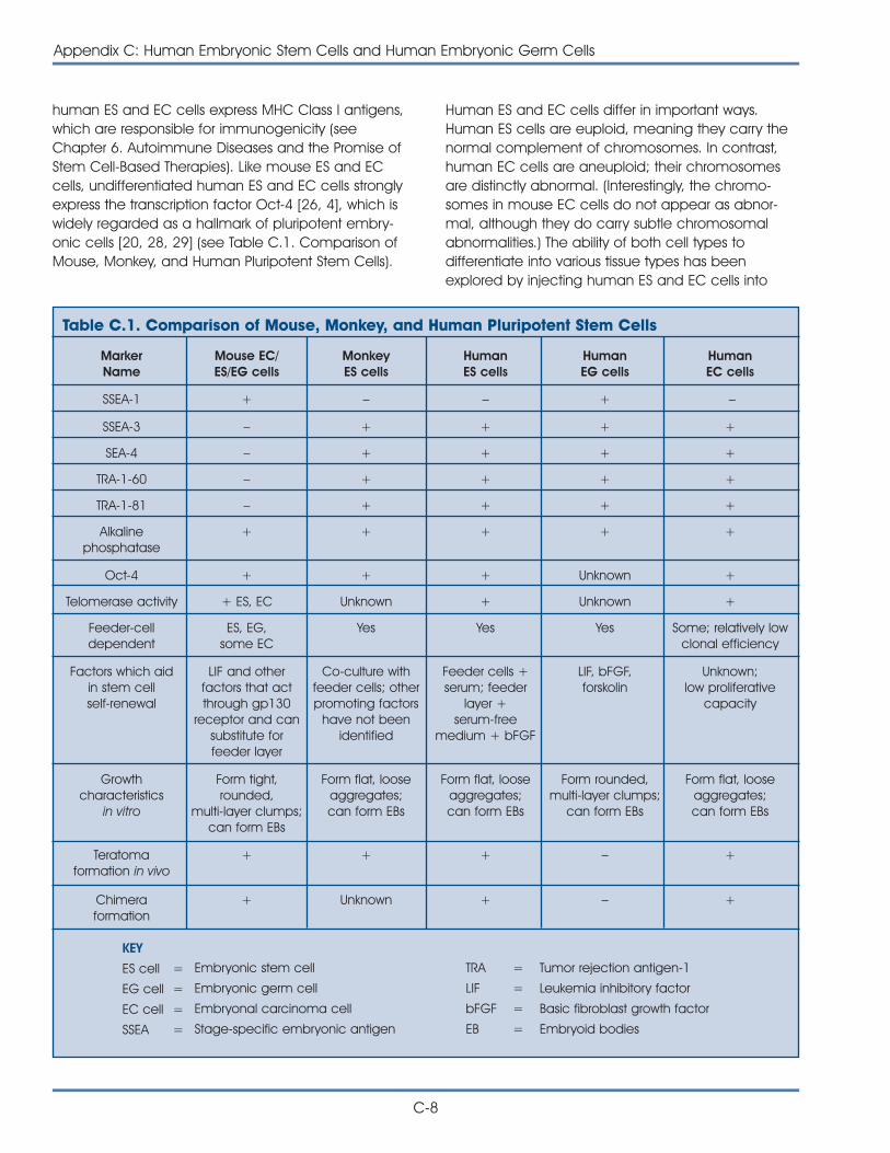
human ES and EC cells express MHC Class I antigens,which are responsible for immunogenicity (seeChapter 6. Autoimmune Diseases and the Promise ofStem Cell-Based Therapies). Like mouse ES and ECcells, undifferentiated human ES and EC cells stronglyexpress the transcription factor Oct-4 [26, 4], which iswidely regarded as a hallmark of pluripotent embry-onic cells [20, 28, 29] (see Table C.1. Comparison ofMouse, Monkey, and Human Pluripotent Stem Cells).
Human ES and EC cells differ in important ways.Human ES cells are euploid, meaning they carry thenormal complement of chromosomes. In contrast,human EC cells are aneuploid; their chromosomesare distinctly abnormal. (Interestingly, the chromo-somes in mouse EC cells do not appear as abnor-mal, although they do carry subtle chromosomalabnormalities.) The ability of both cell types todifferentiate into various tissue types has beenexplored by injecting human ES and EC cells into
C-8
Appendix C: Human Embryonic Stem Cells and Human Embryonic Germ Cells
MarkerName
Mouse EC/ES/EG cells
MonkeyES cells
HumanES cells
HumanEG cells
HumanEC cells
SSEA-1 + – – + –
Table C.1. Comparison of Mouse, Monkey, and Human Pluripotent Stem Cells
SSEA-3 – + + + +
SEA-4 – + + + +
TRA-1-60 – + + + +
TRA-1-81 – + + + +
Alkalinephosphatase
+ + + + +
Oct-4 + + + Unknown +
Telomerase activity + ES, EC Unknown + Unknown +
Feeder-celldependent
ES, EG,some EC
Yes Yes Yes Some; relatively lowclonal efficiency
Factors which aidin stem cellself-renewal
LIF and otherfactors that actthrough gp130
receptor and cansubstitute forfeeder layer
Co-culture withfeeder cells; otherpromoting factors
have not beenidentified
Feeder cells +serum; feeder
layer +serum-free
medium + bFGF
LIF, bFGF,forskolin
Unknown;low proliferative
capacity
Growthcharacteristics
in vitro
Form tight,rounded,
multi-layer clumps;can form EBs
Form flat, looseaggregates;can form EBs
Form flat, looseaggregates;can form EBs
Form rounded,multi-layer clumps;
can form EBs
Form flat, looseaggregates;can form EBs
Teratomaformation in vivo
+ + + – +
Chimeraformation
+ Unknown + – +
KEY
ES cell =
EG cell =
EC cell =
SSEA =
Embryonic stem cell
Embryonic germ cell
Embryonal carcinoma cell
Stage-specific embryonic antigen
TRA =
LIF =
bFGF =
EB =
Tumor rejection antigen-1
Leukemia inhibitory factor
Basic fibroblast growth factor
Embryoid bodies
immunocompromised mice. Injected human ES cellswill form embryonic stem cell teratomas in mice, andthe tumors consist of cells derived from all threeprimary germ layers [36]. In contrast, human EC celllines vary in their ability to differentiate in vivo, but ingeneral are more limited than are ES cells. Forexample, NTERA2 cl.D1 cells (which are derived fromhuman TERA2 EC cells) generate only a few kinds oftissues, including primitive gut-like structures, andneural tissue after injection into immunocompromisedmice [2].
The in vitro growth characteristics of human ES andEC cells are also being compared. Both cell typesgrow well in serum-containing medium on feederlayers of mouse embryonic fibroblasts that havebeen treated to block their proliferation. It is difficultto induce human ES cells to proliferate in theabsence of feeder layers, unless conditioned medi-um from feeder cells cultures is added. However,many human EC cells lines, such as the NTERA2 line,are not dependent on feeder layers [2].
If human ES cells are removed from their feederlayers, they differentiate spontaneously into manycell types. Mouse ES cells, after removal from feederlayers, can be stimulated to divide and preventedfrom differentiating by adding LIF (leukemia inhibitoryfactor); neither human ES nor EC cells show thisresponse to LIF. Instead, if human ES cells grow toconfluence (where the cells grow to completelycover the culture plates), the cells aggregate andbegin to differentiate spontaneously [26, 35]. Also,human ES cells grown in suspension cultures at highdensity will form embryoid bodies. Embryoid bodiesare clumps or groupings of cells that form whencultured in plates or media and do not occur innature. Embryoid bodies contain undifferentiated andpartially differentiated cells [14]. However, human ECcells remain undifferentiated when grown at highdensity [4]. Whether these apparent differences in thein vitro growth characteristics of human ES and ECcells are meaningful or real is subject to debate [5].
The pluripotency of human EC cells does not equalthat of human ES cells. Human ES cells can differen-tiate into a wide range of cell types in vitro, and canform teratomas with many cell types after injectioninto immune-deficient mice. The differentiationpotential of most lines of human EC cells is morelimited, both in vitro and in vivo. One human EC cellline, however, TERA2, differentiates easily in vitro. The
well-studied morphogen, retinoic acid, induces TERA2cells (and the subline NTERA2) to differentiate intoneural precursors, which can then become matureneurons [4]. But when human ES cells are exposed toretinoic acid, they differentiate into a wider array ofcell types than do human EC cells. As yet, it is notclear how the mechanism of action of retinoic aciddiffers in human ES cells versus human EC cells. Itmay be that, because of their tumor origin, humanEC cells carry genetic variations linked to tumorigene-sis that restrict their capacity for differentiation [5].
Thus, the in vitro and in vivo characteristics of humanEC cells resembles that of human ES cells in certainrespects, but not in others. Although ES cells will likelyprove to be a better model for understanding humandevelopment than will EC cells [27], there may besome aspects of development that EC cells willreveal that ES cells will not [5].
REFERENCES1. Amit, M., Carpenter, M.K., Inokuma, M.S., Chiu, C.P., Harris,
C.P., Waknitz, M.A., Itskovitz-Eldor, J., and Thomson, J.A.(2000). Clonally derived human embryonic stem cell linesmaintain pluripotency and proliferative potential forprolonged periods of culture. Dev. Biol. 227, 271-278.
2. Andrews, P.W., Damjanov, I., Simon, D., Banting, G.S., Carlin,C., Dracopoli, N.C., and Fogh, J. (1984). Pluripotent embry-onal carcinoma clones derived from the human teratocar-cinoma cell line Tera-2. Differentiation in vivo and in vitro.Lab. Invest. 50, 147-162.
3. Andrews, P.W., Casper, J., Damjanov, I., Duggan-Keen,M., Giwercman, A., Hata, J., von Keitz, A., Looijenga, L.H.,Millan, J.L., Oosterhuis, J.W., Pera, M., Sawada, M., Schmoll,H.J., Skakkebaek, N.E., van Putten, W., and Stern, P. (1996).Comparative analysis of cell surface antigens expressed bycell lines derived from human germ cell tumours. Int. J.Cancer. 66, 806-816.
4. Andrews, P.W. (1998). Teratocarcinomas and humanembryology: pluripotent human EC cell lines. Reviewarticle. APMIS. 106, 158-167.
5. Andrews, P.W., personal communication.
6. Assady, S., Maor, G., Amit, M., Itskovitz-Eldor, J., Skorecki, K.L.,and Tzukerman, M. (2001). Insulin production by humanembryonic stem cells. Diabetes. 50.
7. Badcock, G., Pigott, C., Goepel, J., and Andrews, P.W.(1999). The human embryonal carcinoma marker antigenTRA-1-60 is a sialylated keratan sulfate proteoglycan.Cancer Res. 59, 4715-4719.
8. Drukker, M., Schuldiner, M., Eiges, R., Eden, A., Yanuka, O.,Itskovitz-Eldor, J., and Benvenisty, N. Keystone symposia.Pluripotent stem cells: biology and applications. Inducedneuronal differentiation of human embryonic stem cells.Poster abstracts. 207.
Appendix C: Human Embryonic Stem Cells and Human Embryonic Germ Cells
C-9
9. Eiges, R., Schuldiner, M., Drukker, M., Yanuka, O., Itskovitz-Eldor, J., and Benvenisty, N. (2001). Establishment of humanembryonic stem cell-transduced clones carrying a markerof undifferentiated cells. Curr. Biol. 11, 514-518.
10. Firpo, M., personal communication.
11. Gearhart, J., personal communication.
12. Hanley, N., personal communication.
13. Inokuma, M.S., Denham, J., Mujtaba, T., Rao, M., andCarpenter, M.K. Keystone symposia. Pluripotent stem cells:biology and applications. Human embryonic stem cellsdifferentiate into neural cells in vitro. Poster abstracts. 312.
14. Itskovitz-Eldor, J., Schuldiner, M., Karsenti, D., Eden, A.,Yanuka, O., Amit, M., Soreq, H., and Benvenisty, N. (2000).Differentiation of human embryonic stem cells into embry-oid bodies comprising the three embryonic germ layers.Mol. Med. 6, 88-95.
15. Kahan, B.W., Jacobson, L.M., Hullett, D.A., Thomson, J.,and Odorico, J.S. Keystone symposia. Pluripotent stem cells:biology and applications. In vitro differentiation of humanembryonic stem (ES) cell lines: expression of endoderm-and pancreatic islet-specific genes. Poster abstract. 117.
16. Kehat, I., Kenyagin-Karsenti, D., Druckmann, M., Segev, H.,Amit, M., Gepstein, A., Livne, E., Binah, O., Itskovitz-Eldor, J.,and Gepstein, L. (2001). Human embryonic stem cellscan differentiate into myocytes portraying cardiomyocyticstructural and functional properties. J. Clin. Invest. (In press)
17. Kerr, D.A., Llado, J., Shamblott, M., Maragakis, N., Irani,D.N., Dike, S., Sappington, A., Gearhart, J., and Rothstein, J.(2001). Human embryonic germ cell derivatives facillitatemotor recovery of rats with diffuse motor neuron injury.
18. Lebkowski, J.S., Gold, J., Chiu, C.P., Xu, C., Inokuma, M.,Hassanipour, M., Denham, J., Piderit, A., Rosler, E., Golds,K., and Carpenter, M. ASGT Meeting. Targeted GeneExpression I. Human Embryonic Stem Cells: GeneticModification and Differentiation into Cell Types for PotentialTransplantation Applications. Poster Abstract. 205.
19. Odorico, J.S., Kaufman, D.S., and Thomson, J.A. (2001).Multilineage Differentiation from Human Embryonic StemCell Lines. Stem Cells. 19, 193-204.
20. Okamoto, K., Okazawa, H., Okuda, A., Sakai, M.,Muramatsu, M., and Hamada, H. (1990). A novel octamerbinding transcription factor is differentially expressed inmouse embryonic cells. Cell. 60, 461-472.
21. Panicker, M., personal communication.
22. Pera, M., personal communication.
23. Rao, M., personal communication.
24. Reubinoff BE, Pera, M., Fong, C.Y., and Trounson, A. (2000).Research Errata. Nat. Biotechnol. 18, 559.
25. Reubinoff, B.E., Pera, M.F., Fong, C.Y., Trounson, A., andBongso, A. (2000). Embryonic stem cell lines from humanblastocysts: somatic differentiation in vitro. Nat. Biotechnol.18, 399-404.
26. Roach, S., Cooper, S., Bennett, W., and Pera, M.F. (1993).Cultured cell lines from human teratomas: windows intotumour growth and differentiation and early humandevelopment. Eur. Urol. 23, 82-87.
27. Rosner, J.L. (1990). Reflections of science as a product.Nature. 345, 108.
28. Schöler, H.R., Ruppert, S., Suzuki, N., Chowdhury, K., andGruss, P. (1990). New type of POU domain in germ line-specific protein Oct-4. Nature. 344, 435-439.
29. Schuldiner, M., Yanuka, O., Itskovitz-Eldor, J., Melton, D., andBenvenisty, N. (2000). Effects of eight growth factors on thedifferentiation of cells derived from human embryonic stemcells. Proc. Natl. Acad. Sci. U. S. A. 97, 11307-11312.
30. Shamblott, M.J., Axelman, J., Wang, S., Bugg, E.M.,Littlefield, J.W., Donovan, P.J., Blumenthal, P.D., Huggins,G.R., and Gearhart, J.D. (1998). Derivation of pluripotentstem cells from cultured human primordial germ cells.Proc. Natl. Acad. Sci. U. S. A. 95, 13726-13731.
31. Shamblott, M.J., Axelman, J., Littlefield, J.W., Blumenthal,P.D., Huggins, G.R., Cui, Y., Cheng, L., and Gearhart, J.D.(2001). Human embryonic germ cell derivatives expressa broad range of develpmentally distinct markers andproliferate extensively in vitro. Proc. Natl. Acad. Sci. U. S. A.98, 113-118.
32. Thomson, J., personal communication.
33. Thomson, J.A., Kalishman, J., Golos, T.G., Durning, M., Harris,C.P., Becker, R.A., and Hearn, J.P. (1995). Isolation of aprimate embryonic stem cell line. Proc. Natl. Acad. Sci. U.S. A. 92, 7844-7848.
34. Thomson, J.A., Itskovitz-Eldor, J., Shapiro, S.S., Waknitz, M.A.,Swiergiel, J.J., Marshall, V.S., and Jones, J.M. (1998).Embryonic stem cell lines derived from human blastocysts.Science. 282, 1145-1147.
35. Thomson, J.A. and Odorico, J.S. (2000). Human embryonicstem cell and embryonic germ cell lines. Trends.Biotechnol. 18, 53-57.
36. Totey, S., personal communication.
37. Xu, C., Inokuma, M.S., Denham, J., Golds, K., Kundu, P.,Gold, J.D., and Carpenter, M.K. Keystone symposia.Pluripotent stem cells: biology and applications. Growth ofundifferentiated human embryonic stem cells on definedmatrices with conditioned medium. Poster abstract. 133.
38. Zhang, S.U., Wernig, M., Duncan, I.D., Brüstle, O., andThomson, J. Keystone symposia. Pluripotent stem cells:biology and applications. Directed differentiation of humanES cells to neural epithelia. Poster abstracts. 235.
C-10
Appendix C: Human Embryonic Stem Cells and Human Embryonic Germ Cells
COMPENDIUM OF SCIENTIFIC PUBLICATIONS REGARDING THEISOLATION AND CHARACTERIZATION OF STEM CELLS
considered progenitor ore precursor cells or thosethat can proliferate without the demonstrated abilityto generate cell types of other tissues.
The tables list the tissue from which the cells werederived, the types of cells that developed, theconditions under which differentiation occurred, themethods by which the cells were characterized, andthe primary references for the information.
D-1
APPENDIX D:
STEM CELL TABLESSTEM CELL TABLES
The following tables provide an overview of informa-tion about stem cells that have been derived frommice and humans. The tables summarize publishedresearch that characterizes cells that are capable ofdeveloping into cells of multiple germ layers (i.e.,multipotent or pluripotent) or that can generate thedifferentiated cell types of another tissue (i.e., plasticity)such as a bone marrow cell becoming a neuronalcell. The tables do not include information about cells
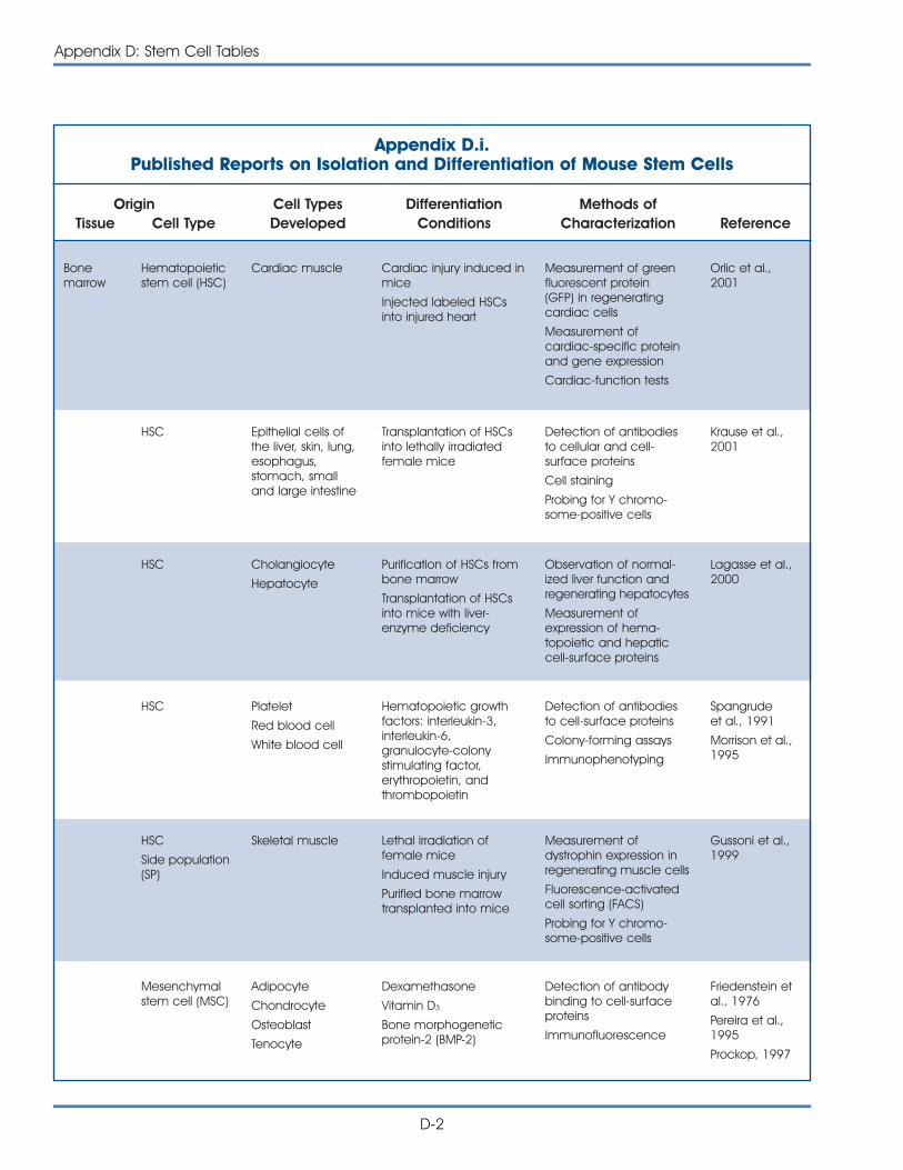
D-2
Appendix D: Stem Cell Tables
Origin Cell Types Differentiation Methods ofTissue Cell Type Developed Conditions Characterization Reference
Bonemarrow
Hematopoieticstem cell (HSC)
Cardiac muscle Cardiac injury induced inmice
Injected labeled HSCsinto injured heart
Measurement of greenfluorescent protein(GFP) in regeneratingcardiac cells
Measurement ofcardiac-specific proteinand gene expression
Cardiac-function tests
Orlic et al.,2001
HSC Epithelial cells ofthe liver, skin, lung,esophagus,stomach, smalland large intestine
Transplantation of HSCsinto lethally irradiatedfemale mice
Detection of antibodiesto cellular and cell-surface proteins
Cell staining
Probing for Y chromo-some-positive cells
Krause et al.,2001
Adult Human Stem CellsAppendix D.i.Published Reports on Isolation and Differentiation of Mouse Stem Cells
HSC Cholangiocyte
Hepatocyte
Purification of HSCs frombone marrow
Transplantation of HSCsinto mice with liver-enzyme deficiency
Observation of normal-ized liver function andregenerating hepatocytes
Measurement ofexpression of hema-topoietic and hepaticcell-surface proteins
Lagasse et al.,2000
HSC Platelet
Red blood cell
White blood cell
Hematopoietic growthfactors: interleukin-3,interleukin-6,granulocyte-colonystimulating factor,erythropoietin, andthrombopoietin
Detection of antibodiesto cell-surface proteins
Colony-forming assays
Immunophenotyping
Spangrudeet al., 1991
Morrison et al.,1995
HSC
Side population(SP)
Skeletal muscle Lethal irradiation offemale mice
Induced muscle injury
Purified bone marrowtransplanted into mice
Measurement ofdystrophin expression inregenerating muscle cells
Fluorescence-activatedcell sorting (FACS)
Probing for Y chromo-some-positive cells
Gussoni et al.,1999
Mesenchymalstem cell (MSC)
Adipocyte
Chondrocyte
Osteoblast
Tenocyte
Dexamethasone
Vitamin D3
Bone morphogeneticprotein-2 (BMP-2)
Detection of antibodybinding to cell-surfaceproteins
Immunofluorescence
Friedenstein etal., 1976
Pereira et al.,1995
Prockop, 1997
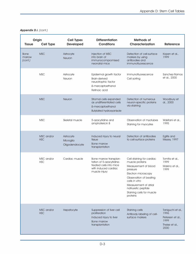
Appendix D: Stem Cell Tables
D-3
Origin Cell Types Differentiation Methods ofTissue Cell Type Developed Conditions Characterization Reference
Bonemarrow(cont.)
MSC Astrocyte
Neuron
Injection of MSCinto brain ofimmunocompromisedneonatal mice
Detection of cell-surfacemarkers by usingantibodies andimmunofluorescence
Kopen et al.,1999
MSC Astrocyte
Neuron
Epidermal growth factor
Brain-derivedneurotrophic factor
ß-mercaptoethanol
Retinoic acid
Immunofluorescence
Cell sorting
Sanchez-Ramoset al., 2000
MSC Neuron Stromal cells expandedas undifferentiated cells
ß-mercaptoethanol
Butylated hydroxyanisole
Detection of numerousneuron-specific proteinsvia staining
Woodbury etal., 2000
MSC Skeletal muscle 5-azacytidine andamphotericin B
Observation of myotubes
Staining for myocytes
Wakitani et al.,1995
MSC and/orHSC
Astrocyte
Microglia
Oligodendrocyte
Induced injury to neuraltissue
Bone marrowtransplantation
Detection of antibodiesto cell-surface proteins
Eglitis andMezey, 1997
Appendix D.i. (cont.)
MSC and/orHSC
Cardiac muscle Bone marrow transplan-tation of 5-azacytidine-treated cells into micewith induced cardiacmuscle injury
Cell staining for cardiacmuscle proteins
Measurement of bloodpressure
Electron microscopy
Observation of beatingcells in vitro
Measurement of atrialnatriuretic peptide
Staining cells for muscleproteins
Tomita et al.,1999
Makino et al.,1999
MSC and/orHSC
Hepatocyte Suppression of liver cellproliferation
Induced injury to liver
Bone marrowtransplantation
Staining cells
Antibody labeling of cell-surface markers
Taniguchi et al.,1996
Petersen et al.,1999
Theise et al.,2000
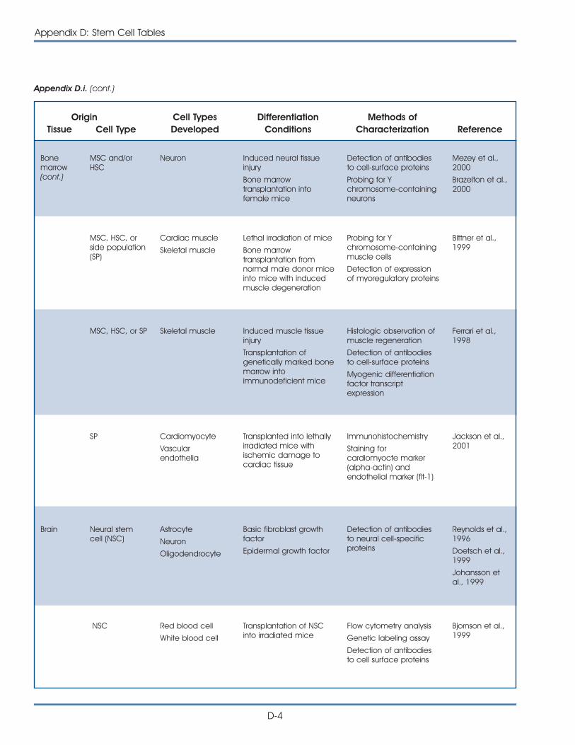
Origin Cell Types Differentiation Methods ofTissue Cell Type Developed Conditions Characterization Reference
Appendix D.i. (cont.)
D-4
Appendix D: Stem Cell Tables
Bonemarrow(cont.)
MSC and/orHSC
Neuron Induced neural tissueinjury
Bone marrowtransplantation intofemale mice
Detection of antibodiesto cell-surface proteins
Probing for Ychromosome-containingneurons
Mezey et al.,2000
Brazelton et al.,2000
MSC, HSC, orside population(SP)
Cardiac muscle
Skeletal muscle
Lethal irradiation of mice
Bone marrowtransplantation fromnormal male donor miceinto mice with inducedmuscle degeneration
Probing for Ychromosome-containingmuscle cells
Detection of expressionof myoregulatory proteins
Bittner et al.,1999
MSC, HSC, or SP Skeletal muscle Induced muscle tissueinjury
Transplantation ofgenetically marked bonemarrow intoimmunodeficient mice
Histologic observation ofmuscle regeneration
Detection of antibodiesto cell-surface proteins
Myogenic differentiationfactor transcriptexpression
Ferrari et al.,1998
SP Cardiomyocyte
Vascularendothelia
Transplanted into lethallyirradiated mice withischemic damage tocardiac tissue
Immunohistochemistry
Staining forcardiomyocte marker(alpha-actin) andendothelial marker (flt-1)
Jackson et al.,2001
Brain Neural stemcell (NSC)
Astrocyte
Neuron
Oligodendrocyte
Basic fibroblast growthfactor
Epidermal growth factor
Detection of antibodiesto neural cell-specificproteins
Reynolds et al.,1996
Doetsch et al.,1999
Johansson etal., 1999
NSC Red blood cell
White blood cell
Transplantation of NSCinto irradiated mice
Flow cytometry analysis
Genetic labeling assay
Detection of antibodiesto cell surface proteins
Bjornson et al.,1999
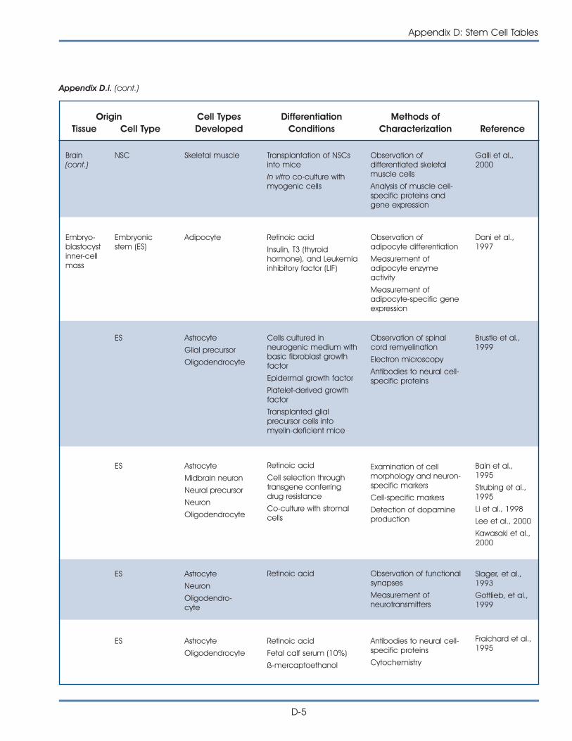
Appendix D: Stem Cell Tables
D-5
Origin Cell Types Differentiation Methods ofTissue Cell Type Developed Conditions Characterization Reference
Brain(cont.)
NSC Skeletal muscle Transplantation of NSCsinto mice
In vitro co-culture withmyogenic cells
Observation ofdifferentiated skeletalmuscle cells
Analysis of muscle cell-specific proteins andgene expression
Galli et al.,2000
Embryo-blastocystinner-cellmass
Embryonicstem (ES)
Adipocyte Retinoic acid
Insulin, T3 (thyroidhormone), and Leukemiainhibitory factor (LIF)
Observation ofadipocyte differentiation
Measurement ofadipocyte enzymeactivity
Measurement ofadipocyte-specific geneexpression
Dani et al.,1997
ES Astrocyte
Glial precursor
Oligodendrocyte
Cells cultured inneurogenic medium withbasic fibroblast growthfactor
Epidermal growth factor
Platelet-derived growthfactor
Transplanted glialprecursor cells intomyelin-deficient mice
Observation of spinalcord remyelination
Electron microscopy
Antibodies to neural cell-specific proteins
Brustle et al.,1999
ES Astrocyte
Midbrain neuron
Neural precursor
Neuron
Oligodendrocyte
Retinoic acid
Cell selection throughtransgene conferringdrug resistance
Co-culture with stromalcells
Examination of cellmorphology and neuron-specific markers
Cell-specific markers
Detection of dopamineproduction
Bain et al.,1995
Strubing et al.,1995
Li et al., 1998
Lee et al., 2000
Kawasaki et al.,2000
ES Astrocyte
Neuron
Oligodendro-cyte
Retinoic acid Observation of functionalsynapses
Measurement ofneurotransmitters
Slager, et al.,1993
Gottlieb, et al.,1999
Appendix D.i. (cont.)
ES Astrocyte
Oligodendrocyte
Retinoic acid
Fetal calf serum (10%)
ß-mercaptoethanol
Antibodies to neural cell-specific proteins
Cytochemistry
Fraichard et al.,1995
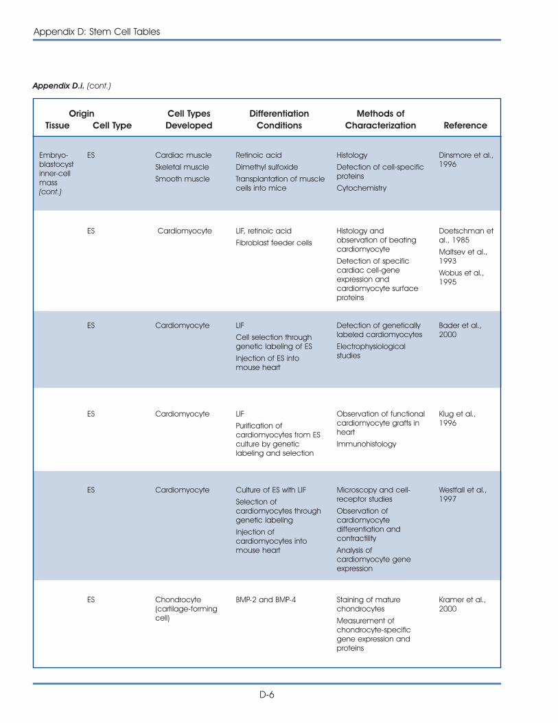
Origin Cell Types Differentiation Methods ofTissue Cell Type Developed Conditions Characterization Reference
Appendix D.i. (cont.)
D-6
Appendix D: Stem Cell Tables
ES Cardiomyocyte LIF, retinoic acid
Fibroblast feeder cells
Histology andobservation of beatingcardiomyocyte
Detection of specificcardiac cell-geneexpression andcardiomyocyte surfaceproteins
Doetschman etal., 1985
Maltsev et al.,1993
Wobus et al.,1995
ES Cardiomyocyte LIF
Cell selection throughgenetic labeling of ES
Injection of ES intomouse heart
Detection of geneticallylabeled cardiomyocytes
Electrophysiologicalstudies
Bader et al.,2000
ES Cardiomyocyte LIF
Purification ofcardiomyocytes from ESculture by geneticlabeling and selection
Observation of functionalcardiomyocyte grafts inheart
Immunohistology
Klug et al.,1996
ES Cardiomyocyte Culture of ES with LIF
Selection ofcardiomyocytes throughgenetic labeling
Injection ofcardiomyocytes intomouse heart
Microscopy and cell-receptor studies
Observation ofcardiomyocytedifferentiation andcontractility
Analysis ofcardiomyocyte geneexpression
Westfall et al.,1997
ES Chondrocyte(cartilage-formingcell)
BMP-2 and BMP-4 Staining of maturechondrocytes
Measurement ofchondrocyte-specificgene expression andproteins
Kramer et al.,2000
Embryo-blastocystinner-cellmass(cont.)
ES Cardiac muscle
Skeletal muscle
Smooth muscle
Retinoic acid
Dimethyl sulfoxide
Transplantation of musclecells into mice
Histology
Detection of cell-specificproteins
Cytochemistry
Dinsmore et al.,1996
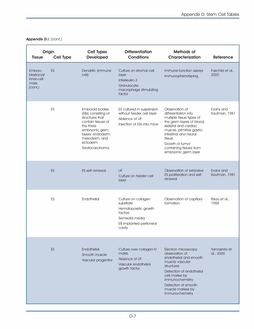
Appendix D: Stem Cell Tables
D-7
Origin Cell Types Differentiation Methods ofTissue Cell Type Developed Conditions Characterization Reference
Embryo-blastocystinner-cellmass(cont.)
ES Dendritic (immunecell)
Culture on stromal celllayer
Interleukin-3
Granulocyte-macrophage stimulatingfactor
Immune-function assays
Immunophenotyping
Fairchild et al.,2000
ES Embryoid bodies(EBs) consisting ofstructures thatcontain tissues ofthe threeembryonic germlayers: endoderm,mesoderm, andectoderm
Teratocarcinoma
ES cultured in suspensionwithout feeder cell layer
Absence of LIF
Injection of ESs into mice
Observation ofdifferentiation intomultiple tissue types ofthe germ layers of blood,skeletal and cardiacmuscle, primitive gastro-intestinal and neuraltissue
Growth of tumorcontaining tissues fromembryonic germ layer
Evans andKaufman, 1981
ES ES self–renewal LIF
Culture on feeder celllayer
Observation of extensiveES proliferation and self-renewal
Evans andKaufman, 1981
ES Endothelial Culture on collagensubstrate
Hematopoietic growthfactors
Semisolid media
EB implanted peritonealcavity
Observation of capillaryformation
Risau et al.,1988
ES Endothelial
Smooth muscle
Vascular progenitor
Culture over collagen-IVmatrix
Absence of LIF
Vascular endothelialgrowth factor
Electron microscopy:observation ofendothelial and smoothmuscle vascularstructures
Detection of endothelialcell marker byimmunochemistry
Detection of smoothmuscle markers byimmunochemistry
Yamashita etal., 2000
Appendix D.i. (cont.)
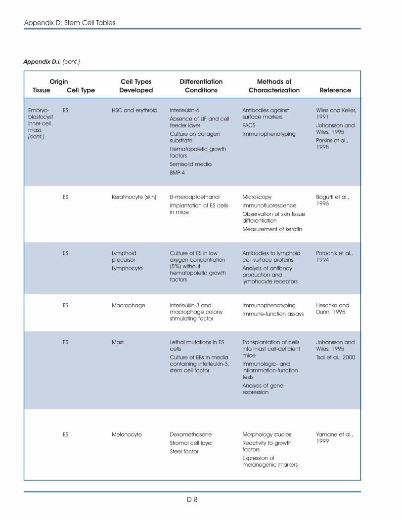
Origin Cell Types Differentiation Methods ofTissue Cell Type Developed Conditions Characterization Reference
Appendix D.i. (cont.)
D-8
Appendix D: Stem Cell Tables
ES Keratinocyte (skin) ß-mercaptoethanol
Implantation of ES cells in mice
Microscopy
Immunofluorescence
Observation of skin tissuedifferentiation
Measurement of keratin
Bagutti et al.,1996
ES Lymphoidprecursor
Lymphocyte
Culture of ES in lowoxygen concentration(5%) withouthematopoietic growthfactors
Antibodies to lymphoidcell-surface proteins
Analysis of antibodyproduction andlymphocyte receptors
Potocnik et al.,1994
ES Macrophage Interleukin-3 andmacrophage colonystimulating factor
Immunophenotyping
Immune-function assays
Lieschke andDunn, 1995
ES Mast Lethal mutations in EScells
Culture of EBs in mediacontaining interleukin-3,stem cell factor
Transplantation of cellsinto mast cell-deficientmice
Immunologic- andinflammation-functiontests
Analysis of geneexpression
Johansson andWiles, 1995
Tsai et al., 2000
ES Melanocyte Dexamethasone
Stromal cell layer
Steel factor
Morphology studies
Reactivity to growthfactors
Expression ofmelanogenic markers
Yamane et al.,1999
Embryo-blastocystinner-cellmass(cont.)
ES HSC and erythroid Interleukin-6
Absence of LIF and cellfeeder layer
Culture on collagensubstrate
Hematopoietic growthfactors
Semisolid media
BMP-4
Antibodies againstsurface markers
FACS
Immunophenotyping
Wiles and Keller,1991
Johansson andWiles, 1995
Perkins et al.,1998
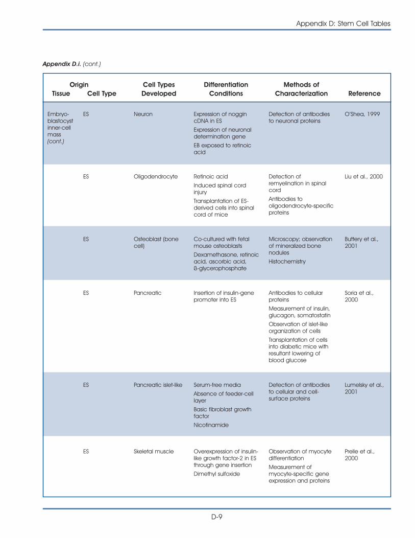
Appendix D: Stem Cell Tables
D-9
Origin Cell Types Differentiation Methods ofTissue Cell Type Developed Conditions Characterization Reference
Embryo-blastocystinner-cellmass(cont.)
ES Neuron Expression of noggincDNA in ES
Expression of neuronaldetermination gene
EB exposed to retinoicacid
Detection of antibodiesto neuronal proteins
O’Shea, 1999
ES Oligodendrocyte Retinoic acid
Induced spinal cordinjury
Transplantation of ES-derived cells into spinalcord of mice
Detection ofremyelination in spinalcord
Antibodies tooligodendrocyte-specificproteins
Liu et al., 2000
ES Osteoblast (bonecell)
Co-cultured with fetalmouse osteoblasts
Dexamethasone, retinoicacid, ascorbic acid, ß-glycerophosphate
Microscopy; observationof mineralized bonenodules
Histochemistry
Buttery et al.,2001
ES Pancreatic Insertion of insulin-genepromoter into ES
Antibodies to cellularproteins
Measurement of insulin,glucagon, somatostatin
Observation of islet-likeorganization of cells
Transplantation of cellsinto diabetic mice withresultant lowering ofblood glucose
Soria et al.,2000
ES Pancreatic islet-like Serum-free media
Absence of feeder-celllayer
Basic fibroblast growthfactor
Nicotinamide
Detection of antibodiesto cellular and cell-surface proteins
Lumelsky et al.,2001
Appendix D.i. (cont.)
ES Skeletal muscle Overexpression of insulin-like growth factor-2 in ESthrough gene insertion
Dimethyl sulfoxide
Observation of myocytedifferentiation
Measurement ofmyocyte-specific geneexpression and proteins
Prelle et al.,2000
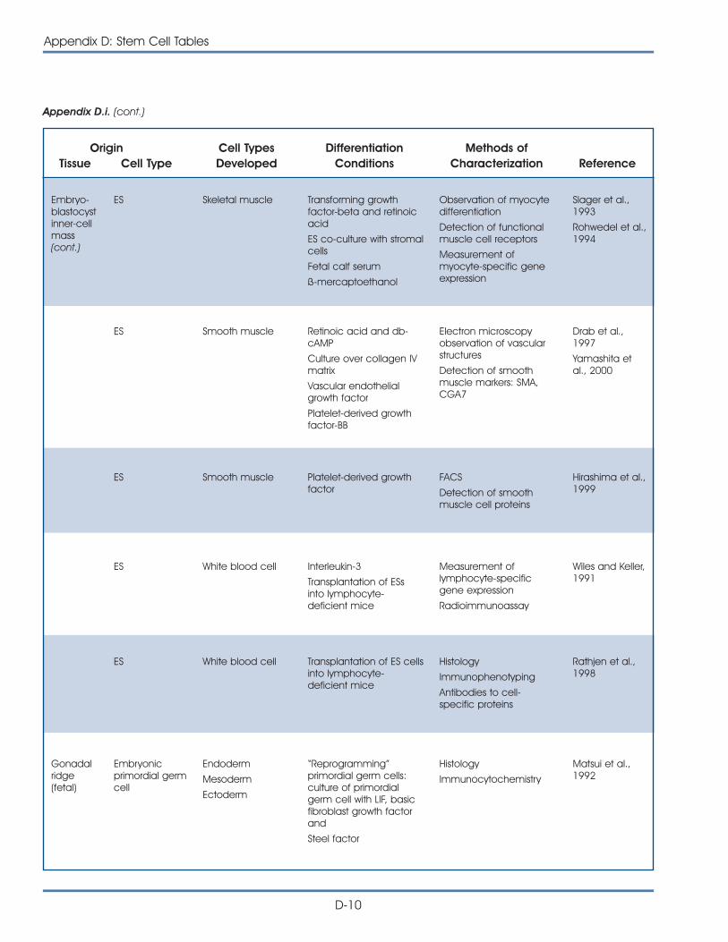
Origin Cell Types Differentiation Methods ofTissue Cell Type Developed Conditions Characterization Reference
Appendix D.i. (cont.)
D-10
Appendix D: Stem Cell Tables
Embryo-blastocystinner-cellmass(cont.)
ES Skeletal muscle Transforming growthfactor-beta and retinoicacid
ES co-culture with stromalcells
Fetal calf serum
ß-mercaptoethanol
Observation of myocytedifferentiation
Detection of functionalmuscle cell receptors
Measurement ofmyocyte-specific geneexpression
Slager et al.,1993
Rohwedel et al.,1994
ES Smooth muscle Retinoic acid and db-cAMP
Culture over collagen IVmatrix
Vascular endothelialgrowth factor
Platelet-derived growthfactor-BB
Electron microscopyobservation of vascularstructures
Detection of smoothmuscle markers: SMA,CGA7
Drab et al.,1997
Yamashita etal., 2000
ES Smooth muscle Platelet-derived growthfactor
FACS
Detection of smoothmuscle cell proteins
Hirashima et al.,1999
ES White blood cell Interleukin-3
Transplantation of ESsinto lymphocyte-deficient mice
Measurement oflymphocyte-specificgene expression
Radioimmunoassay
Wiles and Keller,1991
ES White blood cell Transplantation of ES cellsinto lymphocyte-deficient mice
Histology
Immunophenotyping
Antibodies to cell-specific proteins
Rathjen et al.,1998
Gonadalridge(fetal)
Embryonicprimordial germcell
Endoderm
Mesoderm
Ectoderm
“Reprogramming”primordial germ cells:culture of primordialgerm cell with LIF, basicfibroblast growth factorand
Steel factor
Histology
Immunocytochemistry
Matsui et al.,1992
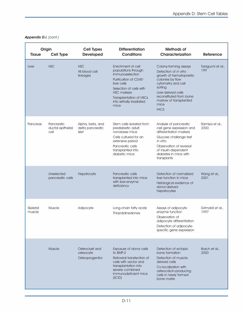
Appendix D: Stem Cell Tables
D-11
Origin Cell Types Differentiation Methods ofTissue Cell Type Developed Conditions Characterization Reference
Liver HSC HSC
All blood celllineages
Enrichment of cellpopulations throughimmunoselection
Purification of CD45+
liver cells
Selection of cells withHSC markers
Transplantation of HSCsinto lethally irradiatedmice
Colony-forming assays
Detection of in vitrogrowth of hematopoieticcolonies by flowcytometry and cellsorting
Liver-derived cellsreconstituted from bonemarrow of transplantedmice
FACS
Taniguchi et al.,199
Unselectedpancreatic cells
Hepatocyte Pancreatic cellstransplanted into micewith liver-enzymedeficiency
Detection of normalizedliver function in mice
Histological evidence ofdonor-derivedhepatocytes
Wang et al.,2001
Skeletalmuscle
Muscle Adipocyte Long-chain fatty acids
Thiazolidinediones
Assays of adipocyteenzyme function
Observation ofadipocyte differentiation
Detection of adipocyte-specific gene expression
Grimaldi et al.,1997
Muscle Osteoclast andosteocyte
Osteoprogenitor
Exposure of donor cellsto BMP-2
Retroviral transfection ofcells with vector andtransplantation intosevere combinedimmunodeficient mice(SCID)
Detection of ectopicbone formation
Detection of muscle-derived cells
Co-localization withosteocalcin-producingcells in newly formedbone matrix
Bosch et al.,2000
Appendix D.i. (cont.)
Pancreas Pancreaticductal epithelialcell
Alpha, beta, anddelta pancreaticislet
Stem cells isolated fromprediabetic adult,nonobese mice
Cells cultured for anextensive period
Pancreatic cellstransplanted intodiabetic mice
Analysis of pancreaticcell gene expression anddifferentiation markers
Glucose challenge testin vitro
Observation of reversalof insulin-dependentdiabetes in mice withtransplants
Ramiya et al.,2000
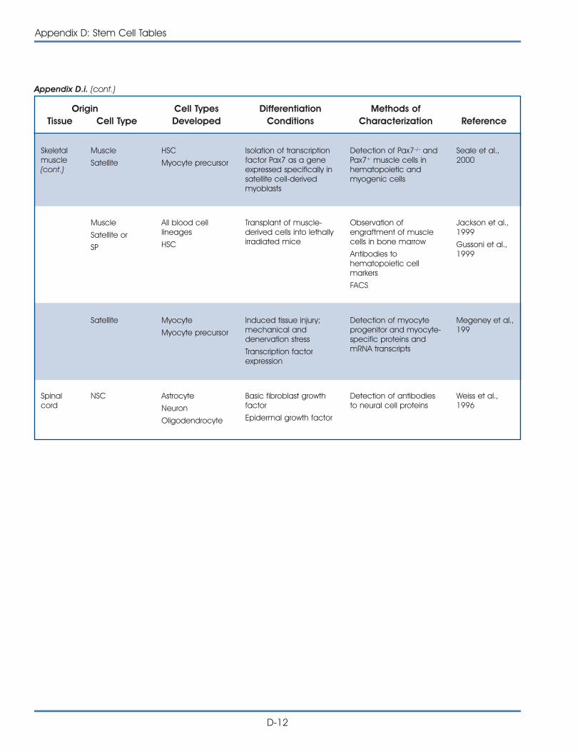
Origin Cell Types Differentiation Methods ofTissue Cell Type Developed Conditions Characterization Reference
Appendix D.i. (cont.)
D-12
Appendix D: Stem Cell Tables
Skeletalmuscle (cont.)
Muscle
Satellite
HSC
Myocyte precursor
Isolation of transcriptionfactor Pax7 as a geneexpressed specifically insatellite cell-derivedmyoblasts
Detection of Pax7–/– andPax7+ muscle cells inhematopoietic andmyogenic cells
Seale et al.,2000
Muscle
Satellite or
SP
All blood celllineages
HSC
Transplant of muscle-derived cells into lethallyirradiated mice
Observation ofengraftment of musclecells in bone marrow
Antibodies tohematopoietic cellmarkers
FACS
Jackson et al.,1999
Gussoni et al.,1999
Satellite Myocyte
Myocyte precursor
Induced tissue injury;mechanical anddenervation stress
Transcription factorexpression
Detection of myocyteprogenitor and myocyte-specific proteins andmRNA transcripts
Megeney et al.,199
Spinalcord
NSC Astrocyte
Neuron
Oligodendrocyte
Basic fibroblast growthfactor
Epidermal growth factor
Detection of antibodiesto neural cell proteins
Weiss et al.,1996
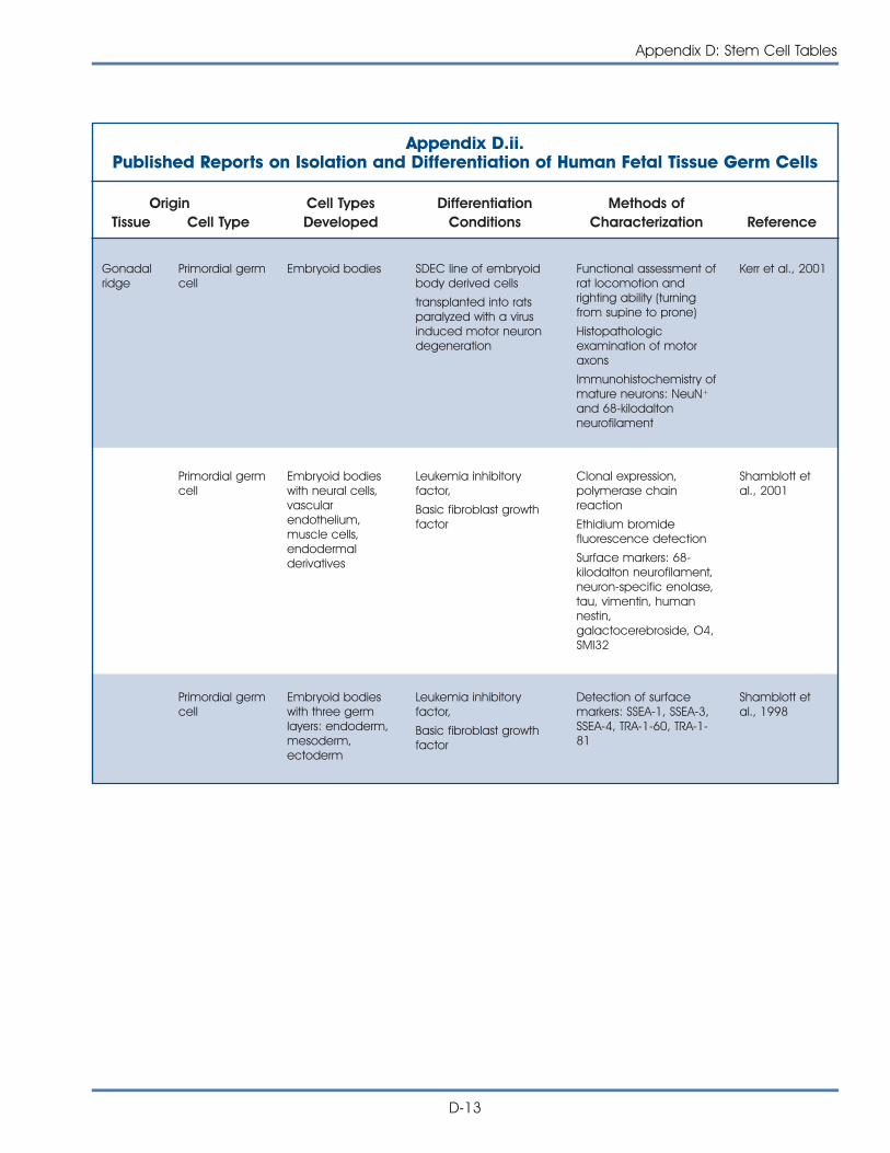
Appendix D: Stem Cell Tables
D-13
Origin Cell Types Differentiation Methods ofTissue Cell Type Developed Conditions Characterization Reference
Gonadalridge
Primordial germcell
Embryoid bodies SDEC line of embryoidbody derived cells
transplanted into ratsparalyzed with a virusinduced motor neurondegeneration
Functional assessment ofrat locomotion andrighting ability (turningfrom supine to prone)
Histopathologicexamination of motoraxons
Immunohistochemistry ofmature neurons: NeuN+
and 68-kilodaltonneurofilament
Kerr et al., 2001
Primordial germcell
Embryoid bodieswith neural cells,vascularendothelium,muscle cells,endodermalderivatives
Leukemia inhibitoryfactor,
Basic fibroblast growthfactor
Clonal expression,polymerase chainreaction
Ethidium bromidefluorescence detection
Surface markers: 68-kilodalton neurofilament,neuron-specific enolase,tau, vimentin, humannestin,galactocerebroside, O4,SMI32
Shamblott etal., 2001
Primordial germcell
Embryoid bodieswith three germlayers: endoderm,mesoderm,ectoderm
Leukemia inhibitoryfactor,
Basic fibroblast growthfactor
Detection of surfacemarkers: SSEA-1, SSEA-3,SSEA-4, TRA-1-60, TRA-1-81
Shamblott etal., 1998
Appendix D.ii.Published Reports on Isolation and Differentiation of Human Fetal Tissue Germ Cells
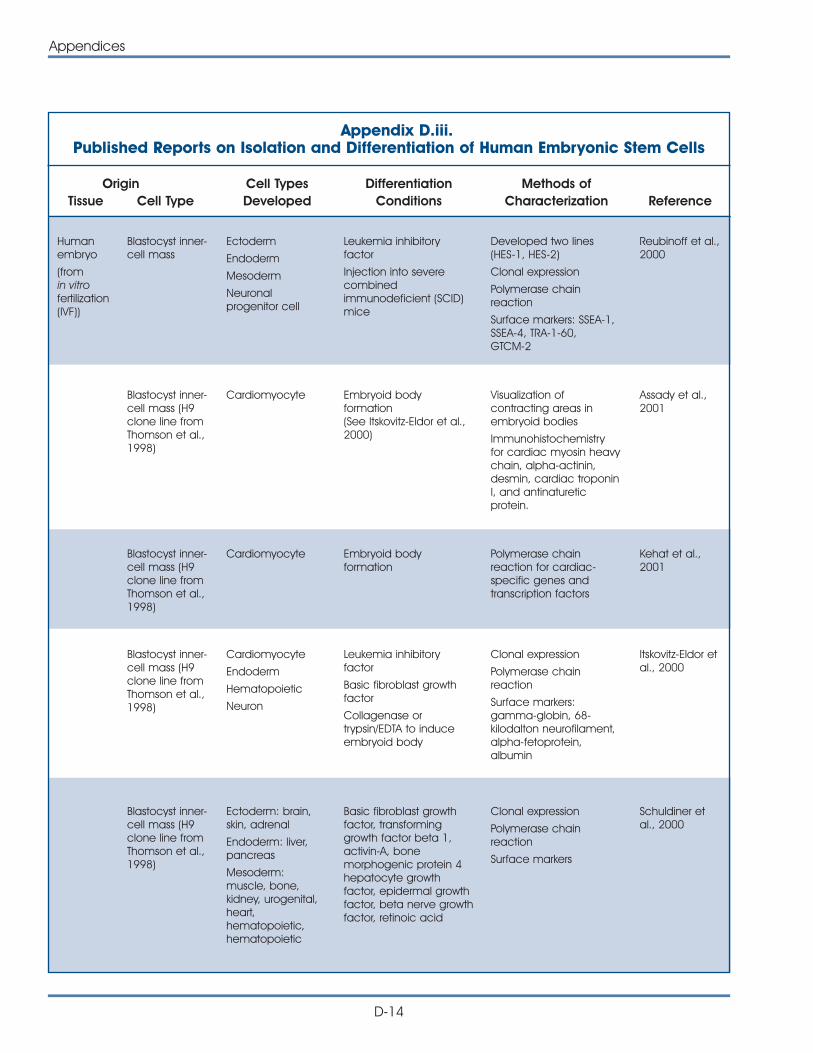
D-14
Appendices
Appendix D.iii.Published Reports on Isolation and Differentiation of Human Embryonic Stem Cells
Origin Cell Types Differentiation Methods ofTissue Cell Type Developed Conditions Characterization Reference
Humanembryo
(from in vitrofertilization(IVF))
Blastocyst inner-cell mass
Ectoderm
Endoderm
Mesoderm
Neuronalprogenitor cell
Leukemia inhibitoryfactor
Injection into severecombinedimmunodeficient (SCID)mice
Developed two lines(HES-1, HES-2)
Clonal expression
Polymerase chainreaction
Surface markers: SSEA-1,SSEA-4, TRA-1-60,GTCM-2
Reubinoff et al.,2000
Blastocyst inner-cell mass (H9clone line fromThomson et al.,1998)
Cardiomyocyte Embryoid bodyformation(See Itskovitz-Eldor et al.,2000)
Visualization ofcontracting areas inembryoid bodies
Immunohistochemistryfor cardiac myosin heavychain, alpha-actinin,desmin, cardiac troponinI, and antinatureticprotein.
Assady et al.,2001
Blastocyst inner-cell mass (H9clone line fromThomson et al.,1998)
Cardiomyocyte Embryoid bodyformation
Polymerase chainreaction for cardiac-specific genes andtranscription factors
Kehat et al.,2001
Blastocyst inner-cell mass (H9clone line fromThomson et al.,1998)
Cardiomyocyte
Endoderm
Hematopoietic
Neuron
Leukemia inhibitoryfactor
Basic fibroblast growthfactor
Collagenase ortrypsin/EDTA to induceembryoid body
Clonal expression
Polymerase chainreaction
Surface markers:gamma-globin, 68-kilodalton neurofilament,alpha-fetoprotein,albumin
Itskovitz-Eldor etal., 2000
Blastocyst inner-cell mass (H9clone line fromThomson et al.,1998)
Ectoderm: brain,skin, adrenal
Endoderm: liver,pancreas
Mesoderm:muscle, bone,kidney, urogenital,heart,hematopoietic,hematopoietic
Basic fibroblast growthfactor, transforminggrowth factor beta 1,activin-A, bonemorphogenic protein 4hepatocyte growthfactor, epidermal growthfactor, beta nerve growthfactor, retinoic acid
Clonal expression
Polymerase chainreaction
Surface markers
Schuldiner etal., 2000
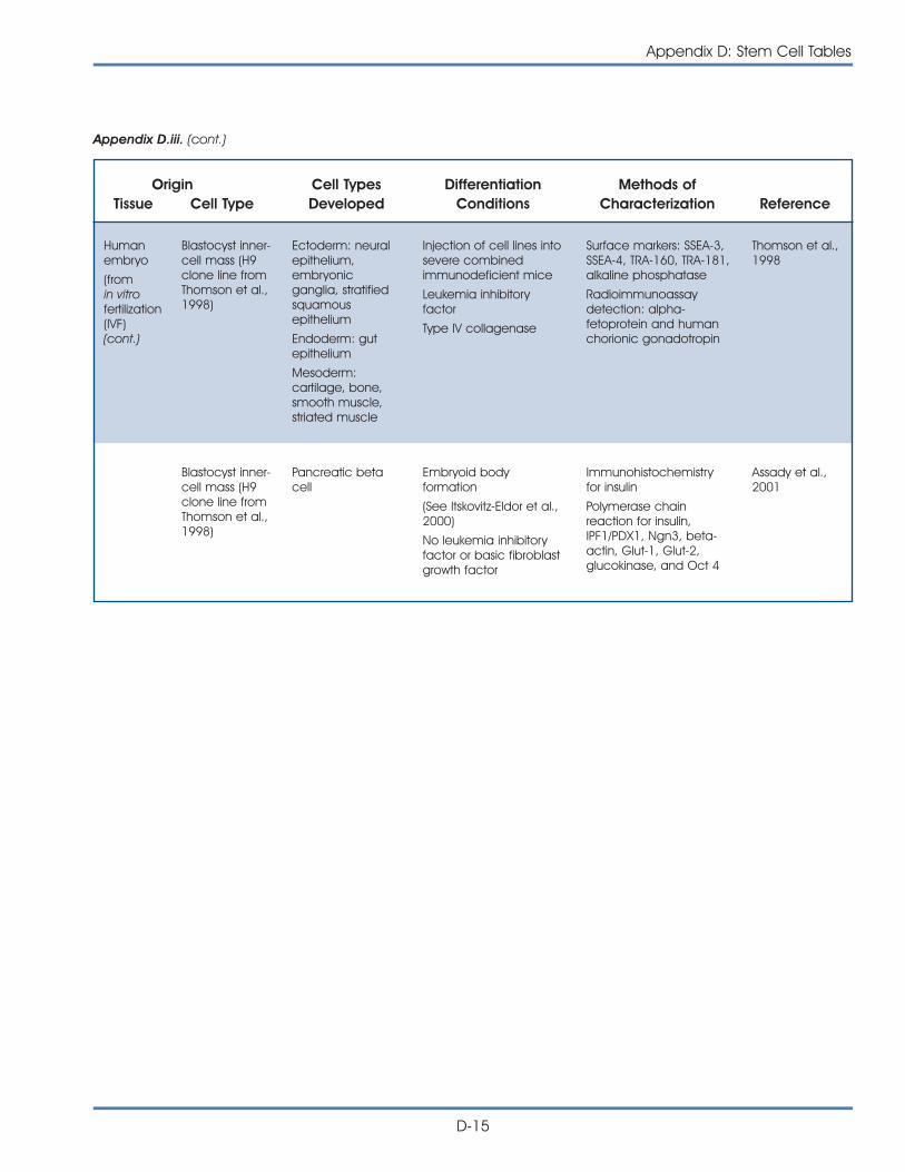
Appendix D: Stem Cell Tables
D-15
Origin Cell Types Differentiation Methods ofTissue Cell Type Developed Conditions Characterization Reference
Blastocyst inner-cell mass (H9clone line fromThomson et al.,1998)
Pancreatic betacell
Embryoid bodyformation
(See Itskovitz-Eldor et al.,2000)
No leukemia inhibitoryfactor or basic fibroblastgrowth factor
Immunohistochemistryfor insulin
Polymerase chainreaction for insulin,IPF1/PDX1, Ngn3, beta-actin, Glut-1, Glut-2,glucokinase, and Oct 4
Assady et al.,2001
Appendix D.iii. (cont.)
Humanembryo
(from in vitrofertilization(IVF)(cont.)
Blastocyst inner-cell mass (H9clone line fromThomson et al.,1998)
Ectoderm: neuralepithelium,embryonicganglia, stratifiedsquamousepithelium
Endoderm: gutepithelium
Mesoderm:cartilage, bone,smooth muscle,striated muscle
Injection of cell lines intosevere combinedimmunodeficient mice
Leukemia inhibitoryfactor
Type IV collagenase
Surface markers: SSEA-3,SSEA-4, TRA-160, TRA-181,alkaline phosphatase
Radioimmunoassaydetection: alpha-fetoprotein and humanchorionic gonadotropin
Thomson et al.,1998
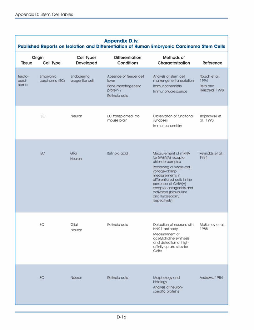
D-16
Appendix D: Stem Cell Tables
Origin Cell Types Differentiation Methods ofTissue Cell Type Developed Conditions Characterization Reference
Terato-carci-noma
Embryoniccarcinoma (EC)
Endodermalprogenitor cell
Absence of feeder celllayer
Bone morphogeneticprotein-2
Retinoic acid
Analysis of stem cellmarker-gene transcription
Immunochemistry
Immunofluorescence
Roach et al.,1994
Pera andHerszfeld, 1998
EC Neuron EC transplanted intomouse brain
Observation of functionalsynapses
Immunochemistry
Trojanowski etal., 1993
Adult Human Stem CellsAppendix D.iv.Published Reports on Isolation and Differentiation of Human Embryonic Carcinoma Stem Cells
EC Glial
Neuron
Retinoic acid Measurement of mRNAfor GABA(A) receptor-chloride complex
Recording of whole-cellvoltage-clampmeasurements indifferentiated cells in thepresence of GABA(A)receptor antagonists andactivators (bicucullineand flurazepam,respectively)
Reynolds et al.,1994
EC Glial
Neuron
Retinoic acid Detection of neurons withHNK-1 antibody
Measurement ofacetylcholine synthesisand detection of high-affinity uptake sites forGABA
McBurney et al.,1988
EC Neuron Retinoic acid Morphology andhistology
Analysis of neuron-specific proteins
Andrews, 1984
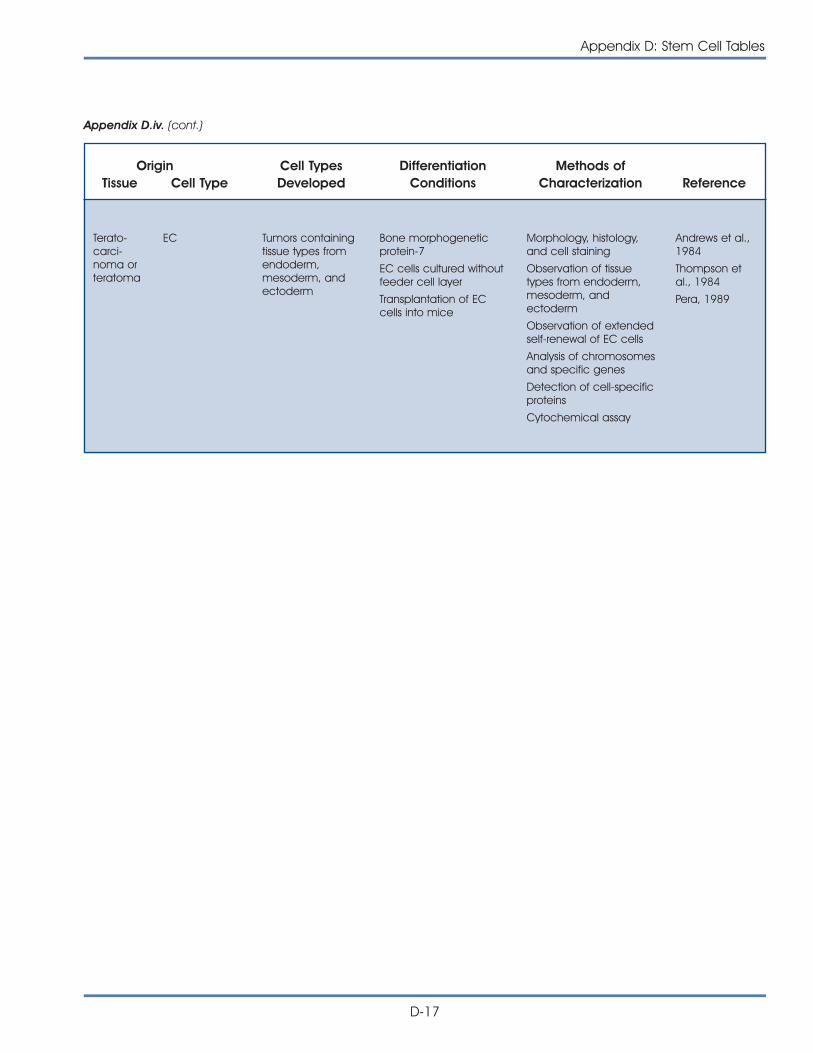
Appendix D: Stem Cell Tables
D-17
Origin Cell Types Differentiation Methods ofTissue Cell Type Developed Conditions Characterization Reference
Terato-carci-noma orteratoma
EC Tumors containingtissue types fromendoderm,mesoderm, andectoderm
Bone morphogeneticprotein-7
EC cells cultured withoutfeeder cell layer
Transplantation of ECcells into mice
Morphology, histology,and cell staining
Observation of tissuetypes from endoderm,mesoderm, andectoderm
Observation of extendedself-renewal of EC cells
Analysis of chromosomesand specific genes
Detection of cell-specificproteins
Cytochemical assay
Andrews et al.,1984
Thompson etal., 1984
Pera, 1989
Appendix D.iv. (cont.)
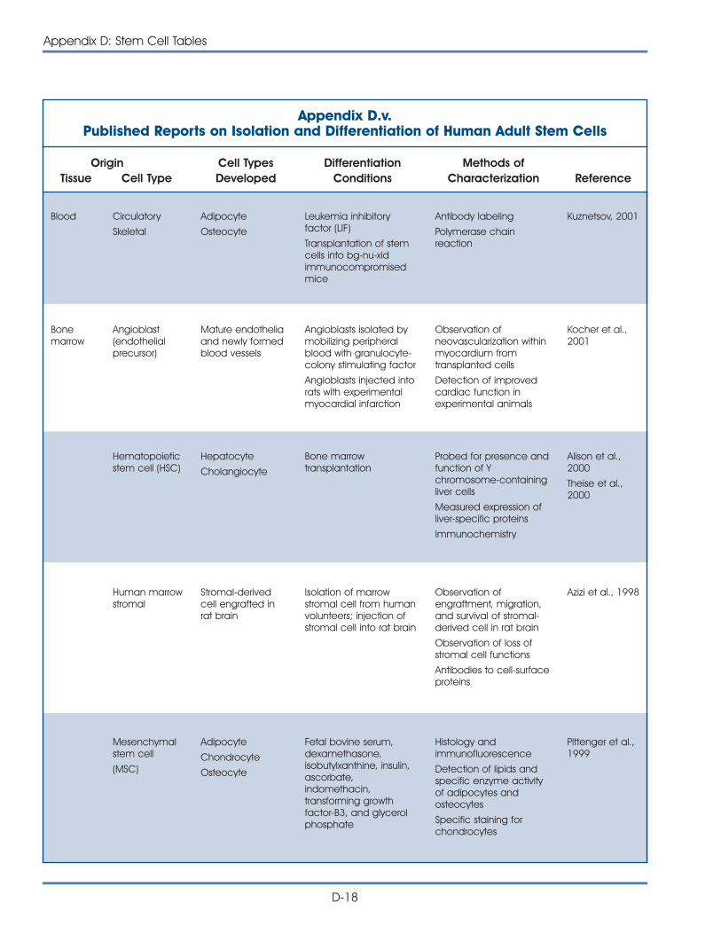
D-18
Appendix D: Stem Cell Tables
Origin Cell Types Differentiation Methods ofTissue Cell Type Developed Conditions Characterization Reference
Blood Circulatory
Skeletal
Adipocyte
Osteocyte
Leukemia inhibitoryfactor (LIF)
Transplantation of stemcells into bg-nu-xidimmunocompromisedmice
Antibody labeling
Polymerase chainreaction
Kuznetsov, 2001
Bonemarrow
Angioblast(endothelialprecursor)
Mature endotheliaand newly formedblood vessels
Angioblasts isolated bymobilizing peripheralblood with granulocyte-colony stimulating factor
Angioblasts injected intorats with experimentalmyocardial infarction
Observation ofneovascularization withinmyocardium fromtransplanted cells
Detection of improvedcardiac function inexperimental animals
Kocher et al.,2001
Adult Human Stem CellsAppendix D.v.Published Reports on Isolation and Differentiation of Human Adult Stem Cells
Hematopoieticstem cell (HSC)
Hepatocyte
Cholangiocyte
Bone marrowtransplantation
Probed for presence andfunction of Ychromosome-containingliver cells
Measured expression ofliver-specific proteins
Immunochemistry
Alison et al.,2000
Theise et al.,2000
Human marrowstromal
Stromal-derivedcell engrafted inrat brain
Isolation of marrowstromal cell from humanvolunteers; injection ofstromal cell into rat brain
Observation ofengraftment, migration,and survival of stromal-derived cell in rat brain
Observation of loss ofstromal cell functions
Antibodies to cell-surfaceproteins
Azizi et al., 1998
Mesenchymalstem cell
(MSC)
Adipocyte
Chondrocyte
Osteocyte
Fetal bovine serum,dexamethasone,isobutylxanthine, insulin,ascorbate,indomethacin,transforming growthfactor-B3, and glycerolphosphate
Histology andimmunofluorescence
Detection of lipids andspecific enzyme activityof adipocytes andosteocytes
Specific staining forchondrocytes
Pittenger et al.,1999
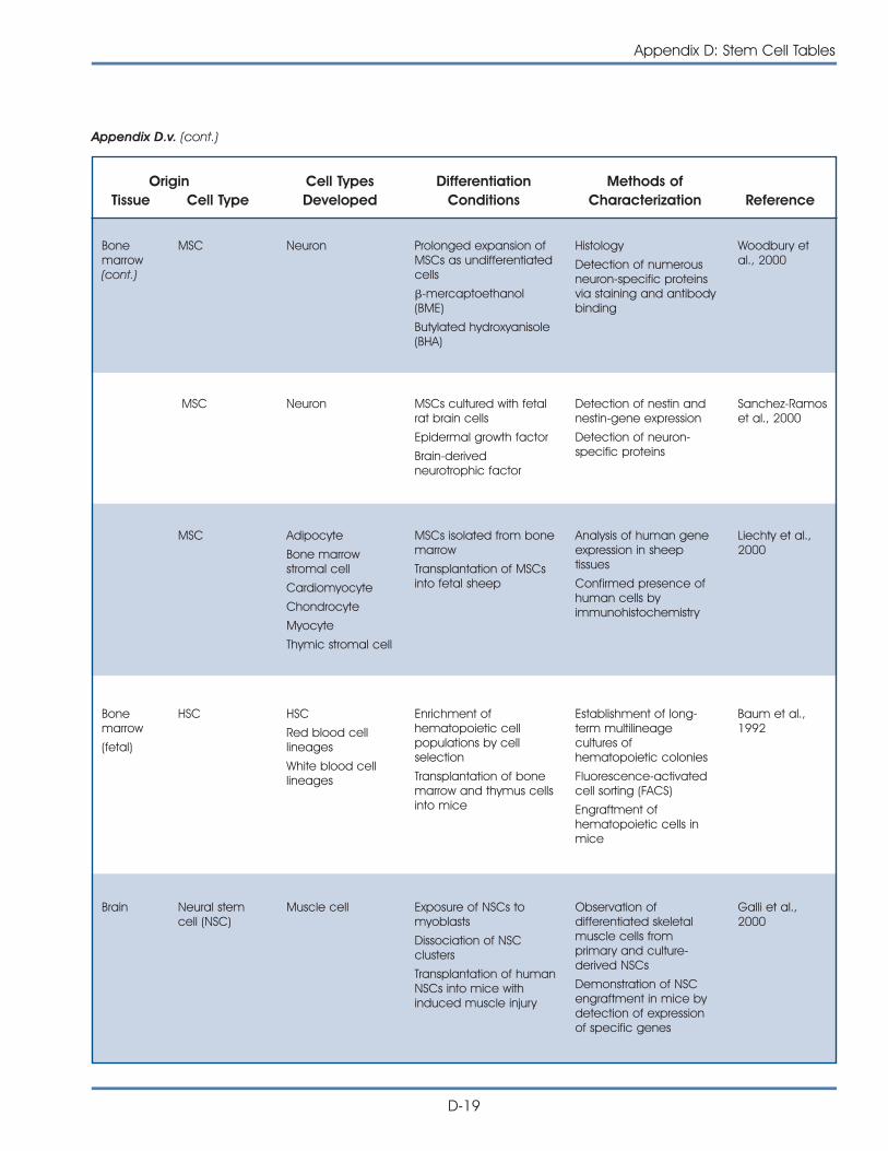
Appendix D: Stem Cell Tables
D-19
Origin Cell Types Differentiation Methods ofTissue Cell Type Developed Conditions Characterization Reference
Bonemarrow(cont.)
MSC Neuron Prolonged expansion ofMSCs as undifferentiatedcells
�-mercaptoethanol(BME)
Butylated hydroxyanisole(BHA)
Histology
Detection of numerousneuron-specific proteinsvia staining and antibodybinding
Woodbury etal., 2000
MSC Neuron MSCs cultured with fetalrat brain cells
Epidermal growth factor
Brain-derivedneurotrophic factor
Detection of nestin andnestin-gene expression
Detection of neuron-specific proteins
Sanchez-Ramoset al., 2000
MSC Adipocyte
Bone marrowstromal cell
Cardiomyocyte
Chondrocyte
Myocyte
Thymic stromal cell
MSCs isolated from bonemarrow
Transplantation of MSCsinto fetal sheep
Analysis of human geneexpression in sheeptissues
Confirmed presence ofhuman cells byimmunohistochemistry
Liechty et al.,2000
Bonemarrow
(fetal)
HSC HSC
Red blood celllineages
White blood celllineages
Enrichment ofhematopoietic cellpopulations by cellselection
Transplantation of bonemarrow and thymus cellsinto mice
Establishment of long-term multilineagecultures ofhematopoietic colonies
Fluorescence-activatedcell sorting (FACS)
Engraftment ofhematopoietic cells inmice
Baum et al.,1992
Brain Neural stemcell (NSC)
Muscle cell Exposure of NSCs tomyoblasts
Dissociation of NSCclusters
Transplantation of humanNSCs into mice withinduced muscle injury
Observation ofdifferentiated skeletalmuscle cells fromprimary and culture-derived NSCs
Demonstration of NSCengraftment in mice bydetection of expressionof specific genes
Galli et al.,2000
Appendix D.v. (cont.)
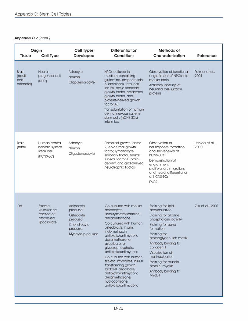
Origin Cell Types Differentiation Methods ofTissue Cell Type Developed Conditions Characterization Reference
Appendix D.v. (cont.)
D-20
Appendix D: Stem Cell Tables
Fat Stromalvascular cellfraction ofprocessedlipoaspirate
Adipocyteprecursor
Osteocyteprecursor
Chondrocyteprecursor
Myocyte precursor
Co-cultured with mouseadipocytes,isobutylmethylxanthine,dexamethasone
Co-cultured with humanosteoblasts, insulin,indomethacin,antibiotic/antimycoticdexamethasone,ascorbate, b-glycerophosphate,antibiotic/antimycotic
Co-cultured with humanskeletal myocytes, insulin,transforming growthfactor-B, ascorbate,antibiotic/antimycoticdexamethasone,hydrocortisone,antibiotic/antimycotic
Staining for lipidaccumulation
Staining for alkalinephosphatase activity
Staining for boneformation
Staining forproteoglycan-rich matrix
Antibody binding tocollagen II
Visualization ofmultinucleation
Staining for muscleprotein: myosin
Antibody binding toMyoD1
Zuk et al., 2001
Brain(adultandneonatal)
Neuralprogenitor cell
(NPC)
Astrocyte
Neuron
Oligodendrocyte
NPCs cultured inmedium containingglutamine, amphotericin-B, antibiotics, fetal calfserum, basic fibroblastgrowth factor, epidermalgrowth factor, andplatelet-derived growthfactor AB
Transplantation of humancentral nervous systemstem cells (hCNS-SCs)into mice
Observation of functionalengraftment of NPCs intomouse brain
Antibody labeling ofneuronal cell-surfaceproteins
Palmer et al.,2001
Brain(fetal)
Human centralnervous systemstem cell
(hCNS-SC)
Astrocyte
Neuron
Oligodendrocyte
Fibroblast growth factor-2, epidermal growthfactor, lymphocyteinhibitory factor, neuralsurvival factor-1, brain-derived and glial-derivedneurotrophic factors
Observation ofneurosphere formationand self-renewal ofhCNS-SCs
Demonstration ofengraftment,proliferation, migration,and neural differentiationof hCNS-SCs
FACS
Uchida et al.,2000
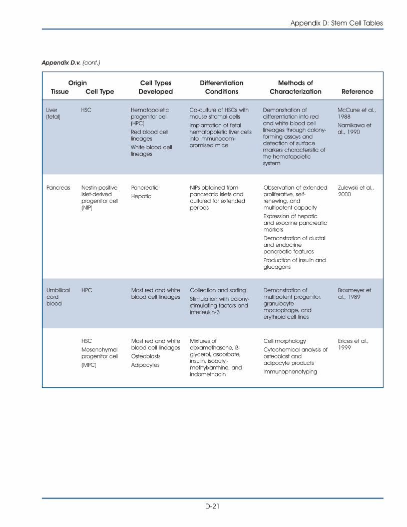
Appendix D: Stem Cell Tables
D-21
Origin Cell Types Differentiation Methods ofTissue Cell Type Developed Conditions Characterization Reference
Pancreas Nestin-positiveislet-derivedprogenitor cell(NIP)
Pancreatic
Hepatic
NIPs obtained frompancreatic islets andcultured for extendedperiods
Observation of extendedproliferative, self-renewing, andmultipotent capacity
Expression of hepaticand exocrine pancreaticmarkers
Demonstration of ductaland endocrinepancreatic features
Production of insulin andglucagons
Zulewski et al.,2000
HSC
Mesenchymalprogenitor cell
(MPC)
Most red and whiteblood cell lineages
Osteoblasts
Adipocytes
Mixtures ofdexamethasone, ß-glycerol, ascorbate,insulin, isobutyl-methylxanthine, andindomethacin
Cell morphology
Cytochemical analysis ofosteoblast andadipocyte products
Immunophenotyping
Erices et al.,1999
Umbilicalcordblood
HPC Most red and whiteblood cell lineages
Collection and sorting
Stimulation with colony-stimulating factors andinterleukin-3
Demonstration ofmultipotent progenitor,granulocyte-macrophage, anderythroid cell lines
Broxmeyer etal., 1989
Appendix D.v. (cont.)
Liver(fetal)
HSC Hematopoieticprogenitor cell(HPC)
Red blood celllineages
White blood celllineages
Co-culture of HSCs withmouse stromal cells
Implantation of fetalhematopoietic liver cellsinto immunocom-promised mice
Demonstration ofdifferentiation into redand white blood celllineages through colony-forming assays anddetection of surfacemarkers characteristic ofthe hematopoieticsystem
McCune et al.,1988
Namikawa etal., 1990
Appendix D.vi.
REFERENCES1. Alison, M.R., Poulsom, R., Jeffery, R., Dhillon, A.P., Quaglia,
A., Jacob, J., Novelli, M., Prentice, G., Williamson, J., andWright, N.A. (2000). Hepatocytes from non-hepatic adultstem cells. Nature. 406, 257.
2. Andrews, P.W., Damjanov, I., Simon, D., Banting, G.S., Carlin,C., Dracopoli, N.C., and Fogh, J. (1984). Pluripotent embry-onal carcinoma clones derived from the human teratocar-cinoma cell line Tera-2. Differentiation in vivo and in vitro.Lab. Invest. 50, 147-162.
3. Assady, S., Maor, G., Amit, M., Itskovitz-Eldor, J., Skorecki, K.L.,and Tzukerman, M. (2001). Insulin production by humanembryonic stem cells. Diabetes, 50,http://www.diabetes.org/Diabetes_Rapids/Suheir_Assady_06282001.pdf.
4. Azizi, S.A., Stokes, D., Augelli, B.J., DiGirolamo, C., andProckop, D.J. (1998). Engraftment and migration of humanbone marrow stromal cells implanted in the brains of albi-no rats—similarities to astrocyte grafts. Proc. Natl. Acad. Sci.U. S. A. 95, 3908-3913.
5. Bader, A., Al Dubai, H., and Weitzer, G. (2000). Leukemiainhibitory factor modulates cardiogenesis in embryoid bod-ies in opposite fashions. Circ. Res. 86, 787-794.
6. Bagutti, C., Wobus, A.M., Fassler, R., and Watt, F.M. (1996).Differentiation of embryonal stem cells into keratinocytes:comparison of wild-type and ß(1) integrin-deficient cells.Dev. Biol. 179, 184-196.
7. Bain, G., Kitchens, D., Yao, M., Huettner, J.E., and Gottlieb,D.I. (1995). Embryonic stem cells express neuronal proper-ties in vitro. Dev. Biol. 168, 342-357.
8. Baum, C.M., Weissman, I.L., Tsukamoto, A.S., Buckle, A.M.,and Peault, B. (1992). Isolation of a candidate humanhematopoietic stem-cell population. Proc. Natl. Acad. Sci.U. S. A. 89, 2804-2808.
9. Bittner, R.E., Schofer, C., Weipoltshammer, K., Ivanova, S.,Streubel, B., Hauser, E., Freilinger, M., Hoger, H., Elbe-Burger,A., and Wachtler, F. (1999). Recruitment of bone-marrow-derived cells by skeletal and cardiac muscle in adult dys-trophic mdx mice. Anat. Embryol. (Berl) 199, 391-396.
10. Bjornson, C.R., Rietze, R.L., Reynolds, B.A., Magli, M.C., andVescovi, A.L. (1999). Turning brain into blood: a hematopoi-etic fate adopted by adult neural stem cells in vivo.Science. 283, 534-537.
11. Bosch, P., Musgrave, D.S., Lee, J.Y., Cummins, J., Shuler, F.,Ghivizzani, S.C., Evans, C., Robbins, P.D., and Huard, J.(2000). Osteoprogenitor cells within skeletal muscle. J.Orthop. Res. 18, 933-944.
12. Brazelton, T.R., Rossi, F.M., Keshet, G.I., and Blau, H.M.(2000). From marrow to brain: expression of neuronal phe-notypes in adult mice. Science. 290, 1775-1779.
13. Broxmeyer, H.E., Douglas, G.W., Hangoc, G., Cooper, S.,Bard, J., English, D., Arny, M., Thomas, L., and Boyse, E.A.(1989). Human umbilical cord blood as a potential sourceof transplantable hematopoietic stem/progenitor cells.Proc. Natl. Acad. Sci. U. S. A. 86, 3828-3832.
14. Brustle, O., Jones, K.N., Learish, R.D., Karram, K., Choudhary,K., Wiestler, O.D., Duncan, I.D., and McKay, R.D. (1999).Embryonic stem cell-derived glial precursors: a source ofmyelinating transplants. Science. 285, 754-756.
15. Buttery, L.D., Bourne, S., Xynos, J.D., Wood, H., Hughes, F.J.,Hughes, S.P., Episkopou, V., and Polak, J.M. (2001).Differentiation of osteoblasts and in vitro bone formationfrom murine embryonic stem cells. Tissue Eng. 7, 89-99.
16. Dani, C., Smith, A.G., Dessolin, S., Leroy, P., Staccini, L.,Villageois, P., Darimont, C., and Ailhaud, G. (1997).Differentiation of embryonic stem cells into adipocytes invitro. J. Cell Sci. 110, 1279-1285.
17. Dinsmore, J., Ratliff, J., Deacon, T., Pakzaban, P., Jacoby, D.,Galpern, W., and Isacson, O. (1996). Embryonic stem cellsdifferentiated in vitro as a novel source of cells for trans-plantation. Cell Transplant. 5, 131-143.
18. Doetsch, F., Caille, I., Lim, D.A., Garcia-Verdugo, J.M., andAlvarez-Buylla, A. (1999). Subventricular zone astrocytes areneural stem cells in the adult mammalian brain. Cell. 97,703-716.
19. Doetschman, T., Eistetter, H., Katz, M., Schmit, W., andKemler, R. (1985). The in vitro development of blastocyst-derived embryonic stem cell lines: formation of visceralyolk sac, blood islands and myocardium. J. Embryol. Exp.Morph. 87, 27-45.
20. Drab, M., Haller, H., Bychkov, R., Erdmann, B., Lindschau, C.,Haase, H., Morano, I., Luft, F.C., and Wobus, A.M. (1997).From totipotent embryonic stem cells to spontaneouslycontracting smooth muscle cells: a retinoic acid and db-cAMP in vitro differentiation model. FASEB J. 11, 905-915.
21. Eglitis, M.A. and Mezey, E. (1997). Hematopoietic cells dif-ferentiate into both microglia and macroglia in the brainsof adult mice. Proc. Natl. Acad. Sci. U. S. A. 94, 4080-4085.
22. Erices, A., Conget, P., and Minguell, J.J. (1999).Mesenchymal progenitor cells in human umbilical cordblood. Br. J. Haematol. 109, 235-242.
23. Evans, M.J. and Kaufman, M.H. (1981). Establishment in cul-ture of pluripotential cells from mouse embryos. Nature.292, 154-156.
24. Fairchild, P.J., Brook, F.A., Gardner, R.L., Graca, L., Strong, V.,Tone, Y., Tone, M., Nolan, K.F., and Waldmann, H. (2000).Directed differentiation of dendritic cells from mouseembryonic stem cells. Curr. Biol. 10, 1515-1518.
25. Ferrari, G., Cusella-De Angelis, G., Coletta, M., Paolucci, E.,Stornaiuolo, A., Cossu, G., and Mavilio, F. (1998). Muscleregeneration by bone marrow-derived myogenic progeni-tors. Science. 279, 1528-1530.
D-22
Appendix D: Stem Cell Tables
26. Fraichard, A., Chassande, O., Bilbaut, G., Dehay, C.,Savatier, P., and Samarut, J. (1995). In vitro differentiation ofembryonic stem cells into glial cells and functional neu-rons. J. Cell Sci. 108, 3181-3188.
27. Friedenstein, A.J., Gorskaja, U.F., and Kulagina, N.N. (1976).Fibroblast precursors in normal and irradiated mousehematopoietic organs. Exp. Hematol. 4, 267-274.
28. Galli, R., Borello, U., Gritti, A., Minasi, M.G., Bjornson, C.,Coletta, M., Mora, M., De Angelis, M.G., Fiocco, R., Cossu,G., and Vescovi, A.L. (2000). Skeletal myogenic potential ofhuman and mouse neural stem cells. Nat. Neurosci. 3,986-991.
29. Gottlieb, D.I. and Huettner, J.E. (1999). An in vitro pathwayfrom embryonic stem cells to neurons and glia. CellsTissues Organs. 165, 165-172.
30. Grimaldi, P.A., Teboul, L., Inadera, H., Gaillard, D., and Amri,E.Z. (1997). Trans-differentiation of myoblasts to adipoblasts:triggering effects of fatty acids and thiazolidinediones.Prostaglandins. Leukot. Essent. Fatty. Acids. 57, 71-75.
31. Gussoni, E., Soneoka, Y., Strickland, C.D., Buzney, E.A., Khan,M.K., Flint, A.F., Kunkel, L.M., and Mulligan, R.C. (1999).Dystrophin expression in the mdx mouse restored by stemcell transplantation. Nature. 401, 390-394.
32. Hirashima, M., Kataoka, H., Nishikawa, S., Matsuyoshi, N.,and Nishikawa, S. (1999). Maturation of embryonic stemcells into endothelial cells in an in vitro model of vasculo-genesis. Blood. 93, 1253-1263.
33. Itskovitz-Eldor, J., Schuldiner, M., Karsenti, D., Eden, A.,Yanuka, O., Amit, M., Soreq, H., and Benvenisty, N. (2000).Differentiation of human embryonic stem cells into embry-oid bodies comprising the three embryonic germ layers.Mol. Med. 6, 88-95.
34. Jackson, K.A., Mi, T., and Goodell, M.A. (1999).Hematopoietic potential of stem cells isolated from murineskeletal muscle. Proc. Natl. Acad. Sci. U. S. A. 96, 14482-14486.
35. Jackson, K., Majka SM, Wang H, Pocius J, Hartley CJ,Majesky MW, Entman ML, Michael LH, Hirschi KK, and andGoodell MA (2001). Regeneration of ischemic cardiacmuscle and vascular endothelium by adult stem cells. J.Clin. Invest. 107, 1-8.
36. Johansson, B.M. and Wiles, M.V. (1995). Evidence forinvolvement of activin A and bone morphogenetic protein4 in mammalian mesoderm and hematopoietic develop-ment. Mol. Cell Biol. 15, 141-151.
37. Johansson, C.B., Momma, S., Clarke, D.L., Risling, M.,Lendahl, U., and Frisen, J. (1999). Identification of a neuralstem cell in the adult mammalian central nervous system.Cell. 96, 25-34.
38. Kawasaki, H., Mizuseki, K., Nishikawa, S., Kaneko, S., Kuwana,Y., Nakanishi, S., Nishikawa, S.I., and Sasai, Y. (2000).Induction of midbrain dopaminergic neurons from ES cellsby stromal cell-derived inducing activity. Neuron. 28, 31-40.
39. Kehat, I., Kenyagin-Karsenti, D., Druckmann, M., Segev, H.,Amit, M., Gepstein, A., Livne, E., Binah, O., Itskovitz-Eldor, J.,and Gepstein, L. (2001). Human embryonic stem cells candifferentiate into myocytes portraying cardiomyocytic struc-tural and functional properties. J. Clin. Invest. (in press).
40. Kerr, D.A., Llado, J., Shamblott, M., Maragakis, N., Irani, D.N.,Dike, S., Sappington, A., Gearhart, J., and Rothstein, J.(2001). Human embryonic germ cell derivatives facillitatemotor recovery of rats with diffuse motor neuron injury.
41. Klug, M.G., Soonpaa, M.H., Koh, G.Y., and Field, L.J. (1996).Genetically selected cardiomyocytes from differentiatingembryonic stem cells form stable intracardiac grafts. J.Clin. Invest. 98, 216-224.
42. Kocher, A.A., Schuster, M.D., Szabolcs, M.J., Takuma, S.,Burkhoff, D., Wang, J., Homma, S., Edwards, N.M., andItescu, S. (2001). Neovascularization of ischemic myocardi-um by human bone-marrow-derived angioblasts preventscardiomyocyte apoptosis, reduces remodeling andimproves cardiac function. Nat. Med. 7, 430-436.
43. Kopen, G.C., Prockop, D.J., and Phinney, D.G. (1999).Marrow stromal cells migrate throughout forebrain andcerebellum, and they differentiate into astrocytes afterinjection into neonatal mouse brains. Proc. Natl. Acad.Sci. U. S. A. 96, 10711-10716.
44. Kramer, J., Hegert, C., Guan, K., Wobus, A.M., Muller, P.K.,and Rohwedel, J. (2000). Embryonic stem cell-derivedchondrogenic differentiation in vitro: activation by BMP-2and BMP-4. Mech. Dev. 92, 193-205.
45. Krause, D.S., Theise, N.D., Collector, M.I., Henegariu, O.,Hwang, S., Gardner, R., Neutzel, S., and Sharkis, S.J. (2001).Multi-organ, multi-lineage engraftment by a single bonemarrow-derived stem cell. Cell. 105, 369-377.
46. Kuznetsov, S.A., Mankani, M.H., Gronthos, S., Satomura, K.,Bianco, P., and Robey P.G. (2001). Circulating skeletal stemcells. J. Cell Biol. 153, 1133-40.
47. Lagasse, E., Connors, H., Al Dhalimy, M., Reitsma, M.,Dohse, M., Osborne, L., Wang, X., Finegold, M., Weissman,I.L., and Grompe, M. (2000). Purified hematopoietic stemcells can differentiate into hepatocytes in vivo. Nat. Med.6, 1229-1234.
48. Lee, S.H., Lumelsky, N., Studer, L., Auerbach, J.M., andMcKay, R.D. (2000). Efficient generation of midbrain andhindbrain neurons from mouse embryonic stem cells. Nat.Biotechnol. 18, 675-679.
49. Li, M., Pevny, L., Lovell-Badge, R., and Smith, A. (1998).Generation of purified neural precursors from embryonicstem cells by lineage selection. Curr. Biol. 8, 971-974.
50. Liechty, K.W., MacKenzie, T.C., Shaaban, A.F., Radu, A.,Moseley, A.B., Deans, R., Marshak, D.R., and Flake, A.W.(2000). Human mesenchymal stem cells engraft anddemonstrate site-specific differentiation after in utero trans-plantation in sheep. Nat. Med. 6, 1282-1286.
Appendix D: Stem Cell Tables
D-23
51. Lieschke, G.J. and Dunn, A.R. (1995). Development of func-tional macrophages from embryonal stem cells in vitro.Exp. Hematol. 23, 328-334.
52. Liu, S., Qu, Y., Stewart, T.J., Howard, M.J., Chakrabortty, S.,Holekamp, T.F., and McDonald, J.W. (2000). Embryonicstem cells differentiate into oligodendrocytes and myeli-nate in culture and after spinal cord transplantation. Proc.Natl. Acad. Sci. U. S. A. 97, 6126-6131.
53. Lumelsky, N., Blondel, O., Laeng, P., Velasco, I., Ravin, R.,and McKay, R. (2001). Differentiation of Embryonic StemCells to Insulin-Secreting Structures Similiar to PancreaticIslets. Science. 292, 1389-1394.
54. Makino, S., Fukuda, K., Miyoshi, S., Konishi, F., Kodama, H.,Pan, J., Sano, M., Takahashi, T., Hori, S., Abe, H., Hata, J.,Umezawa, A., and Ogawa, S. (1999). Cardiomyocytes canbe generated from marrow stromal cells in vitro. J. Clin.Invest. 103, 697-705.
55. Maltsev, V.A., Rohwedel, J., Hescheler, J., and Wobus, A.M.(1993). Embryonic stem cells differentiate in vitro into car-diomyocytes representing sinusnodal, atrial and ventricularcell types. Mech. Dev. 44, 41-50.
56. Matsui, Y., Zsebo, K., and Hogan, B.L. (1992). Derivation ofpluripotential embryonic stem cells from murine primordialgerm cells in culture. Cell. 70, 841-847.
57. McBurney, M.W., Reuhl, K.R., Ally, A.I., Nasipuri, S., Bell, J.C.,and Craig, J. (1988). Differentiation and maturation ofembryonal carcinoma-derived neurons in cell culture. J.Neurosci. 8, 1063-1073.
58. McCune, J.M., Namikawa, R., Kaneshima, H., Shultz, L.D.,Lieberman, M., and Weissman, I.L. (1988). The SCID-humouse: murine model for the analysis of human hema-tolymphoid differentiation and function. Science. 241,1632-1639.
59. Megeney, L.A., Kablar, B., Garrett, K., Anderson, J.E., andRudnicki, M.A. (1996). MyoD is required for myogenic stemcell function in adult skeletal muscle. Genes Dev. 10, 1173-1183.
60. Mezey, E., Chandross, K.J., Harta, G., Maki, R.A., andMcKercher, S.R. (2000). Turning blood into brain: cellsbearing neuronal antigens generated in vivo from bonemarrow. Science. 290, 1779-1782.
61. Morrison, S.J., Uchida, N., and Weissman, I.L. (1995). Thebiology of hematopoietic stem cells. Annu. Rev. Cell. Dev.Biol. 11, 35-71.
62 Namikawa, R., Weilbaecher, K.N., Kaneshima, H., Yee, E.J.,and McCune, J.M. (1990). Long-term human hematopoiesisin the SCID-hu mouse. J. Exp. Med. 172, 1055-1063.
63. O’Shea, K.S. (1999). Embryonic stem cell models of devel-opment. Anat. Rec. 257, 32-41.
64. Orlic, D., Kajstura, J., Chimenti, S., Jakoniuk, I., Anderson,S.M., Li, B., Pickel, J., McKay, R., Nadal-Ginard, B., Bodine,D.M., Leri, A., and Anversa, P. (2001). Bone marrow cellsregenerate infarcted myocardium. Nature. 410, 701-705.
65. Palmer, T.D., Schwartz, P.H., Taupin, P., Kaspar, B., Stein, S.A.,and Gage, F.H. (2001). Progenitor cells from human brainafter death. Nature. 411, 42-43.
66. Pera, M.F., Cooper, S., Mills, J., and Parrington, J.M. (1989).Isolation and characterization of a multipotent clone ofhuman embryonal carcinoma cells. Differentiation. 42,10-23.
67. Pera, M.F. and Herszfeld, D. (1998). Differentiation of humanpluripotent teratocarcinoma stem cells induced by bonemorphogenetic protein-2. Reprod. Fertil. Dev. 10, 551-555.
68. Pereira, R.F., Halford, K.W., O’Hara, M.D., Leeper, D.B.,Sokolov, B.P., Pollard, M.D., Bagasra, O., and Prockop, D.J.(1995). Cultured adherent cells from marrow can serve aslong-lasting precursor cells for bone, cartilage, and lungin irradiated mice. Proc. Natl. Acad. Sci. U. S. A. 92,4857-4861.
69. Perkins, A.C. (1998). Enrichment of blood from embryonicstem cells in vitro. Reprod. Fertil. Dev. 10, 563-572.
70. Petersen, B.E., Bowen, W.C., Patrene, K.D., Mars, W.M.,Sullivan, A.K., Murase, N., Boggs, S.S., Greenberger, J.S., andGoff, J.P. (1999). Bone marrow as a potential source ofhepatic oval cells. Science. 284, 1168-1170.
71. Pittenger, M.F., Mackay, A.M., Beck, S.C., Jaiswal, R.K.,Douglas, R., Mosca, J.D., Moorman, M.A., Simonetti, D.W.,Craig, S., and Marshak, D.R. (1999). Multilineage potentialof adult human mesenchymal stem cells. Science.284, 143-147.
72. Potocnik, A.J., Nielsen, P.J., and Eichmann, K. (1994). In vitrogeneration of lymphoid precursors from embryonic stemcells. EMBO J. 13, 5274-5283.
73. Prelle, K., Wobus, A.M., Krebs, O., Blum, W.F., and Wolf, E.(2000). Overexpression of insulin-like growth factor-II inmouse embryonic stem cells promotes myogenic differen-tiation. Biochem. Biophys. Res. Commun. 277, 631-638.
74. Prockop, D.J. (1997). Marrow stromal cells as stem cells fornonhematopoietic tissues. Science. 276, 71-74.
75. Ramiya, V.K., Maraist, M., Arfors, K.E., Schatz, D.A., Peck,A.B., and Cornelius, J.G. (2000). Reversal of insulin-depend-ent diabetes using islets generated in vitro from pancreaticstem cells. Nat. Med. 6, 278-282.
76. Rathjen, P.D., Lake, J., Whyatt, L.M., Bettess, M.D., andRathjen, J. (1998). Properties and uses of embryonic stemcells: prospects for application to human biology andgene therapy. Reprod. Fertil. Dev. 10, 31-47.
77. Reubinoff, B.E., Pera, M.F., Fong, C.Y., Trounson, A., andBongso, A. (2000). Embryonic stem cell lines from humanblastocysts: somatic differentiation in vitro. Nat. Biotechnol.18, 399-404.
78. Reynolds, B.A. and Weiss, S. (1996). Clonal and populationanalyses demonstrate that an EGF-responsive mammalianembryonic CNS precursor is a stem cell. Dev. Biol. 175,1-13.
D-24
Appendix D: Stem Cell Tables
79. Reynolds, J.N., Ryan, P.J., Prasad, A., and Paterno, G.D.(1994). Neurons derived from embryonal carcinoma (P19)cells express multiple GABA(A) receptor subunits and fullyfunctional GABA(A) receptors. Neurosci. Lett. 165, 129-132.
80. Risau, W., Sariola, H., Zerwes, H.G., Sasse, J., Ekblom, P.,Kemler, R., and Doetschman, T. (1988). Vasculogenesis andangiogenesis in embryonic-stem-cell-derived embryoidbodies. Development. 102, 471-478.
81. Roach, S., Schmid, W., and Pera, M.F. (1994). Hepatocytictranscription factor expression in human embryonal carci-noma and yolk sac carcinoma cell lines: expression ofHNF-3� in models of early endodermal cell differentiation.Exp. Cell. Res. 215, 189-198.
82. Rohwedel, J., Maltsev, V., Bober, E., Arnold, H.H., Hescheler,J., and Wobus, A.M. (1994). Muscle cell differentiation ofembryonic stem cells reflects myogenesis in vivo: develop-mentally regulated expression of myogenic determinationgenes and functional expression of ionic currents. Dev. Biol.164, 87-101.
83. Sanchez-Ramos, J., Song, S., Cardozo-Pelaez, F., Hazzi, C.,Stedeford, T., Willing, A., Freeman, T.B., Saporta, S., Janssen,W., Patel, N., Cooper, D.R., and Sanberg, P.R. (2000). Adultbone marrow stromal cells differentiate into neural cells invitro. Exp. Neurol. 164, 247-256.
84. Schuldiner, M., Yanuka, O., Itskovitz-Eldor, J., Melton, D., andBenvenisty, N. (2000). Effects of eight growth factors on thedifferentiation of cells derived from human embryonic stemcells. Proc. Natl. Acad. Sci. U. S. A. 97, 11307-11312.
85. Seale, P., Sabourin, L.A., Girgis-Gabardo, A., Mansouri, A.,Gruss, P., and Rudnicki, M.A. (2000). Pax7 is required for thespecification of myogenic satellite cells. Cell. 102, 777-786.
86. Shamblott, M.J., Axelman, J., Littlefield, J.W., Blumenthal,P.D., Huggins, G.R., Cui, Y., Cheng, L., and Gearhart, J.D.(2001). Human embryonic germ cell derivatives express abroad range of develpmentally distinct markers and pro-liferate extensively in vitro. Proc. Natl. Acad. Sci. U. S. A.98, 13-118.
87. Shamblott, M.J., Axelman, J., Wang, S., Bugg, E.M.,Littlefield, J.W., Donovan, P.J., Blumenthal, P.D., Huggins,G.R., and Gearhart, J.D. (1998). Derivation of pluripotentstem cells from cultured human primordial germ cells.Proc. Natl. Acad. Sci. U. S. A. 95, 13726-13731.
88. Slager, H.G., Van Inzen, W., Freund, E., Van den Eijnden-VanRaaij A.J.M., and Mummery, C.L. (1993). Transforminggrowth factor-beta in the early mouse embryo: implicationsfor the regulation of muscle formation and implantation.Dev. Genet. 14, 212-224.
89. Soria, B., Roche, E., Berna, G., Leon-Quinto, T., Reig, J.A.,and Martin, F. (2000). Insulin-secreting cells derived fromembryonic stem cells normalize glycemia in streptozotocin-induced diabetic mice. Diabetes. 49, 157-162.
90. Spangrude, G.J., Smith, L., Uchida, N., Ikuta, K., Heimfeld,S., Friedman, J., and Weissman, I.L. (1991). Mousehematopoietic stem cells. Blood. 78, 1395-1402.
91. Strubing, C., Ahnert-Hilger, G., Shan, J., Wiedenmann, B.,Hescheler, J., and Wobus, A.M. (1995). Differentiation ofpluripotent embryonic stem cells into the neuronal lineagein vitro gives rise to mature inhibitory and excitatoryneurons. Mech. Dev. 53, 275-287.
92. Taniguchi, H., Toyoshima, T., Fukao, K., and Nakauchi, H.(1996). Presence of hematopoietic stem cells in the adultliver. Nat. Med. 2, 198-203.
93. Theise, N.D., Nimmakayalu, M., Gardner, R., Illei, P.B.,Morgan, G., Teperman, L., Henegariu, O., and Krause, D.S.(2000). Liver from bone marrow in humans. Hepatology.32, 11-16.
94. Thompson, S., Stern, P.L., Webb, M., Walsh, F.S., Engstrom,W., Evans, E.P., Shi, W.K., Hopkins, B., and Graham, C.F.(1984). Cloned human teratoma cells differentiate intoneuron-like cells and other cell types in retinoic acid. J.Cell. Sci. 72, 37-64.
95. Thomson, J.A., Itskovitz-Eldor, J., Shapiro, S.S., Waknitz, M.A.,Swiergiel, J.J., Marshall, V.S., and Jones, J.M. (1998).Embryonic stem cell lines derived from human blasto-cysts. Science. 282, 1145-1147.
96. Tomita, S., Li, R.K., Weisel, R.D., Mickle, D.A., Kim, E.J., Sakai,T., and Jia, Z.Q. (1999). Autologous transplantation ofbone marrow cells improves damaged heart function672. Circulation. 100 (Suppl. II), 11247-11256.
97. Trojanowski, J.Q., Mantione, J.R., Lee, J.H., Seid, D.P., You,T., Inge, L.J., and Lee, V.M. (1993). Neurons derived from ahuman teratocarcinoma cell line establish molecular andstructural polarity following transplantation into the rodentbrain. Exp. Neurol. 122, 283-294.
98. Tsai, M., Wedemeyer, J., Ganiatsas, S., Tam, S.Y., Zon, L.I.,and Galli, S.J. (2000). In vivo immunological function ofmast cells derived from embryonic stem cells: anapproach for the rapid analysis of even embryonic lethalmutations in adult mice in vivo. Proc. Natl. Acad. Sci. U. S. A. 97, 9186-9190.
99. Uchida, N., Buck, D.W., He, D., Reitsma, M.J., Masek, M.,Phan, T.V., Tsukamoto, A.S., Gage, F.H., and Weissman, I.L.(2000). Direct isolation of human central nervous systemstem cells. Proc. Natl. Acad. Sci. U. S. A. 97, 14720-14725.
100. Wakitani, S., Saito, T., and Caplan, A.I. (1995). Myogeniccells derived from rat bone marrow mesenchymal stemcells exposed to 5-azacytidine 754. Muscle. Nerve. 18,1417-1426.
101. Wang, X., Al-Dhalimy, M., Lagasse, E., Finegold, M., andGrompe, M. (2001). Liver repopulation and correction ofmetabolic liver disease by transplanted adult mouse pan-creatic cells. Am. J. Pathol. 158, 571-579.
102. Weiss, S., Dunne, C., Hewson, J., Wohl, C., Wheatley, M.,Peterson, A.C., and Reynolds, B.A. (1996). Multipotent CNSstem cells are present in the adult mammalian spinalcord and ventricular neuroaxis. J. Neurosci. 16,7599-7609.
Appendix D: Stem Cell Tables
D-25
103. Westfall, M.V., Pasyk, K.A., Yule, D.I., Samuelson, L.C., andMetzger, J.M. (1997). Ultrastructure and cell-cell couplingof cardiac myocytes differentiating in embryonic stemcell cultures. Cell. Motil. Cytoskeleton. 36, 43-54.
104. Wiles, M.V. and Keller, G. (1991). Multiple hematopoieticlineages develop from embryonic stem (ES) cells in cul-ture. Development. 111, 259-267.
105. Wobus, A.M., Rohwedel, J., Maltsev, V., and Hescheler, J.(1995). Development of cardiomyocytes expressing car-diac-specific genes, action potentials, and ionic channelsduring embryonic stem cell-derived cardiogenesis. Ann.N. Y. Acad. Sci. 752, 460-469.
106. Woodbury, D., Schwarz, E.J., Prockop, D.J., and Black, I.B.(2000). Adult rat and human bone marrow stromal cellsdifferentiate into neurons. J. Neurosci. Res. 61, 364-370.
107. Yamane, T., Hayashi, H., Mizoguchi, M., Yamazaki, H., andKunisada, T. (1999). Derivation of melanocytes fromembryonic stem cells in culture. Dev. Dyn. 216, 450-458.
108. Yamashita, J., Itoh, H., Hirashima, M., Ogawa, M.,Nishikawa, S., Yurugi, T., Naito, M., Nakao, K., andNishikawa, S. (2000). Flk1-positive cells derived fromembryonic stem cells serve as vascular progenitors.Nature. 408, 92-96.
109. Zuk, P.A., Zhu, M., Mizuno, H., Huang, J., Futrell, J.W., Katz,A.J., Benhaim, P., Lorenz, H.P., and Hedrick, M.H. (2001).Multilineage cells from human adipose tissue: implicationsfor cell- based therapies. Tissue Eng. 7, 211-228.
110. Zulewski, H., Abraham, E.J., Gerlach, M.J., Daniel, P.B.,Moritz, W., Muller, B., Vallejo, M., Thomas, M.K., andHabener, J.F. (2001). Multipotential nestin-positive stemcells isolated from adult pancreatic islets differentiate exvivo into pancreatic endocrine, exocrine, and hepaticphenotypes. Diabetes. 50, 521-533.
D-26
Appendix D: Stem Cell Tables
Appendix E.i.
HOW DO RESEARCHERS USEMARKERS TO IDENTIFY STEM CELLS?In recent years, scientists have discovered a widearray of stem cells that have unique capabilities toself-renew, grow indefinitely, and differentiate ordevelop into multiple types of cells and tissues.Researchers now know that many different types ofstem cells exist but they all are found in very smallpopulations in the human body, in some cases 1stem cell in 100,000 cells in circulating blood. And,when scientists examine these cells under a micro-scope, they look just like any other cell in the tissuewhere they are found. So, how do scientists identifythese rare type of cells found in many different cellsand tissues—a process that is much akin to finding aneedle in a haystack? The answer is rather simplethanks to stem cell “markers.” This feature describesstem cell marker technology and how it is used in theresearch laboratory. Following this is a listing of someof the commonly used stem cell markers (seeAppendix E.ii. Markers Commonly Used to IdentifyStem Cells and to Characterize DifferentiatedCell Types).
What are stem cell markers? Coating the surface ofevery cell in the body are specialized proteins, calledreceptors, that have the capability of selectively bind-ing or adhering to other “signaling” molecules. Thereare many different types of receptors that differ intheir structure and affinity for the signaling molecules.Normally, cells use these receptors and the mole-cules that bind to them as a way of communicatingwith other cells and to carry out their proper functionsin the body. These same cell surface receptors arethe stem cell markers. Each cell type, for example aliver cell, has a certain combination of receptors ontheir surface that makes them distinguishable from
other kinds of cells. Scientists have taken advantageof the biological uniqueness of stem cell receptorsand chemical properties of certain compounds totag or “mark” cells. Researchers owe much of thepast success in finding and characterizing stem cellsto the use of markers.
Stem cell markers are given short-hand names basedon the molecules that bind to the stem cell surfacereceptors. For example, a cell that has the receptorstem cell antigen – 1, on its surface, is identified asSca-1. In many cases, a combination of multiplemarkers is used to identify a particular stem cell type.So now, researchers often identify stem cells in short-hand by a combination of marker names reflectingthe presence (+) or absence (–) of them. For example,a special type of hematopoietic stem cell fromblood and bone marrow called “side population” or“SP” is described as (CD34–/low, c-Kit+, Sca-1+) [4].
Researchers use the signaling molecules that selec-tively adhere to the receptors on the surface of thecell as a tool that allows them to identify stem cells.Many years ago, a technique was developed toattach to the signaling molecule another molecule(or the tag) that has the ability to fluoresce or emitlight energy when activated by an energy sourcesuch as an ultraviolet light or laser beam (see FigureE.i.1. Identifying Cell Surface Markers UsingFluorescent Tags). At the researchers’ disposal aremultiple fluorescent tags with emitted light that differin color and intensity.
Described here are two approaches of howresearchers use the combination of the chemicalproperties of fluorescence and unique receptor pat-terns on cell surfaces to identify specific populationsof stem cells. One approach for using markers as aresearch tool is with a technique known as fluores-cence-activated cell sorting (FACS) (see Figure E.i.2.
E-1
APPENDIX E:
STEM CELL MARKERSSTEM CELL MARKERS
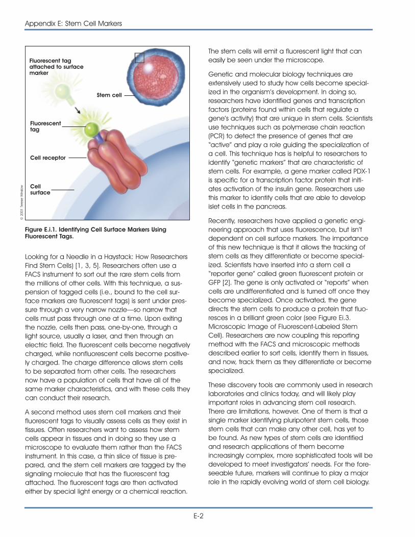
E-2
Appendix E: Stem Cell Markers
Looking for a Needle in a Haystack: How ResearchersFind Stem Cells) [1, 3, 5]. Researchers often use aFACS instrument to sort out the rare stem cells fromthe millions of other cells. With this technique, a sus-pension of tagged cells (i.e., bound to the cell sur-face markers are fluorescent tags) is sent under pres-sure through a very narrow nozzle—so narrow thatcells must pass through one at a time. Upon exitingthe nozzle, cells then pass, one-by-one, through alight source, usually a laser, and then through anelectric field. The fluorescent cells become negativelycharged, while nonfluorescent cells become positive-ly charged. The charge difference allows stem cellsto be separated from other cells. The researchersnow have a population of cells that have all of thesame marker characteristics, and with these cells theycan conduct their research.
A second method uses stem cell markers and theirfluorescent tags to visually assess cells as they exist intissues. Often researchers want to assess how stemcells appear in tissues and in doing so they use amicroscope to evaluate them rather than the FACSinstrument. In this case, a thin slice of tissue is pre-pared, and the stem cell markers are tagged by thesignaling molecule that has the fluorescent tagattached. The fluorescent tags are then activatedeither by special light energy or a chemical reaction.
The stem cells will emit a fluorescent light that caneasily be seen under the microscope.
Genetic and molecular biology techniques areextensively used to study how cells become special-ized in the organism’s development. In doing so,researchers have identified genes and transcriptionfactors (proteins found within cells that regulate agene’s activity) that are unique in stem cells. Scientistsuse techniques such as polymerase chain reaction(PCR) to detect the presence of genes that are“active” and play a role guiding the specialization ofa cell. This technique has is helpful to researchers toidentify “genetic markers” that are characteristic ofstem cells. For example, a gene marker called PDX-1is specific for a transcription factor protein that initi-ates activation of the insulin gene. Researchers usethis marker to identify cells that are able to developislet cells in the pancreas.
Recently, researchers have applied a genetic engi-neering approach that uses fluorescence, but isn’tdependent on cell surface markers. The importanceof this new technique is that it allows the tracking ofstem cells as they differentiate or become special-ized. Scientists have inserted into a stem cell a“reporter gene” called green fluorescent protein orGFP [2]. The gene is only activated or “reports” whencells are undifferentiated and is turned off once theybecome specialized. Once activated, the genedirects the stem cells to produce a protein that fluo-resces in a brilliant green color (see Figure Ei.3.Microscopic Image of Fluorescent-Labeled StemCell). Researchers are now coupling this reportingmethod with the FACS and microscopic methodsdescribed earlier to sort cells, identify them in tissues,and now, track them as they differentiate or becomespecialized.
These discovery tools are commonly used in researchlaboratories and clinics today, and will likely playimportant roles in advancing stem cell research.There are limitations, however. One of them is that asingle marker identifying pluripotent stem cells, thosestem cells that can make any other cell, has yet tobe found. As new types of stem cells are identifiedand research applications of them becomeincreasingly complex, more sophisticated tools will bedeveloped to meet investigators’ needs. For the fore-seeable future, markers will continue to play a majorrole in the rapidly evolving world of stem cell biology.
Fluorescent tagattached to surfacemarker
Fluorescenttag
Cell receptor
Cellsurface
Stem cell
Figure E.i.1. Identifying Cell Surface Markers UsingFluorescent Tags.
© 2
001
Tere
se W
inslo
w
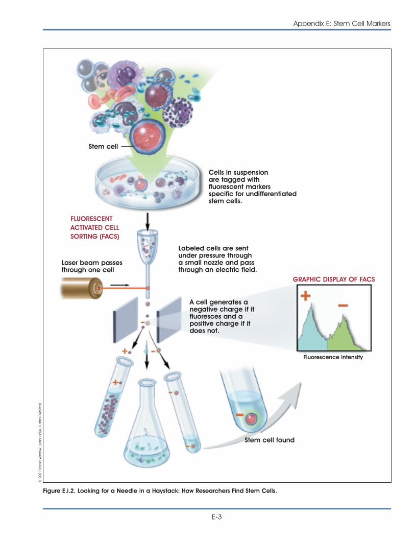
Appendix E: Stem Cell Markers
E-3
Figure E.i.2. Looking for a Needle in a Haystack: How Researchers Find Stem Cells.
Stem cell
Cells in suspensionare tagged withfluorescent markersspecific for undifferentiatedstem cells.
Labeled cells are sentunder pressure througha small nozzle and passthrough an electric field.
A cell generates anegative charge if itfluoresces and apositive charge if itdoes not.
Stem cell found
Laser beam passesthrough one cell
Fluorescence intensity
FLUORESCENTACTIVATED CELLSORTING (FACS)
GRAPHIC DISPLAY OF FACS
© 2
001
Tere
se W
inslo
w, L
ydia
Kib
iuk,
Ca
itlin
Duc
kwa
ll
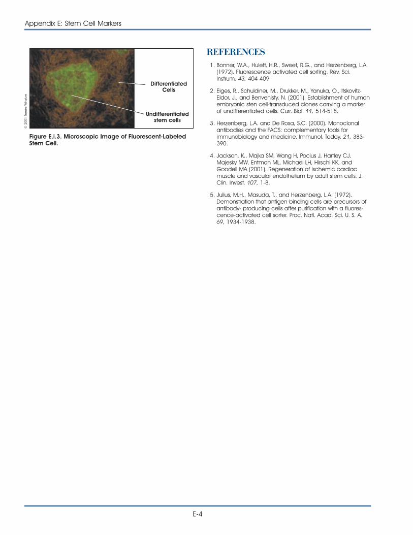
E-4
Appendix E: Stem Cell Markers
REFERENCES1. Bonner, W.A., Hulett, H.R., Sweet, R.G., and Herzenberg, L.A.
(1972). Fluorescence activated cell sorting. Rev. Sci.Instrum. 43, 404-409.
2. Eiges, R., Schuldiner, M., Drukker, M., Yanuka, O., Itskovitz-Eldor, J., and Benvenisty, N. (2001). Establishment of humanembryonic sten cell-transduced clones carrying a markerof undifferentiated cells. Curr. Biol. 11, 514-518.
3. Herzenberg, L.A. and De Rosa, S.C. (2000). Monoclonalantibodies and the FACS: complementary tools forimmunobiology and medicine. Immunol. Today. 21, 383-390.
4. Jackson, K., Majka SM, Wang H, Pocius J, Hartley CJ,Majesky MW, Entman ML, Michael LH, Hirschi KK, andGoodell MA (2001). Regeneration of ischemic cardiacmuscle and vascular endothelium by adult stem cells. J.Clin. Invest. 107, 1-8.
5. Julius, M.H., Masuda, T., and Herzenberg, L.A. (1972).Demonstration that antigen-binding cells are precursors ofantibody- producing cells after purification with a fluores-cence-activated cell sorter. Proc. Natl. Acad. Sci. U. S. A.69, 1934-1938.
Figure E.i.3. Microscopic Image of Fluorescent-LabeledStem Cell.
Undifferentiatedstem cells
DifferentiatedCells
© 2
001
Tere
se W
inslo
w
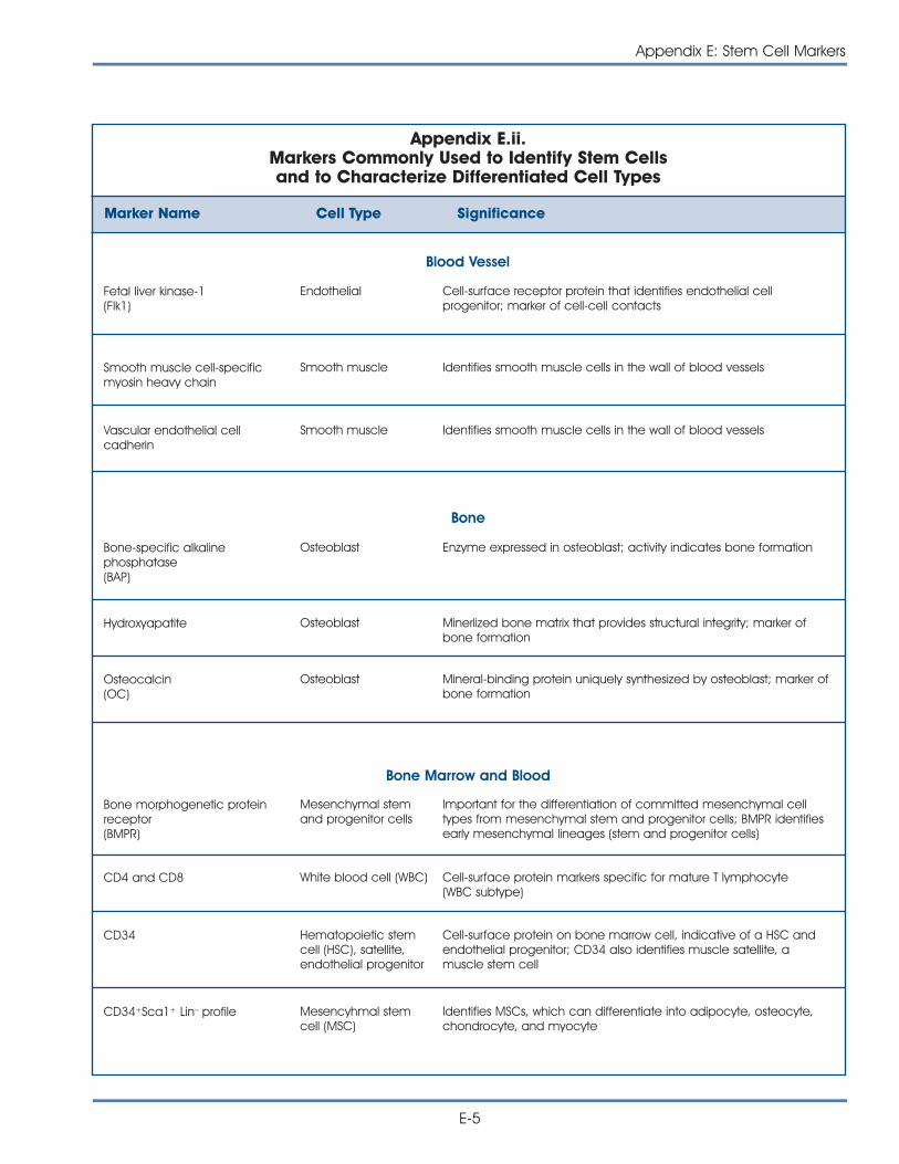
Appendix E: Stem Cell Markers
E-5
Bone
Appendix E.ii.Markers Commonly Used to Identify Stem Cells and to Characterize Differentiated Cell Types
Marker Name Cell Type Significance
Fetal liver kinase-1 (Flk1)
Endothelial Cell-surface receptor protein that identifies endothelial cellprogenitor; marker of cell-cell contacts
Blood Vessel
Smooth muscle cell-specificmyosin heavy chain
Smooth muscle Identifies smooth muscle cells in the wall of blood vessels
Vascular endothelial cellcadherin
Smooth muscle Identifies smooth muscle cells in the wall of blood vessels
Bone-specific alkalinephosphatase (BAP)
Osteoblast Enzyme expressed in osteoblast; activity indicates bone formation
Hydroxyapatite Osteoblast Minerlized bone matrix that provides structural integrity; marker ofbone formation
Osteocalcin(OC)
Osteoblast Mineral-binding protein uniquely synthesized by osteoblast; marker ofbone formation
Bone morphogenetic proteinreceptor(BMPR)
Mesenchymal stemand progenitor cells
Important for the differentiation of committed mesenchymal celltypes from mesenchymal stem and progenitor cells; BMPR identifiesearly mesenchymal lineages (stem and progenitor cells)
CD4 and CD8 White blood cell (WBC) Cell-surface protein markers specific for mature T lymphocyte (WBC subtype)
CD34 Hematopoietic stemcell (HSC), satellite,endothelial progenitor
Cell-surface protein on bone marrow cell, indicative of a HSC andendothelial progenitor; CD34 also identifies muscle satellite, amuscle stem cell
CD34+Sca1+ Lin– profile Mesencyhmal stemcell (MSC)
Identifies MSCs, which can differentiate into adipocyte, osteocyte,chondrocyte, and myocyte
Bone Marrow and Blood
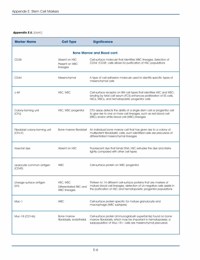
E-6
Appendix E: Stem Cell Markers
Marker Name Cell Type Significance
Bone Marrow and Blood cont.
CD38 Absent on HSC
Present on WBClineages
Cell-surface molecule that identifies WBC lineages. Selection ofCD34+/CD38– cells allows for purification of HSC populations
CD44 Mesenchymal A type of cell-adhesion molecule used to identify specific types ofmesenchymal cells
c-Kit HSC, MSC Cell-surface receptor on BM cell types that identifies HSC and MSC;binding by fetal calf serum (FCS) enhances proliferation of ES cells,HSCs, MSCs, and hematopoietic progenitor cells
Colony-forming unit (CFU)
HSC, MSC progenitor CFU assay detects the ability of a single stem cell or progenitor cellto give rise to one or more cell lineages, such as red blood cell(RBC) and/or white blood cell (WBC) lineages
Fibroblast colony-forming unit (CFU-F)
Bone marrow fibroblast An individual bone marrow cell that has given rise to a colony ofmultipotent fibroblastic cells; such identified cells are precursors ofdifferentiated mesenchymal lineages
Hoechst dye Absent on HSC Fluorescent dye that binds DNA; HSC extrudes the dye and stainslightly compared with other cell types
Leukocyte common antigen(CD45)
WBC Cell-surface protein on WBC progenitor
Lineage surface antigen(Lin)
HSC, MSC
Differentiated RBC andWBC lineages
Thirteen to 14 different cell-surface proteins that are markers ofmature blood cell lineages; detection of Lin-negative cells assists inthe purification of HSC and hematopoietic progenitor populations
Mac-1 WBC Cell-surface protein specific for mature granulocyte andmacrophage (WBC subtypes)
Muc-18 (CD146) Bone marrowfibroblasts, endothelial
Cell-surface protein (immunoglobulin superfamily) found on bonemarrow fibroblasts, which may be important in hematopoiesis; asubpopulation of Muc-18+ cells are mesenchymal precursors
Appendix E.ii. (cont.)
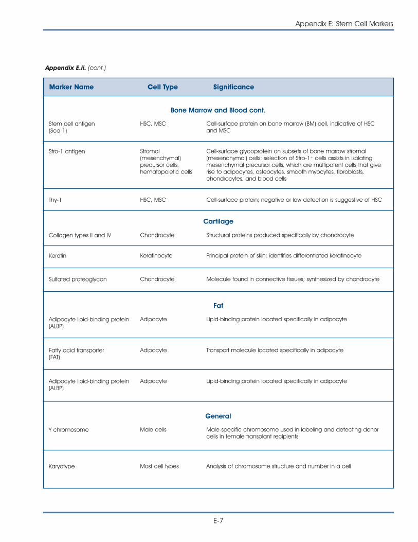
Appendix E: Stem Cell Markers
E-7
Appendix E.ii. (cont.)
Marker Name Cell Type Significance
Stem cell antigen (Sca-1)
HSC, MSC Cell-surface protein on bone marrow (BM) cell, indicative of HSC and MSC
Bone Marrow and Blood cont.
Stro-1 antigen Stromal(mesenchymal)precursor cells,hematopoietic cells
Cell-surface glycoprotein on subsets of bone marrow stromal(mesenchymal) cells; selection of Stro-1+ cells assists in isolatingmesenchymal precursor cells, which are multipotent cells that giverise to adipocytes, osteocytes, smooth myocytes, fibroblasts,chondrocytes, and blood cells
Thy-1 HSC, MSC Cell-surface protein; negative or low detection is suggestive of HSC
Collagen types II and IV Chondrocyte Structural proteins produced specifically by chondrocyte
Keratin Keratinocyte Principal protein of skin; identifies differentiated keratinocyte
Sulfated proteoglycan Chondrocyte Molecule found in connective tissues; synthesized by chondrocyte
Adipocyte lipid-binding protein(ALBP)
Adipocyte Lipid-binding protein located specifically in adipocyte
Fatty acid transporter (FAT)
Adipocyte Transport molecule located specifically in adipocyte
Adipocyte lipid-binding protein(ALBP)
Adipocyte Lipid-binding protein located specifically in adipocyte
Cartilage
Fat
General
Y chromosome Male cells Male-specific chromosome used in labeling and detecting donorcells in female transplant recipients
Karyotype Most cell types Analysis of chromosome structure and number in a cell
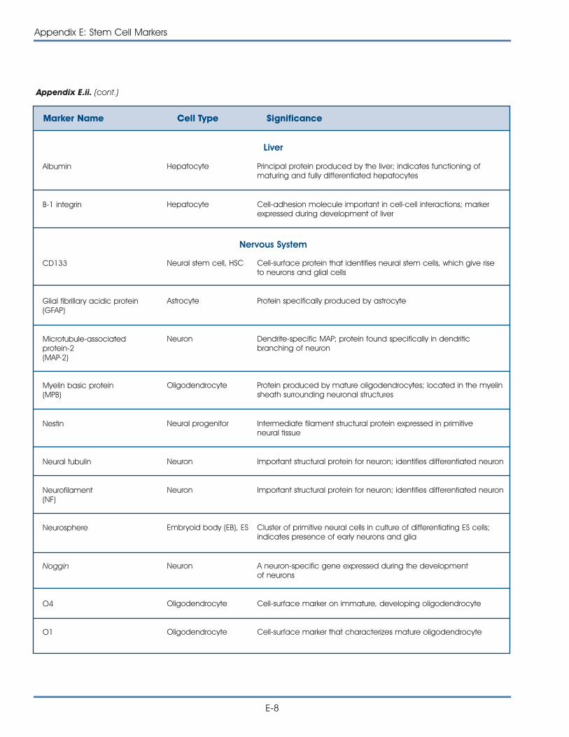
E-8
Appendix E: Stem Cell Markers
Appendix E.ii. (cont.)
Marker Name Cell Type Significance
Albumin Hepatocyte Principal protein produced by the liver; indicates functioning ofmaturing and fully differentiated hepatocytes
B-1 integrin Hepatocyte Cell-adhesion molecule important in cell-cell interactions; markerexpressed during development of liver
CD133 Neural stem cell, HSC Cell-surface protein that identifies neural stem cells, which give riseto neurons and glial cells
Glial fibrillary acidic protein (GFAP)
Astrocyte Protein specifically produced by astrocyte
Microtubule-associated protein-2 (MAP-2)
Neuron Dendrite-specific MAP; protein found specifically in dendriticbranching of neuron
Myelin basic protein (MPB)
Oligodendrocyte Protein produced by mature oligodendrocytes; located in the myelinsheath surrounding neuronal structures
Nestin Neural progenitor Intermediate filament structural protein expressed in primitive neural tissue
Neural tubulin Neuron Important structural protein for neuron; identifies differentiated neuron
Liver
Nervous System
Neurofilament (NF)
Neuron Important structural protein for neuron; identifies differentiated neuron
Neurosphere Embryoid body (EB), ES Cluster of primitive neural cells in culture of differentiating ES cells;indicates presence of early neurons and glia
Noggin Neuron A neuron-specific gene expressed during the development of neurons
O4 Oligodendrocyte Cell-surface marker on immature, developing oligodendrocyte
O1 Oligodendrocyte Cell-surface marker that characterizes mature oligodendrocyte
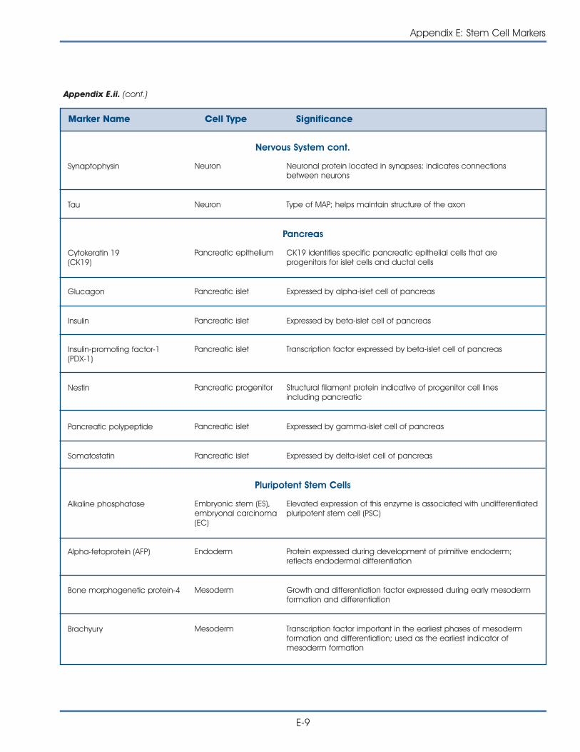
Appendix E: Stem Cell Markers
E-9
Appendix E.ii. (cont.)
Marker Name Cell Type Significance
Nervous System cont.
Synaptophysin Neuron Neuronal protein located in synapses; indicates connectionsbetween neurons
Tau Neuron Type of MAP; helps maintain structure of the axon
Cytokeratin 19(CK19)
Pancreatic epithelium CK19 identifies specific pancreatic epithelial cells that areprogenitors for islet cells and ductal cells
Glucagon Pancreatic islet Expressed by alpha-islet cell of pancreas
Insulin Pancreatic islet Expressed by beta-islet cell of pancreas
Pancreas
Insulin-promoting factor-1 (PDX-1)
Pancreatic islet Transcription factor expressed by beta-islet cell of pancreas
Nestin Pancreatic progenitor Structural filament protein indicative of progenitor cell lines including pancreatic
Pancreatic polypeptide Pancreatic islet Expressed by gamma-islet cell of pancreas
Somatostatin Pancreatic islet Expressed by delta-islet cell of pancreas
Alkaline phosphatase Embryonic stem (ES),embryonal carcinoma(EC)
Elevated expression of this enzyme is associated with undifferentiatedpluripotent stem cell (PSC)
Alpha-fetoprotein (AFP) Endoderm Protein expressed during development of primitive endoderm;reflects endodermal differentiation
Pluripotent Stem Cells
Bone morphogenetic protein-4 Mesoderm Growth and differentiation factor expressed during early mesodermformation and differentiation
Brachyury Mesoderm Transcription factor important in the earliest phases of mesodermformation and differentiation; used as the earliest indicator ofmesoderm formation
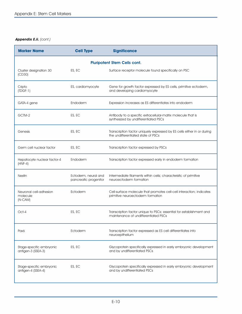
Appendix E: Stem Cell Markers
E-10
Appendix E.ii. (cont.)
Marker Name Cell Type Significance
Pluripotent Stem Cells cont.
Cluster designation 30(CD30)
ES, EC Surface receptor molecule found specifically on PSC
Cripto (TDGF-1)
ES, cardiomyocyte Gene for growth factor expressed by ES cells, primitive ectoderm,and developing cardiomyocyte
GATA-4 gene Endoderm Expression increases as ES differentiates into endoderm
GCTM-2 ES, EC Antibody to a specific extracellular-matrix molecule that issynthesized by undifferentiated PSCs
Genesis ES, EC Transcription factor uniquely expressed by ES cells either in or duringthe undifferentiated state of PSCs
Germ cell nuclear factor ES, EC Transcription factor expressed by PSCs
Hepatocyte nuclear factor-4 (HNF-4)
Endoderm Transcription factor expressed early in endoderm formation
Nestin Ectoderm, neural andpancreatic progenitor
Intermediate filaments within cells; characteristic of primitiveneuroectoderm formation
Neuronal cell-adhesionmolecule(N-CAM)
Ectoderm Cell-surface molecule that promotes cell-cell interaction; indicatesprimitive neuroectoderm formation
Oct-4 ES, EC Transcription factor unique to PSCs; essential for establishment andmaintenance of undifferentiated PSCs
Pax6 Ectoderm Transcription factor expressed as ES cell differentiates intoneuroepithelium
Stage-specific embryonicantigen-3 (SSEA-3)
ES, EC Glycoprotein specifically expressed in early embryonic developmentand by undifferentiated PSCs
Stage-specific embryonicantigen-4 (SSEA-4)
ES, EC Glycoprotein specifically expressed in early embryonic developmentand by undifferentiated PSCs
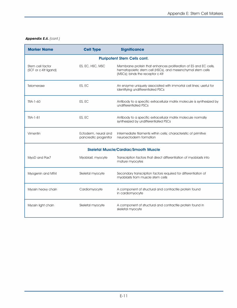
Appendix E: Stem Cell Markers
E-11
Appendix E.ii. (cont.)
Marker Name Cell Type Significance
Pluripotent Stem Cells cont.
Stem cell factor (SCF or c-Kit ligand)
ES, EC, HSC, MSC Membrane protein that enhances proliferation of ES and EC cells,hematopoietic stem cell (HSCs), and mesenchymal stem cells(MSCs); binds the receptor c-Kit
Telomerase ES, EC An enzyme uniquely associated with immortal cell lines; useful foridentifying undifferentiated PSCs
TRA-1-60 ES, EC Antibody to a specific extracellular matrix molecule is synthesized byundifferentiated PSCs
TRA-1-81 ES, EC Antibody to a specific extracellular matrix molecule normallysynthesized by undifferentiated PSCs
Vimentin Ectoderm, neural andpancreatic progenitor
Intermediate filaments within cells; characteristic of primitiveneuroectoderm formation
MyoD and Pax7 Myoblast, myocyte Transcription factors that direct differentiation of myoblasts intomature myocytes
Myogenin and MR4 Skeletal myocyte Secondary transcription factors required for differentiation ofmyoblasts from muscle stem cells
Myosin heavy chain Cardiomyocyte A component of structural and contractile protein found in cardiomyocyte
Myosin light chain Skeletal myocyte A component of structural and contractile protein found in skeletal myocyte
Skeletal Muscle/Cardiac/Smooth Muscle

E-12
Appendix E: Stem Cell Markers
This page intentionally left blank
F-1
Appendix F.i.
GLOSSARY
Adipocyte – Fat cell.
Adult stem cell – An undifferentiated cell found in adifferentiated tissue that can renew itself and (withcertain limitations) differentiate to yield all the special-ized cell types of the tissue from which it originated.
Allogenic – Two or more individuals (or cell lines) arestated to be allogeneic to one another when thegenes at one or more loci are not identical insequence in each organism.
Amnion – The innermost intrauterine membranearound the fetus and the amniotic fluid.
Anterior visceral endoderm (AVE) – Specific tissuestructure arising in the early embryo that helpsestablish the anterior-posterior axis of the organism.
Antibody – A Y-shaped protein secreted by B cells inresponse to an antigen. An antibody binds specifi-cally to the antigen that induced its production.Antibodies directed against antigens on the surfaceof infectious organisms help eliminate those organ-isms from the body.
Antigen – A substance (often a protein) that inducesthe formation of an antibody. Antigens are commonlyfound on the surface of infectious organisms, trans-fused blood cells, and transplanted organs.
Antigen presenting cells (APCs) – One of a variety ofcells within the body that can process antigens anddisplay them on their surface in a form recognizableby T cells.
Apoptosis – Genetically programmed cell death.
Astrocyte – One of the large neuroglia cells ofnervous tissue.
Autoantibody – An antibody that reacts with antigensfound on the cells and tissues of an individual’sownbody. Autoantibodies can cause autoimmunediseases.
Autoimmune disease – A condition that results fromT cells and/or antibodies that attack the cells ortissues of an individual’s own body.
Autologous transplant – Transplanted tissue derivedfrom the intended recipient of the transplant. Such atransplant helps avoid complications of immunerejection.
Axis – A straight line passing through a spherical bodybetween its two poles. The central line of the body orany of its parts. The vertebral column. The centralnervous system. An artery that when created, imme-diately divides into a number of branches.
B cells – Also known as B lymphocytes. Each B cell iscapable of making one specific antibody. Whenstimulated by antigen and helper T cells, B cellsmature into plasma cells that secrete large amountsof their specific antibody.
Blastocoel – The cavity in the blastula of thedeveloping embryo.
Blastocyst – A preimplantation embryo of 30-150cells. The blastocyst consists of a sphere made up ofan outer layer of cells (the trophectoderm), a fluid-filled cavity (the blastocoel), and a cluster of cellson the interior (the inner cell mass).
Blastula – An early stage in the development of anovum consisting of a hollow sphere of cells enclosinga cavity called the blastocoel.
GLOSSARY AND TERMS
APPENDIX F:
GLOSSARY AND TERMS
Bone marrow – The soft, living tissue that fills mostbone cavities and contains hematopoietic stemcells, from which all red and white blood cells evolve.The bone marrow also contains mesenchymal stemcells that a number of cells types come from,including chondrocytes, which produce cartilage.
Bone marrow (BM) cell – Refers to bothhematopoietic and mesenchymal (stromal) cells.
Bone marrow stem cell (BMSC) – One of at least twotypes of multipotient stem cells: hematopoietic stemcell and mesenchymal stem cell.
Bone marrow transplantation (BMTx) – Trans-planta-tion of bone marrow from one individual to another.Autologous BMTx is a process in which a patient’shealthy bone marrow is withdrawn and preserved,then injected back into the patient to restore the pro-duction of healthy blood and immune cells by thebone marrow. This strategy is often used in patientswith certain types of cancer who have undergoneradiation therapy or chemotherapy that destroys thebone marrow cells.
Bone morphogenetic proteins (BMPs) – Proteinsthatare involved in the formation of embryonic bone.BMPs operate at several stages in this formation ofbone, beginning with the early stages of morpho-genesis and continuing to late postnatal life. BMPsalso play a critical role in the development of thecentral nervous system.
Brain-derived neurotrophic factor (BDNF) – A growthfactor synthesized in the brain that stimulates neuriteoutgrowth and supports survival of neurons.
Cavitation – A process that occurs during the forma-tion of the blastocyst and establishes the polarity ofembryonic cells.
Cell cycle – The orderly sequence of events by whichthe cell duplicates its contents and divides into two.
Chimera – An organism composed of cells derivedfrom at least two genetically different zygotes.Theoretically, the zygote could be from separatespecies.
Chondrocytes – Cartilage cells.
Chorion – The multilayered, outermost fetalmembrane. As pregnancy progresses, part of thechorion becomes the placenta.
Chromosomes – Nucleic acid-protein structures inthe nucleus of a cell. Chromosomes are composedchiefly of DNA, the carrier of hereditary information.Chromosomes contain genes, working subunits ofDNA that carry the genetic code for specific proteins,interspersed with large amounts of DNA of unknownfunction. A normal human body cell contains 46chromosomes; a normal human gamete, 23chromosomes.
Cleavage – The process of cell division in the veryearly embryo before it becomes a blastocyst.
Clonality – A line of cells that is genetically identicalto the originating cell; in this case, a stem cell.
Cluster differentiation (CD) – Cell membranemolecules used to classify leukocytes into subsets.
Colony-forming cells – Groups of cells growing on asolid nutrient surface with each group being createdfrom the multiplication of an individual cell.
Colony-stimulating factors – Diffusible proteins thatstimulate the proliferation of hematopoietic stemcells.
Cripto – Transcription factor expressed by pluripotentstem cells and early embryos.
Cyclin-dependent kinase (Cdk protein) – Proteinkinase that has to be complexed with a cyclin proteinin order to act; different Cdk-cyclin complexes arethought to trigger different steps in the cell-divisioncycle by phosphorylating specific target proteins.
Cytokines – A generic term for a large variety ofregulatory proteins produced and secreted by cellsand used to communicate with other cells. One classofcytokines is the interleukins, which act as inter-cellular mediators during the generation of animmune response.
Cytoplasm – The contents of a cell other than thenucleus; cytoplasm consists of a fluid containingnumerous structures, known as organelles, that carryout essential cell functions.
Decidual cells – A cellular matrix that first surroundsan implanted embryo and later occupies most of theendometrium.
Dendrite – Extension of a nerve cell, typicallybranched and relatively short, that receives stimulifrom other nerve cells.
F-2
Appendix F: Glossary and Terms
Differentiation – The process whereby an unspecial-ized early embryonic cell acquires the features of aspecialized cell such as a heart, liver, or muscle cell.
Diploid – A cell or tissue having two chromosomesets, as opposed to the haploid situation of gametes,which have only one chromosome set.
DNA – Deoxyribonucleic acid, a chemical foundprimarily in the nucleus of cells. DNA carries theinstructions for making all the structures and materialsthe body needs to function.
DNA methylation – A type of chemical modificationof DNA that regulates gene expression.
Ectoderm – The upper, outermost of the threeprimitive germ layers of the embryo; it gives rise toskin, nerves, and brain.
Egg cylinder – An asymmetric embryonic structurethat helps to determine the body plan of the mouse.
Embryo – In humans, the developing organism fromthe time of fertilization until the end of the eighthweek of gestation, when it becomes known as afetus.
Embryoid bodies (EBs) – Clumps of cellular structuresthat arise when embryonic stem cells are cultured.Embryoid bodies contain tissue from all three of thegerm layers: endoderm, mesoderm, and ectoderm.Embryoid bodies are not part of normal develop-ment and occur only in in vitro conditions.
Embryonal carcinoma (EC) cells – A type of pluri-potent stem cell derived from teratocarcinoma(usually a testis tumor).
Embryonic disk – A group of cells derived from theinner cell mass of the blastocyst, which later developsinto an embryo. The disc consists of three germ layersknown as the endoderm, mesoderm, and ectoderm.
Embryonic germ (EG) cells – Cells found in aspecific part of the embryo/fetus called the gonadalridge that normally develop into mature gametes.
Embryonic stem (ES) cells – Primitive (undifferen-tiated) cells from the embryo that have the potentialto become a wide variety of specialized cell types.
Endoderm – Lower layer of a group of cells derivedfrom the inner cell mass of the blastocyst; it laterbecomes the lungs and digestive organs.
Epiblast – Gives rise to the ectoderm andmesoderm. The mesoderm then displaces thehypoblast cells and forms the entodermal cell layeron its inner surface.
Epidermal growth factor (EGF) – A protein that stimu-lates epidermal and various other cells to divide.
Epithelium – The layer of cells forming the epidermisof the skin. These cells serve the general functions ofprotection, absorption, and secretion, and play aspecialized role in moving substances through ducts,in the production of germ cells, and in the receptionof stimuli. Their ability to regenerate is excellent; theepithelium may replace itself as frequently as every24 hours.
Erythroid cell – Red blood cells.
Ex vivo – Outside the living body.
Extracellular matrix – The microenvironment next to acell that allows for structural support, orientation, andconnections for cell-to-cell interactions and formationof connective tissues.
Extraembryonic tissues – Intrauterine tissues thatsupport the embryoís placenta, umbilical cord, andamniotic sac.
Feeder cell layer – Cells that are utilized in co-cultureto maintain pluripotent stem cells. Cells usually consistof mouse embryonic fibroblasts.
Fertilization – The process whereby male and femalegametes unite.
Fetal calf serum – A type of culture medium oftenused in the culture of stem cells. It provides a numberof growth factors.
Fibroblast – Cells that give rise to connective tissue.
Fluorescence-activated cell sorting (FACS) – Atechnique that can separate and analyze cells,which are labeled with fluorochrome-conjugatedantibody, by their fluorescence and light scatteringpatterns.
Follistatin – An inhibitory factor produced duringembryonic development that affects the growth anddifferentiation of the pancreas.
Gap junctions – Communicating cell-cell junctionsthat allow ions and small molecules to pass from thecytoplasm of one cell to the cytoplasm of another cell.
Appendix F: Glossary and Terms
F-3
Gastrula – Animal embryo at an early stage of devel-opment in which cells are enclosed in a sheath toform the beginning of a gut cavity.
Gene – A functional unit of heredity that is a segmentof DNA located in a specific site on a chromosome.A gene Directs the formation of an enzyme of otherprotein.
Genital Ridge – Formation of a genital ridge requiresat least two genes, WT-1, which is also important inearly kidney formation, and SF-1, required for thedevelopment of both the gonads and adrenalglands.
Genome – The complete genetic material of anorganism.
Genomic imprinting – A biochemical phenomenonthat determines, for certain specific genes, which oneof the pair of identical genes, the motherís or thefatherís, will be active in that individual.
Germ cell – A sperm or egg, or a cell that canbecome a sperm or egg. All other body cells arecalled somatic cells.
Gestation – The period of development of anorganism from fertilization of the ovum until birth.
Glia – The nonneuronal or supporting tissue(neuroglia) of the brain and spinal cord.
Glial cells – Supporting cells of the nervous system,including oligodendrocytes and astrocytes in thevertebrate central nervous system and Schwann cellsin the peripheral nervous system.
Glial fibrillary acidic protein (GFAP) – A structuralprotein specifically produced by astrocytes. GFAP isoften used as a marker of astrocytes.
Glucagon – A hormone consisting of a straight chainof proteins composed of 29 amino acid residues thatcan be extracted from certain pancreatic cells.
Glycoprotein – A compound consisting of acarbohydrate and a protein.
Gonadal ridge –Anatomic site in the early fetuswhere primordial germ cells (PGCs) are formed.
Gonads – The embryonic sex gland before itbecomes a definitive testis or ovary.
Goosecoid – Gene that encodes a transcriptionfactor that is important for determining craniofacialorientation and features in the vertebrate embryo.
Graft-versus-host disease (GVHD) – A condition thatoccurs following bone marrow transplantation inwhich the donor-derived T cells attack the host’stissues.
Granulocyte – A type of white blood cell filled withmicroscopic granules that are little sacs containingenzymes, compounds that digest microorganisms.Neutrophils, eosinophils and basophils are all types ofgranulocytes. They are named by the stainingfeatures of their granules in the laboratory.
Granulose cells – Cells surrounding and maintainingthe ovarian follicle.
Green fluorescent protein (GFP) – Fluorescent-proteindye used to tag and trace particular genes and cellsof interest.
Hanging drop method – A technique used to cultureembryonic stem cells so that they develop intoembryoid bodies.
Haploid – Refers to a gamete having one chromo-some set, as opposed to the diploid situation of cellsor tissues, where there are two chromosome sets.
hCNS – Human central nervous system stem cell.
Hematopoiesis – Generation of blood cells, mainly inthe bone marrow.
Hematopoietic stem cell (HSC)– A stem cell fromwhich all red and white blood cells evolve.
Hepatic – Relating to the liver.
Hepatocyte – Liver cell.
hES cell – Human embryonic stem cell; a type ofpluripotent stem cell.
Hoechst dye – A dye used to identify hematopoieticstem cells (HSCs).
Hox genes – Consists of at least 38 encodednucleotides that contain genes found in four clusterson four different chromosomes. An important functionof hox genes in blood is the regulation of cellproliferation.
F-4
Appendix F: Glossary and Terms
HSC markers – Cell-surface molecules that are usedto identify hematopoietic stem cells.
Hybridoma – A hybrid cell produced by the fusion ofan antibody-producing cell and a multiple myelomacell. The cell has the capability to produce acontinuous supply of identical antibodies.
Hydroxyapatite – A natural mineral structure thatcontains calcium and phosphate ions that providethe power for the formation of bones and teeth.
Hypoblast – The inner cell layer, or endoderm, whichdevelops during the formation of the embryonicgerm layers.
Identical twinning – Process in which geneticallyidentical organisms arise from symmetrical divisionand separation of totipotent cells.
Immune-function assay – A general term for anumber of tests based on an immune cell’s ability tocarry out a particular immune function.
Immune system cells – White blood cells or leuko-cytes that originate from the bone marrow. Theyinclude antigen presenting cells, such as dendriticcells, T and B lymphocytes, and neutrophils, amongmany others.
Immunocompromised mice – These geneticallyaltered mice are used for transplantation experimentsbecause they usually do not reject the transplantedtissue.
Immunofluorescence – The detection of antibodiesby using special proteins labeled with fluorescein.When present, the specific organism or antibody isobserved as a fluorescent material when examinedmicroscopically while illuminated with a fluorescentlight source.
Immunogenic – Relating to or producing an immuneresponse.
Immunohistology – Examination of tissues throughspecific immunostaining techniques.
Immunophenotyping – Identification of various typesof immune cells by sorting them according to theircell-surface markers.
In utero – In the uterus.
In vitro – Literally, “in glass;” in a laboratory dish or testtube; an artificial environment.
In vitro fertilization (IVF) – An assisted reproductiontechnique in which fertilization is accomplishedoutside the body.
In vivo – In the living subject; in a natural environment.
Indomethacin – An anti-inflammatory, antipain, andantifever drug. Its primary use is in rheumatoid arthritisand degenerative joint disease when aspirin-basedproducts are ineffective or cannot be tolerated.
Inner cell mass – The cluster of cells inside theblastocyst. These cells give rise to the embryonic diskof the later embryo and, ultimately, the fetus.
Insulin-promoting factor 1 – A transcription factorexpressed in the pancreas and necessary for theproduction of insulin.
Interleukin – Selected peptide or protein thatprimarily mediates local interactions between whiteblood cells.
Irradiate – Application of radiation from a source(heat, light, Xrays) to a structure or organism.
Karyotype – The full set of chromosomes of a cellarranged with respect to size, shape, and number.
Keratin –An extremely tough protein substance foundin hair, nails, skin, and cornea.
Keratinocytes – Cells that synthesize keratin and arefound in the skin, hair, and nails. A fibrous protein isproduced by keratinocytes and may be hard or soft.The hard keratin is found in hair and nails. The softkeratin is found in the epidermis of the skin in the formof flattened non-nucleated scales that sloughcontinually.
Knock-out mouse – A mouse that has had one or both copies of a specific gene deleted orinactivated.
Lacunae – The spaces occupied by cells (e.g.,chondrocytes and osteocytes) of calcified tissues.
Lefty – A developmental factor that helps determineright-left asymmetry in vertebrates.
Leptin –A hormone produced by the placenta andfetal tissues that acts as a growth factor andmodulator of metabolic and immune functions.
Leukemia inhibitory factor (LIF) – A growth factornecessary for maintaining mouse embryonic stemcells in a proliferative, undifferentiated state.
Appendix F: Glossary and Terms
F-5
Leukocyte – A white blood cell or corpuscle.Leukocytes are formed from undifferentiated stemcells that give rise to all blood cells.
Leukocyte common antigen – Cell-surface mole-cule found on white blood cells and white blood cellprogenitors. Also referred to as CD45.
Lineage surface antigen (Lin) – A mixture of mono-clonal antibodies that are directed against antigensfound on mature hematopoietic cells of differentlineages. A usual Lin mix includes eight differentantibodies directed against B and T cells, myeloidcells, and erythroid cells.
Lipase – An enzyme produced by many tissues.Lipase is an important regulator of fat in the blood.A deficiency of this enzyme leads to low levels ofhigh-density lipoproteins (HDLs).
Lipid – Any one of a group of fats or fatlike sub-stances characterized by their insolubility in water andsolubility in fat solvents such as alcohol, ether, andchloroform.
Lymph nodes – Widely distributed lymphoid organswithin the lymphatic system where many immunecells are concentrated.
Lymphatic system – A network of lymph vessels andnodes that drain and filter antigens from tissue fluidsbefore returning lymphocytes to the blood.
Lymphocyte – A cell present in the blood andlymphatic tissue.
Lymphoid – A shape or form that resembles lymph orlymph tissue.
Macrophage – A monocyte that has left thecirculation and settled and matured in a tissue.Because of their placement in the lymphoid tissues,macrophages serve as the major scavenger of theblood, clearing it of abnormal or old cells andcellular debris as well as pathogenic organisms.
Major histocompatibility complex (MHC) – A groupof genes that code for cell-surface histocompatibilityantigens. These antigens are the primary reason whyorgan and tissue transplants from incompatibledonors fail.
Marker – See Surface marker.
F-6
Appendix F: Glossary and Terms
Mast cell – A large tissue cell that does not circulatein the blood. They are also important in producingthe signs and symptoms of hypersensitivity reaction,such as those of an insect sting, and certain forms ofasthma.
Maternal gene product – A product in the maleorganism of a gene from the X chromosome.
Meiosis – A process where two successive cells divideand produce cells, eggs, or sperm that contain halfthe number of chromosomes in the somaticcells.During fertilization, the nuclei of the sperm andovum fuse and produce a zygote with the fullchromosome complements.
Melanocyte – A cell that produces the dark pigmentmelanin; responsible for the pigmentation of skin andhair.
Memory – The ability of antigen-specific T or B cellsto“recall” prior exposure to an antigen and respondquickly without the need to be activated again byCD4 helper T cells.
Memory cells – A subset of antigen-specific T or Bcells that “recall” prior exposure to an antigen andrespond quickly without the need to be activatedagain by CD4 helper T cells.
Mesenchymal stem cells (MSCs) – Cells from theimmature embryonic connective tissue. A number ofcell types come from mesenchymal stem cells,including chondrocytes, which produce cartilage.
Mesoderm – The middle layer of the embryonic disk,which consists of a group of cells derived from theinner cell mass of the blastocyst. This middle germlayer is known as gastrulation and is the precursor tobone, muscle, and connective tissue.
Metaphase– A stage of mitosis where chromosomesare firmly attached to the mitotic spindle at itsequator but have not yet segregated toward oppo-site poles.
Microtubule – An elongated, hollow tubular structurepresent in the cell. Microtubules help certain cellsmaintain their rigidity, convert chemical energy intowork, and provide a means of transportation ofsubstances in different directions within a cell.
Monoclonal – From a single cell.
Monoclonal antibody (MoAb) – An exceptionallypure and specific antibody derived from hybridomacells. Because each of the clones is derived from asingle B cell, all of the antibody molecules it makesare identical.
Monocyte – A white blood cell derived from myeloidstem cells.
Mononucleocyte – A cell containing a singlenucleus. Generally refers to a white blood cell.
Morphology – The shape and structural makeup of acell, tissue, or organism.
Morula – A solid mass of cells that resembles amulberry and result from the cleavage of an ovum.
Mouse embryonic fibroblast (MEF) – Mouseembryonic fibroblast cells are used as feeder cellswhen culturing pluripotent stem cells.
Multipotent stem cells – Stem cells that have thecapability of developing cells of multiple germ layers.
Myelin – A fatty sheath that covers axons of nervecells. It is produced by oligodendrocytes andprovides an insulation for nerve conduction throughthe axons.
Myelin basic protein (MPB) –A structural protein withinthe myelin sheath surrounding neurons.
Myelin sheath – Insulating layer of specialized cellmembrane wrapped around vertebrate axons. Thissheath is produced by oligodendrocytes in thecentral nervous system and by Schwann cells in theperipheral nervous system.
Myeloid – Marrow-like, but not necessarily originatingfrom bone marrow.
Myeloid stem cells – Precursors to the other lines ofblood cells: erythrocytes, granulocytes, monocytes,and platelets. The second-generation cells are stillpluripotent but their developmental potency is limitedbecause neither can form an offspring of the othertype.
Myocyte – A muscular tissue cell.
MyoD1 – A group of four basic myogenic regulatoryfactors (helix-loop-helix transcription) and a newlydiscovered factor called muscle enhancer factor-2which appears to work away from the other threefactors. However, all four of the factors in this MyoDfamily have the capacity of converting nonmuscle cellsinto cells expressing the full range of muscle proteins.
Myosin – A protein in muscle fibers.
Myosin light chain – There are four light chainsubunits containing complex molecules that formcontractile units in skeletal muscle.
Nestin – An intermediate filament protein found incells such as neural and pancreatic precursors.
Neural crest – A band of cells that extend lengthwisealong the neural tube of an embryo and give rise tocells that form the cranial, spinal, and autonomicganglia, as well as becoming odontoblasts, whichform the calcified part of the teeth.
Neural plate – A thickened band of ectoderm alongthe dorsal surface of an embryo. The nervous systemdevelops from this tissue.
Neural stem cell (NSC) – A stem cell found in adultneural tissue that can give rise to neurons, astrocytes,and oligodendrocytes.
Neural tube – The embryological forerunner of thecentral nervous system.
Neuroectoderm – The central region of the earlyembryonic ectoderm, which later forms the brainand spinal cord, as well as evolving into nerve cells ofthe peripheral nervous system
Neuroepithelium – A specialized epithelial structurethat forms the termination of a nerve of a specialsense, i.e., olfactory cells, hair cells of the inner ear,and the rods and cones of the retina. It is theembryonic layer of the epiblast that develops intothe cerebrospinal axis.
Neurofilament (NF) – A type of intermediate filamentfound in nerve cells.
Appendix F: Glossary and Terms
F-7
Neuron – A nerve cell, the structural and functionalunit of the nervous system. A neuron consists of a cellbody and its processes, an axon, and one or moredendrites. Neurons function by the initiation and con-duction of impulses and transmit impulses to otherneurons or cells by releasing neurotransmitters atsynapses.
Neurosphere –A primitive neural tissue that ariseswhen embryonic stem cells are grown in certainculture conditions.
NMDA receptor – (N-methyl-d-aspartate receptor).A neurotransmitter receptor for excitatory synapses.
Nodal – A knob-like protrusion.
Node – A knot, knob; a protrusion or swelling; a con-stricted region; a small, rounded organ or structure.
Notochord – Forms the axial skeleton in embryos ofall chordates. In vertebrates, it is replaced partially orcompletely by vertebrae.
Oligodendrocyte – Cell that provides insulation tonerve cells by forming a myelin sheath around axons.
Oocyte – Developing egg; usually a large andimmobile cell.
Osteocalcin (OC) – A cytokine produced byosteoblasts that promotes bone formation.
Osteoclast – A giant multi-nuclear cell formed in thebone marrow of growing bones.
Osteocyte – A cell from the bone tissue.
Osteoprogenitor – A cell-type that differentiates intoa mature osteocyte.
Ovarian follicle – An external, fluid-filled portion of theovary in which oocytes mature before ovulation.
Oviduct – The passage through which the ova travelfrom the ovary into the uterus.
Pancreatic polypeptide – An endocrine proteinproduced by islet cells of the pancreas.
Paracrine factors – Cytokines or hormones that acton cells or tissues within an extremely limited area.
Passage – A round of cell growth and proliferationin culture.
Placenta – The oval or discoid spongy structure in theuterus from which the fetus derives its nourishmentand oxygen.
Plasticity – The ability of stem cells from one adulttissue to generate the differentiated types of anothertissue.
Pluripotent stem cell (PSC) – A single stem cell thathas the capability of developing cells of all germlayers (endoderm, ectoderm, and mesoderm).
Polarity – The presence of an axial, non symmetricgradient along a cell or tissue.
Population doublings – A doubling in the number ofcells when grown in culture.
Precursor Cells – In fetal or adult tissues, these arepartly differentiated cells that divide and give rise todifferentiated cells.Also known as progenitor cells.
Pre-implantation embryo – The very early, free-floating Embryo, from the time the egg is fertilizeduntil implantation in the mother ’s womb is complete.
Primary germ layers – The three initial embryonicgerm layers–endoderm, mesoderm, andectoderm–from which all other somatic tissue-typesdevelop.
Primitive streak – The initial band of cells from whichthe embryo begins to develop. The primitive streakestablishes and reveals the embryo’s head-tail andleft-right orientations.
Radioimmunoassay – A sensitive method of deter-mining the concentration of a substance, particularlya protein-bound hormone, in blood plasma.
Retinoic acid – A metabolite of vitamin A.
Ribonucleic acid (RNA) – A chemical that is similar instructure to DNA. One of its main functions is to trans-late the genetic code of DNA into structural proteins.
Ribosome – Any of the RNA- and protein-rich cyto-plasmic organelles that are sites of protein synthesis.
Schwann cell – In the embryo, Schwann cells growaround the nerve fiber, forming concentric layers ofcell membrane (the myelin sheath).
F-8
Appendix F: Glossary and Terms
Side population (SP) stem cell – Two examples ofmultipotent stem cell populations found in bonemarrow and skeletal muscle. SPs are not yet fullycharacterized. Their significance is their unexpectedability to differentiate into cell types that are distinctfrom their tissue of origin.
Signal transduction pathways – Relay of a signal bythe conversion from one physical or chemical formto another. In cell biology, signal transduction is theprocess in which a cell converts an extracellularsignal into a response.
Somatic cell nuclear transfer – The transfer of a cell
nucleus from a somatic cell into an egg from whichthe nucleus has been removed.
Somatic cells – Any cell of a plant or animal otherthan a germ cell or germ cell precursor.
Somatostatin – A hormone that inhibits the secretionof insulin and gastrin.
Steel factor – See stem cell factor.
Stem cell – A cell that has the ability to divide forindefinite periods in culture and to give rise tospecialized cells.
Stem cell antigen 1 (Sca-1) – Cell-surface protein onbone marrow cell, indicative of hematopoietic stemcells and mesenchymal stem cells.
Stem cell factor (SCF) – Relatively undifferentiatedcell that can continue dividing indefinitely, throwingoff daughter cells that can undergo terminal differ-entiation into particular cell types. (Also known assteel factor).
Stromal cell – A non-blood cell that is derived fromblood organs, such as bone marrow or fetal liver,which is capable of supporting growth of blood cellsin vitro. Stromal cells that make this matrix within thebone marrow are also derived from mesenchymalstem cells.
Sulfated proteoglycan – Molecules found primarily inconnective tissues and joint fluids and that providelubrication.
Surface marker – Surface proteins that are unique tocertain cell types capable of detection by antibodiesor other detection methods.
Syncytiotrophoblast – A multinucleated cell formedfrom the cells of the trophoblast. Only a small area ofthe syncytiotrophoblast is evident at the start of theformation of the embryo, but this cell tissue is highlyinvasive, quickly expands and soon surrounds theentire embryo.
Syncytium – A mass of cytoplasm containing manynuclei that are enclosed by a single plasma mem-brane. This is usually the result of either cell fusion or aseries of incomplete division cycles in which thenuclei divide but the cell does not.
T cells – A type of white blood cell that is of crucialimportance to the immune system. Immature T cellsmigrate to the thymus gland in the upper chestcavity, where they mature and differentiate intovarious types of mature T cells and become active inthe immune system in response to a hormone calledthymosin and other factors. T-cells that are potentiallyactivated against the body ’s own tissues are normallykilled or changed (“down-regulated”) during thismaturation process.
Telomerase – An enzyme that is composed of acatalytic protein component and an RNA templateand that synthesizes DNA at the ends of chromo-somes and confers replicative immortality to cells.
Telomere – The end of a chromosome, associatedwith a characteristic DNA sequence that is replicatedin a special way. A telomere counteracts thetendency of the chromosome to shorten with eachround of replication.
Tenocyte – Tendon-producing cell.
Teratocarcinoma – A tumor that occurs mostly inthe testis.
Teratogen – A drug or other agent that raises theincidence of congenital malformations.
Teratoma – A tumor composed of tissues from thethree embryonic germ layers. Usually found in ovaryand testis. Produced experimentally in animals byinjecting pluripotent stem cells, in order to determinethe stem cells’ abilities to differentiate into varioustypes of tissues.
Thiazolidinediones – A class of antidiabetes drugsthat enhances the activity of insulin.
Appendix F: Glossary and Terms
F-9
Thrombopoietin – Growth factor for the proliferationand differentiation of platelet forming cells calledmegakaryocytes.
Thymus – A lymphoid organ located in the upperchest cavity. Maturing T cells go directly to thethymus, where they are “educated” to discriminatebetween self and foreign proteins. (See toleranceinduction.)
Tissue culture – Growth of tissue in vitro on an artificialmedium for experimental research.
Tolerance – A state of specific immunologic unre-ponsiveness. Individuals are normally tolerant to theirown cells and tissues. Autoimmune diseases occurwhen tolerance fails.
Tolerance induction – The “education” process thatT cells undergo to discriminate between self andforeign proteins. This process takes place primarily inthe thymus. In addition to inactivating or deleting self-reactive T cells, those T cells that can recognize thebody ’s MHC proteins, but not be activated solely bythis recognition, are also selected to leave the thy-mus (circulate through the body).
Totipotent – Having unlimited capability. The totipo-tent cells of the very early embryo have the capacityto differentiate into extra embryonic membranes andtissues, the embryo, and all postembryonic tissuesand organs.
Transaminase – An enzyme that catalyzes chemicalreactions in the body in which an amino group istransferred from a donor molecule to a recipientmolecule.
Transcription – Making an RNA copy from asequence of DNA (a gene). Transcription is the firststep in gene expression.
Transcription factor – Molecules that bind to RNApolymerase III and aid in transcription.
Transgene – A gene that has been incorporatedfrom one cell or organism and passed on tosuccessive generations.
Translation – The process of forming a proteinmolecule at a ribosomal site of protein synthesis frominformation contained in messenger RNA.
Trophectoderm – The outer layer of the developingblastocyst that will ultimately form the embryonic sideof the placenta.
Trophoblast – The extraembryonic tissue responsiblefor negotiating implantation, developing into theplacenta, and controlling the exchange of oxygenand metabolites between mother and embryo.
Trypsin – An enzyme that digests proteins. Often usedto separate cells.
Undifferentiated – Not having changed to becomea specialized cell type.
Unipotent – Refers to a cell that can only develop ina specific way to produce a certain end result.
Vascular – Composed of, or having to do with, bloodvessels.
Villi – Projections from the surface, especially of amucous membrane. If the projection is minute, as ina cell surface, it is called a microvillus.
Vimentin – The major polypeptide that joins withother subunits to form the intermediate filamentcytoskeleton of mesenchymal cells. Vimentin mayalso have a role in maintaining the internalorganization of certain cells.
White blood cell (WBC) – The primary effector cellsagainst infection and tissue damage. WBCs areformed from the undifferentiated stem cell that cangive rise to all blood cells. Also known as a leukocyte.
X inactivation – The normal inactivation of one of thetwo X chromosomes in females.
Y chromosome – The chromosome which determinesmale gender.
Yolk sac – Vital to the embryo for the formation ofprimordial and other cells that form the embryo. Inmammals, it is small and devoid of a yolk.
Zona pellucida – A thick, transparent noncellularlayer that surrounds and protects the oocyte.
Zygote – A cell formed by the union of male andfemale germ cells (sperm and egg, respectively).
F-10
Appendix F: Glossary and Terms
AGM – The region where the aorta, gonads, and fetalkidney mesh.
ALS – A myotrophic lateral sclerosis. Also known asLou Gehrig’s disease.
BME – Beta-mercaptoethanol.
BMP-1 to BMP-9 – Bone morphogenetic proteins thatare signaling molecules.
BRCA1 – Breast Cancer Gene 1.
BRCA2 – Breast Cancer Gene 2.
C/EBC – CCAAT/Enhancer binding protein.
CD4 – Helper T cells that are instrumental in initiatingan immune response by supplying help in the formof special cytokines to both CD 8 cytotoxic T cellsand B cells.
CD8 – Cytotoxic (killer) T cells that are capable ofkilling infected cells once activated by cytokinessecreted by antigen-specific CD4 helper T cells.
CMV – Cytomegalovirus.
EBs – Embryoid bodies.
EG – Embryonic germ cell.
ES – Embryonic stem cell.
FACS – Fluorescence-activated cell sorting.
Fas receptor (CD95) – Fatty acid synthase.
FGF-1 to FGF-10 – Fibroblast growth factor 1 to 10.A growth factor molecule.
GATA4 – Transcription factor. Important in embryonicstem differentiation into yolk sac endoderm.
GATA6 – Important for embryonic stem celldifferentiation into heart smooth muscle.
GCSF – Granulocyte-colony stimulating factor.
Gdf-5 – Growth/differentiation factor – 5. A growthfactor molecule.
GDNF – Glial cell-derived neurotrophic factor. Agrowth factor molecule.
GFP – Green fluorescent protein.
Gp130 – Glycoprotein. Signal transducing receptorof Cytokines.
Gsc – Goosecoid. A signaling molecule.
hCNS-SC – Human central nervous system stem cell.
Hesx1 – Pituitary transcription factor.
Hex – Hexosaminidase. Enzyme for processing lipid (fat).
HGF – Hepatic growth factor molecule. Also a scatter factor.
HLAs – Human leukocyte antigens.
Hoxa-d – Homeobox-containing a to d.A transcription factor.
HPC – Hematopoietic progenitor cell.
HSC – Hematopoietic stem cell.
ICM – Inner Cell Mass.
IVF – In vitro fertilization.
LIF – Leukemia inhibitory factor. A growth factormolecule.
Lim1 – A transcription factor molecule.
Mac-1 (CD11b) – Antigen found in blood cells.Indicative of murine and progenitor cells.
MPC – Mesenchymal progenitor cell.
MR4 – Metabolic regulator. Important for electrontransportand ATP synthesis.
MSC – Mesenchymal stem cell.
Myf-5 – Myogenic regulatory factor molecule.
NK – Natural killer lymphocytes.
NSC – Neural stem cell.
Oct4 – Octamer binding gene. Important for germcell generation.
Otx2 – A transcription factor molecule.
Pax-1 to Pax-9 – Paired box 1-9. A transcription factormolecule.
PDGF – Platelet-derived growth factor.
Appendix F: Glossary and Terms
F-11
Appendix F.ii.
TERMS
PDX-1 – A transcription factor molecule.
PECAM 1 – Platelet. Endothelial cell adhesionmolecule.
SDF-1/CXCR4 – Stromal-derived factor and itsreceptor.
SHH – Sonic hedgehog.
SMA – Alpha-smooth muscle actin.
SP – Side population stem cell.
Stat 3 – Signal Transducers and Activators ofTranscription 3.
T3 – Triiodothyronine. A thyroid hormone important forhematopietic cells.
TGF-�1 to TGF-�5 – Transforming growth factors.
TPO/mpl – Thrombopoietin and receptor.
VEGF – Vascular endothelial growth factor.
Wnt1 – A signaling molecule.
XIST – X-inactive specific transcript. Uncertainfunction.
F-12
Appendix F: Glossary and Terms
G-1
Peter AndrewsUniversity of SheffieldSheffield, UK
Piero AnversaNew York Medical CollegeValhalla, NY
Nissim BenvenistyHebrew UniversityTel Aviv, Israel
Christopher BjornsonUniversity of WashingtonSeattle, WA
Helen BlauStanford UniversityPalo Alto, CA
David Bodine National Human Genome
Research InstituteBethesda, MD
Ariff BongsoNational University HospitalSingapore
Susan Bonner-WeirHarvard UniversityCambridge, MA
Richard BurtNorthwestern Medical CenterChicago, IL
Karen Chandross Aventis Pharmaceuticals Inc.Bridgewater, NJ
Richard ChildsNational Heart, Lung, and Blood
InstituteBethesda, MD
Dennis ChoiWashington University School of
MedicineSt. Louis, MO
Alan ColmanPPL, LtdBlacksburg, VA
Jonathan DinsmoreDiacrin, Inc.Charlestown, MA
Cynthia DunbarNational Heart, Lung, and Blood
Institute Bethesda, MD
Stephen DunnettCardiff UniversityCardiff, Wales
Elaine DzierzakErasmus UniversityRotterdam, Holland
Connie EavesUniversity of British ColumbiaVancouver, BC Canada
Chris EvansHarvard UniversityCambridge, MA
APPENDIX G:
Appendix G.i.
PERSONS INTERVIEWED
INFORMATIONALRESOURCESINFORMATIONALRESOURCES
James FallonUniversity of California, IrvineIrvine, CA
Gary FathmanStanford University Palo Alto, CA
Meri FirpoUniversity of California at
San FranciscoSan Francisco, CA
Itzak FischerTemple UniversityPhiladelphia, PA
Curt Freed University of Colorado Denver, CO
Fred GageSalk InstituteLa Jolla, CA
Richard GardnerUniversity of OxfordOxford, UK
John GearhartJohns Hopkins UniversityBaltimore, MD
Steven GoldmanCornell UniversityNew York, NY
Margaret GoodellBaylor College of MedicineHouston, TX
Joel HabenerHarvard UniversityCambridge, MA
Neil HanleyUniversity of SouthamptonSouthampton, UK
Alberto HayekUniversity of California, San DiegoSan Diego, CA
Robert Hawley American Red CrossRockville, MD
Thomas HoNeuronyx CorporationMalvern, PA
Ronald HoffmanUniversity of Illinois Cancer CenterChicago, IL
Ole IsacsonHarvard Medical SchoolBoston, MA
Silviu ItescuColumbia UniversityNew York, NY
Josef Itskovitz-EldorTechnion-Israel Institute of
TechnologyHaifa, Israel
Douglas KerrJohns Hopkins UniversityBaltimore, MD
Jeffrey KordowerRush Presbyterian Medical CenterChicago, IL
Kristy KraemerNational Institute of Allergy andInfectious DiseasesBethesda, MD
William LangstonParkinsonís InstituteSunnyvale, CA
Robert LanzaAdvanced Cell TechnologiesCambridge, MA
Ihor Lemischka Princeton UniversityPrinceton, NJ
Fred LevineUniversity of California, San DiegoSan Diego, CA
John McDonaldWashington University School of
MedicineSt. Louis, MO
Ron D. McKayNational Institute of Neurological
Disorders and StrokeBethesda, MD
Jeffrey MacklisHarvard Medical SchoolBoston, MA
Douglas MeltonHarvard UniversityCambridge, MA
Eva MezeyNational Institute of Neurological
Disorders and StrokeBethesda, MD
Shin-Ichi NishikawaKyoto University Medical SchoolKyoto, Japan
Jon Odorico University of WisconsinMadison, WI
Warren OlanowMt. Sinai School of MedicineNew York, NY
Thomas OkarmaGeron Inc.Menlo Park, CA
Donald OrlicNational Human Genome
Research InstituteBethesda, MD
G-2
Appendix G: Information Resources
Mitradas PanickerNational Centre for Biological
SciencesBangalore, India
Ammon PeckUniversity of FloridaGainesville, FL
Roger Pedersen University of California at San
FranciscoSan Francisco, CA
Martin Pera Monash UniversityMelbourne, Australia
Vijayakumar RamiyaIxion BiotechnologyAlachua, FL
Mahendra RaoNational Institute on AgingBethesda, MD
Eugene RedmondYale UniversityNew Haven, CT
Juan ReigUniversidad Miguel HernandezAlicante, Spain
Camillo RicordiUniversity of MiamiMiami, FL
Pamela Gehron RobeyNational Institute of Dental and Craniofacial ResearchBethesda, MD
Janet RossantUniversity of TorontoOntario, Canada
Jeffrey RothsteinJohns Hopkins University Baltimore, MD
Manfred RuedigerCardion, Inc.Erkrath, Germany
Catherine VerfallieUniversity of MinnesotaMinneapolis, MN
Inder M. VermaThe Salk InstituteLa Jolla, CA
Irving WeissmanStanford University Medical SchoolStanford, CA
Esmail ZanjaniUniversity of NevadaReno, NV
Leonard ZonHarvard Medical SchoolBoston, MA
Appendix G: Information Resources
G-3
Hans R. ScholerUniversity of PennsylvaniaKennett Square, PA
James ShapiroUniversity of AlbertaEdmonton, Canada
Karl SkoreckiTechnion ñ Israel Institute of
TechnologyHaifa, Israel
Austin SmithUniversity of EdinburghEdinburgh, Scotland
Evan SnyderHarvard Medical SchoolBoston, MA
James ThomsonUniversity of Wisconsin ñ MadisonMadison, WI
Satish ToteyNational Institute of ImmunologyNew Delhi, India
Pantelis TsoulfasUniversity of Miami School of
MedicineMiami, FL
Ann TsukamotoStem Cells, Inc.Palo Alto, CA
Gary Van ZantUniversity of Kentucky Medical
CenterLexington, KY
G-4
Appendix G: Information Resources
John GearhartJohns Hopkins UniversityBaltimore, MD
Ron D. McKayNational Institute of NeurologicalDisorders and StrokeBethesda, MD
Pamela Gehron RobeyNational Institute of Dental andCraniofacial ResearchBethesda, MD
Janet RossantUniversity of TorontoOntario, Canada
James ThomsonUniversity of Wisconsin – MadisonMadison, WI
Appendix G.ii.
SPECIAL CONTRIBUTIONS
Appendix G: Information Resources
G-5
Appendix G.iii.
SPECIAL ACKNOWLEDGMENTS
Cynthia AllenSilver Spring, MD
Deborah M. BarnesBethesda, MD
Marty BrotemarkleOffice of Science Policy &
Planning, NIHBethesda, MD
Elizabeth Miller DeanOffice of Science Policy &
Planning, NIHBethesda, MD
Gregory J. DowningOffice of Science Policy &
Planning, NIHBethesda, MD
Donald M. Fink, Jr.U.S. Food and Drug AdministrationBethesda, MD
Bruce FuchsOffice of Science Education, NIHBethesda, MD
Charles Anderson Goldthwaite, Jr.Charlottesville, VA
Mary GroeschOffice of Biotechnology Activities,
NIHBethesda, MD
Celia HooperOffice of Intramural Research, NIHBethesda, MD
Robin I. KawazoeOffice of Science Policy &
Planning, NIHBethesda, MD
Kristy KraemerNational Institute of Allergy and
Infectious DiseasesBethesda, MD
Robert LevinOffice of Science Policy &
Planning, NIHBethesda, MD
Marina O’ReillyOffice of Biotechnology Activities,
NIHBethesda, MD
Amy P. PattersonOffice of Biotechnology Activities,
NIHBethesda, MD
Peggy SchnoorOffice of Science Policy &
Planning, NIHBethesda, MD
Robert TaylorFalls Church, VA
Dat TranOffice of Science Policy &
Planning, OSPBethesda, MD
Nancy TouchetteScience Designs, inc.Monkton, MD
Jeff WalkerSutter DesignLanham, MD
MEDICAL ILLUSTRATIONS
Terese WinslowMedical IllustrationAlexandria, VA
Caitlin DuckwallDuckwall ProductionsBaltimore, MD
Lydia KibiukBaltimore, MD
Rob DuckwallDuckwall ProductionsBaltimore, MD

G-6
Appendix G: Information Resources
This page intentionally left blank

National Institutes of Health
Department of Health and Human ServicesJune 2001