bjmp december 2011 volume 4 issue 4
DESCRIPTION
British Journal of Medical PractitionersTRANSCRIPT
1
BJMP
Volume 4 Number 4
December 2011
www.bjmp.org
ISSN: 1757-8515
British Journal of Medical Practitioners

© BJMP.org
British Journal of Medical Practitioners Volume 4 Number 4 (December 2011)
http://www.bjmp.org
Editorial Board Managing Editors
• Dr Javed Latoo, UK
• Dr Nadeem Mazi-Kotwal, UK
Medical Editor
• Dr M.Y. Latoo, UK
Associate Editors
• Professor Ken Brummel-Smith, USA
• Dr Nasseer Masoodi, USA
• Dr Ramesh Mehta, UK
Assistant Editor
• Dr Minal Mistry, UK
• Dr Mehraj Shah, UK
Editorial Advisors
• Prof Raman Bedi, Director of Global Child Dental Health
Taskforce, UK
• Dr Francis Dunne, Consultant Psychiatrist and Honorary
Senior Lecturer, UK
• Prof Rajan Madhok,Medical Director of NHS Manchester,
UK
• Prof Elisabeth Paice, Dean Director of Postgraduate
Medical & Dental Education for London, UK
• Prof Arnie Purushotham, Professor of Surgery, UK
• Prof Khalid J Qazi, Professor of clinical Medicine, USA
• Dr Abid Rajah, Consultant Anaesthetics and Critical Care
Medicine, UK
• Prof A A Riaz, Professor of Surgery, UK
• Prof Robert Thomas, Professor of Oncology, UK
Editorial Board
Internal Medicine and allied Specialties
• Dr John Ellis Agens, Jr, Associate Professor of Medicine,
USA
• Dr Mohammed Azher, Consultant Physician, UK
• Dr Rajith deSilva, Consultant Neurologist, UK
• Dr Indrajit Gupta, Consultant Physician, UK
• Dr Amir Jaffer, Associate Professor of Medicine, USA
• Dr Roop Kaw, Assistant Professor of Internal Medicine,
USA
• Dr Ajay Kumar, Medical Director, Internal Medicine
Preoperative Center, US
• Prof Ghulam J Mufti, Professor and Head of
Haematological Medicine, UK
• Prof Claudio Puoti, Chief, Internal Medicine and Liver
Unit, Marino, Italy
• Prof G V Sherbet, Cancer and Molecular Medicine, UK
• Dr Yili Zhou, Neurologist and Interventional Pain
Management Specialist, USA
Surgery and allied Specialties
• Prof Leif Bergkvist, Professor of Surgery, Sweden
• Mr Habib Charfare, Consultant Surgeon, UK
• Prof Jorg Haier, Professor of Surgery, Germany
• Mr Sanjiv Manjure, Consultant Orthopaedic Surgeon, UK
• Mr Patrick Omotoso, Consultant Surgeon, UK
• Mr Anup Kumar Saha MP, Laparascopic Surgeon and
Member of Parliament of India, India
• Mr Yadu K Shankarappa, Counsultant Trauma and
Orthopaedic Surgeon, UK
• Mr Harbinder Sharma, Consultant Surgeon and Urologist,
UK
• Mr Manoj Sood, Consultant Orthopaedic Surgeon, UK
Anaesthesia and Critical Care Medicine
• Dr Mehmood A Durrani, Vice Chair of Anaesthesia and
Chief of Cardiiac Anaesthesia, USA
• Dr Faisal Salim, Consultant Anaesthetics, UK
Psychiatry
• Dr Charlotte Feinman, Consultant Psychiatrist, UK
• Dr Saad Ghalib, Consultant Psychiatrist , UK
• Dr Hameen Markar, Consultant Psychiatrist & Medical
Director , UK
• Dr Chris McEvedy, Consultant Psychiatrist, UK
• Dr Kabir Padamsee, Consultant Child Psychiatrist, UK
• Dr Saoud Sultan, Consultant Psychiatrist and College
Tutor, UK
• Prof Malcolm Weller, Emeritus Consultant Psychiatrist,
UK
Family Medicine
• Dr Anita Sharma, Family Physician, UK
Paediatrics
• Dr Raghvan Kadalraja, Consultant Paediatrician, UK
Gynaecology & Obstetrics
• Mr Dilip Patil, Consultant Obstetrician & Gynaecologist,
UK
1

© BJMP.org
Radiology
• Dr M I Shaikh, Consultant Radiologist, UK
Research & Development Advisors
• Dr Sam Tothill, Associate Dean of the Faculty of Medicine
& Biosciences Crainfield University, UK
• Dr Mohammed Wasil,Assistant Director of Research &
Development & Clinical Fellow Crainfield University ,
UK
Legal Advisor
• Fazl Syed, Consultant International law, UK
Attorney at Law -New York USA, Solicitor-Supreme Court
of England & Wales-UK
Other Editorial Staff Marketing Advisors
• Dr Mohamed Abeid, Egypt
• Dr Shafi Shali, UK
Trainee Editors
• Dr Sripurna Basu, UK
• Dr Farida Jan, UK
• Dr Minaz Mazi Kotwal, UK
• Dr Prabhu Nesargarikar, UK
• Dr Daljit Sura, UK
Proof Readers
• Dr Diana Ayoola Mabayoje, UK
• Dr Tabassum Malik, UK
• Dr Cristal Oxley, UK
• Dr Claire Pocklington, UK
• Dr Natasha Quader, UK
• Dr Farheen Zulfiquer, UK
Instructions to authors Please visit: http://bjmp.org/content/guidance-authors
Submit an article Please visit: http://bjmp.org/content/submit-articles
Contact us Please visit: http://www.bjmp.org/contact
Publishers JMN Medical Education Ltd
1 Waltham Drive
Elstow
Bedford, United Kingdom
MK429FY
The British Journal of Medical Practitioners (BJMP) is a
quarterly peer-reviewed online international medical journal
published by JMN Medical Education Ltd UK. The
information, opinions and views presented in the British
Journal of Medical Practitioners reflect the views of the authors
and contributors of the articles and not of the British Journal of
Medical Practitioners or the Editorial Board or its publishers.
The British Journal of Medical Practitioners and/or its
publisher cannot be held responsible for any errors or for any
consequences arising from the use of the information contained
in this journal.
2

British Journal of Medical Practitioners Volume 4 Number 4 (December 2011)
BJMP BJMP BJMP BJMP DecemberDecemberDecemberDecember 2011 Volume 4 Number 2011 Volume 4 Number 2011 Volume 4 Number 2011 Volume 4 Number 4444
EditorialEditorialEditorialEditorial
Efficacy and safety of dietary supplement use in the primary prevention of chronic disease in the general nonEfficacy and safety of dietary supplement use in the primary prevention of chronic disease in the general nonEfficacy and safety of dietary supplement use in the primary prevention of chronic disease in the general nonEfficacy and safety of dietary supplement use in the primary prevention of chronic disease in the general non----pregnant Unitpregnant Unitpregnant Unitpregnant United ed ed ed
States adult populationStates adult populationStates adult populationStates adult population
4
Nasseer A Masoodi
Research ArticlesResearch ArticlesResearch ArticlesResearch Articles
Seroprotection after Hepatitis B Vaccination in Expanded Programme Seroprotection after Hepatitis B Vaccination in Expanded Programme Seroprotection after Hepatitis B Vaccination in Expanded Programme Seroprotection after Hepatitis B Vaccination in Expanded Programme on Immunisationon Immunisationon Immunisationon Immunisation
6
Mohammad Afzal, Khaliq Naveed, Shabbir Hussain, Shaukat Mehmood Qureshi, Asifa Majeed, Zia Farooqi, Alizay Gohar and Abdul
Wahab
Dexmedetomidine versus ketamine combined with midazolam; a comparison of anxiolytic and sedative premedication in childrenDexmedetomidine versus ketamine combined with midazolam; a comparison of anxiolytic and sedative premedication in childrenDexmedetomidine versus ketamine combined with midazolam; a comparison of anxiolytic and sedative premedication in childrenDexmedetomidine versus ketamine combined with midazolam; a comparison of anxiolytic and sedative premedication in children 12
Mohamed A. Daabiss and Mohamed Hashish
Comparison of trauma and elective income in a district general hospitalComparison of trauma and elective income in a district general hospitalComparison of trauma and elective income in a district general hospitalComparison of trauma and elective income in a district general hospital 17
Hussain Anthony Kazi and Ashutosh Acharya
Older people with longOlder people with longOlder people with longOlder people with long----term mental illness. A survey in a community rehabilitation service using the Camberwterm mental illness. A survey in a community rehabilitation service using the Camberwterm mental illness. A survey in a community rehabilitation service using the Camberwterm mental illness. A survey in a community rehabilitation service using the Camberwell Assessment of ell Assessment of ell Assessment of ell Assessment of
Needs for the Elderly (CANE)Needs for the Elderly (CANE)Needs for the Elderly (CANE)Needs for the Elderly (CANE)
22
Saoud Sultan, Dirk Claassen and Stephen Stansfeld
Review ArticlesReview ArticlesReview ArticlesReview Articles
Latest diagnosis and management of Latest diagnosis and management of Latest diagnosis and management of Latest diagnosis and management of diverticulitisdiverticulitisdiverticulitisdiverticulitis
26
Stephen O’Neill, Phillip Ross, Philip McGarry and Satheesh Yalamarthi
Case Reports/SeriesCase Reports/SeriesCase Reports/SeriesCase Reports/Series
Massive Hepatic Necrosis due to Massive Hepatic Necrosis due to Massive Hepatic Necrosis due to Massive Hepatic Necrosis due to Hepatic Abscesses after Transplantation.Hepatic Abscesses after Transplantation.Hepatic Abscesses after Transplantation.Hepatic Abscesses after Transplantation.
34
Seif Fadi, Gholam Pierre and Montenegro Hugo
ViewpointViewpointViewpointViewpoint
The Care Programme Approach: first you have to prove The Care Programme Approach: first you have to prove The Care Programme Approach: first you have to prove The Care Programme Approach: first you have to prove you are illyou are illyou are illyou are ill
37
Francis J Dunne
Medical ImagesMedical ImagesMedical ImagesMedical Images
Medicine in Pictures: Purple Urine Bag SyndromeMedicine in Pictures: Purple Urine Bag SyndromeMedicine in Pictures: Purple Urine Bag SyndromeMedicine in Pictures: Purple Urine Bag Syndrome
39
Capt Gary Chow, Katerina Achilleos and Col Hem Goshai
3
British Journal of Medical Practitioners, December 2011, Volume 4, Number 4
BJMP.org
BJMP 2011;4(4):a442
Efficacy and safety of dietary supplement use in the primary prevention of chronic
disease in the general non-pregnant United States adult population
Nasseer A Masoodi
The use of dietary supplements has grown rapidly over the past
several decades, and are now used by more than half of the
adult population in the United States (US).1 In 1994, the
Dietary Supplements Health and Education Act (DSHEA)
significantly changed the Food and Drug Administration’s
(FDA) role in regulating supplement labeling. According to the
DSHEA dietary supplements may contain products taken by
mouth including vitamins, minerals, herbs or other botanicals,
amino acids, other dietary substances, or combinations or
extracts of any of these ‘dietary ingredients.’ The DSHEA
reaffirmed that dietary supplements are to be regulated as foods
and not as drugs. Annual sales of supplements to Americans are
now reported at about $23 billion, a substantial share of which
is spent on vitamins and minerals.
The purpose of this review is to present the discussion from
available research to internists and other clinicians to help guide
their decisions behind the efficacy and safety of dietary supplement
use in primary prevention of chronic disease in the general non-
pregnant adult population.
Profile of a dietary supplement user
In general dietary supplements are used by individuals who
practise healthier lifestyles. Its use is higher among women and
the children of women who use supplements; in elderly persons;
among people with more education, higher income, healthier
diets, and lower body mass indices; and among residents of the
western US.2 Individuals with chronic illnesses, or those who
are seeking to prevent recurrence of a serious disease (for
example, cancer) also tend to be more frequent supplement
users.3 Many dietary supplement users perceive their health as
better.
Why use dietary supplements?
The growth in supplement use has accelerated rapidly with
marketing spurred by claims that chronic conditions could be
prevented or treated by supplement use. The commonly used
over-the-counter multivitamin and mineral supplements
contain at least 10 vitamins and 10 minerals. On a daily basis
consumers receive advertising and promotional material of
unproven claims made about dietary supplements or other
products and the medical wonders they can achieve. Some of
the promotional material makes a consumer feel guilty if he or
she is not using one. Many users feel so strongly about the
potential health benefits of some of these products that they
reported that they would continue to take them even if they
were shown to be ineffective in scientifically conducted clinical
studies.4 More than half of American adults take dietary
supplements in the belief that they will make them feel better,
give them greater energy, improve their health, and prevent and
treat disease.
Is there clinical evidence for use of dietary supplements?
Most studies do not provide strong evidence for beneficial
health-related effects of supplements taken singly, in pairs, or in
combinations of 3 or more.5 In some studies, or subgroups of
the study populations, there is encouraging evidence of health
benefits such as increased bone mineral density and decreased
fractures in postmenopausal women who use calcium and
vitamin D supplements.
Huang et al 5 performed a systematic review to synthesize the
published literature on the efficacy of multivitamin and mineral
supplements and certain commonly used single vitamin or
mineral supplements in the primary prevention of cancer and
chronic disease in the general adult population. The authors
concluded that the strength of evidence for the efficacy of
multivitamin/mineral supplementation in the general adult US
population was very low for primary prevention of cancer,
cardiovascular disease, and hypertension; and low for cataract
and age-related macular degeneration.
The National Institutes of Health (NIH) consensus panel
statement2 on ‘multivitamin/mineral supplements and chronic
disease prevention’ did not find any strong evidence for
beneficial health-related effects of supplements taken singly, in
pairs, or in combinations of 3 or more. The panel concluded
that the present evidence is insufficient to recommend either for
or against the use of dietary supplements by the American
public to prevent chronic disease. It also concluded that the
current level of public assurance of the safety and quality of
dietary supplements is inadequate, given the fact that
manufacturers of these products are not required to report
adverse events and the FDA has no regulatory authority to
Editorial
4
British Journal of Medical Practitioners, December 2011, Volume 4, Number 4
BJMP.org
require labeling changes or to help inform the public of these
issues and concerns.
A recent study published in Archives of Internal
Medicine6 raised some disturbing concerns. In this large
prospective study, 38,772 older women in the Iowa Women's
Health Study were followed up for a mean time of 19.0 years.
The authors found that most of the supplements studied were
not associated with a reduced total mortality rate in older
women. In contrast, they found that several commonly used
dietary vitamin and mineral supplements, including
multivitamins, vitamins B6, and folic acid, as well as the
minerals iron, magnesium, zinc, and copper, were associated
with a higher risk of total mortality. Of particular concern,
supplemental iron was strongly and dose dependently associated
with increased total mortality risk. The association was
consistent across shorter intervals, strengthened with multiple
use reports and with increasing age at reported use.
Supplemental calcium was consistently inversely related to total
mortality rate; however, no clear dose-response relationship was
observed. The strengths of this study include the large sample
size and longitudinal design. In addition, the use of dietary
supplements was queried three times: at baseline in 1986, in
1997, and in 2004. The use of repeated measures enabled
evaluation of the consistency of the findings and decreased the
risk that the exposure was misclassified.
Summary
The use of dietary supplements has grown rapidly over the past
several decades even though clinical deficiency of vitamins or
minerals, other than iron, is now uncommon in the
US.2 Fortification of foods has led to the remediation of
vitamin and mineral deficits. The cumulative effects of
supplementation and fortification have also raised safety
concerns about exceeding upper levels besides interactions of
dietary supplements with the prescriptions drugs taken by a
consumer. There is no evidence-based data about what the
optimal compositions and dose of a multivitamin and mineral
supplement should be. Though dietary supplements are
perceived to be safe, that should not be sufficient reason for
using them without a valid medical need. Providers should take
into consideration their efficacy and cost-effectiveness. There
are also no outcomes data or data about quality adjusted life
years gained by using dietary supplements taken singly, in pairs,
or in combinations. The current data available on the efficacy
and safety of dietary supplements is conflicting. Clinicians
considering the use of dietary supplements should be aware of
their risks, consider the likelihood of the adverse effects,
interaction with prescription medications, safety, efficacy, costs,
and possibility of unintended effectsof dietary supplements.
Conclusion
The conclusion from the available data (new and old) is that
consumption of dietary supplements for prolonged periods
appears not to be safe and is not cost-effective in primary
prevention of chronic disease in the general non-pregnant adult
US population. Practitioners should evaluate each case
individually and take a decision based on available evidence-
based data when considering dietary supplements in this
population. Given the potential for widespread use of dietary
supplements, there is a need for robust study methods in the
future.
Competing Interests
Consultant, Pfizer Vaccines Primary Care Practice Advisory Board. Specialist
Editor, DynaMed. Member Performance Measures Committee, American College
of Physicians (non-paid).
Author Details
NASEER A MASOODI MD, MBA, FACP, CMD, CPE, Associate Professor
Clinical Sciences, FSU College of Medicine, Tallahassee, FL. Vice President/Chief
Medical Informatics Officer, Medical Services, ACV Inc., Dowling Park-FL Ph:
386 658 5300, Fax: 386 658 5130. http://www.acvillage.net
CORRESSPONDENCE: NASEER A MASOODI MD, MBA, FACP, CMD,
CPE,Associate Professor Clinical Sciences, FSU College of Medicine, Tallahassee,
FL.
Email: [email protected]
REFERENCES
1. Radimer K, Bindewald B, Hughes J et al: Dietary supplement use by US
adults: Data from the National Health and Nutrition Examination Survey,
1999-2000. Am J Epidemiol. 2004;160:339-349.
2. NIH State-of-the-Science Conference Statement on
multivitamin/mineral supplements and chronic disease prevention. NIH
Consens State Sci Statements. 2006 May 15-17;23(2):1-30.
3. Velicer CM, Ulrich CM. Vitamin and mineral supplement use among
US adults after cancer diagnosis: a systematic review. J Clin Oncol. 2008
Feb 1;26(4):665-673.
4. Blendon RJ, DesRoches CM, Benson JM et al. Americans' Views on the
Use and Regulation of Dietary Supplements. Arch Intern Med.
2001;161:805-810.
5. Huang H, Caballero B, Chang S et al. The efficacy and safety of
multivitamin and mineral supplement use to prevent cancer and chronic
disease in adults: A systematic review for a National Institutes of Health
State-of-the-Science Conference. Ann Intern Med. 2006;145:372-385.
6. Mursu J, Robien K, Harnack LJ et al. Dietary supplements and
mortality rate in older women: the Iowa Women's Health Study. Arch
Intern Med. 2011 Oct 10; 171(18):1625-1633.
5

British Journal of Medical Practitioners, December 2011, Volume 4, Number 4
BJMP.org
BJMP 2011;4(4):a445
Seroprotection after Hepatitis B Vaccination in Expanded Programme on
Immunisation
Mohammad Afzal, Khaliq Naveed, Shabbir Hussain, Shaukat Mehmood Qureshi, Asifa Majeed, Zia Farooqi, Alizay
Gohar and Abdul Wahab
ABSTRACT
Aim: As part of a global strategy, Pakistan included the Hepatitis B (HB) vaccine in the national Expanded Programme on Immunisation (EPI) in 2004.
The aim of this study was to know the status of seroprotection amongst those receiving HB vaccination in the EPI in Pakistan.
Introduction: Hepatitis B vaccination has produced very convincing results in reducing disease burden in the developed world. As per the World Health
Organisation (WHO) recommendations, most countries have included HB vaccination in the national EPI schedules. Pakistan included the HB
vaccination in the EPI in 2004. There are various factors affecting seroprotection after HB vaccination done in the EPI, for example dosing schedule,
maintenance of the cold chain and missing the birth dose, etc. There are no published studies to date regarding seroprotection status and anti-HBs
antibodies levels after receiving the HB vaccination in the EPI in Pakistan.
Methods: This study was conducted at the paediatric departments of Military Hospital (MH) and Combined Military Hospital (CMH), Rawalpindi from
1st January 2010 to 31st December 2010. One hundred and ninety-four children ranging from 9 months to 2 years of age, who had received HB
vaccination according to the EPI schedule, were included. Blood samples were taken and tested for anti–HBs antibody levels by enzyme-linked
immunosorbent assay (ELISA) at the Department of Biochemistry of Army Medical College, Rawalpindi. Anti–HBs antibody titres >10 IU/L was taken as
seroprotection level as per WHO and kit manufacturers’ standards.
Results: Out of 194 children, 133 (68.6%) had anti–HBs titres > 10 IU/L (seroprotected) while 61 (31.4%) had anti–HBs titres <10 IU/L (non-
protected). GMT achieved among seroprotected vaccine recipients was 85.81 IU/L. One hundred and twenty-nine were male children and of them 95
(73.6%) had a protective level and 34 (26.4%) were non- protected. Sixty-five were female children and out of them 38 (58.5%) had a protective level
while 27 (41.5%) were non-protected. The difference was significant between males and females (p value= 0.032). One hundred and eighty-four children
received the vaccine procured through the public sector, out of which 123 (68.5%) developed anti-HBs levels >10 IU/L (protected) and 61 (23.2%) had
anti-HBs titres <10 IU/L (non-protected). However, 10 children received privately procured HB vaccines of whom all developed anti-HB titres >10 IU/L
(protected). The difference was significant between the public sector procured and privately procured vaccine (p-value= 0.028).
One hundred and thirty-two children received the HB vaccination at army vaccination centres (MH & CMH). Out of them 96 (72.7%) developed anti-
HBs levels >10 IU/L (protected) and 36 (27.3%) had antibody titres <10 IU/L (non protected). Sixty-two children were vaccinated at civil health facilities
and at home by vaccination teams. Out of them 38 (58.5%) developed anti-HBs levels >10 IU/L (protected) while 27 (41.5%) had antibody titres <10
IU/L (non protected).
Conclusion: Seroprotection achieved after HB vaccination received in the EPI at 6, 10 and 14 weeks in combination vaccination form was 68.6%. This is
low as compared to results reported internationally. Geometric mean titre (GMT) levels achieved in seroprotected vaccine recipients are also low (85.81
IU/L) when compared with international data. There is a need to look into relevant aspects of HB vaccination in the EPI to improve seroprotection in
future.
KEYWORDS : Hepatitis B, Hepatitis B vaccine, seroprotection, EPI
Introduction
Hepatitis B (HB) is a major disease and is a serious global
public health problem. About 2 billion people (latest figures so
far by WHO) are infected with the hepatitis B virus (HBV) all
over the world. Interestingly, rates of new infection and acute
disease are highest among adults, but chronic infection is more
likely to occur in persons infected as infants or young children,
which leads to cirrhosis and hepatocellular carcinoma in later
life. More than 350 million persons are reported to have
chronic infection globally at present1,2. These chronically
infected people are at high risk of death from cirrhosis and liver
cancer. This virus kills about 1 million persons each year. For a
newborn infant whose mother is positive for both HB surface
antigen (HBsAg) and HB e antigen (HBeAg), the risk of
chronic HB Virus (HBV) infection is 70% - 90% by the age of
6 months in the absence of post-exposure immunoprophylaxis3.
HB vaccination is the only effective measure to prevent HBV
infection and its consequences. Since its introduction in 1982,
recommendations for HB vaccination have evolved into a
comprehensive strategy to eliminate HBV transmission
globally4. In the United States during 1990–2004, the overall
incidence of reported acute HB declined by 75%, from 8.5 to
2.1 per 100,000 population. The most dramatic decline
occurred in children and adolescents. Incidence among children
aged <12 years and adolescents aged 12-19 years declined by
94% from 1.1 to 0.36 and 6.1 to 2.8 per 100,000 population,
respectively2,5.
Population of countries with intermediate and high endemicity
rates are at high risk of acquiring HB infection. Pakistan lies in
an intermediate endemic region with a prevalence of 3-4% in
the general population6. WHO has included the HB vaccine in
the Expanded Programme on Immunisation (EPI) globally
Research A
rticle
6

British Journal of Medical Practitioners, December 2011, Volume 4, Number 4
BJMP.org
since 1997. Pakistan included the HB vaccination in the EPI in
2004. Primary vaccination consists of 3 intramuscular doses of
the HB vaccine. Studies show seroprotection rates of 95% with
standard immunisation schedule at 0, 1 and 6 months using a
single antigen HB vaccine among infants and children7,8.
Almost similar results have been reported with immunisation
schedules giving HB injections (either single antigen or in
combination vaccines) at 6, 10 and 14 weeks along with other
vaccines in the EPI schedule. But various factors like age,
gender, genetic and socioenvironmetal influences, are likely to
affect seroprotection rates9.So there is need to know actual
seroprotection rates in our population where different vaccines,
EPI procured and privately procured incorporated in different
schedules are used. This study has been conducted to know the
real status of seroprotection against HB in our children. Results
will help in future policy-making, highlighting our
shortcomings, comparing our programme with international
standards and moreover augment future confidence in
vaccination programmes.
Materials and Methods
This study was conducted at vaccinations centres and
paediatrics OPDs (Outpatient Departments) of CMH and
MH, Rawalpindi, Pakistan. Children reporting for measles
vaccination at vaccination centres at 9 months of age were
included. Their vaccination cards were examined and ensured
that they had received 3 doses of HB vaccine according to the
EPI schedule, duly endorsed in their cards. They included
mainly children of soldiers but some civilians also who were
invited for EPI vaccination at the MH vaccination centre.
Children of officers were similarly included from the CMH
vaccination centre and vaccination record was ensured by
examining their vaccination cards. Some civilians who received
private HB vaccination were included from paediatric OPDs .
Some children beyond 9 months and less than 2 years of age
who reported for non-febrile minor illnesses in the paediatric
OPD at CMH and MH, were also included and their
vaccination status was confirmed by examining their
vaccination cards.
Inclusion Criteria
1) Male and female children >9 months and <2 years of age.
2) Children who had received 3 doses of HBV according to
the EPI schedule at 6,10 and 14 weeks.
3) Children who had a complete record of vaccination- duly
endorsed in vaccination cards.
4) Childen who did not have history of any chronic illness.
Exclusion Criteria
1) Children who did not have proper vaccination records
endorsed in their vaccination cards.
2) Interval between last dose of HBV and sampling was <1
month.
3) Children suffering from acute illness at time of sampling.
4) Children suffering from chronic illness or on
immunosuppressive drugs.
Informed consent for blood sample collection was obtained
from the parents or guardians. The study and the informed
consent form was approved by the institutional ethical review
board. Participants were informed about results of HBs
antibody screening. After proper antiseptic measures, blood
samples (3.5 ml) were obtained by venepuncture. Autodisabled
syringes were used. Collected blood samples were taken in
vaccutainers and labelled by identification number and name of
child. Samples were immediately transported to the
Biochemistery Department of Army Medical College. Samples
were kept upright for half an hour and then centrifuged for 10
minutes. Supernatant serum was separated and stored at -20 0C
in 1.5 ml eppendorf tubes till the test was performed. Samples
were tested using ELISA (DiaSorin S.p.A Italy kit) for detection
of anti-HBs antibodies according to manufacturers’
instructions. The diagnostic specificity of this kit is 98.21%
(95% confidence interval 97.07-99.00%) and diagnostic
sensitivity is 99.11% (95% confidence interval 98.18-99.64%)
as claimed by the manufacturer. Anti-HBs antibody
enumeration was done after all 3 doses of vaccination (at least 1
month after the last dose was received).
As per WHO standards, anti-HBs antibody titres of >10 IU/L
is taken as protective and samples showing antibody titres <10
IU/L were considered as non-protected. Samples having
antibody titres >10 IU/L were taken as seroprotected against
HB infection. All relevant information was entered in a
predesigned data sheet and used accordingly at the time of
analysis. Items entered included age, gender, place of
vaccination, type of vaccination (private or government
procured), number of doses and entitlement status (dependent
of military personnel or civilian). The study was conducted
from 1st January 2010 to 31st Dec 2010.
Statistical Analysis
Data was analysed using SPSS version 15. Descriptive statistics
were used to describe the data, i.e. mean and standard deviation
(SD) for quantitative variables, while frequency and percentages
were used for qualitative. Quantitative variables were compared
through independent samples’ t-test and qualitative variables
were compared through the chi-square test between both the
groups. A P-value <0.05 was considered as significant.
The mean age of the children was 13.7 months. The overall
frequency of children with titres <10 IU/L was 61 (31.4%)
while frequency of children with titres >10 IU/L was 133
(68.6%). Geometric mean titres (GMT) were 85.81 for the
seroprotected (>10 IU/L) category.
Results
One hundred and ninety-four children, who had received HB
vaccination according to EPI schedule, were tested for anti-HBs
7
British Journal of Medical Practitioners, December 2011, Volume 4, Number 4
BJMP.org
titres. Out of them 61 (31.4%) had anti-HBs titres less than 10
IU/L (non-protective level) while 133 (68.6%) had anti-HBs
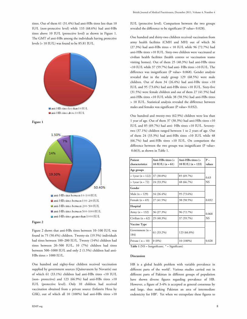
titres above 10 IU/L (protective level) as shown in Figure 1.
The GMT of anti-HBs among the individuals having protective
levels (> 10 IU/L) was found to be 85.81 IU/L.
Figure 1
Figure 2
Figure 2 shows that anti-HBs titres between 10–100 IU/L was
found in 75 (50.4%) children. Twenty-six (19.5%) individuals
had titres between 100–200 IU/L. Twenty (14%) children had
titres between 20–500 IU/L, 10 (7%) children had titres
between 500–1000 IU/L and only 2 (1.5%) children had anti-
HBs titres > 1000 IU/L.
One hundred and eighty-four children received vaccination
supplied by government sources (Quinevaxem by Novartis) out
of which 61 (33.1%) children had anti-HBs titres <10 IU/L
(non- protective) and 123 (66.9%) had anti-HBs titres >10
IU/L (protective level). Only 10 children had received
vaccination obtained from a private source (Infanrix Hexa by
GSK), out of which all 10 (100%) had anti-HBs titres >10
IU/L (protective level). Comparison between the two groups
revealed the difference to be significant (P value= 0.028).
One hundred and thirty-two children received vaccination from
army health facilities (CMH and MH) out of which 36
(27.3%) had anti-HBs titres < 10 IU/L while 96 (72.7%) had
anti-HBs titres >10 IU/L. Sixty-two children were vaccinated at
civilian health facilities (health centres or vaccination teams
visiting homes). Out of them 25 (40.3%) had anti-HBs titres
<10 IU/L while 37 (59.7%) had anti- HBs titres >10 IU/L. The
difference was insignificant (P value= 0.068). Gender analysis
revealed that in the study group 129 (68.5%) were male
children. Out of them 34 (26.4%) had anti-HBs titres <10
IU/L and 95 (73.6%) had anti-HBs titres >10 IU/L. Sixty-five
(31.5%) were female children and out of them 27 (41.5%) had
anti-HBs titres <10 IU/L while 38 (58.5%) had anti-HBs titres
> 10 IU/L. Statistical analysis revealed the difference between
males and females was significant (P value= 0.032).
One hundred and twenty-two (62.9%) children were less than
1 year of age. Out of them 37 (30.3%) had anti-HBs titres <10
IU/L and 85 (69.7%) had anti- HBs titres >10 IU/L. Seventy-
two (37.1%) children ranged between 1 to 2 years of age. Out
of them 24 (33.3%) had anti-HBs titres <10 IU/L while 48
(66.7%) had anti-HBs titres >10 IU/L. On comparison the
difference between the two groups was insignificant (P value=
0.663), as shown in Table 1.
Patient
characteristics
Anti-HBs titres (<
10 IU/L) (n = 61)
Anti-HBs titres (>
10 IU/L) (n = 133)
P –
values
Age groups
0.63
NS
< 1year (n = 122) 37 (30.0%) 85 (69.7%)
> 1year (n = 72) 24 (33.3%) 48 (66.7%)
Gender
0.032
Male (n = 129) 34 (26.4%) 95 (73.6%)
Female (n = 65) 27 (41.5%) 38 (58.5%)
Hospital
0.068
NS
Army (n = 132) 36 (27.3%) 96 (72.7%)
Civilian (n = 62) 25 (40.3%) 37 (59.7%)
Vaccine Type
0.028
Government (n =
184) 61 (33.2%) 123 (66.8%)
Private ( n = 10) 0 (0%) 10 (100%)
Table 1 (NS = Insignificant; * = Significant)
Discussion
HB is a global health problem with variable prevalence in
different parts of the world1. Various studies carried out in
different parts of Pakistan in different groups of population
have shown diverse figures regarding prevalence of HB.
However, a figure of 3-4% is accepted as general consensus by
and large, thus making Pakistan an area of intermediate
endemicity for HB6. Yet when we extrapolate these figures to
8

British Journal of Medical Practitioners, December 2011, Volume 4, Number 4
BJMP.org
our population, it is estimated that Pakistan hosts about seven
million carriers of HB which is about 5% of the worldwide 350
million carriers of HB10,11.
Age at the time of infection plays the most important role in
acquisition of acute or chronic HBV disease. HBV infection
acquired in infancy is responsible for a very high risk of chronic
liver disease due to HBV in later life12. HB is a preventable
disease and fortunately vaccination at birth and during infancy
can eradicate the disease globally, if vaccination strategy is
effectively implemented13. This can be claimed as the first anti-
cancer vaccine which prevents hepatocellular carcinoma in later
life.
In Pakistan, the HB vaccine was included in the EPI in 2004,
given along with DPT (Diphtheria, Pertussis, Tetanus) at 6, 10
and 14 weeks of age. The vaccine is provided through
government health infrastructure to health facilities. Private HB
vaccines supplied as a single antigen or in combination vaccines
are also available in the market. The efficacy of these
recombinant vaccines is claimed to be more than 95% among
children and 90% among normal healthy adults14. The
immunity of the HB vaccination is directly measured by
development of anti-HBs antibodies more than 10 IU/L, which
is considered as a protective level15. However, it is estimated
that 5–15 % of vaccine recipients may not develop this
protective level and remain non-responders due to
undermentioned reasons.16 Published studies regarding
antibody development in relation to various factors in terms of
immunogenicity and seroprotection, show highly varied results.
Multiple factors like dose, dosing schedules, sex, storage, site
and route of administration, obesity, genetic factors, diabetes
mellitus and immunosupression, affect HB antibodies
development response17.
Although the HB vaccine was included in the EPI in 2004 in
Pakistan, until now no published data showing seroconversion
and seroprotection among vaccine recipients of this programme
is available on a national level to our knowledge. Our study has
revealed that out of 194 children, only 133 (68.6%) had anti-
HBs titres in the protective range (>10 IU/L) while 61 (31.4%)
did not develop seroprotection. These results are low as
compared to other international studies. A study from
Bangladesh among EPI vaccinated children shows a
seroprotection rate of 92.2%13while studies from Brazil18 and
South Africa19 have separately reported seroprotection rates of
90.0% and 86.6%, respectively. Studies from Pakistan carried
out in adults also show seroprotection rates (anti-HBe titres >10
IU/L) of more than 95% in Karachi University students14 and
86% in health care workers of Agha Khan University
Hospital20, respectively. However, in these studies the dosing
schedule was 0, 1 and 6 months, and participants were adults.
These results are consistent with international reports.
The gravity of low seroprotection after HB vaccination is
further aggravated when we extrapolate these figures to our
overall low vaccination coverage rates of 37.6% to 45% as
shown in studies at Peshawar and Karachi respectively21,22. One
can imagine a significantly high percentage of individuals
vulnerable to HBV infection even after receiving HB vaccine in
an extensive national EPI programme. Therefore, a large
population still remains exposed to risk of HBV infection, and
national and global eradication of HBV infection will remain a
dream. Failure of seroprotection after receiving the HBV
vaccination in the EPI will also be responsible for projecting a
sense of false protection among vaccine recipients.
Dosing schedule is an important factor in the development of
an antibody response and titre levels. According to the Advisory
Committee on Immunization Practices (ACIP) of America,
there should be a minimum gap of 8 weeks between the second
and third doses and at least 16 weeks between the first and third
doses of the HB vaccination23. To minimize frequent visits and
improve compliance, the dosing schedule has been negotiated in
the EPI to 6, 10 and 14 weeks24. Although some studies have
shown this schedule to be effective, the GMT of anti-HBs
antibodies achieved was lower than that achieved by the
standard WHO schedule25. This may be one explanation of
lower rates of seroprotection in our study. The GMT achieved
in our study among the children having protective levels of
antibodies is 85.81 IU/L which is lower than most other
studies. This supports the observation that GMT achieved in
this schedule is lower than that produced by the standard
WHO schedule. This may result in breakthrough infection of
HB in vaccinated individuals in later life due to waning
immunity. However, the immune memory hypothesis supports
protection of vaccinated individuals in later life in spite of low
anti-HBs antibody titres26. Yet further studies are required to
dispel this risk.
Another shortcoming of this schedule is to miss the dose at
birth (‘0 dose’). It has been reported that the 0 dose of the HB
vaccine alone is 70% - 95% effective as post-exposure
prophylaxis in preventing perinatal HBV transmission without
giving HB immunoglobulins27. This may also be a factor
contributing to lower rates of seroprotection in our study as we
have not done HBsAg and other relevant tests to rule out HBV
infection in these children. Moreover pregnant ladies by and
large are not screened for HBV infection in Pakistan routinely
in the public sector except in a few big cities like Islamabad,
Lahore or Krachi. Therefore, we do not know the HB status of
pregnant mothers and the risk of transmission to babies remains
high. Different studies have reported much varied figures of HB
status in pregnant ladies. A study from Karachi reports 1.57%
pregnant ladies are positive for HBsAg while a study from
Rahim Yar Khan reports this figure to be up to 20%28,29. A
study by Waheed et al regarding the transmission of HBV
infection from mother to infants reports the risk to be up to
90%30. All of these studies support the importance of the birth
dose of the HB vaccination and augment the fact that control
and eradication of HB with the present EPI schedule is not
9
British Journal of Medical Practitioners, December 2011, Volume 4, Number 4
BJMP.org
possible. Jain from India has reported a study using an
alternative schedule of 0, 6 weeks and 9 months. He has
reported it to be comparable to the standard WHO schedule of
0, 1, 6 months in regards to seroprotection and GMT levels
achieved31. This schedule can be synchronised with the EPI
schedule, avoiding extra visits and incorporating the birth dose.
A similar schedule can also be incorporated in our national EPI.
In our study, seroprotection rates were found to be low in the
female gender and the difference was significant. This finding
differs with other studies which report lower seroprotection
rates in males32. Although the number of female children was
less, there is no plausible explanation for this observation. The
site of inoculation of the HB vaccine is also very important for
an adequate immune response. Vaccines given in the buttocks
or intradermally produce lower antibody titres than
intramuscular injections given in the outer aspect of the thigh
in children, due to poor distribution and absorption of the
vaccine within the host body. The practice of giving
vaccinations in the buttocks by vaccinators is a common
observation which they feel convenient for intramuscular
injection in children. This may also be one reason for low
seroprotection rates in our study, as we picked the children at
random who had received vaccination at public health facilities
except a small number of private cases.
The effectiveness of the vaccine also depends on the source of
procurement and proper maintenance of the cold chain. In this
study 100% seroprotection was observed in children who
received the HB vaccine procured from a private source.
Although the number of private cases was less, this factor of
source and the cold chain also needs attention. To address this
issue proper training of EPI teams regarding maintenance of
temperature, injection techniques, motivation and monitoring
can improve outcomes substantially.
The findings of this study are different from published
literature because this is a cross-sectional observational study.
This reports the actual seroprotection rates after receiving the
HB vaccination in the EPI schedule. While most other studies
show the results after ensuring control of influencing factors
such as type of vaccine, dose, schedule, route of administration,
training and monitoring of local EPI teams and health status of
vaccine recipients, etc. Therefore, this is an effort to look at a
practical scenario and evaluate outcomes which can help in
framing future guidelines to achieve the goal of control and
eradication of HB infection. Further studies are required at a
large scale to determine the effect of HB vaccination at a
national level.
Conclusion
The HB vaccination programme has decreased the global
burden of HBV infection, but evidence of decreased burden is
not uniform amongst world population.Of course figures
witness marked decrease in developed world while in
developing world statistics show little change. Unfortunately,
implementation of this programme is not uniformly effective in
all countries, thus resvoirs of infection and the source of
continued HBV transmission persists. HBV infection is
moderately endemic in Pakistan. The HB vaccine has been
included in the national EPI since 2004. The present study
shows seroprotection rates of only 68.6% in vaccine recipients,
which is low when compared with other studies; 31.4% of
vaccine recipients remain unprotected even after vaccination.
Moreover GMT achieved in seroprotected vaccine recipients is
also low (85.81 IU/L). There can be multiple reasons for these
results, such as type of vaccine used, maintenance of the cold
chain, route and site of administration, training and monitoring
of EPI teams and dosing schedule. In present practice, the very
important birth dose is also missing. These observations warrant
review of the situation and appropriate measures to be taken to
rectify the above mentioned factors, so that desired
seroprotection rates after HB vaccination in the EPI can be
achieved among vaccine recipients.
Acknowledgements We are thankful to the administration team of National University of Sciences
and Technology (NUST), Islamabad, Pakistan for financing this study project.
We are also thankful to the Department of Biochemistry and Molecular Biology,
Army Medical College, for conducting laboratory tests. Mr. Ghulam Husnain of
Army Medical College deserves special thanks for typing the manuscript. Special
thanks also to Miss Irum, statistician of Army Medical College, for carrying our
statistical analysis.
Author Details MOHAMMAD AFZAL, Associate Professor of Paediatrics, Army Medical
College, Rawalpindi. KHALIQ NAVEED, Head of Dept of Biochemistry, Army
Medical College Rawalpindi. SHABBIR HUSSAIN, Assistant Professor of
Paediatrics, Armed Forces Postgraduate Medical Institute, Rawalpindi.
SHAUKAT MEHMOOD QURESHI, Associate Professor of Community
Medicine, Army Medical College, Rawalpindi. ASIFA MAJEED, Assistant Prof
Biochemistry, Army Medical College, Rawalpindi. ZIA FAROOQI, Research
Technologist, Dept of Biochemistry and Molecular Biology, Army Medical
College, Rawalpindi. ALIZAY GOHAR, 4th year MBBS student, Foundation
University Medical College, Rawalpindi. ABDUL WAHAB, 3rd year MBBS
student, Army Medical College, Rawalpindi. CORRESSPONDENCE: Dr.Mohammad Afzal, Associate Professor of
Paediatrics, Army Medical College, Rawalpindi. Email: [email protected]
REFERENCES
1. World Health Organization. HB vaccines: Weekly Epidemiol Rec
2009;40:405-420.
2. A Comprehensive Immunization Strategy to Eliminate Transmission of
Hepatitis B Virus Infection in the United States. MMWR
2005;54/No.RR-16:1-32.
3. Okada K, Kamiyama I, Inomata M, Imai M, Miyakawa Y. e antigen and
anti-e in the serum of asymptomatic carrier mothers as indicators of positive
and negative transmission of HB virus to their infants. N Engl J Med
1976;294:746-9.
4. CDC. Recommendation of the Advisory Committee on Immunization
Practices (ACIP): Inactivated HB virus vaccine. MMWR 1982;31:317-7,
327-8.
5. CDC. National, State and urban area vaccination coverage among
children aged 19-35 months United States, 2004. MMWR2004:54:717-21.
6. Abbas Z, Jafri W, Shah SHA, Khokar N, Zuberi SJ. Members of the
consensus panel. PSG consensus statement on management of HB virus
infection – 2003. J Pak Med Assoc 2004, 54:150-8.
10

British Journal of Medical Practitioners, December 2011, Volume 4, Number 4
BJMP.org
7. Kane M. Global Program for control of hepatitis B infection. Vaccine
1995;13:S47-49
8. World Health Organization. Immunization Policy
WHO/EPI/GEN/95.3.1995.
9. Gomber Sunil, Sharma Rajesh, Ramachandran VG Talwar Vibha, Singh
Bharat. Immunogenicity of HB Vaccine incorporatd into the Expanded
Programme of immunization schedule. Indian Paediatr 2000;37:411-413.
10. Waseem J, Nadeem J, Yakoob K, Mohammad I, Tirmizi SFA, et al. HB
& C: prevalence and risk factors associated with seropositivity among
children in Karachi, Pakistan. BMC Infectious disease. 2006;6:101.
11. Zaman AS. 2003.Daily Dawn internet addition. 23rd January.
12. Stevens CE, Toy PT, Tong MJ, et al. Perinatal HB virus transmission in
the United States: prevention by passive – active immunization JAMA
1985;253:1740-5.
13. Guho A, Ahaad A, Salam A, Aleem A, Haq AE, Islam QE.
Seroconversion after recombinant HB vaccination. J MEDICINE
2010;11:143-150.
14. Hakeem ST, Nadeem SG, Kazmi SU. Comparative evaluation of four
HB vaccines in Pakistan. Reactogenicity & Immunogenicity. BJMP
2009;2:30-34.
15. Jacj ADM, Hall AJ, Maine M, Whittle HC. What level of hepatitis B
antibody is protective? J infect Dis 1999;179:489-92.
16. John TJ. HB immunization. Indian Pediatr 1995;32:609-613.
17. Shaw FE, Jr, Guess HA, Roets JK et al. Effect of anatomic injection site,
age and smoking on the immune response to HB vaccination. Vaccine
1989;7:425-30.
18. Ribero TM and Azevedo RS. Seroconversion of HB vaccine in infants
related to the mothers serostatus in a community of Sao Jose dos Campos,
state of Sao Paulo, Brazil. Clinics 2006;61:5
19. Tsebes KV, Burnett RJ, Hlungwani NP, et al. The first five years of
universal HB vaccination in South Africa; evidence for elimination of
HBsAg carriage in under-5-year-children. Vaccine 2001;19:3919-3926
20. Zeeshan M, Jabeen K, Ali ANA, Ali AW, Farooqi SZ, Mehraj V, Zafar
A. Evaluation of immune response to HB vaccine in health care workers at a
tertiary care hospital in Pakistan: an observational prospective study. BMC
Infectious Diseases 2007,7:120.
21. Rehman H, Arshad S. Immunization status of children admitted in
Pediatrics department Lady Reading Hospital Peshawar. Med Channel Jan-
Mar 2007;13:36-8.
22. Siddiqui N, Khan A, Nisar N, Siddiqui AA. Assessment of
EPI(Expanded Program on Immunization) vaccine coverage in a peri-urban
area. J Pak Med Assoc 2007;57:391-5.
23. CDC. General recommendations of immunization: recommendations of
the Advisory Committee on immunization Practices (ACIP) and the
American Academy of the Family Physicians (AAFP). MMWR 2002;51;No
RR-2):1-35
24. Expanded Programme on Immunization: Framework for evaluating a
vaccine for the EPI, WHO Documet WHO/EPI/GEN/93.5;1993g.
25. Mittal SK, Rao S, Agarwal V, Parkash C, Thirupuram S. Simultaneous
administration of HB vaccine with other EPI vaccines. Indian J Pediatr
1994;61:183-8.
26. Resti M, Azzari C,Mannelli F, Rossi ME, Lioneti P, Vierucci A, Ten-
year follow up study of neonatal HB immunization: are booster injections
indicatd? Vaccine 197; 15:1338-40.
27. Pichichero ME, Blater MM, Reisinger KS, et al. Impact of a birth dose
of HB vaccine on the reactogenicity and immunogenicity of diphtheria-
tetanus-acellular pertussis –HB-inactivated poliovirus-Haemophilus
influenzae type b combination vaccination. Pediatr Infect Dis J
2002;21:854-9.
28. Ali S, Memon A. Prevalence of HB infection in pregnant women in
tertiary care hospital. Infect dis J 2007;16:36-8.
29. Hakeem A, KS, Abdullah M, RA, Hashmi I. Prevalence of HB surface
antigen and anti HCV in pregnant ladies attending antenatal clinic at Sheikh
Zayed Medical Complex Rahim Yar Khan. Esculapio J Services Inst Med Sci
2006;2:6-8
30. Kazmi K, Ghafoor A, Qureshi AW. Mother infant transmission of HB
in Pakistan. Pak J Med Res 2003;42:152-6.
31. Jain KA, Mittal SK, Ramji S and Chakravarti A. HB vaccine in the EPI
Schedule. Indian J Pediatr 2004; 72(8):661-664.
32. Zuckerman JN, Sabin C, Craig FM, et al. Immune response to a new
hepatitis B vaccine in healthcare workers who had not responded to standard
vaccine : randomized blind dose-response study. Br Med J 1997;314: 329-
33.
33. CDC. Suboptimal response to HB vaccine given by injection into the
buttock. MMWR 1985;34:105-9,113.
34. Craven DE, Awedh ZL, Kunches LM,Yunis EJ, Diestag JL, Werner BJ,
Polk BF, snydman DR, Platt R, Crumpacker CS, Grady GF, Alper CA,
Non responsiveness to HB vaccine in healthcare workers. Ann Inter Med
1986; 105:356-360.
11
British Journal of Medical Practitioners, December 2011, Volume 4, Number 4
BJMP.org
BJMP 2011;4(4):a441
Dexmedetomidine versus ketamine combined with midazolam; a comparison of
anxiolytic and sedative premedication in children
Mohamed A. Daabiss and Mohamed Hashish
ABSTRACT Background: Preanaesthetic medication plays an important role in the anaesthetic care of children by allaying anxiety, decreasing vagal stimulation and preventing postoperative psychological sequelae. This study was undertaken to evaluate the efficacy of dexmedetomidine when administered orally as a hypnotic and anxiolytic compared to oral combination ketamine/midazolam as preanaesthetic medication in paediatric patients. Methods: Sixty-six children aged 2-6 years posted for elective surgical procedures were randomly allocated to one of two groups ‘Group D’ and ‘Group MK’. Group D received oral dexmedetomidine 3 µg/kg and group MK received 0.25 mg/kg oral midazolam (up to a maximum of 15 mg) mixed with 2.5 mg/kg oral ketamine. Drug acceptance was noted. Heart rate, arterial pressure, respiratory rate, sedation score and anxiolysis score were noted before drug administration and every 5 min for up to 30 min after drug administration. Parental separation score at 30 min and mask acceptance score in addition to parental satisfaction were also noted. Results: premedication with oral MK appeared to be superior to oral dexmedetomidine, in addition to evident haemodynamic stability and higher degree of parental satisfaction (90%), but 97% of children better accepted oral dexmedetomidine. No significant side effects were attributable to either premedication. Emergence from anaesthesia was comparable between groups. Conclusion: premedication with oral midazolam ketamine appeared to be superior to oral dexmedetomidine, with evident haemodynamic stability and a higher degree of parental satisfaction, although oral dexmedetomidine was more accepted by the children. KEYWORDS : Dexmedetomidine, Midazolam, Ketamine, Paediatric, Premedication
Introduction
Fear of physicians, injections, operations, the operation theatre
and the forced separation from parents make the operative
experience more traumatic for young children and can cause
nightmares and postoperative behavioural abnormalities.
Preanaesthetic medication may decrease the adverse
psychological and physiological sequelae of induction of
anaesthesia in a distressed child1. An important goal of
premedication is to have the child arrive in the operating room
calm and quiet with intactcardiorespiratoryreflexes. Various
drugs have been advocated as premedication to allay anxiety
and facilitate the smooth separation of children from parents.
The idealpremedicantin children should be readily acceptable
and should have a rapid and reliable onset with minimal side
effects. Midazolam has sedative and anxiolytic activities,
provides anterograde amnesia, and has anticonvulsant
properties2. Ketamine, on the other hand, provides well-
documented anaesthesia and analgesia. It has a wide margin of
safety, as the protective reflexes are usually maintainedOral
premedication with midazolam and ketamine became widely
used inpaediatric anaesthesiato reduce emotional trauma and
ensure smooth induction. It provided better premedication than
either oral ketamine or midazolam alone4, but excessive
salivation and hallucination were observed5.
Dexmedetomidine is a highly selective α2-adrenoreceptor
agonist drug. Clinical investigations have demonstrated its
sedative, analgesic and anxiolytic effects after IV administration
to volunteers and postsurgical patients6. It has been used to
sedate infants and children during mechanical ventilation and
also to sedate children undergoing radiological imaging
studies,8In the literature, few articles have used
dexmedetomidine orally for the premedication of children. The
purpose of this study is to evaluate the efficacy of
dexmedetomidine when administered orally as a hypnotic and
anxiolytic agent compared to oral combination
ketamine/midazolam as preanaesthetic medication in
paediatrics.
Methods:
The Hospital Ethics Committee approved the protocol.
Written informed consent was obtained from parents prior to
inclusion. Sixty six children of ASA physical status I or II, aged
between 2 and 6 years and scheduled for elective minor surgery
of more than 30 minutes expected duration were enrolled in
this prospective, randomized, double-blind study. Exclusion
criteria were: a known allergy or hypersensitivity reaction to any
of the study drugs, organ dysfunction, cardiac arrhythmia or
congenital heart disease, and mental retardation.
Children were randomly allocated to one of the two study
groups using computer-generated random numbers. Group D
received oral dexmedetomidine 3 µg/kg and group MK received
0.25 mg/kg oral midazolam (up to a maximum of 15 mg) with
2.5 mg/kg oral ketamine. The oral premedication was mixed
with 3 ml of apple juice as a carrier to be given thirty minutes
before induction of anaesthesia. The oral route was chosen as it
is the most acceptable and familiar mode of drug
Research A
rticle
12

British Journal of Medical Practitioners, December 2011, Volume 4, Number 4
BJMP.org
administration. An independent investigator not involved in the
observation or administration of anaesthesia for the children
prepared all study drugs. Observers and attending anaesthetists
who evaluated the patients for preoperative sedation and
emergence from anaesthesia were blinded to the drug
administered. Children had premedication in the preoperative
holding area in the presence of one parent. All children received
EMLA cream unless contraindicated.
After drugs were administrated, the following conditions were
observed: 1) response to drug and onset of sedation, 2) response
to the family separation circumstance and the entrance to the
operating room, 3) response to the venous line (IV) insertion,
4) ease of mask acceptance during induction of anaesthesia. The
time to recovery from anaesthesia and to achieve satisfactory
Aldrete score were also noted. Onset of sedation was defined as
the minimum time interval necessary for the child to become
drowsy or asleep.
Sedation statuswas assessed every 5 min for up to 30 min with a
five-point scale. A score of three or higher was considered
satisfactory. In addition anxiolysis was assessed on a four-point
scale. An anxiety score of three or four was considered
satisfactory. Cooperation was assessed with a four-point scale. A
cooperation score of three or four was considered satisfactory.
Taste acceptability was evaluated on a four-point scale. A score
of 1–3 was considered satisfactory.
Score Sedation Anxiolysis Cooperation Taste
1 Alert/active Poor Poor Accepted readily
2 Upset/wary Fair Fair Accepted with grimace
3 Relaxed Good Good Accept with
verbalcomplaint
4 Drowsy Excellent Excellent Rejected entirely
5 Asleep
Heart rate, blood pressure, respiratory rate and arterial oxygen
saturation were recorded before premedication, every five
minutes for 30 min preoperatively, and then during induction
of anaesthesia, every 5 min intra-operatively, every 15 min in
recovery room and every 30 min in day-case unit until time of
discharge.
The anaesthetic agents administered were
standardized.Children were induced with sevoflurane, nitrous
oxide in oxygen and fentanyl 1-2 µg/Kg and maintained with
the same drugs. The trachea was intubated after administering
cisataracurium 0.1 mg/kg.
At the end of the procedure, the neuromuscular blockade was
reversed with neostigmine with glycopyrolate and the child was
extubated. After that, they were kept in the recovery room
(PACU) under observation until discharge. The time to
recovery from anaesthesia and to achieve satisfactory Aldrete
score were noted. The discharge time was also noted and
postprocedure instructions were given. Children were called for
checkups the following day, when parents were asked to answer
a questionnaire about the surgical experience of the parent and
child and side effects experienced, if any.
Statistical analysis was performed using SPSS version 17. All
values were reported as mean ± SD and range. Data analysis for
numerical data was performed by unpaired Student’s t-test to
detect the differences between the groups for age, weight, onset
of anxiolysis and sedation. Data analysis for categorical data was
performed by Fisher’s exact test to detect differences for the
scores. Other data are reported as mean ± SD or frequency (%).
A P value < 0.05 was considered statistically significant. Prior to
the study, we chose the null hypothesis (i.e.
nosignificantsedation scores between the groups). The number
of patients required in each group was determined using power
analysis based on previous studies. Assuming that 79% of
patients would become drowsy or asleep in the
midazolam/ketamine group (15 patients), a sample size of 30
patients per group would have an 80% power of detecting a
20% difference in sedation (from 79% to 99%) at the 0.05 level
ofsignificance. We decided to study 66 patients to account for
possible dropouts.
Results:
Sixty-six patients were enrolled; four did not receive the study
medication and two did not have surgery on the same day,
leaving 60 subjects who fulfilled the criteria for the
study.Groups were comparable regarding age, sex, weight, ASA
physical status, surgical interventions and duration of
anaesthesia (Table 1). Operative procedures were evenly
distributed and included inguinalherniorrhaphy, hydrocele
repair or orchidopexy.
Table 1: Demographic characteristics and duration of
anaesthesia:
Group D Group MK
No of patients 33 33
No of patients excluded 4 2
Age (years) 4.02±1.98 4.2±1.45
Gender (female/male) 13/16 15/16
ASA (I/II) 25/4 25/6
Weight (Kg) 17.72±4.4 16.56±5.1
Duration of Anaesthesia (min) 35.17±5.9 32.7±8.4
Data are expressed as mean ± SD (range). P > 0.05. No significant
difference among groups.
Dex group (D). Midazolam Ketamine group (MK). ASA,
American Society of Anesthesiology physical status.
Onset of sedation was significantly faster after premedication
with midazolam/ketamine (Fig1), and the level of sedation was
significantly better after premedication with
13

British Journal of Medical Practitioners, December 2011, Volume 4, Number 4
BJMP.org
midazolam/ketamine 30 minutes after ingestion of the
premedicant.
The anxiolysis score revealed 84 % of children in group MK as
being friendly and only 51% of children in group D have
similar behaviour (Table 2). The taste of oral dexmedetomidine
was judged as significantly better; 13% of children rejected the
oral midazolam/ketamine combination (Table 2).
Table 2: Distribution of behaviour and sedation status at time
of induction:
Group D Group MK P
Time to onset of sedation (min) 24.52 ± 3.1 18.36 ± 2.6 0.015*
Preoperative sedation score 1.6±0.5 3.1±0.8 0.003*
% asleep at induction 61% 90% 0.024*
Preoperative anxiolysis score 1.4±0.6 2.9±0.7 0.016*
% Face mask acceptance 58% 88% 0.033*
% Venous line insertion acceptance 72% 90% 0.005*
% Satisfactory parental separation 50% 80% 0.04*
% Parental satisfaction 70% 90% 0.036*
% Taste acceptance 97% 87% 0.002*
Data are expressed as mean ± SD (range) or percentage. Dex group
(D). Midazolam Ketamine group (MK).
* significantP <0.05.
Application of a facemask at induction of anaesthesia was
accepted more readily in patients of group MK (Fig 2).Overall,
satisfactory cooperation with venous line insertion was found in
90% of children in group MK, while comparatively 72% of
children in group D showed satisfactory cooperation with
insertion of a venous line (Table 2). Moreover, most of the MK
treated children were more calm and sedated than the D-treated
group at the time of separation from parents. Parental
satisfaction was significantly higher in group MK.
The time interval from end of surgery to spontaneous eye
opening in the PACU was significantly less in group D (Fig 1),
while the time to discharge from the PACU to ward was similar
for groups (Table 3).
Table 3: Time to eye opening and PACU discharge
Group D Group MK P
Time to eye opening (min) 21±4.3 30±6.1 0.032*
Time of PACU discharge (min) 30± 3.9 28.12±5.5 0.316
Data are expressed as median ± SD (range). Dex group (D).
Midazolam Ketamine group (MK).
* significantP < 0.05.
While no child experienced respiratory complications or arterial
oxygendesaturationbefore induction, heart rate and systolic
blood pressure were marginally higher after administration of
MK. On the other hand, the mean heart rate and systolic blood
pressure measurements were 15% lower (than preoperative
values) in group D at the same study periods. However, during
recovery, haemodynamic responses were similar.
Adverse events were recorded for the three periods. Two
children in group MK as well as one in group D experienced
nausea but only one patient in group MK vomited before
induction. Hallucination was recorded in 10 % of patients in
group MK. Excessive salivation occurred in 12% of children
receiving the combination of drugs, compared to 7% in D-
treated children.
Discussion:
Our study proved that midazolam/ketaminereceiving patients
were significantly calmer and more cooperative compared to
dexmedetomidine receiving patients during the preoperative
period, the insertion of a venous line, during separation from
parents and also during the application of a facemask at
induction. Several studies have been published demonstrating
the advantage of the midazolam/ketamine combination in
paediatric premedication4,9, while others have reported
superiority of oral dexmedetomidine premedication to oral
midazolam10,11.
Based on their experience with using oral dexmedetomidineas a
preanaesthetic in children, Kamal et al10 and Zub et
al12 reported that the dose of 3 µg/kg could be safely and
effectively applied without haemodynamic side effects.
Midazolam is currently the most commonly usedpaediatric
premedication due to easy application, rapid onset, short
duration of action and a lack of significant side effects13.
Meanwhile oral ketamine was used in the 1970s by dentists to
facilitate the treatment of mentally handicapped children. In
1982, Cetina found that rectal or oral preanaesthetic ketamine
is an excellent analgesic and amnesic agent with no incidence
ofdysphoric reactions, possibly related to its high rate of first-
pass metabolism14. The metabolite norketamine has
approximately one-third the potency of ketamine, but reaches
higher blood concentration and also causes sedation and
analgesia 15. The use of midazolam and ketamine in
combination as a premedicant combines their properties of
sedation and analgesia and attenuates drug induced
deliriumGhai et al and Funk et al have also reported that a
combination of midazolam and ketamine results in better
premedication than the individual drugs given alone4,9.
Like clonidine, dexmedetomidine possesses a high ratio of
specificity for the α2 versus the α1 receptor (200: 1 for clonidine
and 1600: 1 for dexmedetomidine). Through presynaptic
activation of the α2 adrenoceptor, it inhibits the release of
norepinephrine and decreases sympathetic tone. There is also an
attenuation of the neuroendocrine and haemodynamic
responses to anaesthesia and surgery, thereby leading to sedation
and analgesia16. One of the highest densities of α2 receptors has
14
British Journal of Medical Practitioners, December 2011, Volume 4, Number 4
BJMP.org
been detected in the locus coeruleus, the predominant
noradrenergic nucleus in the brain and an important modulator
of vigilance. The hypnotic and sedative effects of α2-
adrenoceptor activation have been attributed to this site in the
CNS16. This allows psychomotor function to be preserved while
letting the patient rest comfortably, so patients are able to
return to their baseline level of consciousness when
stimulated17. Clonidine and dexmedetomidine seems to offer
the beneficial properties, but dexmedetomidine has a shorter
half-life, which might be more suitable for day surgery. Zuband
his colleagues reported that dexmedetomidine may be an
effective oral premedicant prior to anaesthesia induction or
procedural sedation and it was effective even in patients with
neurobehavioural disorders in whom previous attempts at
sedation had failedAlso Sakurai et al reported that oral
dexmedetomidine could be applied safely and effectively as a
preanaesthetic in children18.
While dexmedetomidine is tasteless and odourless17 , with 82%
bioavailability after extravascular doses in healthy human
adults19, oral midazolam formulations have a bitter taste and
were usually prepared by mixing the IV midazolam with a
variety of sweet additives. In our study, children judged the
taste of oral dexmedetomidine as significantly better than oral
midazolam ketamine mixture, although both drugs were given
with the same sweet tasting syrup. This observation probably
might also reflect the developmental age of these patients and
the difficulty of gaining their cooperation in swallowing
something that they did not wish to swallow. Recently, new
commercially prepared oral midazolam formulations are
reported to be more palatable20, but unfortunately, it is not
available yet in our country.
Our data confirmed that onset of sedation and peak sedative
effect was significantly slower after oral dexmedetomidine
compared to oral midazolam ketamine. These results are
consistent with studies by Kamal et al and Schmidt et al who
reported slow onset of action of oral dexmedetomidine,21In
addition, Anttila et al reported that, in adults after oral
administration, peak plasma concentration is achieved at 2.2 ±
0.5 h after a lag-time of 0.6 ± 0.3 h19.
In this study, dexmedetomidine premedication with the present
study design resulted in slight hypotension and bradycardia,
which could be attributed to postsynaptic activation of
α2 adrenoceptors in the central nervous system (CNS) that
inhibit sympathetic activity and thus can decrease blood
pressure and heart rate22. In a finding consistent with our
results, Khan et al and Aantaa et al reported that
useofdexmedetomidine can beassociatedwithsome
cardiovascular side effects including hypotension and
bradycardia,24Conversely, Ray and Tobias did not find
significant haemodynamic changes when used
dexmedetomidine in providing sedation during
electroencephalographic analysis in children with autism and
seizure disorders25.
There were some limitations to this study; the bioavailability of
oral dexmedetomidine is based on the adult dataWe need to
decide the timing of the oral administration as
apremedicantbased on the data in children. Therefore, the
bioavailability of oral dexmedetomidine needs to be studied in
children. The premedication period was 30 min, however, if a
longer premedication period had been allowed, possibly more
subjects could have attained satisfactory sedation at separation
from parents and at induction of anaesthesia.
Conclusion:
In this study, premedication with oral
midazolam/ketamineappeared to be superior to oral
dexmedetomidine with evident haemodynamic stability and a
higher degree of parental satisfaction demonstrated, although
oral dexmedetomidinewas more accepted by the children. No
significant side effects were attributable to either premedication.
Emergence from anaesthesia was comparable between groups.
Competing Interests None declared Author Details MOHAMED A. DAABISS, Riyadh Armed Forces Hospital, Department of Anaesthesia; Riyadh, Saudi Arabia. MOHAMED HASHISH, Armed Forces Hospital, King Abdulaziz Airbase Hospital, Dhahran, Department of Anaesthesia; Riyadh, Saudi Arabia. CORRESSPONDENCE: MOHAMED DAABISS, Department of Anaesthesia, Riyadh Armed Forces Hospital, Mailbox: 7897-D186 Riyadh 11159 Saudi Arabia Email: [email protected]
REFERENCES
1. Kain ZN, Caldwell-Andrews AA, Krivutza DM, et al. Trends in the
practice of parental presence during induction of anaesthesia and the use of
preoperative sedative premedication in the United States, 1995–2002: results
of a follow-up national survey. Anesth Analg 2004;98:1252–9.
2. Kupietzky A, Houpt MI. Midazolam: A review of its uses for conscious
sedation of children. Pediatr Dent 1993;15:237-41.
3. Sekerci C, D φnmez A, Ate Y, et al. Oral ketamine premedication in
children (placebo controlled double-blind study). Eur J Anaesthesiol
1997;13:606-11.
4. Ghai B, Grandhe RP, Kumar A, et al. Comparative evaluation of
midazolam and ketamine with midazolam alone as oral premedication.
Pediatr Anesth 2005; 15(7): 554-9.
5. Roelofse JA, Joubert JJ, Roelofse PG. A double-blind randomized
comparison of midazolam alone and midazolam combined with ketamine
for sedation of paediatric dental patients. J oromaxillofacial surg 1996;
54(7): 838-44.
6. Taittonen MT, Kirvela OA, Aantaa R, et al. Effect of clonidine and
dexmedetomidine premedication on perioperative oxygen consumption and
haemodynamic state. Br J Anaesth 1997; 78: 400-6.
7. Tobias JD, Berkenbosch JW. Sedation during mechanical ventilation in
infants and children: dexmedetomidine versus midazolam. South Med J
2004; 97: 451-5.
8. Mason KP, Zgleszewski SE, Dearden JL, et al. Dexmedetomidine for
paediatric sedation for computed tomography imaging studies. Anesth Analg
2006;103:57-62.
9. Funk W, Jakob W, Riedl T, et al. Oral preanesthetic medication for
children: double-blind randomized study of a combination of midazolam
and ketamine vs. midazolam or ketamine alone. Br J Anaesth 2000;
84(3):335-40.
15
British Journal of Medical Practitioners, December 2011, Volume 4, Number 4
BJMP.org
10. Kamal K, Soliman D, Zakaria D. Oral dexmedetomidine versus oral
midazolam as premedication in children. Ain Shams J anaesth 2008;1: 1-18.
11. Üstün Y, Gündüz M, ErdoğanO, et al. Dexmedetomidine versus
Midazolam in Outpatient Third Molar Surgery. J oromaxillofacial surg
2006; 64(9): 1353-8.
12. Zub D, Berkenbosch J, Tobias J. Preliminary experience with oral
dexmedetomidine for procedural and anesthetic premedication. Pediatr
Anesth 2005;15(11): 932-8.
13. McMillan CO, Spahr-Schopfer IA, Sikich N, et al. Premedication of
children with oral midazolam. Can J Anaeth 1992;39: 545-50.
14. Cetina J. Schonende Narkoseeinleitung bel kindern durch orale oder
rektale Ketamin-Dehydrobenzperidol-Applikation. Anaesthetist
1982;31:277-9.
15. Grant IS, Nimmo WS, Clements JA. Pharmacokinetics and analgesic
effects of intramuscular and oral ketamine. Br J Anaesth 1981; 53: 805-10.
16. Hunter JC, Fontana DJ, Hedley LR, et al. Assessment of the role of
alpha 2-adrenoceptor subtypes in the antinociceptive, sedative and
hypothermic action of dexmedetomidine in transgenic mice. Br J Pharmacol.
1997;122:1339–44.
17. Hall JE, Uhrich TD, Barney JA, et al. Sedative, amnestic, and analgesic
properties of small-dose dexmedetomidine infusions. Anesth Analg.
2000;90:699–705.
18. Sakurai Y, Terui K, Obata T, et al. Buccal administration of
dexmedetomidine as a preanesthetic in children. Anaesthesia 2010; 24:49–
53.
19. Anttila M, Penttila J, Helminen A, et al. Bioavailability of
dexmedetomidine after extravascular doses in healthy subjects. J Clin
Pharmacol 2003;56:691–3.
20. Cote ` CJ, Cohen IT, Suresh S, et al. A comparison of three doses of
commercially prepared oral midazolam syrup in children. Anesth Analg
2002; 94: 37–43.
21. Schmidt AP, Valinetti EA, Bandeira D, et al. Effects of preanesthetic
administration of midazolam, clonidine, or dexmedetomidine on
postoperative pain and anxiety in children. Pediatr Anesth 2007;17: 667 -74.
22. Dyck JB, Maze M, Haack C, et al. The pharmacokinetics and
haemodynamic effects of intravenous and intramuscular dexmedetomidine
hydrochloride in adult human volunteers. Anesthesiol 1993;78:813–20
23. Khan ZP, Ferguson CN, Jones RM. Alpha-2 and imidazoline receptor
agonists. Their pharmacology and therapeutic role. Anaesthesia 1999;
54:146-165.
24. Aantaa R, Jaakola ML, Kallio A, et al. comparison of dexmedetomidine,
an alpha2-adrenoceptor agonist, and midazolam as i.m. premedication for
minor gynecological surgery. Br J Anaesth 1991; 67(4): 402-9.
25. Ray T ,Tobias J. Dexmedetomidine for sedation during
electroencephalographic analysis in children with autism, pervasive
developmental disorders, and seizure disorders. J Clin Anesth 2008; 20(5):
364-8.
16
British Journal of Medical Practitioners, December 2011, Volume 4, Number 4
BJMP.org
BJMP 2011;4(4):a440
Comparison of trauma and elective income in a district general hospital
Hussain Anthony Kazi and Ashutosh Acharya
ABSTRACT We aimed to investigate the income of trauma and elective work in our unit and compare inpatient stay and resource allocation. We performed a
prospective study of trauma and elective admissions for a one-week period. We calculated the income received using health resource group coding. 48
trauma patients were admitted of which 36 required operative intervention. This generated £134,321 for primary procedures followed by an extra £18,141
for those requiring no surgery. The total income for the week was £171,941. 71 elective patients underwent surgery. The total income generated was
£150,318 for this week. This was a typical week in a busy unit. No consultants were on leave. Although the income was higher in the trauma group this
was loss making due to the length of stay of those patients with hip fractures. Attempts at profitability should include enhanced rehabilitation services and
more realistic tariff for proximal femur fractures.
KEYWORDS : Trauma, Elective, Income, Health Resource Group Codes, Economics, Coding
Introduction:
Payment by results was introduced across the National Health
Service (NHS) in 2005. It’s aim was to provide a pricing
structure (tariff) for the whole country with some allowance for
geographical variation1-2. The system uses Healthcare Resource
Group codes (HRG) in which treatments in similar cost
brackets have the same codeA price / tariff is derived from each
hospital patient episode and the patient’s registered Primary
Care Trust (PCT) is billed accordingly.
In order to generate an HRG code data is collected by the
hospital clinical coding department including primary
diagnosis, comorbidity (which incurs an extra charge if
applicable), and complications, surgical procedure, age and
duration of stay4. Diagnoses (either primary, co morbidities or
complications) are coded using ICD-10 codes. Surgical
procedure is defined using OPCS-4 codesA piece if software is
then utilised to allocate the HRG code. Each HRG code
represents a tariff, which is the average cost of a treatment
nationwide. Minor regional adjustments are made to reflect the
cost of living2.
Payment by results covers all admissions, attendance in accident
& emergency departments and outpatients attendances5. The
2004 NHS Improvement plan designated 18 weeks as a target
for referral to treatment (RTT)6. It is a common misconception
that trauma patients do not account for considerable income
within the NHS. Trauma is often seen as the poor relation
when compared with elective work where a target based culture
now prevails. Elective targets must be met or hospital trusts can
incur financial penalty. This situation is not apparent for
trauma due to the acute nature of service delivery in the
majority of cases. The burden of trauma work can block elective
admissions and is seen by some as a barrier to target attainment.
At least 36% of orthopaedic surgeons in the United Kingdom
describe trauma as part of their sub-specialist interestWe aimed
to assess the throughput and income generated from one week
of trauma workload and compared this with the elective
throughput in our unit for the same week. This was performed
by means of a prospective study. We are not aware of any
published work in this specific area.
Methods:
We followed all acute patients admitted to our trauma unit
between 21/02/2008 and 28/02/2008. This represented a
“trauma week” which is how the consultant rota is organised in
our trust. We then compared this with the throughput in our
elective unit for the same calendar period. No surgeons were on
leave this week and no theatre sessions were cancelled other
than the on call trauma consultant’s elective operating sessions.
Our trust is a busy district general hospital with over 500 beds
and approximately 55,000 emergency attendances per year. The
orthopaedic directorate is staffed by ten full time consultants
and serves a population of 315,000 patients.
All patient details were recorded prospectively and followed
until the end of their inpatient episode. Case notes were then
reviewed with the coding department and ICD-10 and OPCS-4
codes were generated. Their length of stay and other required
variables were reviewed in order to generate the correct HRG
code. Once the analysis was complete income for the trauma
and elective groups were calculated.
Results:
Trauma:
48 patients were admitted (22 male) of which 36 required
operative intervention. This utilised 14 theatre sessions. Mean
age was 53.75 years (range: 7-93, median: 59). Median stay was
4 days with a mean of 13.3. The median and mean trim points
Research Article
17
British Journal of Medical Practitioners, December 2011, Volume 4, Number 4
BJMP.org
(expected duration of stay before extra charges incurred by
PCT) were 14.5 and 26.7 days respectively. Other consultants
operated on 6 patients. This was either due to expertise in a
specific area or space on an elective list utilised to reduce
backlog. The income generated by these cases is included in the
trauma total due to them being acute trauma interventions
rather than elective cases. These results are summarised in tables
1 and 2.
Table 1: Demographic & Income Data of Trauma and Elective Patients
Trauma Elective
Median age (yrs) 59 47
Number of Patients 47 71
No of Males 26 30
Median stay (days) 4 1
Range of stay (days) 1 – 107 1 - 7
Total bed days 637 118
Estimated Bed Costs (£) 203,840 26,550
Mean income per pt (£) 3658.32 2117.15
Total Income (£) 171,941 150,318
Of the 48 patients admitted 12 required no operative
intervention. These cases were general ‘run of the mill’
admissions such as soft tissue infections for intravenous
antibiotics, undisplaced fractures where home circumstances
obstructed discharge, soft tissue injuries for further investigation
and back pain. These will not be discussed further but the
income generated (£31,127) does go towards the total. The
median stay was 2 days with a mean stay 8.5 days (range: 1 –
47). This reflects the broad comorbidities and social
circumstances of this subset.
The group requiring operative intervention included hip
fractures (11 patients). Of these, seven required dynamic hip
screw fixation but were deemed “complex” due to their
comorbidities and therefore attracted the higher tariff rate
(£6685). One displaced intracapsular fracture required total hip
replacement, attracting a tariff of £7261. One patient required
revision from a dynamic hip screw to an intramedullary device
and then revision to a total hip arthroplasty. The tariff price was
£19,479. The remaining fractured neck of femur patients
attracted between £4379 and £6711 dependent on operative
procedure. The median stay was 26 days (mean: 14, range 9 –
107). One patient required closed manipulation of a dislocated
total hip replacement attracting a tariff price of £1034 and an
inpatient stay of one day. In addition one acetabular fracture
was sustained requiring open reduction and internal fixation. It
attracted a tariff price of £4262 and an inpatient stay of
seventeen days.
One patient required open reduction and internal fixation of a
patella fracture attracting a tariff of £2405 and was an inpatient
for 10 days. Another patient with septic arthritis required two
arthroscopic knee washouts, attracting a tariff of £5941 and was
an inpatient for 26 days.
Seven ankle fractures were admitted requiring operative
intervention, all of these attracted a tariff of £2405 except one,
which attracted £4262 due to co morbidity and complexity of
injury. The median stay in this group was six days (mean: 4.9,
range: 2-7).
Thirteen patients sustained hand and wrist injuries requiring
operative intervention. Of these there were two tendon repairs,
two abscesses drained and one digital terminalisation. Five wrist
fractures required either manipulation and plaster application,
closed reduction and Kirschner wiring or open reduction and
internal fixation by means of a volar plate. Three fractures of
the base of the thumb were manipulated and percutaneously K-
wired. These patients attracted a tariff of between £1048 and
£3227. Median stay was one day (mean: 1.36, range: 1 – 3).
Three of these cases were managed by our hand surgeon on a
trauma list.
One patient admitted with cauda equina syndrome required
microdiscectomy attracting a tariff of £1271 and was an
inpatient for one day. This was performed by one of our spinal
surgeons on a trauma list.
Elective:
71 procedures were performed (36 female). This utilised 22
theatre sessions. Mean age was 49.51 years, (11 – 87 median:
47). Mean stay was 2.3 days. The median and mean trimpoints
were 2 and 6.35 days respectively. Cases were divided by
anatomical region. A table of income for both trauma and
elective patients by anatomical region is included (Table 2).
Twelve patients had hip procedures performed. These included
hip injections (n=2, tariff £615), sciatic nerve exploration (n=1,
tariff £1217), cemented total hip arthroplasty (n=2, tariff
£4304), uncemented total hip arthroplasty (n=1, £5305),
resurfacing hip arthroplasty (n=5, £4023) and revision hip
arthroplasty (n=1, £7185).
Twelve patients had knee procedures performed. These
consisted of total knee replacements (n=3, tariff £5613),
unicompartmental knee replacements (n=4, £5613), one
anterior cruciate ligament reconstruction (£1863), knee
arthroscopies (n=2, tariff £1063), one removal of metal work
(tariff £1063) and one scar revision (tariff £1091).
Four patients had foot and ankle procedures performed and
these all attracted £1217 tariff price. They consisted of one
ganglion excision, one hallux valgus correction, one excision of
Morton’s neuroma and one ankle arthroscopy.
18
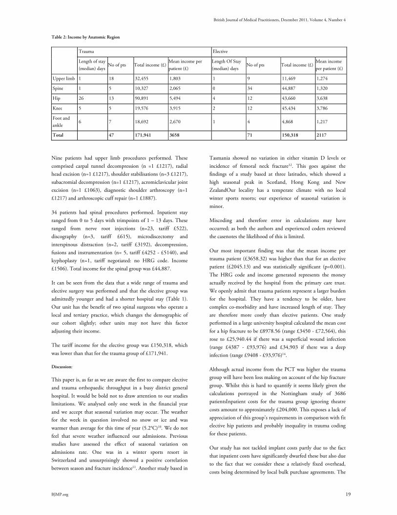
British Journal of Medical Practitioners, December 2011, Volume 4, Number 4
BJMP.org
Table 2: Income by Anatomic Region
Trauma Elective
Length of stay
(median) days No of pts Total income (£)
Mean income per
patient (£)
Length Of Stay
(median) days No of pts Total income (£)
Mean income
per patient (£)
Upper limb 1 18 32,455 1,803 1 9 11,469 1,274
Spine 1 5 10,327 2,065 0 34 44,887 1,320
Hip 26 13 90,891 5,494 4 12 43,660 3,638
Knee 5 5 19,576 3,915 2 12 45,434 3,786
Foot and
ankle 6 7 18,692 2,670 1 4 4,868 1,217
Total
47 171,941 3658
71 150,318 2117
Nine patients had upper limb procedures performed. These
comprised carpal tunnel decompression (n =1 £1217), radial
head excision (n=1 £1217), shoulder stabilisations (n=3 £1217),
subacromial decompression (n=1 £1217), acromiclavicular joint
excision (n=1 £1063), diagnostic shoulder arthroscopy (n=1
£1217) and arthroscopic cuff repair (n=1 £1887).
34 patients had spinal procedures performed. Inpatient stay
ranged from 0 to 5 days with trimpoints of 1 – 13 days. These
ranged from nerve root injections (n=23, tariff £522),
discography (n=3, tariff £615), microdiscectomy and
interspinous distraction (n=2, tariff £3192), decompression,
fusions and instrumentation (n= 5, tariff £4252 - £5140), and
kyphoplasty (n=1, tariff negotiated: no HRG code. Income
£1506). Total income for the spinal group was £44,887.
It can be seen from the data that a wide range of trauma and
elective surgery was performed and that the elective group was
admittedly younger and had a shorter hospital stay (Table 1).
Our unit has the benefit of two spinal surgeons who operate a
local and tertiary practice, which changes the demographic of
our cohort slightly; other units may not have this factor
adjusting their income.
The tariff income for the elective group was £150,318, which
was lower than that for the trauma group of £171,941.
Discussion:
This paper is, as far as we are aware the first to compare elective
and trauma orthopaedic throughput in a busy district general
hospital. It would be bold not to draw attention to our studies
limitations. We analysed only one week in the financial year
and we accept that seasonal variation may occur. The weather
for the week in question involved no snow or ice and was
warmer than average for this time of year (5.2°C)10. We do not
feel that severe weather influenced our admissions. Previous
studies have assessed the effect of seasonal variation on
admissions rate. One was in a winter sports resort in
Switzerland and unsurprisingly showed a positive correlation
between season and fracture incidence11. Another study based in
Tasmania showed no variation in either vitamin D levels or
incidence of femoral neck fracture12. This goes against the
findings of a study based at three latitudes, which showed a
high seasonal peak in Scotland, Hong Kong and New
ZealandOur locality has a temperate climate with no local
winter sports resorts; our experience of seasonal variation is
minor.
Miscoding and therefore error in calculations may have
occurred; as both the authors and experienced coders reviewed
the casenotes the likelihood of this is limited.
Our most important finding was that the mean income per
trauma patient (£3658.32) was higher than that for an elective
patient (£2045.13) and was statistically significant (p=0.001).
The HRG code and income generated represents the money
actually received by the hospital from the primary care trust.
We openly admit that trauma patients represent a larger burden
for the hospital. They have a tendency to be older, have
complex co-morbidity and have increased length of stay. They
are therefore more costly than elective patients. One study
performed in a large university hospital calculated the mean cost
for a hip fracture to be £8978.56 (range £3450 - £72,564), this
rose to £25,940.44 if there was a superficial wound infection
(range £4387 - £93,976) and £34,903 if there was a deep
infection (range £9408 - £93,976)14.
Although actual income from the PCT was higher the trauma
group will have been loss making on account of the hip fracture
group. Whilst this is hard to quantify it seems likely given the
calculations portrayed in the Nottingham study of 3686
patientsInpatient costs for the trauma group ignoring theatre
costs amount to approximately £204,000. This exposes a lack of
appreciation of this group’s requirements in comparison with fit
elective hip patients and probably inequality in trauma coding
for these patients.
Our study has not tackled implant costs partly due to the fact
that inpatient costs have significantly dwarfed these but also due
to the fact that we consider these a relatively fixed overhead,
costs being determined by local bulk purchase agreements. The
19

British Journal of Medical Practitioners, December 2011, Volume 4, Number 4
BJMP.org
consequence on overall study outcome would be minimal given
that trauma implants are several orders of magnitude cheaper
than elective joint prostheses.
It became apparent to us during the course of our study that
trauma can be under resourced when compared with elective
care. The background team currently provided for trauma
patients include the on call medical team (Consultant
Orthopaedic Surgeon, Specialist Registrar and Senior House
officer). In addition there are ward nursing staff, anaesthetist,
theatre staff, occupational therapists and physiotherapists. On
the elective side there are 4 waiting list clerks, 3 surgical
assistants, 3 preoperative clinic sisters as well as reception staff
and the background medical team (anaesthetist, consultant
orthopaedic surgeon, specialist registrar and senior house
officer). In the elective setting the aim is identification and
optimisation of comorbidities pre-operatively and discharge
planning to ensure throughput and turnover of patients. We
admit that pre admission screening is not applicable to trauma
but faster throughput could ensure improved efficiency and
reduced duration of stay.
Our elective patients have a 30-bed ward with an additional 8-
bed day case unit; the trauma ward has 24 inpatient beds. The
elective unit has 7 registered nurses and 4 health care assistants;
on the trauma ward this figure is 4 and 3 respectively. Our
elective patients have 2.5 full time equivalent physiotherapists
whilst our trauma patients have 1.5.
This situation is probably not dissimilar to the situation in
many units elsewhere in the country. This work has shown that
trauma income is higher than that for elective work and from
this we can infer that if resources were directed accordingly then
length of stay could be reduced and profit could be a possibility.
A recent paper using hospital episode statistics (HESS) data has
shown that length of stay fell quickly once payment by results
was implemented15. What was unclear was whether this
represented a real change in efficiencies or simply a change in
data manipulation by trusts. HESS data has repeatedly been
noted to be inaccurate with a range from 10 to 98% dependent
on region and disease group.16-17. In a 2006 statement by the
then Health Minister Mr. A Burnham it was quoted that £88m
pounds was being wasted from 390,000 extra unnecessary bed
days18. This was based on the cost of an elective bed being £225
per day with acute beds being significantly more (approximately
£320 in one study)The total stay for 66 elective patients was
118 days whereas that for 48 trauma patients was 637 days.
Several outliers hugely increased the figure for trauma. Ten
trauma patients represented 464 days of inpatient care. If the
inpatient stay was reduced by one day for fractured neck of
femur patients alone, this amounts to 500 less days per year and
approximately £160,000 per year reduction in overhead costs
for the trust.
One study in the USA assessed the use of a caseworker to
expedite discharge for elderly patients with hip fractures19. The
study did not utilise extra physiotherapy and occupational
therapy support. Findings were increased theatre, anaesthetic
and blood product costs in elderly patients. Increasing age did
not correlate with length of stay, cost of stay or income for the
hospital. They found that a case manager did reduce the average
stay but did not reduce the overall cost. The NHS would do
well to note these findings - in many trusts patient flow
practitioners are being employed to try and expedite discharge
and increase patient turnover. We feel that this money could be
channelled into rehabilitation services to effect prompt
rehabilitation and discharge.
One final issue is the variation in income between secondary
and tertiary centres for certain injuries. One acetabular fracture
underwent fixation generating £4262. If this had been referred
to a tertiary centre a supplementary specialised service code
would have been applicable generating more income (up to
70% in some cases) when intervention was identical. We agree
that certain injuries require tertiary treatment by a team with
high volume experience and specialised skills. There is an
income chasm between the income generated between
secondary and tertiary centres for the same injury, which seems
perverse.
Overall trauma income was higher than elective income, but
still ran at a loss. This was on account of the length of stay of
the hip fracture patients and current coding underestimating
their true cost to the trust. There is a disparity between
rehabilitation services provided for trauma and elective patients,
which needs to be addressed to improve efficiency.
Competing Interests
None declared
Author Details
HUSSAIN ANTHONY KAZI, MBChB(Hons)BSc(Hons)FRCS(Tr&Orth),
Specialist Registrar.
ASHUTOSH ACHARYA, MS(Orth),FRCS,MCh(Orth),FRCS (Trauma &
Orth) Consultant Orthopaedic Surgeon, Warrington & Halton Hospitals NHS
Foundation Trust, Department of Orthopaedic Surgery Warrington Hospital,
United Kingdom
CORRESSPONDENCE: HUSSAIN ANTHONY KAZI, Specialist Registrar,
Warrington & Halton Hospitals NHS Foundation Trust, Department of
Orthopaedic Surgery, Warrington Hospital, Lovely Lane, Warrington, Cheshire,
WA5 1QG, United Kingdom
Email: [email protected]
REFERENCES
1. Department of Health. Delivering the NHS Plan:next steps on
investment: next steps on reform.
http://www.dh.gov.uk/en/Publicationsandstatistics/Publications/Public
ationsPolicyAndGuidance/Browsable/DH_4097252 (date last accessed
15/04/2009).
2. Newbold D. How to save the NHS. Nurs Stand 2006;20:20-3.
3. The information centre. HRG4 questionaire: the debrief report.
www.ic.nhs.uk/webfiles/Services/casemix/prev%20HRG4/
HRG4%20questionnaire%202006.doc (date last accessed 15/04/2009).
4. NHS Information Authority: clinical Coding Instruction Manual.
September 2000.
www.connectingforhealth.nhs.uk/systemsandservices/data/clinicalcodin
g/ codingstandards/publications/ccim (date last accessed 15/04/2009).
20
British Journal of Medical Practitioners, December 2011, Volume 4, Number 4
BJMP.org
5. No authors listed. Department of Health - Payment by Results
Background, June 2009.
http://www.dh.gov.uk/en/Managingyourorganisation/Financeandplann
ing/NHSFinancialReforms/DH_077259(date last accessed
15/02/2010).
6. Department of Health. The NHS Improvement Plan: Putting People
at the Heart of Public Services, The Stationary Office 24th June 2004.
ISBN: 0-10-162682-7.
7. Department of Health. Chief Executive Report to NHS. DoH, June
2006.
http://www.dh.gov.uk/en/Publicationsandstatistics/Publications/Public
ationsPolicyAndGuidance/DH_4135795 (date last accessed
15/04/2009).
8. Davies R. On the road to success delivering shorter elective pathways
for patients. London 7th May 2009. Department of Health.
http://www.18weeks.nhs.uk/Asset.ashx?path=/Orthopaedics/2009_MS
K_events/RobinDavis_LondonMSK_070509.pdf(Date last accessed
5th December 2009).
9. British Orthopaedic Association Manpower Census 2008. BOA
5th June 2009, available from:
http://www.boa.ac.uk/en/publications/orthopaedic-manpower-census/.
(Date last accessed 12/02/2010).
10. BBC Weather Summary – February
2008.http://www.bbc.co.uk/weather/ukweather/year_review/reviews/fe
bruary2008_review_england.shtml. Accessed 18/03/2009.
11. Matter-Walstra K, Widmer M, Busato A. Seasonal variation in
orthopaedic health services utilisation in Switzerland: the impact of
winter sports tourism. BMC Health Serv Res 2006 mar 3;6:25.
12. Inderjeeth CA, Barrett T, Al-Lahham Y, Mulford J, Nicklason F,
Reberger C. Seasonal variation, hip fracture and vitamin D levels in
Southern Tasmania. N Z Med J 2002 Apr 26;115(1152):183-5.
13. Douglas S, Bunyan A, Chiu KH, Twaddle B, Maffuli N. Seasonal
variation in hip fracture at three latitudes. Injury 2000 Jan;31(1):11-9.
14. Edwards C, Counsell A, Boulton C, Moran CG. Early infection after
hip fracture surgery. J Bone Joint Surg [Br] 2008;90-B:770-7.
15. Farrar S, Yi D, Sutton M, Chalkley M et al. Has payment by results
affected the way that English hospitals are providing care? Difference in
difference analysis. BMJ 2009;339:b3047.
16. Williams JG, Mann RY. Hospital episode statistics, time for clinicians
to get involved? Clin Med JRCPL 2002;2:34-37.
17. Korner E. First Report to the Secretary of State of the steering group
on health services information. London: HMSO, 1987.
18. Department of Health. News Release 12th June 2006: NHS could
save more than £700m by increasing productivity.
http://www.dh.gov.uk/en/Publicationsandstatistics/Pressreleases/DH_4
135963(date last accessed 19/04/2009).
19. Jacobs MJ, Markel DC. Geriatric intertrochanteric hip fractures: an
economic analysis. Am J Orthop 1999:Oct;28(10)573-6.
21
British Journal of Medical Practitioners, December 2011, Volume 4, Number 4
BJMP.org
BJMP 2011;4(4):a438
Older people with long-term mental illness. A survey in a community rehabilitation
service using the Camberwell Assessment of Needs for the Elderly (CANE)
Saoud Sultan, Dirk Claassen and Stephen Stansfeld
ABSTRACT Aims: Following the Royal College of Psychiatrists' recommendations, assessments were carried out by the community rehabilitation team in Newham,
East London, using the Camberwell Assessment of Needs for the Elderly (CANE) to assess the needs and quality of service provided to all patients over the
age of 65 years.
Results: 49 patients were screened using the CANE. The majority of needs appeared to be met by the current service provision. However, certain needs
remained unmet: daytime activity (in 36.7% of patients), lack of company (in 22.4% of patients), help with eyesight and hearing difficulties (in 20.4% of
patients) and help with money and budgeting (in 18.4 % of patients) were the most prominent.
Implications: Whereas most psychiatric and practical needs were well met, service planning needs to focus on outreach day activity and befriending services.
Mental health services need to closely monitor physical problems which are more specific to older people e.g. eyesight and hearing, and include these in
their care plans.
Introduction
The Royal College of Psychiatrists defines the 'graduates' as
people who have had enduring or episodic severe mental
disorder in adulthood and have reached the age of 65 years.
Estimates of the most severely affected range from 11 to 60 per
100 000.1 This group of people seems to be uniquely disabled
by a combination of social, mental health and physical
disadvantages and there is a risk of falling between general
adult, rehabilitation services and old age psychiatry.2
There has been an ongoing debate about identifying the best
practice in the management of this group of patients who often
have spent most of their lives in the old psychiatric asylums.
The recommendations include identifying all graduates within
the service followed by a full assessment of the patients' health
and social care needs and the implementation of a care plan to
meet these needs, to be reviewed at least annually. According to
the report, the medical responsibility will rest with a principal
in general practice or a consultant psychiatrist, and maintenance
of continuous review should be the responsibility of the case
manager.1
The Recovery and Rehabilitation Team (RRT) in Newham was
founded in 1988 to facilitate the discharge of groups of patients
from Goodmayes hospital (Essex). Patients discharged to
residential care units and other supported schemes usually had
spent many years in the institution and the team's remit after
relocation into the community was mainly monitoring of
mental health by conducting multiprofessional reviews in the
care homes, crisis intervention, and the promotion of social
networks and leisure activities. Over the following years, the
team also received many referrals from Community Mental
Health Teams (CMHT) for continuing care of people suffering
from long term and severe mental illness. Today, a considerable
proportion of these patients have 'graduated' into old age and
the current percentage of the total caseload is now nearly 25%.
Our survey was carried out following an independent review by
the Health and Social Care Advisory Service (HASCAS) in
January 2005 for the rehabilitation services provided in the
London Borough of Newham. The recommendations included
an assessment of needs for all patients 65 years of age or over,
using the Camberwell Assessment of Needs for the Elderly
(CANE) .3This is a comprehensive needs assessment tool
suitable for use in a variety of settings. It has been successfully
used for older people in primary care, sheltered
accommodation, residential homes, nursing homes, and mental
health services for older people. However, it has not been used
before to specifically assess the needs of older people who have
graduated within the general adult mental health or
rehabilitation services. CANE was found to be a valid and
reliable tool and easy to use by different professions.4
Method:
The RRT database was searched for all patients aged 65 years or
over. This yielded 52 names, who were then approached
between June and September 2005 for a comprehensive
assessment after an explanation about the survey. CMHTs were
asked for numbers of graduates in their services, obtained from
the respective databases.
The CANE is a structured, 24-item questionnaire covering
different areas (see table 2), including social, psychological,
mental health and physical needs. It is easily applicable by
different professions and requires on average about one hour of
assessment time. It measures met and unmet needs and obtains
Research A
rticle
22
British Journal of Medical Practitioners, December 2011, Volume 4, Number 4
BJMP.org
views from patients, carers, staff and the rater. Assessments were
carried out by members of the multi-disciplinary team that
consists of a consultant psychiatrist, the team manager, two
senior clinical medical officers, two clinical psychologists, two
occupational therapists, two social workers, five community
psychiatric nurses and four community support workers. All
raters had received a one day training provided by Juanita Hoe,
one of the contributors in producing the CANE.
The collected data were analysed using Microsoft Excel.
Results
The total number of patients aged 65 years and above under the
care of the rehabilitation services was 52 (24.5% of the total
caseload of 212 patients). There were a further ten patients
under the care of the adult CMHTs in Newham. Attempts
were also made to determine the number of the graduates under
the care of mental health services for older people, but these
were unsuccessful.
Out of the 52 patients, 50 could be assessed using the CANE,
two patients declined the assessment and the assessment sheet of
one patient could not be traced, giving a total of 49 patients
and a response rate of 79% of all known 'graduate' patients
under the care of adult mental health services.
Results describing patient characteristics including mean age,
gender, type of accommodation and diagnosis, are summarized
in Table 1.
Table: 1 Demographic Details
Variable
Mean Age (years)
72.16
Gender (n(%))
Female 16(32.65%)
Male 33(67.34%)
Type of accommodation (n(%))
Residential care 25(51%)
Supported accommodation 13(26.53%)
Private accommodation 12(24.48%)
Diagnosis (n(%))
Schizophrenia 33(67.34%)
Schizoaffective Disorder 6(12.24%)
Bipolar Affective Disorder 5(10.20%)
Depression 2(4.08%)
Personality Disorder 1(2.04)
OCD 1(2.04%)
Dysthymic Disorder 1(2.04%)
Nearly two-thirds of patients were female, three-quarters of this
population were living in supported living or residential care
and 90% were suffering from a severe mental illness (two-thirds
from schizophrenia).
The met and unmet needs of this population are described in
table 2.
Regarding unmet needs, the highest value (nearly 37%) was on
daytime activities, which 18/49 people scored. This is followed
by company (22.5%), which was a problem for 11 people.
Eyesight or hearing also scored strongly (20.5%), followed by
money (18.4%) and different problems in areas such as food
and self-care, physical health and psychological distress (each
12%). Problems with suicidal behaviour and drug or alcohol
abuse were not evident in terms of unmet needs.
Discussion
Our results show that the majority of needs identified by the
CANE were adequately met by the current service provision or
were only identified as unmet needs by a tiny minority (table
2). Since the vast majority of the patients were living in either
residential or supported accommodation (25.51% and 26.53%
respectively), items associated with domestic needs and activities
appeared to be met to a great extent, e.g. accommodation
(44.90% no need, 44.90% met need).
In terms of items related to mental state, the majority of
patients seemed to be satisfactorily managed and receiving
appropriate treatment. The raised number of patients who
suffered from psychological distress could be explained by other
psychosocial factors such as lack of daytime activities and lack of
company which have been identified as the major unmet needs
in our population.
A recent article,5 named risk of harm, unpredictability of
behaviour, poor motivation, lack of insight and low public
acceptability as the major reasons for social disability. However,
in our review, over one-third of people clearly expressed the
wish for more daytime activities, where the named disabilities
might prevent a more active and satisfied lifestyle. In the
interviews, it transpired that people mostly wished for an
outreach service providing social contact, befriending and
activities. The majority of people in our population seemed to
be rather reluctant to access general facilities, like day centres
for the elderly.
As we have assessed most of the patients under the care of adult
mental health services, this survey should be able to inform
service planning about the needs of this population. The
development of an outreach service offering day time activities
including a befriending component could be a challenge for the
responsible service providers, e.g. social services, adult
community mental health services and old age psychiatry.
23
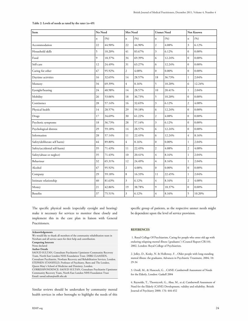
British Journal of Medical Practitioners, December 2011, Volume 4, Number 4
BJMP.org
Table 2: Levels of needs as rated by the rater (n=49)
Item No Need Met Need Unmet Need Not Known
n (%) n (%) n (%) n (%)
Accommodation 22 44.90% 22 44.90% 2 4.08% 3 6.12%
Household skills 5 10.20% 41 83.67% 3 6.12% 0 0.00%
Food 9 18.37% 34 69.39% 6 12.24% 0 0.00%
Self-care 12 24.49% 31 63.27% 6 12.24% 0 0.00%
Caring for other 47 95.92% 2 4.08% 0 0.00% 0 0.00%
Daytime activities 16 32.65% 14 28.57% 18 36.73% 1 2.04%
Memory 34 69.39% 4 8.16% 5 10.20% 6 12.24%
Eyesight/hearing 24 48.98% 14 28.57% 10 20.41% 1 2.04%
Mobility 26 53.06% 18 36.73% 5 10.20% 0 0.00%
Continence 28 57.14% 16 32.65% 3 6.12% 2 4.08%
Physical health 14 28.57% 29 59.18% 6 12.24% 0 0.00%
Drugs 17 34.69% 30 61.22% 2 4.08% 0 0.00%
Psychotic symptoms 18 36.73% 28 57.14% 3 6.12% 0 0.00%
Psychological distress 29 59.18% 14 28.57% 6 12.24% 0 0.00%
Information 28 57.14% 11 22.45% 6 12.24% 4 8.16%
Safety(deliberate self harm) 44 89.80% 4 8.16% 0 0.00% 1 2.04%
Safety(accidental self-harm) 35 71.43% 11 22.45% 2 4.08% 2 4.08%
Safety(abuse or neglect) 35 71.43% 10 20.41% 4 8.16% 1 2.04%
Behaviour 32 65.31% 12 24.49% 4 8.16% 1 2.04%
Alcohol 47 95.92% 2 4.08% 0 0.00% 0 0.00%
Company 29 59.18% 8 16.33% 11 22.45% 1 2.04%
Intimate relationship 40 81.63% 3 6.12% 4 8.16% 2 4.08%
Money 21 42.86% 19 38.78% 9 18.37% 0 0.00%
Benefits 37 75.51% 3 6.12% 4 8.16% 5 10.20%
The specific physical needs (especially eyesight and hearing)
make it necessary for services to monitor these closely and
implement this in the care plan in liaison with General
Practitioners.
Acknowledgements
We would like to thank all members of the community rehabilitation team in
Newham and all service users for their help and contribution.
Competing Interests
None declared
Author Details
SAOUD SULTAN, Consultant Psychiatrist Upminster Community Recovery
Team, North East London NHS Foundation Trust. DIRK CLAASSEN,
Consultant Psychiatrist, Newham Recovery and Rehabilitation Services, London.
STEPHEN STANSFELD, Professor of Psychiatry, Barts and The London,
Queen Mary’s School of Medicine and Dentistry, London.
CORRESSPONDENCE: SAOUD SULTAN, Consultant Psychiatrist Upminster
Community Recovery Team, North East London NHS Foundation Trust
Email: [email protected]
Similar reviews should be undertaken by community mental
health services in other boroughs to highlight the needs of this
specific group of patients, as the respective unmet needs might
be dependent upon the level of service provision.
REFERENCES
1. Royal College Of Psychiatrists, Caring for people who enter old age with
enduring relapsing mental illness (‘graduates`) (Council Report CR110).
2002, London: Royal College of Psychiatrists.
2. Jolley, D., Kosky, N. & Holloway, F. , Older people with long-standing
mental illness: the graduates, Advances in Psychiatric Treatment, 2004; 10:
29-34
3. Orrell, M., & Hancock, G. , CANE: Camberwell Assessment of Needs
for the Elderly, London: Gaskell 2004
4. Reynolds, T., Thornicroft, G., Abas, M., et al, Camberwell Assessment of
Need for the Elderly (CANE) Development, validity and reliability. British
Journal of Psychiatry 2000; 176: 444-452
24

British Journal of Medical Practitioners, December 2011, Volume 4, Number 4
BJMP.org
5. Karim, S. & Burns, A. Invited commentary on: Older people with long-
standing mental illness: the graduates. Advances in Psychiatric Treatment
2004; 10: 34-36
25

British Journal of Medical Practitioners, December 2011, Volume 4, Number 4
BJMP.org
BJMP 2011;4(4):a443
Latest diagnosis and management of diverticulitis
Stephen O’Neill, Phillip Ross, Philip McGarry and Satheesh Yalamarthi
Abstract Diverticular disease is extremely common especially amongst the elderly. It mainly presents as sigmoid diverticulitis but there is potential for serious
complications. In the acute setting Computed Tomography is the gold standard investigation and helps classify the stage. Evidence to support outpatient
treatment of uncomplicated diverticulitis is appearing however hospital admission and treatment with intravenous antibiotics is often required and is highly
effective. The decision to proceed with elective surgery is judged on an individual basis with a long-term conservative approach suitable for most. For
elective surgery there is evidence to advocate a laparoscopic approach. In Hinchey stage III or IV disease, laparotomy followed by either a Hartmann’s
procedure or ideally, a resection followed by primary anastomosis may be required. Radiologically guided drainage of an abscess is an established alternative
and laparoscopic lavage is another less invasive option that has emerged. Following successful acute medical management, colonoscopy is usually performed
several weeks after resolution to rule out other colonic pathology.
Keywords: Diverticulitis, Diagnosis, Management, Surgery
Introduction
A colonic diverticulum is defined as a sac-like protrusion of
mucosa through the muscular component of the colonic wall1.
The terms “diverticulosis” and “diverticular disease” are used to
express the presence of diverticula without associated
inflammation. While the term “diverticulitis” indicates there is
inflammation of a diverticulum or diverticula, which is
commonly accompanied by either microscopic or macroscopic
perforation2.
In the developed world, diverticular disease of the colon is
widespread and in those aged over 65 years of age it is present
in greater than 65%3. The incidence increases dramatically with
time and while only 5% of the western population are affected
in the fifth decade this rises steeply to over 50% by the eight
decade and 60% in the ninth 4.
Although diverticulosis is extremely common, complications
requiring surgery only occur in 1% of patients overall 5 and
10% of those admitted to hospital as an emergency for
treatment6. Despite this, there is a substantial healthcare burden
inflicted by diverticular disease and within the United States
alone it accounts for 312,000 hospital admissions, 1.5 million
days of inpatient treatment and a total estimated cost of 2.6
billion dollars per annum 7.
The aetiology of the diverticulosis is poorly understood but it is
probably a multi-factorial process involving dietary habits
(specifically low fibre intake) as well as changes in colonic
pressure, motility and wall structure that are associated with
ageing8. The pathogenesis of diverticulitis is also uncertain,
however stasis or obstruction in a narrow necked diverticulum
leading to overgrowth of pathogens and local tissue ischemia is
thought likely 2.
This review will discuss the common presentations,
investigations and current treatment strategies utilised in the
management of acute diverticulitis and its complications as well
as providing an up to date synopsis of existing
recommendations for follow up and prevention.
Symptoms and Signs
In Western nations, diverticula are most commonly situated in
the left colon9 and 99% of patients will have some element of
sigmoid involvement10. Therefore patients commonly present
with sigmoid diverticulitis that typically displays features of left
iliac fossa pain and fever with raised inflammatory markers (see
below). Physical exam will disclose left lower quadrant
peritonism for simple disease, but in complicated cases physical
examination findings may reveal a palpable abdominal mass,
evidence of fistulas or obstruction, or widespread peritonitis11.
In cases of complicated diverticulosis, a stricture may lead to
obstructive symptoms with complaints of nausea, vomiting and
distension being present. If a fistula has developed, a history of
recurrent urinary tract infection, pneumaturia and faecaluria
may also be elicited12. In a female with a previous history of
hysterectomy suspicion will be further raised as colovesical and
colovaginal fistulas are rare in females with their uterus in place.
If a patient reports passing stools per vagina, insertion of a
vaginal speculum and inspection may confirm this latter
diagnosis12.
Differential diagnosis
The differential diagnosis for diverticulitis and its complications
is extensive and includes irritable bowel syndrome,
inflammatory bowel disease, ischaemic or infective colitis, pelvic
inflammatory disease and malignancy. It is obviously most
Review Article
26

British Journal of Medical Practitioners, December 2011, Volume 4, Number 4
BJMP.org
imperative to exclude the latter differential 4, particularly in the
case of a stricture that is impassable on colonoscopy, as many of
these specimens following resection (32% in one series13) will
transpire to be adenocarcinoma4. It should also be noted that
sigmoid diverticulitis may also masquerade as acute appendicitis
if the colon is long and redundant or otherwise situated within
the abdomen or pelvis such that the inflamed segment lies in
the suprapubic region, right iliac fossa or McBurney’s point2.
Complications
Although diverticulosis is present in nearly two thirds of the
elderly population, the vast majority of patients will remain
entirely asymptomatic. Even so, an estimated 20% of those
affected will manifest symptomatology, mainly as diverticulitis,
but potentially with further complications of perforation,
abscesses, fistulas, and obstruction, as well as bleeding per
rectum6.
The European Association for Endoscopic Surgeons (EAES)
developed a classification scheme based upon the severity of
diverticulitis, which broadly classifies patients into either simple
symptomatic or complicated disease (Table 1)14. Where an
abscess or perforation develops the Hinchey classification is
used as a staging tool and can provide prognostic information
on the likely outcome (Table 2)15.
Table 1 - European Association for Endoscopic Surgeons classification
system for diverticulitis 14
Grade of
disease
Clinical
explanation of
grade
Clinical state of the patient
I
Symptomatic
uncomplicated
disease
Pyrexia, abdominal pain, CT findings
consistent with diverticulitis
II
Recurrent
symptomatic
disease
Recurrence of Grade I
III Complicated
disease
Bleeding, abscess formation, phlegmon,
colonic perforation, purulent and faecal
peritonitis, stricturing, fistula and
obstruction
Table 2 – Hinchey classification of perforated diverticulitis 15
Hinchey
stage Features of disease
Risk of
death71
Stage I* Diverticulitis with a pericolic abscess 5%
Stage II** Diverticulitis with a distant abscess (this may
be retroperitoneal or pelvic) 5%
Stage III Purulent peritonitis 13%
Stage IV Faecal peritonitis 43%
*Stage I has been divided into Ia Phlegmon and Ib confined pericolic
abscess in later modifications38, 72
** Stage II has been divided into IIa abscesses amenable to percutaneous
drainage and IIb complex abscess with or without fistula in later
modifications14, 73
Perforation is probably the most feared complication and the
annual prevalence of perforated diverticulitis within a northern
European population is currently thought to stand at 3.8 per
100,000 of the population, which is a figure that is increasing16.
Despite this only 1-2% of patients who attend for urgent
assessment and treatment will have a gross perforation2 but for
80% this will be their first presentation so a high index of
suspicion is still required17.
Blood investigations
In clinical practice, inflammatory markers, commonly the
White Blood Cell (WBC) count and C-Reactive Protein (CRP)
level, are frequently employed to assist in diagnosing
diverticulitis and its complications. In a recent retrospective
study, a White Blood Cell (WBC) count >10,000/µL was
present in 62% of patients with Computed Tomography (CT)
confirmed diverticulitis and the presence of leukocytosis was
significantly more common in patients with diverticulitis and
associated perforation than without (86% v 65%, p=0.01)18.
CRP has also been shown to be of considerable benefit in the
diagnosis of acute left sided colonic diverticulitis 19. A recently
established diagnostic nomogram with a reported accuracy of
86% that was developed to improve the clinical diagnosis of
diverticulitis includes an elevated CRP >50mg/l as well other
variables including age, previous episodes, aggravation of pain
on movement, absence of vomiting and localization of
symptoms and tenderness in the left iliac fossa19.
In addition, it has been demonstrated that in acute sigmoid
diverticulitis a CRP below 50mg/l is unlikely to correlate with
an associated perforation (negative predictive value 79%) while
a CRP above 200mg/l is an indicator that the patient may have
a perforation (positive predictive value 69%)20. In this latter
study, CRP also had the highest diagnostic accuracy in
diagnosing perforation in acute sigmoid diverticulitis across a
range of parameters assessed that included WBC count as well
as less commonly used tests like bilirubin and alkaline
phosphatase20.
Imaging investigations
In the acute phase of diverticulitis the extent of the extramural
component of inflammation is more important than the degree
of the intramural inflammation and as such CT associated with
the use of intravenous and oral contrast and, in ideal
conditions, rectal contrast is the gold standard means of
investigation21.
CT can accurately identify extra-luminal complications such as
an abscess, phlegmon, adjacent organ involvement, or fistula, as
well as recognising other alternative diagnoses such as
appendicitis, pelvic inflammatory disease, tubo-ovarian abscess
or inflammatory bowel disease22.
27
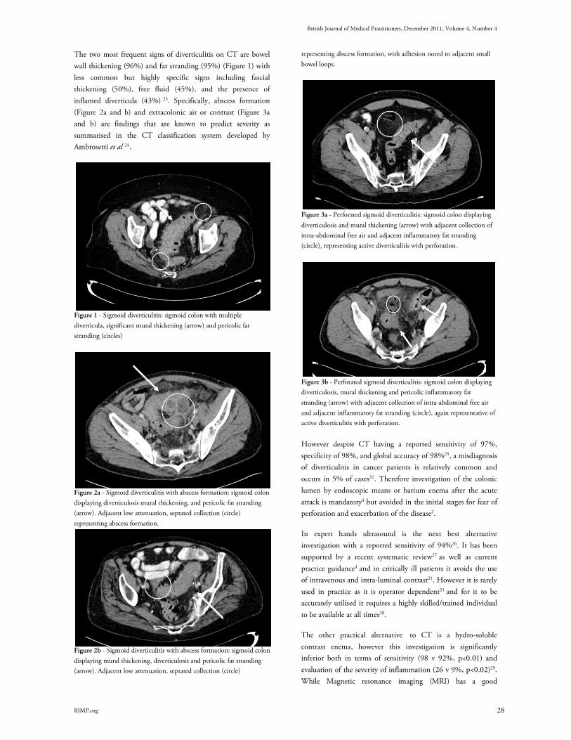
British Journal of Medical Practitioners, December 2011, Volume 4, Number 4
BJMP.org
The two most frequent signs of diverticulitis on CT are bowel
wall thickening (96%) and fat stranding (95%) (Figure 1) with
less common but highly specific signs including fascial
thickening (50%), free fluid (45%), and the presence of
inflamed diverticula (43%) 23. Specifically, abscess formation
(Figure 2a and b) and extracolonic air or contrast (Figure 3a
and b) are findings that are known to predict severity as
summarised in the CT classification system developed by
Ambrosetti et al 24.
Figure 1 - Sigmoid diverticulitis: sigmoid colon with multiple
diverticula, significant mural thickening (arrow) and pericolic fat
stranding (circles)
Figure 2a - Sigmoid diverticulitis with abscess formation: sigmoid colon
displaying diverticulosis mural thickening, and pericolic fat stranding
(arrow). Adjacent low attenuation, septated collection (circle)
representing abscess formation.
Figure 2b - Sigmoid diverticulitis with abscess formation: sigmoid colon
displaying mural thickening, diverticulosis and pericolic fat stranding
(arrow). Adjacent low attenuation, septated collection (circle)
representing abscess formation, with adhesion noted to adjacent small
bowel loops.
Figure 3a - Perforated sigmoid diverticulitis: sigmoid colon displaying
diverticulosis and mural thickening (arrow) with adjacent collection of
intra-abdominal free air and adjacent inflammatory fat stranding
(circle), representing active diverticulitis with perforation.
Figure 3b - Perforated sigmoid diverticulitis: sigmoid colon displaying
diverticulosis, mural thickening and pericolic inflammatory fat
stranding (arrow) with adjacent collection of intra-abdominal free air
and adjacent inflammatory fat stranding (circle), again representative of
active diverticulitis with perforation.
However despite CT having a reported sensitivity of 97%,
specificity of 98%, and global accuracy of 98%25, a misdiagnosis
of diverticulitis in cancer patients is relatively common and
occurs in 5% of cases21. Therefore investigation of the colonic
lumen by endoscopic means or barium enema after the acute
attack is mandatory4 but avoided in the initial stages for fear of
perforation and exacerbation of the disease2.
In expert hands ultrasound is the next best alternative
investigation with a reported sensitivity of 94%26. It has been
supported by a recent systematic review27 as well as current
practice guidance4 and in critically ill patients it avoids the use
of intravenous and intra-luminal contrast21. However it is rarely
used in practice as it is operator dependent21 and for it to be
accurately utilised it requires a highly skilled/trained individual
to be available at all times28.
The other practical alternative to CT is a hydro-soluble
contrast enema, however this investigation is significantly
inferior both in terms of sensitivity (98 v 92%, p<0.01) and
evaluation of the severity of inflammation (26 v 9%, p<0.02)29.
While Magnetic resonance imaging (MRI) has a good
28

British Journal of Medical Practitioners, December 2011, Volume 4, Number 4
BJMP.org
sensitivity of 94% and a specificity of 87%30, in the acute
setting it may be impractical both in terms of examination time
and patient co-operation21. Finally, laparoscopy can also be
helpful for diagnostic purposes but again in practical terms,
with the increasing availability of cross-sectional imaging, it is
rarely required for this purpose4.
Outpatient treatment
Evidence for successful and economical outpatient treatment of
uncomplicated diverticulitis is beginning to emerge. In a
prospective study of 70 patients classified on the basis of an
ultrasound examination as having mild-to-moderate acute
colonic diverticulitis (as defined by either limited inflammation
within a diverticulum extending up to an abscess < 2 cm in
diameter), 68 patients were successfully treated with oral
antibiotics with an initial liquid diet and this led to a cost saving
on inpatient treatment of 80%31.
In a further retrospective analysis, among a cohort of patients
who were referred for outpatient treatment it was found that
such treatment was effective for 94% of patients, with women
and those with free fluid on CT scan appearing to be at higher
risk for treatment failure32.
In reality the prospect of outpatient treatment in uncomplicated
cases of acute diverticulitis is determined largely by access to the
necessary investigative tools for accurate diagnosis and staging
of disease, the general fitness of the patient, their ability to
maintain adequate oral intake, the possibility of further
outpatient review, patient compliance with medications,
satisfactory social support and ability to plan for endoscopic
follow up21.
In broad terms, if symptoms are not severe and the patient has
no significant co-morbidities and is compliant with medical
treatment, then a course of broad spectrum antibiotics can be
administered orally on an outpatient basis and the patient
followed up at subsequent outpatient clinics. However if the
patient is systemically unwell, elderly, has significant co-
morbidities or there are any other concerns it is safer to arrange
for a hospital admission and treatment with intravenous
antibiotics12.
Conservative inpatient treatment
Simple diverticulitis requiring hospital admission is usually
treated by rehydration, symptomatic relief and intravenous
antibiotics. Most patients with uncomplicated disease respond
well to medical treatment and generally experience significant
improvement in their abdominal pain, temperature and
inflammatory markers within two days of initiation of
antibiotic treatment33. If this is not the case or there is clinical
concern a repeat CT is advocated and operative intervention or
percutaneous drainage considered (see below)2.
It should be noted at this stage while the use of broad spectrum
antibiotics in acute uncomplicated diverticulitis is supported by
guidelines34 there is no actual evidence mandating the routine
use of antibiotics in mild uncomplicated diverticulitis35 and in
some European countries it is not routine36.
High-quality evidence regarding the most effective type of
antibiotic is also lacking35. However anaerobic bacteria (usually
bacteroides, clostridium, fusobacterium and
peptostreptococcus) are the most commonly cultured organisms
with gram-negative aerobes, especially Escherichia coli, and
facultative gram-positives, such as streptococci, often grown as
well37. Therefore coverage against both Gram-negative and
anaerobic bacteria is widely advocated2 21 38.
If combination antibiotics are selected, Metronidazole provides
excellent anaerobic cover with less risk of clostridium
difficle infection than alternatives4. However use of single agent
may be more cost effective39. Local protocols are likely to
influence selection but the patient may be safely switched from
intravenous to oral therapy when they can tolerate a diet and
oral medicines22 as intravenous antibiotics are not felt to be
vastly superior40. Seven to ten days of antibiotic therapy is an
acceptable treatment period22 however evidence is emerging to
support shorter courses41.
Elective surgery
In a recent position statement from the Association of
Coloproctology of Great Britain and Ireland (ASCPGBI) it was
concluded that the majority of patients, whether young or old,
presenting with acute diverticulitis could be managed with a
conservative, medical approach in the longer term. Previous
blanket recommendations for elective resection e.g. following
two acute episodes of diverticulitis14 were challenged in this
statement and it was proposed that the decision on elective
resection should be made on an individual basis4. The
traditional practice of waiting for a period of 4-6 weeks after a
diverticulitis attack before performing an elective operation was
not disputed12.
Surgery in the elective setting can be by either an open or
laparoscopic technique with a recent randomised trial
identifying a 27% reduction in major morbidity42 along with
less pain, improved quality of life and shorter hospitalization at
the cost of a longer operating time with the laparoscopic
approach43. In expert centres conversion rates as low as 2.8%
and median hospitals stays of 4 days can be achieved44 and
individual case reports of resections using single laparoscopic
port access have also emerged45. However if a laparoscopic
resection is considered, it is currently recommended that
patients should be treated after full recovery from the acute
episode of inflammation as there is evidence to suggest lower
complication and conversion rates can be achieved4.
The principles for both approaches are the same. A colorectal
anastomosis is a predictor of lower recurrence rates after elective
29

British Journal of Medical Practitioners, December 2011, Volume 4, Number 4
BJMP.org
sigmoid resection for uncomplicated diverticulitis46. Therefore
it is recommended that the distal resection margin is taken onto
the rectum as opposed to the distal sigmoid and the splenic
flexure is fully mobilised to facilitate this4, however in the case
of a long redundant left colon this may not be necessary12. The
proximal resection margin is less clear but should be made onto
soft compliant bowel4 34. Often it is possible to identify the
ureters intra-operatively however, there may be cases of
complicated diverticulitis in which the extent and degree of
inflammatory changes warrant the use of pre-operatively placed
ureteric stents to help aid their identification and avoid injury12.
Emergency surgery for complicated diverticulitis
The indications for emergency operative intervention in acute
diverticulitis include the presence of generalised peritonitis,
uncontained visceral perforation, gross uncontrollable sepsis, a
large undrainable or inaccessible abscess, bowel obstruction and
lack of improvement or clinical deterioration with initial
medical management 2.
Historically, perforated diverticulitis was treated with a three-
stage procedure consisting of faecal diversion with a stoma,
resection of the diseased segment of bowel, followed by
takedown of the stoma and restoration of intestinal continuity.
This then shifted to performing a Hartmann’s procedure which
includes a primary resection of the diseased segment and end
colostomy followed by subsequent colostomy reversal at a
second operation11. In this case reconstruction generally
involves a second laparotomy because although laparoscopic
reconstruction is effective, it is infrequently performed47-48. As a
result reversal is often permanently deferred.
In selected cases the ideal therapeutic option in colonic
perforation is a one-stage procedure with resection followed by
primary anastomosis, which adds the benefits of being a
definitive treatment with the avoidance of the morbidity and
mortality associated with a stoma and its reversal49. A protective
ileostomy after resection and primary anastomosis is viewed as a
valid additional step in patients at high risk of an anastomotic
leak (immunosuppression, American Society of
Anaesthesiologists (ASA) grade IV, faecal peritonitis)21 but a
Hartmann’s procedure may also be selected.
Particularly in cases where there is a stricture causing
obstruction and significant faecal loading, a resection in
conjunction with on-table colonic lavage and primary
anastomosis may be used. This technique has also been
described as facilitating a primary anastomosis in the case of a
perforation50. However in certain patients with obstruction
depending on the viability of the proximal colon a subtotal
colectomy with ileorectal anastomosis may be required12 and
because small-bowel obstruction may also occur, especially in
the presence of a large diverticular abscess, this may also warrant
further treatment2.
The use of endoscopic colonic stenting as a treatment of acute
obstruction of the large bowel secondary to colonic cancer has
been well documented in the literature either as a definitive
procedure or as a bridge to surgery and can effectively
decompresses the obstructed colon in 90% of cases51. However
the use of stents in benign disease is less well documented , with
it used mainly as a bridge to surgery52 and because is associated
with a higher incidence of complications in acute diverticular
disease53 it cannot as yet be recommended.
Laparoscopic surgery in the emergency setting
There have been a number of recent reports of laparoscopic
lavage with or without the placement of an intra-abdominal
drain for patients with acute diverticulitis and perforation, with
the reported advantages including the avoidance of an acute
resection and the possibility of a stoma 4. The evidence that has
been produced thus far to support its case is highly promising.
A recent systematic review of laparoscopic lavage for perforated
colonic diverticulitis identified two prospective cohort studies,
nine retrospective case series and two case reports with
231patients and the vast majority of patients (77%) had
Hinchey grade III purulent peritonitis. Laparoscopic peritoneal
lavage successfully controlled abdominal and systemic sepsis in
95.7% of patients, mortality was 1.7%, morbidity 10.4% and
only four (1.7%) patients received a colostomy54.
In the largest series in the literature to date, Myers et al reported
100 patients with perforated diverticulitis and generalised
peritonitis. Eight patients with Hinchey IV disease required
conversion to an open procedure, with the overall mortality
being 4% and recurrence rates only 2% over a median time
period of 36 months55.
Percutaneous therapy
The appropriate management of diverticular abscesses is a
matter of some debate. However according to the American
Society of Colon and Rectal Surgeons (ASCRS) radiologically
guided percutaneous drainage is usually the most appropriate
treatment for patients with a large diverticular abscess as it
avoids the need for emergency surgery and possibility of a
colostomy34.
When the abscess diameter is over 5 cm, percutaneous CT
guided drainage, in combination with antibiotics, is the
standard treatment and offers rapid improvement in symptoms
in over 90% of cases, albeit with a high recurrence rate in more
severe cases38 and higher likelihood of surgery being needed in
those involving the pelvis56.
In practical terms diverticular abscesses less than 3 cm in
diameter usually cannot be successfully drained, as the diameter
of the pigtail of most drainage catheters will be a similar
dimension28. Also for smaller abscesses21, especially those less
than 2cm resolution usually occurs with the use intravenous
30

British Journal of Medical Practitioners, December 2011, Volume 4, Number 4
BJMP.org
antibiotics alone34. However if a drain is sited it is advisable that
before it is removed, resolution of the abscess should be
confirmed and a potential bowel fistula excluded by a further
contrast study28.
Finally, diverticular disease of the colon is also a relatively
common cause of acute lower gastrointestinal bleeding and is in
fact the diagnosis in 23% of cases57. This usually settles with
conservative management but if the bleeding is profuse
angiography and endovascular intervention may be helpful,
with surgery very rarely required for this indication4.
Follow up
Following successful medical management of an acute episode
of diverticulitis, colonoscopy, flexible sigmoidoscopy or barium
enema should be performed several weeks after the resolution of
symptoms to confirm the diagnosis and rule out other colonic
pathology such as malignancy, inflammatory bowel disease, or
ischemia22.
Following surgery there is reported to be a high incidence of the
order of 25% for recurrent symptoms, which is put down to the
diagnostic overlap that exists with irritable bowel syndrome58.
However any suspicion of recurrent diverticulitis following
surgical resection should be confirmed by CT scan after which
antibiotic treatment should be initiated, as for a case of primary
uncomplicated disease12. If this is excluded the high incidence
(17.6%) of symptomatic anastomotic stenosis after elective
laparoscopic sigmoidectomy should be borne in mind with the
possibility of endoscopic dilatation considered if applicable59.
Summary points
• CT scan is the gold standard means of investigation for
acute diverticulitis and helps classify the stage of disease.
• Evidence to support outpatient treatment of uncomplicated
diverticulitis is beginning to appear, however hospital
admission and treatment with broad spectrum intravenous
antibiotics is often required and is highly effective.
• The decision to proceed with elective surgery is judged on
an individual basis and there is evidence gathering to
advocate a laparoscopic approach.
• In Hinchey stage III or IV disease, emergency laparotomy
followed by either a Hartmann’s procedure or ideally in
selected patients a resection followed by primary
anastomosis may be required.
• In certain cases percutaneous radiologically guided drainage
of abscesses is an established alternative to open surgery
with laparoscopic lavage another less invasive and highly
promising option.
Lifestyle modifications and prevention
Following treatment weight loss, rationalisation of certain
medications and exercise are recommended as obesity is
significantly associated with an increased incidence of both
diverticular bleeding and diverticulitis60, as are non-steroidal
anti-inflammatory drugs and paracetamol61, with physical
activity significantly associated with a reduction in the risk of
complications62.
Whilst dietary fibre, particularly cellulose63, is
recommended22 the evidence that supports these
recommendations is not particularly strong64. However
foodstuffs such as nuts, seeds, popcorn and corn that are usually
discouraged have no evidence to support the theory that they
may lead to increased complications65.
Small studies without control groups suggest that probiotics
may have a positive effect on the recurrence of symptomatic
diverticular disease66-67. Long term administration of the non-
absorbable antibiotic Rifaxamin has also been used with
reported success68 as has the anti-inflammatory mesalazine69.
However none of these medications have a strong evidence base
and as a result are not in routine use70.
Competing Interests None declared Author Details S O’NEILL, Surgical Registrar MB BCh BAO (dist) MSc (dist) MRCSEd
Department of Colorectal Surgery, Queen Margaret Hospital, Dunfermline.
P ROSS, Medical Student, Queens University, Belfast. P MCGARRY, Radiology
Registrar MB BCh BAO (dist), Department of Radiology, Altnagelvin Hospital,
Londonderry. S YALAMARTHI, Consultant Surgeon MS FRCS, Department of
Colorectal Surgery, Queen Margaret Hospital, Dunfermline
CORRESSPONDENCE: Mr STEPHEN O’NEILL, 6 Dean Park Mews,
Edinburgh EH4 1EF.
Email: [email protected]
REFERENCES
1. Young-Fadok TM, Roberts PL, Spencer MP et al. Colonic diverticular
disease. Curr Probl Surg 2000;37(7):457-514.
2. Jacobs DO. Clinical practice. Diverticulitis. N Engl J Med
2007;357(20):2057-66.
3. West AB. The pathology of diverticulitis. J Clin Gastroenterol
2008;42(10):1137-8.
4. Fozard JB, Armitage NC, Schofield JBet al. ACPGBI position statement
on elective resection for diverticulitis. Colorectal Dis 2011;13 Suppl 3:1-11.
5. Roberts PL, Veidenheimer MC. Current management of diverticulitis.
Adv Surg 1994;27:189-208.
6. Stollman NH, Raskin JB. Diverticular disease of the colon. J Clin
Gastroenterol 1999;29(3):241-52.
7. Etzioni DA, Mack TM, Beart RW, Jr.et al. Diverticulitis in the United
States: 1998-2005: changing patterns of disease and treatment. Ann Surg
2009;249(2):210-7.
8. Heise CP. Epidemiology and pathogenesis of diverticular disease. J
Gastrointest Surg 2008;12(8):1309-11.
9. Vermeulen J, van der Harst E, Lange JF. Pathophysiology and prevention
of diverticulitis and perforation. Neth J Med 2010;68(10):303-9.
10. Hughes LE. Postmortem survey of diverticular disease of the colon. II.
The muscular abnormality of the sigmoid colon. Gut 1969;10(5):344-51.
11. Lopez DE, Brown CV. Diverticulitis: the most common colon
emergency for the acute care surgeon. Scand J Surg 2010;99(2):86-9.
12. Stocchi L. Current indications and role of surgery in the management of
sigmoid diverticulitis. World J Gastroenterol 2010;16(7):804-17.
13. King DW, Lubowski DZ, Armstrong AS. Sigmoid stricture at
colonoscopy--an indication for surgery. Int J Colorectal Dis 1990;5(3):161-
3.
14. Kohler L, Sauerland S, Neugebauer E. Diagnosis and treatment of
diverticular disease: results of a consensus development conference. The
31
British Journal of Medical Practitioners, December 2011, Volume 4, Number 4
BJMP.org
Scientific Committee of the European Association for Endoscopic Surgery.
Surg Endosc 1999;13(4):430-6.
15. Hinchey EJ, Schaal PG, Richards GK. Treatment of perforated
diverticular disease of the colon. Adv Surg 1978;12:85-109.
16. Makela J, Kiviniemi H, Laitinen S. Prevalence of perforated sigmoid
diverticulitis is increasing. Dis Colon Rectum 2002;45(7):955-61.
17. Hart AR, Kennedy HJ, Stebbings WSet al. How frequently do large
bowel diverticula perforate? An incidence and cross-sectional study. Eur J
Gastroenterol Hepatol 2000;12(6):661-5.
18. Mounday A, Aubin C, Lewis L. Absence of fever and elevated white
blood count does not exclude the diagnosis of diverticulitis as determined by
CT in the emergency department. Ann Emerg Med 2007;50(3):S77.
19. Andeweg CS, Knobben L, Hendriks JCet al. How to diagnose acute left-
sided colonic diverticulitis: proposal for a clinical scoring system. Ann Surg
2011;253(5):940-6.
20. Kaser SA, Fankhauser G, Glauser PMet al. Diagnostic value of
inflammation markers in predicting perforation in acute sigmoid
diverticulitis. World J Surg 2010;34(11):2717-22.
21. Biondo S, Borao JL, Millan Met al. Current status of the treatment of
acute colonic diverticulitis: a systematic review. Colorectal Dis 2011.
22. Beckham H, Whitlow CB. The medical and nonoperative treatment of
diverticulitis. Clin Colon Rectal Surg 2009;22(3):156-60.
23. Kircher MF, Rhea JT, Kihiczak Det al. Frequency, sensitivity, and
specificity of individual signs of diverticulitis on thin-section helical CT with
colonic contrast material: experience with 312 cases. AJR Am J Roentgenol
2002;178(6):1313-8.
24. Ambrosetti P, Grossholz M, Becker Cet al. Computed tomography in
acute left colonic diverticulitis. Br J Surg 1997;84(4):532-4.
25. Werner A, Diehl SJ, Farag-Soliman Met al. Multi-slice spiral CT in
routine diagnosis of suspected acute left-sided colonic diverticulitis: a
prospective study of 120 patients. Eur Radiol 2003;13(12):2596-603.
26. Ripolles T, Agramunt M, Martinez MJet al. The role of ultrasound in
the diagnosis, management and evolutive prognosis of acute left-sided
colonic diverticulitis: a review of 208 patients. Eur Radiol
2003;13(12):2587-95.
27. Liljegren G, Chabok A, Wickbom Met al. Acute colonic diverticulitis: a
systematic review of diagnostic accuracy. Colorectal Dis 2007;9(6):480-8.
28. Baker ME. Imaging and interventional techniques in acute left-sided
diverticulitis. J Gastrointest Surg 2008;12(8):1314-7.
29. Ambrosetti P, Jenny A, Becker Cet al. Acute left colonic diverticulitis--
compared performance of computed tomography and water-soluble contrast
enema: prospective evaluation of 420 patients. Dis Colon Rectum
2000;43(10):1363-7.
30. Heverhagen JT, Sitter H, Zielke Aet al. Prospective evaluation of the
value of magnetic resonance imaging in suspected acute sigmoid
diverticulitis. Dis Colon Rectum 2008;51(12):1810-5.
31. Mizuki A, Nagata H, Tatemichi Met al. The out-patient management of
patients with acute mild-to-moderate colonic diverticulitis. Aliment
Pharmacol Ther 2005;21(7):889-97.
32. Etzioni DA, Chiu VY, Cannom RRet al. Outpatient treatment of acute
diverticulitis: rates and predictors of failure. Dis Colon Rectum
2010;53(6):861-5.
33. Evans J, Kozol R, Frederick Wet al. Does a 48-hour rule predict
outcomes in patients with acute sigmoid diverticulitis? J Gastrointest Surg
2008;12(3):577-82.
34. Rafferty J, Shellito P, Hyman NHet al. Practice parameters for sigmoid
diverticulitis. Dis Colon Rectum 2006;49(7):939-44.
35. de Korte N, Unlu C, Boermeester MAet al. Use of antibiotics in
uncomplicated diverticulitis. Br J Surg 2011;98(6):761-7.
36. de Korte N, Klarenbeek BR, Kuyvenhoven JPet al. Management of
diverticulitis. Results of a survey among gastroenterologists and surgeons.
Colorectal Dis 2011.
37. Brook I, Frazier EH. Aerobic and anaerobic microbiology in intra-
abdominal infections associated with diverticulitis. J Med Microbiol
2000;49(9):827-30.
38. Kaiser AM, Jiang JK, Lake JPet al. The management of complicated
diverticulitis and the role of computed tomography. Am J Gastroenterol
2005;100(4):910-7.
39. Kellum JM, Sugerman HJ, Coppa GFet al. Randomized, prospective
comparison of cefoxitin and gentamicin-clindamycin in the treatment of
acute colonic diverticulitis. Clin Ther 1992;14(3):376-84.
40. Ridgway PF, Latif A, Shabbir Jet al. Randomized controlled trial of oral
vs intravenous therapy for the clinically diagnosed acute uncomplicated
diverticulitis. Colorectal Dis 2009;11(9):941-6.
41. Schug-Pass C, Geers P, Hugel Oet al. Prospective randomized trial
comparing short-term antibiotic therapy versus standard therapy for acute
uncomplicated sigmoid diverticulitis. Int J Colorectal Dis 2010;25(6):751-9.
42. Klarenbeek BR, Bergamaschi R, Veenhof AAet al. Laparoscopic versus
open sigmoid resection for diverticular disease: follow-up assessment of the
randomized control Sigma trial. Surg Endosc 2011;25(4):1121-6.
43. Klarenbeek BR, Veenhof AA, Bergamaschi Ret al. Laparoscopic sigmoid
resection for diverticulitis decreases major morbidity rates: a randomized
control trial: short-term results of the Sigma Trial. Ann Surg
2009;249(1):39-44.
44. Jones OM, Stevenson AR, Clark Det al Laparoscopic resection for
diverticular disease: follow-up of 500 consecutive patients. Ann Surg
2008;248(6):1092-7.
45. Leroy J, Cahill RA, Asakuma Met al. Single-access laparoscopic
sigmoidectomy as definitive surgical management of prior diverticulitis in a
human patient. Arch Surg 2009;144(2):173-9; discussion 79.
46. Thaler K, Baig MK, Berho Met al. Determinants of recurrence after
sigmoid resection for uncomplicated diverticulitis. Dis Colon Rectum
2003;46(3):385-8.
47. Caselli G, Bambs C, Pinedo Get al. [Laparoscopic approach for
intestinal passage reconstruction after Hartmann's operation: experience
with 30 patients]. Cir Esp 2010;88(5):314-8.
48. Siddiqui MR, Sajid MS, Baig MK. Open vs laparoscopic approach for
reversal of Hartmann's procedure: a systematic review. Colorectal Dis
2010;12(8):733-41.
49. Trenti L, Biondo S, Golda Tet al. Generalized peritonitis due to
perforated diverticulitis: Hartmann's procedure or primary anastomosis? Int
J Colorectal Dis 2011;26(3):377-84.
50. Biondo S, Perea MT, Rague JM et al. One-stage procedure in non-
elective surgery for diverticular disease complications. Colorectal Dis
2001;3(1):42-5.
51. Khot UP, Lang AW, Murali Ket al. Systematic review of the efficacy and
safety of colorectal stents. Br J Surg 2002;89(9):1096-102.
52. Small AJ, Young-Fadok TM, Baron TH. Expandable metal stent
placement for benign colorectal obstruction: outcomes for 23 cases. Surg
Endosc 2008;22(2):454-62.
53. Forshaw MJ, Sankararajah D, Stewart Met al. Self-expanding metallic
stents in the treatment of benign colorectal disease: indications and
outcomes. Colorectal Dis 2006;8(2):102-11.
54. Toorenvliet BR, Swank H, Schoones JW et al. Laparoscopic peritoneal
lavage for perforated colonic diverticulitis: a systematic review. Colorectal
Dis 2010;12(9):862-7.
55. Myers E, Hurley M, O'Sullivan GCet al. Laparoscopic peritoneal lavage
for generalized peritonitis due to perforated diverticulitis. Br J Surg
2008;95(1):97-101.
56. Ambrosetti P, Chautems R, Soravia Cet al. Long-term outcome of
mesocolic and pelvic diverticular abscesses of the left colon: a prospective
study of 73 cases. Dis Colon Rectum 2005;48(4):787-91.
57. Machicado GA, Jensen DM. Acute and chronic management of lower
gastrointestinal bleeding: cost-effective approaches. Gastroenterologist
1997;5(3):189-201.
58. Egger B, Peter MK, Candinas D. Persistent symptoms after elective
sigmoid resection for diverticulitis. Dis Colon Rectum 2008;51(7):1044-8.
59. Ambrosetti P, Francis K, De Peyer Ret al Colorectal anastomotic stenosis
after elective laparoscopic sigmoidectomy for diverticular disease: a
prospective evaluation of 68 patients. Dis Colon Rectum 2008;51(9):1345-
9.
32
British Journal of Medical Practitioners, December 2011, Volume 4, Number 4
BJMP.org
60. Strate LL, Liu YL, Aldoori WHet al. Obesity increases the risks of
diverticulitis and diverticular bleeding. Gastroenterology 2009;136(1):115-
22 e1.
61. Aldoori WH, Giovannucci EL, Rimm EBet al. Use of acetaminophen
and nonsteroidal anti-inflammatory drugs: a prospective study and the risk
of symptomatic diverticular disease in men. Arch Fam Med 1998;7(3):255-
60.
62. Strate LL, Liu YL, Aldoori WHet al. Physical activity decreases
diverticular complications. Am J Gastroenterol 2009;104(5):1221-30.
63. Aldoori W, Ryan-Harshman M. Preventing diverticular disease. Review
of recent evidence on high-fibre diets. Can Fam Physician 2002;48:1632-7.
64. Unlu C, Daniels L, Vrouenraets BCet al. A systematic review of high-
fibre dietary therapy in diverticular disease. Int J Colorectal Dis 2011.
65. Strate LL, Liu YL, Syngal Set al. Nut, corn, and popcorn consumption
and the incidence of diverticular disease. JAMA 2008;300(8):907-14.
66. Fric P, Zavoral M. The effect of non-pathogenic Escherichia coli in
symptomatic uncomplicated diverticular disease of the colon. Eur J
Gastroenterol Hepatol 2003;15(3):313-5.
67. Tursi A, Brandimarte G, Giorgetti GMet al. Mesalazine and/or
Lactobacillus casei in maintaining long-term remission of symptomatic
uncomplicated diverticular disease of the colon. Hepatogastroenterology
2008;55(84):916-20.
68. Colecchia A, Vestito A, Pasqui Fet al Efficacy of long term cyclic
administration of the poorly absorbed antibiotic Rifaximin in symptomatic,
uncomplicated colonic diverticular disease. World J Gastroenterol
2007;13(2):264-9.
69. Di Mario F, Aragona G, Leandro Get al. Efficacy of mesalazine in the
treatment of symptomatic diverticular disease. Dig Dis Sci 2005;50(3):581-
6.
70. Maconi G, Barbara G, Bosetti Cet al. Treatment of diverticular disease
of the colon and prevention of acute diverticulitis: a systematic review. Dis
Colon Rectum 2011;54(10):1326-38.
71. Schwesinger WH, Page CP, Gaskill HV, 3rdet al. Operative
management of diverticular emergencies: strategies and outcomes. Arch Surg
2000;135(5):558-62; discussion 62-3.
72. Wasvary H, Turfah F, Kadro Oet al. Same hospitalization resection for
acute diverticulitis. Am Surg 1999;65(7):632-5; discussion 36.
73. Sher ME, Agachan F, Bortul Met al. Laparoscopic surgery for
diverticulitis. Surg Endosc 1997;11(3):264-7.
33
British Journal of Medical Practitioners, December 2011, Volume 4, Number 4
BJMP.org
BJMP 2011;4(4):a439
Massive Hepatic Necrosis due to Hepatic Abscesses after Transplantation
Seif Fadi , Gholam Pierre and Montenegro Hugo
ABSTRACT Delayed hepatic artery thrombosis (HAT) is a rare complication of orthotopic liver transplantion (OLT) that may present with biliary sepsis or remain
asymptomatic. Sonography is extremely sensitive for the detection of HAT in symptomatic patients during the immediate postoperative period. Magnetic
resonance angiography (MRA) is a useful adjunct in patients with indeterminate ultrasound exams, those who have renal insufficiency or an allergy to
iodinated contrast.
In the absence of hepatic failure, conservative treatment appears to be effective for patients with HAT. Whole-graft orthotopic liver transplantation (OLT),
Roux-en-Y biliary reconstruction, cold ischaemia and operative times, the use of blood and plasma, and the use of aortic conduits in arterial reconstruction
are all risk factors associated with HAT.
We present a patient with a case of delayed HAT three years after liver transplantation who presented with mild symptoms and was later found to have a
massive hepatic abscess and significant necrosiswith positive cultures forClostridium perfringens. There was evidence of complete occlusion of the hepatic
artery 2 cm from its origin.
Introduction:
The clinical features of early HAT are well defined, yet the
features of delayed HAT are less clear. Delayed HAT is a rare
complication of OLT that may present with biliary sepsis or
remain asymptomatic. Sonography is extremely sensitive for the
detection of HAT in symptomatic patients during the
immediate postoperative period. However, the sensitivity of
ultrasonography diminishes as the interval between
transplantation and diagnosis of HAT increases due to collateral
arterial flow. MRA is a useful adjunct in patients with
indeterminate ultrasound exams and in those who have renal
insufficiency or an allergy to iodinated contrast.
In the absence of hepatic failure, conservative treatment appears
to be effective for patients with HAT but retransplantation may
be necessary as a definitive treatment.
Case Presentation:
A 52 year old male with a history of whole graft OLT for
primary sclerosing cholangitis presented with two days of fever,
nausea, and mild abdominal discomfort.
One week prior to presentation, he was seen in the liver clinic
for regular follow-up. At that time, he was totally asymptomatic
and his laboratory workup including liver function tests were
within normal range.
He has undergone OLT three years prior. At the time of
transplant he required transfusion of 120 units of packed red
blood cells, 60 units of fresh frozen plasma and 100 units of
platelets due to extensive intraoperative bleeding secondary to
chronic changes of pancreatitis and severe portal hypertension,
but had an otherwise uneventful postoperative recovery.
On physical examination the temperature was 39C, heart rate
was 125 beats per minute, respiratory rate was 22 bpm. Initial
laboratory workup revealed a white blood cell count of
25,000/mm3, AST of 6230 U/L, ALT of 2450 U/L, total
bilirubin of 11 mg/dL , BUN 55 mg/dL and Creatinine of 4.5
mg/dL. Lactate level was 5 mmol/L. Doppler ultrasonography
revealed an extensive intrahepatic gas (Image 1A). Computed
tomography of the abdomen and pelvis revealed extensive area
of hepatic necrosis with abscess formation measuring 19x14 cm
with extension of gas into the peripheral portal vein branches
(Image 1B,C). Upon admission to the hospital, the patient
required endotracheal intubation, mechanical ventilator support
and aggressively fluid resuscitation. He was started on broad-
spectrum antibiotics and a percutaneous drain was placed that
drained dark, foul smelling fluid. Cultures from the blood and
the drain grew Clostridium perfringens.
Magnetic resonance imaging (MRI), MRA revealed occlusion of
the hepatic artery 2 cm from its origin and also evidence of
collaterals (Image 2A,B).
Following drain placement, the patient’s clinical condition
markedly improved with significant reduction of liver function
test values. Retransplantation was considered but delayed in the
setting of infection and significant clinical and laboratory
testing improvement.
The patient was transferred to the medical floor in stable
condition, and the drain was then removed.
A week later the patient developed low grade fevers and
tachycardia. One day later he began to experience mild
abdominal discomfort and high grade fevers. Repeat CT of the
abdomen revealed worsening hepatic necrosis and formation of
Case Report
34
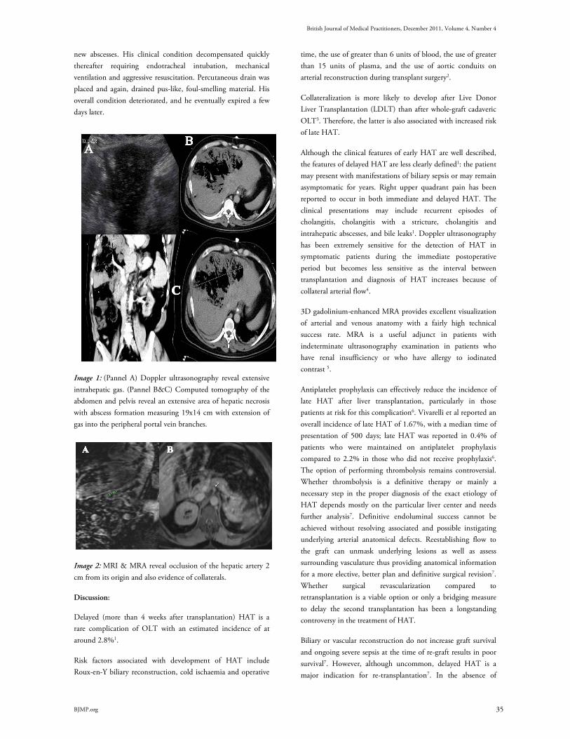
British Journal of Medical Practitioners, December 2011, Volume 4, Number 4
BJMP.org
new abscesses. His clinical condition decompensated quickly
thereafter requiring endotracheal intubation, mechanical
ventilation and aggressive resuscitation. Percutaneous drain was
placed and again, drained pus-like, foul-smelling material. His
overall condition deteriorated, and he eventually expired a few
days later.
Image 1: (Pannel A) Doppler ultrasonography reveal extensive
intrahepatic gas. (Pannel B&C) Computed tomography of the
abdomen and pelvis reveal an extensive area of hepatic necrosis
with abscess formation measuring 19x14 cm with extension of
gas into the peripheral portal vein branches.
Image 2: MRI & MRA reveal occlusion of the hepatic artery 2
cm from its origin and also evidence of collaterals.
Discussion:
Delayed (more than 4 weeks after transplantation) HAT is a
rare complication of OLT with an estimated incidence of at
around 2.8%1.
Risk factors associated with development of HAT include
Roux-en-Y biliary reconstruction, cold ischaemia and operative
time, the use of greater than 6 units of blood, the use of greater
than 15 units of plasma, and the use of aortic conduits on
arterial reconstruction during transplant surgery2.
Collateralization is more likely to develop after Live Donor
Liver Transplantation (LDLT) than after whole-graft cadaveric
OLT3. Therefore, the latter is also associated with increased risk
of late HAT.
Although the clinical features of early HAT are well described,
the features of delayed HAT are less clearly defined1: the patient
may present with manifestations of biliary sepsis or may remain
asymptomatic for years. Right upper quadrant pain has been
reported to occur in both immediate and delayed HAT. The
clinical presentations may include recurrent episodes of
cholangitis, cholangitis with a stricture, cholangitis and
intrahepatic abscesses, and bile leaks1. Doppler ultrasonography
has been extremely sensitive for the detection of HAT in
symptomatic patients during the immediate postoperative
period but becomes less sensitive as the interval between
transplantation and diagnosis of HAT increases because of
collateral arterial flow4.
3D gadolinium-enhanced MRA provides excellent visualization
of arterial and venous anatomy with a fairly high technical
success rate. MRA is a useful adjunct in patients with
indeterminate ultrasonography examination in patients who
have renal insufficiency or who have allergy to iodinated
contrast 5.
Antiplatelet prophylaxis can effectively reduce the incidence of
late HAT after liver transplantation, particularly in those
patients at risk for this complication6. Vivarelli et al reported an
overall incidence of late HAT of 1.67%, with a median time of
presentation of 500 days; late HAT was reported in 0.4% of
patients who were maintained on antiplatelet prophylaxis
compared to 2.2% in those who did not receive prophylaxis6.
The option of performing thrombolysis remains controversial.
Whether thrombolysis is a definitive therapy or mainly a
necessary step in the proper diagnosis of the exact etiology of
HAT depends mostly on the particular liver center and needs
further analysis7. Definitive endoluminal success cannot be
achieved without resolving associated and possible instigating
underlying arterial anatomical defects. Reestablishing flow to
the graft can unmask underlying lesions as well as assess
surrounding vasculature thus providing anatomical information
for a more elective, better plan and definitive surgical revision7.
Whether surgical revascularization compared to
retransplantation is a viable option or only a bridging measure
to delay the second transplantation has been a longstanding
controversy in the treatment of HAT.
Biliary or vascular reconstruction do not increase graft survival
and ongoing severe sepsis at the time of re-graft results in poor
survival7. However, although uncommon, delayed HAT is a
major indication for re-transplantation7. In the absence of
35

British Journal of Medical Practitioners, December 2011, Volume 4, Number 4
BJMP.org
hepatic failure, conservative treatment appears to be effective for
patients with hepatic artery thrombosis.
C. perfringensis an anaerobic, gram-positive rod frequently
isolated from the biliary tree and gastrointestinal tract.
Inoculation of Clostridium spores into necrotic tissue is
associated with formation of hepatic abscess8.
Necrotizing infections of the transplanted liver are rare. There
have been around 20 cases of gas gangrene or necrotizing
infections of the liver reported in the literature. Around 60% of
these infections were caused by clostridial species with C.
perfringens accounting for most of them. Around 80% of
patients infected with Clostridium died, frequently within hours
of becoming ill9,10. Those who survived underwent prompt
retransplantation and the infection had not resulted in shock or
other systemic changes that significantly decreased the
likelihood of successful retransplantation8.
Because the liver has contact with the gastrointestinal tract via
the portal venous system, intestinal tract bacteria may enter the
liver via translocation across the intestinal mucosa into the
portal venous system. Clostridial species can also be found in
the bile of healthy individuals undergoing cholecystectomy9,10.
The donor liver can also be the source of bacteria. Donors may
have conditions that favor the growth of bacteria in bile or the
translocation of bacteria into the portal venous blood. These
conditions include trauma to the gastrointestinal tract,
prolonged intensive care unit admissions, periods of
hypotension, use of inotropic agents, and other conditions that
increase the risk of potential infection 8,9,10. C. perfringens sepsis
in OLT recipients has been uniformly fatal without emergent
retransplantation. Survival from C. perfringens sepsis managed
without exploratory laparotomy or emergency treatment has
been extremely rarely reported8. In those patients who survived,
and in whom the infection has not resulted in shock or multiple
organ failure, retransplantation may be successful8.
Although our patient survived his intensive care course, his
recovery was tenuous as he quickly developed additional hepatic
abscesses that led to his eventual demise. Post-mortem
examination in our patient revealed intra-hepatic presence
of Clostridium perfringens.
He was managed conservatively since he markedly improved
both clinically and by liver function tests. Because of this,
retransplantation was delayed. He was also already on
antiplatelet prophylaxis.
Conclusion:
We report an interesting case of Clostridium
perfringens hepatic abscess due to late HAT following OLT.
Although the patient initially improved with non-surgical
treatment, he eventually died. In similar cases, besides aggressive
work-up and medical management retransplantation may be
necessary for a better long term outcome.
Competing Interests
None declared
Author Details
Seif Fadi, M.D. Fellow, Pulmonary Critical Care and Sleep Medicine, University
Hospitals Case Medical Center, Case Western Reserve University School of
Medicine
Gholam Pierre, MD. Associate Professor of Medicine Medical Director,
Hepatology. Division of Gastroenterology and Liver Disease, University Hospitals
Case Medical Center, Case Western Reserve University School of Medicine
Montenegro Hugo, M.D. Professor, Pulmonary Critical Care and Sleep
Medicine, University Hospitals Case Medical Center, Case Western Reserve
University School of Medicine. Louis Stokes Cleveland VA Medical Center.
CORRESSPONDENCE: Montenegro Hugo University Hospitals Case Medical
Center, Case Western Reserve, Case Western Reserve University School of
Medicine. Louis Stokes Cleveland VA Medical Center.
Email: [email protected]
REFERENCES
1. Bhattacharjya S, Gunson BK, Mirza DF, et al. Delayed hepatic artery
thrombosis in adult orthotopic liver transplantation-a 12-year experience.
Transplantation 2001; 71(11):1592-6.
2. Silva M, Jambulingam P, Gunson B, et al. Hepatic artery thrombosis
following orthotopic liver transplantation: A 10-year experience from a
single centre in the United Kingdom. Liver Transplantation 2006; 12:146-
151.
3. Tian MG, Tso WK, Lo CM et al. Treatment of hepatic artery
thrombosis after orthotopic liver transplantation. Asian J Surg. 2004;
27(3):213-7.
4. Horrow M, Blumenthal B, Reich D et al. Sonographic Diagnosis and
Outcome of Hepatic Artery Thrombosis After Orthotopic Liver
Transplantation in Adults. AJR 2007; 189:346-351.
5. Ishigami K, Stolpen A, Al-kass F et al. Diagnostic Value of Gadolinium-
Enhanced 3D Magnetic Resonance Angiography in Patients With Suspected
Hepatic Arterial Complications After Liver Transplantation. Journal of
Computer Assisted Tomography 2005; 29(4):464-471.
6. Vivarelli M, La Barba G, Cucchetti A, et al. Can antiplatelet prophylaxis
reduce
the incidence of hepatic artery thrombosis after liver transplantation? Liver
Transpl 2007; 13:651-654.
7. Perkins J. Thrombolysis for early hepatic artery thrombosis: Definitive
therapy or diagnostic aid? Liver Transplantation 2007; 13:927– 931.
8. Diaz G, Boyer T, Renz J. Survival of Clostridium perfringens. Sepsis in a
Liver Transplant Recipient. Liver Transplantation 2009; 15:1469-1472.
9. Doblecki-Lewis S, Palaios E, Bejarano PA et al. Hepatic gas gangrene
following orthotopic liver transplantation: three cases treated with re-
transplantation and a review of the literature. Transpl Infect Dis.2008;
10:280–285.
10. Eigneberger B, Königsrainer I, Kendziorra H et al. Fulminant liver
failure due to Clostridium perfringens sepsis 9 years after liver
transplantation. Transpl Int.2006; 19:172–173.
36
British Journal of Medical Practitioners, December 2011, Volume 4, Number 4
BJMP.org
BJMP 2011;4(4):a437
Care Programme Approach: first you have to prove you are ill
Francis J Dunne
The Care Programme Approach (CPA) was introduced in
England in 1993 to co-ordinate the care of patients with mental
health disorders.1 Its aim was to ensure that there was a full
assessment of the patient’s needs, that a care co-ordinator would
see that the care was delivered, regular checks would be carried
out to review progress, there would be collaboration between
health and social services, and that patients (or the term used to
‘demedicalise’ them in psychosocial Newspeak 2 namely, ‘service
users’) and carers (they also use the service) would have a greater
say in the written management plan. Targets were set.
What has happened since? Prior to this I recall that most
psychiatrists carried out full assessment needs, regular checks
reviewed progress (outpatients), social services were involved
when necessary, and patients and their families were nearly
always involved in the discussion of after-care, when
appropriate. Despite the condescending manner in which
patients and carers were treated by the hierarchy, i.e. they
would not understand the difference between social services
management and other after-care, it was always quite clear to
doctors that patients had no difficulty with the concepts of
medical intervention (investigations, diagnosis, treatment),
psychological therapies, and social help (housing, work, family,
finances).
Rather than simplifying the process we now have two tiers of
CPA, namely, standard and enhanced. Where the patient has
‘complex needs’ or is a ‘complicated case’ then you are in the
enhanced bracket. For the rest – back to the General
Practitioner (GP)! Not enough resources apparently. Not ill
enough more likely. Remember – you have to have a severe and
enduring mental health disorder – nothing else counts.
Nowadays the GP is expected to be a specialist in mental health
and run a risk assessment on every ‘psychiatric’ patient. The GP
is frowned upon by the ‘experts in living’ should he/she for
example, dare refer a mild or moderately (yes - those
descriptions again!) ill patient to the Mental Health Services.
Because there is no bottomless pit of money, the scenario was
changed in 2008 so that those receiving only standard CPA
were no longer entitled to it. However, not to appear callous
and indifferent to the plight of those suffering from ‘less severe’
mental health problems, the usual lip service was paid to
patients, assuring them that they should be respected and
supported, and that their carers be also recognised as having
‘needs’. All the buzz words were put in place again – integrated
care pathways, working together, reviews about the reviews,
good practice, better training, and so forth. Now there is the
Supervised Community Treatment Order, (whether you like it
or not) and those subject to the new ‘order’ will be entitled to
the ‘new’ CPA. Wonderful in theory.
So what happens to a patient who is not on CPA? We are
informed that such patients should still be open to secondary
mental health services, should continue to receive clinical
support, that reviews should take place regularly, and a social
assessment should be available under the new guidance to local
authorities FACS (Fair Access to Care Services), readily
available on the Internet. The truth of the matter is that only
those patients on enhanced CPA will receive immediate
support, the rest will have to jump through the usual hurdles to
prove they have a severe, enduring mental illness (enduring is
not enough) in order to gain access to NHS ‘support’ facilities.
Some patients are seen as more deserving than others, for
example, those admitted to hospital under the Mental Health
Act (voluntary admission may count against you), current or
potential risk (theoretically, any patient with a mental health
disorder, which seems to defeat the purpose of the exercise) or
the presence of a dual diagnosis (depression with alcoholism, or
is it the other way round?). Anyway, if in doubt, the patient is
entitled to a formal reassessment CPA and may be admitted to
the ‘new’ CPA list. If all fails, the patient (remember, one with
severe, enduring mental health symptoms) may make a
complaint to the local authority or even hire a lawyer.
What is the true state of affairs? To begin with, many patients
have enduring mental health problems which are not severe, are
not life-threatening, and despite the hardship and drudgery
endured, manage to trundle through work, relationships, and
family life. Years of talking therapies or psychotropic
medication, indeed both, may have only taken the edge off their
symptoms. Often symptoms resurge and require alterations or
adjustments in medication; sometimes a different psychological
approach needs to be considered. Such patients are best left to
the fountain of all wisdom, the GP, so it seems. Rather akin to
telling the GP to treat for example, a ‘minor’ cardiac problem
(say, palpitations) because the ‘specialist unit’ only deals with
Viewpoint
37
British Journal of Medical Practitioners, December 2011, Volume 4, Number 4
BJMP.org
severe arrhythmias, severe pain, severe disability, ‘severe
everything’. It is unfair to expect GPs to make informed
decisions concerning psychotropic medication (no more than
they should about adjusting chemotherapy drugs) and most
would be familiar only with specific therapies such as Cognitive
Behavioural Therapy (CBT) or Anger Management, where
appropriate. The type of patients described here comprise the
majority of those seen in outpatients, yet there is now a growing
trend to discharge such patients back to the GP, because he/she
is not ‘care co-ordinated’ on enhanced CPA. The burden is on
the GP. It does not seem to have registered with politicians or
management (doctors included) that chronic schizophrenia is
not the same as chronic gastro-oesophageal reflux.
The trend now is for the setting up of Community Clinics (the
patient does not necessarily get to see a doctor) where ‘all the
other psychiatric problems’ are dealt with. The traditional
psychiatric outpatient department is to be abolished, unless of
course, GPs do something about this torrid state of affairs now.
It could only happen in Psychiatry which seems to me a
specialty doomed to oblivion. Family doctors are becoming
increasingly irritated by a system or discipline (Psychiatry
especially) which seems to ignore their concerns and is more
preoccupied with targets (nothing has changed) and outcomes
(back to the GP). Even referrals from GPs, who want a medical
opinion, are filtered in order to weed out those not worthy to
enter the hallowed walls of the Mental Health Institution.
Those patients who ‘know the system’ or who are vociferous
and make complaints (‘I know my rights’) get to be seen by the
Great and Good. Lesser mortals, usually those with serious
mental illnesses, do not make any undue demands and are
therefore often forgotten or fall by the wayside. A patient with
bipolar disorder on lithium is discharged back to a GP who is
unsure whether or not the medication needs ‘fine tuning’ at
times, should be discontinued, or reinstated were compliance is
a problem in one heading for a relapse. As a corollary of that, I
am sure most hospital doctors would not know what the
acronym ABVD means in the chemotherapy treatment of
Hodgkin’s disease. Adjusting psychotropic medication is not
quite the same as adjusting an antihypertension regime.
Unfortunately, if the patient needs to be referred back into the
system the whole Kafkaesque scenario begins again.
A medical colleague once bemoaned to me that psychiatrists are
totally out of touch with Medicine. Alas, it seems they are also
now out of touch with their medical colleagues.
Competing Interests
None declared
Author Details
FRANCIS J DUNNE, FRCPsych, Consultant Psychiatrist and Honorary Senior
Lecturer, University College London, North East London Foundation Trust,
United Kingdom.
CORRESSPONDENCE: FRANCIS J DUNNE, FRCPsych, Consultant
Psychiatrist and Honorary Senior Lecturer, University College London, North
East London Foundation Trust, United Kingdom.
Email: [email protected]
REFERENCES
1. Refocusing the Care Programme Approach: policy and positive practice
guidance. Department of Health 2008; www.dh.gov.uk
2. Dunne FJ. Psychiatry in limbo: new ways of talking. British Journal of
Medical Practitioners BJMP 2010; 3:( 2):319
38
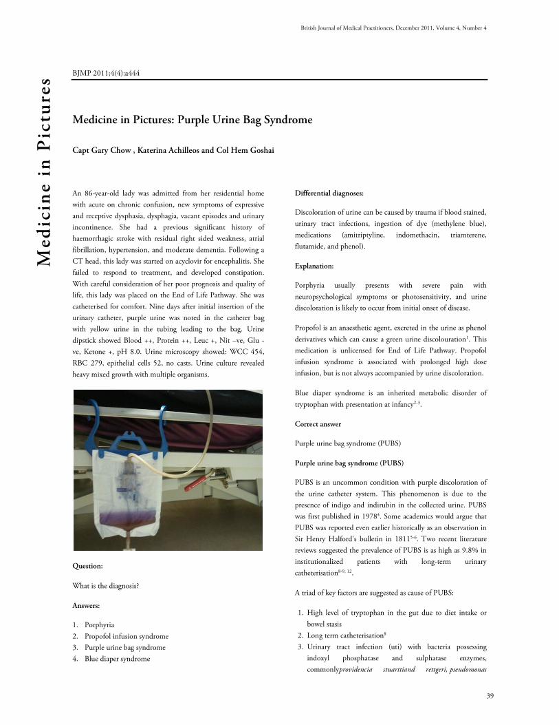
British Journal of Medical Practitioners, December 2011, Volume 4, Number 4
BJMP 2011;4(4):a444
Medicine in Pictures: Purple Urine Bag Syndrome
Capt Gary Chow , Katerina Achilleos and Col Hem Goshai
An 86-year-old lady was admitted from her residential home
with acute on chronic confusion, new symptoms of expressive
and receptive dysphasia, dysphagia, vacant episodes and urinary
incontinence. She had a previous significant history of
haemorrhagic stroke with residual right sided weakness, atrial
fibrillation, hypertension, and moderate dementia. Following a
CT head, this lady was started on acyclovir for encephalitis. She
failed to respond to treatment, and developed constipation.
With careful consideration of her poor prognosis and quality of
life, this lady was placed on the End of Life Pathway. She was
catheterised for comfort. Nine days after initial insertion of the
urinary catheter, purple urine was noted in the catheter bag
with yellow urine in the tubing leading to the bag. Urine
dipstick showed Blood ++, Protein ++, Leuc +, Nit –ve, Glu -
ve, Ketone +, pH 8.0. Urine microscopy showed: WCC 454,
RBC 279, epithelial cells 52, no casts. Urine culture revealed
heavy mixed growth with multiple organisms.
Question:
What is the diagnosis?
Answers:
1. Porphyria
2. Propofol infusion syndrome
3. Purple urine bag syndrome
4. Blue diaper syndrome
Differential diagnoses:
Discoloration of urine can be caused by trauma if blood stained,
urinary tract infections, ingestion of dye (methylene blue),
medications (amitriptyline, indomethacin, triamterene,
flutamide, and phenol).
Explanation:
Porphyria usually presents with severe pain with
neuropsychological symptoms or photosensitivity, and urine
discoloration is likely to occur from initial onset of disease.
Propofol is an anaesthetic agent, excreted in the urine as phenol
derivatives which can cause a green urine discolouration1. This
medication is unlicensed for End of Life Pathway. Propofol
infusion syndrome is associated with prolonged high dose
infusion, but is not always accompanied by urine discoloration.
Blue diaper syndrome is an inherited metabolic disorder of
tryptophan with presentation at infancy2-3.
Correct answer
Purple urine bag syndrome (PUBS)
Purple urine bag syndrome (PUBS)
PUBS is an uncommon condition with purple discoloration of
the urine catheter system. This phenomenon is due to the
presence of indigo and indirubin in the collected urine. PUBS
was first published in 19784. Some academics would argue that
PUBS was reported even earlier historically as an observation in
Sir Henry Halford's bulletin in 18115-6. Two recent literature
reviews suggested the prevalence of PUBS is as high as 9.8% in
institutionalized patients with long-term urinary
catheterisation8-9, 12.
A triad of key factors are suggested as cause of PUBS:
1. High level of tryptophan in the gut due to diet intake or
bowel stasis
2. Long term catheterisation8
3. Urinary tract infection (uti) with bacteria possessing
indoxyl phosphatase and sulphatase enzymes,
commonlyprovidencia stuarttiand rettgeri, pseudomonas
Medicine in Pictures
39

British Journal of Medical Practitioners, December 2011, Volume 4, Number 4
BJMP.org
auruginosa, proteus mirabilis, escherichia coli, klebsiella
pneumoniae,morganella, citrobacter species, group b
streptococci and enterococci8, 13.
It is understood that bowel stasis causes accumulation of
tryptophan, which leads to an increase in urinary indoxyl
sulphate (UIS). In the presence of indoxyl phosphatase and
sulphatase enzyme activities, whilst collected in the catheter
system, UIS is degraded to form a mixture of indigo and
dissolved indirubin in the plastic11, coating the catheter system
with a purple appearance. Intensity of discoloration is deeper
the longer the urine is in contact with the catheter plastic7, 10-12.
The urine does not appear purple prior to entering the catheter.
Recent literature7-8 also suggested female gender, alkaline urine,
bed bound debilitated patient population, PVC material7 and
institutionalization are further predisposing factors of PUBS.
Management of PUBS requires catheter change and treatment
of underlying UTI.
Good catheter hygiene and shorter duration of catheterisation
can reduce PUBS1.
Competing Interests
None declared
Author Details
Capt GARY CHOW, Medical Officer, 2 Medical Regiment, Glyn Hughs Bks,
Hohne. KATERINA ACHILLEOS, MBBS BSc(Hons) MRCP, CT2 Acute
Medicine, Southend University Hospital, UK. Col HEM GOSHAI, MBE
MBCHB BSc, Garrison Clinical Director, Hohne Garrison.
CORRESSPONDENCE: Capt GARY CHOW, Medical Officer, 2 Medical
Regiment, Glyn Hughs Brks, Hohne, BFPO 30.
Email: [email protected]
REFERENCES
1. Foot C, Fraser JF. Uroscopic Rainbow: Modern Matula Medicine.
Postgrad Med J 2006;82(964):126–129.
2. Drummond K, Miachael A, Ulstrom R, et al. The Blue Diaper
Syndrome: Familial Hypercalcemia with Nephrocalcinosis and
Indicanuria; A new familial disease, with definition of the metabolic
abnormality. Am J Med. 1964 Dec;37:928-48.
3. Cochat P, Pichault V, Bacchetta J, et al. NephrolithiasisRelated to
Inborn Metabolic Diseases. Pediatr Nephrol 2010;25(3):415–424.
4. Barlow G, Dickson J. Purple Urine Bags. Lancet 1978;1:220-221.
5. Arnold W. King George III's Urine and Indigo Blue. Lancet
1996;347:1811-1813.
6. Macalpine I, Hunter R. The "Insanity" of King George III: A Classic
Case of Porphyria. BMJ 1966;1:65-71.
7. Su F, Chung S, Chen MH, et al. Case Analysis of Purple Urine-Bag
Syndrome at a Long-Term Care Service in a Community Hospital.
Chang Gung Med J 2005;28:636-642.
8. Khan F, Chaudhry M, Qureshi N, et al. PurpleUrine Bag Syndrome:
An Alarming Hue? A Brief Review of the Literature. Int J Nephrol.
2011;2011:419213.
9. Dealler S, Belfield P, Bedford M, et al. Purple Urine Bags. J
Urol1989;142(3):769–770.
10. Al-Sardar H, Haroon D, Purple urinary bag syndrome. Am J Med
2009;122(10):e1-2.
11. Dealler S, Hawkey P, Millar M. Enzymatic Degradation of Urinary
Indoxyl Sulfate by Providencia Stuartii and Klebsiella Pneumoniae
Causes the Purple Urine Bag Syndrome. J Clin Microbiol
1988;26:2152-2156.
12. Lin C, Huang H, Chien C, et al. Purple Urine Bag Syndrome in
Nursing Homes: Ten Elderly Case Reports and a Literature Review.
Clin Interv Aging 2008;3(4):729-734.
13. Vallego-Manzur F, Mireles-Cabodevila E, Varon J. Purple Urine Bag
Syndrome. Am J Emerg Med 2005;23:521-524.
40