blackwell science, ltd ovicell structure in callopora ... fileovicell structure in callopora...
TRANSCRIPT
Acta Zoologica
(Stockholm)
84
: 15–24 (January 2003)
© 2003 The Royal Swedish Academy of Sciences
Abstract
Ostrovsky, A.N. and Schäfer P. 2002. Ovicell structure in
Callopora dumerilii
and
C. lineata
(Bryozoa: Cheilostomatida). —
Acta Zoologica
(Stockholm)
84
: 15–24
Anatomical and SEM-studies of the brood-chambers (ovicells) in two bryozoans(
Callopora dumerilii
and
C. lineata
) were undertaken to resolve a long-termcontroversy existing in the literature about the origin of the ovicells. In contrastwith the interpretation of Silén (1945), both species investigated possesshyperstomial ovicells with the ooecium formed by the distal (daughter) zooid.The ooecial coelomic cavity communicates with the zooidal coelom through apore-like canal or canals remaining after the closure of an arch-shaped slit. Theslit forms during ovicellogenesis. The communication canals are normallyplugged by epithelial cells, however incompletely closed canals were also foundin
Callopora lineata.
SEM-studies of noncleaned, air-dried specimens showeda relationship between membranous and calcified parts during earlyovicellogenesis. It starts from a transverse wall as the calcification of theproximal part of the daughter zooid frontal wall, and has the shape of two flatrounded plates. There are no knobs or any other outgrowths. Conditions andphenomenology of hyperstomial ovicell formation are discussed.
Dr A. Ostrovsky, Prof. P. Schäfer, Institut für Geowissenschaften, Christian-Albrechts-Universität zu Kiel, Olshausenstr. 40, 24118 Kiel, Germany, Fax +49 431 880 4376. E-mail: [email protected]
Blackwell Science, Ltd
Ovicell structure in
Callopora dumerilii
and
C. lineata
(Bryozoa: Cheilostomatida)
A. N. Ostrovsky
1
and P. Schäfer
2
1
Department of Invertebrate Zoology, Faculty of Biology and Soil Science, St. Petersburg State University, Universitetskaja nab. 7/9, St. Petersburg, 199034, Russia;
2
Institut für Geowissenschaften, Christian-Albrechts-Universität zu Kiel, Olshausenstr. 40, 24118 Kiel, Germany
Keywords:
anatomy, development, brooding, ovicells,
Callopora
, Cheilostomatida, Bryozoa
Accepted for publication:
1 February 2002
Introduction
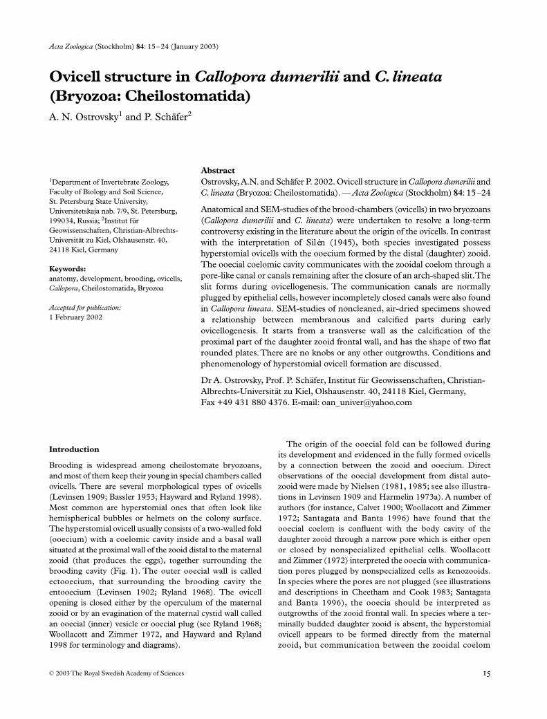
Brooding is widespread among cheilostomate bryozoans,and most of them keep their young in special chambers calledovicells. There are several morphological types of ovicells(Levinsen 1909; Bassler 1953; Hayward and Ryland 1998).Most common are hyperstomial ones that often look likehemispherical bubbles or helmets on the colony surface.The hyperstomial ovicell usually consists of a two-walled fold(ooecium) with a coelomic cavity inside and a basal wallsituated at the proximal wall of the zooid distal to the maternalzooid (that produces the eggs), together surrounding thebrooding cavity (Fig. 1). The outer ooecial wall is calledectooecium, that surrounding the brooding cavity theentooecium (Levinsen 1902; Ryland 1968). The ovicellopening is closed either by the operculum of the maternalzooid or by an evagination of the maternal cystid wall calledan ooecial (inner) vesicle or ooecial plug (see Ryland 1968;Woollacott and Zimmer 1972, and Hayward and Ryland1998 for terminology and diagrams).
The origin of the ooecial fold can be followed duringits development and evidenced in the fully formed ovicellsby a connection between the zooid and ooecium. Directobservations of the ooecial development from distal auto-zooid were made by Nielsen (1981, 1985; see also illustra-tions in Levinsen 1909 and Harmelin 1973a). A number ofauthors (for instance, Calvet 1900; Woollacott and Zimmer1972; Santagata and Banta 1996) have found that theooecial coelom is confluent with the body cavity of thedaughter zooid through a narrow pore which is either openor closed by nonspecialized epithelial cells. Woollacottand Zimmer (1972) interpreted the ooecia with communica-tion pores plugged by nonspecialized cells as kenozooids.In species where the pores are not plugged (see illustrationsand descriptions in Cheetham and Cook 1983; Santagataand Banta 1996), the ooecia should be interpreted asoutgrowths of the zooid frontal wall. In species where a ter-minally budded daughter zooid is absent, the hyperstomialovicell appears to be formed directly from the maternalzooid, but communication between the zooidal coelom
Ovicell structure
•
Ostrovsky and Schäfer Acta Zoologica
(Stockholm)
84
: 15–24 (January 2003)
© 2003 The Royal Swedish Academy of Sciences
and the ovicellar coelom is usually through pores withspecial pore-cell complexes, and ooecia of this type musttherefore be considered as true kenozooids (Ostrovsky1998).
Silén (1944, 1945) reported that ovicells situated on thefrontal wall of the distal zooid in several species are formedas direct outgrowths from the maternal zooid, and he pro-posed that this is the general structure of hyperstomial ovice-lls. An uncertainty about this has therefore crept into theliterature (see, for example, Ryland 1979; Reed 1991; Mukai
et al
. 1997). It was therefore decided to reinvestigate
Callo-pora dumerilii
(Audouin, 1826) on which Silén based muchof his argumentation, and
C. lineata
(Linnaeus, 1767) (typespecies of the genus
Callopora
Gray 1848), in order to checkthe basis for his generalization. A detailed description of thehistory of investigation of ovicell formation and structure isgiven in Ostrovsky (in press).
Materials and methods
Colonies of
Callopora dumerilii
were collected 05 August1997 by trawling at 16–29 m depth on the stone reef HerthasFlak (North Kattegat, Baltic Sea), and 12 June 1997 bySCUBA from the 22 m depth near Riou Island (WestMediterranean). Colonies of
C. lineata
were collected 14June 1995, 03 July 1995 and 17 August 1996 by dredgingand SCUBA from 3 to 7 m depth near Sredniy and MatreninIslands in the Chupa Inlet (Kandalaksha Bay, White Sea).Specimens were fixed in Bouin’s fluid without acetic acid(sometimes with formalin neutralized by calcium carbonate)and 70% alcohol. For light microscopy, colonies decalcifiedin Bouin’s fluid were embedded in plastic (epon), sectioned(1–2.5
µ
m thick) and stained with toluidine blue usingstandard methods. For SEM studies, colonies fixed inalcohol were cleaned in 7.5% solution of sodium hypoclorite,
Fig. 1—Schema of the longitudinal section of decaleified specimen of Callopora dumerilii (the partly plugged communication canal between the ooecial coelom and the general body cavity is arrowed). Abbreviations: bc = brooding cavity, cec = calcified part of ectooecium, dz = coelomic cavity of daughter autozooid, en = entooecium, fs = funicular strands, fw = frontal wall of distal zooid, m = muscle bundles of ooecial vesicle, mec = membranous part of ectooecium, mz = coelomic cavity of maternal autozooid, oc = ooecial coelomic cavity, of = vicellular floor, op = operculum, ov = ooecial vesicle, s = sclerite, tw = transverse zooidal wall, v = vestibulum.
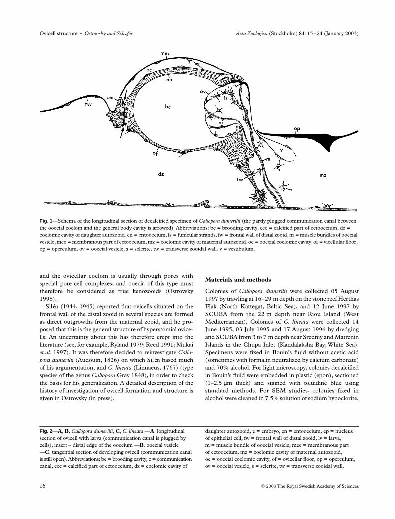
Fig. 2
—
A
,
B
.
Callopora dumerilii
,
C
,
C. lineata
—
A
. longitudinal section of ovicell with larva (communication canal is plugged by cells), insert – distal edge of the ooecium —
B
. ooecial vesicle —
C
. tangential section of developing ovicell (communication canal is still open). Abbreviations: bc = brooding cavity, c = communication canal, cec = calcified part of ectooecium, dz = coelomic cavity of
daughter autozooid, e = embryo, en = entooecium, ep = nucleus of epithelial cell, fw = frontal wall of distal zooid, lv = larva, m = muscle bundle of ooecial vesicle, mec = membranous part of ectooecium, mz = coelomic cavity of maternal autozooid, oc = ooecial coelomic cavity, of = ovicellar floor, op = operculum, ov = ooecial vesicle, s = sclerite, tw = transverse zooidal wall.

Acta Zoologica
(Stockholm)
84
: 15–24 (January 2003)
Ostrovsky and Schäfer
•
Ovicell structure
© 2003 The Royal Swedish Academy of Sciences

Ovicell structure
•
Ostrovsky and Schäfer Acta Zoologica
(Stockholm)
84
: 15–24 (January 2003)
© 2003 The Royal Swedish Academy of Sciences

Acta Zoologica
(Stockholm)
84
: 15–24 (January 2003)
Ostrovsky and Schäfer
•
Ovicell structure
© 2003 The Royal Swedish Academy of Sciences
dehydrated in absolute alcohol, air-dried and coated withgold. Also some colonies fixed in ‘neutral’ Bouin’s fluid weredehydrated and either air or critical-point dried for SEMstudy.
Results
Ovicell structure
Anatomical investigations showed that both
Callopora dumer-ilii
and
C. lineata
possess hyperstomial ovicells with ooeciaformed by the distal (daughter) zooid (Figs 1–5). In the com-plete ovicell the basis of the ooecium is a continuation of theproximal gymnocyst of the daughter zooid. The majority ofthe ovicells was formed by autozooids, but one ovicellformed by a kenozooid was also found in
C. dumerilii
fromthe Mediterranean locality (Fig. 4B) (a similar case is shownin Zabala and Maluquer 1988; Pl. 3C). In this species thedevelopment of ovicell-bearing zooids was sometimes sup-ressed by the formation of an adjacent zooidal row (Fig. 4C).In one such case two fused ovicells with brooding cavitiesseparated by a calcified wall were encountered (Fig. 4C,D).
Ovicells are helmet-shaped, and their chambers areslightly flattened dorsoventrally. The opening of the ovicell isnot closed by an operculum (acleithral type) but is pluggedby an ooecial vesicle (Figs 1, 2A, 3A).
The ooecial fold consists of two walls: outer (ectooecium)and inner (entooecium), with the coelomic lumen in between(Figs 1–3). In
C. dumerilii
the ectooecium is membranousexcept for the narrow basal part (Figs 2A, 4E, 4F). In
C. lineata
the calcified ectooecium is characterized by a big membranouswindow (lucida) near the ooecial edge (Figs 3B, 5A) (see alsoRyland and Hayward 1977 for comparison). The ectooecialcuticle is markedly thinner than the cuticle of the frontalzooidal wall (Fig. 2A).
The entooecium is always completely calcified and thickenedalong the ooecial orifice (Figs 1, 2A and insert). In olderzooids this thickening appears as a prominent lip. Its edge hasa flattened area at the point of contact with the ooecial vesi-cle. The surface of the entooecium facing the brooding cavityis smooth (Figs 4H, 5E), with concentric lines of growth andoften nondistinct radial wrinkles. The coelomic surface of theentooecium is either more or less even (
C. lineata
) or granu-lated (
C. dumerilii
). Prenant and Bobin (1996) described andpictured a short medial suture in the entooecium of
C. line-ata
, and a similar, small medial furrow was observed in oneovicell of this species in the material studied here.
The coelomic cavity of the ooecium is lined by epidermaland peritoneal cells (Fig. 2A insert). Extensions of the lattersometimes cross the cavity. As calcification increases, thelumen becomes narrower, finally slit-like, but is neverreduced completely. The ooecial cavity communicates with acavity of the daughter zooid through a flattened pore-likecanal. The opening of the canal into the zooidal coelom isusually placed near the left or, sometimes, the right ‘corner’of the ooecial fold basis (Fig. 4G). In two cases, the canal wasseen to communicate with the zooidal cavity by a couple ofsuch openings in
C. dumerilii
(Fig. 4H). Each communica-tion canal is a rudiment of an arch-shaped slit, formed duringovicellogenesis and connecting ooecial and zooidal coelomsduring that time. In younger zooids with complete ovicellsthis slit is already closed, but still clearly seen (Fig. 5F). Inolder zooids it is recognizible by an arch-shaped groove(Fig. 4G,H). As a rule, the canal is plugged by epithelialcells (not special pore-cell complexes) (Figs 2A, 3, 5F), sofree circulation of the coelomic fluid is obviously absent.However, some completely formed ovicells of
C. lineata
hadan open the communication canal, lined by epithelial cells,but not plugged (Fig. 3A). The ooecial vesicle is a hollowevagination of the distal wall of the maternal autozooid thatplugs the ovicell opening (Figs 1, 2A,B, 3A,B). In manycases observed, the vesicle was recumbent on the distal edgeof the operculum. The cuticle of the vesicle wall that facesinto the ovicell cavity is thinnest. The part of the vesicle incontact with the flattened area of the entooecium edge ischaracterized by a thickened cuticle forming a sclerite (seeSantagata and Banta 1996). Its outer surface bears cuticularribs, making the closure of the ovicell tighter (Fig. 1). Theinner surface possesses a transverse (triangular in section) ribto which the upper bundle of the muscles is fixed (Figs 2A,3A,B).
Paired sets of the bilateral muscles of the ooecial vesicleoriginate laterally either at the basal wall of the maternalzooid (
C. dumerilii
from the Baltic locality) (Fig. 1) or at thetransverse wall (
C. lineata
), near the corner between thesewalls. In
C. dumerilii
from the Mediterranean the muscleswere found originating at the basal wall in some cases, and atthe transverse wall in others; the transverse wall was moreinclined, and more often the muscles were fixed at the basalwall. Sometimes the muscles originated exactly in the cornerbetween the walls. The upper (larger) muscle set inserts onthe sclerite rib (Figs 1, 2A,B, 3A). The middle set consistsof four bundles which distally insert on the inner wall of theooecial vesicle in its middle part. Lower (smaller) set distally
Fig. 3
—
A
–
B
,
Callopora lineata
,
C
,
C. dumerilii
—
A
. longitudinal section of ovicell with embryo (communication canal is open) —
B
,
C
. tangential section of ovicell (communication canal is plugged by cells, nuclei of epithelial cells arrowed). Abbreviations: a = avicularium, bc = brooding cavity, c = communication canal, cec = calcified part of ectooecium, dz = coelomic cavity of daughter autozooid, e = embryo, en = entooecium, ep = nucleus of
epithelial cell, fs = funicular strand, fw = frontal wall of distal zooid, lv = larva, m = muscle bundle of ooecial vesicle, mec = membranous part of ectooecium, mz = coelomic cavity of maternal autozooid, nm = noncellular material within brooding cavity, oc = ooecial coelomic cavity, of = ovicellar floor, op = operculum, ov = ooecial vesicle, s = sclerite, tw = transverse zooidal wall, v = vestibulum.

Ovicell structure
•
Ostrovsky and Schäfer Acta Zoologica
(Stockholm)
84
: 15–24 (January 2003)
© 2003 The Royal Swedish Academy of Sciences
Fig. 4 —Callopora dumerilii. —A. ovicells formed by distal autozooids —B. ovicell formed by distal kenozooid (ooecium bordered by two avicularia) —C, D. fused ovicells (C, ovicell of the zooid supressed by the formation of an adjacent zooidal row arrowed) —E. ovicell distal view (membranous part of the ectooecium detached) —F. slit between endooecium and calcified
basal part of ectooecium (arrowed) —G, H. detached ooecia with one (G) and two (H) communication pores (arrowed) (trace of closed arc-shaped slit is clearly seen). Abbreviations: cec = calcified part of ectooecium, d = daughter autozooid, en = entooecium, k = distal kenozooid, m = maternal autozooid, mec = membranous part of ectooecium, o = ovicell.

Acta Zoologica
(Stockholm)
84
: 15–24 (January 2003)
Ostrovsky and Schäfer
•
Ovicell structure
© 2003 The Royal Swedish Academy of Sciences
inserts on this wall in its lower part. The distance betweenthese smaller sets (middle and lower) varies depending onthe ovicell. In
C. dumerilii
they sometimes insert in upperand middle parts of the vesicle. Compared with the parietalmuscle bands of the frontal zooidal wall, the muscles ofthe ooecial vesicle are much wider, having longer zones ofattachment to the body wall. Silén (1945) believed that thereis a single muscle bundle.
The wall of the ooecial vesicle is lined by flat epithelial andperitoneal cells, which are in contact with funicular strandsthat run into the zooidal coelom (Figs 1, 3A). No sign of cellhypertrophy was found in the ooecial vesicles of the ovicellscontaining embryos or in the empty ones. There was also nospecial evagination on the inner wall of the vesicle asdescribed by Silén (1945), and only late embryos sometimesdeformed it. A thin layer of noncellular material was often
Fig. 5 —Callopora lineata —A. part of colony with ovicells —B–D. early stages of ooecial development: —B. distal zooidal bud with bilobate calcification (arrowed) —C. stage of bilobate plate with medial suture (two forming ovicell are seen) —D. stage of
semicircular fold —E. detached ooecium (inner view) —F. arc-shaped communication slit (arrowed) with collapsed epithelial cells. Abbreviations: d = daughter autozooid, m = maternal autozooid, f = forming ovicell.

Ovicell structure
•
Ostrovsky and Schäfer Acta Zoologica
(Stockholm)
84
: 15–24 (January 2003)
© 2003 The Royal Swedish Academy of Sciences
seen on the surface of the vesicle inner wall facing the brood-ing cavity (Fig. 2B). It was clearly seen in the wrinkles of thewall caused by muscle retraction.
Each embryo contained in the ovicell is surrounded by avery thin fertilization envelope. These were also found inempty ovicells, indicating that the brood chambers had beenused at least once.
Early ovicellogenesis
The earliest stages of ovicell formation were investigated in
Callopora lineata
. The ooecial fold originates as a proximaloutgrowth of the frontal wall of the daughter zooid bud.Before folding, an area of its membranous frontal wall beginsto calcify. Calcification starts from the transverse zooidal walland expands distally, having a shape of two rounded flatplates. The plates originate independently of one another andsometimes have different sizes (Fig. 5B). Finally, they fuseforming a bilobate plate with a medial suture (Fig. 5C).Calcification continues to expand centrifugally along thefrontal wall of the zooidal bud, forming a shallow depression– the future floor (basal wall) of the ovicell. At this stage thecalcified area looses its bilobate shape, but the medial sutureis often still seen. It disappears at a later stage when thesemicircular fold of the future ooecium starts to grow(Fig. 5D). The ooecial coelomic cavity communicates withthe zooidal perigastric coelom through the arch-shaped slitthat gradually closes (Figs 2C and 5F).
Discussion
The ovicells of the two species of
Callopora
studied heredefinitely develop as special outgrowths from the frontal wallof the zooid distal to the maternal, not from the maternalzooid itself, as proposed by Silén (1945). An analysis oftext-Fig. 18 in Silén (1944) (representing a longitudinalsection of the ovicell in Scrupocellaria scabra (van Beneden,1848)), and the accompanying description shows that Siléncould not find a communication between an ooecial fold anda distal zooid because of the strong shrinkage of the specimenfixed in alcohol. Studing three other species, Silén (1945) didnot make sections and referred to the wrongly interpretedstructure of Scrupocellaria. On the basis of recent and previousfindings (Levinsen 1909; Nielsen 1981; Lobastova andOstrovsky 1994; see also Nielsen 1985 and Santagata andBanta 1996), Silén’s (1944, 1945) conclusions concerningovicell structure in Scrupocellaria scabra, Callopora dumerilii,Fenestrulina malusii (Audouin, 1826) and Escharella immersa(Fleming, 1828) must be regarded as incorrect, and hisgeneralization rejected.
The picture of brood chamber formation in cheilostomatidsis not simple. Initially, Harmer (1902; p. 248) noted that theooecium can be part of ‘the distal’ zooid (type 1) or belongto the ‘fertile (proximal)’ zooid (type 2), and this has beenconfirmed (reviewed in Reed 1991; Ostrovsky 1998). In con-
trast with the opinion of Silén, type 1 is much more commonthan type 2. In the Cribrilinidae both types can sometimesbe found within one and the same taxon and even in thesame colony (see Ristedt 1985; Bishop and Househam1987). Both types of ovicells are also known in Calloporidae,Romancheinidae and some other families (see illustrations inLevinsen 1909, 1916; Harmelin 1973b; Gordon 1984,1986; Hayward 1995, etc.). Type 2 could have evolved fromtype 1 by a gradual reduction of the distal zooid (discussedin Bishop and Househam 1987; Ostrovsky 1998). Thus, thevast majority of the type 2 ovicells studied are found in spe-cies where a terminally budded daughter zooid is absent, andthis was not the case in the species investigated by Silén. Hebelieved that the development of an ovary triggers ovicellformation in the same zooid. Nielsen (1981) however, clearlyshowed that ovicellogenesis in distal zooids is induced by theproximal ones in fused colonies of Fenestrulina malusii: fertilezooids from one colony had induced zooids from the othercolony to form the ovicells (see also Nielsen 1990). Twofused ovicells on one zooid have been sometimes formed insuch cases (one from maternal, the second from the ‘neigh-bour’), but it is difficult to gauge the reason for the fusion oftwo ovicells in the colony of C. dumerilii studied (see above).
The first sign of the ovicellogenesis in Callopora is a calci-fication in the shape of two flat areas (rounded plates)extending from the proximal edge and along the frontalsurface of the distal zooidal bud (see also Levinsen 1893,1894, 1909; Harmelin 1973a for comparison). Silén (1945)described the first stage as a pair of knobs, but this was prob-ably the result of observing only with a dissecting microscope(see also Ryland 1979). Believing that an ooecium developsfrom the maternal zooid as paired knobs, Silén (1977) sup-ported Harmer’s (1902) hypothesis on the evolution of theooecium from the distal pair of oral spines (see Ryland 1979and Ostrovsky 1998 for further discussion).
In cleaned specimens subsequent stages in calcification ofthe entooecium appear to develop from the transverse wallbetween zooids (see, for instance illustrations in Harmelin1973a; Nielsen 1985, etc.). This probably explains the diver-sity of opinions concerning the mixed origin of the ooecium.Since the paper of Levinsen (1902), it was suggested that theentooecium derives from the maternal zooid, and the ecto-oecium from the daughter zooid (Harmelin 1973a; Soule1973; Cook 1977, 1979, 1985; Ryland 1979; Morris 1980;Cook and Chimonides 1981; Wass and Banta 1981; Ristedt1985). Calcification, however, is only a part, not the wholeof ovicellogenesis, and it is incorrect, we believe, to considerthe maternal zooid as a locus for entooecium only becausethe calcification of the latter starts from the transverse wallequally belonging to both maternal and daughter zooids.Nielsen (1981, Fig. 20) concluded that the entooecium isformed by the distal zooid (see also Ostrovsky 1998 andOstrovsky (in press) for further discussion).
Further growth of the ooecial fold leads to the appearanceof a helmet-shaped ooecium, the lumen of which initially is

Acta Zoologica (Stockholm) 84: 15–24 (January 2003) Ostrovsky and Schäfer • Ovicell structure
© 2003 The Royal Swedish Academy of Sciences
confluent with a cavity of the daughter zooid (Fig. 2C), thus,the ooecium is simply an outgrowth of the zooid, although inmost cases quite complicated. The communication slit isthen subsequently getting narrowed by increasing calcifica-tion of the ooecial walls. If the pore-like canal(s) remainingafter the slit closure is free of cell obstruction (Fig. 3A), theooecium remains as an outgrowth of the zooidal wall as inFigularia figularis (Johnston, 1847), Scrupocellaria ferox(Busk, 1852), and some colonies of Callopora lineata(Cheetham and Cook 1983; Santagata and Banta 1996; ourdata). If the canal is plugged by the epithelial cells (Figs 2A,3B,C), the ooecium transforms to some kind of heterozooid(but not a true kenozooid) as in the cases of Bugula neritina(see Woollacott and Zimmer 1972) and two species of thegenus Callopora studied. The epithelial lining of an ooeciumreceives nutrients probably by intercellular transport. Findingclosed and nonclosed communication canals in completelyformed ovicells of C. lineata can reflect ontogenetical changeswhen older ooecia finally should be ‘closed’ by cells, however,it is also still possible that some canals can stay open duringthe whole existence of particular ovicells. If so, there is onlya formal reason to credit ooecia as being a special heterozooidor ‘morph’ (Woollacott and Zimmer 1972; Ryland 1979;p. 214; see also Nielsen 1985 for discussion).
Ooecia of type 2 start their development in the samemanner as ordinary zooids (Ostrovsky 1998). They bud fromthe maternal zooids, becoming true kenozooids after thedevelopment of the transverse wall with pores closed byspecial pore-cell complexes.
In the calloporid Amphiblestrum flemingii (Busk, 1854) (asMembranipora), Calvet (1900, Fig. 45) pictured cells of the innerwall of the ooecial vesicle as larger than all other epithelialcells. The same impression is gained from the schema of theSecuriflustra securifrons (Pallas, 1766) (as Flustra) (Flustridae)(Calvet 1900, Fig. 44), however there is no sign of extra-embryonal feeding and subsequent hypertrophy of theepithelial cells in two species of the genus Callopora studied(Figs 2A,B, 3A). We suggest that Calvet, being impressed byhis finding of an embryophore in the inner vesicle of Bugulasimplex, incorrectly extrapolated these data to other species. Atthe moment, placental brooding is known in three bryozoanfamilies: Bugulidae, Candidae, and Hippothoidae (Woollacottand Zimmer 1972; 1975; Dyrynda and King 1983; Hughes1987; Santagata and Banta 1996; Ostrovsky 1998). In contrast,an egg receives all the nutrients while still in the ovary inCalloporidae. Silén (1945) noted that embryos died if theywere removed from the brood chamber to the sea-water inC. dumerilii, but the processes involved are completely unknown.
Acknowledgements
We sincerely thank Natalia N. Shunatova, Department ofInvertebrate Zoology, St. Petersburg State University, andDr Jean-George Harmelin, Station Marien d’Endoume,Centre d’Océanologie Marseille, for collecting and sorting
the material, and species identifications. We greatly appreciatethe staff of the Zoological Museum, University of Copenhagen:Professor Claus Nielsen for providing working facilities andmuch encouragement and discussions, Dr Ole Tendal forassistance with collecting, Dr Karen Bille Hansen for helpwith the species identifications, Vibe Lund Hansen and GeertBrovad, for help with making sections and photoworks,Dr Mary E. Petersen for translating and kind help withliterature. Thanks are also given to Ute Schuldt, Institutfür Geowissenschaften, Christian-Albrechts-Universität zuKiel, for assistance with SEM- and photoworks, and DrMary Spencer Jones, The Natural History Museum, London,for help with literature. Drs Dennis P. Gordon, NationalInstitute of Water and Atmospheric Research, Wellington,New Zealand, and Claus Nielsen, Zoological Museum,University of Copenhagen, kindly reviewed the early draft ofthe manuscript. We also thank two anonymous reviewers foruseful comments and criticism. The study was completedduring a postdoctoral fellowship of A. Ostrovsky sponsoredby the Humboldt Foundation (Alexander von Humboldt-Stifting). Deutsche Forschungsgemeinschaft is acknow-ledged for financial support (grant SCHA 355/20–1).
References
Bassler, R. S. 1953. Bryozoa (Part G). In: Treatise on Invertebrate Paleon-tology (ed. R. C. Moore), pp. 1–253. Geological Society of America(New York) and University of Kansas Press (Lawrence, Kansas).
Bishop, J. D. D. and Househam, B. C. 1987. Puellina (Bryozoa:Cheilostomata: Cribrilinidae) from British and adjacent waters.– Bulletin of the British Museum (Natural History), Zoology 53(1):1–63.
Calvet, L. 1900. Contribution à l’histoire naturelle des BryozoairesEctoproctes marins. – Travaux de l’Institut de Zoologie de l’Universitéde Montpellier, Nouvelle Série 8: 1–488.
Cheetham, A. H., Cook, P. L. 1983. General features of the classGymnolaemata. In: Treatise on Invertebrate Paleontology, Vol. 1. (ed.R. A. Robinson), Bryozoa (Part G, revised), pp. 138–207. Geo-logical Society of America (Boulder, Colorado) and University ofKansas (Lawrence).
Cook, P. L. 1977. The genus Tremogasterina Canu (Bryozoa,Cheilostomata). – Bulletin of the British Museum (Natural History),Zoology 32(5): 103–165.
Cook, P. L. 1979. Some problems in interpretation of heteromorphyand colony integration in Bryozoa. In: Biology and Systematics ofColonial Organisms (eds G. Larwood, B. R. Rosen), SystematicsAssociation Special, Vol. 11, pp. 193–210. Academic Press, Lon-don and New York.
Cook, P. L. 1985. Bryozoa from Ghana. – Zoologische WetenschappenMusee Royal l’Afrique Centrale Tervuren, Belgique 238: 1–315.
Cook, P. L. and Chimonides, P. J. 1981. Morphology and systematicsof some rooted cheilostome Bryozoa. – Journal of Natural History15: 97–134.
Dyrynda, P. E. J. and King, P. E. 1983. Gametogenesis in placentaland non-placental ovicellate cheilostome Bryozoa. – Journal ofZoology, London 200: 471–492.
Gordon, D. P. 1984. The marine fauna of New Zealand: Bryozoa:Gymnolaemata from the Kermadec Ridge. – New Zealand Ocea-nographic Institute Memoir 91: 1–198.
Ovicell structure • Ostrovsky and Schäfer Acta Zoologica (Stockholm) 84: 15–24 (January 2003)
© 2003 The Royal Swedish Academy of Sciences
Gordon, D. P. 1986. The marine fauna of New Zealand: Bryozoa:Gymnolaemata (Ctenostomata and Cheilostomata Anasca) fromthe Western South Island continental shelf and slope. – New Zea-land Oceanographic Institute Memoir 95: 1–121.
Harmelin, J.-G. 1973a. Les bryozoaires des peuplement sciaphilesde Méditerranée: le genre Crassimarginatella Canu (ChilostomesAnasca). – Cahiers de Biologie Marine 14: 471–492.
Harmelin, J.-G. 1973b. Callopora minuta n. sp., nouvelle espèce debryozoaire Chilostome (Alderinidae) des côtes françaises deMéditerranée. – Cahiers de Biologie Marine 14: 29–37.
Harmer, S. F. 1902. On the morphology of the Cheilostomata.– Quarterly Journal of Microscopical Science 46(182): 263–350.
Hayward, P. J. 1995. Antarctic Cheilostomatous Bryozoa. OxfordUniversity Press.
Hayward, P. J. and Ryland, J. S. 1998. Cheilostomatous Bryozoa,Part 1. Aetoidea-Cribrilinoidea. – Synopses of the British Fauna. 102nd edn.: 1–366.
Hughes, D. J. 1987. Gametogenesis and embryonic brooding in thecheilostome bryozoan Celleporella hyalina. – Journal of Zoology,London 212: 691–711.
Levinsen, G. M. R. 1893. Polyzoa. In: Det Videnskabelige Udbytte afKanonbaaden ‘Hauchs’ Togter i de Danske Have indenfor Skagen iAarene 1883–86, pp. 243–306. A. F. Høst and Søns Forlag,Kjøbenhavn.
Levinsen, G. M. R. 1894. Mosdyr. – Zoologia Danica 9: 1–105.Levinsen, G. M. R. 1902. Studies on Bryozoa. – Videnskabelige
Meddelelser fra Dansk Naturhistorisk Forening i Kjøbenhavn 54:1–31.
Levinsen, G. M. R. 1909. Morphological and systematic studies on theCheilostomatous Bryozoa. F. Bagge, Copenhagen.
Levinsen, G. M. R. 1916. Bryozoa. – Danmark-Expeditionen TilGrønlands Nordøstkyst 1906–08: 3(16): 433–472.
Lobastova, E. V., Ostrovsky, A. N. 1994. Some new data onanatomy and ovicellogenesis in two cheilostome bryozoans –Scrupocellaria scabra and Callopora aurita from the White Sea.In: Fossil and Living Bryozoa of the Globe (ed. V. P. Ozhgibesov),pp. 44–45. Perm State University, Perm.
Morris, P. A. 1980. The bryozoan family Hippothoidae (Cheilostomata-Ascophora), with emphasis on the genus Hippothoa. – Allan Han-cock Monographs in Marine Biology 10: 1–115.
Mukai, H., Terakado, K. and Reed, C. G. 1997. Bryozoa. In: Micro-scopic Anatomy of Invertebrates, Vol. 13. (ed. F. W. Harrison),pp. 45–206. Wiley-Liss, New York.
Nielsen, C. 1981. On morphology and reproduction of ‘Hippodiplo-sia’ insculpta and Fenestrulina malusii (Bryozoa, Cheilostomata).– Ophelia 20: 91–125.
Nielsen, C. 1985. Ovicell formation in Tegella and four cellularioids(Bryozoa, Cheilostomata). In: Bryozoa, Ordovician to Recent, (edsC. Nielsen, G. P. Larwood), pp. 231–224. Olsen and Olsen,Fredensborg.
Nielsen, C. 1990. Bryozoa Ectoprocta. In: Reproductive Biology ofInveretebrates, Vol. IV. (eds K. G. Adiyodi, R. G. Adiyodi), Part B,pp. 185–200. Oxford and IBH Publishing Co, Pvt. Ltd, NewDelhi, Bombay, Calcutta.
Ostrovsky, A. N. 1998. Comparative studies of ovicell anatomyand reproductive patterns in Cribrilina annulata and Celleporellahyalina (Bryozoa: Cheilostomatida). – Acta Zoologica 79(4):287–318.
Ostrovsky, A. N. (in press) The history of study of ovicells in cheilos-tomate bryozoans. – Annals of Science.
Prenant, M., Bobin, G. Bryozoaires. Deuxième partie. ChilostomesAnasca. – Faune de France 68: 1–398.
Reed, C. G. 1991. Bryozoa. In: Reproduction of Marine Invertebrates,Vol. VI Echinoderms and Lophophorates, (eds A. C. Giese,J. S. Pearse, V. B. Pearse), pp. 85–245. Boxwood Press, PacificGrove.
Ristedt, H. 1985. Cribrilaria-Arten (Bryozoa) des Indopazifiks(Rotes Meer, Seychellen, Philippinen). – Mitteilungen aus demGeologisch-Paläontologischen Institut der Universität Hamburg 59:15–38.
Ryland, J. S. 1968. Terminological problems in Bryozoa. – Atti DellaSocietà Italiana Di Scienze Naturali E Del Museo Civico Di StoriaNaturale Di Milano 108: 225–236.
Ryland, J. S. 1979. Structural and physiological aspects of colonialityin Bryozoa. In: Biology and Systematics of Colonial Organisms,Systematics Association special Vol. 11. (eds G. Larwood, B. R. Rosen),pp. 211–242. − Academic Press, London and New York.
Ryland, J. S. and Hayward, P. J. 1977. British anascan bryozoans. –Synopses of the British Fauna 10: 1–188.
Santagata, S. and Banta, W. C. 1996. Origin of brooding and ovicellsin cheilostome bryozoans: interpretive morphology of Scrupocellariaferox. – Invertebrate Biology 115(2): 170–180.
Silén, L. 1944. The anatomy of Labiostomella gisleni Silén (BryozoaProtocheilostomata). – Kungliga Svenska VetenskapsakademiensHandlingar, Serie 3 21: 1–111.
Silén, L. 1945. The main features of the development of the ovum,embryo and ooecium in the ooecioferous Bryozoa Gymnolae-mata. – Arkiv För Zoologi 35A(17): 1–34.
Silén, L. 1977. Polymorphism. In: Biology of Bryozoans (edsR. M. Woollacott, R. L. Zimmer), pp. 184–232. Academic Press,New York.
Soule, D. F. 1973. Morphogenesis of giant avicularia and ovicellsin some Pacific Smittinidae. In: Living and Fossil Bryozoa (ed.G. P. Larwood), pp. 485–495. Academic Press, London and NewYork.
Wass, R. E. and Banta, W. C. 1981. Catenicellid cheilostome Bryozoa.II. Introduction to ovicell complexes. – Australian Journal ofZoology 29: 365–400.
Woollacott, R. M. and Zimmer, R. L. 1972. Origin and structure ofthe brood chamber in Bugula neritina (Bryozoa). – Marine Biology16: 165–170.
Woollacott, R. M. and Zimmer, R. L. 1975. A simplified placenta-like system for the transport of extraembryonic nutrients duringembryogenesis of Bugula neritina (Bryozoa) – Journal of Morphology147: 355–378.
Zabala, M. and Maluquer, P. 1988. Illustrated keys for the classificationof Mediterranean Bryozoa. – Treballs Del Museu de Zoologia(Barcelona) 4: 1–294.