blood and plasma titanium levels associated with well
TRANSCRIPT

1
Blood and plasma titanium levels associated with well-functioning hip implants
Ilona Swiatkowska1, Nicholas G. Martin2, Johann Henckel3, Hugh Apthorp4, Jane
Hamshere4, Alister J. Hart1,3
1Institute of Orthopaedics and Musculoskeletal Science, University College London, HA7
4LP Stanmore, UK 2Trace Element Laboratory, North West London Pathology, Charing Cross Hospital, W6 8RF
London, UK 3Royal National Orthopaedic Hospital, Stanmore, HA7 4LP Stanmore, UK 4The Horder Centre, TN6 1XP Crowborough, UK
Correspondence and requests for materials should be addressed to I.S. (email:
[email protected], phone number: 020 8416 3412)
ABSTRACT
Background: Hip implants are usually manufactured from cobalt-chromium and titanium
alloys. As the implants wear and corrode, metal debris is released into the surrounding tissue
and blood, providing a potential biomarker for their function. Whilst there are laboratory
reference levels for blood cobalt and chromium in patients with well and poorly functioning
hip implants, there are no such guidelines for titanium. This is despite the increasing use of
titanium implants worldwide.
Patients and methods: We recruited a consecutive series of 95 patients (mean age 71 years,
mean time after surgery 8.5 years) with one hip implant type, inserted by the same surgeon.
We assessed clinical and radiological outcome, and measured blood and plasma titanium
using high resolution inductively-coupled plasma mass spectrometry.
Results: The upper normal reference limit for blood and plasma titanium was 2.20 and 2.56
g L-1, respectively, and did not differ significantly between males and females.
Conclusion: We are the first to propose a laboratory reference level for blood and plasma
titanium in patients with well-functioning titanium hip implants. This is an essential starting
point for further studies to explore the clinical usefulness of blood titanium as a biomarker of
orthopaedic implant performance, and comes at a time of considerable controversy regarding
the use of certain titanium alloys in hip arthroplasty.

2
Keywords: blood titanium; toxicity; TMZF; reference range; HR ICP-MS; total hip
arthroplasty
1. Introduction
Components of joint replacements are usually manufactured from cobalt-chromium (Co-Cr)
or titanium (Ti) alloys. Once implanted, all metals degrade through wear and corrosion,
releasing ions and particles into the surrounding tissue and bloodstream. The greatest wear
usually occurs during the first 1-2 years after surgery [1], which is followed by a low, but
steady, rate of wear over subsequent years. The amount of metal debris released is a surrogate
marker of implant wear [2], and can inform on the risk of local adverse effects and need for a
revision surgery.
In the case of cobalt and chromium, a blood level of 2 µg L-1 implies a well-functioning
metal-on-metal hip implant, while concentrations exceeding 7 µg L-1 indicate potential for
local tissue damage and a failing implant [3]. It is now believed that measurement of titanium
could also be used to gain insights into implant performance [4–7], though “normal” and
“abnormal” blood levels have not been established. This is partly due to the technical
challenges involved in the measurement of titanium in biological samples. Traditional
techniques, such as graphite furnace atomic absorption spectroscopy (GF AAS) and
quadrupole inductively-coupled plasma mass spectrometry, suffer from a range of
interferences, which can lead to overestimation of analyte concentration. Several groups have
reported blood/serum titanium levels associated with different types of well-functioning and
malfunctioning prostheses [8]. However, in addition to unreliable analytical techniques used,
majority of the studies suffered from small sample size.
We present a series of 95 patients with well-functioning, unilateral hip implants inserted by
the same surgeon. We used a high resolution inductively-coupled plasma mass spectrometer

3
(HR ICP-MS) to investigate how much titanium is released by the implants at medium-to-
long term follow up (when the wear rate is thought to have normalised), and established a
normal reference range for blood/plasma titanium in this population.
As uncemented hip replacements and 3D-printed implants are gaining popularity, the use of
titanium in orthopaedics is growing. Additionally, constant ageing of the population means
that the overall demand for total joint replacements is on the rise [9]. Taken together, these
points underscore the potential impact of the present study. The proposed guidelines could be
a useful tool to assess patients with titanium-based implants, and help predict which might
develop clinical problems.
2. Patients and methods
This study protocol was approved by our institutional review board after ethical approval by
Riverside Research Ethics Committee (ref. 07/Q0401/25).
All patients who received one type of titanium alloy femoral stem (see below for details)
between 2007-2014 at a participating institution were identified using the National Joint
Registry database (N=1036). Inclusion criteria stipulated unilateral, primary, uncemented
ceramic-on-ceramic (CoC) hip implants inserted by the same surgeon. All eligible
participants (N=199) were invited, and those willing to take part were booked in for a clinic
visit. The assessment involved 1) a pelvis X-ray; 2) Oxford Hip Score; 3) UCLA Activity
Score, 4) a blood test and 5) a short interview. A complete set of data was obtained for 95
subjects (42 males and 53 females) (Figure 1). The underlying diagnoses leading to the
primary surgery were osteoarthritis (98%) or developmental hip dysplasia (2%).
All patients provided written informed consent to participate in the study.

4
Figure 1. Flowchart outlining participant enrolment and clinical assessment.
2.1. Implants
Implant design was uniform across the series, and comprised a V40 32 mm Al2O3 (alumina)
femoral head articulating against a Trident titanium-backed alumina insert, a commercially
pure titanium Trident PSL acetabular cup and a Ti-12Mo-6Zr-2Fe (TMZF) Accolade I
femoral stem (Figure 2). All the components were manufactured
by Stryker Orthopaedics (Mahwah, NJ) and were inserted without
cement. In several cases, the acetabular component was secured to
the pelvis with a varying number of titanium alloy (Ti-6Al-4V)
screws. 11 (12%) participants had cobalt-chromium-molybdenum
(CoCrMo) alloy knee replacement implants in addition to the hip
implant. Since these did not contain titanium, they were included
in the study.
Figure 2. The implant
design featured in the
current study.

5
Despite good clinical performance and overall high success rate [10], the TMZF Accolade
stem was reported to fail when combined with large diameter (>36 mm) Co-Cr femoral heads
[11,12]. Even though similar problems were not observed with ceramic [13] or smaller Co-Cr
alloy femoral heads, in 2012 the design was replaced with Accolade II stem made of standard
Ti-6Al-4V alloy.
2.2. Radiographs
Standardized anteroposterior radiographs were taken on a Proteus X-ray machine (GE
Healthcare). The films were pseudonymised and reviewed by two observers for evidence of
poor implant positioning, signs of acetabular cup/femoral stem loosening and adverse
reaction to metal (ARMD), such as pseudotumours and tissue necrosis. Factors, such as
acetabular cup abduction/anteversion angle and femoral offset, were found not to influence
blood titanium levels [14,15], so they were not quantified in the current study.
2.3. Blood sample collection and analysis
Blood samples were withdrawn from a forearm vein using a stainless-steel needle surrounded
by an inert plastic cannula. As a precautionary measure, the first 5 mL of blood drawn was
used to rinse the system and then discarded. The subsequent 5 mL of blood were collected
into Vacuette® Trace Elements tubes (Greiner Bio-One International) coated with sodium
heparin as anticoagulant. The specimens were shipped to the Trace Elements Laboratory at
the Charing Cross Hospital on the same day, where they were mixed by inversion and 2.5 mL
of whole blood was aliquoted. The remaining 2.5 mL of blood was centrifuged at 2500 rpm
in a bench-top centrifuge for 10 minutes, to separate the plasma. The samples were
refrigerated at 4°C prior to analysis.

6
Whole blood and plasma samples were analysed separately for titanium content on a Thermo
Element 2 HR ICP-MS instrument (Thermo Fisher Scientific GmBH, Bremen, Germany),
with an estimated limit of detection (LoD) of 0.77 µg L-1 for titanium (calculated as 3 x the
standard deviation of the blank concentrations [16]). Two levels of ClinChek Plasma (Recipe,
Germany) Internal Quality Control (IQC) and two levels of Custom Whole Blood IQC
(UTAK, US) material were included in each batch (see Table A.1 for the IQC results). The
laboratory was enrolled in the Quebec Multielement External Quality Assessment Scheme at
the time this study was carried out (see Table A.2 for the results).
150 µL of each sample or IQC were dispensed into polystyrene assay tubes with 150 µL of
water and 4.5 mL of assay diluent (0.5 % (v/v) tetramethylammonium hydroxide (Electronics
grade, Alpha Aesar, US), 0.005 % (v/v) Triton X-100 (Romil, UK)) and 2.5 µg/L gallium
(Alpha Aesar, US). Calibration standards were prepared by dilution from a custom stock
solution (Qmx Laboratories Limited, Thaxted, UK) with a titanium concentration traceable to
NIST SRM 3162a Lot 130925. The standard concentrations were: 0.00 µg/L, 1.00 µg/L, 4.00
µg/L, 9.99 µg/L, 39.96 µg/L, 99.90 µg/L and 249.75 µg L-1 . Whole blood and serum/plasma
matrix matched calibrations were prepared by dispensing 150 µL of each standard into
polystyrene assay tubes with 4.5 mL of assay diluent and 150 µL and either Defibrinated
Horse Blood (TSC Biosciences, Buckingham, UK) or Foetal Bovine Serum (Sigma-Aldrich,
UK). The diluted samples, calibration standards and IQCs were sequentially sampled using
an ESI-SC FAST autosampler (Elemental Scientific, US) and introduced to the HR ICP-MS
with a PTFE Nebulizer (Elemental Scientific, US) and cyclonic spray chamber (Thermo
Scientific). See Table A.3 for the ICP-MS settings used for each element. The counts per
second (cps) data for Ti47 cps were normalised to Ga71 cps and a calibration curve was plotted
using ordinary linear regression in Microsoft Excel. The regression equation was then applied
to the normalised Ti47 cps for each sample and IQC to give the Ti concentration.

7
2.4. Other outcome measures
The participants were asked to complete an Oxford Hip Score (OHS) to assess pain and
function of the hip in relation to daily activities [17]. The questionnaire consists of 12
questions scored from 0 (worst outcome) to 4 (best outcome), with overall scores running
from 0 to 48. It this scoring system, a total score >42 is considered excellent, 41-34 good, 33-
27 fair and <26 poor [18]. Participants with excellent and good implant function (>34) were
included in the present study, while those with poor scores (<26) were excluded. The OHS is
short, reproducible and sensitive to clinically important changes, and therefore widely used to
assess hip function and pain in THA patients [19]. However, since it was designed as a site-
specific outcome measure, patients can find it hard to discriminate between pain/disability
from the replaced joint and that arising from an existing co-morbidity (such as arthritis in
other joints) [20]. For this reason, participants with fair scores (33-27), who volunteered the
existence of a co-morbidity affecting the score, were also included in the study.
Since increased physical activity has the potential to accelerate implant wear and raise blood
metal levels [21], the participants were classified using the University of California Los
Angeles (UCLA) Activity Score (Figure 3) .
Figure 3. UCLA activity level rating.

8
2.5. Statistical analyses
The Shapiro-Wilk test revealed that age was the only normally-distributed variable in the
dataset. Blood and plasma titanium, OHS and activity scores followed a skewed distribution,
and were analysed with non-parametric tests. Gender differences were assessed using the
independent samples t-test or the Mann-Whitney test, while the influence of age, implant
time in situ, OHS and activity level on titanium levels was assessed with Pearson’s
correlation method. The same method was used to correlate the titanium content of blood and
plasma. SPSS (version 25) was employed for all statistical analyses, with p values <0.05
considered statistically significant.
3. Results
The demographic data and study results are summarised in Table 1.
Table 1. Patient demographics and summary of study results.
Male (n=42) Female (n=53) Overall (n=95) Gender difference?
Age
Mean 72 71 71 No (p=0.44)
Median 73 71 71
SD 8 6 7
Range 54-87 53-84 53-87
IQR 66-77 67-75 67-76
Implant time in situ (months)
Mean 95 107 101
Median 92 107 102 Yes (p=0.009)

9
SD 21 20 21
Range 64-142 64-143 64-143
IQR 81-111 90-121 86-118
Oxford Hip Score (48 being the best possible score)
Mean 45 45 45
Median 46 47 47 No (p=0.22)
SD 4 4 4
Range 32-48 28-48 28-48
IQR 43-48 45-48 45-48
UCLA Activity Score (10 being the most physically active)
Mean 6 6 6
Median 7 6 6 No (p=0.21)
SD 2 1 2
Range 2-10 3-10 2-10
IQR 5-8 6-7 6-8
Blood Ti (g L-1)
Mean 1.32 1.38 1.35
Median 1.20 1.20 1.20 No (p=0.69)
SD 0.39 0.56 0.49
Range 0.8-2.3 0.6-4.4 0.6-4.4
IQR 1.0-1.5 1.1-1.6 1.0-1.5
Plasma Ti (g L-1)
Mean 1.74 1.93 1.85
Median 1.70 1.70 1.70 No (p=0.36)

10
SD 0.50 1.03 0.84
Range 0.7-3.1 0.7-8.6 0.7-8.6
IQR 1.4-2.1 1.6-2.2 1.4-2.1
SD- standard deviation, IQR- interquartile range.
3.1. Trace metal analysis
Blood and plasma titanium levels followed a skewed distribution, with one outlier (a value
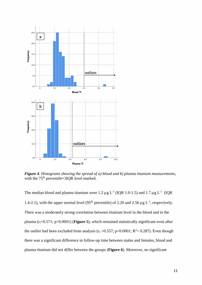
larger than 3IQR from the 75th percentile) which was included in the statistical analyses
(Figure 4).
11
Figure 4. Histograms showing the spread of a) blood and b) plasma titanium measurements,
with the 75th percentile+3IQR level marked.
The median blood and plasma titanium were 1.2 g L-1 (IQR 1.0-1.5) and 1.7 g L-1 (IQR
1.4-2.1), with the upper normal level (95th percentile) of 2.20 and 2.56 g L-1, respectively.
There was a moderately strong correlation between titanium level in the blood and in the
plasma (rs=0.571; p<0.0001) (Figure 5), which remained statistically significant even after
the outlier had been excluded from analysis (rs =0.557; p<0.0001; R2= 0.287). Even though
there was a significant difference in follow-up time between males and females, blood and
plasma titanium did not differ between the groups (Figure 6). Moreover, no significant

12
correlations between blood titanium values and age (r=0.056; p= 0.59), implant months in
situ (rs=-0.151; p=0.14), OHS (rs=-0.01; p=0.92) or activity score (rs=-0.012; p=0.91) were
detected. Similarly, there were no significant correlations between plasma titanium values
and age (r=0.052; p=0.61), OHS (rs=-0.058; p=0.58) and activity score (rs=-0.033; p=0.75).
We detected a weak negative association between plasma titanium and implant years in situ
(rs=-0.313; p=0.002).
Figure 5. Scatter plot of plasma versus blood titanium, showing a statistically significant
positive correlation between the two (R2= 0.583; p<0.0001).
a
a

13
Figure 6. Box plots of titanium concentration in a) blood and b) plasma of study participants.
The boundaries of the box represent the 25th and 75th percentile, with the median line inside
the box. The whiskers extend to maximum and minimum values in each data set. Outliers
(values more than 1.5xIQR from the end of the box) are identified as open circles, while
extreme outliers (values more than 3xIQR from the end of the box) are denoted as asterisks.
3.2. Radiographs
All the hip implants were well-positioned, with no signs of cup loosening or gross soft tissue
changes evident from the radiographs (Figure 7). Two femoral stems (2%) were
radiologically loose, but clinically asymptomatic (Figure 8).
b
Figure 7. A representative anteroposterior radiograph from the current series, showing a
well-fixed hip implant.

14
Figure 8. Radiologically loose stem.
3.3. Oxford Hip Score
84 (88%) patients had excellent hip function and 8 (8%) had good hip function. The
remaining 3 patients with fair function reported that the lower scores were due to severe
arthritis in other joints or spinal stenosis. One patient with unexplained pain in the replaced
hip, and an OHS score of 16, was excluded from the study.
4. Discussion and conclusions
This study proposes laboratory threshold values for blood (2.20 µg L-1) and plasma (2.56 µg
L-1) titanium in patients with well-functioning titanium hip implants at medium-to-long term
follow up. These guidelines are an essential starting point for further studies to explore the
clinical usefulness of blood titanium as a biomarker of orthopaedic implant performance, and
come at a time of considerable controversy regarding the use of certain titanium alloys in hip
arthroplasty.
Compared to cobalt and chromium, relatively little research has been directed to the
biological effects of titanium ions and, unlike for blood cobalt and chromium, a “cut-off”
value has not been defined for titanium. The current Mayo Clinic Laboratory guidelines state

15
that a prosthetic device in good condition should give rise to serum titanium in the range of 1-
3 µg L-1, while concentrations exceeding 10 µg L-1 indicate prosthesis wear. These values are
based on the 1998 works by Jacobs and Liu. Jacobs et al. [22] used GF AAS to quantify
serum titanium in 55 patients with 3 different types of well-functioning hip implants, while
Liu et al. [23] measured blood titanium associated with well-fixed (5 patients) and loose (4
patients) knee prostheses. More recently, Jacobs et al. [4] proposed that well-functioning,
unilateral hip implants should produce serum titanium levels of approximately 4 µg L-1, while
levels exceeding 8 µg L-1 should warrant further investigation. Savarino et al. found that the
upper normal reference limit was 5.13 µg L-1 in the medium term (2-7-year follow up) [24],
and 4.5 µg L-1 in the long term (10-year follow up) [25]. It is important to note that all of the
above studies employed GF AAS for trace metal analysis, which is, as is now known, not
sensitive/selective enough to measure titanium accurately [26]. The instrumental LoD quoted
in the Savarino study (2.91 µg L-1) was likely not low enough to appreciate the subtle
titanium elevations in patients with well-functioning prostheses. Studies employing HR ICP-
MS point to lower values, which generally decrease with increasing length of follow up
(Table 2). Discrepancies between “normal” serum levels measured by GF AAS and the more
powerful HR ICP-MS were previously observed for aluminium, where the latter technique
obtained values one order of magnitude lower than those published before using GF AAS
[27].

16
Table 2. Blood and serum titanium levels associated with different types of well-functioning titanium-based hip implants. 1
Ref Analytical technique LoD (µg L-1) Blood Ti (µg L-1)a Serum Ti (µg L-1)a Implant fixation/ function assessment Follow up
[26] HR ICP-MS 1.1 2.306 (n=11)
1.519 (n=11)
n/a
n/a
Not assessed 14-22 months
70-106 months
[28] HR ICP-MS 0.05 3.0 (n=9 males)
2.2 (n=6 females)
n/a
n/a
Not assessed n/a
n/a
[29] HR ICP-MS <0.1 3.74; 1.40-8.80 (n=34)
2.75; 1.40-4.10 (n=33)
1.83; 0.90-4.60 (n=31)
1.30; 0.35-2.40 (n=24)
n/a
n/a
n/a
n/a
X-Ray/ WOMAC, Merle D’Aubigne and
Postel scores
3 months
6 months
1 years
2 years
[30] HR ICP-MS <0.1 n/a 2.7; 1.1-7.0 (n=6) Not assessed 7-13 months
[31] HR ICP-MS 0.2 n/a 1.8; 1.7-1.9 (n=8) X-Ray/ Harris Hip Score 10 years
[32] HR ICP-MS
<0.17 n/a
n/a
2.54; 2.17-3.10 (n=23)
2.70; 2.11-3.25 (n=23)
Not assessed 1 years
2 years
[33] HR ICP-MS <0.4 2.165 (n=15)
1.359 (n=15)
2.160 (n=15)
n/a
n/a
n/a
X-ray/ Short-Form 12, WOMAC, Harris
Hip Score
1 years
2 years
5 years
[14] HR ICP-MS <2.0 n/a 2.28 (n=74) X-ray/ WOMAC, Harris Hip Score 50 months
aResults are reported as “mean/median, range (sample size)”; WOMAC- Western Ontario and McMaster Universities Osteoarthritis Index, LoD-limit of 2 detection.3

17
4.1. Comparison with other studies using HR ICP-MS 4
The mean titanium level in the current series was 1.35 g L-1 in whole blood and 1.85 g L-1 5
in the plasma, at a mean follow up of 8.5 years (range 5-12). This is consistent with the work 6
of Sarmiento-Gonzalez et al. [26], who investigated 11 patients with titanium-based hip 7
implants, and reported a mean blood level of 1.52 g L-1 at a mean 6.5 years after surgery 8
(range 6-9). Levine et al. [31], who measured serum titanium in 8 patients with well-9
functioning (confirmed with X-rays and Harris Hip Score) metal-on-polyethylene 10
implants, noted that the levels peaked at 3 years and then proceeded to decline until after the 11
9-year interval. Mean serum titanium at 9-year follow up reported by the authors was 12
approximately 1.8 g L-1, which is in very close agreement with our findings. The decreasing 13
trend in plasma titanium over time was also apparent in the present study, and could mean 14
that the rate of implant degradation decreases, or that the efficiency with which titanium ions 15
are excreted improves, with increasing implant time in situ. However, the most likely 16
explanation is that the metal is slowly accumulated in systemic tissue [34]. The clinical 17
implications of titanium deposition, and chronic low-level exposure to titanium ions, are yet 18
to be established. While titanium is considered to be less toxic than cobalt and chromium, the 19
issue of titanium sensitivity/allergy is still under discussion [35]. It has been suggested that 20
those with a pre-operative sensitivity to titanium might be at an increased risk of local 21
adverse reactions to titanium debris [36]. 22
Titanium levels were not influenced by age, gender or OHS, which was expected, since all 23
the implants displayed excellent or good function. Titanium measurements were not 24
correlated with activity level either. Vendittoli et al. [29] was also unable to find a significant 25
correlation between UCLA Activity Score and post-operative blood titanium level at 1-2- 26
year follow up. Taken together, these findings suggest that increased physical activity does 27
not influence the magnitude of titanium release from hip implants. Titanium is not part of the 28

18
bearing surface, and its release is thought to occur via passive corrosion of the acetabular cup 29
and/or the femoral stem, rather than through wear [32]. Corrosion, unlike wear, is unlikely to 30
be affected by physical activity, which helps to explain our findings. 31
32
4.2. Whole blood versus plasma/serum sampling 33
Titanium ions associate with plasma proteins [37], which accounts for the higher titanium 34
content of plasma compared to whole blood [38]. Serum/plasma sampling is preferred by 35
some researchers, because the higher titanium levels are easier to detect and quantify. The 36
official Medicines and Healthcare products Regulatory Agency (MHRA) guidelines call for 37
the measurement of cobalt and chromium in whole blood, but it is still unclear which blood 38
fraction is optimal for monitoring titanium levels in THA patients. Based on our experience, 39
we recommend analysis of whole blood when a high resolution instrument with a low LoD is 40
employed. The extra processing steps required for serum/plasma separation might introduce 41
contaminants into the sample and have a devastating effect on the results of trace element 42
assays, particularly when minute titanium concentrations are being measured [39]. 43
44
4.3. Study strengths 45
The main strengths of our investigation lie in the clean data set we obtained and the state-of-46
the art analytical technique we employed for trace metal analysis. All the participants 47
received the same implant and were operated on by the same team, thus removing surgical 48
variability. Our study also boasts a large sample size, which enabled us to obtain accurate 49
estimates of “normal” titanium levels in THA patients. Correct implant placement and 50
fixation were evaluated radiologically, while good clinical function was ensured with 51
excellent or good OHS, which was not always done in previous studies. The X-rays and 52
questionnaires were taken on the same day as the blood samples to make sure that the trace 53

19
metal analysis was as meaningful as possible. The analytical approach we employed is 54
considered to be one of the most effective for the determination of titanium in biological 55
fluids [31]. HR ICP-MS boasts a short run time, low LoD (1-3 orders of magnitude lower 56
than GF AAS [40,41]) and is able to measure multiple elements/isotopes simultaneously, 57
making it possible to analyse a large number of samples quickly and reliably. Our results are 58
in close agreement with previous smaller studies that used the same technique to determine 59
blood/serum titanium in THA patients with well-functioning implants [26,31]. 60
61
4.4. Study limitations 62
The present study has a number of limitations. First, pre-operative blood titanium 63
measurements were unavailable, so that true influence of the hip implant on the systemic 64
titanium load could not be assessed. As blood/serum titanium level in patients without 65
orthopaedic implants is thought to be lower than 1 µg L-1 [8], the values observed in the 66
current series are considered to be mildly elevated. While we believe that the main source of 67
raised metal levels was the hip implant, it is likely that external sources contributed to it. 68
Titanium dioxide (TiO2) is added in varying levels to many foodstuffs and personal care 69
products [42], as well as being present in ambient air. HR ICP-MS quantifies both ionic and 70
particulate titanium [43], and is unable to discriminate between the two forms of the metal. 71
None of the participants disclosed occupational exposure to titanium, but dietary intake, and 72
the use of titanium-containing products, were impossible to control for. Secondly, recent 73
creatinine clearance data was unavailable, so we could not estimate the participants’ renal 74
function (impaired excretion could lead to increased retention of metals in the body [27]). 75
Titanium is highly insoluble and tends to accumulate in tissue, with only a small fraction 76
excreted in the urine [44,45]. It follows that kidney function is unlikely to influence the 77
systemic titanium load to a great extent. 78

20
79
4.5. Clinical significance 80
The use of titanium in THA is increasing, and there is a renewed interest in blood titanium as 81
a biomarker of implant wear. In particular, it is thought that elevated blood levels could help 82
identify malfunctioning prostheses before they fail [4,5,7], though the normal “cut-off” value 83
to guide diagnosis of malfunctioning implants has not been well-defined. We aimed to bridge 84
this data gap with the present study, which is the largest investigation of systemic titanium 85
levels associated with well-functioning titanium-based implants to date. Future work in this 86
area should involve applying our guidelines (2.20 g L-1 for blood and 2.56 g L-1 for plasma 87
titanium) to other patients, and looking for radiographic signs of ARMD, implant loosening 88
and wear. This approach could be a means of monitoring the tens of thousands of patients 89
with titanium-based hips, and could help predict which might develop clinical problems. 90
91
Acknowledgments 92
The authors would like to thank The Horder Centre for their invaluable support and 93
assistance, with a special thank you to the radiology and phlebotomy teams, and Ms Tracy 94
Young. 95
96
Funding 97
This work was supported by Gwen Fish Orthopaedic Trust and The Horder Centre. The 98
funding sources did not have involvement in the analysis and interpretation of data, in the 99
writing of the report or in the decision to submit the article for publication. 100
101
References 102
[1] O. Posada, R. Tate, R.M. Meek, M. Grant, In vitro analyses of the toxicity, 103 immunological, and gene expression effects of cobalt-chromium alloy wear debris and 104

21
Co ions derived from metal-on-metal hip implants, Lubricants. 3 (2015) 539–568. 105 [2] B. Sampson, A. Hart, Clinical usefulness of blood metal measurements to assess the 106
failure of metal-on-metal hip implants., Ann. Clin. Biochem. 49 (2012) 118–31. 107 doi:10.1258/acb.2011.011141. 108
[3] A.J. Hart, S.A. Sabah, A.S. Bandi, P. Maggiore, P. Tarassoli, B. Sampson, J. A. 109 Skinner, Sensitivity and specificity of blood cobalt and chromium metal ions for 110 predicting failure of metal-on-metal hip replacement, Bone Joint J. 93-B (2011) 1308–111 1313. doi:10.1302/0301-620X.93B10.26249. 112
[4] J.J. Jacobs, A.K. Skipor, P.A. Campbell, N.J. Hallab, R.M. Urban, H.C. Amstutz, Can 113 metal levels be used to monitor metal-on-metal hip arthroplasties?, J. Arthroplasty. 19 114 (2004) 59–65. doi:10.1016/j.arth.2004.09.019. 115
[5] I.P. McAlister, M.P. Abdel, Elevated serum titanium level as a marker for failure in a 116 titanium modular fluted tapered stem, Orthopedics. 39 (2016) e768–e770. 117
[6] H. von Schroeder, D. Smith, A. Gross, R. Piliar, R. Kandel, R. Chernecky, S. 118 Lugowski, Titanemia From Total Knee Arthroplasty, J. Arthroplasty. 11 (1996) 620–119 625. 120
[7] R.M. Urban, J.J. Jacobs, M.J. Tomlinson, J. Gavrilovic, J. Black, M. Peoc’h, 121 Dissemination of wear particles to the liver, spleen, and abdominal lymph nodes of 122 patients with hip or knee replacement, J. Bone Joint Surg. Am. 82-A (2000) 457–476. 123 http://www.ncbi.nlm.nih.gov/pubmed/10761937. 124
[8] I. Swiatkowska, N. Martin, A.J. Hart, Blood titanium level as a biomarker of 125 orthopaedic implant wear, J. Trace Elem. Med. Biol. 53 (2019) 120–128. 126 doi:10.1016/j.jtemb.2019.02.013. 127
[9] S.M. Kurtz, E. Lau, K. Ong, K. Zhao, M. Kelly, K.J. Bozic, Future young patient 128 demand for primary and revision joint replacement: National projections from 2010 to 129 2030, Clin. Orthop. Relat. Res. 467 (2009) 2606–2612. doi:10.1007/s11999-009-0834-130 6. 131
[10] D.S. Casper, G.K. Kim, C. Restrepo, J. Parvizi, R.H. Rothman, Primary Total Hip 132 Arthroplasty With an Uncemented Femoral Component. Five- to Nine-Year Results, J. 133 Arthroplasty. 26 (2011) 838–841. doi:10.1016/j.arth.2011.02.010. 134
[11] M.M. Morlock, E.C. Dickinson, K.-P. Günther, D. Bünte, V. Polster, Head taper 135 corrosion causing head bottoming out and consecutive gross stem taper failure in total 136 hip arthroplasty, J. Arthroplasty. xxx (2018) 1–10. 137
[12] J. Spanyer, J. Hines, C.M. Beaumont, J. Yerasimides, Catastrophic Femoral Neck 138 Failure after THA with the Accolade®I Stem in Three Patients, Clin. Orthop. Relat. 139 Res. 474 (2016) 1333–1338. doi:10.1007/s11999-015-4438-z. 140
[13] S.M. Kurtz, S.B. Kocagöz, J.A. Hanzlik, R.J. Underwood, J.L. Gilbert, D.W. 141 MacDonald, G.C. Lee, M.A. Mont, M.J. Kraay, G.R. Klein, J. Parvizi, C.M. Rimnac, 142 Do ceramic femoral heads reduce taper fretting corrosion in hip arthroplasty? A 143 retrieval study, Clin. Orthop. Relat. Res. 471 (2013) 3270–3282. doi:10.1007/s11999-144 013-3096-2. 145
[14] Z. Yi, Z. Bo, S. Bin, Y. Jing, Z. Zongke, P. Fuxing, Clinical Results and Metal Ion 146 Levels After Ceramic-on-Metal Total Hip Arthroplasty: A Mean 50-Month 147 Prospective Single-Center Study, J. Arthroplasty. 31 (2016) 438–441. 148 doi:10.1016/j.arth.2015.09.034. 149
[15] P.-A. Vendittoli, A. Roy, S. Mottard, J. Girard, D. Lusignan, M. Lavigne, Metal ion 150 release from bearing wear and corrosion with 28 mm and large-diameter metal-on-151 metal bearing articulations: a follow-up study, J. Bone Jt. Surgery-British Vol. 92-B 152 (2010) 12–19. 153
[16] Eurachem Method Validation Working Group, The fitness for purpose of analytical 154

22
methods a laboratory guide to method validation and related topics (Second Edition), 155 2014. doi:10.1016/S0014-2999(99)00500-2. 156
[17] D.W. Murray, R. Fitzpatrick, K. Rogers, H. Pandit, D.J. Beard, A.J. Carr, J. Dawson, 157 The use of the Oxford hip and knee scores, J. Bone Joint Surg. Br. 89-B (2007) 1010–158 1014. 159
[18] Y. Kalairajah, K. Azurza, C. Hulme, S. Molloy, K.J. Drabu, Health outcome measures 160 in the evaluation of total hip arthroplasties- a comparison between the harris hip score 161 and the Oxford hip score, J. Arthroplasty. 20 (2005) 1037–1041. 162
[19] J. Dawson, R. Fitzpatrick, A. Carr, D. Murray, Questionnaire on the perceptions of 163 patients about total hip replacement, J. Bone Joint Surg. Br. 78-B (1996) 185–190. 164
[20] V. Wylde, I. Learmonth, V. Cavendish, The Oxford hip score: the patient’s 165 perspective, Health Qual. Life Outcomes. 3 (2005) 1–8. 166
[21] P. Campbell, R. Urban, I. Catelas, A. Skipor, T. Schmalzried, Autopsy analysis thirty 167 years after metal-on-metal total hip replacement, J. Bone Jt. Surg. 85 (2003) 2218–168 2222. 169
[22] J. Jacobs, A. Skipor, L. Patterson, N. Hallab, W. Paprosky, J. Black, J. Galante, Metal 170 Release in Patients Who Have Had a Primary Total Hip Arthroplasty. A Prospective, 171 Controlled, Longitudinal Study, J. Bone Jt. Surg. 80-A (1998) 1447–1458. 172 doi:10.1016/0142-9612(85)90030-4. 173
[23] T.-K. Liu, S.-H. Liu, C.-H. Chang, R.-S. Yang, Concentration of metal elements in the 174 blood and urine in the patients with cementless total knee arthroplasty, J. Exp. Med. 175 185 (1998) 253–262. 176
[24] L. Savarino, M. Greco, E. Cenni, L. Cavasinni, R. Rotini, N. Baldini, A. Giunti, 177 Differences in ion release after ceramic-on-ceramic and metal-on-metal total hip 178 replacement, J. Bone Joint Surg. Br. 88-B (2006) 472–476. 179
[25] L. Savarino, G. Padovani, M. Ferretti, M. Greco, E. Cenni, G. Perrone, F. Greco, N. 180 Baldini, A. Giunti, Serum ion levels after ceramic-on-ceramic and metal-on-metal total 181 hip arthroplasty: 8-Year minimum follow-up, J. Orthop. Res. 26 (2008) 1569–1576. 182 doi:10.1002/jor.20701. 183
[26] A. Sarmiento-González, J.M. Marchante-Gayón, J.M. Tejerina-Lobo, J. Paz-Jiménez, 184 A. Sanz-Medel, High-resolution ICP-MS determination of Ti, V, Cr, Co, Ni, and Mo 185 in human blood and urine of patients implanted with a hip or knee prosthesis, Anal. 186 Bioanal. Chem. 391 (2008) 2583–2589. doi:10.1007/s00216-008-2188-4. 187
[27] C.S. Muñiz, J.L. Fernández-Martin, J.M. Marchante-Gayón, J.I.G. Alonso, J.B. 188 Cannata-Andía, A. Sanz-Medel, Reference values for trace and ultratrace elements in 189 human serum determined by double-focusing ICP-MS, Biol. Trace Elem. Res. 82 190 (2001) 259–272. doi:10.1385/BTER:82:1-3:259. 191
[28] Y. Nuevo Ordóñez, M. Montes-Bayón, E. Blanco-González, J. Paz-Jiménez, J.M. 192 Tejerina-Lobo, J.M. Peña-López, A. Sanz-Medel, Metal release in patients with total 193 hip arthroplasty by DF-ICP-MS and their association to serum proteins, J. Anal. At. 194 Spectrom. 24 (2009) 1037–1043. doi:10.1039/b820339c. 195
[29] P.-A. Vendittoli, A. Roy, S. Mottard, J. Girard, D. Lusignan, M. Lavigne, Metal ion 196 release from bearing wear and corrosion with 28 mm and large-diameter metal-on-197 metal bearing articulations: A follow-up study, J. Bone Jt. Surg. - Br. Vol. 92-B (2010) 198 12–19. doi:10.1302/0301-620X.92B1.22226. 199
[30] G.W. Omlor, J.P. Kretzer, J. Reinders, M.R. Streit, T. Bruckner, T. Gotterbarm, P.R. 200 Aldinger, C. Merle, In vivo serum titanium ion levels following modular neck total hip 201 arthroplasty-10 year results in 67 patients, Acta Biomater. 9 (2013) 6278–6282. 202 doi:10.1016/j.actbio.2012.12.001. 203
[31] B.R. Levine, A.R. Hsu, A.K. Skipor, N.J. Hallab, W.G. Paprosky, J.O. Galante, J.J. 204

23
Jacobs, Ten-Year Outcome of Serum Metal Ion Levels After Primary Total Hip 205 Arthroplasty, J. Bone Jt. Surg. 95 (2013) 512–518. doi:10.2106/JBJS.L.00471. 206
[32] W. Gofton, P.E. Beaule, Serum metal ions with a titanium modular neck total hip 207 replacement system, J. Arthroplasty. 30 (2015) 1781–1786. 208
[33] D. Nam, J.A. Keeney, R.M. Nunley, S.R. Johnson, J.C. Clohisy, R.L. Barrack, Metal 209 ion concentrations in young, active patients following total hip arthroplasty with the 210 use of modern bearing couples, J. Arthroplasty. 30 (2015) 2227–2232. 211
[34] I. Swiatkowska, J.F.W. Mosselmans, T. Geraki, C.C. Wyles, J.J. Maleszewski, J. 212 Henckel, B. Sampson, D.B. Potter, I. Osman, R.T. Trousdale, A.J. Hart, Synchrotron 213 analysis of human organ tissue exposed to implant material, J. Trace Elem. Med. Biol. 214 46 (2018) 128–137. doi:10.1016/j.jtemb.2017.12.007. 215
[35] M. Goutam, C. Giriyapura, S. Mishra, S. Gupta, Titanium allergy: A literature review, 216 Indian J. Dermatol. 59 (2014) 630. doi:10.4103/0019-5154.143526. 217
[36] E.J. Mcpherson, M. V Dipane, S.M. Sherif, Massive pseudotumor in a 28mm ceramic- 218 polyethylene revision THA: a case report, JISRF Reconstr. Rev. 4 (2014) 11–17. 219
[37] Y. Nuevo-Ordonez, M. Montes-Bayon, E. Blanco Gonzalez, A. Sanz-Medel, Y. 220 Nuevo-Ordoñez, M. Montes-Bayón, E. Blanco González, A. Sanz-Medel, Titanium 221 preferential binding sites in human serum transferrin at physiological concentrations., 222 Met. Integr. Biometal Sci. 3 (2011) 1297–303. doi:10.1039/c1mt00109d. 223
[38] C.F. Harrington, C. Mckibbin, M. Rahanu, D. Langton, Titanium in hip replacement 224 patients by Inductively Coupled Plasma Optical Emission Spectroscopy (ICP-OES), 225 Ann. Clin. Biochem. 0 (2016) 1–8. doi:10.1177/0004563216662292. 226
[39] J. Versieck, The Collection and Preparation of Human-Blood Plasma or Serum for 227 Trace-Element Analysis, J. Res. Natl. Bur. Stand. (1934). 91 (2012) 87. 228 doi:10.6028/jres.091.014. 229
[40] J. Jacobs, A. Skipor, J. Black, R. Urban, J. Galante, Release and excretion of metal in 230 patients who have a total hip-replacement component made of titanium-base alloy, J. 231 Bone Jt. Surg. 73-A (1991) 1475–1486. 232
[41] J. Jacobs, A. Skipor, L. Patterson, N. Hallab, W. Paprosky, J. Black, J. Gallante, Metal 233 release in patients who have had a primary total hip arthroplasty, J. Bone Jt. Surg. 80-234 A (1998) 1447–1458. 235
[42] A. Weir, P. Westerhoff, L. Fabricius, K. Hristovski, N. Von Goetz, Titanium dioxide 236 nanoparticles in food and personal care products, Environ. Sci. Technol. 46 (2012) 237 2242–2250. doi:10.1021/es204168d. 238
[43] D. Koller, P. Bramhall, J. Devoy, H. Goenaga-Infante, C.F. Harrington, E. Leese, J. 239 Morton, S. Nuñez, J. Rogers, B. Sampson, J.J. Powell, Analysis of soluble or titanium 240 dioxide derived titanium levels in human whole blood: consensus from an inter-241 laboratory comparison, Analyst. (2018). doi:10.1039/C8AN00824H. 242
[44] K. Merritt, S.A. Brown, Storage and elimination of titanium, aluminum, and vanadium 243 salts in vivo, J. Biomed. Mater. Res. 26 (1992) 1503–1515. 244
[45] S. Takai, N. Yoshino, Y. Kusaka, Y. Watanabe, Y. Hirasawa, Dissemination of metals 245 from a failed patellar component made of titanium-base alloy, J. Arthroplasty. 18 246 (2003) 931–935. 247
248
24
249 250 Appendix 251 252 Table A.1. Internal Quality Control results. 253 254
IQC material
Mean
measured
concentration
(µg L-1)
Manufacturer's
target (µg L-1)
Bias to
manufacturer's
target (%)
ClinChek Plasma 1 11.5 12.4 -6.9
ClinChek Plasma 2 49.3 47.3 4.2
Custom Whole Blood 1 0.6 N/A N/A
Custom Whole Blood 2 10.7 N/A N/A
255 Table A.2. Results submitted by the laboratory to the Quebec Multielement External Quality 256 Assessment Scheme (QMEQAS), the value assigned by QMEQAS, and the range considered 257 acceptable by QMEQAS (See the QMEQAS participants manual for details). The percentage 258 bias of the submitted results compared to the assigned value is also shown. 259
Sample
Submitted results
(nmol L-1)
Assigned value
(nmol L-1)
Acceptable
range
Bias (%)
QM-B-Q1901 196 207 123 - 291 -5.3
QM-B-Q1902 319 307 181 - 433 +3.9
QM-B-Q1903 247 246 143 - 349 +0.4
260 Table A.3. Isotopes measured and ICP-MS settings used. 261 262
Isotope Ti47 Ga71
Mass range 46.948 - 46.954 70.921 - 70.928
Mass window 50 40
Settling time 0.300 0.050
Sample time 0.7500 0.0500
Samples per peak 25 20
Search window 50 40
Integration window 50 40
Scan type EScan EScan
Resolution mode Medium Medium
Runs 4 4
Passes 1 1
263