blood vessels restrain pancreas branching,...
TRANSCRIPT
4743RESEARCH ARTICLE
INTRODUCTIONThe primary function of blood vessels is to provide organs withoxygen and nutrients that are essential for tissue growth andmaintenance. In recent years, additional roles of vascularendothelial cells in organ development and tissue homeostasis havebeen discovered. In the case of the embryonic liver and pancreas,seminal studies have shown that blood vessels provide perfusion-independent paracrine signals, which are crucial for early stages oforgan development and differentiation (Jacquemin et al., 2006;Lammert et al., 2001; Matsumoto et al., 2001; Yoshitomi and Zaret,2004). Paracrine factors secreted from endothelial cells also supportadult liver regeneration (LeCouter et al., 2003). Survival and properfunction of adult pancreatic -cells were shown to depend onextracellular matrix produced by blood vessels (Nikolova et al.,2006; Zaret, 2006). In addition, it has been proposed that a‘vascular niche’ maintains self-renewal in several adult stem cellsystems, including the brain, testis, bone marrow and fat (Butler etal., 2010b; Palmer et al., 2000; Shen et al., 2008; Tang et al., 2008;Tavazoie et al., 2008; Yoshida et al., 2007). At least for neuronaland hematopoietic stem cells, blood vessels are able to impact self-renewal in vitro in the absence of blood flow, demonstrating theimportance of paracrine signals from endothelial cells (Butler et al.,2010b; Shen et al., 2004). The effects of blood vessels on self-renewal of hematopoietic stem cells appear to involve theactivation of Notch signaling within stem cells (Butler et al.,2010b).
Notably, the net effect of blood vessels on tissue development inall these cases is seemingly a positive one. Endothelial cellsprovide important cues for early organ formation, maintain stem
cell potential and help carrying the limiting nutrients needed fortissue growth beyond the minimal size afforded by passivediffusion.
Here, we describe a series of experiments that examine the roleof blood vessels during the morphogenetic stages of pancreasdevelopment, using both endothelial cell ablation and forcedhypervascularization. During these stages (embryonic days 10-13in the mouse), the pancreas starts to undergo an extensive processof branching morphogenesis, growth and the beginning ofdifferentiation (Cleaver and MacDonald, 2009; Gittes, 2009;Jorgensen et al., 2007; Murtaugh, 2007; Oliver-Krasinski andStoffers, 2008). At the tip of each branch, rapidly dividing cells thatexpress Cpa1 and Ptf1a are multipotent progenitors, which leavebehind ‘trunk’ cells (Zhou et al., 2007). As shown by time-lapsestudies (Cleaver and MacDonald, 2009; Puri and Hebrok, 2007;Solar et al., 2009) and lineage tracing (Solar et al., 2009), trunkscan give rise to new lateral branches headed by tips. Endocrineprogenitor cells expressing neurogenin 3 (Ngn3) emerge in thetrunks, delaminate and differentiate to hormone-producing cellsthat will coalesce to form the islets of Langerhans. Trunk cellseventually form the differentiated ductal epithelium. Tip cellsdifferentiate later in development to acinar cells, which secretedigestive enzymes to the ducts (Zhou et al., 2007). We show thatin this context, perfusion-independent signals from blood vesselssurprisingly act to slow down organ growth and have a net negativeeffect on organ size. The underlying mechanism is vascularsustenance of a simple tubular pancreatic epithelium, and therestriction of pancreas tip and endocrine progenitor cell formation.Consequently, blood vessels favor reduced branching and inhibitendocrine and exocrine differentiation.
MATERIALS AND METHODSMicePdx1-tTA mice (Holland et al., 2002) were the generous gift from RayMacDonald (University of Texas Southwestern, TX, USA). TET-VEGF(Dor et al., 2001), TET-sFLT1 (Dor et al., 2001; May et al., 2008), Flk1-lacZ (Shalaby et al., 1997) and Pdx1-GFP (Gu et al., 2004) mice have beendescribed previously. Embryos were genoyped by PCR using the followingprimers: Pdx1-tTA, 5�-TAGATGTGCTTTACTAAGTCATCGCG-3� and
Development 138, 4743-4752 (2011) doi:10.1242/dev.066548© 2011. Published by The Company of Biologists Ltd
1Department of Developmental Biology and Cancer Research, The Institute forMedical Research Israel-Canada, The Hebrew University-Hadassah Medical School,Jerusalem 91120, Israel. 2Cyclotron/Radiochemistry Unit, Hadassah-HebrewUniversity Medical Center, Jerusalem, 91120, Israel. 3Department of MolecularBiology, UT Southwestern Medical Center, Dallas, TX 75235, USA.
*Author for correspondence ([email protected])
Accepted 26 August 2011
SUMMARYHow organ size and form are controlled during development is a major question in biology. Blood vessels have been shown to beessential for early development of the liver and pancreas, and are fundamental to normal and pathological tissue growth. Here,we report that, surprisingly, non-nutritional signals from blood vessels act to restrain pancreas growth. Elimination of endothelialcells increases the size of embryonic pancreatic buds. Conversely, VEGF-induced hypervascularization decreases pancreas size. Thegrowth phenotype results from vascular restriction of pancreatic tip cell formation, lateral branching and differentiation of thepancreatic epithelium into endocrine and acinar cells. The effects are seen both in vivo and ex vivo, indicating a perfusion-independent mechanism. Thus, the vasculature controls pancreas morphogenesis and growth by reducing branching anddifferentiation of primitive epithelial cells.
KEY WORDS: Pancreas development, VEGF, Blood vessels, Branching, Organ size, Vascular niche, Mouse
Blood vessels restrain pancreas branching, differentiationand growthJudith Magenheim1, Ohad Ilovich2, Alon Lazarus1, Agnes Klochendler1, Oren Ziv1, Roni Werman1, Ayat Hija1,Ondine Cleaver3, Eyal Mishani2, Eli Keshet1 and Yuval Dor1,*
DEVELO
PMENT
4744
5�-GAGATCGAGCGGGCCCTCGATGGTAG-3�; TET-VEGF, 5�-CGCGAAGCTTCCACCATGGACTTTCTGCTCTCTTGGGT-3� and 5�-CGCGGATATCACCGCCTTGGCTTGTCACA-3�; Flk1-lacZ, 5�-CAACAGTTGCGCAGCCTGAATGG-3� and 5�-AAATCGCTGAT -TTGTGTAGTCGGT-3�; Pdx1-GFP, 5�-TCGTGGAACTGGA TGG -CGATG-3� and 5�-CTTCAGCTCGATTCTATTCACCA-3�. The jointethics committee (IACUC) of the Hebrew University and HadassahMedical Center approved the study protocol for animal welfare. TheHebrew University is an AAALAC International accredited institute.
Pancreatic explantsPancreatic buds were cultured on filters, in the liquid-air interface, aspreviously described (van Eyll et al., 2004). Briefly, the dorsal pancreaticbud from embryonic day (E)12.5 mouse embryos was excised under adissecting microscope in Hank’s balanced salts solution (HBSS).Alternatively, the gut and pancreas from E9.5 embryos were isolated. Theexplants were cultured in DMEM supplied with 10% fetal bovine serum,containing 100 U/ml penicillin and 100 mg/ml streptomycin. Explants werecultured on microporous membranes and no medium was added on top ofthe filter, so that the tissue grew at the air/medium interface. Medium waschanged every other day.
To ablate endothelial cells in pancreatic explants, we used 1-[4-(6,7-Dimethoxy-quinolin-4-yloxy)-3-fluoro-phenyl]-3-(2-fluoro-phenyl)-urea(Ilovich et al., 2008) (VEGFR2i). The molecule has a low IC50 (5-15 nM)to VEGFR2 and PDGFRb, and a high IC50 (>500 nM) for IGF1R, EGFRand Her2. Drug was dissolved in DMSO (stock solution 25 mM) and addedto explants at a final concentration of 100 nM. -Secretase inhibitor XX(Calbiochem, catalog number 565789), was added to explants at a finalconcentration of 100 nM.
Immunostaining, antibodies and microscopyWhole-mount immunostaining was performed essentially as describedpreviously (Ahnfelt-Ronne et al., 2007b). Alternatively, staining wasperformed on 5 mm paraffin sections of dissected guts (E15.5) or isolatedpancreata (E15.5-P3). Tissue was fixed in 4% buffered zinc-formalin for 2hours at 4°C, dehydrated in an ethanol series, cleared in histoclear andembedded in Paraplast (Kendall). When required, antigen retrieval wasperformed using a pressure cooker. Primary antibodies used in this studyincluded: guinea pig anti-insulin (1:500; DAKO), armenian hamster anti-Muc1 (1:250; Labvision), rat anti-CD31/Pecam (1:50; BD), rabbit anti-amylase (1:100; Sigma), rabbit anti-carboxypeptidase 1 (1:200; Rockland),guinea pig, goat and rabbit anti-Pdx1 (1:2500; a generous gift from ChrisWright, Vanderbilt University, TN, USA), rabbit anti-ptf1a (1:2000; BetaCell Biology Consortium), rabbit anti-Ngn3 (1:500; Beta Cell BiologyConsortium), guinea pig anti-Ngn3 (1:500; a generous gift from MaikeSander, UCSD, CA, USA), mouse anti-Nkx2.2 (1:50; DevelopmentalStudies Hybridoma Bank), rabbit anti Nkx6.1 (1:800; Beta Cell BiologyConsortium), rabbit anti phospho histone H3 (PHH3) (1:100; CellSignaling), rabbit anti active caspase 3 (1:100; Cell Signaling), andHypoxyprobe (Hypoxyprobe Kit, Chemicon). For DNA counterstain weused DAPI (Sigma). Secondary antibodies were from JacksonImmunoResearch, used at 1:200. Immunofluorescence images werecaptured on a Nikon C1 confocal microscope or Olympus FV1000. ForFACS analysis, E15.5 pancreata from Pdx1-GFP embryos were dissociatedto single cells with Collagenase P (2 mg/ml, Roche), and stained with eitherApc-conjugated anti-Flk1 (1:20; BD) or rabbit anti-Flt1 (1:100; SantaCruz) followed by a secondary antibody Cy5 anti-rabbit (1:800; Jackson).To measure proliferation by FACS, E12.5 explants treated for 3 days withVEGFRi or vehicle were dissociated and stained with antibodies againstPdx1 and Ki67, followed by fluorophore-conjugated secondary antibodies.
RNAFor RT-PCR, total RNA was prepared using Qiagen RNeasy microkitaccording to the manufacturer’s protocol. Total RNA (100 ng) was used forfirst-strand cDNA synthesis using random primers (Roche) and reversetranscriptase (ImProm-II, Promega). Quantitative real-time PCR wasperformed with SYBR Green PCR master mix (Applied Biosystems) in 96-well plates using the 7900HT instrument (Applied Biosystems). Allreactions were performed in triplicates. The relative amount of Ngn3
mRNA was calculated using the comparative CT method afternormalization to Pdx1. Primer sequences were: Ngn3, 5�-ACTGACCTGCTGCTCTCTATTCTTT-3� and ACTGACC TGC -TGCTCTCTATTCTTT-3�; Pdx1, 5�-AGGCCAGTGGGCAGGAG-3� and5�-CTCTTGTTTTCCTCGGGTTCC-3�. For transcriptome analysis, RNAwas isolated using QIAGEN RNeasy micro Kit from pancreatic buds ofPdx1-tTA (n3) and littermate Pdx1-tTA; TET-VEGF (n3) E12.5embryos, or from E12.5 wild-type pancreatic buds explanted and treatedwith VEGFR2i (n5) or vehicle (n5) for 2 days. Affymetrix mouse gene1.0 st arrays were used. The arrays were RMA normalized using PartekGenomic Suite 6.5. Differentially regulated genes were selected based onP-values and ratios using t-test. Data are deposited in Gene ExpressionOmnibus (Accession Number GSE32098).
AnalysisTo measure the area of explants stained in whole mount we obtainedconfocal z-stacks of the whole tissue. The fraction of tissue covered byPdx1 or Muc1 staining was determined using NIS-Elements software.Significance was determined using two-tailed Student’s t-test. A P-valueless than 0.05 was considered significant. Data are presented asmean±s.e.m.
RESULTSBlood vessels restrict the size of the developingpancreasTo examine the role of blood vessels in pancreas morphogenesis andgrowth, we manipulated the vasculature of the embryonic pancreas.To enhance the formation of blood vessels, we crossed Pdx1-tTA andTET-VEGF transgenic mice (Dor et al., 2001; Holland et al., 2002)to drive expression of vascular endothelial growth factor (VEGF)specifically in the pancreas. Importantly, VEGF receptors are notexpressed in the pancreatic epithelium (see Fig. S1 in thesupplementary material), indicating that any effect of VEGF on theepithelium will be indirect, via the vasculature. As expected, VEGFexpression led to massive hypervascularization of the pancreas,evident by the intense staining for the endothelial cell marker Pecam(Fig. 1A). Surprisingly, the pancreas of VEGF-expressing mice wassignificantly smaller compared with littermate controls, duringembryonic development (see Fig. S2 in the supplementary material)as well as immediately after birth (Fig. 1B,C). Organ size wasassessed by documenting total organ size and weight (in newborns)or total Pdx1-stained area (in embryos). As the absolute mass ofblood vessels in transgenic mice was increased, the smaller organsize represented an even more dramatic inhibition of growth of theparenchymal component of the pancreas.
To rule out a possible non-specific toxic effect ofhypervascularization, we performed reciprocal loss of functionexperiments. Pancreata were isolated from E12.5 wild-type embryosand cultured on filters, in the liquid-gas interface (van Eyll et al.,2004). Explants were treated with a small molecule inhibitor ofVEGFR2 (VEGFR2i) (Ilovich et al., 2008), which led to a rapid andtotal ablation of endothelial cells, seen as a complete loss of stainingfor Pecam (Fig. 1D). To validate that endothelial cells wereeliminated and have not simply lost Pecam expression, we treatedFlk1-LacZ explants with the same inhibitor. Loss of X-Gal stainingconfirmed that endothelial cells were indeed efficiently ablated (Fig.1D). In agreement with the TET-VEGF experiments, elimination ofendothelial cells led to a significant increase in organ size after 3days of culture, evident from the larger area stained for the pancreaticmarker Pdx1 relative to vehicle-treated explants (Fig. 1E,F). Vehicleand VEGFR2i-treated explants had similar heights, as determined byconfocal slices along the z axis, indicating that avascular buds had alarger total volume (see Fig. S3 in the supplementary material). As
RESEARCH ARTICLE Development 138 (21)
DEVELO
PMENT

with the VEGF-expressing pancreas, the larger organ size in the faceof reduced vasculature suggests that our measurementsunderestimate the impact on growth of the pancreas.
Finally, we took an independent genetic approach to eliminateblood vessels in the pancreas. Using the same Pdx1-tTA driver linedescribed above, we overexpressed in the pancreatic epithelium asoluble receptor of VEGF, which acts as an efficient VEGF-trap(TET-sFLT1) (May et al., 2008). Expression of the sFLT1transgene led to a dramatic reduction of blood vessel density in thedeveloping pancreas (see Fig. S4 in the supplementary material).In vivo, this led to tissue hypoxia beyond embryonic day 11.5,followed by massive apoptosis, failure of pancreas developmentand death of newborn mice (see Fig. S4 in the supplementarymaterial). The likely contribution of hypoxic stress to thisphenotype precluded the analysis of Pdx1-tTA; TET-sFLT1 micein vivo. However, explant cultures of E9.5 pancreatic buds fromPdx1-tTA; TET-sFLT1 mice grew to a size significantly larger thancontrol littermates (see Fig. S5 in the supplementary material),consistent with the results obtained using the pharmacologicalinhibitor of VEGFR2.
Taken together, these results reveal that, contrary to expectation,blood vessels restrict the growth of the embryonic pancreas. Thefunction of blood vessels is perfusion independent, as the sameeffects are seen in vivo and in explants, cultured in ambient oxygenand standard culture medium in the absence of blood flow.
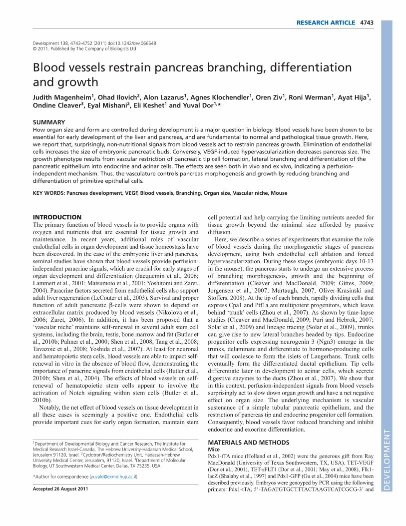
Reduced branching and differentiation inhypervascularized pancreataTo study the basis for the surprising negative effect of blood vesselson pancreas size, we examined how the patterns of branchingmorphogenesis and differentiation are affected by VEGFoverexpression. Examination of VEGF-expressing pancreatarevealed a striking block in branching at E12.5, apparent fromimmunostaining for either Muc1 (Fig. 2A) or Pdx1 (Fig. 2B andsee Fig. S6 in the supplementary material). Transgenic pancreatahad fewer branches, and instead presented with elongatedunbranched tubes.
To determine whether multi-potent tip cells were affected byVEGF overexpression, we examined the expression pattern of the tipcell marker Cpa1. As shown in Fig. 2B, transgenic pancreata had a
4745RESEARCH ARTICLEVasculature restrains pancreas growth and branching
Fig. 1. The vasculature restricts pancreas growth. (A)Hypervascularization of pancreata overexpressing VEGF. Whole-mount staining for Pecam(green) and Pdx1 (red) in pancreata from an E13.5 Pdx1-tTA; TET-VEGF embryo and a control littermate. Scale bar: 100mm. (B)Smaller pancreas innewborn mice (postnatal day 1) overexpressing VEGF. Dotted lines mark the pancreas. Among control animals, top image is from a Pdx1-tTA animaland bottom image is from a TET-VEGF animal. All images are from the same litter. (C)Reduced pancreas weight in newborn mice overexpressingVEGF. n describes the number of mice analyzed. Graphs show cumulative data from three litters. In each litter, wild-type pancreas weight wasnormalized to 1, to correct for inter-litter differences. Data are mean±s.e.m. (D)Elimination of endothelial cells upon treatment of pancreaticexplants with VEGFR2 inhibitor. Pancreatic buds from E12.5 wild-type embryos were cultured for 3 days in the presence or absence of VEGFR2i. Inthe lower panels, mice contained a Flk1-LacZ reporter allele. X-gal stains blood vessels in such mice. Scale bars: 200mm (top); 80mm (bottom).(E)Larger size of E12.5 pancreatic buds cultured for 3 days in the presence of VEGFR2i. Images show confocal z-stacks of explants in whole-mountstained for Pdx1. Shown are representative images from three individual explants. Yellow numbers describe the area stained for Pdx1. Scale bar:200mm. (F)Quantification of explant size after treatment with VEGFR2i, as in E. n describes the total number of explants analyzed. Graphs showcumulative data from ten experiments using ten independent litters. Data are mean±s.e.m.
DEVELO
PMENT
4746
dramatic reduction in Cpa1 staining. Similar results were obtainedwith another tip marker, the transcription factor Ptf1a (see Fig. S6 inthe supplementary material). Both Cpa1 and Ptf1a are also expressedin differentiated acinar cells, and thus changes in their expressioncould reflect a delay in acinar cell differentiation, rather than achange in tip cell formation. To examine this possibility, we stainedembryonic pancreata for the definitive acinar cell marker amylase,which is absent from multipotent tip cells. Amylase expression wasabsent in E12.5 pancreata (see Fig. S7 in the supplementarymaterial), consistent with Cpa1 and Ptf1a expression markingmultipotent tip cells, rather than differentiated acinar cells. Thus,hypervascularization blocks tip cell formation and branching.
We then examined endocrine lineage development in transgenicembryos. Strikingly, expression of the key marker of endocrineprogenitor cells, Ngn3, was nearly abolished in E12.5 VEGF-expressing pancreata (Fig. 2A). This suggests that blood vesselsinhibit the development of both exocrine and endocrine lineages inthe pancreas.
Finally, we characterized the unbranched tubes in Pdx1-tTA;TET-VEGF pancreata. Epithelial cells in E13.5 transgenicpancreata expressed key markers of undifferentiated trunkepithelium (Cleaver and MacDonald, 2009; Zhou et al., 2007),including Pdx1 (Fig. 2B), Nkx2.2 and Nkx6.1 (see Fig. S8 in the
supplementary material). These results suggest thathypervascularization does not induce duct differentiation, but rathersupports the maintenance of trunk tubular epithelium.
To validate the immunofluorescence results at the mRNA level,we harvested pancreata from E12.5 VEGF-overexpressing embryosand control littermates, prepared RNA and compared theirtranscriptomes using Affymetrix microarrays. The analysis oftranscriptome data confirmed the finding that VEGFoverexpression led to a dramatic increase in the number of vascularendothelial cells, and a concomitant decrease in the level ofendocrine progenitor cells, as well as their differentiation products.Interestingly, although markers of differentiated acinar cells are notdetected at this age at the protein level in wild-type embryos,mRNA for acinar genes is already expressed at significant levels.Similar to its impact on differentiated endocrine cell markers,VEGF overexpression led to a decrease in the level of acinar cellmarkers such as Amylase and elastase (see Fig. S9, Table S1 andTable S2 in the supplementary material).
Together, these results show that VEGF-drivenhypervascularization reduces the formation of multipotent tip cells,reduces branching, and prevents endocrine and exocrinedifferentiation of the embryonic pancreas, while sustainingprimitive undifferentiated (proto-differentiated) epithelial cells.These combined effects can account for the unexpected smallersize of transgenic pancreata shown above.
The effects of VEGF on branching anddifferentiation are perfusion independentTo determine whether the effects of VEGF and hypervascularizationon pancreas branching and differentiation depend on blood flow, weisolated pancreatic buds from VEGF-expressing embryos andcultured them on filters in complete medium and ambient oxygen.As shown in Fig. 3A, transgenic E10.5 buds explanted for 3 dayswere smaller compared with wild-type littermates, had an alteredpattern of Muc1 staining, indicative of defective branching, and hadonly faint staining for Ngn3. Similarly, VEGF-expressing E12.5 budsexplanted for 2 days had a dramatic reduction in size and branchingcompared with control littermates (Fig. 3B). In addition, transgenicexplants had fewer Ngn3+ cells and fewer insulin+ cells, showingthat the reduction of endocrine progenitor cells results in fewerdownstream differentiation products.
To control for potential early effects of VEGF expression invivo, we treated pregnant females with tetracycline to represstransgenic VEGF expression until the time of culture at E10.5.Transgenic embryos at E10.5 were indistinguishable from wild-type littermates (Pecam, Pdx1 and Ngn3 expression patterns),suggesting efficient repression of the VEGF transgene. However,after 3 days of culture in the absence of tetracycline, VEGF-expressing explants had more blood vessels (not shown) and adramatic decrease in Ngn3+ cells (see Fig. S10 in thesupplementary material).
These results show that the repressive effect of blood vessels onpancreas branching and endocrine differentiation is not mediatedthrough blood flow, circulating cells or plasma factors, nutrients oroxygen. Rather, factor(s) produced by vascular endothelial cells arelikely to be responsible for this effect.
Ablation of endothelial cells triggers tip cellformation and differentiationThe experiments with transgenic mice overexpressing VEGFsuggest a novel role for blood vessels in restraining branchingand differentiation. We therefore used our loss-of-function
RESEARCH ARTICLE Development 138 (21)
Fig. 2. Reduced branching, tip cell formation and endocrinespecification in pancreata from E12.5 embryos overexpressingVEGF. (A)Whole-mount immunostaining for Muc1 (red) and Ngn3(green), showing reduced branching and fewer Ngn3+ cells in VEGF-expressing pancreata. (B)Whole-mount immunostaining for Pdx1(green), Pecam (red) and Cpa1 (blue), showing elongated unbranchedtubes and fewer tip cells in VEGF-expressing pancreata. Scale bars:200mm. Images are z-stacks of serial confocal sections.
DEVELO
PMENT

approach to examine whether the endogenous vasculature hassimilar effects during pancreas development. E12.5 pancreaticexplants from wild-type mice were cultured for 2-3 days in thepresence of VEGFR2i, which totally ablated vascular endothelialcells as shown in Fig. 1D. We then compared branching anddifferentiation in VEGFR2i-treated and control explants, usingwhole-mount immunostaining. We used the pattern of Muc1staining as a convenient readout for tips and trunks. In normalexplants, tips located in the periphery show strong and densestaining for Muc1, overlapping with established tip markers suchas Cpa1 and Ptf1a, whereas large trunks in central areas stainmore weakly for Muc1 (see Fig. S11 in the supplementarymaterial). In further support of the use of Muc1 pattern as atip/trunk indicator, the key trunk marker Hnf1 colocalized withweakly stained Muc1, whereas the dense clusters stronglyexpressing Muc1 were largely Hnf1 negative (see Fig. S12 inthe supplementary material). VEGFR2i treatment eliminated thecenter/periphery dichotomy in Muc1 staining, and essentiallytransformed the whole explant into a homogenous fieldexhibiting a peripheral pattern of Muc1 staining (Fig. 4A). Asimilar trend was seen in the pattern of the tip cell marker Ptf1a.Although in control explants cultured for 2 days Ptf1a was
expressed in discrete foci, vascular ablation caused a moreextensive and uniform distribution of Ptf1a protein, consistentwith widespread formation of tips in central areas of the bud(Fig. 4B).
Tip cells eventually give rise to acinar cells. To examinewhether endothelial cell ablation triggered acinar differentiation,we stained explants for amylase. In E12.5 buds cultured for 3days, amylase+ cells appear mostly in the periphery, reflectingthe location of tip cells. By contrast, in VEGFR2i-treatedexplants the domain of amylase expression expanded into centralareas of the bud, normally containing mostly trunks (Fig. 4C).Quantification of amylase staining intensity relative to Pdx1staining showed a significant, >2 fold, increase in the amylase-stained area (Fig. 4C). Consistent with this finding, westernblotting revealed a striking 2.2-fold increase in the amount ofamylase protein in explants that were exposed to VEGFR2i,when normalized to -actin (Fig. 4D). In agreement with thesedata, expression of the tip/acinar marker Cpa1 expanded beyondperipheral tips, to abundant expression in central areas inVEGFRi-treated explants (see Fig. S13 in the supplementarymaterial). Last, the mRNA levels of multiple acinar genes weresignificantly increased upon vascular ablation (Fig. 4E).
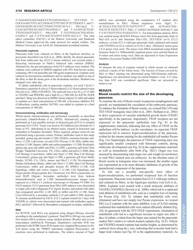
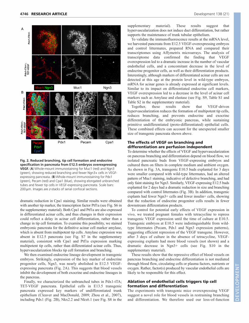
We then asked whether the formation of endocrine progenitorcells was also affected by ablation of blood vessels. Wild-type budswere cultured in the presence or absence of VEGFR2i, and stainedin whole mount for endocrine progenitor cells. In cultures of E12.5buds, vascular ablation had no detectable effect on endocrineprogenitor cells (not shown). However, E9.5 explants cultured for6 days in the presence of VEGFR2i had more Ngn3+ cells (Fig.5A). To validate and quantify these results, we extracted RNA fromremaining explants of the same litter, and determined the levels ofNgn3 mRNA using quantitative RT-PCR. In agreement withimmunostaining results, avascular explants had a significantincrease in Ngn3 mRNA (Fig. 5B). Thus, blood vessels restrain theformation of Ngn3+ endocrine progenitor cells during normaldevelopment of the pancreas.
Finally, we used microarray analysis to identify transcriptomechanges in pancreatic explants upon vascular ablation. As expected,E12.5 explants treated with VEGFR2i for 2 days showed adramatic reduction in the expression of vascular endothelial cellmarkers, reflecting efficient ablation of blood vessels (see Fig. S9,Table S3 and Table S4 in the supplementary material). Althoughendocrine cell markers showed little or no change in these samples,acinar cell markers such as amylase, elastase and Rnase1 weresignificantly upregulated compared with control explants (see Fig.S9 in the supplementary material), consistent with the western blotand immunofluorescence data. In summary, these results show thatablation of vascular endothelial cells induces the formation of tipcells and acinar differentiation, as well as the formation ofendocrine progenitor cells.
Evidence for involvement of Notch pathway inendothelial to epithelial signalingWhat is the molecular nature of the endothelial signals that restrainepithelial branching and differentiation? Delta-Notch signalingrepresents an attractive candidate for a mediator of this tissueinteraction. Notch is a well-recognized regulator of pancreasdifferentiation decisions, acting to restrict Ngn3 cell formation andacinar cell differentiation; in fact, the phenotype of pancreataexpressing a constitutively active Notch transgene is reminiscentof VEGF-expressing pancreata, in that endocrine and exocrinedifferentiation is inhibited (Hald et al., 2003; Murtaugh et al.,
4747RESEARCH ARTICLEVasculature restrains pancreas growth and branching
Fig. 3. Reduced branching and endocrine differentiation inexplanted Pdx1-tTA; TET-VEGF pancreatic buds, suggestingperfusion-independent effects of hypervascularization.(A)Whole-mount immunostaining for Pdx1 (green), Muc1 (red) andNgn3 (blue) in E10.5 buds cultured for 3 days. (B)Whole-mountimmunostaining for insulin (green), Muc1 (red) and Ngn3 (blue) inE12.5 buds cultured for 2 days. Note smaller size, reduced branchingand reduced numbers of -cells in VEGF-expressing pancreata. Scalebars: 200mm.
DEVELO
PMENT

4748
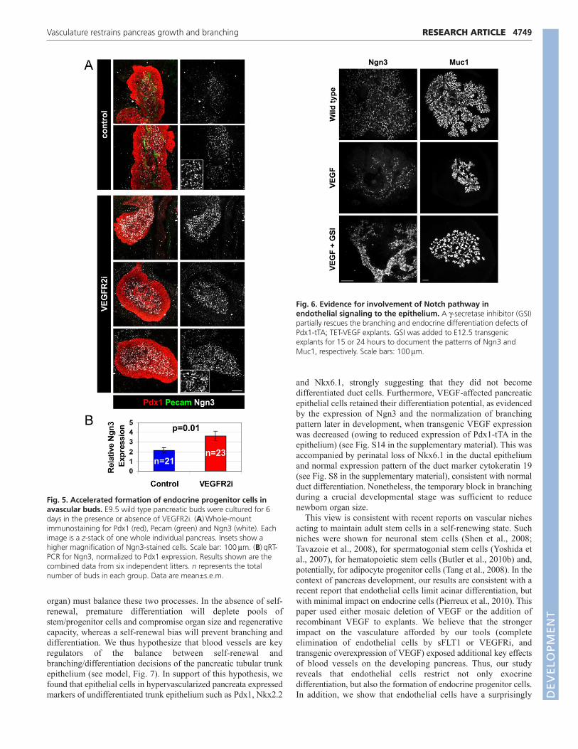
2003). Furthermore, Notch signaling has been recently implicatedin vascular control of stem cell dynamics in neuronal (Shen et al.,2004) and hematopoietic stem cells (Butler et al., 2010b). We thushypothesized that blood vessels affect branching and differentiationby modulating Notch signaling in the epithelium, either directly orvia a relay (Jacquemin et al., 2006). To begin and examine thishypothesis, we treated hypervascular pancreatic explants with apharmacological -secretase inhibitor (GSI), which prevents theactivation of Notch. As shown in Fig. 6, GSI partially rescued thedefects in Ngn3 cell formation and branching in VEGF-expressingexplants. GSI induced the rapid expression of Ngn3 in the majorityof epithelial cells in VEGF transgenics (Fig. 6), as well as in wild-type explants (Magenheim et al., 2011). Longer exposure to GSIcaused thinning of tubes and excessive tip formation in both VEGFtransgenics (Fig. 6) and wild-type explants (Magenheim et al.,2011). Therefore, the epithelium in hypervascularized pancreaticbuds remains sensitive to Notch inhibition, supporting the idea thatthe vasculature could be acting by modulating an upstream step inepithelial Notch signaling. These results are consistent with thehypothesis that the vasculature restrains pancreas differentiationand branching at least in part via modulation of Notch signalingwithin the epithelium.
DISCUSSIONVascular control of pancreas branching anddifferentiationWe show here that during pancreas development, blood vesselsrestrain pancreas tip cell formation and branching morphogenesis,and antagonize differentiation of epithelial cells into exocrine and
endocrine fates. Consequently, the surprising net effect of bloodvessels on pancreas development is inhibition of growth and arestriction of final organ size. The signals that mediate thisresponse are independent of blood flow, circulating plasma factorsand the provision of nutrients and oxygen, as manipulations of thevasculature lead to similar effects in vivo and in explants culturedin complete medium and ambient oxygen.
These findings seem counterintuitive and run against the well-established notion that blood vessels are positive regulators oftissue growth during development and postnatal life, as well as inpathologies such as cancer. How can our results be reconciled withthis view? More specifically, how do our findings fit into thecurrent understanding of pancreas development andmorphogenesis, in particular the ‘tip-trunk’ model describingbranching, progenitor cell dynamics and differentiation (Zhou etal., 2007)? We propose that epithelial cells in the developingpancreas, most of which reside in the trunks, face a fundamentalchoice. They can either divide symmetrically to generate twoidentical daughters, or take the differentiation/morphogenesis path,involving either the generation of a new, rapidly dividing tip cellfollowed by formation of a lateral branch (Puri and Hebrok, 2007;Solar et al., 2009), or the expression of Ngn3, followed bydelamination and endocrine differentiation (Ahnfelt-Ronne et al.,2007a; Apelqvist et al., 1999).
Taking the first path, which we propose represents ‘self-renewal’, the epithelium generates elongated, unbranched tubes andretains multipotentiality. Taking the second path, the epitheliumgenerates the branched pancreas, including its typical differentiatedcell types. Proper development of the pancreas (and indeed, any
RESEARCH ARTICLE Development 138 (21)
Fig. 4. Excessive formation of tips and acinar differentiation upon ablation of endothelial cells in explanted pancreatic buds. E12.5wild-type buds were cultured with or without VEGFR2i and stained in whole mount. (A)Staining for Muc1 after culturing for 3 days. (B)Staining forPtf1a after culturing for 2 days. Upper panels, z-stacks of 11 confocal sections. Bottom panels, individual confocal sections. (C)Top: staining foramylase after culturing for 3 days. Bottom: quantification of amylase-stained area in explants, normalized to the Pdx1-stained area. n denotes thenumber of individual explants analyzed. Data are mean±s.e.m. Bottom panels in A and C show higher magnification images of the areas marked inyellow in the upper panels. Scale bars: 200mm. (D)Western blot showing increased amylase expression in avascular buds cultured as in C. Each lanerepresents a pool of three explants. The intensity of the amylase band relative to -actin was increased 2.2-fold in the avascular samples. (E)Foldchange in the mRNA levels of selected acinar cells upon vascular ablation. Data are extracted from microarray data (see Tables S1-4 and Fig. S9 inthe supplementary material).
DEVELO
PMENT
organ) must balance these two processes. In the absence of self-renewal, premature differentiation will deplete pools ofstem/progenitor cells and compromise organ size and regenerativecapacity, whereas a self-renewal bias will prevent branching anddifferentiation. We thus hypothesize that blood vessels are keyregulators of the balance between self-renewal andbranching/differentiation decisions of the pancreatic tubular trunkepithelium (see model, Fig. 7). In support of this hypothesis, wefound that epithelial cells in hypervascularized pancreata expressedmarkers of undifferentiated trunk epithelium such as Pdx1, Nkx2.2
and Nkx6.1, strongly suggesting that they did not becomedifferentiated duct cells. Furthermore, VEGF-affected pancreaticepithelial cells retained their differentiation potential, as evidencedby the expression of Ngn3 and the normalization of branchingpattern later in development, when transgenic VEGF expressionwas decreased (owing to reduced expression of Pdx1-tTA in theepithelium) (see Fig. S14 in the supplementary material). This wasaccompanied by perinatal loss of Nkx6.1 in the ductal epitheliumand normal expression pattern of the duct marker cytokeratin 19(see Fig. S8 in the supplementary material), consistent with normalduct differentiation. Nonetheless, the temporary block in branchingduring a crucial developmental stage was sufficient to reducenewborn organ size.
This view is consistent with recent reports on vascular nichesacting to maintain adult stem cells in a self-renewing state. Suchniches were shown for neuronal stem cells (Shen et al., 2008;Tavazoie et al., 2008), for spermatogonial stem cells (Yoshida etal., 2007), for hematopoietic stem cells (Butler et al., 2010b) and,potentially, for adipocyte progenitor cells (Tang et al., 2008). In thecontext of pancreas development, our results are consistent with arecent report that endothelial cells limit acinar differentiation, butwith minimal impact on endocrine cells (Pierreux et al., 2010). Thispaper used either mosaic deletion of VEGF or the addition ofrecombinant VEGF to explants. We believe that the strongerimpact on the vasculature afforded by our tools (completeelimination of endothelial cells by sFLT1 or VEGFRi, andtransgenic overexpression of VEGF) exposed additional key effectsof blood vessels on the developing pancreas. Thus, our studyreveals that endothelial cells restrict not only exocrinedifferentiation, but also the formation of endocrine progenitor cells.In addition, we show that endothelial cells have a surprisingly
4749RESEARCH ARTICLEVasculature restrains pancreas growth and branching
Fig. 5. Accelerated formation of endocrine progenitor cells inavascular buds. E9.5 wild type pancreatic buds were cultured for 6days in the presence or absence of VEGFR2i. (A)Whole-mountimmunostaining for Pdx1 (red), Pecam (green) and Ngn3 (white). Eachimage is a z-stack of one whole individual pancreas. Insets show ahigher magnification of Ngn3-stained cells. Scale bar: 100mm. (B)qRT-PCR for Ngn3, normalized to Pdx1 expression. Results shown are thecombined data from six independent litters. n represents the totalnumber of buds in each group. Data are mean±s.e.m.
Fig. 6. Evidence for involvement of Notch pathway inendothelial signaling to the epithelium. A -secretase inhibitor (GSI)partially rescues the branching and endocrine differentiation defects ofPdx1-tTA; TET-VEGF explants. GSI was added to E12.5 transgenicexplants for 15 or 24 hours to document the patterns of Ngn3 andMuc1, respectively. Scale bars: 100mm.
DEVELO
PMENT
4750
negative effect on organ size, and propose a general mechanism forthe activity of the vascular niche in controlling pancreasdifferentiation, morphogenesis and growth.
We thus propose that a conserved function of endothelial cells ismaintenance of progenitor pools. In the context of late pancreasdevelopment (but not in earlier stages prior to branching, seebelow), the net effect of vasculature-driven excessive self-renewaland deficient branching/differentiation is a smaller organ size. Thesurprising conclusion that blood vessels restrict organ size isconsistent with a recent paper (Sand et al., 2011), which used S1P1deficiency and vascular ablation in mice to show that endothelialcell hyperplasia negatively influences growth of the pancreas,stomach and liver.
It will be interesting to test whether signals from endothelialcells act in a similar manner in the context of adult tissueregeneration and tumorigenesis (Butler et al., 2010a). Progress inthis direction may offer new insights into the role of the vasculaturein regenerative biology, and into the mode of action of anti-angiogenic cancer therapy.
We note that an important aspect of the vascular effect onpancreas size remains unresolved. Differences in size could resultfrom differences in cell number, cell size or the extracellular space.We could not find significant differences in the rate of cellproliferation and apoptosis between the epithelium of VEGF-expressing mice and control littermates, and between theepithelium of VEGFRi-treated and vehicle-treated explants (seeFig. S15 in the supplementary material). However, we believe thatsmall differences in replication and apoptosis could be missed by
this analysis and still have a significant effect. We also examinedthe possibility that differences in organ size were the result ofchanges in the proportion of acinar cells, which are known to bemuch larger than other cell types in the adult pancreas. However,amylase+ cells in embryonic explants were not larger than adjacentamylase–; Pdx1– cells, in both control and VEGFRi-treatedexplants (see Fig. S16 in the supplementary material). This resultargues against the possibility that differences in cell size accountfor the differences in organ size upon vascular manipulations.
It is important to acknowledge a limitation of our experiments.Since all our manipulations targeted VEGF signaling, it istheoretically possible that the effect on pancreas development wasmediated through a non-endothelial VEGFR+ compartment, andthat the effect on the vasculature was an epiphenomenon. Toaddress this possibility, we showed that VEGF receptors are notexpressed in pancreatic epithelial cells, suggesting that VEGFcannot act directly on the epithelium. However it is more difficultto exclude the possibility that VEGF affects pancreas developmentvia a non-endothelial, non-epithelial VEGFR+ compartment suchas macrophages. While we could not rule out this formalpossibility, we believe it is unlikely given the bulk of accumulatedknowledge on the biology of vascular niches in multiple tissues.
Early and late effects of blood vessels on pancreasdevelopmentAlthough our results appear at odds with previous studies whichidentified an essential role of blood vessels in pancreasdevelopment (Lammert et al., 2001; Yoshitomi and Zaret, 2004),we note that these studies addressed an earlier developmental stage,after organ specification but before the onset of branchingmorphogenesis and differentiation. It is possible that eitherendothelial cells produce different signaling molecules at differentdevelopmental stages, or alternatively, that molecular responses ofthe epithelium to the same signals change as developmentproceeds, as recently suggested (Zaret and Grompe, 2008). Wefavor a third alternative, whereby the same endothelial signals andthe same signaling responses have different consequences at thetissue level depending on context.
During early stages of pancreas formation, self-renewingdivisions are essential to generate a pool of stem/progenitor cellsfrom which branches and differentiated products will emerge;interfering with this process by vascular ablation leads to apremature exhaustion of this pool and developmental failure, asreported before (Lammert et al., 2001; Yoshitomi and Zaret, 2004).During later stages of development, cell choices must graduallyshift to branching (including the formation of rapidly dividing tipcells) and differentiation; in this setting, ablation of blood vesselswill accelerate organ growth (if hypoxic effects are avoided, as inexplants), and hypervascularization will restrain branching,differentiation and growth, as we observe. More experiments willbe required to distinguish between these possibilities.
Involvement of Notch signaling in vascularrestriction of branching and differentiationThe molecular mechanism underlying the perfusion-independentinteraction of blood vessels with the developing pancreas duringearly stages remains unknown to date (Jacquemin et al., 2006;Lammert et al., 2001; Yoshitomi and Zaret, 2004). As a first steptowards identifying the molecular basis for the later endothelial-epithelial interaction that we report here, we considered Notchsignaling. Of the central signaling pathways known to be involvedin pancreas development, the Notch system represents a
RESEARCH ARTICLE Development 138 (21)
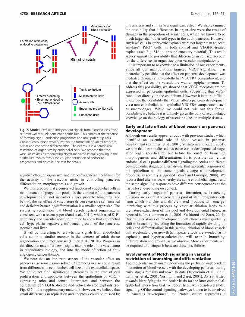
Fig. 7. Model. Perfusion-independent signals from blood vessels favorself-renewal of trunk pancreatic epithelium. This comes at the expenseof forming Ngn3+ endocrine progenitors and multipotent tip cells.Consequently, blood vessels restrain the formation of lateral branches,acinar and endocrine differentiation. The net result is a paradoxicalrestriction of organ size by endothelial cells. We propose that thevasculature acts by modulating Notch-mediated lateral signaling in theepithelium, which favors the coupled formation of endocrineprogenitors and tip cells. See text for details.
DEVELO
PMENT
particularly attractive candidate for mediating the effects of thevasculature on self-renewal versus differentiation (Esni et al., 2004;Hald et al., 2003; Murtaugh et al., 2003). The effects of forcedNotch activation closely resemble the phenotype of VEGFoverexpression, namely inhibition of endocrine differentiation andbranching (Murtaugh et al., 2003). Moreover, Notch signalingwithin vascular niches has been implicated in the control ofneuronal and hematopoietic stem cells (Butler et al., 2010b; Shenet al., 2004).
We found that pharmacological inhibition of intracellular Notchactivation can partially rescue the VEGF phenotype, with regard toboth endocrine differentiation and branching. Thus, the epitheliumin VEGF-expressing pancreata remain sensitive to Notchinhibition, suggesting that blood vessels affect an upstream step inNotch signaling in the epithelium. This view is consistent with therecent report of Pierruex et al. that blood vessels positively regulatethe Notch target genes Hey1 and Hey2 (Pierreux et al., 2010).
In light of these findings, we propose the following model(Fig. 7). Within the pancreatic epithelium, Notch-mediatedlateral interactions cause the coupled formation of endocrineprogenitor cells (cells downregulating Notch) and tip cells(adjacent cells upregulating Notch). We hypothesize that,normally, signals from blood vessels restrain these lateralinteractions, perhaps by providing a constant low level of anendothelial Notch ligand (e.g. Dll4) that dampens the tendencyof adjacent epithelial cells to undergo lateral inhibition, eitherlosing or gaining Notch signaling. When blood vessels areeliminated, lateral interactions are uninhibited and lead topremature divergence of adjacent epithelial cells into low notch(endocrine) and high notch (tips). Hypervascularization preventsthe Notch-controlled lateral divergence to endocrine/tip cells,and maintains the epithelium in a primitive proliferative statethat might be termed ‘self-renewal’. Genetic and gene expressionstudies are ongoing to test this model. Finally, it is entirelypossible that additional signaling pathways affect theendothelial-epithelial interaction reported here. For example,endothelial cells could signal via the extracellular matrix andintegrins (Kesavan et al., 2009; Nikolova et al., 2006) or via Fgfreceptors (Hart et al., 2003; Jacquemin et al., 2006; Norgaard etal., 2003). More studies will be needed to test these ideas.
Implications for directed differentiation ofembryonic stem cells to beta cellsThese findings may have practical implications for the derivationof transplantable, insulin-producing -cells from embryonic stemcells (ESC) (D’Amour et al., 2006; Kroon et al., 2008). Our datamay explain the failure of early attempts to use co-cultures of ESCand vascular endothelial cells for the derivation of -cells (O.C.,unpublished). It is becoming increasingly evident that thecontribution of endothelial cell signals to pancreas and -celldevelopment is much more dynamic than previously assumed, andits practical use will have to take this complexity into account.Specifically, we propose that endothelial cells may positivelyinfluence the expansion of ESC-derived, multipotent pancreaticprogenitor cells, but final differentiation may require the timedremoval of endothelial cells at distinct intervals.
In summary, we show that blood vessels act to restrain pancreasbranching and differentiation, leading to an overall restriction oforgan growth. We propose that these effects reflect a conserved,perfusion-independent function of vascular endothelial cells, actingto favor self-renewal at the expense of branching anddifferentiation.
AcknowledgementsWe thank Chaya Kalcheim, Ben Stanger and Jay Rajagopal for discussions andcomments on the manuscript. We are grateful to Chris Wright and MaikeSander for the generous gifts of Pdx1 and Ngn3 antisera, and to RayMacDonald for providing Pdx1-tTA mice. The 74.5A5 Nkx2.2 monoclonalantibody developed by T. Jessell and S. Brenner-Morton was obtained from theDevelopmental Studies Hybridoma Bank developed under the auspices of theNICHD and maintained by The University of Iowa, Department of Biology, IowaCity, IA 52242.
FundingSupported by grants from JDRF [to Y.D., E.K. and O.C.], the HelmsleyFoundation, European Union Seventh Framework Programme [241883], ERCstarting grant and the Dutch Friends of Hebrew University (Y.D.). O.Z. is a NewYork Stem Cell Foundation-Druckenmiller Fellow.
Supplementary materialSupplementary material for this article is available athttp://dev.biologists.org/lookup/suppl/doi:10.1242/dev.066548/-/DC1
ReferencesAhnfelt-Ronne, J., Hald, J., Bodker, A., Yassin, H., Serup, P. and Hecksher-
Sorensen, J. (2007a). Preservation of proliferating pancreatic progenitor cells byDelta-Notch signaling in the embryonic chicken pancreas. BMC Dev. Biol. 7, 63.
Ahnfelt-Rønne, J., Jørgensen, M. C., Hald, J., Madsen, O. D., Serup, P. andHecksher-Sørensen, J. (2007b). An improved method for three-dimensionalreconstruction of protein expression patterns in intact mouse and chickenembryos and organs. J. Histochem. Cytochem. 55, 925-930.
Apelqvist, A., Li, H., Sommer, L., Beatus, P., Anderson, D. J., Honjo, T., Hrabede Angelis, M., Lendahl, U. and Edlund, H. (1999). Notch signalling controlspancreatic cell differentiation. Nature 400, 877-881.
Butler, J. M., Kobayashi, H. and Rafii, S. (2010a). Instructive role of the vascularniche in promoting tumour growth and tissue repair by angiocrine factors. Nat.Rev. Cancer 10, 138-146.
Butler, J. M., Nolan, D. J., Vertes, E. L., Varnum-Finney, B., Kobayashi, H.,Hooper, A. T., Seandel, M., Shido, K., White, I. A., Kobayashi, M. et al.(2010b). Endothelial cells are essential for the self-renewal and repopulation ofNotch-dependent hematopoietic stem cells. Cell Stem Cell 6, 251-264.
Cleaver, O. and MacDonald, R. J. (2009). Developmental molecular biology ofthe pancreas. In Handbook of Pancreatic Cancer (ed. J. Neoptolemos, J.Abbruzzese, M. Buchler and R. Urrutia). New York: Springer.
D’Amour, K. A., Bang, A. G., Eliazer, S., Kelly, O. G., Agulnick, A. D., Smart,N. G., Moorman, M. A., Kroon, E., Carpenter, M. K. and Baetge, E. E.(2006). Production of pancreatic hormone-expressing endocrine cells fromhuman embryonic stem cells. Nat. Biotechnol. 24, 1392-1401.
Dor, Y., Camenisch, T. D., Itin, A., Fishman, G. I., McDonald, J. A., Carmeliet,P. and Keshet, E. (2001). A novel role for VEGF in endocardial cushionformation and its potential contribution to congenital heart defects.Development 128, 1531-1538.
Esni, F., Ghosh, B., Biankin, A. V., Lin, J. W., Albert, M. A., Yu, X., MacDonald,R. J., Civin, C. I., Real, F. X., Pack, M. A. et al. (2004). Notch inhibits Ptf1function and acinar cell differentiation in developing mouse and zebrafishpancreas. Development 131, 4213-4224.
Gittes, G. K. (2009). Developmental biology of the pancreas: a comprehensivereview. Dev. Biol. 326, 4-35.
Gu, G., Wells, J. M., Dombkowski, D., Preffer, F., Aronow, B. and Melton, D.A. (2004). Global expression analysis of gene regulatory pathways duringendocrine pancreatic development. Development 131, 165-179.
Hald, J., Hjorth, J. P., German, M. S., Madsen, O. D., Serup, P. and Jensen, J.(2003). Activated Notch1 prevents differentiation of pancreatic acinar cells andattenuate endocrine development. Dev. Biol. 260, 426-437.
Hart, A., Papadopoulou, S. and Edlund, H. (2003). Fgf10 maintains notchactivation, stimulates proliferation, and blocks differentiation of pancreaticepithelial cells. Dev. Dyn. 228, 185-193.
Holland, A. M., Hale, M. A., Kagami, H., Hammer, R. E. and MacDonald, R. J.(2002). Experimental control of pancreatic development and maintenance. Proc.Natl. Acad. Sci. USA 99, 12236-12241.
Ilovich, O., Jacobson, O., Aviv, Y., Litchi, A., Chisin, R. and Mishani, E. (2008).Formation of fluorine-18 labeled diaryl ureas-labeled VEGFR-2/PDGFR dualinhibitors as molecular imaging agents for angiogenesis. Bioorg. Med. Chem.16, 4242-4251.
Jacquemin, P., Yoshitomi, H., Kashima, Y., Rousseau, G. G., Lemaigre, F. P.and Zaret, K. S. (2006). An endothelial-mesenchymal relay pathway regulatesearly phases of pancreas development. Dev. Biol. 290, 189-199.
Jorgensen, M. C., Ahnfelt-Ronne, J., Hald, J., Madsen, O. D., Serup, P. andHecksher-Sorensen, J. (2007). An illustrated review of early pancreasdevelopment in the mouse. Endocr. Rev. 28, 685-705.
4751RESEARCH ARTICLEVasculature restrains pancreas growth and branching
DEVELO
PMENT
4752
Kesavan, G., Sand, F. W., Greiner, T. U., Johansson, J. K., Kobberup, S., Wu,X., Brakebusch, C. and Semb, H. (2009). Cdc42-mediated tubulogenesiscontrols cell specification. Cell 139, 791-801.
Kroon, E., Martinson, L. A., Kadoya, K., Bang, A. G., Kelly, O. G., Eliazer, S.,Young, H., Richardson, M., Smart, N. G., Cunningham, J. et al. (2008).Pancreatic endoderm derived from human embryonic stem cells generatesglucose-responsive insulin-secreting cells in vivo. Nat. Biotechnol. 26, 443-452.
Lammert, E., Cleaver, O. and Melton, D. (2001). Induction of pancreaticdifferentiation by signals from blood vessels. Science 294, 564-567.
LeCouter, J., Moritz, D. R., Li, B., Phillips, G. L., Liang, X. H., Gerber, H. P.,Hillan, K. J. and Ferrara, N. (2003). Angiogenesis-independent endothelialprotection of liver: role of VEGFR-1. Science 299, 890-893.
Magenheim, J., Klein, A. M., Stanger, B. Z., Ashery-Padan, A., Sosa-Pineda,B., Gu, G. and Dor, Y. (2011). Ngn3+ endocrine progenitor cells control thefate and morphogenesis of pancreatic ductal epithelium. Dev. Biol. doi:10.1016/j.ydbio.2011.08.006.
Matsumoto, K., Yoshitomi, H., Rossant, J. and Zaret, K. S. (2001). Liverorganogenesis promoted by endothelial cells prior to vascular function. Science294, 559-563.
May, D., Gilon, D., Djonov, V., Itin, A., Lazarus, A., Gordon, O., Rosenberger,C. and Keshet, E. (2008). Transgenic system for conditional induction andrescue of chronic myocardial hibernation provides insights into genomicprograms of hibernation. Proc. Natl. Acad. Sci. USA 105, 282-287.
Murtaugh, L. C. (2007). Pancreas and beta-cell development: from the actual tothe possible. Development 134, 427-438.
Murtaugh, L. C., Stanger, B. Z., Kwan, K. M. and Melton, D. A. (2003). Notchsignaling controls multiple steps of pancreatic differentiation. Proc. Natl. Acad.Sci. USA 100, 14920-14925.
Nikolova, G., Jabs, N., Konstantinova, I., Domogatskaya, A., Tryggvason, K.,Sorokin, L., Fassler, R., Gu, G., Gerber, H. P., Ferrara, N. et al. (2006). Thevascular basement membrane: a niche for insulin gene expression and Beta cellproliferation. Dev. Cell 10, 397-405.
Norgaard, G. A., Jensen, J. N. and Jensen, J. (2003). FGF10 signaling maintainsthe pancreatic progenitor cell state revealing a novel role of Notch in organdevelopment. Dev. Biol. 264, 323-338.
Oliver-Krasinski, J. M. and Stoffers, D. A. (2008). On the origin of the beta cell.Genes Dev. 22, 1998-2021.
Palmer, T. D., Willhoite, A. R. and Gage, F. H. (2000). Vascular niche for adulthippocampal neurogenesis. J. Comp. Neurol. 425, 479-494.
Pierreux, C. E., Cordi, S., Hick, A. C., Achouri, Y., Ruiz de Almodovar, C.,Prevot, P. P., Courtoy, P. J., Carmeliet, P. and Lemaigre, F. P. (2010). Epithelial:Endothelial cross-talk regulates exocrine differentiation in developing pancreas.Dev. Biol. 347, 216-227.
Puri, S. and Hebrok, M. (2007). Dynamics of embryonic pancreas developmentusing real-time imaging. Dev. Biol. 306, 82-93.
Sand, F. W., Hornblad, A., Johansson, J. K., Loren, C., Edsbagge, J.,Stahlberg, A., Magenheim, J., Ilovich, O., Mishani, E., Dor, Y. et al. (2011).Growth-limiting role of endothelial cells in endoderm development. Dev. Biol.352, 267-277.
Shalaby, F., Ho, J., Stanford, W. L., Fischer, K. D., Schuh, A. C., Schwartz, L.,Bernstein, A. and Rossant, J. (1997). A requirement for Flk1 in primitive anddefinitive hematopoiesis and vasculogenesis. Cell 89, 981-990.
Shen, Q., Goderie, S. K., Jin, L., Karanth, N., Sun, Y., Abramova, N., Vincent,P., Pumiglia, K. and Temple, S. (2004). Endothelial cells stimulate self-renewaland expand neurogenesis of neural stem cells. Science 304, 1338-1340.
Shen, Q., Wang, Y., Kokovay, E., Lin, G., Chuang, S. M., Goderie, S. K.,Roysam, B. and Temple, S. (2008). Adult SVZ stem cells lie in a vascular niche:a quantitative analysis of niche cell-cell interactions. Cell Stem Cell 3, 289-300.
Solar, M., Cardalda, C., Houbracken, I., Martin, M., Maestro, M. A., DeMedts, N., Xu, X., Grau, V., Heimberg, H., Bouwens, L. et al. (2009).Pancreatic exocrine duct cells give rise to insulin-producing beta cells duringembryogenesis but not after birth. Dev. Cell 17, 849-860.
Tang, W., Zeve, D., Suh, J. M., Bosnakovski, D., Kyba, M., Hammer, R. E.,Tallquist, M. D. and Graff, J. M. (2008). White fat progenitor cells reside in theadipose vasculature. Science 322, 583-586.
Tavazoie, M., Van der Veken, L., Silva-Vargas, V., Louissaint, M., Colonna, L.,Zaidi, B., Garcia-Verdugo, J. M. and Doetsch, F. (2008). A specialized vascularniche for adult neural stem cells. Cell Stem Cell 3, 279-288.
van Eyll, J. M., Pierreux, C. E., Lemaigre, F. P. and Rousseau, G. G. (2004).Shh-dependent differentiation of intestinal tissue from embryonic pancreas byactivin A. J. Cell Sci. 117, 2077-2086.
Yoshida, S., Sukeno, M. and Nabeshima, Y. (2007). A vasculature-associatedniche for undifferentiated spermatogonia in the mouse testis. Science 317,1722-1726.
Yoshitomi, H. and Zaret, K. S. (2004). Endothelial cell interactions initiate dorsalpancreas development by selectively inducing the transcription factor Ptf1a.Development 131, 807-817.
Zaret, K. S. (2006). Pancreatic beta cells: responding to the matrix. Cell Metab. 3,148-150.
Zaret, K. S. and Grompe, M. (2008). Generation and regeneration of cells of theliver and pancreas. Science 322, 1490-1494.
Zhou, Q., Law, A. C., Rajagopal, J., Anderson, W. J., Gray, P. A. and Melton,D. A. (2007). A multipotent progenitor domain guides pancreaticorganogenesis. Dev. Cell 13, 103-114.
RESEARCH ARTICLE Development 138 (21)
DEVELO
PMENT