bmc chemical biology biomed central - universität kasselnbn:de:hebis:... · bmc chemical biology...
TRANSCRIPT
BioMed CentralBMC Chemical Biology
ss
Open AcceMethodology articleChemical tools selectively target components of the PKA systemDaniela Bertinetti†1, Sonja Schweinsberg†1, Susanne E Hanke1, Frank Schwede2, Oliver Bertinetti1, Stephan Drewianka3, Hans-Gottfried Genieser2 and Friedrich W Herberg*1Address: 1Department of Biochemistry, University of Kassel, Heinrich-Plett-Str. 40, 34132 Kassel, Germany, 2Biolog Life Science Institute, Flughafendamm 9a, P.O. Box 107125, Bremen, Germany and 3Biaffin GmbH & Co KG, Heinrich-Plett-Str. 40, 34132 Kassel, Germany
Email: Daniela Bertinetti - [email protected]; Sonja Schweinsberg - [email protected]; Susanne E Hanke - [email protected]; Frank Schwede - [email protected]; Oliver Bertinetti - [email protected]; Stephan Drewianka - [email protected]; Hans-Gottfried Genieser - [email protected]; Friedrich W Herberg* - [email protected]
* Corresponding author †Equal contributors
AbstractBackground: In the eukaryotic cell the cAMP-dependent protein kinase (PKA) is a key enzyme insignal transduction and represents the main target of the second messenger cAMP. Here wedescribe the design, synthesis and characterisation of specifically tailored cAMP analogs which canbe utilised as a tool for affinity enrichment and purification as well as for proteomics based analysesof cAMP binding proteins.
Results: Two sets of chemical binders were developed based on the phosphorothioate derivativesof cAMP, Sp-cAMPS and Rp-cAMPS acting as cAMP-agonists and -antagonists, respectively. Thesecompounds were tested via direct surface plasmon resonance (SPR) analyses for their bindingproperties to PKA R-subunits and holoenzyme. Furthermore, these analogs were used in an affinitypurification approach to analyse their binding and elution properties for the enrichment andimprovement of cAMP binding proteins exemplified by the PKA R-subunits. As determined by SPR,all tested Sp-analogs provide valuable tools for affinity chromatography. However, Sp-8-AEA-cAMPS displayed (i) superior enrichment properties while maintaining low unspecific binding toother proteins in crude cell lysates, (ii) allowing mild elution conditions and (iii) providing thecapability to efficiently purify all four isoforms of active PKA R-subunit in milligram quantities within8 h. In a chemical proteomics approach both sets of binders, Rp- and Sp-cAMPS derivatives, can beemployed. Whereas Sp-8-AEA-cAMPS preferentially binds free R-subunit, Rp-AHDAA-cAMPS,displaying antagonist properties, not only binds to the free PKA R-subunits but also to the intactPKA holoenzyme both from recombinant and endogenous sources.
Conclusion: In summary, all tested cAMP analogs were useful for their respective application asan affinity reagent which can enhance purification of cAMP binding proteins. Sp-8-AEA-cAMPS wasconsidered the most efficient analog since Sp-8-AHA-cAMPS and Sp-2-AHA-cAMPS, demonstratedincomplete elution from the matrix, as well as retaining notable amounts of bound proteincontaminants. Furthermore it could be demonstrated that an affinity resin based on Rp-8-AHDAA-cAMPS provides a valuable tool for chemical proteomics approaches.
Published: 12 February 2009
BMC Chemical Biology 2009, 9:3 doi:10.1186/1472-6769-9-3
Received: 10 September 2008Accepted: 12 February 2009
This article is available from: http://www.biomedcentral.com/1472-6769/9/3
© 2009 Bertinetti et al; licensee BioMed Central Ltd. This is an Open Access article distributed under the terms of the Creative Commons Attribution License (http://creativecommons.org/licenses/by/2.0), which permits unrestricted use, distribution, and reproduction in any medium, provided the original work is properly cited.
Page 1 of 15(page number not for citation purposes)

BMC Chemical Biology 2009, 9:3 http://www.biomedcentral.com/1472-6769/9/3
BackgroundThe cAMP-dependent protein kinase (PKA) is a key regu-lator protein in eukaryotic signal transduction and isinvolved in several cellular processes during growth anddevelopment. Via phosphorylation of its substrate pro-teins PKA controls metabolic processes, cAMP mediatedgene expression, cell differentiation and/or apoptosis [1].The enzymatic activity of the PKA catalytic (C) subunit iscontrolled by a set of four different regulatory (R) subunitisoforms, i.e. type I and type II, both with two isoforms (αand β) each. Thus, in its inactive state PKA forms a heter-otetrameric holoenzyme complex (R2C2), containing anR-subunit dimer and two C-subunit monomers. The PKAholoenzyme is activated upon cooperative binding of fourmolecules of the second messenger cAMP to the R-subu-nits, thus releasing the now active C-subunits [2]. Theexpression pattern of single PKA isoforms as well as theisoform specific composition of the holoenzyme and tis-sue specific distribution of PKA isoforms allows a tightregulation of the catalytic activity of PKA. In turn, PKAprovides an important model system for kinases, allowinginvestigation of the molecular mechanisms of kinasefunction as well as the development of tools for diagnosticpurposes which in turn enables their use as biomarkers[3].
Historically, purification of PKA holoenzyme from bio-logical material was performed via anion exchange chro-matography (DEAE, [4]); later, a second step based onaffinity purification utilising cAMP resins was added [5,6].However, most strategies so far resulted in either partlydegraded or insoluble protein with limited yield. There-fore we set out to design novel cAMP affinity matrices forsimple and rapid purification of cAMP binding proteins.These resins should provide a chemical tool that targetsproteins containing the conserved cAMP bindingdomains, specifically PKA R-subunits while fulfilling thefollowing criteria required for efficient affinity binders:
1. Purify high quantities of selected protein of interest;
2. yield functionally active protein;
3. provide a purification procedure with mild but efficientelution conditions while retaining high yields of protein;
4. obtain nucleotide-free proteins which can easily beused for further interaction studies and biochemicalassays;
5. provide an easy-to-use procedure applicable in chemi-cal proteomics.
In addition to cAMP's role as a general activator of allholoenzyme isoforms, with PKA representing the main
intracellular effector of cAMP, there are still other targetsof cAMP like nucleotide-gated ion channels [7], cAMPdegrading phosphodiesterases [8] or Epac, the cAMP-reg-ulated guanine nucleotide-exchange factor for Rap 1 and2 [9], all containing one or more highly conserved cyclicnucleotide binding (CNB) domains [10].
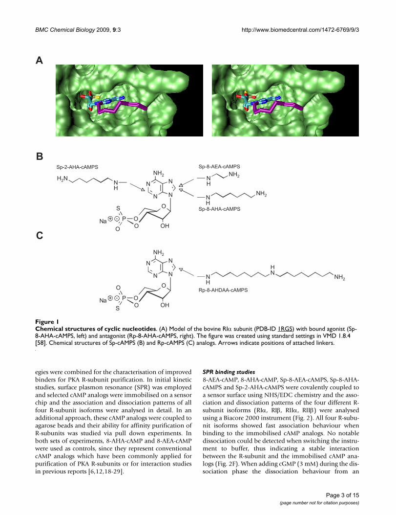
PKA R-subunits and specific components of the cAMP sig-nalling network can be precisely targeted from complexprotein mixtures using highly specific cAMP analogs cov-alently coupled to agarose beads. Generally, two groups ofchemical tools are used for the affinity purification offunctional complexes: agonist and antagonist binders(Fig. 1A). Agonist binders are synthetic cyclic nucleotideswhich bind the R-subunits that are complexed with inter-action partners (e.g. AKAPs), however, these agonistscause the PKA holoenzyme to dissociate. Antagonist bind-ers, interacting preferentially with the intact, non dissoci-ated PKA holoenzyme, can be utilised to identifyinteraction partners that bind to the entire complex(R2C2), including proteins interacting with the C-subu-nits.
In the present study several cyclic nucleotide analogs weresynthesised and optimised for their binding properties tothe respective components of the PKA system using aniterative approach based on direct SPR binding studies.Subsequently, these cAMP analogs were coupled to a solidsupport and tested for binding and elution in a one steppurification procedure probing all four recombinantlyexpressed R-subunit isoforms. Furthermore, we testedoptimised cyclic nucleotides analogs for their applicationin a chemical proteomics approach addressing either theR-subunits or the intact PKA holoenzyme complex alongwith physiological interaction partners derived from ani-mal tissue.
ResultsIn previous studies, hundreds of cyclic nucleotides weredeveloped and characterised regarding their binding abil-ity to proteins containing cyclic nucleotide binding (CNB)domains [11-14].
Improved cAMP analogs as novel tools for PKA R-subunit purificationFor our developments we used optimised binders basedon Sp-cAMPS (Fig. 1A left panel, 1B), a cAMP analogwhere the axial exocyclic oxygen atom of the cyclic phos-phate is replaced by a sulphur atom, resulting in anapproximately 10-fold reduced affinity towards the R-sub-unit when compared to cAMP alone [15-17]. In order tocouple Sp-cAMPS to a solid phase, analogs with differentspacers in positions 2 or 8 of the adenine base (Sp-8-AEA-cAMPS, Sp-8-AHA-cAMPS, Sp-2-AHA-cAMPS, Fig. 1B)were designed and synthesised. Two complementary strat-
Page 2 of 15(page number not for citation purposes)
BMC Chemical Biology 2009, 9:3 http://www.biomedcentral.com/1472-6769/9/3
egies were combined for the characterisation of improvedbinders for PKA R-subunit purification. In initial kineticstudies, surface plasmon resonance (SPR) was employedand selected cAMP analogs were immobilised on a sensorchip and the association and dissociation patterns of allfour R-subunit isoforms were analysed in detail. In anadditional approach, these cAMP analogs were coupled toagarose beads and their ability for affinity purification ofR-subunits was studied via pull down experiments. Inboth sets of experiments, 8-AHA-cAMP and 8-AEA-cAMPwere used as controls, since they represent conventionalcAMP analogs which have been commonly applied forpurification of PKA R-subunits or for interaction studiesin previous reports [6,12,18-29].
SPR binding studies8-AEA-cAMP, 8-AHA-cAMP, Sp-8-AEA-cAMPS, Sp-8-AHA-cAMPS and Sp-2-AHA-cAMPS were covalently coupled toa sensor surface using NHS/EDC chemistry and the asso-ciation and dissociation patterns of the four different R-subunit isoforms (RIα, RIβ, RIIα, RIIβ) were analysedusing a Biacore 2000 instrument (Fig. 2). All four R-subu-nit isoforms showed fast association behaviour whenbinding to the immobilised cAMP analogs. No notabledissociation could be detected when switching the instru-ment to buffer, thus indicating a stable interactionbetween the R-subunit and the immobilised cAMP ana-logs (Fig. 2F). When adding cGMP (3 mM) during the dis-sociation phase the dissociation behaviour from an
Chemical structures of cyclic nucleotidesFigure 1Chemical structures of cyclic nucleotides. (A) Model of the bovine RIα subunit (PDB-ID 1RGS) with bound agonist (Sp-8-AHA-cAMPS, left) and antagonist (Rp-8-AHA-cAMPS, right). The figure was created using standard settings in VMD 1.8.4 [58]. Chemical structures of Sp-cAMPS (B) and Rp-cAMPS (C) analogs. Arrows indicate positions of attached linkers.
OS
O
O
O
P
N
N
N
N
HO
NH2
Na + -
NH
H2N NH
NH2
NH
NH2
OO
O
O
S
P
N
N
N
N
HO
NH2
Na + -
NH
NNH2
H
NH
NH2
A
B
C
Sp-8-AEA-cAMPS
Sp-8-AHA-cAMPS
Sp-2-AHA-cAMPS
Rp-8-AHA-cAMPS
Rp-8-AHDAA-cAMPS
Page 3 of 15(page number not for citation purposes)

BMC Chemical Biology 2009, 9:3 http://www.biomedcentral.com/1472-6769/9/3
Page 4 of 15(page number not for citation purposes)
Interaction of R-subunit isoforms with immobilised cAMP analogs investigated by SPRFigure 2Interaction of R-subunit isoforms with immobilised cAMP analogs investigated by SPR. Binding of all four R-subu-nit isoforms (100 nM) to immobilised 8-AEA-cAMP (A), 8-AHA-cAMP (B), Sp-8-AEA-cAMPS (C) and Sp-8-AHA-cAMPS (D) Sp-2-AHA-cAMPS (E) using a flow rate of 10 μL/min in buffer A containing 0.005% P20. Association and dissociation of hRIα (blue), hRIβ (red), hRIIα (green) and rRIIβ (black) were monitored for 5 and 10 min, respectively. The dissociation was moni-tored in the presence of 3 mM cGMP. (F) hRIα binding to five different cAMP analogs. 100 nM hRIα was injected to immobi-lised 8-AHA-cAMP (red), 8-AEA-cAMP (orange), Sp-2-AHA-cAMPS (green), Sp-8-AEA-cAMPS (blue) and Sp-8-AHA-cAMPS (black). Dissociation was initiated either by buffer A containing 0.005% P20 (dotted lines) or by 3 mM cGMP in the same buffer (solid lines). Experimental setup was performed as described above.
BMC Chemical Biology 2009, 9:3 http://www.biomedcentral.com/1472-6769/9/3
immobilised cyclic nucleotide analog was affected by (i)the exchange of the exocyclic oxygen to a sulphur in theimmobilised cAMP analog, (ii) the chosen linker of thecAMP analog and (iii) the R-subunit isoform. In general,dissociation in the presence of cGMP was slower for con-ventional cAMP analogs compared to the newly designedSp-cAMPS analogs. When comparing dissociation fromall Sp-cAMPS analogs, a slower dissociation was observedfrom the 2-substituted analog (Fig. 2E) than from the 8-substituted analog (Fig. 2C, D). Among the R-subunit iso-forms, RIIα displayed the slowest dissociation from allfive analog surfaces tested.
Generally, fast binding to the affinity matrix as well asrapid and complete dissociation under elution conditionsare preferred for purification, without the dissociation ofprotein from the affinity matrix under washing condi-tions. From the observed elution patterns, Sp-8-AEA-cAMPS was considered the best candidate for efficient elu-tion of hRIα (Fig. 2C, F), especially when compared to 8-AHA-cAMP and 8-AEA-cAMP which conventionally havebeen employed (Fig. 2A, B) [18,27-29]. Panels A and B(Fig. 2) clearly demonstrate that the RIβ and RIIα dissoci-ate rather slow from the non Sp-cAMPS analogs, even inthe presence of cGMP. In general, for the R-subunit iso-forms RIβ, RIIα and RIIβ, the dissociation with cGMP isfast and almost identical for the 8-substituted cAMPS ana-logs.
As a consequence of the high surface ligand density theassociation and dissociation patterns of the R-subunits tothe immobilised cAMP analogs were severely affected bymass transfer limitations. These limitations are due todepletion of the analyte (here the R-subunits) being inclose proximity to the sensor surface during association,thus prohibiting the quantitative analyses of associationand dissociation kinetics. Furthermore, rebinding effectsin the dissociation phase are intensified by the presence offour cAMP binding sites per R-subunit dimer [30-32].Still, the high surface density of cAMP analogs on the sen-
sor surface very well represents the situation that is occur-ring on the agarose beads during binding and elution.
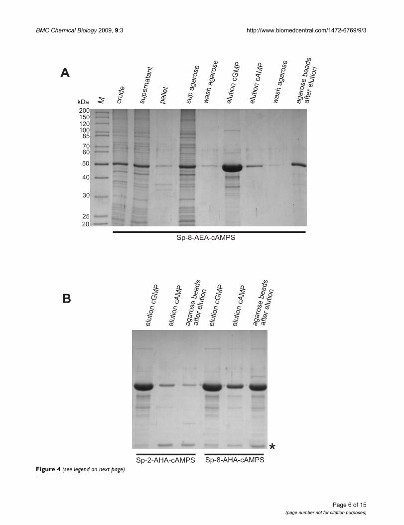
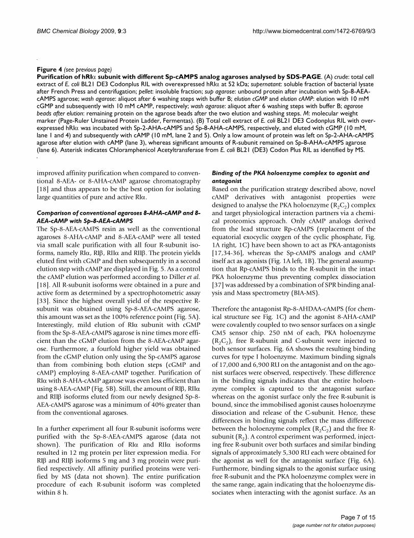
Purification of the R-subunit isoforms using Sp-cAMPS agarosesThe analogs tested in SPR (8-AEA-cAMP, 8-AHA-cAMP,Sp-8-AEA-cAMPS, Sp-8-AHA-cAMPS and Sp-2-AHA-cAMPS) were coupled to agarose beads using NHS chem-istry (Fig. 3). Bacterial cell lysate containing overexpressedRIα was incubated with each analog coupled to agarose (arepresentative purification procedure with Sp-8-AEA-cAMPS is shown in Fig. 4A). After several washing stepswere performed, no notable dissociation of the RIα subu-nit from the agarose was detected. Elution of the proteinwith 10 mM cGMP resulted in a protein fraction contain-ing highly pure and active R-subunit as determined bySDS-PAGE and spectrophotometric activity assay [33],yielding 12 mg protein per approximately 400 μL of agar-ose slurry. Only little additional protein could be elutedsubsequently with cAMP, still leaving some protein on theagarose beads (Fig. 4A).
cGMP elution from the other two agaroses (Sp-8-AHA-cAMPS and Sp-2-AHA-cAMPS) resulted in half theamount of purified PKA R-subunit (7.2 mg protein perapproximately 400 μL of agarose slurry, Fig. 4B) whencompared to the yield from the Sp-8-AEA-cAMPS agarose.Furthermore, hRIα could only be partially eluted from theSp-8-AHA-cAMPS agarose with 10 mM cAMP, while leav-ing a significant amount of protein on the beads, thusindicating that RIα binds too tight to this analog andtherefore can not be used for this affinity purification pro-cedure. Additionally, both the Sp-2-AHA-cAMPS and Sp-8-AHA-cAMPS agarose showed a 25 kDa protein coelutedwith cGMP, it was identified by mass spectrometry (MS)as E. coli Chloramphenicol Acetyltransferase which isabundant in the BL21 (DE3) Codon Plus RIL strain (Fig.4B).
Thus, from the three affinity resins tested, only Sp-8-AEA-cAMPS fulfils and exceeds all necessary requirements for
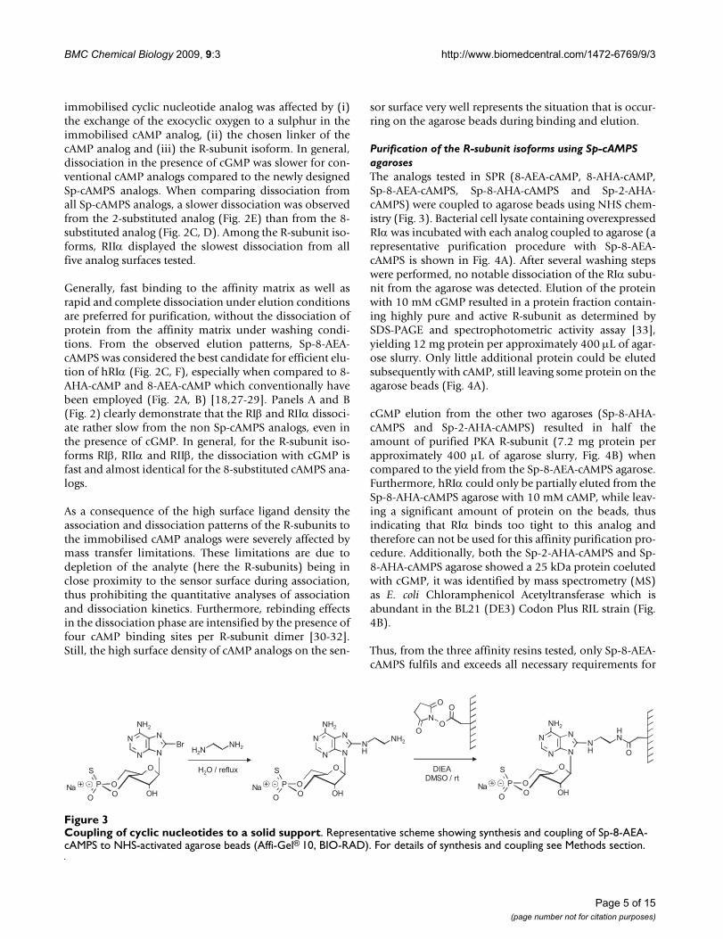
Coupling of cyclic nucleotides to a solid supportFigure 3Coupling of cyclic nucleotides to a solid support. Representative scheme showing synthesis and coupling of Sp-8-AEA-cAMPS to NHS-activated agarose beads (Affi-Gel® 10, BIO-RAD). For details of synthesis and coupling see Methods section.
OS
O
O
O
P
N
N
N
N
HO
NH2
Na + -
H2NNH2Br
OS
O
O
O
P
N
N
N
N
HO
NH2
Na + -
NH
NH2
H2O / reflux OS
O
O
O
P
N
N
N
N
HO
NH2
Na + -
NH
HN
O
N
O
OO
O
DIEADMSO / rt
Page 5 of 15(page number not for citation purposes)
BMC Chemical Biology 2009, 9:3 http://www.biomedcentral.com/1472-6769/9/3
Figure 4 (see legend on next page)
MkDa
40
30
25
6070
85100120150200
20
50
Sp-8-AEA-cAMPS
cru
de
supern
ata
nt
pelle
t
sup
agaro
se
wash
agaro
se
elu
tion
cG
MP
elu
tion
cA
MP
wash
agaro
se
agaro
se
beads
after
elu
tion
Sp-2-AHA-cAMPS Sp-8-AHA-cAMPS
elu
tion
cG
MP
elu
tion
cA
MP
elu
tion
cG
MP
elu
tion
cA
MP
*
agaro
se
beads
after
elu
tion
agaro
se
beads
after
elu
tion
A
B
Page 6 of 15(page number not for citation purposes)
BMC Chemical Biology 2009, 9:3 http://www.biomedcentral.com/1472-6769/9/3
improved affinity purification when compared to conven-tional 8-AEA- or 8-AHA-cAMP agarose chromatography[18] and thus appears to be the best option for isolatinglarge quantities of pure and active RIα.
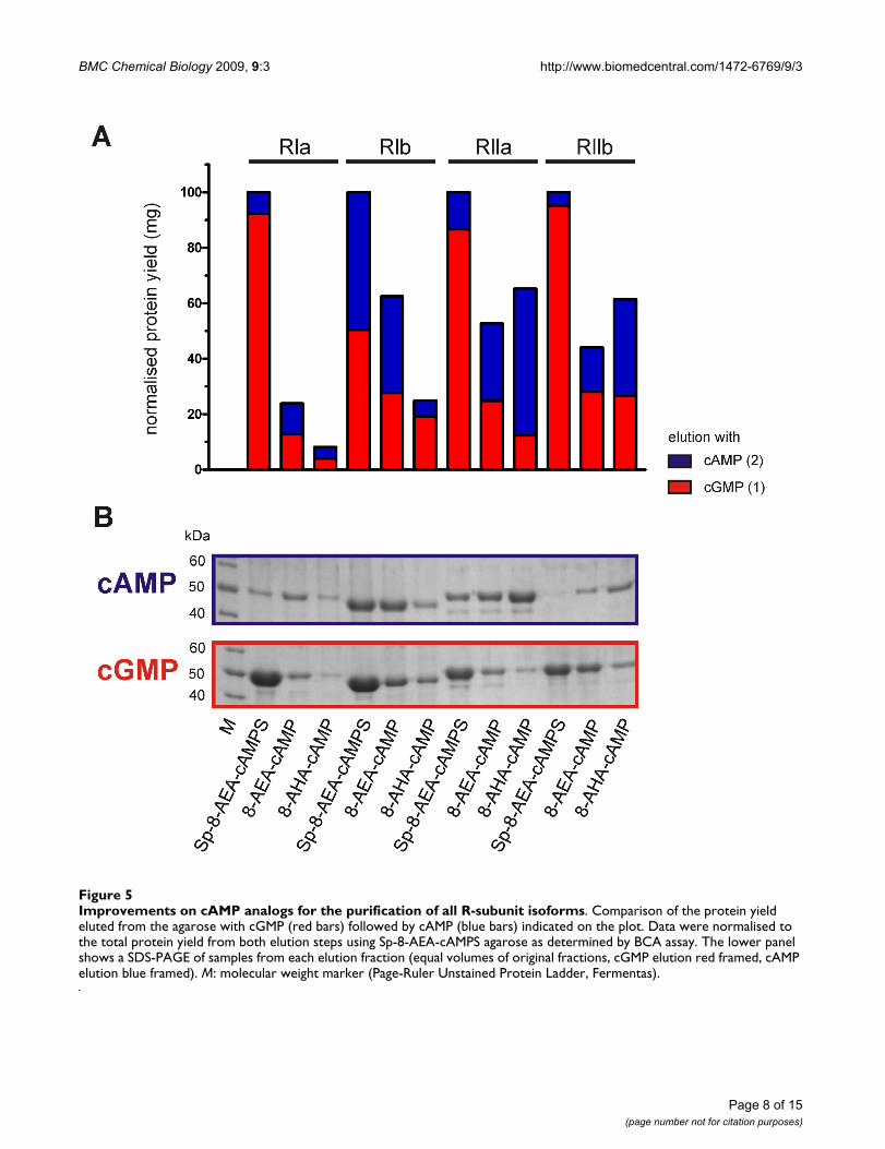
Comparison of conventional agaroses 8-AHA-cAMP and 8-AEA-cAMP with Sp-8-AEA-cAMPSThe Sp-8-AEA-cAMPS resin as well as the conventionalagaroses 8-AHA-cAMP and 8-AEA-cAMP were all testedvia small scale purification with all four R-subunit iso-forms, namely RIα, RIβ, RIIα and RIIβ. The protein yieldseluted first with cGMP and then subsequently in a secondelution step with cAMP are displayed in Fig. 5. As a controlthe cAMP elution was performed according to Diller et al.[18]. All R-subunit isoforms were obtained in a pure andactive form as determined by a spectrophotometric assay[33]. Since the highest overall yield of the respective R-subunit was obtained using Sp-8-AEA-cAMPS agarose,this amount was set as the 100% reference point (Fig. 5A).Interestingly, mild elution of RIα subunit with cGMPfrom the Sp-8-AEA-cAMPS agarose is nine times more effi-cient than the cGMP elution from the 8-AEA-cAMP agar-ose. Furthermore, a fourfold higher yield was obtainedfrom the cGMP elution only using the Sp-cAMPS agarosethan from combining both elution steps (cGMP andcAMP) employing 8-AEA-cAMP together. Purification ofRIα with 8-AHA-cAMP agarose was even less efficient thanusing 8-AEA-cAMP (Fig. 5B). Still, the amount of RIβ, RIIαand RIIβ isoforms eluted from our newly designed Sp-8-AEA-cAMPS agarose was a minimum of 40% greater thanfrom the conventional agaroses.
In a further experiment all four R-subunit isoforms werepurified with the Sp-8-AEA-cAMPS agarose (data notshown). The purification of RIα and RIIα isoformsresulted in 12 mg protein per liter expression media. ForRIβ and RIIβ isoforms 5 mg and 3 mg protein were puri-fied respectively. All affinity purified proteins were veri-fied by MS (data not shown). The entire purificationprocedure of each R-subunit isoform was completedwithin 8 h.
Binding of the PKA holoenzyme complex to agonist and antagonistBased on the purification strategy described above, novelcAMP derivatives with antagonist properties weredesigned to analyse the PKA holoenzyme (R2C2) complexand target physiological interaction partners via a chemi-cal proteomics approach. Only cAMP analogs derivedfrom the lead structure Rp-cAMPS (replacement of theequatorial exocyclic oxygen of the cyclic phosphate, Fig.1A right, 1C) have been shown to act as PKA-antagonists[17,34-36], whereas the Sp-cAMPS analogs and cAMPitself act as agonists (Fig. 1A left, 1B). The general assump-tion that Rp-cAMPS binds to the R-subunit in the intactPKA holoenzyme thus preventing complex dissociation[37] was addressed by a combination of SPR binding anal-ysis and Mass spectrometry (BIA-MS).
Therefore the antagonist Rp-8-AHDAA-cAMPS (for chem-ical structure see Fig. 1C) and the agonist 8-AHA-cAMPwere covalently coupled to two sensor surfaces on a singleCM5 sensor chip. 250 nM of each, PKA holoenzyme(R2C2), free R-subunit and C-subunit were injected toboth sensor surfaces. Fig. 6A shows the resulting bindingcurves for type I holoenzyme. Maximum binding signalsof 17,000 and 6,900 RU on the antagonist and on the ago-nist surfaces were observed, respectively. These differencein the binding signals indicates that the entire holoen-zyme complex is captured to the antagonist surfacewhereas on the agonist surface only the free R-subunit isbound, since the immobilised agonist causes holoenzymedissociation and release of the C-subunit. Hence, thesedifferences in binding signals reflect the mass differencebetween the holoenzyme complex (R2C2) and the free R-subunit (R2). A control experiment was performed, inject-ing free R-subunit over both surfaces and similar bindingsignals of approximately 5,300 RU each were obtained forthe agonist as well for the antagonist surface (Fig. 6A).Furthermore, binding signals to the agonist surface usingfree R-subunit and the PKA holoenzyme complex were inthe same range, again indicating that the holoenzyme dis-sociates when interacting with the agonist surface. As an
Purification of hRIα subunit with different Sp-cAMPS analog agaroses analysed by SDS-PAGEFigure 4 (see previous page)Purification of hRIα subunit with different Sp-cAMPS analog agaroses analysed by SDS-PAGE. (A) crude: total cell extract of E. coli BL21 DE3 Codonplus RIL with overexpressed hRIα at 52 kDa; supernatant: soluble fraction of bacterial lysate after French Press and centrifugation; pellet: insoluble fraction; sup agarose: unbound protein after incubation with Sp-8-AEA-cAMPS agarose; wash agarose: aliquot after 6 washing steps with buffer B; elution cGMP and elution cAMP: elution with 10 mM cGMP and subsequently with 10 mM cAMP, respectively; wash agarose: aliquot after 6 washing steps with buffer B; agarose beads after elution: remaining protein on the agarose beads after the two elution and washing steps. M: molecular weight marker (Page-Ruler Unstained Protein Ladder, Fermentas). (B) Total cell extract of E. coli BL21 DE3 Codonplus RIL with over-expressed hRIα was incubated with Sp-2-AHA-cAMPS and Sp-8-AHA-cAMPS, respectively, and eluted with cGMP (10 mM, lane 1 and 4) and subsequently with cAMP (10 mM, lane 2 and 5). Only a low amount of protein was left on Sp-2-AHA-cAMPS agarose after elution with cAMP (lane 3), whereas significant amounts of R-subunit remained on Sp-8-AHA-cAMPS agarose (lane 6). Asterisk indicates Chloramphenicol Acetyltransferase from E. coli BL21 (DE3) Codon Plus RIL as identified by MS.
Page 7 of 15(page number not for citation purposes)
BMC Chemical Biology 2009, 9:3 http://www.biomedcentral.com/1472-6769/9/3
Page 8 of 15(page number not for citation purposes)
Improvements on cAMP analogs for the purification of all R-subunit isoformsFigure 5Improvements on cAMP analogs for the purification of all R-subunit isoforms. Comparison of the protein yield eluted from the agarose with cGMP (red bars) followed by cAMP (blue bars) indicated on the plot. Data were normalised to the total protein yield from both elution steps using Sp-8-AEA-cAMPS agarose as determined by BCA assay. The lower panel shows a SDS-PAGE of samples from each elution fraction (equal volumes of original fractions, cGMP elution red framed, cAMP elution blue framed). M: molecular weight marker (Page-Ruler Unstained Protein Ladder, Fermentas).

BMC Chemical Biology 2009, 9:3 http://www.biomedcentral.com/1472-6769/9/3
Page 9 of 15(page number not for citation purposes)
Rp-cAMPS analogs bind the intact PKA holoenzyme complexFigure 6Rp-cAMPS analogs bind the intact PKA holoenzyme complex. The entire PKA type I holoenzyme complex (R2C2) binds highly specific to Rp-8-AHDAA-cAMPS as demonstrated by SPR (A), BIA MS (B) as well as affinity purification followed by SDS-PAGE (C) and Western blot analysis (D). (A) SPR binding pattern of type I holoenzyme, hRIα and Cα (as indicated, 250 nM each) on antagonist (Rp-8-AHDAA-cAMPS) and agonist (8-AHA-cAMP) surfaces. Measurements were performed in buffer A containing 0.005% P20, 1 mM ATP and 10 mM MgCl2 at a flow rate of 5 μL/min. (B) BIA-MS: SDS-PAGE analysis of protein recovered from the experiments depicted in (A). Type I holoenzyme, R-subunit and C-subunit were injected to the agonist and antagonist surfaces as described and were eluted with 0.2% SDS after a 4 min wash step. The eluted material from 15 repetitive runs was pooled and analysed by SDS-PAGE, MS and Western blot analysis (not shown). C-subunit (42 kDa) and R-subunit hRIα (50 kDa) are indicated. M: molecular weight marker (Page-Ruler, unstained protein ladder, Fermentas). (C) Binding of PKA type I holoenzyme complex (R2C2, left panel), free C-subunit (center panel) and free R-subunit (right panel) to antagonist agarose (Rp-8-AHA-cAMPS, 600 pmoles, for chemical structure see Fig. 1C) determined with SDS-PAGE. (D) Immunoblot analysis of SDS gels from panel (C) using anti-C-subunit antibody.
R-SU
C-SUR-SUdegradation
50
40
30
607085
100120150
kDa
25
20
M Holo
enzym
e
wash
1
wash
7
agaro
se
beads
elu
tion
cA
MP
C-S
U
wash
1
wash
7
RIa
-SU
wash
1
wash
7
C-SU
50
40
C
D
Holoenzyme C subunit R subunit
agaro
se
beads
after
elu
tion
agaro
se
beads
after
elu
tion
agaro
se
beads
after
elu
tion
0 100 200 300 400 500 600 700
0
5000
10000
15000
20000
C/agonist
C/antagonist
Holo/agonist
Holo/antagonist
R/agonist
R/antagonist
Time (s)
SP
Rsig
nal
(RU
)
A
50
60
kDa R/a
gonis
t
Holo
/agonis
t
Holo
/anta
gonis
t
R/a
nta
gonis
t
C/a
nta
gonis
t
C/a
gonis
t
R-SU
M
40C-SU
B

BMC Chemical Biology 2009, 9:3 http://www.biomedcentral.com/1472-6769/9/3
additional control C-subunit alone was injected over bothanalog surfaces. No binding was observed, demonstratingthat the C-subunit does not contribute to the binding sig-nal.
To prove that the intact holoenzyme complex was boundto the antagonist sensor surface a BIA MS experiment wasperformed (Fig. 6A, B). 250 nM of each, PKA holoenzyme(R2C2), free R-subunit and C-subunit were injected andafter a 4 min wash, the sensor surface was incubated with0.2% SDS. Proteins bound to the nucleotide surface wereeluted using the microrecovery function (Biacore 3000Control Software 4.1) and the content of 15 subsequentelutions was subjected to SDS-PAGE (Fig. 6B), displayingtwo distinct bands. These bands correspond to R- and C-subunits, as identified by LC-ESI MS/MS and Western blotanalysis (data not shown). Both, R- and C-subunit, weredetected in equal amounts according to Coomassie stain-ing (Fig. 6B), strongly supporting the hypothesis thatfunctionally intact holoenzyme complex (R2C2) wasbound to the antagonist surface and subsequently eluted.For a control the same amount of holoenzyme was alsoinjected to the agonist surface and analysed as describedabove. Only R-subunit, but no C-subunit, could bedetected by SDS-PAGE (Fig. 6B), MS and Western blotanalysis (data not shown).
Based on the BIA-MS results (Fig. 6B), the antagonist wascoupled to agarose beads via NHS chemistry (Fig. 3).These beads were then incubated with purified holoen-zyme complex. Samples of each step of the pull downexperiment were analysed via a SDS-PAGE (Fig. 6C) aswell as analysed by Western blot with an anti-C-subunitantibody (Fig. 6D), proving that Rp-cAMPS derivatisedagarose provides an extremely valuable tool for holoen-zyme purification. An additional control was performedwith free R- and C-subunit. Both were incubated with theantagonist beads and then analysed with SDS-PAGE. Onlythe R-subunit but not C-subunit was detected verifyingthe BIA-MS results.
Targeting the PKA interactome from pig brain using agonist and antagonist agarosesIn a proof of principle experiment agonist (Sp-8-AEA-cAMPS) and antagonist agarose (Rp-8-AHDAA-cAMPS)were incubated with pig brain lysate in order to investi-gate, if endogenously expressed R-subunit and PKAholoenzyme can be pulled out of biological material.After incubation of the soluble protein fraction with theaffinity resins, the beads were washed six times and thebound proteins were subsequently eluted with 20 mMcAMP and analysed by SDS-PAGE (Fig. 7).
Using the agonist as affinity reagent, all four R-subunitisoforms were pulled from pig brain lysate. When using
the antagonist, additionally all major C-subunit isoforms(Cα, Cβ and Cγ; Fig. 7) were identified based on MS (datanot shown). In addition to the PKA subunits other distinctsets of proteins were pulled out from pig brain tissue usingeither agonist or antagonist agaroses (Bertinetti et al. inpreparation). This indicates, that functional complexes ofcAMP interactome can be pulled out of tissue lysates withthese new chemical tools, proving the opportunity for thediscovery of additional components of the cAMP signal-ling pathway.
DiscussionDuring the last 30 years several approaches have beenused to selectively purify fully functional R-subunit iso-forms of PKA, either endogenous from animal tissues [6]or recombinantly expressed in bacteria [38]. Most purifi-
Affinity enrichment of components of the cAMP-pathway using agonist and antagonist agarosesFigure 7Affinity enrichment of components of the cAMP-pathway using agonist and antagonist agaroses. Pro-teins from pig brain tissue were enriched using agonist (Sp-8-AEA-cAMPS) and antagonist (Rp-8-AHDAA-cAMPS) agar-oses and subsequently eluted with cAMP. Fractions were separated on a 12% SDS-PAGE. supernatant: soluble protein fraction of pig brain after lysis, filtration and centrifugation; agonist/antagonist: protein eluted from the agonist/antagonist agarose with 25 mM cAMP. C-subunits (42 kDa), R-subunits RI (50 kDa) and RII (52 kDa) subunits are indicated, each identified by MS (data not shown). M: molecular weight marker (Page-Ruler, unstained protein ladder, Fermentas).
kDa
40
30
25
607085
100
120
150200
20
50
M agonis
t
supern
ata
nt
anta
gonis
t
RI-SU
C-SU
RII-SU
15
Page 10 of 15(page number not for citation purposes)
BMC Chemical Biology 2009, 9:3 http://www.biomedcentral.com/1472-6769/9/3
cation strategies have been based on ion exchange chro-matography. There were also early attempts to use cAMPanalogs linked via the N6 or C8 position of the adeninering to an agarose matrix for affinity purification of R-sub-units from several tissues [5]. In most cases, however, theaffinity of the R-subunit to the various cAMP analogs usedwas too high to subsequently and successfully elute theprotein efficiently. Only under harsh conditions could theR-subunit be removed from the matrix either by one of thetwo procedures: 7 M urea [6,39] or cAMP elution at ele-vated temperatures [23,38]; while still yielding not onlyless protein than using ion exchange chromatography butalso protein that was insoluble and/or proteolyticallydegraded [26]. In order to increase the yield of nucleotide-free and highly active R-subunit attempts to use fusiontags for purification [40-42]. However, fusion tags to theN-terminus as well as to the C-terminus of the R-subunitturned out to be problematic. Modifications of the N-ter-minus can interfere with R-subunit dimerisation andbinding of A kinase anchoring proteins [43]. Since fusiontags on the C-terminus are located in close proximity tothe cAMP binding domains, the cyclic nucleotide bindingproperties can be affected. Cleavage of the tag is an addi-tional purification step which may lead to a loss of proteinas well as cause additional proteolytic products.
We synthesised 3 different Sp-cAMPS analogs (Sp-8-AEA-cAMPS, Sp-8-AHA-cAMPS, Sp-2-AHA-cAMPS) and charac-terised the binding to all four R-subunit isoforms in directinteractions studies using SPR. Subsequently these ago-nists were coupled to solid supports and used for affinitypurification of recombinantly expressed R-subunits. Allthree analogs displayed superior purification strategieswhen compared to conventional cAMP analogs (8-AEA-cAMP, 8-AHA-cAMP).
Using Sp-8-AEA-cAMPS agarose it is now possible toobtain large yields of active and nucleotide-free R-subunitwithout the use of denaturants. This is especially impor-tant since remaining nucleotide in the CNB domainwould interfere with subsequent studies. Furthermoredecreased stability of Urea unfolded and refolded proteinoccurs [44].
In general, affinity reagents, addressing a specific subset ofproteins or protein complexes, are of growing importancefor many applications in proteomics. This includesenrichment of proteins or subcellular components as wellas removal of unwanted cell debris subcellular compo-nents, proteins and metabolites. Therefore, our describedcAMP analogs not only provide a highly selective tool asan affinity material for purification of PKA R-subunits, butcan also serve as a tool that particularly targets the cAMPsub-proteome (Bertinetti et al. in preparation). Here wecan demonstrate that components of cAMP signalling
pathways were selectively complexed with their physio-logical interaction partners thus demonstrating that phos-phorothioate cAMP analog agaroses are extremelyvaluable for comparable chemical proteomics studies.Furthermore, phosphorothioate cAMP analogs provide anadditional advantage as they were shown to be highly sta-ble against phosphodiesterase activity [45] and are there-fore well suited for use with cellular lysates.
In a recent approach, Scholten et al. [24] used conven-tional cyclic nucleotide agaroses to pull out componentsof the cGMP/cAMP interactome from rat heart tissue inorder to identify cyclic nucleotide binding proteins andtheir interaction partners. Since elution with cAMP fromthe 8-AEA-cAMP agarose was not sufficient for subsequentMS analysis, those studies had to be performed with mate-rial that was obtained by boiling the incubated agarosebeads in SDS loading buffer. With the newly designed Sp-and Rp-cAMPS agaroses described here, specific bindingand elution of cAMP binding proteins and a reduction offalse positives can be achieved since elution with cAMPcan be considered as an additional purification step. Theuse of these novel tools now enables us to also pull outinteraction partners of intact holoenzyme, which is espe-cially important for identification of novel interactionpartners of the C-subunits of PKA such as the recentlydescribed A-kinase interacting proteins (AKIPs, [46]) inthe holoenzyme form.
ConclusionNovel cAMP analogs based on phosphorothioate wereoptimised in an iterative approach combining design,synthesis and direct SPR binding studies. After coupling toa solid support, two sets of chemical binders were testedfor highly efficient purification procedure of all isoformsof PKA R-subunit. Sp-8-AEA-cAMPS was identified as themost valuable tool for purification purposes, yielding mil-ligram amounts of both highly pure and active R-subunitwithin 8 h. Furthermore, Sp- and Rp-cAMPS derived ana-logs have the potential to be employed in a chemical pro-teomics approach targeting distinct cAMP sub-proteomes.
MethodsChemicalsAll chemicals used were of the purest grade available andwere obtained either from Sigma-Aldrich (Seelze), Roth(Karlsruhe) or Applichem (Darmstadt). The followingcAMP-analogs were synthesised by Biolog LSI (Bremen):8-AHA-cAMP, 8-AEA-cAMP, Sp-cAMPS, Sp-8-AHA-cAMPS, Sp-2-AHA-cAMPS, Sp-8-AEA-cAMPS, Rp-cAMPS,Rp-8-AHA-cAMPS, Rp-8-AHDAA-cAMPS. A representativescheme with synthesis and coupling of Sp-8-AEA-cAMPSto agarose beads is provided in Fig. 3.
Page 11 of 15(page number not for citation purposes)
BMC Chemical Biology 2009, 9:3 http://www.biomedcentral.com/1472-6769/9/3
Synthesis of cyclic nucleotide derivatives and coupling to agaroseAll cAMP- and ω-aminoalkyl-substituted Sp-cAMPS ana-logs (for chemical structure see Fig. 1B) were synthesisedas described [22,47] with some minor modifications. Typ-ically, 100 μmoles of (Sp)-8-Br-cAMP(S), (Sp)-2-Cl-cAMP(S) or (Rp)-8-Br-cAMP(S) (Biolog) and 1,000μmoles of 1,ω-diaminoalkan (Fluka) were dissolved in 10mL water and refluxed until no starting material wasdetectable by HPLC analysis (first step in the reactionpathway Fig. 3). The reaction mixtures were neutralisedwith diluted HCl, concentrated by rotary evaporationunder reduced pressure, and subsequently purified bymeans of semi-preparative reversed phase HPLC (YMCODS-A 120–11, YMC). The column was washed with 100mM NaH2PO4, pH 7, followed by water. Each cAMP(S)analog was eluted with a gradient from 100% water to100% acetonitrile. The product containing fractions werecollected and evaporated under reduced pressure toobtain 8-AHA-cAMP, 8-AEA-cAMP, Sp-8-AHA-cAMPS, Sp-8-AEA-cAMPS, Sp-2-AHA-cAMPS and Rp-8-AHDAA-cAMPS in yields of 60–80% with purities > 99% (byHPLC). The structure of each cAMP(S) analog was con-firmed by UV/VIS spectrometry and FAB/MS or ESI/MSanalysis.
cAMP(S) analogs were coupled to NHS-activated agarosebeads (Affi-Gel® 10, BIO-RAD) according to the manufac-turer's instructions (Fig. 3). Briefly, 6.6 μmoles ofcAMP(S) analog and 7.26 μmoles ethyldiisopropylaminewere added per mL settled gel, suspended in DMSO. Reac-tion mixture was carefully shaken for 2–18 h at ambienttemperature until no further consumption of startingmaterial was detectable by analytical HPLC monitoring.Any unreacted NHS-groups of the agarose gels wereblocked by addition of 20 μmoles ethanolamine per mLsettled gel by incubation for 1 h. After filtration and mul-tiple washing with subsequently 2 × 25 mL 20% ethanol,2 × 25 mL H2O and 2 × 25 mL 30 mM NaH2PO4, pH 7,each agarose gel was stored in 30 mM NaH2PO4, 1%NaN3, pH 7 at 4°C. Ligand densities were 6 μmoles/mL 8-AHA-cAMP, 6 μmoles/mL 8-AEA-cAMP, 4 μmoles/mL Sp-8-AEA-cAMPS agarose, 5 μmoles/mL Sp-8-AHA-cAMPSagarose, 6 μmoles/mL Sp-2-AHA-cAMPS agarose and 6μmoles/mL Rp-8-AHDAA-cAMPS agarose.
Direct binding studies of Sp-cAMPS analogs using SPRSp-2-AHA-cAMPS, Sp-8-AEA-cAMPS, Sp-8-AHA-cAMPS(for chemical structures see Fig. 1B), 8-AHA-cAMP and 8-AEA-cAMP were dissolved in 100 mM HEPES-KOH pH 8by cautious heating (max. 70°C) and filtered. The concen-trations of the stock solutions were determined via theirrespective extinction coefficient. CM5 sensor chip surfaces(research grade, Biacore AB) were activated for 10 minwith NHS/EDC according to the manufacturer's instruc-
tions (amine coupling kit, Biacore AB). The analogs (3mM) were injected for 7 min (running buffer: 100 mMHEPES, pH 8). Deactivation of the surface was performedwith 1 M ethanolamine-HCl, pH 8.5. Each flow cell wasactivated, coupled and deactivated individually with aflow rate of 5 μL/min at 20°C. A reference cell (Flow cell1) was activated and deactivated without ligand immobi-lisation.
All interaction analyses were performed at 20°C in 150mM NaCl, 20 mM MOPS, pH 7 (buffer A) containing0.005% (v/v) surfactant P20, using a Biacore 2000 instru-ment (Biacore AB). Binding analyses were performed byinjection of 100 nM hRIα, hRIβ, hRIIα and rRIIβ (all pro-teins were purified classically by DEAE cellulose chroma-tography [48]) at a flow rate of 10 μL/min. Associationand dissociation were monitored for 5 min and 10 min,respectively. Dissociation was performed in buffer A con-taining 0.005% P20 in the presence or absence of 3 mMcGMP. The sensor surfaces were regenerated after eachbinding cycle by two short injections of 3 M guanidiniumHCl. After subtracting the reference cell signal, bindingdata were normalised (Fig. 2).
Purification of hRIα using Sp-cAMPS agarosesBacterial cells overexpressing R-subunit were lysed using aFrench Pressure Cell (Thermo Electron) in lysis buffercontaining 20 mM MOPS pH 7, 100 mM NaCl, 1 mM β-mercaptoethanol, 2 mM EDTA and 2 mM EGTA (bufferA). The crude lysate was centrifuged at 27,000 g for 30 minat 4 C. Three different Sp-cAMPS agaroses (Sp-8-AHA-cAMPS, Sp-8-AEA-cAMPS and Sp-2-AHA-cAMPS agarose)were tested side by side in a one step purification strategy.1.2 μmoles of coupled analog were used for each purifica-tion, corresponding to approximately 400 μL of agaroseslurry. 12 mL supernatant from 500 mL bacterial culturewere incubated with the respective affinity matrices. Bind-ing was carried out in a batch format by gently rotatingover night at 4°C. After washing the agarose seven timeswith 1.25 mL lysis buffer each, the protein was eluted with1.25 mL of 10 mM cGMP in buffer B (buffer A plus 1 mMβ-mercaptoethanol) by gentle rotation at 4°C for 1 h fol-lowed by an elution using 10 mM cAMP in buffer Binstead of cGMP. Excess of nucleotide was removed usinga PD10 gel filtration column (Amersham Pharmacia).cGMP bound to the cyclic nucleotide binding pockets wasremoved by extensive dialysis against buffer B.
The purification strategy of RIβ with Sp-8-AEA-cAMPS fol-lows in principle the procedure described for RIα.
Purification of type II R-subunits using Sp-8-AEA-cAMPS agaroseThe purification strategy of RII isoforms follows the pro-cedure described for hRIα except cell lysis was performed
Page 12 of 15(page number not for citation purposes)
BMC Chemical Biology 2009, 9:3 http://www.biomedcentral.com/1472-6769/9/3
in buffer containing 20 mM MES pH 6.5, 100 mM NaCl,5 mM EDTA, 5 mM EGTA and 5 mM β-mercaptoethanol(buffer C) including the protease inhibitors Leupeptin(0,025 mg/100 mL, Biomol), TPCK and TLCK (each 1 mg/100 mL, Biomol). After cell lysis, the soluble protein frac-tion was incubated in a batch format with Sp-8-AEA-cAMPS agarose (1.4 μmol analog). The agarose waswashed twice with 10 mL buffer D (20 mM MES pH 7, 1M NaCl, 5 mM β-mercaptoethanol) and subsequentlywith buffer C containing protease inhibitors. Two elutionsteps were performed with 1 mL 25 mM cGMP in buffer Cand exchanged to 20 mM MES pH 6.5, 150 mM NaCl, 2mM EDTA, 2 mM EGTA and 1 mM β-mercaptoethanolusing a PD10 column (Amersham).
Compared purification of all R-subunits using different cAMP analog agarosesThe side by side comparison of Sp-8-AEA-cAMPS, 8-AHA-cAMP and 8-AEA-cAMP for purification was performed asdescribed for the RI and RII purification. Crude lysatefrom one litre expression culture was divided into threeequal aliquots and incubated in a small scale experimentwith 100 μL of the respective agaroses. The cGMP elutionwas followed by a second elution step with 40 mM cAMPat room temperature for 30 minutes according to [18].
BIA-MSRp-8-AHDAA-cAMPS (for chemical structure see Fig. 1C)and 8-AHA-cAMP were dissolved by cautious heating(max. 60°C) in 100 mM Borate pH 8.5 with 20% DMSOand 100 mM HEPES pH 8, respectively, filtered and cou-pled to the sensor surface as described above. All microre-covery experiments were performed in buffer A containing10 mM MgCl2, 1 mM ATP with 0.005% (v/v) surfactantP20 on a Biacore 3000 instrument. 250 nM PKA type Iholoenzyme (R2C2), free RIα subunit or C-subunit wereinjected over the analog sensor surfaces in separate exper-iments. The association phase was monitored for 5 minfollowed by a 4 min washing step with running buffer.Bound protein was eluted by incubating the sensor surfacewith 0.2% SDS for 90 s and the eluted material was recov-ered in a capped vial. The sensor surfaces were regeneratedby three subsequent injections of 3 M guanidinium HCl.
The recovered material from 15 repetitive cycles waspooled and applied to a 12% SDS-PAGE [49] (Fig. 6B) forprotein identification via MS.
Fishing of PKA holoenzyme complex with Rp-cAMPS agarosePurified recombinant mCα, hRIα and PKA holoenzymewere each incubated with Rp-8-AHDAA-cAMPS agarose(600 pmoles coupled analog, for chemical structure seeFig. 1C) for 2 h at 4°C. The agarose was washed seventimes with 1 mL buffer E (buffer B containing 1 mM ATP
and 10 mM MgCl2). Protein was eluted with 1 mL 20 mMcAMP in buffer E by gentle rotation at room temperaturefor 1 h. The entire supernatant of each step was precipi-tated with TCA and applied to SDS-PAGE (Fig. 6C). ForWestern blotting, the samples were transferred to PVDFmembrane and immunoblotted with anti-PKA C-subunitantibody (Santa Cruz Biotechnology, PKAα cat C-20) vis-ualised by enhanced chemiluminescence (Fig. 6D).
Fishing of PKA holoenzyme complex with agonist and antagonist agarose from pig brain lysateFresh pig brain tissue was homogenised in buffer F (bufferE in the presence of protease inhibitors (complete, EDTAfree, Roche), 2 mM NADH and 20 mM sucrose). After cen-trifugation at 13,700 g for 25 min, the supernatant was fil-tered and incubated with 150 μL agarose (correspondingto 1 μmole of Sp-8-AEA-cAMPS or Rp-8-AHDAA-cAMPS)over night at 4°C. The beads were washed six times with1.5 mL buffer F. Elution was carried out with 1 mL ofbuffer E containing 20 mM cAMP by gentle rotation atroom temperature for 1 h. All samples were precipitatedwith TCA for SDS-PAGE (Fig. 7).
Biochemical characterisation of the proteinsThe purification of the R-subunits was analysed by 12%SDS-PAGE [49] unless otherwise noted and proteins werestained with colloidal Coomassie Brilliant Blue dye mod-ified after Neuhoff et al. [50-52]. After electrophoresis,remaining SDS was removed by heating and rinsing indistilled water. Overnight staining with 0.1% Coomassie®
Brilliant Blue G 250 in 5% aluminium sulphate octadec-ahydrate and 2% phosphoric acid resulted in intense bluebands with low background (Fig. 4, 5, 6, 7). Protein con-centration was determined by a colorimetric assay usingBSA as a standard [53]. The biological activity of the pro-teins was verified by a spectrophotometric phosphotrans-ferase assay using the substrate peptide Kemptide(LRRASLG, Biosynthan) according to Cook et al. [33].
Mass spectrometry analysisProtein bands were excised from one-dimensional SDS-PAGE [49] and digested in gel with trypsin according topublished procedures [54], modified by omitting all pre-washing steps. After equilibrating in water, the gel pieces(Fig. 4, 6B, 7) were excised and homogenised via centrifu-gation (1,6000 g) through 10 μL pipet tips (MBP) and col-lected in small reaction vials (CS-ChromatographieService). Destaining, reduction and alkylation were omit-ted and 25 μL digestion buffer containing 50 mMNH4HCO3, 100 ng/μL of trypsin (sequencing grade,Promega) were added directly to the gel slurry and incu-bated at 50°C for a minimum 1 h [55]. After a short cen-trifugation the supernatants were diluted with 40 μL of0.3% formic acid for analysis in a nanoLC-ESI-MS/MS(nanoLC-Ultimate HPLC-system, LC Packings, Dionex
Page 13 of 15(page number not for citation purposes)
BMC Chemical Biology 2009, 9:3 http://www.biomedcentral.com/1472-6769/9/3
coupled online to a linear ion trap mass spectrometer4000 QTRAP™, Applied Biosystems), as described in[56,57]. The MS/MS spectra were searched against a non-redundant sequence database (MSDB) using MASCOT(Matrix Science), version 1.9.05. Taxonomy was restrictedto mammals with variable modifications on deamidation(NQ), myristoylation (N-Term. G), oxidation (M) andphosphorylation (ST).
Authors' contributionsDB, SS and SEH performed the expression and purifica-tion of the recombinant proteins as well as the biochemi-cal characterisation and the chemical proteomicsexperiments. FS and HGG synthesised and purified thecyclic nucleotide analogs and performed coupling to theagarose. SD carried out the SPR measurements, OB themass spectrometry analyses, SS the BIA-MS experimentsand DB, SS and FWH wrote the manuscript and preparedthe figures.
AcknowledgementsWe thank Michaela Hansch, Marko Knoll, Antje Badel and Maike Vetter (Kassel University) for technical assistance as well as Undine Manzau and Ursula Havemann (Biolog LSI) for technical support. We thank Maria McGlone for carefully reading the manuscript. This work was supported by grants from the Deutsche Forschungsgemeinschaft (DFG, He1818/4) to FWH and EU (LSHB-CT-2006-037189, thera-cAMP) to FWH, HGG, FS and SD. SS and SEH were supported by the Graduate Program of Kassel University. FWH's group is a member in the EU FP6 ProteomeBinders con-sortium.
References1. Shabb JB: Physiological substrates of cAMP-dependent pro-
tein kinase. Chem Rev 2001, 101(8):2381-2411.2. Kim C, Vigil D, Anand G, Taylor SS: Structure and dynamics of
PKA signaling proteins. Eur J Cell Biol 2006, 85(7):651-654.3. Taylor SS, Kim C, Cheng CY, Brown SH, Wu J, Kannan N: Signaling
through cAMP and cAMP-dependent protein kinase: diversestrategies for drug design. Biochim Biophys Acta 2008,1784(1):16-26.
4. Rubin CS, Erlichman J, Rosen OM: Molecular forms and subunitcomposition of a cyclic adenosine 3',5'-monophosphate-dependent protein kinase purified from bovine heart muscle.J Biol Chem 1972, 247(1):36-44.
5. Wilchek M, Salomon Y, Lowe M, Selinger Z: Conversion of proteinkinase to a cyclic AMP independent form by affinity chroma-tography on N 6-caproyl 3',5'-cyclic adenosine monophos-phate-sepharose. Biochem Biophys Res Commun 1971,45(5):1177-1184.
6. Ramseyer J, Kaslow HR, Gill GN: Purification of the cAMP recep-tor protein by affinity chromatography. Biochem Biophys ResCommun 1974, 59(2):813-821.
7. Craven KB, Zagotta WN: CNG and HCN channels: two peas,one pod. Annu Rev Physiol 2006, 68:375-401.
8. Omori K, Kotera J: Overview of PDEs and their regulation. CircRes 2007, 100(3):309-327.
9. de Rooij J, Rehmann H, van Triest M, Cool RH, Wittinghofer A, BosJL: Mechanism of regulation of the Epac family of cAMP-dependent RapGEFs. J Biol Chem 2000, 275(27):20829-20836.
10. Berman HM, Ten Eyck LF, Goodsell DS, Haste NM, Kornev A, TaylorSS: The cAMP binding domain: an ancient signaling module.Proc Natl Acad Sci USA 2005, 102(1):45-50.
11. Biskup C, Kusch J, Schulz E, Nache V, Schwede F, Lehmann F, HagenV, Benndorf K: Relating ligand binding to activation gating inCNGA2 channels. Nature 2007, 446(7134):440-443.
12. Schweinsberg S, Moll D, Burghardt NC, Hahnefeld C, Schwede F,Zimmermann B, Drewianka S, Werner L, Kleinjung F, Genieser HG,et al.: Systematic interpretation of cyclic nucleotide bindingstudies using KinetXBase. Proteomics 2008, 8(6):1212-1220.
13. Schwede F, Christensen A, Liauw S, Hippe T, Kopperud R, Jastorff B,Doskeland SO: 8-Substituted cAMP analogues reveal markeddifferences in adaptability, hydrogen bonding, and chargeaccommodation between homologous binding sites (AI/AIIand BI/BII) in cAMP kinase I and II. Biochemistry 2000,39(30):8803-8812.
14. Christensen AE, Selheim F, de Rooij J, Dremier S, Schwede F, DaoKK, Martinez A, Maenhaut C, Bos JL, Genieser HG, et al.: cAMP ana-log mapping of Epac1 and cAMP kinase. Discriminating ana-logs demonstrate that Epac and cAMP kinase actsynergistically to promote PC-12 cell neurite extension. J BiolChem 2003, 278(37):35394-35402.
15. de Wit RJ, Hoppe J, Stec WJ, Baraniak J, Jastorff B: Interaction ofcAMP derivatives with the 'stable' cAMP-binding site in thecAMP-dependent protein kinase type I. Eur J Biochem 1982,122(1):95-99.
16. de Wit RJ, Hekstra D, Jastorff B, Stec WJ, Baraniak J, Van Driel R, VanHaastert PJ: Inhibitory action of certain cyclophosphate deriv-atives of cAMP on cAMP-dependent protein kinases. Eur J Bio-chem 1984, 142(2):255-260.
17. Dostmann WR, Taylor SS, Genieser HG, Jastorff B, Døskeland SO,Øgreid D: Probing the cyclic nucleotide binding sites ofcAMP-dependent protein kinases I and II with analogs ofadenosine 3',5'-cyclic phosphorothioates. J Biol Chem 1990,265(18):10484-10491.
18. Diller TC, Xuong NH, Taylor SS: Type II beta regulatory subunitof cAMP-dependent protein kinase: purification strategies tooptimize crystallization. Protein Expr Purif 2000, 20(3):357-364.
19. Moll D, Schweinsberg S, Hammann C, Herberg FW: Comparativethermodynamic analysis of cyclic nucleotide binding to Pro-tein Kinase A. Biol Chem 2007, 388(2):163-172.
20. Corbin JD, Sugden PH, West L, Flockhart DA, Lincoln TM, McCarthyD: Studies on the properties and mode of action of the puri-fied regulatory subunit of bovine heart adenosine 3':5'-monophosphate-dependent protein kinase. J Biol Chem 1978,253(11):3997-4003.
21. Dills WL Jr, Beavo JA, Bechtel PJ, Krebs EG: Purification of rabbitskeletal muscle protein kinase regulatory subunit usingcyclic adenosine-3':5'-monophosphate affinity chromatogra-phy. Biochem Biophys Res Commun 1975, 62(1):70-77.
22. Dills WL Jr, Beavo JA, Bechtel PJ, Myers KR, Sakai LJ, Krebs EG: Bind-ing of adenosine 3',5'-monophosphate dependent proteinkinase regulatory subunit to immobilized cyclic nucleotidederivatives. Biochemistry 1976, 15(17):3724-3731.
23. Ramseyer J, Kanstein CB, Walton GM, Gill G: The use of affinitychromatography in purification of cyclic nucleotide receptorproteins. Biochim Biophys Acta 1976, 446(2):358-370.
24. Scholten A, Poh MK, van Veen TA, van Breukelen B, Vos MA, HeckAJ: Analysis of the cGMP/cAMP interactome using a chemi-cal proteomics approach in mammalian heart tissue vali-dates sphingosine kinase type 1-interacting protein as agenuine and highly abundant AKAP. J Proteome Res 2006,5(6):1435-1447.
25. Scholten A, Visser NF, Heuvel RH van den, Heck AJ: Analysis ofprotein-protein interaction surfaces using a combination ofefficient lysine acetylation and nanoLC-MALDI-MS/MSapplied to the E9:Im9 bacteriotoxin – immunity proteincomplex. J Am Soc Mass Spectrom 2006, 17(7):983-994.
26. Potter RL, Stafford PH, Taylor S: Regulatory subunit of cyclicAMP-dependent protein kinase I from porcine skeletal mus-cle: purification and proteolysis. Arch Biochem Biophys 1978,190(1):174-180.
27. Vigil D, Lin JH, Sotriffer CA, Pennypacker JK, McCammon JA, TaylorSS: A simple electrostatic switch important in the activationof type I protein kinase A by cyclic AMP. Protein Sci 2006,15:113-121.
28. Vigil D, Blumenthal DK, Heller WT, Brown S, Canaves JM, Taylor SS,Trewhella J: Conformational differences among solutionstructures of the type Ialpha, IIalpha and IIbeta proteinkinase A regulatory subunit homodimers: role of the linkerregions. J Mol Biol 2004, 337(5):1183-1194.
Page 14 of 15(page number not for citation purposes)
BMC Chemical Biology 2009, 9:3 http://www.biomedcentral.com/1472-6769/9/3
Publish with BioMed Central and every scientist can read your work free of charge
"BioMed Central will be the most significant development for disseminating the results of biomedical research in our lifetime."
Sir Paul Nurse, Cancer Research UK
Your research papers will be:
available free of charge to the entire biomedical community
peer reviewed and published immediately upon acceptance
cited in PubMed and archived on PubMed Central
yours — you keep the copyright
Submit your manuscript here:http://www.biomedcentral.com/info/publishing_adv.asp
BioMedcentral
29. Stokka AJ, Gesellchen F, Carlson CR, Scott JD, Herberg FW, TaskenK: Characterization of A-kinase-anchoring disruptors using asolution-based assay. Biochem J 2006, 400(3):493-499.
30. Hall DR, Cann JR, Winzor DJ: Demonstration of an upper limitto the range of association rate constants amenable to studyby biosensor technology based on surface plasmon reso-nance. Anal Biochem 1996, 235(2):175-184.
31. Schuck P, Minton AP: Analysis of mass transport-limited bind-ing kinetics in evanescent wave biosensors. Anal Biochem 1996,240(2):262-272.
32. Herberg FW, Zimmermann B: Analysis of protein kinase interac-tions using biomolecular interaction analysis. In Protein Phos-phorylation – A Practical Approach Second edition. Edited by: HardieDG. Oxford University Press; 1999:335-371.
33. Cook PF, Neville ME Jr, Vrana KE, Hartl FT, Roskoski R Jr: Adenos-ine cyclic 3',5'-monophosphate dependent protein kinase:kinetic mechanism for the bovine skeletal muscle catalyticsubunit. Biochemistry 1982, 21(23):5794-5799.
34. Van Haastert PJ, Van Driel R, Jastorff B, Baraniak J, Stec WJ, De WitRJ: Competitive cAMP antagonists for cAMP-receptor pro-teins. J Biol Chem 1984, 259(16):10020-10024.
35. Dostmann WR, Taylor SS: Identifying the molecular switchesthat determine whether (Rp)-cAMPS functions as an antag-onist or an agonist in the activation of cAMP-dependent pro-tein kinase I. Biochemistry 1991, 30(35):8710-8716.
36. Dostmann WR: (RP)-cAMPS inhibits the cAMP-dependentprotein kinase by blocking the cAMP-induced conforma-tional transition. FEBS Lett 1995, 375(3):231-234.
37. Das R, Melacini G: A Model for Agonism and Antagonism in anAncient and Ubiquitous cAMP-binding Domain. J Biol Chem2007, 282(1):581-593.
38. Saraswat LD, Filutowicz M, Taylor SS: Expression of the type Iregulatory subunit of cAMP-dependent protein kinase inEscherichia coli. J Biol Chem 1986, 261(24):11091-11096.
39. Builder SE, Beavo JA, Krebs EG: Stoichiometry of cAMP and1,N6-etheno-cAMP binding to protein kinase. J Biol Chem1980, 255(6):2350-2354.
40. Kopperud R, Christensen AE, Kjarland E, Viste K, Kleivdal H, Doske-land SO: Formation of inactive cAMP-saturated holoenzymeof cAMP-dependent protein kinase under physiological con-ditions. J Biol Chem 2002, 277(16):13443-13448.
41. Dao KK, Teigen K, Kopperud R, Hodneland E, Schwede F, Chris-tensen AE, Martinez A, Doskeland SO: Epac1 and cAMP-depend-ent Protein Kinase Holoenzyme Have Similar cAMP Affinity,but Their cAMP Domains Have Distinct Structural Featuresand Cyclic Nucleotide Recognition. J Biol Chem 2006,281(30):21500-21511.
42. Solberg R, Tasken K, Wen W, Coghlan VM, Meinkoth JL, Scott JD,Jahnsen T, Taylor SS: Human regulatory subunit RI beta ofcAMP-dependent protein kinases: expression, holoenzymeformation and microinjection into living cells. Exp Cell Res1994, 214(2):595-605.
43. Herberg FW, Maleszka A, Eide T, Vossebein L, Tasken K: Analysisof A-kinase anchoring protein (AKAP) interaction with pro-tein kinase A (PKA) regulatory subunits: PKA isoform specif-icity in AKAP binding. J Mol Biol 2000, 298(2):329-339.
44. Canaves JM, Leon DA, Taylor SS: Consequences of cAMP-bind-ing site mutations on the structural stability of the type I reg-ulatory subunit of cAMP-dependent protein kinase.Biochemistry 2000, 39(49):15022-15031.
45. Schaap P, van Ments-Cohen M, Soede RD, Brandt R, Firtel RA, Dost-mann W, Genieser HG, Jastorff B, van Haastert PJ: Cell-permeablenon-hydrolyzable cAMP derivatives as tools for analysis ofsignaling pathways controlling gene regulation in Dictyostel-ium. J Biol Chem 1993, 268(9):6323-6331.
46. Sastri M, Barraclough DM, Carmichael PT, Taylor SS: A-kinase-interacting protein localizes protein kinase A in the nucleus.Proc Natl Acad Sci USA 2005, 102(2):349-354.
47. Long RA, Robins RK, Townsend LB: Purine nucleosides. XV. Thesynthesis of 8-amino and 8-substituted aminopurine nucleo-sides. J Org Chem 1967, 32(9):2751-2756.
48. Corbin JD, Keely SL, Park CR: The distribution and dissociationof cyclic adenosine 3':5'-monophosphate-dependent proteinkinases in adipose, cardiac, and other tissues. J Biol Chem 1975,250(1):218-225.
49. Laemmli UK: Cleavage of structural proteins during theassembly of the head of bacteriophage T4. Nature 1970,227(5259):680-685.
50. Neuhoff V, Stamm R, Eibl H: Clear background and highly sensi-tive protein staining with Coomassie Blue dyes in polyacry-lamide gels: A systematic analysis. Electrophoresis 1985,6(9):427-448.
51. Neuhoff V, Arold N, Taube D, Ehrhardt W: Improved staining ofproteins in polyacrylamide gels including isoelectric focusinggels with clear background at nanogram sensitivity usingCoomassie Brilliant Blue G-250 and R-250. Electrophoresis1988, 9(6):255-262.
52. Kang DH, Gho YS, Suh MK, Kang CH: Highly sensitive and fastprotein detection with coomassie brilliant blue in sodiumdodecyl sulfate-polyacrylamide gel electrophoresis. BullKorean Chem Soc 2002, 23(11):1511-1512.
53. Bradford MM: A rapid and sensitive method for the quantita-tion of microgram quantities of protein utilizing the princi-ple of protein-dye binding. Anal Biochem 1976, 72:248-254.
54. Rosenfeld J, Capdevielle J, Guillemot JC, Ferrara P: In-gel digestionof proteins for internal sequence analysis after one- or two-dimensional gel electrophoresis. Anal Biochem 1992,203(1):173-179.
55. Finehout EJ, Cantor JR, Lee KH: Kinetic characterization ofsequencing grade modified trypsin. Proteomics 2005,5(9):2319-2321.
56. Gesellchen F, Bertinetti O, Herberg FW: Analysis of posttransla-tional modifications exemplified using protein kinase A. Bio-chim Biophys Acta 2006, 1764(12):1788-1800.
57. Schaefer H, Chamrad DC, Marcus K, Reidegeld KA, Bluggel M, MeyerHE: Tryptic transpeptidation products observed in proteomeanalysis by liquid chromatography-tandem mass spectrome-try. Proteomics 2005, 5(4):846-852.
58. Humphrey W, Dalke A, Schulten K: VMD: visual moleculardynamics. J Mol Graph 1996, 14(1):27-28.
Page 15 of 15(page number not for citation purposes)