bone & cartilage
TRANSCRIPT

Cartilage • Specialized dense connective tissue • Semi rigid ,designed to give support, bear
weight & withstand tension ,torsion & bending • Devoid of blood vessels and not innervated
by nerve • Most of them are calcified in old age. • Cartilage can grow by interstitial &
appositional growth

Composition of cartilage
• Perichondrium • Ground substance-Highly hydrated Contains hyaluronic
acid glucoseaminoglycans • Cells- chondroblasts,
chondrocytes • Fibers- collagen , elastic fibers
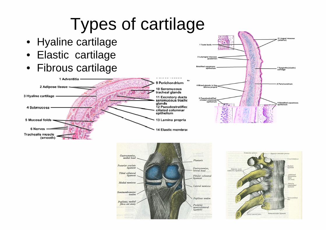
Types of cartilage • Hyaline cartilage • Elastic cartilage • Fibrous cartilage

Hyaline cartilage • Most common type • Makes the skeletal
model of most bones in embryo
• Gradually replaced by bone in grown ups except at the articular surface of bones, ends of the ribs, nose, larynx, trachea and bronchi

• In living conditions looks translucent & bluish white in colour
• Covered with perichondrium. Articular cartilage is not covered by perichondrium
• Matrix is homogenous which consists of chondroitin sulphate & collagen fibers
• Cells are chondrocytes arranged in groups in lacunae • Collagen fibers are not visible in matrix because of the
same refractive index as that of matrix
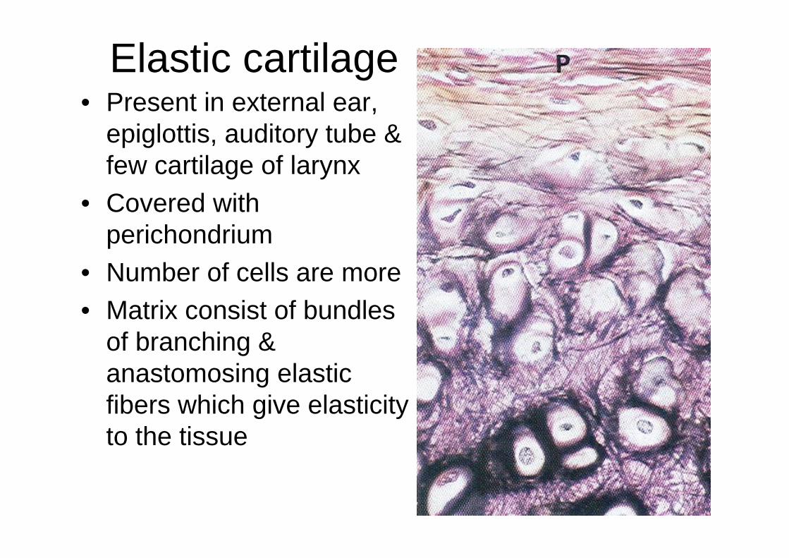
Elastic cartilage • Present in external ear,
epiglottis, auditory tube & few cartilage of larynx
• Covered with perichondrium
• Number of cells are more • Matrix consist of bundles
of branching & anastomosing elastic fibers which give elasticity to the tissue

Fibrous cartilage • Found in intervertebral disc,
pubic symphysis, intrarticular disc of certain joints, menisci of knee joint & articular cartilage of temporomandibular cartilage
• Consists of bundles of collagen fibers embedded in minimal amount of matrix
• Cells are usually placed single in between the bundles of collagen fibers
• Not covered with perichondrium

BONE • Specializes form of dense
connective tissue • Makes supportive frame work • Support & transmit weight of
the body • Provide the levers for
locomotion by formingarticulations
• Give attachment to muscles & ligaments
• Provide mechanical protection to the vital organ
• Store calcium • Form blood in their marrow

Classification of bones According to position
Axial Appendiculer

Number of bones
• Total 206 bones • Upper limbs - 64 • Lower limbs – 62 • Vertebrae – 26(33) • Skull – 29(26Skull bones
+ hyoid +6 ossicle) • Ribs – 24 • Sternum

• Appendicular-Upper limb
Lower limb
64
62

• Long bones • Short bones • Short bones • Flat bones • Irregular bones • Pneumatic bones • Sesamoid bones • Accessory bones
According to size & shape

According to gross structure
• Compact (Lamellar) bone
• Spongy (cancellous) bone
• Diploic bones

According todevelopment
• Membranous bones-Bone is laid down directly in the fibrousmembrane e.g. bonesof vault of skull, mandible
• Cartilaginous bones-Formation of bone is proceeded by theformation of a cartilage,which is later replacedby a bone e.g. femur,tibia

Membranous ossification • Bone is formed in mesenchyme • The cells in mesenchyme secrete
ground substance & collagen fiber around themselves
• Thus ground substance, fiber & cells form a membrane
• Vascularization of membrane & differentiation of osteoblast cells
• Formation of osteoid matrix • Formation of calcified matrix • Formation of trabeculae, bone
cells (osteocytes) & lacunae • Subperiosteal ossification

Development Endochondral ossification • Condensation of mesenchymal
cells occur at the site of bone formation
• Mesen. Cells are transformed in to chondroblast which now form hyaline cartilage
• Formation of perichondrium which is highly vascular
• Hypertrophy of cartilage cells & formation of calcified matrix
• Subperiosteal ossification • Vascular invasion & osteogenesis
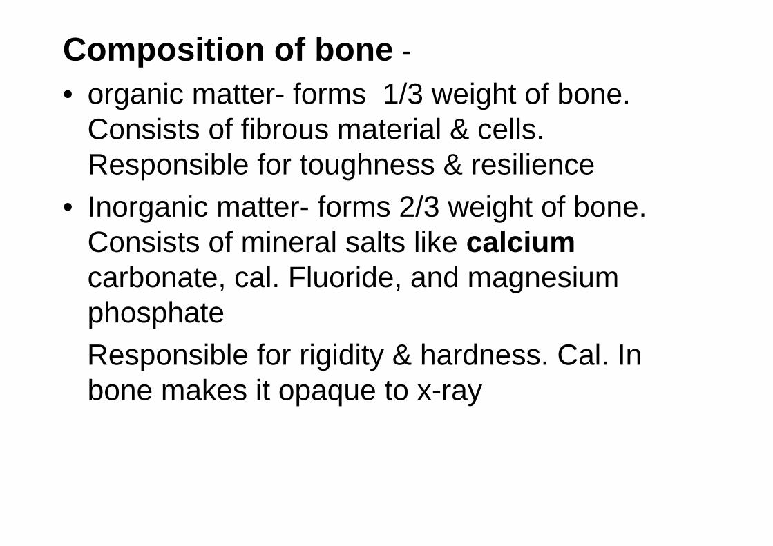
Composition of bone -• organic matter- forms 1/3 weight of bone.
Consists of fibrous material & cells. Responsible for toughness & resilience
• Inorganic matter- forms 2/3 weight of bone. Consists of mineral salts like calcium carbonate, cal. Fluoride, and magnesium phosphate Responsible for rigidity & hardness. Cal. In bone makes it opaque to x-ray

Macroscopic structure of living adult bone
• Compact bone • Cancellous bone

Microscopic structure of bone

Parts of a developing long bone • Diaphysis-
intermediate region or shaft
• Metaphysis-developing extraepiphyseal regions of shaft
• Epiphysis- ends of bone which ossify with a separate centre of ossifi. (secondary)

Epiphyseal cartilage • Zone of resting
cartilage • Zone of proliferating
cartilage • Zone of
hypertrophied cartilage
• Zone of calcified cartilage

Centers of ossification • Primary center • Secondary center • Epiphyseal line
Types of epiphysis Pressure epiphysis- articular & take part in
transmission of weight e.g. head of femur, lower end of radius
Traction epiphysis- Nonarticular & does not take part in the transmission of the weight.
• Tendons are attached here which exert a traction on the epiphysis
• Ossify later then the pressure epiphysis e.g. trochanters of tubercles of humerus
Atavistic epiphysis- femur, Phylogenetically an independent bone which in man become fused to another bone e.g. coracoid process of scapula & os trigonum
Aberrant epiphysis- Not always present e.g. epiphysis at the head of first metacarpal & at the base of other metacarpal bones

Blood supply of bone • Nutrient artery • Periosteal vessels • Metaphyseal vessels • Epiphyseal vessels

• Lymphatic supply- lymphatics present only in periosteum & Haversian system
• Accompany blood vessels • No lymphatic in the bone marrow • Lymphatic of the haversian system drain in to
periosteal vessels • Nerve supply- Most numerous at the articular
ends of the long bones vertebrae & flat bones • Distributed freely to the periosteum & with the
branches of nutrient artery. • Consist of both sensory & autonomic fibers
(blood vessels)

Some important points about ossification
• Ossification begins constantly at a prefixed spot & at a fairly constant time
• Centre may be primary or secondary • Primary center may be single or multiple
but appear & as a rule appear before birth • Between 6 to 8th wk of fetal life. Exception
cuneiform & navicular bones • Secondary center usually multiple &
appear after birth. Exception are lower end of femur

• Most long bones have epiphysis at both ends the epiphysis which ossifies first unites with the diaphysis last & the epiphysis which ossifies last fuses first. Exceptions. Lower end of fibula where epiphysis ossifies first, also fuses last with shaft
• The end of the long bone where epiphysis appear first & fuses last is called the growing end of the bone
• The direction of the nutrient artery is always away from the growing end of the bone given away by rhyme,
To the elbow I go, from the knee I flee”

• The different centers of ossification first unite together & then they unite with the shaft
• In long bones growing ends of the bone fuses with the shaft at about 20 years & the opposite end at about 18 years i.e. 2 years earlier
• Fusion of epiphysis with diaphysis occurs 2 years earlier in women than in men. Epiphysis also appear earlier in women
• Epiphysis in bones other than long bones fuses with main part of the bone between 20-25 years

Estimation of age, sex &height from the bones
• Timing of eruption of milk teeth & permanent teeth can estimate age up to18 years
• Age at which epiphysis of the bone appears and fuses with the diaphysis es fairlyconstant. This can provides the age till 25 years
• After 25 years age is estimated by the closing of cranial sutures &changes occuring at themedial surface of pubic bones. this age canbe estimated till 60 years

• Sex can be determined by studying morphological feature of the bone & the measurement of skull & pelvis
• Race can be determined with85-90% accuracy by metrical & nonmetrical data developed from cranial &other parts of skeleton.